Article

Heterologous Expression Reveals Ancient Properties of Tei3—A VanS Ortholog from the Teicoplanin Producer Actinoplanes teichomyceticus

1
Department of Biotechnology and Life Sciences, University of Insubria, 21100 Varese, Italy
2
Department of Genetics and Biotechnology, Ivan Franko National University of Lviv, 79005 Lviv, Ukraine
*
Author to whom correspondence should be addressed.
Int. J. Mol. Sci. 2022, 23(24), 15713; https://doi.org/10.3390/ijms232415713
Submission received: 7 November 2022 / Revised: 3 December 2022 / Accepted: 6 December 2022 / Published: 11 December 2022
(This article belongs to the Special Issue Microbial Resistance Mechanisms)

Abstract

:
Glycopeptide antibiotics (GPAs) are among the most clinically successful antimicrobials. GPAs inhibit cell-wall biosynthesis in Gram-positive bacteria via binding to lipid II. Natural GPAs are produced by various actinobacteria. Being themselves Gram-positives, the GPA producers evolved sophisticated mechanisms of self-resistance to avoid suicide during antibiotic production. These self-resistance genes are considered the primary source of GPA resistance genes actually spreading among pathogenic enterococci and staphylococci. The GPA-resistance mechanism in Actinoplanes teichomyceticus—the producer of the last-resort-drug teicoplanin—has been intensively studied in recent years, posing relevant questions about the role of Tei3 sensor histidine kinase. In the current work, the molecular properties of Tei3 were investigated. The setup of a GPA-responsive assay system in the model Streptomyces coelicolor allowed us to demonstrate that Tei3 functions as a non-inducible kinase, conferring high levels of GPA resistance in A. teichomyceticus. The expression of different truncated versions of tei3 in S. coelicolor indicated that both the transmembrane helices of Tei3 are crucial for proper functioning. Finally, a hybrid gene was constructed, coding for a chimera protein combining the Tei3 sensor domain with the kinase domain of VanS, with the latter being the inducible Tei3 ortholog from S. coelicolor. Surprisingly, such a chimera did not respond to teicoplanin, but indeed to the related GPA A40926. Coupling these experimental results with a further in silico analysis, a novel scenario on GPA-resistance and biosynthetic genes co-evolution in A. teichomyceticus was hereby proposed.

1. Introduction

Glycopeptide antibiotics (GPAs) [1], particularly dalbaheptides [2], belong to a clinically relevant group of natural compounds produced by soil-dwelling and mycelium-forming Gram-positive actinobacteria, traditionally named actinomycetes [3,4]. As with other natural compounds, genes responsible for the biosynthesis of GPAs are grouped in large biosynthetic gene clusters (BGCs) [5]. GPAs are successfully utilized to treat severe infections caused by multi-drug-resistant (MDR) staphylococcal and enterococcal strains. Two natural and three semisynthetic GPAs are currently approved for clinical use [3,6,7]. Natural first-generation GPAs are vancomycin and teicoplanin, produced by the actinobacteria Amycolatopsis orientalis (various strains) [8] and Actinoplanes teichomyceticus ATCC 31121 [9], respectively. Vancomycin was introduced in clinics first (in 1958), followed by teicoplanin (1988) in Europe and then in Japan (1998) [3]. Semisynthetic second-generation GPAs include telavancin—a vancomycin derivative [10], oritavancin—a derivative of the natural GPA chloroeremomycin [11], and dalbavancin—a derivative of the other natural GPA, A40926 [12].
Although GPAs of both generations are still clinically useful, resistant pathogens are inevitably emerging [13,14,15,16]. Peculiarly, resistance mechanisms in GPA producers and pathogens are very similar [17]. It is likely that pathogens acquired GPA resistance genes from GPA producers, or more generally from non-producing actinobacteria, where GPA resistance determinants are abundant [18,19]. GPAs interact with d-alanyl-d-alanine (d-Ala-d-Ala) termini of the nascent peptidoglycan (PG), impeding upstream transpeptidation and transglycosylation reactions and consequently blocking cell wall biosynthesis [20]. The resistance mechanisms shared between GPA producers and pathogens lead to the modification of the d-Ala-d-Ala termini, reducing the affinity of GPAs for their target. d-Ala-d-Ala termini in PG precursors are replaced by d-Ala-d-lactate (d-Lac) [17,21] or truncated to a single d-Ala residue [22,23,24]. The first mechanism requires a three-gene operon—vanHAX, where vanH codes for a d-Lac dehydrogenase responsible for generating a d-Lac pool, vanX—for a d,d-dipeptidase whose role is reducing the intracellular pool of d-Ala-d-Ala [25,26], and vanA—for a d-Ala-d-Lac ligase [21,27]. The second involves a single d,d-carboxypeptidase named VanY, which trims the terminal d-Ala residue. In both cases, PG precursors still undergo transpeptidation reactions, but this process is not affected by GPAs [22,23,24]. The expression of vanY or vanHAX might be constitutive or inducible. The latter requires a two-component regulatory system consisting of a sensor histidine kinase (SHK) and a transcriptional response regulator (RR), also known as VanS and VanR, respectively [28,29,30,31]. VanS can specifically recognize extracellular GPAs and phosphorylate VanR, which, in turn, activates the expression of other van genes [29,30,31,32,33].
Actinobacterial GPA producers such as A. teichomyceticus, Amycolatopsis balhimycina DSM 5908 (balhimycin producer), Nonomuraea gerenzanensis ATCC 39727 (A40926 producer), Streptomyces toyocaensis NRRL 15009 (A47934 producer), as well as the GPA non-producing Streptomyces coelicolor A3(2) represent well-studied models of GPA resistance mechanisms (recently reviewed in [34]), although certain aspects are not fully elucidated yet and merit further investigation. In this work, the properties of Tei3—the VanS ortholog from the teicoplanin producer A. teichomyceticus—were investigated in vivo. Notably, A. teichomyceticus is resistant to very high concentrations of teicoplanin, likely due to the strong constitutive expression of the vanHAX orthologs—tei7-6-5, which was previously reported during the transcription analyses of the tei BGC [35,36,37]. It was speculated that Tei3 acts as a constitutive SHK [35]. We aimed to experimentally investigate whether Tei3 really acts as a constitutive SHK and if any GPA is eventually recognized by its sensor domain (SD). Conducting a series of experiments in S. coelicolor as a heterologous host, it was found out that Tei3 does not require any ligand to function. Strikingly, the SD of Tei3 appeared to recognize not teicoplanin but the structurally related GPA A40926. Finally, reconstruction of the phylogeny of glycosyltransferases (GTFs) coded within GPA BGCs allowed us to build a possible scenario explaining the obtained results.

2. Results

The main goals of this work were to test if Tei3 acts as a non-inducible phosphorylase and to investigate which ligand could be sensed by Tei3. Considering A. teichomyceticus a challenging microorganism for gene-engineering manipulations, a series of genetic experiments were designed and performed in heterologous hosts such as S. coelicolor M512 [38] and S. coelicolor J3200 [31]. As anticipated in the introduction, S. coelicolor has a full set of van genes conferring vancomycin resistance, although it does not produce any GPAs [39]. S. coelicolor M512 does not produce the pigmented antibiotics actinorhodin and undecylprodigiosin [38], permitting the use of a β-glucuronidase (GusA)-based reporter assay, as described below. The second strain—J3200—is the ΔvanSSc mutant, in which the host vanS gene was knocked out [31]. Thus, the experimental steps described below aimed: (1) to test if the heterologous Tei3 can cross-phosphorylate the host VanRSc; (2) to build a gusA-based reporter S. coelicolor strain, able to convert X-Gluc (5-bromo-4-chloro-3-indolyl-β-d-glucuronide) to 5,5′-dibromo-4,4′-dichloro-indigo following GPA induction; (3) to utilize the created reporter strain to show if Tei3 needs the presence of GPAs or not; (4) to replace the SD of the host VanSSc with its counterpart from Tei3 and test which GPAs may act as ligands for Tei3 SD.

2.1. Heterologous Expression of Tei3 SHK Leads to Teicoplanin and A40926 Resistance in S. coelicolor M512

VanRS-like two-component regulatory pairs are conserved enough among actinobacteria to show a certain degree of cross-talking [33,40]. For example, it was shown that VanRSc (coming from S. coelicolor) could be phosphorylated in vivo by VanSSt (from A47934 producer S. toyocaensis), but not vice versa, implying that VanRSc is accessible for non-cognate SHKs [33]. In this case, SHKs and RRs both came from Streptomyces spp. Instead, it was necessary to check if Tei3 SHK from A. teichomyceticus (order Micromonosporales) could phosphorylate VanRSc. Overall, Tei2 and Tei3 share a high percentage of aa sequence identity with VanRSc and VanSSc: 91% and 67%, respectively. Tei3 and VanSSc are collinear, sharing highly similar transmembrane helix (TMHs) regions and conserved putative autophosphorylation sites (Figure 1a). The most divergent region between Tei3 and VanSSc is the extracytoplasmic sensory loop (ESL), implying that quite different ligands should be recognized by the two proteins (Figure 1a). At the same time, Tei2 and VanRSc were almost identical (Figure 1b). Thus, it seems plausible that Tei3 would be able to phosphorylate VanRSc in vivo.
To test if Tei3 can phosphorylate the non-cognate RR—VanRSc, tei3 was cloned into the pSET152A vector, giving pGP101, and then transferred into S. coelicolor M512. The obtained recombinant strain—S. coelicolor M101—gained teicoplanin and A40926 resistance, in addition to the endogenous vancomycin resistance (Figure 2). Consequently, it could be concluded that Tei3 phosphorylates VanRSc in vivo. However, this experiment did not clarify whether Tei3 acts as a constitutive phosphorylase, considering that teicoplanin and A40926 added to the plates could serve as its activators.

2.2. Tei3 Acts as a Non-Inducible Phosphorylase

Expression of tei2-3-4 and tei7-6-5 operons remains stable throughout the life cycle of A. teichomyceticus, independently from the teicoplanin concentration [35,37], pathway-specific regulation [36], and growth phase [37]. Previous papers speculated about the need of a strong constitutive promoter driving tei2-3-4 expression [37] or of the presence of specific mutations in Tei3 that might explain its function as a constitutive phosphorylase, working independently from the presence of any extracellular GPA [35]. Protein sequence comparison indicated that Tei3 carries single aa substitutions in two sites where analogous mutations transform the vancomycin-inducible VanSSc into a constitutive phosphorylase [35]. Specifically, these are L216P and G271V substitutions [31]. Homologous sites in Tei3 are N215 and R270 (Figure 1a). However, there are multiple other sites within the putative ATPase domain (ATPaseD) of Tei3, which significantly diverged from VanSSc (see Figure 1a).
To experimentally verify the GPA inducibility of Tei3, a bioassay responding to the inducers of van genes was first developed in S. coelicolor. Other authors previously described a reporter S. coelicolor strain, where the endogenous vanJ promoter (vanJp) was cloned into a multicopy plasmid, fused with the kanamycin/neomycin resistance gene—neo—in a way that induction of vanJp by vancomycin conferred resistance to both neomycin and kanamycin [39]. Using this experience, either vanJp or the other endogenous vancomycin-responsive S. coelicolor vanHAX promoter (vanHp) was fused with the gusA gene, coding for a β-glucuronidase in the pGUS chassis [44]. vanJp or vanHp activation by GPAs in the reporter strain should activate the chromogenic conversion of the X-Gluc substrate into the green-colored 5,5′-dibromo-4,4′-dichloro-indigo [44]. Thus, plasmids pGHp (carrying vanHp-gusA) and pGJp (carrying vanJp-gusA) were transferred to S. coelicolor M512 by means of intergeneric conjugation with Escherichia coli ET12567 pUZ8002+. The inducibility of the two generated reporter strains S. coelicolor pGHp+ and pGJp+ was first tested in liquid medium and then in solid plates. When vancomycin was added at 10 µg/mL to 50 h old cultures in TSB liquid medium, the basal glucuronidase activity of the mycelia was increased by, ca., twenty- and forty-fold, in S. coelicolor pGHp+ and pGJp+, respectively (Figure S1), indicating that vanJp seems more responsive to vancomycin than vanHp. In solid media containing 25 µg/mL of X-Gluc, the presence of vancomycin induced vanJp-mediated glucuronidase activity in recombinant strains, yielding green halos around the Whatman discs soaked in antibiotic solution (Figure 3 and Figure S2). S. coelicolor pGJp+ acted very well as a reporter, giving a detectable chromogenic conversion in response to very low concentrations of vancomycin (250 ng, Figure S2), whereas S. coelicolor pGHp+ resulted as much less responsive to vancomycin (data not shown) and, thus, it was not further used.
The next step was transferring Tei3 into S. coelicolor pGJp+ to test if its activity is inducible by GPAs. As either pGJp or pGP101 is a distant derivative of pSET152 [45], both plasmids use the φC31 attB site for integration and are not compatible. To solve this issue, the pRT801 plasmid [46] was used to create a φBT1-based derivative of pGP101, named pGP111. The M512 derivative carrying pGP111 (carrying tei3) was named M111, while the strain carrying both pGP111 and pGJp was named M1J. It was expected that if Tei3 functions as a constitutive phosphorylase, M1J would express a constitutive glucuronidase activity in the presence of X-Gluc, not depending on the presence or absence of GPAs. This assumption was correct as M1J converted X-Gluc independently from the presence of any GPA (Figure 3), confirming in vivo that Tei3 functions as a constitutive phosphorylase. As a control, it was shown that no induction was observed in those strains not carrying vanJp-gusA (M512 and M111), whereas the GPA induction was observed in S. coelicolor pGJp+ due to the action of the endogenous VanSSc.

2.3. Loss of Extracytoplasmic Sensory Loop and Transmembrane Helices Renders Tei3 and VanSSc Nonfunctional

Considering the non-inducible properties of Tei3, we were wondering whether TMHs and ESL are still necessary for its function, or whether ATPaseD would be the only required domain. To answer this question, a series of plasmids was created carrying truncated versions of tei3 or of vanSSc lacking (i) TMH1 and ESL (tei3′, vanSSc′); (ii) TMH1, ESL, and TMH2 (tei3″, vanSSc″) (Figure S3). These plasmids were named pGP104 (tei3′), pGP105 (vanSSc′), pGP106 (tei3″), and pGP107 (vanSSc″). Plasmids were transferred into S. coelicolor M512 (resistant to vancomycin but sensitive to teicoplanin and A40926) and S. coelicolor J3200 (this last one—ΔvanSSc—is constitutively resistant to all GPAs), generating M104-107 and J104-107 strains. It was expected that if ATPaseD of Tei3 would be able to function alone, the expression of tei3′ and tei3″ might lead to constitutive GPA resistance in M512. On the contrary, the absence of the SD in VanSSc might render J3200 constitutively sensitive to GPAs. However, the obtained results indicated that ATPaseDs of neither Tei3 nor VanSSc can function alone: the phenotypes of M104-107 and J104-107 strains in the presence of vancomycin, teicoplanin, and A40926 were the same as the parental M512 and J3200 strains, respectively (Figure 4).

2.4. Tei3 Sensor Domain Is Sensitive to A40926 but Not to Teicoplanin and Vancomycin

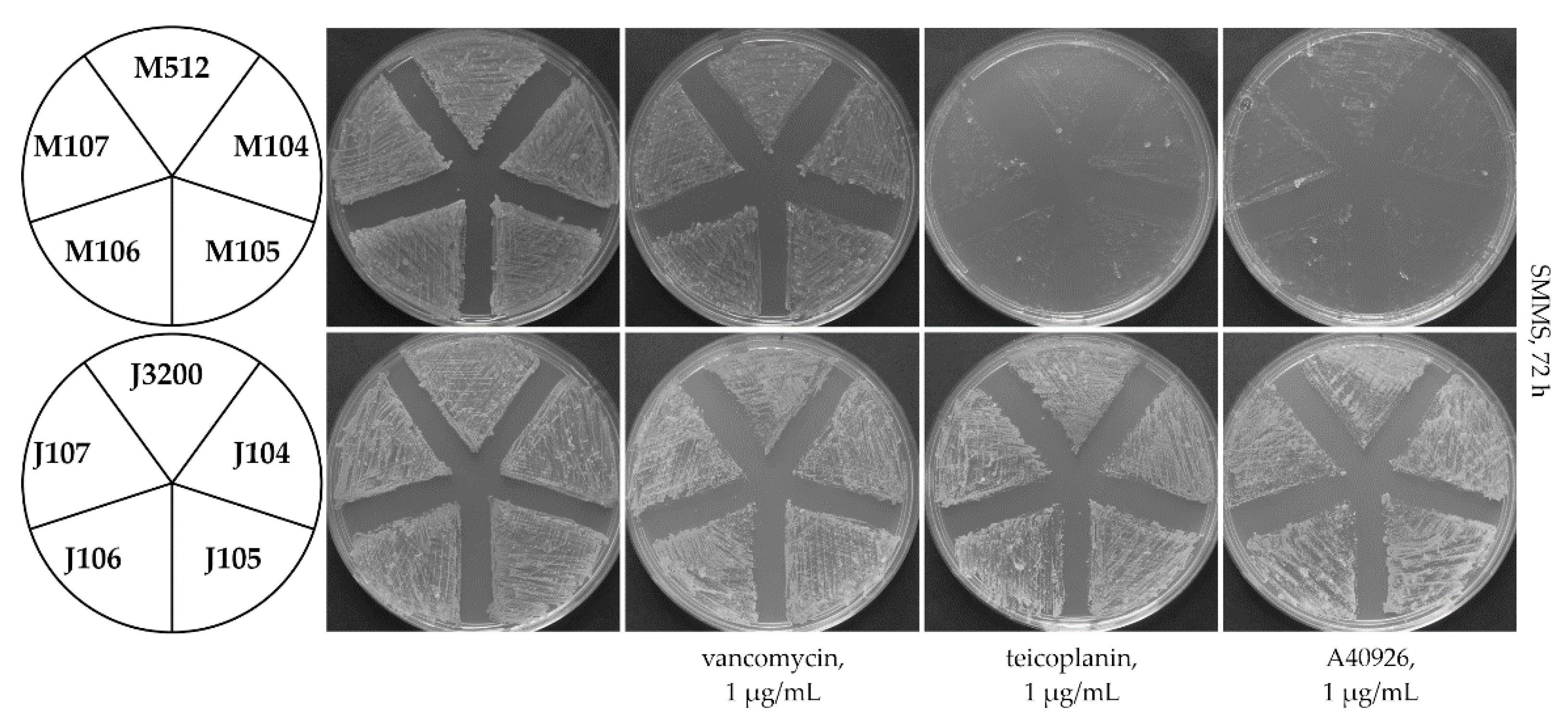
Logically, teicoplanin should be the original ligand for Tei3. However, direct experimental verification of this assumption is impossible due to the non-inducible properties of Tei3. It is possible that maybe Tei3 became a constitutive phosphorylase in the course of the evolution, but clues for Tei3 sensitivity might have remained “fossilized” within its SD. To answer this question, a hybrid gene coding for a chimeric SHK combining the SD of Tei3 with the ATPaseD of VanSSc was created. vanSSc and tei3 (Figure S4) were collinear, facilitating such an exchange. Luckily, a unique PaeI recognition site was found only 4 bp after the vanSSc region coding for SD. This allowed us to use this site for replacing the vanSSc region coding for the SD with the corresponding one from tei3 (see Section 4 for details). The obtained hybrid gene—tei3-vanSSc (Figure S4)—was cloned into the pSET152A plasmid generating pGP103. Next, pGP103 was transferred into M512 and J3200 generating the recombinant strains M103 and J103, respectively. In the first case, a merodiploid strain was generated, carrying the native vanSSc together with the hybrid tei3-vanSSc allele, while in J3200, the knockout of vanSSc was complemented by the added tei3-vanSSc. GPA resistance phenotypes of these recombinants showed that both M103 and J103 became sensitive to vancomycin, implying that this GPA is not an inducer for the Tei3 SD (Figure 5).
Surprisingly, M103 and J103 were also sensitive to teicoplanin, excluding its possible role as a ligand for the Tei3 SD. Adding a final detail to these puzzling results, both M103 and J103 were resistant to A40926, implying that the Tei3 SD recognizes A40926 as a ligand.

2.5. Establishing a Link between the Evolution of Glycosylation Pattern of Teicoplanin and Properties of Tei3 SD

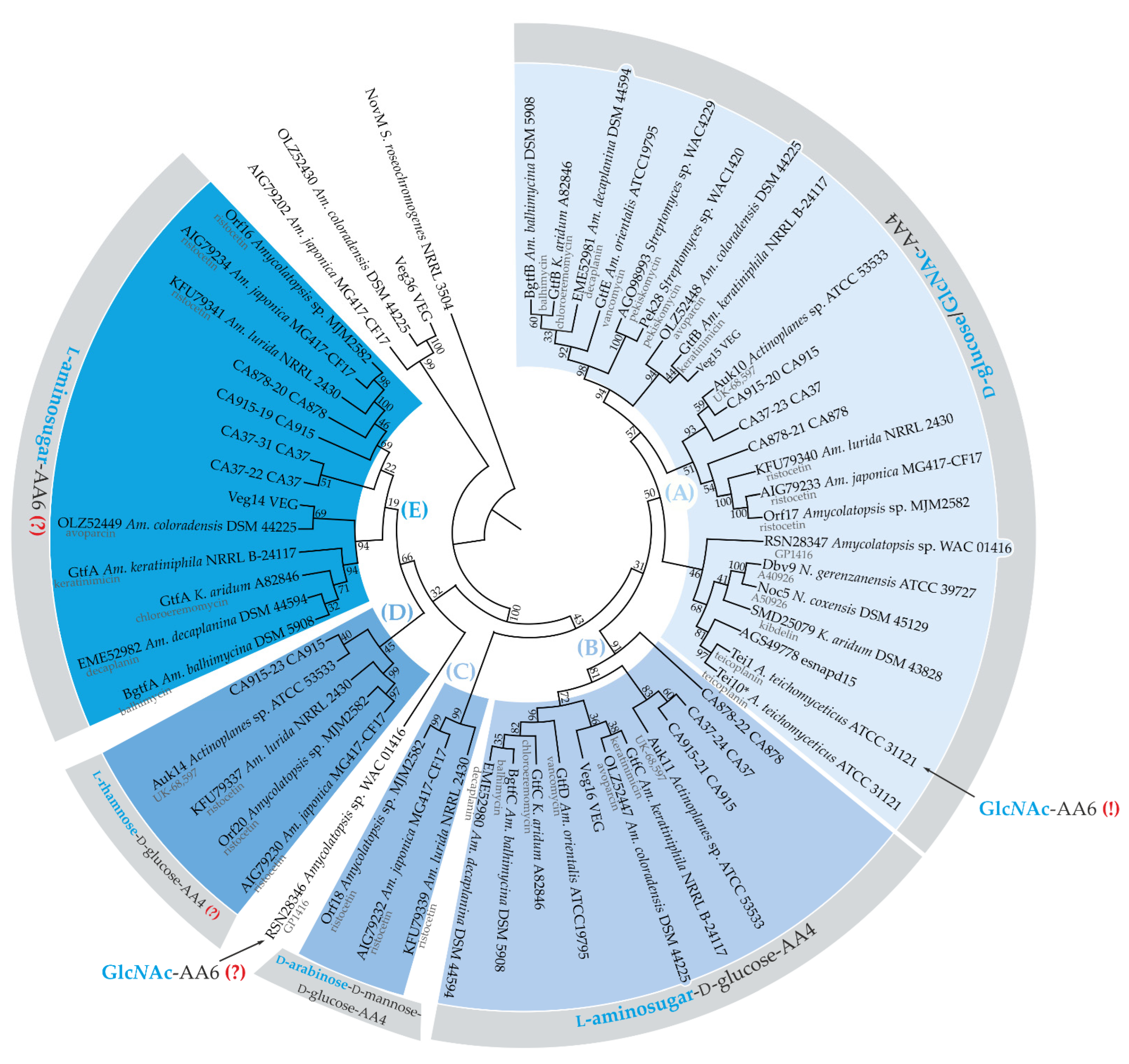
Teicoplanin and A40926 are chemically similar GPAs that likely emerged in the course of convergent evolution (Figure S5) [47,48,49]. Differences lie in the chlorination and methylation pattern; notably, A40926 also lacks a N-acetyl glucosamine (GlcNAc) moiety attached to the aglycone of teicoplanin at the aa position 6 (AA6) (Figure S5). The glycosylation pattern might be important for binding VanS, in accordance with previous results reporting that VanS from S. toyocaensis was unable to recognize vancomycin, sensing only the non-glycosylated A47934 [33]. Hence, it could be speculated that the responsiveness of Tei3 to a GPA lacking a GlcNAc residue might be an ancient property from the times when the ancestral teicoplanin BGC did not carry a gene for the attachment of the GlcNAc moiety at aglycone AA6. To understand this better, the phylogeny of glycosyl transferases (GTFs) coming from experimentally studied GPA BGCs was reconstructed (Figure 6).
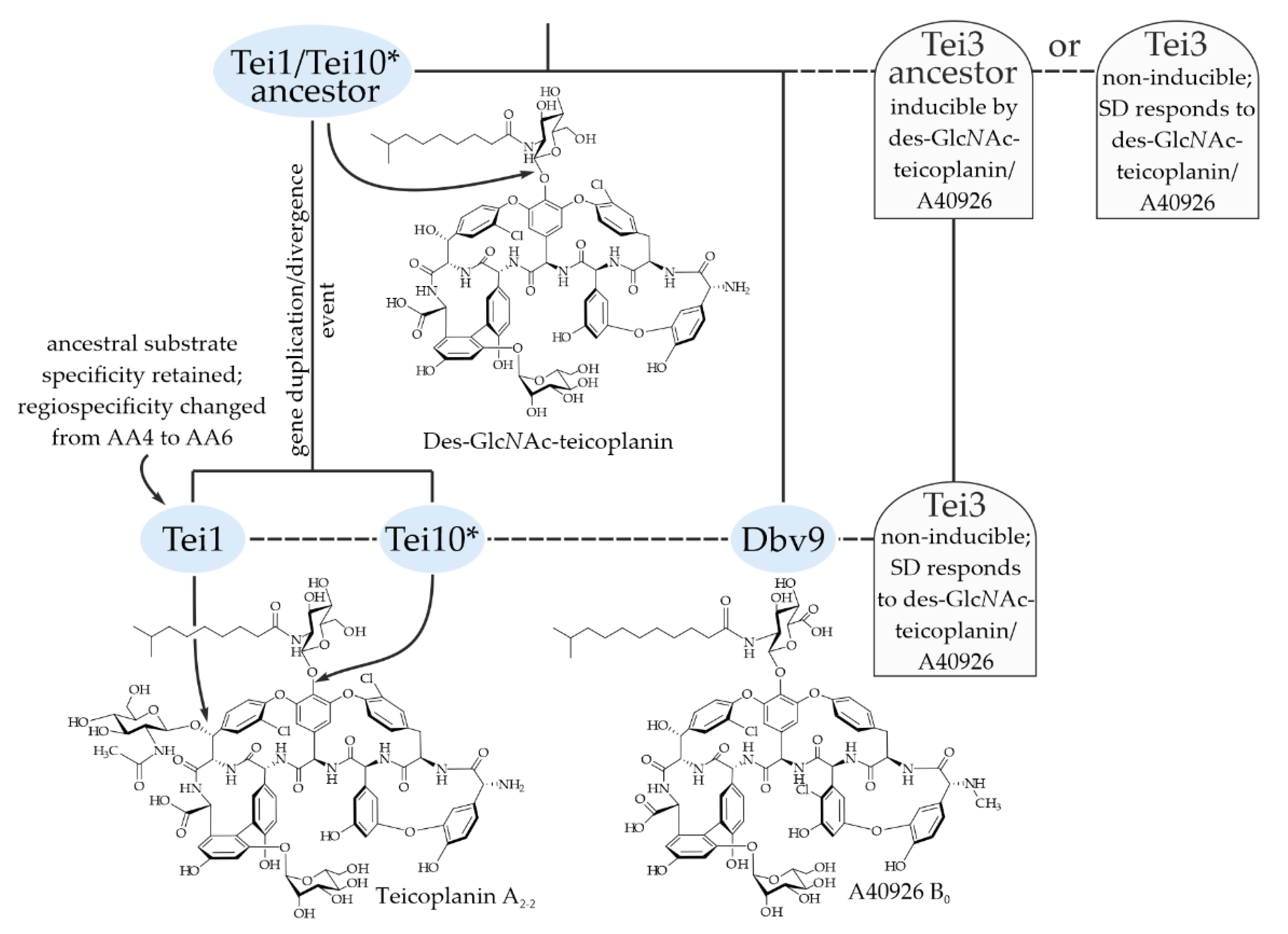
Five clades (A–D) might be delineated on the obtained tree (Figure 6) and they seem to correspond to the regiospecificity of GTFs well. The regiospecificity of clades (A–C) GTFs could be predicted with high confidence as, for many members of these clades, experimental evidence exists. Thus, clade (A) comprehended GTFs attaching either d-glucose or GlcNAc to AA4 of the GPA aglycone (see [50] for the review of such GTFs); clade (B) GTFs are responsible for the attachment of l-aminosugars to AA4 d-glucose [50]; clade (C) included GTFs from ristocetin BGCs likely attaching d-arabinose to AA4 d-mannosyl-d-glucose [51] (Figure 6). The substrate- and regiospecificity of GTFs from clades (D) and (E) were dubious: clade (D) GTFs probably attach l-rhamnose to the AA4 d-glucose, while clade (E) GTFs may attach l-aminosugars to aglycone AA6. To our surprise, Tei1—known to attach the GlcNAc moiety at the teicoplanin aglycone AA6 [52]—was found deep in clade (A), being a sister branch to Tei10* (known to attach GlcNAc at AA4). One other GPA—GP1416 from Amycolatopsis sp. WAC01416 [53]—is known to closely resemble the teicoplanin structure, bearing the GlcNAc moiety at AA6. However, the GTF that is likely responsible for this did not belong to any of the clades and was located far from either Tei1 or Tei10* (Figure 6). The presence of the GlcNAc moiety at AA6 of teicoplanin and GP1416 could, thus, be considered an example of the convergent evolution of GPAs. Considering all mentioned above, one could reasonably assume that Tei1 is a recent product of the Tei10* duplication/divergence event. In the course of evolution, it is likely that Tei1 changed its regiospecificity but retained the substrate specificity. A direct ancestor of teicoplanin BGC likely coded the biosynthesis of des-GlcNAc-teicoplanin, which structurally resembles A40926 and was recognized by the Tei3 SD (see Figure 7 and the discussion below for a possible reconstruction of co-evolution of GTFs encoded in teicoplanin BGC and Tei3).

3. Discussion

vanHAXRS genes were experimentally shown to provide GPA resistance in (i) GPA-producing actinobacteria [34], (ii) actinobacterial GPA non-producers [31], (iii) GPA-resistant pathogens [28], and (iv) other soil bacteria [19,54]. In the majority of the known cases, the VanRS two-component regulatory system is utilized to sense extracellular GPAs [29] (or perhaps the GPA-lipid II complex [30]) and activate the expression of functional van genes. Pathogens and GPA-non-producing actinobacteria generally have inducible van-resistant phenotypes, because the constitutive expression of van genes comes at a cost of decreased fitness of the cells [55,56].
Figure 6. Maximum-likelihood phylogenetic tree of 59 GTFs coded within or near experimentally studied GPA BGCs and NovM from clorobiocin BGC [57], used as an outgroup (the tree is not drawn to scale). MEGA 11 was used for the analysis [58]. Evolution was inferred by using the JTT matrix-based model [59]. A discrete γ distribution was used to model evolutionary rate differences among sites (5 categories). Numbers at nodes indicate bootstrap-support values derived from 1000 replications. Five clades were delineated on this tree (AE); clades correlated with substrate- and regiospecificity of GTFs. Veg36-OLZ52430-AIG79202 seemed to be a clade of few additional irrelevant GTFs coded nearby the corresponding BGCs. Notably, Tei1 (marked with “GlcNAc-AA6 (!)” label) appeared in the clade (A) grouping GTFs attaching either d-glucose or GlcNAc to aglycone AA4, while the enzyme was experimentally shown to attach GlcNAc to aglycone AA6. “(?)” signs indicate presumed regiospecificity (lack of experimental evidence).
Ijms 23 15713 g006
In contrast, the GPA resistance phenotype is commonly constitutive in GPA producers, as it was shown in A. teichomyceticus and Am. balhimycina [34]. In Am. balhimycina, vanHAX orthologs are the main contributors to GPA resistance [40,60]. However, vanHAX are not situated within the borders of balhimycin BGC (as commonly occurs in the other actinomycetes producing GPAs), and they are not co-localized with vanRS orthologs [40]. Hence, the expression of vanHAX is devoid of vanRS-mediated regulation and is constitutive. It should be noted that Am. balhimycina also possesses additional GPA-resistance mechanisms that are not van-mediated [61].
Figure 7. A scheme illustrating possible scenarios for the co-evolution of GTFs encoded in teicoplanin BGC and Tei3. Please refer to the main text for more detail.
Ijms 23 15713 g007
The most striking outcome of our work was that the SD of Tei3 did not respond to teicoplanin, but recognized A40926: the expression of the hybrid SHK carrying the Tei3 SD fused with the catalytic part from VanSSc changed the constitutive GPA-resistance phenotype of S. coelicolor J3200, making the strain teicoplanin- and vancomycin-sensitive, but A40926-resistant. This prompted us to focus on the chemical differences in the structure of A40926 and teicoplanin (the presence of a GlcNAc residue in teicoplanin, which is absent in A40296) and on the possible evolution of the teicoplanin glycosylation pattern and of the whole tei BGC. Reconstruction of the phylogeny of GTFs coded within the known GPA BGCs allowed us to build possible scenarios explaining the obtained results (Figure 7).
If our reconstruction of the evolution of Tei1/Tei10* is correct, the ancestor of the teicoplanin BGC coded the biosynthesis of des-GlcNAc-teicoplanin, which structurally resembles A40926 and was recognized by the Tei3 SD. Tei3 SHK initially responded to des-GlcNAc-teicoplanin (modeled with A40926 in the experiment presented in this work) (Figure 7) and it was probably inducible. A further two alternative scenarios are possible. The first implies that the non-inducibility of Tei3 evolved as an adaptation to the appearance of Tei1 because A. teichomyceticus was required to rapidly gain constitutive GPA resistance as Tei3 was unable to sense teicoplanin. The second scenario implies that Tei3 in the des-GlcNAc-teicoplanin producer became non-inducible at the first place, consequently giving the BGC room for further evolution and structural changes of the produced GPA. Considering that, in the first hypothesis, fewer mutations would probably be required to give Tei3 constitutive properties than to remodel its SD, the second scenario is more likely in our opinion. Hence, mutations leading to the non-inducibility of Tei3 might have served as a preadaptation, which allowed teicoplanin to emerge in its current form, carrying the GlcNAc residue at AA6 (Figure 7).
To conclude, our results proved the non-inducibility of Tei3, showed that its SD is able to respond to A40926, but not to teicoplanin or vancomycin, and demonstrated a novel example of how BGCs, antibiotic structures, and antibiotic resistance genes might have co-evolved. Further investigations will include mutational analysis of Tei3 to elucidate which particular aa changes in its ATPaseD lead to non-inducible properties. Finally, as a byproduct, a S. coelicolor-based chromogenic assay for vancomycin detection was developed capable of sensing vancomycin at the ng range.

4. Materials and Methods

4.1. Plasmids, Bacterial Strains, and Cultivation Conditions

All plasmids and bacterial strains utilized or generated in the course of the current study are summarized in Table 1. For routine maintenance, A. teichomyceticus was cultivated on ISP3 agar [62] at 30 °C, and S. coelicolor strains were cultivated on SFM [45] agar at 30 °C. For genomic DNA isolation, A. teichomyceticus was cultivated in ISP2 liquid medium [47] in 50 mL baffled flasks on an orbital shaker at 200 rpm and 30 °C; S. coelicolor strains for genomic DNA isolation were cultivated under the same conditions in TSB medium [62]. Antibiotic susceptibility tests with S. coelicolor strains were performed on SMMS medium [45]. Apramycin sulfate (50 μg/mL), spectinomycin hydrochloride (50 μg/mL), and nalidixic acid (30 μg/mL) were used for the selection and maintenance of S. coelicolor recombinant strains. Vancomycin, teicoplanin, and A40926 were added to solid or liquid cultures at the desired concentrations reported in Section 2. E. coli DH5α was used as a general cloning host, while E. coli ET12567 pUZ8002+ was used as a donor for intergeneric matings. E. coli strains were cultivated at 37 °C in lysogeny broth or agar media supplemented with 100 μg/mL of apramycin sulfate, 50 μg/mL of kanamycin sulfate, and 25 μg/mL of chloramphenicol when appropriate. All antibiotics were purchased from Sigma-Aldrich (Merck Group, Darmstadt, Germany).

4.2. Generation of Recombinant Plasmids for Gene Expression and Promoter-Probe Vectors

In all cases described below, Q5 High-Fidelity DNA Polymerase (NEB, Ipswich, MA, USA) was used as a DNA polymerase of choice in PCRs according to the recommendations of the supplier. Restriction endonucleases and T4 DNA ligase from Thermo Fisher Scientific (Waltham, MA, USA) were always used for DNA digestion and ligation according to the supplier’s recommendations. Chromosome DNA, utilized as a PCR template, was isolated according to the Kirby procedure [45].
pGP101, pGP111. The coding sequence of the tei3 gene was amplified from the chromosome DNA of A. teichomyceticus using the tei3_F/R primer pair (Table 2); the obtained 1133 bp amplicon was digested with EcoRI/EcoRV restriction endonucleases and cloned into the pSET152A vector digested with the same restriction endonucleases yielding pGP101. Further, pGP101 was digested with BamHI/XhoI restriction endonuclease, and a 3247 bp DNA fragment carrying aac(3)IVp-tei3 was purified; in parallel, the pRT801 plasmid was also digested with BamHI/XhoI and the 3362 bp DNA fragment was purified. Both obtained fragments were then ligated, generating pGP111.
pGP102, pKC1132-102, pKC1132-103, and pGP103. The coding sequence of vanSsc (SCO3589) was amplified using the chromosome DNA of S. coelicolor M512 as a template with the SCO3859_F/R primer pair; the obtained 1130 bp amplicon was digested with EcoRI/EcoRV restriction endonucleases and cloned into pSET152A via the same sites, giving pGP102. pGP102 was digested with PvuII and the 1753 bp fragment (carrying aac(3)IVp-SCO3589) was cloned into pKC1132 digested with the same restriction endonuclease, generating pKC1132-102. Then, a fragment of SCO3589 coding for the SD was exchanged for the corresponding fragment of tei3. The fragment of the tei3 sequence coding for the SD was amplified from pGP101 using the tei3_F and tei3_sdomain_PaeI primers pair; the obtained 285 bp amplicon was digested with EcoRV and PaeI restriction endonucleases. In parallel, pKC1132-102 was also digested with EcoRV and PaeI and a 4289 bp fragment was selected; this latter fragment was ligated with the PaeI-digested tei3-SD amplicon, yielding pKC1132-103. Finally, the hybrid ORF of SCO3589 with the SD-coding fragment exchanged with tei3-SD was excised from pKC1132-103 using EcoRV and EcoRI restriction endonucleases, and cloned into pSET152A via the same recognition sites generating pGP103.
pGP104, pGP105, pGP106, and pGP107. These vectors carried truncated versions of tei3 and SCO3589 lacking parts of the sequence coding for (i) TMh1 (tei3′—pGP104; SCO3589′—pGP105) and (ii) whole SD (tei3″—pGP106; SCO3589″—pGP107). All these coding sequences were amplified using primers listed in Table 2, generating: tei3′—956 bp; tei3″—879 bp; SCO3589′—959 bp; SCO3589″—878 bp. Amplicons were digested with EcoRV/EcoRI and cloned into pSET152A via the same recognition sites.
pGJp and pGHp. DNA fragments including promoter regions of S. coelicolor vanJ (SCO3592) and vanH (SCO3594) genes were amplified from the chromosome DNA of S. coelicolor M512 using vanJp_KpnI_F/vanJp_SpeI_R and vanHp_KpnI_F/vanHp_SpeI_R primers pairs (Table 2). The obtained amplicons for vanJp (321 bp) and vanHp (539 bp) were digested with KpnI/SpeI restriction endonucleases and ligated with pGUS digested by the same enzymes, yielding pGJp and pGHp promoter-probe vectors.
All generated plasmids were verified by restriction mapping and sequencing.

4.3. Conjugal Transfer of Recombinant Plasmids to S. coelicolor Strains

A standard protocol for the conjugal transfer of plasmids to S. coelicolor strains was utilized [45]. All necessary plasmids were individually transferred into the non-methylating E. coli ET12567 pUZ8002+ and the resulting derivatives were used as donor strains for intergeneric conjugation, while spores of S. coelicolor were used as acceptors. Circa 106 spores were mixed with, ca., 109 donor cells and plated on ISP3 agar supplemented with MgCl2 (10 mM); after 12 h of incubation at 30 °C, each plate was overlaid with 1 mL of sterile water with 1.25 mg of apramycin-sulfate and 750 μg of nalidixic acid. Transconjugants were selected as resistant to 50 μg/mL of apramycin sulfate or 50 μg/mL of spectinomycin hydrochloride. All recombinant strains were tested with PCR using chromosome DNA isolated according to the Kirby procedure [45]. aac(3)IV was amplified from chromosome DNA of transconjugants carrying pSET152A and pRT801 derivatives with the aac(3)IV_F/R primer pair. Transconjugants carrying promoter-probe vectors were tested by amplifying the 1000 bp internal region of gusA with the gusA_ver_F/R primer pair (Table 2).

4.4. Qualitative and Quantitative Glucuronidase Assays

Qualitatively, the β-glucuronidase (GusA) activity in S. coelicolor was assessed by adding 25 mg/mL of 5-bromo-4-chloro-3-indolyl-β-d-glucuronide (X-Gluc, Thermo Fisher Scientific, Waltham, MA, USA) to SMMS agar. The chromogenic conversion of X-Gluc into the green-colored 5,5′-dibromo-4,4′-dichloro-indigo was then monitored. Quantitative measurement was performed as follows. A number of 107 spores of S. coelicolor pGJp+ and pGHp+ were inoculated into a baffled 300 mL Erlenmeyer flask containing 50 mL of TSB and incubated without and with vancomycin as an inducer. Mycelium obtained in this way was used to prepare cell-free lysates as reported previously [63]. Glucuronidase activity was measured in cell-free lysates as described previously [44,63] utilizing a spectrophotometric assay to detect the conversion of the colorless p-nitrophenyl-β-d-glucuronide (Thermo Fisher Scientific, Waltham, MA, USA) into the colored p-nitrophenol at 415 nm using a Unicam UV 500 UV-Visible Spectrometer (Thermo, Waltham, MA, USA). Glucuronidase activity was normalized to the weight of dry biomass, and one unit of activity was considered as the amount of enzyme able to convert 1 μM of the substrate in 1 min.

4.5. Tools for In Silico Analysis

Clustal Omega (EMBL-EBI) [41] was used for pairwise alignment of aa and nucleic acid sequences. CD-Search was used to identify conserved domain regions [42]. Geneious 4.8.5 was utilized for routine analysis of aa and nucleic acid sequences [64]. MEGA11 (v.11.0.13) was used to perform phylogenetic reconstruction [58].

Supplementary Materials

The following supporting information can be downloaded at: https://www.mdpi.com/article/10.3390/ijms232415713/s1.

Author Contributions

Conceptualization, O.Y., B.O., V.F. and F.M.; formal analysis, O.Y.; investigation, O.Y. and K.Z.; writing—original draft, O.Y. and F.M. All authors have read and agreed to the published version of the manuscript.

Funding

This work was supported by the public grant “Fondo di Ateneo per la Ricerca” 2020 and 2021 to F.M., and by the BG-09F grant of the Ministry of Education and Science of Ukraine to V.F.

Institutional Review Board Statement

Not applicable.

Informed Consent Statement

Not applicable.

Data Availability Statement

All the data are available from the corresponding author upon reasonable request.

Acknowledgments

We thank Mark Buttner (John Innes Centre) for kindly providing us with S. coelicolor M512 and J3200 strains and Margaret Smith (University of York) for kindly providing us with the pRT801 vector.

Conflicts of Interest

The authors declare no conflict of interest. The funders had no role in the design of the study; in the collection, analyses, or interpretation of data; in the writing of the manuscript, or in the decision to publish the results.

References

  1. Nicolaou, K.C.; Boddy, C.N.C.; Bräse, S.; Winssinger, N. Chemistry, biology, and medicine of the glycopeptide antibiotics. Angew. Chem. Int. Ed. 1999, 38, 2096–2152. [Google Scholar] [CrossRef]
  2. Parenti, F.; Cavalleri, B. Proposal to name the vancomycin-ristocetin like glycopeptides as dalbaheptides. J. Antibiot. 1989, 42, 1882–1883. [Google Scholar] [CrossRef] [PubMed]
  3. Marcone, G.L.; Binda, E.; Berini, F.; Marinelli, F. Old and new glycopeptide antibiotics: From product to gene and back in the post-genomic era. Biotechnol. Adv. 2018, 36, 534–554. [Google Scholar] [CrossRef] [PubMed]
  4. Binda, E.; Marinelli, F.; Marcone, G.L. Old and new glycopeptide antibiotics: Action and resistance. Antibiotics 2014, 3, 572–594. [Google Scholar] [CrossRef] [PubMed] [Green Version]
  5. Thaker, M.N.; Wright, G.D. Opportunities for synthetic biology in antibiotics: Expanding glycopeptide chemical diversity. ACS Synth. Biol. 2015, 4, 195–206. [Google Scholar] [CrossRef] [PubMed]
  6. Butler, M.S.; Hansford, K.A.; Blaskovich, M.A.T.; Halai, R.; Cooper, M.A. Glycopeptide antibiotics: Back to the future. J. Antibiot. 2014, 67, 631–644. [Google Scholar] [CrossRef] [Green Version]
  7. Van Bambeke, F. Lipoglycopeptide antibacterial agents in gram-positive infections: A comparative review. Drugs 2015, 75, 2073–2095. [Google Scholar] [CrossRef] [Green Version]
  8. McCormick, M.H.; McGuire, J.M.; Pittenger, G.E.; Pittenger, R.C.; Stark, W.M. Vancomycin, a new antibiotic. I. Chemical and biologic properties. Antibiot. Annu. 1955, 3, 606–611. [Google Scholar]
  9. Malabarba, A.; Strazzolini, P.; Depaoli, A.; Landi, M.; Berti, M.; Cavalleri, B. Teicoplanin, antibiotics from Actinoplanes teichomyceticus nov. sp. VI. Chemical degradation: Physico-chemical and biological properties of acid hydrolysis products. J. Antibiot. 1984, 37, 988–999. [Google Scholar] [CrossRef] [Green Version]
  10. Leadbetter, M.R.; Adams, S.M.; Bazzini, B.; Fatheree, P.R.; Karr, D.E.; Krause, K.M.; Lam, B.M.T.; Linsell, M.S.; Nodwell, M.B.; Pace, J.L.; et al. Hydrophobic vancomycin derivatives with improved ADME properties: Discovery of telavancin (TD-6424). J. Antibiot. 2004, 57, 326–336. [Google Scholar] [CrossRef] [Green Version]
  11. Cooper, R.D.; Snyder, N.J.; Zweifel, M.J.; Staszak, M.A.; Wilkie, S.C.; Nicas, T.I.; Mullen, D.L.; Butler, T.F.; Rodriguez, M.J.; Huff, B.E.; et al. Reductive alkylation of glycopeptide antibiotics: Synthesis and antibacterial activity. J. Antibiot. 1996, 49, 575–581. [Google Scholar] [CrossRef] [PubMed]
  12. Steiert, M.; Schmitz, F.-J. Dalbavancin (Biosearch Italia/Versicor). Curr. Opin. Investig. Drugs 2002, 3, 229–233. [Google Scholar] [PubMed]
  13. Melegh, S.; Nyul, A.; Kovács, K.; Kovács, T.; Ghidán, Á.; Dombrádi, Z.; Szabó, J.; Berta, B.; Lesinszki, V.; Pászti, J.; et al. Dissemination of VanA-type Enterococcus faecium isolates in Hungary. Microb. Drug Resist. 2018, 24, 1376–1390. [Google Scholar] [CrossRef] [PubMed]
  14. Gold, H.S. Vancomycin-resistant enterococci: Mechanisms and clinical observations. Clin. Infect. Dis. 2001, 33, 210–219. [Google Scholar] [CrossRef]
  15. Boneca, I.G.; Chiosis, G. Vancomycin resistance: Occurrence, mechanisms and strategies to combat it. Expert Opin. Ther. Targets 2003, 7, 311–328. [Google Scholar] [CrossRef]
  16. Pootoolal, J.; Neu, J.; Wright, G.D. Glycopeptide antibiotic resistance. Annu. Rev. Pharmacol. Toxicol. 2002, 42, 381–408. [Google Scholar] [CrossRef]
  17. Marshall, C.G.; Broadhead, G.; Leskiw, B.K.; Wright, G.D. d-Ala-d-Ala ligases from glycopeptide antibiotic-producing organisms are highly homologous to the enterococcal vancomycin-resistance ligases VanA and VanB. Proc. Natl. Acad. Sci. USA 1997, 94, 6480–6483. [Google Scholar] [CrossRef] [Green Version]
  18. Andreo-Vidal, A.; Binda, E.; Fedorenko, V.; Marinelli, F.; Yushchuk, O. Genomic insights into the distribution and phylogeny of glycopeptide resistance determinants within the Actinobacteria phylum. Antibiotics 2021, 10, 1533. [Google Scholar] [CrossRef]
  19. Yushchuk, O.; Binda, E.; Fedorenko, V.; Marinelli, F. Occurrence of vanHAX and related genes beyond the Actinobacteria phylum. Genes 2022, 13, 1960. [Google Scholar] [CrossRef]
  20. Williams, D.H. The glycopeptide story—How to kill the deadly “superbugs”. Nat. Prod. Rep. 1996, 13, 469–477. [Google Scholar] [CrossRef]
  21. Arthur, M.; Molinas, C.; Bugg, T.D.H.; Wright, G.D.; Walsh, C.T.; Courvalin, P. Evidence for in vivo incorporation of d-lactate into peptidoglycan precursors of vancomycin-resistant enterococci. Antimicrob. Agents Chemother. 1992, 36, 867–869. [Google Scholar] [CrossRef] [PubMed]
  22. Marcone, G.L.; Beltrametti, F.; Binda, E.; Carrano, L.; Foulston, L.; Hesketh, A.; Bibb, M.; Marinelli, F. Novel mechanism of glycopeptide resistance in the A40926 producer Nonomuraea sp. ATCC 39727. Antimicrob. Agents Chemother. 2010, 54, 2465–2472. [Google Scholar] [CrossRef] [Green Version]
  23. Marcone, G.L.; Binda, E.; Carrano, L.; Bibb, M.; Marinelli, F. Relationship between glycopeptide production and resistance in the actinomycete Nonomuraea sp. ATCC 39727. Antimicrob. Agents Chemother. 2014, 58, 5191–5201. [Google Scholar] [CrossRef] [Green Version]
  24. Binda, E.; Marcone, G.L.; Pollegioni, L.; Marinelli, F. Characterization of VanYn, a novel d,d-peptidase/d,d- carboxypeptidase involved in glycopeptide antibiotic resistance in Nonomuraea sp. ATCC 39727. FEBS J. 2012, 279, 3203–3213. [Google Scholar] [CrossRef] [PubMed]
  25. Reynolds, P.E.; Depardieu, F.; Dutka-Malen, S.; Arthur, M.; Courvalin, P. Glycopeptide resistance mediated by enterococcal transposon Tn1546 requires production of VanX for hydrolysis of d-alanyl-d-alanine. Mol. Microbiol. 1994, 13, 1065–1070. [Google Scholar] [CrossRef] [PubMed]
  26. Wu, Z.; Wright, G.D.; Walsh, C.T. Overexpression, purification, and characterization of VanX, a d-,d-dipeptidase which is essential for vancomycin resistance in Enterococcus faecium BM4147. Biochemistry 1995, 34, 2455–2463. [Google Scholar] [CrossRef] [PubMed]
  27. Bugg, T.D.; Wright, G.D.; Dutka-Malen, S.; Arthur, M.; Courvalin, P.; Walsh, C.T. Molecular basis for vancomycin resistance in Enterococcus faecium BM4147: Biosynthesis of a depsipeptide peptidoglycan precursor by vancomycin resistance proteins VanH and VanA. Biochemistry 1991, 30, 10408–10415. [Google Scholar] [CrossRef]
  28. Arthur, M.; Quintiliani, R., Jr. Regulation of VanA- and VanB-type glycopeptide resistance in enterococci. Antimicrob. Agents Chemother. 2001, 45, 375–381. [Google Scholar] [CrossRef] [Green Version]
  29. Koteva, K.; Hong, H.J.; Wang, X.D.; Nazi, I.; Hughes, D.; Naldrett, M.J.; Buttner, M.J.; Wright, G.D. A vancomycin photoprobe identifies the histidine kinase VanSsc as a vancomycin receptor. Nat. Chem. Biol. 2010, 6, 327–329. [Google Scholar] [CrossRef]
  30. Kwun, M.J.; Novotna, G.; Hesketh, A.R.; Hill, L.; Hong, H.J. In vivo studies suggest that induction of VanS-dependent vancomycin resistance requires binding of the drug to d-Ala-d-Ala termini in the peptidoglycan cell wall. Antimicrob. Agents Chemother. 2013, 57, 4470–4480. [Google Scholar] [CrossRef] [Green Version]
  31. Hutchings, M.I.; Hong, H.J.; Buttner, M.J. The vancomycin resistance VanRS two-component signal transduction system of Streptomyces coelicolor. Mol. Microbiol. 2006, 59, 923–935. [Google Scholar] [CrossRef] [PubMed]
  32. Hong, H.J.; Paget, M.S.B.; Buttner, M.J. A signal transduction system in Streptomyces coelicolor that activates expression of a putative cell wall glycan operon in response to vancomycin and other cell wall-specific antibiotics. Mol. Microbiol. 2002, 44, 1199–1211. [Google Scholar] [CrossRef] [PubMed] [Green Version]
  33. Novotna, G.B.; Kwun, M.J.; Hong, H.J. In vivo characterization of the activation and interaction of the VanR-VanS two-component regulatory system controlling glycopeptide antibiotic resistance in two related Streptomyces species. Antimicrob. Agents Chemother. 2016, 60, 1627–1637. [Google Scholar] [CrossRef] [PubMed] [Green Version]
  34. Yushchuk, O.; Binda, E.; Marinelli, F. Glycopeptide antibiotic resistance genes: Distribution and function in the producer actinomycetes. Front. Microbiol. 2020, 11, 1173. [Google Scholar] [CrossRef]
  35. Beltrametti, F.; Consolandi, A.; Carrano, L.; Bagatin, F.; Rossi, R.; Leoni, L.; Zennaro, E.; Selva, E.; Marinelli, F. Resistance to glycopeptide antibiotics in the teicoplanin producer is mediated by van gene homologue expression directing the synthesis of a modified cell wall peptidoglycan. Antimicrob. Agents Chemother. 2007, 51, 1135–1141. [Google Scholar] [CrossRef] [Green Version]
  36. Yushchuk, O.; Horbal, L.; Ostash, B.; Marinelli, F.; Wohlleben, W.; Stegmann, E.; Fedorenko, V. Regulation of teicoplanin biosynthesis: Refining the roles of tei cluster-situated regulatory genes. Appl. Microbiol. Biotechnol. 2019, 103, 4089–4102. [Google Scholar] [CrossRef]
  37. Yushchuk, O.; Homoniuk, V.; Ostash, B.; Marinelli, F.; Fedorenko, V. Genetic insights into the mechanism of teicoplanin self-resistance in Actinoplanes teichomyceticus. J. Antibiot. 2020, 73, 255–259. [Google Scholar] [CrossRef]
  38. Floriano, B.; Bibb, M. afsR is a pleiotropic but conditionally required regulatory gene for antibiotic production in Streptomyces coelicolor A3(2). Mol. Microbiol. 1996, 21, 385–396. [Google Scholar] [CrossRef]
  39. Hong, H.J.; Hutchings, M.I.; Neu, J.M.; Wright, G.D.; Paget, M.S.B.; Buttner, M.J. Characterization of an inducible vancomycin resistance system in Streptomyces coelicolor reveals a novel gene (vanK) required for drug resistance. Mol. Microbiol. 2004, 52, 1107–1121. [Google Scholar] [CrossRef]
  40. Kilian, R.; Frasch, H.J.; Kulik, A.; Wohlleben, W.; Stegmann, E. The VanRS homologous two-component system VnlRSAb of the glycopeptide producer Amycolatopsis balhimycina activates transcription of the vanHAXSc genes in Streptomyces coelicolor, but not in A. balhimycina. Microb. Drug Resist. 2016, 22, 499–509. [Google Scholar] [CrossRef] [Green Version]
  41. Sievers, F.; Higgins, D.G. Clustal omega. Curr. Protoc. Bioinform. 2014, 48, 1–33. [Google Scholar] [CrossRef] [PubMed]
  42. Marchler-Bauer, A.; Bryant, S.H. CD-Search: Protein domain annotations on the fly. Nucleic Acids Res. 2004, 32, 327–331. [Google Scholar] [CrossRef] [PubMed]
  43. Binda, E.; Cappelletti, P.; Marinelli, F.; Marcone, G.L. Specificity of induction of glycopeptide antibiotic resistance in the producing actinomycetes. Antibiotics 2018, 7, 36. [Google Scholar] [CrossRef] [Green Version]
  44. Myronovskyi, M.; Welle, E.; Fedorenko, V.; Luzhetskyy, A. β-Glucuronidase as a sensitive and versatile reporter in Actinomycetes. Appl. Environ. Microbiol. 2011, 77, 5370–5383. [Google Scholar] [CrossRef] [PubMed] [Green Version]
  45. Kieser, T.; Bibb, M.J.; Buttner, M.J.; Chater, K.F.; Hopwood, D.A. Practical Streptomyces Genetics; John Innes Foundation: Norwich, UK, 2000. [Google Scholar]
  46. Gregory, M.A.; Till, R.; Smith, M.C.M. Integration site for Streptomyces phage φBT1 and development of site-specific integrating vectors. J. Bacteriol. 2003, 185, 5320–5323. [Google Scholar] [CrossRef] [PubMed] [Green Version]
  47. Yushchuk, O.; Vior, N.M.; Andreo-Vidal, A.; Berini, F.; Rückert, C.; Busche, T.; Binda, E.; Kalinowski, J.; Truman, A.W.; Marinelli, F. Genomic-led discovery of a novel glycopeptide antibiotic by Nonomuraea coxensis DSM 45129. ACS Chem. Biol. 2021, 16, 915–928. [Google Scholar] [CrossRef]
  48. Yushchuk, O.; Ostash, B.; Truman, A.W.; Marinelli, F.; Fedorenko, V. Teicoplanin biosynthesis: Unraveling the interplay of structural, regulatory, and resistance genes. Appl. Microbiol. Biotechnol. 2020, 104, 3279–3291. [Google Scholar] [CrossRef]
  49. Donadio, S.; Sosio, M.; Stegmann, E.; Weber, T.; Wohlleben, W. Comparative analysis and insights into the evolution of gene clusters for glycopeptide antibiotic biosynthesis. Mol. Genet. Genom. 2005, 274, 40–50. [Google Scholar] [CrossRef]
  50. Yushchuk, O.; Zhukrovska, K.; Berini, F.; Fedorenko, V.; Marinelli, F. Genetics behind the glycosylation patterns in the biosynthesis of dalbaheptides. Front. Chem. 2022, 10, 858708. [Google Scholar] [CrossRef]
  51. Truman, A.W.; Kwun, M.J.; Cheng, J.; Yang, S.H.; Suh, J.W.; Hong, H.J. Antibiotic resistance mechanisms inform discovery: Identification and characterization of a novel Amycolatopsis strain producing ristocetin. Antimicrob. Agents Chemother. 2014, 58, 5687–5695. [Google Scholar] [CrossRef] [Green Version]
  52. Yushchuk, O.; Ostash, B.; Pham, T.H.; Luzhetskyy, A.; Fedorenko, V.; Truman, A.W.; Horbal, L. Characterization of the post-assembly line tailoring processes in teicoplanin biosynthesis. ACS Chem. Biol. 2016, 11, 2254–2264. [Google Scholar] [CrossRef] [PubMed]
  53. Xu, M.; Wang, W.; Waglechner, N.; Culp, E.J.; Guitor, A.K.; Wright, G.D. GPAHex—A synthetic biology platform for type IV–V glycopeptide antibiotic production and discovery. Nat. Commun. 2020, 11, 5232. [Google Scholar] [CrossRef] [PubMed]
  54. Kruse, T.; Levisson, M.; de Vos, W.M.; Smidt, H. vanI: A novel d-Ala-d-Lac vancomycin resistance gene cluster found in Desulfitobacterium hafniense. Microb. Biotechnol. 2014, 7, 456–466. [Google Scholar] [CrossRef]
  55. Foucault, M.-L.; Courvalin, P.; Grillot-Courvalin, C. Fitness cost of VanA-type vancomycin resistance in methicillin-resistant Staphylococcus aureus. Antimicrob. Agents Chemother. 2009, 53, 2354–2359. [Google Scholar] [CrossRef] [PubMed] [Green Version]
  56. Foucault, M.-L.; Depardieu, F.; Courvalin, P.; Grillot-Courvalin, C. Inducible expression eliminates the fitness cost of vancomycin resistance in enterococci. Proc. Natl. Acad. Sci. USA 2010, 107, 16964–16969. [Google Scholar] [CrossRef] [Green Version]
  57. Heide, L. The aminocoumarins: Biosynthesis and biology. Nat. Prod. Rep. 2009, 26, 1241–1250. [Google Scholar] [CrossRef]
  58. Tamura, K.; Stecher, G.; Kumar, S. MEGA11: Molecular Evolutionary Genetics Analysis Version 11. Mol. Biol. Evol. 2021, 38, 3022–3027. [Google Scholar] [CrossRef]
  59. Jones, D.T.; Taylor, W.R.; Thornton, J.M. The rapid generation of mutation data matrices from protein sequences. Bioinformatics 1992, 8, 275–282. [Google Scholar] [CrossRef]
  60. Schäberle, T.F.; Vollmer, W.; Frasch, H.J.; Hüttel, S.; Kulik, A.; Röttgen, M.; Von Thaler, A.K.; Wohlleben, W.; Stegmann, E. Self-resistance and cell wall composition in the glycopeptide producer Amycolatopsis balhimycina. Antimicrob. Agents Chemother. 2011, 55, 4283–4289. [Google Scholar] [CrossRef] [Green Version]
  61. Frasch, H.J.; Kalan, L.; Kilian, R.; Martin, T.; Wright, G.D.; Stegmann, E. Alternative pathway to a glycopeptide-resistant cell wall in the balhimycin producer Amycolatopsis balhimycina. ACS Infect. Dis. 2016, 1, 243–252. [Google Scholar] [CrossRef]
  62. Koshla, O.; Lopatniuk, M.; Rokytskyy, I.; Yushchuk, O.; Dacyuk, Y.; Fedorenko, V.; Luzhetskyy, A.; Ostash, B. Properties of Streptomyces albus J1074 mutant deficient in tRNALeuUAA gene bldA. Arch. Microbiol. 2017, 199, 1175–1183. [Google Scholar] [CrossRef] [PubMed]
  63. Horbal, L.; Kobylyanskyy, A.; Yushchuk, O.; Zaburannyi, N.; Luzhetskyy, A.; Ostash, B.; Marinelli, F.; Fedorenko, V. Evaluation of heterologous promoters for genetic analysis of Actinoplanes teichomyceticus—Producer of teicoplanin, drug of last defense. J. Biotechnol. 2013, 168, 367–372. [Google Scholar] [CrossRef] [PubMed]
  64. Kearse, M.; Moir, R.; Wilson, A.; Stones-Havas, S.; Cheung, M.; Sturrock, S.; Buxton, S.; Cooper, A.; Markowitz, S.; Duran, C.; et al. Geneious Basic: An integrated and extendable desktop software platform for the organization and analysis of sequence data. Bioinformatics 2012, 28, 1647–1649. [Google Scholar] [CrossRef] [PubMed] [Green Version]
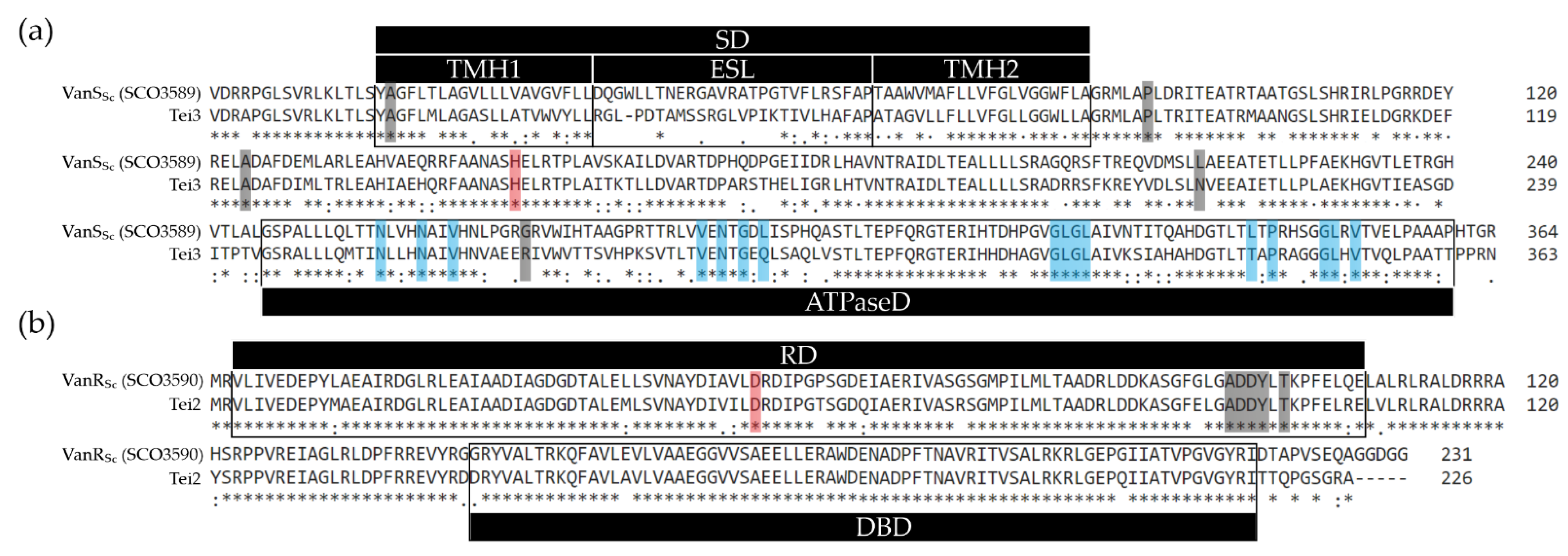
Figure 1. Clustal Omega [41] pairwise alignments of aa sequences of Tei3 and VanSSc (a) as well as of Tei2 and VanRSc (b). Domains and sequence features were annotated according to [31] and CD-Search tool [42]; SD—sensor domain, TMH1/2—first and second transmembrane α-helices, ESL—extracytoplasmic sensory loop, ATPaseD—ATPase domain, RD—response domain, DBD—DNA binding domain. In (a): mutation sites that were shown to impair VanSSc function [31] are highlighted in gray; the autophosphorylation site is highlighted in red; conserved ATP-binding sites are in blue. In (b): residues putatively involved in dimerization are highlighted in gray, while the phosphorylation site is in red.
Ijms 23 15713 g001
Figure 2. Expression of tei3 led to teicoplanin and A40926 resistance in S. coelicolor M512. A number of 106 spores of S. coelicolor M512 and M101 were inoculated at each sector of SMMS agar plates added with vancomycin, teicoplanin, and A40926. Plates were examined after 72 h of incubation. S. coelicolor M512 was resistant to vancomycin and sensitive to teicoplanin and A40926, as previously reported [43], whereas M101 was resistant to vancomycin, A40926, and teicoplanin.
Ijms 23 15713 g002
Figure 3. gusA-based GPA-inducible reporter assay revealed a constitutive phosphorylase activity of Tei3. S. coelicolor strains pGJp+ and M1J were inoculated on SMMS agar added with 25 µg/mL of X-Gluc along with their gusA-control strains, i.e., the teicoplanin/A40926-sensitive M512 and the teicoplanin/A40926-resistant M111. Notably, in pGJp+, the chromogenic conversion of X-Gluc (observed as green halos) occurred only upon GPA induction, while the background of M1J remained completely green, implying that Tei3 did not require GPA induction of its activity. A number of 107 spores of each strain were used for inoculation and the plate was examined after 48 h of incubation.
Ijms 23 15713 g003
Figure 4. Loss of the gene fragments coding for transmembrane helices and extracytoplasmic sensory loops makes tei3 and vanSSc non-functional. A number of 106 spores were inoculated at each sector of SMMS agar plates added with vancomycin, teicoplanin, or A40926; plates were examined after 72 h of incubation. All the recombinant strains deriving from M512 were vancomycin-resistant and teicoplanin- and A40926-sensitive, whereas all the recombinant strains deriving from J3200 were resistant to all the tested GPAs.
Ijms 23 15713 g004
Figure 5. Hybrid SHK containing SD from Tei3 and ATPaseD from VanSSc made S. coelicolor J3200 sensitive to teicoplanin and vancomycin, and S. coelicolor M512 sensitive to vancomycin, but resistant to A40926, implying that Tei3 SD recognized A40926 as a ligand. A number of 106 spores were inoculated at each sector of SMMS agar plates added with vancomycin, teicoplanin, or A40926; plates were examined after 72 h of incubation.
Ijms 23 15713 g005
Table 1. Plasmids and bacterial strains used or generated in this study.
NameCharacteristicReference
Plasmids:
pSET152AφC31-based integrative plasmid, pSET152 derivative carrying aac(3)IVp from pIJ773, Amr[63]
pKC1132suicide vector, Amr[45]
pRT801φBT1-based integrative plasmid, Amr[46]
pGUSφC31-based integrative plasmid, pSET152 derivative containing promoterless GusA, Amr, Spr[44]
pGP101pSET152A derivative carrying tei3this work
pGP111pRT801 derivative carrying tei3this work
pGP102pSET152A derivative carrying SCO3589 (vanSSc)this work
pKC1132-102pKC1132 derivative carrying SCO3589 (vanSSc)this work
pKC1132-103pKC1132 derivative carrying hybrid SHK gene with tei3-SD and vanSSc-ATPaseDthis work
pGP103pSET152A derivative carrying hybrid SHK gene with tei3-SD and vanSSc-ATPaseDthis work
pGP104pSET152A derivative carrying tei3this work
pGP105pSET152A derivative carrying SCO3589′this work
pGP106pSET152A derivative carrying tei3this work
pGP107pSET152A derivative carrying SCO3589″this work
pGJppGUS derivative carrying vanJpthis work
pHJppGUS derivative carrying vanHpthis work
Strains:
A. teichomyceticus ATCC 31121wild type, teicoplanin producerATCC
S. coelicolor M512A3(2) derivative, ΔredD ΔactII-ORF4 SCP1 SCP2[38]
S. coelicolor J3200A3(2) derivative, ΔSCO3589 (vanSSc)[31]
S. coelicolor M101M512 derivative carrying pGP101this work
S. coelicolor M111M512 derivative carrying pGP111this work
S. coelicolor pGJp+M512 derivative carrying pGJpthis work
S. coelicolor pGHp+M512 derivative carrying pGHpthis work
S. coelicolor M1JM512 derivative carrying pGJp and pGP111this work
S. coelicolor M103M512 derivative carrying pGP103this work
S. coelicolor M104M512 derivative carrying pGP104this work
S. coelicolor M105M512 derivative carrying pGP105this work
S. coelicolor M106M512 derivative carrying pGP106this work
S. coelicolor M107M512 derivative carrying pGP107this work
S. coelicolor J103J3200 derivative carrying pGP103this work
S. coelicolor J104J3200 derivative carrying pGP104this work
S. coelicolor J105J3200 derivative carrying pGP105this work
S. coelicolor J106J3200 derivative carrying pGP106this work
S. coelicolor J107J3200 derivative carrying pGP107this work
Table 2. Oligonucleotide primers used in this study.
NameSequence (5′-3′) *Purpose
tei3_sdomain_PaeICGAGCATGCGACCGGCCAGReverse primer for cloning the tei3 region coding for the sensor domain
tei3_FTTTGATATCGGAGGGAGACCGTGGACCGAGCCCCloning of the tei3 and its truncated versions
tei3’_FTTTGATATCGGAGGGAGACCGTGTTCGCCCCGGCGACGG-//-
tei3”_FTTTGATATCGGAGGAGACCGTGGGTCGGATGCTCGCTCCTCT-//-
tei3_RTTTGAATTCGCGGTGGGCGGTTCAGTTT-//-
SCO3589_FTTTGATATCGGAGGGCGACGGTGGATAGGCGCCCloning of the SCO3589 (vanSSc) and its truncated versions
SCO3589’_FTTTGATATCGGAGGGAGACCGTGCTTCGCAGTTTCGCCC-//-
SCO3589’’_FTTTGATATCGGAGGGCGACGGTGGGACGCATGCTCGCCCCCCT-//-
SCO3589_RTTTGAATTCTGGCGCTCACCTGCCGGTG-//-
vanHp_KpnI_FTTTGGTACCTACGTCCACACCGCCGAGCCloning of vanHSc promoter region into pGUS
vanHp_SpeI_RTTTACTAGTGCCGTCCCGTATGCGCTTT-//-
vanJp_KpnI_FTTTGGTACCACACTCAGCAGCTCCAACGCloning of vanJSc promoter region into pGUS
vanJp_SpeI_RTTTACTAGTCTGGCGCCGGTGCGGCCGA-//-
aac(3)IV_FATCGACTGATGTCATCAGCGDiagnostic primers for the amplification of aac(3)IV gene
aac(3)IV_RCGAGCTGAAGAAAGACAAT-//-
gusA_ver_FGGCGGCTACACGCCCTTCGADiagnostic primers for the amplification of 1000 bp internal region of gusA
gusA_ver_RTGATGGGCCGGGTGGGGTC-//-
* artificial ribosome binding sites are highlighted in red and artificial start codons are underlined.
Publisher’s Note: MDPI stays neutral with regard to jurisdictional claims in published maps and institutional affiliations.