Article

A Tea Plant (Camellia sinensis) FLOWERING LOCUS C-like Gene, CsFLC1, Is Correlated to Bud Dormancy and Triggers Early Flowering in Arabidopsis

1
Key Laboratory of Tea Biology and Resources Utilization, Ministry of Agriculture and Rural Affairs of the People’s Republic of China/ National Center for Tea Improvement/Tea Research Institute, Chinese Academy of Agricultural Sciences, Hangzhou 310008, China
2
Instituto de Biología Molecular y Celular de Plantas (IBMCP), Consejo Superior de Investigaciones Científicas-Universidad Politécnica de Valencia, 46022 Valencia, Spain
*
Author to whom correspondence should be addressed.
Int. J. Mol. Sci. 2022, 23(24), 15711; https://doi.org/10.3390/ijms232415711
Submission received: 1 November 2022 / Revised: 6 December 2022 / Accepted: 9 December 2022 / Published: 11 December 2022
(This article belongs to the Section Molecular Plant Sciences)

Abstract

:
Flowering and bud dormancy are crucial stages in the life cycle of perennial angiosperms in temperate climates. MADS-box family genes are involved in many plant growth and development processes. Here, we identified three MADS-box genes in tea plant belonging to the FLOWERING LOCUS C (CsFLC) family. We monitored CsFLC1 transcription throughout the year and found that CsFLC1 was expressed at a higher level during the winter bud dormancy and flowering phases. To clarify the function of CsFLC1, we developed transgenic Arabidopsis thaliana plants heterologously expressing 35S::CsFLC1. These lines bolted and bloomed earlier than the WT (Col-0), and the seed germination rate was inversely proportional to the increased CsFLC1 expression level. The RNA-seq of 35S::CsFLC1 transgenic Arabidopsis showed that many genes responding to ageing, flower development and leaf senescence were affected, and phytohormone-related pathways were especially enriched. According to the results of hormone content detection and RNA transcript level analysis, CsFLC1 controls flowering time possibly by regulating SOC1, AGL42, SEP3 and AP3 and hormone signaling, accumulation and metabolism. This is the first time a study has identified FLC-like genes and characterized CsFLC1 in tea plant. Our results suggest that CsFLC1 might play dual roles in flowering and winter bud dormancy and provide new insight into the molecular mechanisms of FLC in tea plants as well as other plant species.

1. Introduction

Flowering is an important trait that helps plants transition from the vegetative phase to the reproductive phase and involves many complex changes, including those involving physiological, metabolic and molecular processes [1]. Moreover, flowering time is regulated not only by a plant’s intracellular signature but also by environmental factors [2]. Daylength and temperature are two main environmental factors that affect plant reproduction. According to the daylength and temperature, plants can sense the season and whether the time is appropriate to produce flowers and seeds. The vernalization pathway was proposed to explain how temperate angiosperms avoid blooming during winter. FLC is considered a crucial regulator in the vernalization pathway [3]. The function of FLC differs between ecotypes of Arabidopsis thaliana: it represses flowering in late-flowering ecotypes [4], while in early flowering ecotypes, FLC overexpression further delays flowering [5]. In Chinese cabbage (Brassica rapa ssp. pekinensis (Lour.) Hanelt), there are three FLC homologues that have lower expression levels in early flowering varieties [6]. Additionally, in Eustoma grandiflorum (Raf.) Shinners, EgFLC represses flowering [7]. FLC was reported to directly bind to the promoter of SUPPRESSOR OF OVEREXPRESSION OF CONSTANS 1 (SOC1) and the first intron of FLOWERING LOCUS T (FT) to repress expression and delay flowering time [8,9]. FLC expression is reduced by DNA methylation (epigenetic modifications) but induced by acetylation of histones (chromatin remodeling) [10,11,12,13,14,15,16,17,18].
Bud dormancy is necessary for temperate perennials to avoid cellular damage and ensure plant survival during winter. FLC was also related to bud dormancy of perennial plant species such as apple (Malus × domestica Borkh.) and kiwifruit (Actinidia chinensis Planch.), where it is highly expressed during dormancy [19,20]. However, transgenic kiwifruit plants overexpressing AcFLCL displayed earlier budbreak times [20].
Tea plant (Camellia sinensis (L.) Kuntze) is an economically important crop species in many countries and areas of Asia, Africa and Latin America [21]. Its leaves are processed into different kinds of tea, which is famous for its health benefits [22,23,24,25]. Since tea plant is a crop species grown for its leaves, its reproductive phase from flowering to fruit production requires an abundance of plant resources, thus affecting tea plant bud growth and limiting the production and quality of tea. Therefore, breeding improved tea varieties or finding a way to balance vegetative and reproductive growth to improve the efficiency of the tea industry is urgently needed. For this, it is necessary to understand the molecular mechanisms of flowering in tea plant. Tea plant FLC orthologues have not yet been described. In our previous study, the phenotypes of two types of tea plant cultivars, a floriferous type and an oliganthous type, were observed [26]. We found that the meristem was maintained in the vegetative phase and did not switch to reproductive growth in the axillary buds of the oliganthous cultivars, while the transition to the floral meristem occurred in June in the axillary buds of the floriferous tea plant cultivars. The flowering-related genes in these two cultivars were identified, and among them, one MADS-box gene was found that might play an important role in floral organ differentiation and maturation [26]. In temperate areas, tea plant usually undergoes bud dormancy in the winter to overcome low-temperature stress, and when the temperature arises in the spring, the buds emerge. The progression of the bud dormancy–budbreak phase involves many genes, such as transcript factor genes bZIP (basic leucine-zipper) and MIKC-MADS as well as phytohormone-associated genes auxin-, ABA-, GA- and JA-associated genes [27,28,29]. Despite several studies about bud dormancy in tea plant, the role of CsFLC in dormancy is still unknown. Hence, it is necessary for breeders to understand the mechanisms of flowering and dormancy as well as their relationship.
Tea plant is a leaf-used crop, reproductive growth including flowering and fruit production affects vegetative growth and reduces outputting of tea. Therefore, it is necessary to study mechanisms of flowering in tea plant. In annuals, FLC plays key roles in flowering [3,4,5,6]. While in perennials, FLC is associated to bud dormancy [19,20]. Bud dormancy helps tea plant avoid low-temperature damage. FLC-like genes might have dual functions on flowering and bud dormancy in perennials. It’s meaningful to identify and characterize FLC-like genes in tea plant. In this study, we identified three FLC-like genes in tea plant, including CsFLC1, whose expression was correlated with bud dormancy and flower bud development. The RNA transcription of CsFLC1 was monitored throughout the year. Then, we established CsFLC1-overexpressing transgenic Arabidopsis lines to characterize its functionality in a heterologous model plant species. Our study provides a new understanding of flowering as well as bud dormancy in tea plant and a theoretical basis for future breeding programmers to develop novel cultivars with early bud break and few flowers.

2. Results

2.1. Identification of CsFLC-Like Genes in Tea Genome

In our previous work, we have isolated a MADS-box gene candidate that might play an important role in floral organ differentiation and maturation [26]. Then, we further analyzed its putative protein product and found that it shares the highest homology with FUL-like and FLC-like genes. Considering its high degree of sequence divergence, and in order to understand its true identity, we conducted a mixed approach of phylogeny and gene collinearity. A phylogenomic study [30] clarified that one paleo-FLC gene was present in the ancestor of angiosperms and that, in fact, FLC-like genes also exist in monocots but were previously misclassified. Their conserved genomic locations and their tendency to duplicate only by whole-genome duplications can help overcome the limits of phylogenetic analysis. In particular, the ancestral genomic configuration of FLC is in a narrow tandem with a monophyletic group of SQUAMOSA promoter BINDING PROTEIN (SBP) members and also with SEPALLATA3 (SEP3) [30]. Since core eudicots belong to a paleohexaploid ancestor, this SBP-SEP3-FLC cluster should have triplicated in them. Indeed, in the model genome of grape (Vitis vinifera L.) two such clusters have been reported by Ruelen and coworkers [30] in addition to another one that we have found in chromosome 17 (Figure 1A,B). However, FLC and its close homologues MAF genes lost this conserved collinearity in the Arabidopsis family, the Brassicaceae, probably by gene transposition or a massive genome fractionation process [31].
Except for Brassicaceae, we found that the conservation of the three SBP paralogous clades is remarkable among core eudicot species, and most of them reveal an FLC locus in close proximity (Figure 1A,B). In tea plant, two of these SBP genes are located in proximity of a putative FLC locus, and an SEP3-FLC tandem also exists (Figure 1A,B). Despite the tea plant genome undergoing a relatively old whole-genome duplication [32]), only duplicated copies of SEP3 seem to have been retained within the SBP-SEP3-FLC clusters, evidencing multiple gene losses (Figure 1B, Table S1). Finally, a fourth highly diverged MADS-box gene was isolated by BLAST (Genebank XP_028119527.1), without any clear homology to other core eudicot genes we have screened so far, and in a region non collinear to FLC, for which further analysis is needed. In conclusion, three paralogous FLC clades seem to exist in core eudicots, with one member each in the tea plant genome, that we named CsFLC1, CsFLC2 and CsFLC3. Despite the fact that CsFLC1 shares only 32% homology with Arabidopsis FLC, it is 56% homologous and collinear to the AcFLCL (Acc05562) gene recently reported in the closely related kiwifruit [20]. While CsFLC2 is homologous and shares gene collinearity to kiwifruit genes Acc14299 and Acc33776 (data not shown).
Since FLC-like genes have higher expression during dormancy in apple and kiwifruit, we analyzed the expression pattern of CsFLC1, CsFLC2 and CsFLC3 in different tea bud dormant state transcriptomes [29], including endo-dormancy, eco-dormancy, para-dormancy and bud flush (Figure S1). The result shows that only CsFLC1 had high mRNA expression level during winter dormancy (endo- and eco-dormancy) while CsFLC2 did not have any change across the four states and CsFLC3 had changes much lower than CsFLC1. Hence, CsFLC1 was chosen for further analysis.

2.2. CsFLC1 Gene Expression Patterns

To identify the subcellular location of CsFLC1, we constructed a 35S::CsFLC1:eGFP plasmid and transformed it into Agrobacterium to infect nuclear marker (red fluorescence) transgenic tobacco. Through confocal microscopy, we observed green fluorescence merged with red fluorescence to show yellow light. As expected for a putative transcription factor (TF), CsFLC1 was located in the cell nucleus (Figure 2).
To study the expression pattern of CsFLC1, we detected the gene expression level in axillary or floral buds in tea plant (Figure 3A). The result shows that CsFLC1 had two expression peaks (one in 28 December and another in 28 August) in the whole year (from 2016 to 2017). From 1 November to the next 14 March was a dormancy period in tea plant [29]. From 27 May to 26 September was the floral bud differentiation and floral bud development phases [26]. The result shows that CsFLC1 was corresponding to bud dormancy and flowering.
To identify in which tissues CsFLC1 is expressed, we measured its expression in seven different tissues of tea plant, namely, apical buds, axillary buds, flower buds, flowers, mature leaves, stems and roots (Figure 3B). CsFLC1 had the highest expression level in the three kinds of buds, and the next highest expression was in the stems and roots, while the lowest was in the flowers and leaves. Therefore, we further measured the expression in different flower organs (Figure 3C). The results show that CsFLC1 had the highest expression level in the pistils followed by the petals, while it was not detected in stamens (Figure 3D). In pCsFLC1::GUS transgenic Arabidopsis, we found similar results (Figure 3E). GUS was expressed in the apical meristems, pistils, and stamens followed by the vascular tissue but was hardly detected in the roots and leaves. The expression patterns in tea and pCsFLC1::GUS Arabidopsis were common in the apical meristem and pistil but different in the petals, stamens and roots, which might be caused either by an incomplete CsFLC1 promoter region or by differences in the Arabidopsis heterologous system.
In Figure 3A, CsFLC1 was highly expressed during the bud dormancy phase, and to explore if CsFLC1 could respond to low temperature (LT), we detected the expression pattern of CsFLC1 under low-temperature treatment in tea plant.
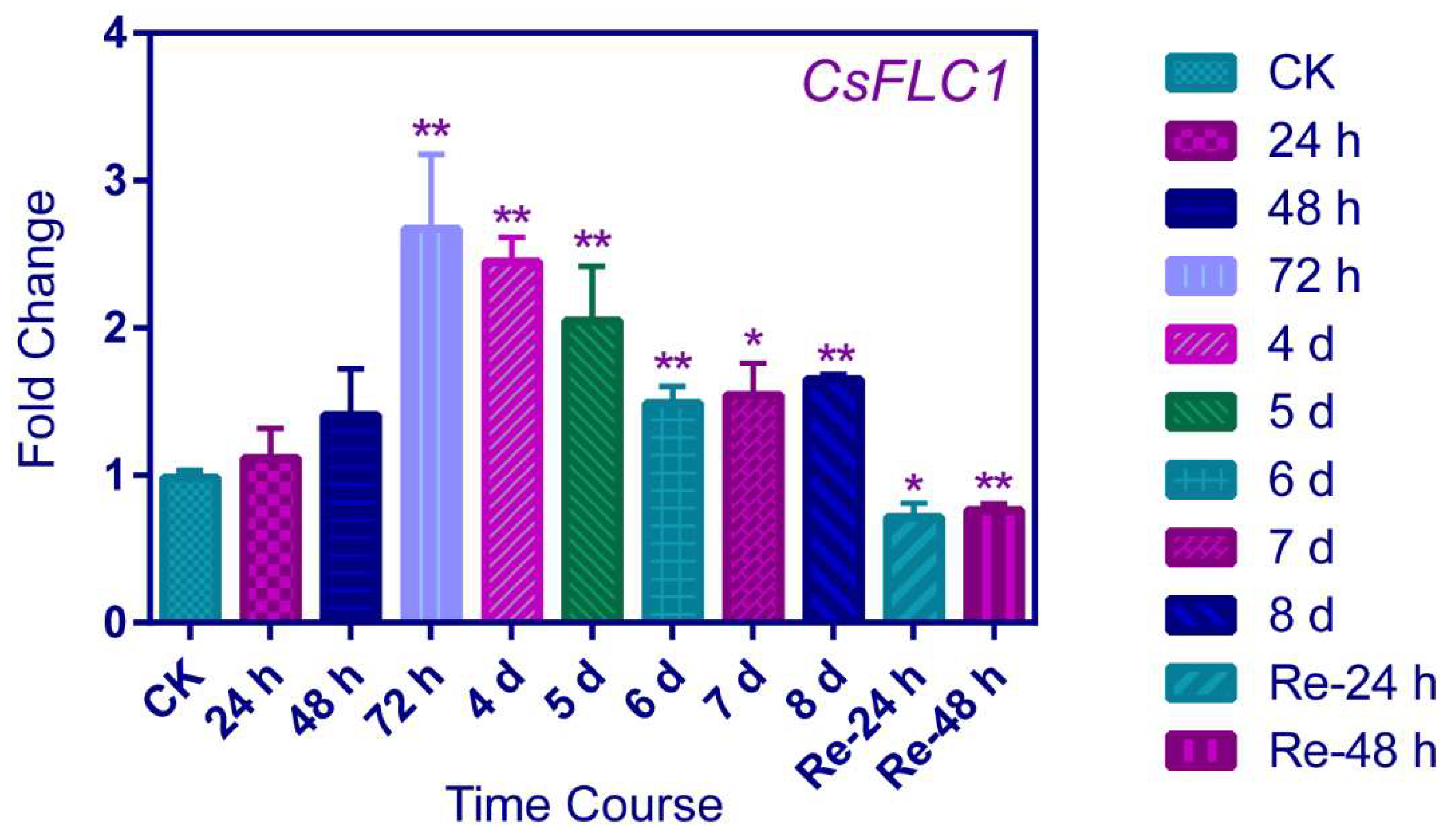
CsFLC1 was significantly responding to LT after 72 h of treatment and then the RNA expression level was lower at 4, 5 and 6 days; later, at 7 and 8 days, the expression levels were stabilized but also higher than CK (0 d) (Figure 4). When recovering to a warm temperature after 24 and 48 h, the expression level of CsFLC1 was significantly lower than CK. This result suggests that CsFLC1 was induced by LT while being reduced by a warm temperature. Combining the expression pattern of CsFLC1 in tea plant during bud dormancy, CsFLC1 might play roles in maintaining bud dormancy.

2.3. The CsFLC1 Promoter Is Responsive to Low Temperature and Photoperiod

To clarify the CsFLC1 response to environmental stimuli, we treated pCsFLC1::GUS transgenic lines with low temperature (4 °C) and different day length, LD (16 h light/8 h dark), medium day MD (12 h light/12 h dark) and short day SD (8 h light/16 h dark), and observed the relative GUS staining pattern. The result shows that the GUS staining in leaf veins, leaf apexes and roots was stronger under low temperature treatment than the control group (Figure 5A–C). GUS expressed highly either in light or dark under MD treatment (Figure 5E,H), while it expressed higher in light than dark under LD treatment (Figure 5D,G), and its behavior was the opposite under SD treatment (Figure 5F,I).

2.4. CsFLC1 Affects Flowering Time and Seed Germination in Transgenic Arabidopsis thaliana

To learn the function of CsFLC1, we tested three independent transgenic Arabidopsis lines of 35S::CsFLC1 (Figure 6B). We observed the phenotype of overexpressed (OE)lines and found the bolting, flowering and aging time of transgenic lines were earlier than Col-0 wild type (WT; Figure 6A,D). In addition, the higher the CsFLC1 expression, the lower the seed germination rate (Figure 5C).
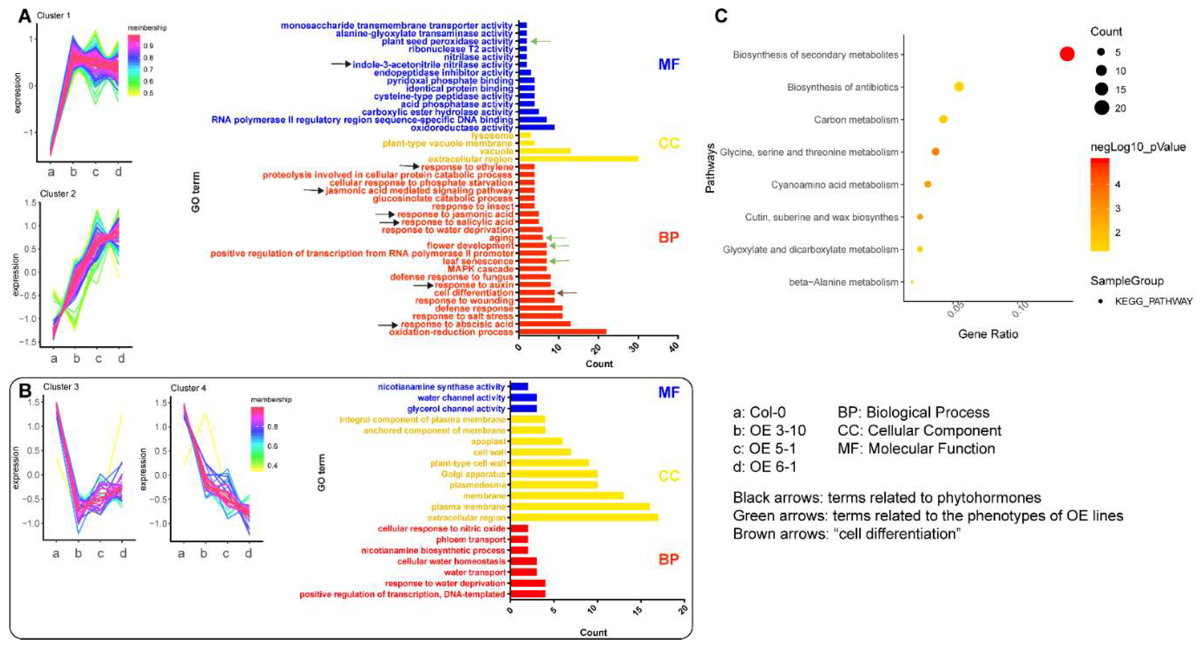
To further study how CsFLC1 plays roles in these physiological processes, we detected gene expression levels of three OE lines and WT Arabidopsis by RNA-sequencing. Compared to WT, 228 differentially expressed genes (DEGs) were common in the three OE lines (Figure S2). In total, 169 DEGs were upregulated, while 59 DEGs were downregulated in OE lines compared with WT (Figure 7A,B). Among the up-regulated gene clusters of OE-CsFLC1 lines, the terms ‘aging’, ‘flower development’ and ‘leaf senescence’ in biological process (BP) were enriched. Moreover, we observed that the OE-CsFLC1 lines were senescent, bolting and blooming earlier than the wild type. Therefore, the phenotype and regulated pathways could be matched. The terms ‘response to abscisic acid’, ‘response to auxin’, ‘response to salicylic acid’, ‘response to jasmonic acid’, ‘jasmonic acid mediated signaling pathway’, ‘response to ethylene’ in BP and ‘indole-3-acetonitrile nitrilase activity’ in molecular function (MF) were enriched (Figure 7A). According to GO analysis in down-regulated gene clusters, the BP terms ‘water channel activity’, ‘glycerol channel activity’ and ‘nicotianamine synthase activity’, some cellular component (CC) terms about membrane and cell wall, as well as ‘cellular water homeostasis’, ‘water transport’ and ‘response to water deprivation’ MF terms were enriched (Figure 7B). The biosynthesis secondary metabolite KEGG pathway was clustered significantly in 169 upregulated DEGs, while no pathway was clustered in 59 downregulated DEGs (Figure 7C).
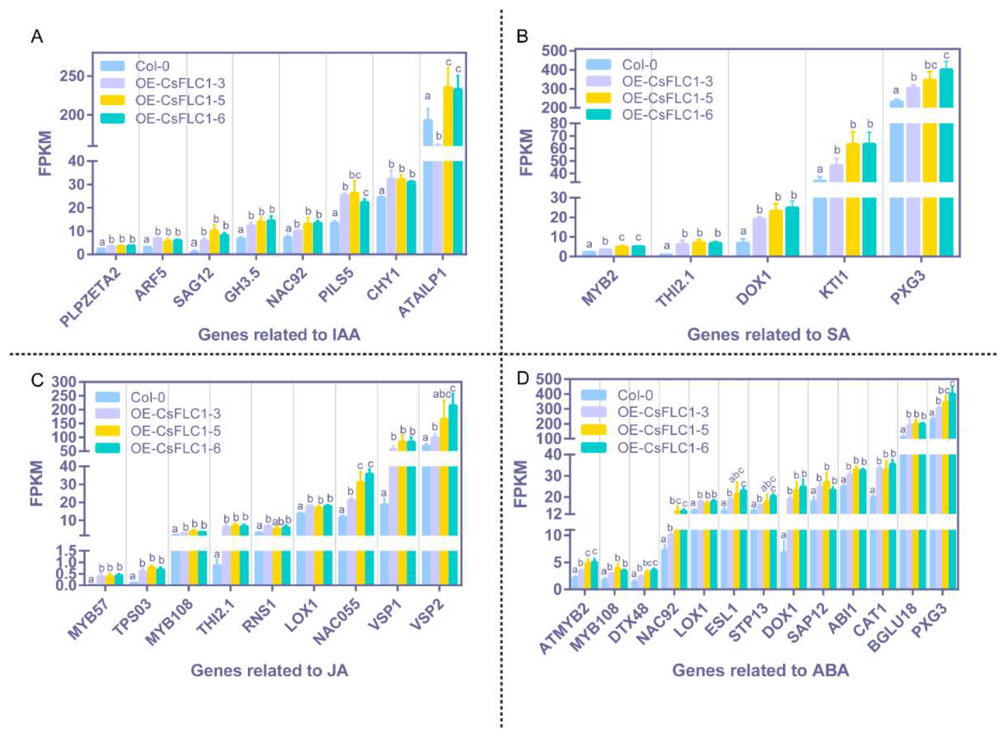
To study which genes respond to auxin, SA, JA and abscisic acid (ABA) were upregulated in the high-level CsFLC1 lines, and we displayed the transcriptional levels in the pathways mentioned above (Figure 8). The result shows that almost all these genes had a higher expression level in OE-CsFLC1 lines compared to WT, but there were no significant differences of expression levels among the OE-CsFLC1 lines. According to the results of transcriptomes, we noticed that three auxin-related genes AUXIN RESPONSE FACTOR 5/MONOPTEROS (ARF5/MP), SENESCENCE-ASSOCIATED GENE 12 (SAG12) and the acyl acid amido synthetase gene Gretchen Hagen 3.5 (GH3.5), three SA-related genes THIONIN 2.1 (THI2.1), MYB2 and DIOXYGENASE 1 (DOX1), four JA-related genes MYB57, TERPENE SYNTHASE 03 (TPS03), NAC055 and VEGETATIVE STORAGE PROTEIN 1 (VSP1) as well as four ABA-related genes DETOXIFICATION 48 (DTX48), NAC92, CATALASE 1 (CAT1) and BETA GLUCOSIDASE 18 (BGLU18) were significantly and obviously upregulated in OE-CsFLC1 lines.
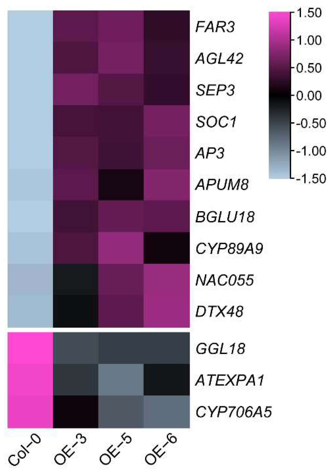
To explore which DEGs might be regulated by CsFLC1 and cause the phenotypes of transgenic Arabidopsis thaliana, we analyzed expression patterns of known AtFLC direct target genes based on ChIP-seq data [33]. There were 13 common genes, among them 10 genes were induced, while 3 genes were repressed in OE-CsFLC1 lines (Table S2). In OE-CsFLC1 lines, four MADS-box family members AGAMOUS-LIKE 42 (AGL42), SEP3, SOC1 and APETALA3 (AP3), three hormone-related genes BGLU18, NAC055 and DTX48, a post-transcriptional regulator PUMILIO 8 (APUM8), a Cytochrome P450 gene CYP89A9 as well as a wax biosynthetic gene FATTY ACID REDUCTASE 3 (FAR3) were up-regulated while GUARD-CELL-ENRICHED GDSL LIPASE 18 (GGL8), a cell-wall-related gene EXPA1 as well as a Cytochrome P450 gene CYP706A5 were down-regulated (Figure 9).
In conclusion, CsFLC1 has functions in controlling bolting and blooming times, seed germination, leaf senescence as well as regulating some auxin, SA-, JA- and ABA-associated genes in transgenic Arabidopsis thaliana.

2.5. Phytohormone Contents of Tea Plants in the Whole Year

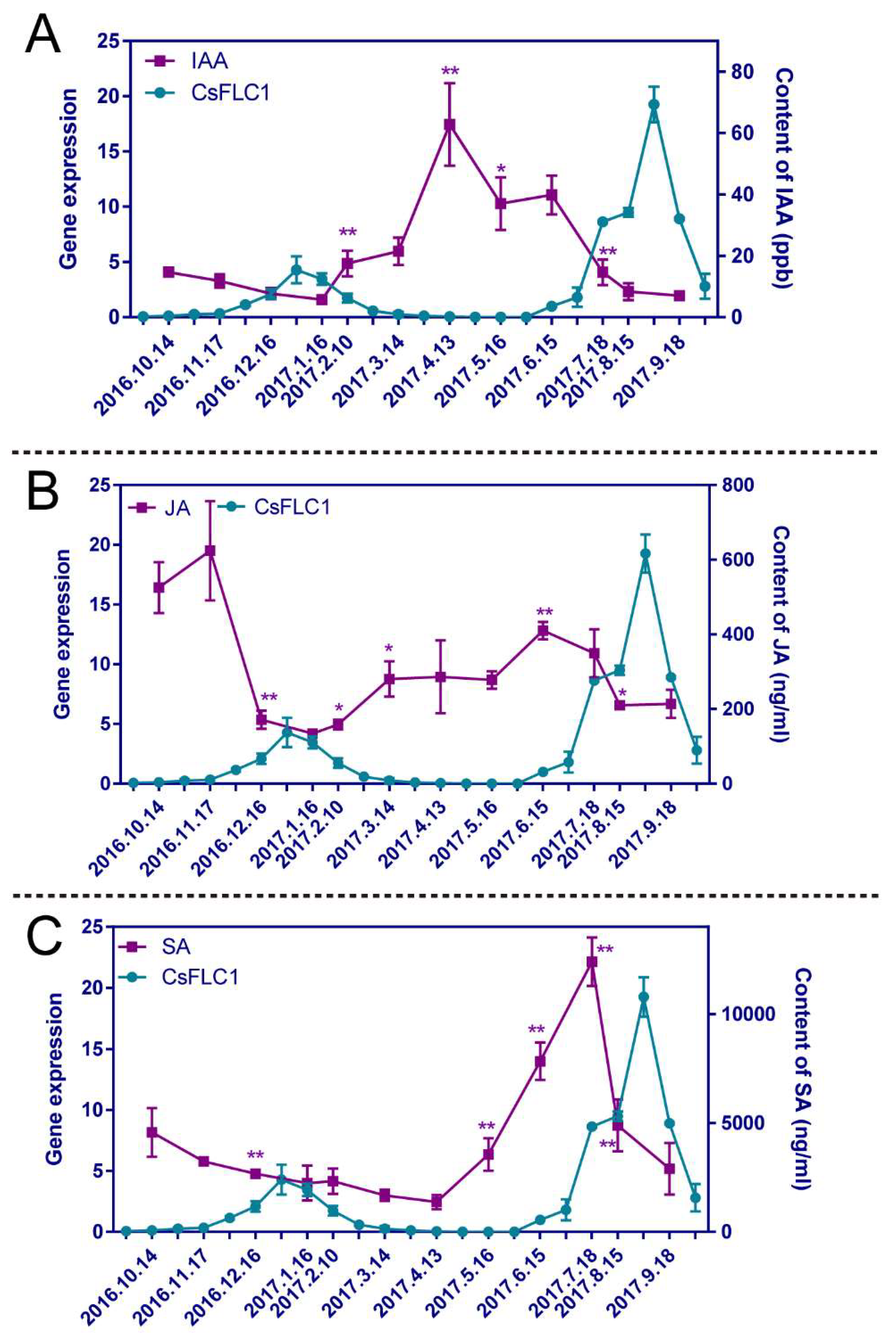
Since we found there were some phytohormone pathways influenced in 35S::CsFLC1 lines, we measured the contents of three hormones (indole-acetic acid (IAA), JA and SA) in tea plant buds throughout the year. The hormone content changes are displayed in Figure 10.
The content of IAA was low from 14 October 2016 to 16 January 2017, and then increased until 13 April. The content peaked; later, it declined until 18 July, and finally, it was stable and low again (Figure 10). The JA content was high on 14 October and 17 November, decreased on 16 December, remained at a low level until 10 February, increased on 14 March, remained at a moderate level from 10 February to 16 May, increased on 15 June, decreased until 15 August, and finally remained low until 18 September (Figure 10B). The content of SA was 4573.43 ng/mL on 14 October 2016, slowly decreased until 13 April 2017, rapidly increased until 18 July and rapidly decreased until 18 September, sharply peaking on 18 July (Figure 10C). The changes in IAA and JA contents were opposite those of the CsFLC1 expression level. There was only one time at which the SA content peak, which occurred one and-a-half months before the peak CsFLC1 expression occurred, which was on 28 August. In conclusion, CsFLC1 expression coincided with low contents of IAA and JA throughout the year but coincided with a high SA content of during flowering.

3. Discussion

3.1. Function of CsFLC in Reproduction Processes

Flowering time is regulated by five main pathways: the autonomous, gibberellin, aging, photoperiod and vernalization pathways [34,35]. These various pathways depended on different genetic regulations [36]. FLC was identified first in 1999 and was considered to encode a repressor of flowering; its expression was upregulated by FRIGIDA (FRI) but downregulated by vernalization in Landsberg erecta (Ler, a late-flowering ecotype) [4,10]. The FLC expression level was found to be an indicator of the extent of the vernalization and was also regulated by the autonomous pathway [3,37]. FLC regulates the circadian clock via autonomous and vernalization pathways to control flowering time, which shows that FLC is a link between vernalization and the circadian rhythm [38]. In addition, by binding to their chromatin, FLC represses the expression of FT and SOC1 to inhibit flowering [8,9]. In apple trees, MdFLC-like is expressed in flower buds and is upregulated during cold accumulation and flower primordium differentiation and development [19].
In our previous study, we found that, during the flowering period, CsFLC1 was specifically expressed during the floral transition stage [26]. In this study, to find out when CsFLC1 was functioning, we detected the expression pattern of CsFLC1 in tea plant. According to the expression patterns, CsFLC1 was highly expressed during flower organ development. Thus, it positively regulated floral development, which coincided with the function of the apple MdFLC-like gene. Based on these findings, we constructed transgenic CsFLC1 OE Arabidopsis in the Col-0 (an early flowering ecotype) background to further study the function of CsFLC1. With respect to 35S::CsFLC1 transgenic lines, bolting and blooming occurred earlier than it did for Col-0. To clarify which pathways and genes were influenced by CsFLC1 in OE Arabidopsis, we performed RNA-seq. According to the GO analysis of the transcriptome data, genes annotated to the ‘flower development’ term were enriched in the OE lines. We measured CsFLC1 expression in the apical and axillary buds, flower buds and pistils of tea plant as well as in the carpel of pCsFLC1::GUS transgenic Arabidopsis to research the tissue expression of CsFLC1. Our results show that CsFLC1 might positively regulate early flowering, floral transition, petal and pistil development reproduction processes. The transcriptome results show that SOC1 was upregulated in OE-CsFLC1 lines. Thus, CsFLC1 might induce flowering by influencing the expression of SOC1 in tea plant, the results of which are the opposite to those of Ler Arabidopsis plants for both phenotype and genetic regulatory relationships. AGL42, a SOC1-like gene that was reported to be a target of SOC1 and FLC, can promote flowering [33,39]. SEP3 is essential for floral meristem determinacy [40]. AP3 is involved in the formation of petals and stamens during flower development [41]. In our study, these four MADS-box genes SOC1, AGL42, SEP3 and AP3 were upregulated by CsFLC1, indicating that CsFLC1 promotes flowering by controlling the expression of these genes in Arabidopsis. While in flc-3 mutant, AtFLC targets SOC1 and SEP3 were up-regulated [33], it was the same in OE-CsFLC1 transgenic Arabidopsis (Figure 9), which means that AtFLC and CsFLC1 have the opposite function on regulating SOC1 and SEP3. The fact that CsFLC1 promotes flowering while AtFLC represses flowering might be because the sequences of FLC are not conserved among species [30], which results in different functions in flowering and gene regulation.

3.2. CsFLC1 Putative Function in Bud Dormancy

In tea plant, CsFLC1 was highly expressed during the winter, which was identified as the endodormancy period of axillary buds (Figure S1). Based on the results of the GUS location patterns, CsFLC1 was highly expressed in light under LDs, while it was induced in the dark under SDs, but it was highly expressed continuously under MDs. Interestingly, CsFLC accumulated in the day under LD conditions but accumulated at night under SD conditions (Figure 3). This means that it plays different roles under different photoperiods. In previous studies, the transcript of FLC was independent of the photoperiod [3,4], indicating that CsFLC1 is a special FLC with dual function in response to different photoperiods. Additionally, in the pCsFLC1::GUS lines as well as LT-treated tea plant (Figure 4 and Figure 5), CsFLC1 could respond to low temperatures. SDs and low temperatures are the signals of winter, and plants enter dormancy when they receive the signals of winter to avoid damage and no longer grow before the environment is suitable for growth.
To further explore whether CsFLC1 was coordinated with bud dormancy, we detected its expression in the late budbreak cultivar ZHDB from 30 September 2016 to 16 May 2017 (Figure S3). The results show that the mRNA transcript of CsFLC1 was no longer detected approximately one month later than in LJ43, which coincided with budbreak time. Combining the results above, we predict that CsFLC1 is a repressor of bud break and maintains the bud dormancy state. In apple and kiwifruit, FLC-like genes were reported to be highly expressed during dormancy, while overexpression AcFLCL transgenic kiwifruit budbreak time is earlier [19,20]. This might be because FLC plays a role like a switch on bud dormancy only if FLC reaches a threshold value when the environmental temperature is favorable, meaning the dormancy could break easily. In tea plant, CsFLC1 was up-regulated from endo- to eco-dormancy (Figure 3A and Figure S1); in addition, CsFLC1 could be a response to low temperatures and down-regulated by the following warm temperature (Figure 4), which indicates that CsFLC1 might be a switch between bud dormancy and budbreak. Phytohormones play important roles in regulating plant bud dormancy [42]. In tea plant, auxin-, ABA-, GA- and JA-associated genes were involved in bud dormancy [29,43]. In OE-CsFLC1 transgenic Arabidopsis, IAA-, JA- and ABA-associated genes were affected (Figure 7A and Figure 8), which suggested that CsFLC1 might control bud dormancy by regulating phytohormone-associated genes.

3.3. Relationship between CsFLC1 Expression and Phytohormones’ Concentration and Response

Phytohormones are important in the growth, development and response to environmental stimuli of plants. It has been reported that flowering and growth are promoted by relatively low concentrations of auxin but inhibited by higher concentrations [44]. According to the GO enrichment of DEGs of OE-CsFLC1 Arabidopsis, several phytohormones, such as auxin, SA and JA were enriched (Figure 7). To clarify the relationship between CsFLC1 and hormones, contents of IAA, SA and JA were detected. The results of whole-year RNA expression and hormone content detection showed that CsFLC1 RNA transcription was correlated with a low content of IAA in both axillary and floral buds of tea plant. ARFs are TFs that bind to AuxREs in the promoters of early auxin response genes [45,46]. ARF5 is an important activator in auxin signaling and is expressed in cells with low levels of auxin [47]. SAG12 is a senescence indicator and is repressed by auxin [48,49,50]. GH3.5 functions in modulating and integrating both auxin and SA signaling [51,52] is expressed in seedlings, roots, stems, buds and blooming flowers of Arabidopsis thaliana [53], and influences root development in rice [54]. ARF5, SAG12 and GH3.5 were significantly upregulated in OE-CsFLC1 lines, which indicated that CsFLC1 might activate them to control auxin signaling and further induce flowering at low concentrations of auxin.
JA plays a critical role in inflorescence, stamen and seed development [55,56]. JA induces the expression of MYB57 to promote stamen filament development [57]. The JA content moderately peaked from 16 May to 15 August when the plants were in the floral transition and floral organ differentiation stages, and MYB57 had an obviously higher transcription level in OE-CsFLC1 lines. NAC055 is a target of FLC and is induced by MeJA [58]; in our study, it was highly expressed in OE-CsFLC1 transgenic Arabidopsis. These results show that both CsFLC1 and JA might control flowering through NAC055.
Cleland and Ajami [59] found that SA could induce flowering in Lemna gibba G3. In this study, the content of SA increased during floral induction and initiation stages in tea plant and then decreased after 18 July. The results suggest that SA may play roles in the floral induction of tea plant, which is in accordance with the results of previous studies on Arabidopsis thaliana [60]. NAC055 could reduce the accumulation of SA [61], and it was upregulated by CsFLC1. Therefore, the subsequent decrease in SA content might be because the high expression of CsFLC1 induces the expression of NAC055. DOX1 is expressed in the roots, anthers, and senescing leaves and induced by SA [62]. According to the results, the above DOX1 expression level was increased in the OE-CsFLC1 lines, which indicated that flowering induction and leaf senescence by CsFLC1 and SA might occur through the DOX1-dependent signaling pathway.
ABA inhibits seed germination while accelerating floral transition and flower development [63,64,65,66]. ABA application can enhance CAT1 expression in maize embryos [67]. Based on the transcriptome results, CAT1 expression was also significantly increased in the OE-CsFLC1 lines. MYB2 is a repressor of proanthocyanidins, and anthocyanin biosynthesis also plays a positive role in seed dormancy [68]. Therefore, we proposed that OE-CsFLC1 lines have defective seed germination, possibly because of the upregulation of ABA signaling genes, including CAT1 and MYB2.
The contents of IAA, JA and SA in tea plant, as well as the RNA expression level of hormone-related genes in OE-CsFLC1 transgenic Arabidopsis, which indicates that CsFLC1 might play roles in flowering through these hormones’ signaling, accumulation and metabolism, have not been reported in other species until now, even though there were some FLC targets associated to phytohormones that existed in Arabidopsis thaliana [33].
In conclusion, CsFLC1 was found to be involved in flowering in Arabidopsis thaliana and tea plant, possibly by influencing flowering-related genes (SOC1, AGL42, SEP3 and AP3) and hormone signaling, accumulation and metabolism.

4. Materials and Methods

4.1. Plant Materials and Growth Conditions

Buds, leaves, flowers, stems and roots of Longjing 43 (LJ43) and buds of Zhenghedabai (ZHDB) tea plant cultivars for gene expression or hormone detection were collected from a tea plantation at the Tea Research Institute of the Chinese Academy of Agricultural Sciences (Hangzhou, China; N30°180′, E120°100′). Axillary buds located at the same positions along the branches from more than 30 individual plants were collected in the afternoon on 30 September, 14 October, 1 November, and 16 December 2016, and on 16 January, 10 February, 1 March, 14 March, 27 March, 13 April, 28 April, 16 May, 27 May, 15 June, 28 June and 18 July 2017. Flower buds were collected in the afternoon on 15 August, 28 August, 18 September and 26 September 2017. The apical, axillary and floral buds as well as the leaves, flowers, stems and roots of LJ43 were collected on 12 April 2019, for tissue expression analysis. There were three biological replicates collected at each time point. All Arabidopsis thaliana plants except those composing the treatment groups were grown under long days (LDs; 16 h light/8 h darkness) at 22 °C and under 70% relative humidity. As for low-temperature treatment tea plant (LJ43), potted plants were grown under LDs (14 h light/10 h darkness) at 25 °C and under 75% relative humidity for 7 d to naturalize in climatic chamber. Then, the tea plant was treated at 4 °C for 8 d, and the temperature recovered to 25 °C to treat the tea plant for 2 d. One bud and three leaves were collected for RNA extraction. There were three biological replicates collected at each time point (4 °C 0 d (CK), 24 h, 48 h, 72 h, 4 d, 5 d, 6 d, 7 d, 8 d as well as recover 24 and 48 h). Tissues of Arabidopsis thaliana and tea plant for RNA extraction were frozen in liquid nitrogen immediately after harvesting and stored at −80 °C before use.

4.2. Phylogenetic Analysis

The two tea plant genome assemblies, namely, GCA_004153795.1 [69] and GCA_013676235.1 [70], available in the NCBI database were screened to identify the FLC genes and their collinear genes. Only the latter assembly was used to study conserved gene collinearity because of its high continuity at the chromosome-scale level. The other genes and genomes were accessed through Phytozome v.13; several incomplete annotations were found that we corrected by screening the NCBI GenBank database. Gene collinearity was assessed by SynFind (https://genomevolution.org/coge/SynFind.pl, accessed on 11 July 2022) and manually.
To construct a phylogenetic tree, SBP proteins were aligned using MAFFT (https://mafft.cbrc.jp/alignment/server/, accessed on 11 July 2022) and then analyzed with MEGA 11 [71]. The evolutionary history was inferred by using the maximum likelihood method and the Jones Taylor Thornton (JTT) matrix-based model. The trees were drawn to scale, with branch lengths equal to the number of substitutions per site. The accessions used are listed in Figure 1 and in Supplementary Table S1.

4.3. Quantitative Real-Time PCR (qRT-PCR) Analysis

Total RNA was isolated from tissues of tea plant as well as Arabidopsis thaliana using an RNAprep Pure Plant Kit (Tiangen Biotech Co., Ltd., Beijing, China) according to the manufacturer’s protocols. For qRT-PCR, 1 μg of total RNA was used to synthesize first-strand cDNA with PrimeScript RT enzyme together with gDNA eraser (Takara, Kyoto, Japan). qRT-PCR was performed on a Roche LightCycler 480 (Roche Diagnostics, Rotkreuz, Switzerland) using LightCycler 480 SYBR Green I Master Reagent (Roche Diagnostics, Rotkreuz, Switzerland). The gene-specific primer pairs used are listed in Table S3. The polypyrimidine tract-binding protein (CsPTB1) gene was used as an internal control [72]. The relative expression levels were calculated using the 2−ΔΔCt method [73].

4.4. Cloning, Plasmid Construction and Transformation

Due to the current lack of efficient tea plant transformation techniques, we used the model plant species Arabidopsis thaliana as a heterologous expression system to validate CsFLC1′s function. The primers that we designed to clone the promoter and coding DNA sequence (CDS) of CsFLC1 are listed in Table S3. The CDS of CsFLC1 was cloned into pCAMBIA1300 (p1300) together with a 35S promoter vector to obtain the 35S::CsFLC1 p1300 plasmid. The 1066 bp region upstream of the ATG codon of CsFLC1 was cloned into p1300 together with a β-glucuronidase (GUS) fragment vector, yielding pCsFLC1::GUS p1300 plasmids. We transformed the plasmids into Agrobacterium tumefaciens GV3101. Transgenic 35S::CsFLC1 and pCsFLC1::GUS Arabidopsis thaliana plants were obtained by the floral dip technique [74]. The resulting T0 generation seeds were germinated on 15 mg/L hygromycin B selective 1/2-strength Murashige and Skoog (MS) plates to select positive plants from among those composing the T1 generation, and then the positive plants were transplanted into soil to collect T1 seeds. The T1 seeds were sown on selective MS plates as described above to select T2-generation seedlings showing a positive:negative selection ratio ≈ 3:1; these seedlings were transplanted into soil to obtain T2-generation seeds. The T2 seeds were plated on selective MS plates as described above to select the lines that were 100% hygromycin-resistant, which constituted T3-generation homozygous lines.

4.5. Subcellular Localization

To obtain 35S::CsFLC1:eGFP recombination plasmids, we cloned the CDS of CsFLC1 into p1300 together with the 35S promoter and a GFP reporter vector. Then, we transformed the resulting plasmids into Agrobacterium tumefaciens GV3101. Transformation of Nicotiana benthamiana with nuclear marker was performed according to a previous description [75]. An Olympus FV1000 confocal laser-scanning microscope (Zeiss, Oberkochen, Germany) was used for imaging.

4.6. GUS Staining

For cold treatment, seven-day-old pCsFLC1::GUS Arabidopsis transgenic lines were exposed to 4 °C for 0, 3 or 4 h. For photoperiod treatment, pCsFLC1::GUS transgenic seedlings were grown under 3 different daylengths: LDs (16 h light/8 h darkness), medium days (MDs; 12 h light/12 h darkness) and short days (SDs; 8 h light/16 h darkness) after germinating for one week. Seedlings were collected at the end of the light or dark phase. Different samples or tissues of transgenic Arabidopsis thaliana pCsFLC1::GUS plants were subjected to GUS staining buffer as previously described [76]. Two independent T3 homozygous lines were used for GUS staining in each experiment. An Olympus SZ61 microscope and a Nikon Eclipse 80i microscope were used for imaging.

4.7. RNA Sequencing and Transcriptome Data Analysis of Arabidopsis Transgenic Lines

Leaves from twenty-two-day-old Arabidopsis Col-0 and three different CsFLC1 overexpression T3 homozygous lines (OE 3-10, OE 5-1 and OE 6-1) were collected to perform RNA sequencing. Twelve plants were pooled as one biological replicate, and three replicates of each line and of ecotype Columbia (Col-0) were used for RNA sequencing. A Venn diagram was generated by TBtools [77]. Gene expression pattern clusters were evaluated via R version 3.6.1. Gene Ontology (GO) enrichment was analyzed by GOEAST tools [78]. Kyoto Encyclopedia of Genes and Genomes (KEGG) functional annotation clustering was performed by using DAVID [79].

4.8. Measurements of Phytohormone Contents in Tea Plant Buds

For hormone extraction and content determination, 0.1 g of buds of tea plant that were the same samples as those used for RNA extraction were ground into powder in liquid nitrogen; there were three biological replicates for each time point. The methods that we used to extract and determine the contents of the phytohormones have been described previously [80]. Diagrams of the results were generated by GraphPad Prism 6 software (USA).

Supplementary Materials

The following supporting information can be downloaded at: https://www.mdpi.com/article/10.3390/ijms232415711/s1.

Author Contributions

Y.L., X.H. and X.W. conceived, designed and supervised the experiment. Y.L., H.Z., X.Z., N.L., K.Z., T.D., L.W. and Y.Y. conducted the experiments, which included collecting the samples, measuring the phytohormone contents and extracting RNA. Y.L. and X.W. analyzed the data. L.D. contributed to the evolutionary analysis. Y.L., L.D. and X.W. wrote the manuscript. All authors have read and agreed to the published version of the manuscript.

Funding

This research was funded by [the National Key Research and Development Program of China] grant number [2018YFD1000601], [the Major Science and Technology Special Project of Variety Breeding of Zhejiang Province] grant number [2016C02067], [the Agricultural Science and Technology Innovation Program of the Chinese Academy of Agricultural Sciences] grant number [CAAS-ASTIP-2021-TRICAAS], [the Central Public-interest Scientific Institution Basal Research Fund] grant number [Y2021PT07] and [the China Agriculture Research System of MOF and MARA] grant number [CARS-19].

Institutional Review Board Statement

Not applicable.

Informed Consent Statement

Not applicable.

Acknowledgments

We would like to thank Cristina Ferrándiz from the Spanish National Research Council for advice on revising the manuscript.

Conflicts of Interest

The authors have no conflict of interests to declare.

Abbreviations

ABAAbscisic acid
AP3APETALA3
ARF5/MPAUXIN RESPOSE FACTOR 5/MONOPTEROS
CAT1CATALASE 1
CALCAULIFLOWER
CDSCoding sequences
DEGsDifferentially expressed genes
DOX1DIOXYGENASE 1
FTFLOWERING LOCUS T
FLCFLOWERING LOCUS C
GOGene ontology
GH3.5Gretchen Hagen 3.5
IAAIndoleacetic acid
JAJasmonic acid
KEGGKyoto Encyclopedia of Genes and Genomes
LDLong day
MDMiddle day
MFMolecular function
OEOverexpression
SASalicylic acid
SAG12SENESCENCE-ASSOCIATED GENE 12
SHPSHATTERPROOF
SAMsShoot apical meristems
SDShort day
SOC1SUPPRESSOR OF OVEREXPRESSION OF CO 1
TPS03TERPENE SYNTHASE 03
THI2.1THIONIN 2.1
TFTranscription factor
VSP1VEGETATIVE STORAGE PROTEIN 1

References

  1. Balanzà:, V.; Martinez-Fernandez, I.; Sato, S.; Yanofsky, M.F.; Kaufmann, K.; Angenent, G.C.; Bemer, M.; Ferrandiz, C. Genetic control of meristem arrest and life span in Arabidopsis by a FRUITFULL-APETALA2 pathway. Nat. Commun. 2018, 9, 565. [Google Scholar] [CrossRef] [PubMed] [Green Version]
  2. Dong, X.; Li, Y.; Guan, Y.; Wang, S.; Luo, H.; Li, X.; Li, H.; Zhang, Z. Auxin-induced AUXIN RESPONSE FACTOR4 activates APETALA1 and FRUITFULL to promote flowering in woodland strawberry. Hort. Res. 2021, 8, 115. [Google Scholar] [CrossRef] [PubMed]
  3. Sheldon, C.C.; Rouse, D.T.; Finnegan, E.J.; Peacock, W.J.; Dennis, E.S. The molecular basis of vernalization: The central role of FLOWERING LOCUS C (FLC). Proc. Natl. Acad. Sci. USA. 2000, 97, 3753–3758. [Google Scholar] [CrossRef] [PubMed]
  4. Michaels, S.D.; Amasino, R.M. FLOWERING LOCUS C encodes a novel MADS domain protein that acts as a repressor of flowering. Plant Cell 1999, 11, 949–956. [Google Scholar] [CrossRef] [Green Version]
  5. Ratcliffe, O.J. Regulation of flowering in Arabidopsis by an FLC homologue. Plant Physiol. 2001, 126, 122–132. [Google Scholar] [CrossRef] [PubMed] [Green Version]
  6. Kim, S.Y.; Park, B.S.; Kwon, S.J.; Kim, J.; Lim, M.H.; Park, Y.D.; Kim, D.Y.; Suh, S.C.; Jin, Y.M.; Ji, H.A. Delayed flowering time in Arabidopsis and Brassica rapa by the overexpression of FLOWERING LOCUS C (FLC) homologs isolated from Chinese cabbage (Brassica rapa L. ssp. pekinensis). Plant Cell Rep. 2007, 26, 327–336. [Google Scholar] [CrossRef]
  7. Nakano, Y.; Kawashima, H.; Kinoshita, T.; Yoshikawa, H.; Hisamatsu, T. Characterization of FLC, SOC1 and FT homologs in Eustoma grandiflorum: Effects of vernalization and post-vernalization conditions on flowering and gene expression. Physiol. Plant. 2011, 141, 383–393. [Google Scholar] [CrossRef]
  8. Searle, I. The transcription factor FLC confers a flowering response to vernalization by repressing meristem competence and systemic signaling in Arabidopsis. Genes Dev. 2006, 20, 898–912. [Google Scholar] [CrossRef] [Green Version]
  9. Seo, E.; Lee, H.; Jin, J.; Park, H.; Kim, J.; Noh, Y.S.; Lee, I. Crosstalk between cold response and flowering in Arabidopsis is mediated through the flowering-time gene SOC1 and its upstream negative regulator FLC. Plant Cell 2009, 21, 3185–3197. [Google Scholar] [CrossRef] [Green Version]
  10. Sheldon, C.C.; Burn, J.E.; Perez, P.P.; Metzger, J.; Dennis, E.S. The FLF MADS Box gene: A repressor of flowering in Arabidopsis regulated by vernalization and methylation. Plant Cell 1999, 11, 445–458. [Google Scholar] [CrossRef]
  11. Questa, J.I.; Song, J.; Geraldo, N.; An, H.; Dean, C. Arabidopsis transcriptional repressor VAL1 triggers Polycomb silencing at FLC during vernalization. Science 2016, 353, 485–488. [Google Scholar] [CrossRef] [PubMed]
  12. He, Y.; Amasino, R.M. Role of chromatin modification in flowering-time control. Trends Plant Sci. 2005, 10, 30–35. [Google Scholar] [CrossRef] [PubMed]
  13. Greb, T.; Mylne, J.S.; Crevillen, P.; Geraldo, N.; An, H.; Gendall, A.R.; Dean, C. The PHD finger protein VRN5 functions in the epigenetic silencing of Arabidopsis FLC. Curr. Biol. 2007, 17, 73–78. [Google Scholar] [CrossRef] [PubMed] [Green Version]
  14. Sheldon, C.C.; Hills, M.J.; Lister, C.; Dean, C.; Dennis, E.S.; Peacock, W.J. Resetting of FLOWERING LOCUS C expression after epigenetic repression by vernalization. Proc. Natl. Acad. Sci. USA. 2008, 105, 2214–2219. [Google Scholar] [CrossRef] [PubMed] [Green Version]
  15. Xiao, J.; Zhang, H.; Xing, L.; Xu, S.; Liu, H.; Chong, K.; Xu, Y. Requirement of histone acetyltransferases HAM1 and HAM2 for epigenetic modification of FLC in regulating flowering in Arabidopsis. Plant Physiol. 2013, 170, 444–451. [Google Scholar] [CrossRef]
  16. Kwak, J.S.; Son, G.H.; Song, J.T.; Seo, H.S. Post-translational modifications of FLOWERING LOCUS C modulate its activity. J. Exp. Bot. 2016, 68, 383–389. [Google Scholar] [CrossRef]
  17. Kwak, J.S.; Son, G.H.; Sung-Il, K.; Song, J.T.; Seo, H.S. Arabidopsis HIGH PLOIDY2 sumoylates and stabilizes FLOWERING LOCUS C through its E3 ligase activity. Front. Plant Sci. 2016, 7, 530. [Google Scholar] [CrossRef]
  18. Nishio, H.; Buzas, D.M.; Nagano, A.J.; Suzuki, Y.; Sugano, S.; Ito, M.; Morinaga, S.I.; Kudoh, H. From the laboratory to the field: Assaying histone methylation at FLOWERING LOCUS C in naturally growing Arabidopsis halleri. Genes Genet. Syst. 2016, 91, 15–26. [Google Scholar] [CrossRef] [Green Version]
  19. Soichiro, N.; Cecile, M.M.; Hisayo, Y.; Chikako, H.; Kazuma, O.; Yosuke, T.; Shigeki, M.; Tao, R. Functional and expressional analyses of apple FLC-like in relation to dormancy progress and flower bud development. Tree Physiol. 2019, 41, 562–570. [Google Scholar] [CrossRef]
  20. Voogd, C.; Brian, L.A.; Wu, R.; Wang, T.; Allan, A.C.; Varkonyi-Gasic, E. A MADS-box gene with similarity to FLC is induced by cold and correlated with epigenetic changes to control budbreak in kiwifruit. N. Phytol. 2022, 233, 2111–2126. [Google Scholar] [CrossRef]
  21. Wang, X.; Feng, H.; Chang, Y.; Ma, C.; Wang, L.; Hao, X.; Li, A.; Cheng, H.; Wang, L.; Cui, P.; et al. Population sequencing enhances understanding of tea plant evolution. Nat. Commun. 2020, 11, 4447. [Google Scholar] [CrossRef] [PubMed]
  22. Malar, D.S.; Prasanth, M.I.; Brimson, J.M.; Sharika, R.; Sivamaruthi, B.S.; Chaiyasut, C.; Tencomnao, T. Neuroprotective properties of green tea (Camellia sinensis) in Parkinson’s Disease: A Review. Molecules 2020, 25, 3926. [Google Scholar] [CrossRef] [PubMed]
  23. Yi, M.; Wu, X.; Zhuang, W.; Xia, L.; Chen, Y.; Zhao, R.; Wan, Q.; Du, L.; Zhou, Y. Tea consumption and health outcomes: Umbrella review of meta-analyses of observational studies in humans. Mol. Nutr. Food Res. 2019, 63, e1900389. [Google Scholar] [CrossRef] [PubMed]
  24. Xu, X.Y.; Zhao, C.N.; Cao, S.Y.; Tang, G.Y.; Gan, R.Y.; Li, H.B. Effects and mechanisms of tea for the prevention and management of cancers: An updated review. Crit. Rev. Food Sci. Nutr. 2020, 60, 1693–1705. [Google Scholar] [CrossRef]
  25. Williams, J.L.; Everett, J.M.; D’Cunha, N.M.; Sergi, D.; Georgousopoulou, E.N.; Keegan, R.J.; McKune, A.J.; Mellor, D.D.; Anstice, N.; Naumovski, N. The effects of green tea amino acid L-theanine consumption on the ability to manage stress and anxiety levels: A systematic review. Plant Foods Hum. Nutr. 2019, 75, 12–23. [Google Scholar] [CrossRef]
  26. Liu, Y.; Hao, X.; Lu, Q.; Zhang, W.; Zhang, H.; Wang, L.; Yang, Y.; Xiao, B.; Wang, X. Genome-wide identification and expression analysis of flowering-related genes reveal putative floral induction and differentiation mechanisms in tea plant (Camellia sinensis). Genomics 2020, 112, 2318–2326. [Google Scholar] [CrossRef]
  27. Wang, X.C.; Hao, X.Y.; Ma, C.L.; Cao, H.L.; Yue, C.; Wang, L.; Zeng, J.M.; Yang, Y.J. Identification of differential gene expression profiles between winter dormant and sprouting axillary buds in tea plant (Camellia sinensis) by suppression subtractive hybridization. Tree Genet. Genomes 2014, 10, 1149–1159. [Google Scholar] [CrossRef]
  28. Chen, Y.; Li, Y.; Ren, H.; Zhou, J.; Wang, L.; Yang, Y.; Hao, X.; Wang, X. Genome-wide identification and expression profiling reveal the diverse role of Methyl-CpG-binding domain proteins in tea plant (Camellia sinensis). Beverage Plant Res. 2021, 1, 1–12. [Google Scholar] [CrossRef]
  29. Hao, X.; Yang, Y.; Yue, C.; Wang, L.; Horvath, D.P.; Wang, X. Comprehensive transcriptome analyses reveal differential gene expression profiles of Camellia sinensis axillary buds at para-, endo-, ecodormancy, and bud flush stages. Front. Plant Sci. 2017, 8, 553. [Google Scholar] [CrossRef] [Green Version]
  30. Ruelens, P.; Maagd, R.A.D.; Proost, S.; Theien, G.; Geuten, K.; Kaufmann, K. FLOWERING LOCUS C in monocots and the tandem origin of angiosperm-specific MADS-box genes. Nat. Commun. 2013, 4, 2280. [Google Scholar] [CrossRef]
  31. Zhao, T.; Holmer, R.; Bruijn, S.D.; Angenent, G.C.; Van, D.; Schranz, M.E. Phylogenomic synteny network analysis of MADS-Box transcription factor genes reveals lineage-specific transpositions, ancient tandem duplications, and deep positional conservation. Plant Cell 2017, 29, 1278–1292. [Google Scholar] [CrossRef] [PubMed] [Green Version]
  32. Chen, J.D.; Zheng, C.; Ma, J.Q.; Jiang, C.K.; Ercisli, S.; Yao, M.J.; Chen, L. The chromosome-scale genome reveals the evolution and diversification after the recent tetraploidization event in tea plant. Hortic. Res. 2020, 7, 63. [Google Scholar] [CrossRef]
  33. Deng, W.; Ying, H.; Helliwell, C.A.; Taylor, J.M.; Peacock, W.J.; Dennis, E.S. FLOWERING LOCUS C (FLC) regulates development pathways throughout the life cycle of Arabidopsis. Proc. Natl. Acad. Sci. USA. 2011, 108, 6680–6685. [Google Scholar] [CrossRef] [Green Version]
  34. Amasino, R.M.; Michaels, S.D. The timing of flowering. Plant Physiol. 2010, 154, 516–520. [Google Scholar] [CrossRef] [PubMed] [Green Version]
  35. He, Y.; Chen, T.; Zeng, X. Genetic and epigenetic understanding of the seasonal timing of flowering. Plant Commun. 2020, 1, 100008. [Google Scholar] [CrossRef] [PubMed]
  36. Bloomer, R.H.; Dean, C. Fine-tuning timing: Natural variation informs the mechanistic basis of the switch to flowering in Arabidopsis thaliana. J. Exp. Bot. 2017, 68, 5439–5452. [Google Scholar] [CrossRef] [Green Version]
  37. Michaels, S.D. Loss of FLOWERING LOCUS C activity eliminates the late-flowering phenotype of FRIGIDA and autonomous pathway mutations but not responsiveness to vernalization. Plant Cell 2001, 13, 935–941. [Google Scholar] [CrossRef] [Green Version]
  38. Salathia, N.; Davis, S.J.; Lynn, J.R.; Michaels, S.D.; Millar, A.J. FLOWERING LOCUS C-dependent and -independent regulation of the circadian clock by the autonomous and vernalization pathways. BMC Plant Biol. 2006, 6, 10. [Google Scholar] [CrossRef] [Green Version]
  39. Dorca-Fornell, C.; Gregis, V.; Grandi, V.; Coupland, G.; Colombo, L.; Kater, M.M. The Arabidopsis SOC1-like genes AGL42, AGL71 and AGL72 promote flowering in the shoot apical and axillary meristems. Plant J. 2011, 67, 1006–1017. [Google Scholar] [CrossRef]
  40. Véronique, H.; Silva, C.S.; Agnès, J.; Arnaud, S.; Quentin, C.; Vanessa, C.; Conn, S.J.; Carles, C.C.; François, P.; Chloe, Z. Tetramerization of MADS family transcription factors SEPALLATA3 and AGAMOUS is required for floral meristem determinacy in Arabidopsis. Nucleic Acids Res. 2018, 46, 4966–4977. [Google Scholar] [CrossRef]
  41. Wuest, S.E.; O"Maoileidigh, D.S.; Rae, L.; Kwasniewska, K.; Raganelli, A.; Hanczaryk, K.; Lohan, A.J.; Loftus, B.; Graciet, E.; Wellmer, F. Molecular basis for the specification of floral organs by APETALA3 and PISTILLATA. Proc. Natl. Acad. Sci. USA. 2012, 109, 13452–13457. [Google Scholar] [CrossRef] [PubMed] [Green Version]
  42. Ito, A.; Tuan, P.A.; Saito, T.; Bai, S.; Kita, M.; Moriguchi, T. Changes in phytohormone content and associated gene expression throughout the stages of pear (Pyrus pyrifolia Nakai) dormancy. Tree Physiol. 2021, 41, 529–543. [Google Scholar] [CrossRef] [PubMed]
  43. Hao, X.; Tang, H.; Wang, B.; Wang, L.; Cao, H.; Wang, Y.; Zeng, J.; Fang, S.; Chu, J.; Yang, Y.; et al. Gene characterization and expression analysis reveal the importance of auxin signaling in bud dormancy regulation in tea plant. J. Plant Growth Regul. 2019, 38, 225–240. [Google Scholar] [CrossRef]
  44. Leopold, A.; Thimann, K. The effect of auxin on flower initiation. Am. J. Bot. 1949, 36, 342–347. [Google Scholar] [CrossRef] [PubMed]
  45. Guilfoyle, T.J.; Hagen, G. Auxin response factors. Curr. Opin. Plant Biol. 2007, 10, 453–460. [Google Scholar] [CrossRef] [PubMed]
  46. Tiwari, S.B.; Hagen, G.; Guilfoyle, T. The roles of auxin response factor domains in auxin-responsive transcription. Plant Cell 2003, 15, 533–543. [Google Scholar] [CrossRef] [PubMed] [Green Version]
  47. Cucinotta, M.; Cavalleri, A.; Guazzotti, A.; Astori, C.; Manrique, S.; Bombarely, A.; Oliveto, S.; Biffo, S.; Weijers, D.; Kater, M.M. Alternative splicing generates a MONOPTEROS isoform required for ovule development. Curr. Biol. 2021, 31, 892–899. [Google Scholar] [CrossRef]
  48. Noh, Y.S.; Amasino, R.M. Identification of a promoter region responsible for the senescence-specific expression of SAG12. Plant Mol. Biol. 1999, 41, 181–194. [Google Scholar] [CrossRef]
  49. Grbić, V. SAG2 and SAG12 protein expression in senescing Arabidopsis plants. Physiol. Plant. 2003, 119, 263–269. [Google Scholar] [CrossRef]
  50. James, M.; Poret, M.; Masclaux-Daubresse, C.; Marmagne, A.; Coquet, L.; Jouenne, T.; Chan, P.; Trouverie, J.; Etienne, P. SAG12, a major cysteine protease involved in nitrogen allocation during senescence for seed production in Arabidopsis Thaliana. Plant Cell Physiol. 2018, 59, 2052–2063. [Google Scholar] [CrossRef]
  51. Staswick, P.E.; Tiryaki, I.; Rowe, M.L. Jasmonate response locus JAR1 and several related Arabidopsis genes encode enzymes of the firefly luciferase superfamily that show activity on jasmonic, salicylic, and indole-3-acetic acids in an assay for adenylation. Plant Cell 2002, 14, 1405–1415. [Google Scholar] [CrossRef] [PubMed] [Green Version]
  52. Zhang, Z.; Li, Q.; Li, Z.; Staswick, P.E.; Wang, M.; Zhu, Y.; He, Z. Dual regulation role of GH3. 5 in salicylic acid and auxin signaling during Arabidopsis-Pseudomonas syringae interaction. Plant Physiol. 2007, 145, 450–464. [Google Scholar] [CrossRef] [PubMed] [Green Version]
  53. Zhang, Z.; Wang, M.; Li, Z.; Li, Q.; He, Z. Arabidopsis GH3. 5 regulates salicylic acid-dependent and both NPR1-dependent and independent defense responses. Plant Signal. Behav. 2008, 3, 537–542. [Google Scholar] [CrossRef] [PubMed] [Green Version]
  54. Fu, J.; Yu, H.; Li, X.; Xiao, J.; Wang, S. Rice GH3 gene family: Regulators of growth and development. Plant Signal. Behav. 2011, 6, 570–574. [Google Scholar] [CrossRef] [PubMed] [Green Version]
  55. Wasternack, C.; Forner, S.; Strnad, M.; Hause, B. Jasmonates in flower and seed development. Biochimie 2013, 95, 79–85. [Google Scholar] [CrossRef] [PubMed]
  56. Yuan, Z.; Zhang, D. Roles of jasmonate signalling in plant inflorescence and flower development. Curr. Opin. Plant Biol. 2015, 27, 44–51. [Google Scholar] [CrossRef]
  57. Cheng, H.; Song, S.; Xiao, L.; Soo, H.M.; Cheng, Z.; Xie, D.; Peng, J. Gibberellin acts through jasmonate to control the expression of MYB21, MYB24, and MYB57 to promote stamen filament growth in Arabidopsis. PLoS Genet. 2009, 5, e1000440. [Google Scholar] [CrossRef] [Green Version]
  58. Hickman, R.; Van Verk, M.C.; Van Dijken, A.; Mendes, M.P.; Vroegop-Vos, I.A.; Caarls, L.; Steenbergen, M.; Van der Nagel, I.; Wesselink, G.J.; Jironkin, A.; et al. Architecture and dynamics of the jasmonic acid gene regulatory network. Plant Cell 2017, 29, 2086–2105. [Google Scholar] [CrossRef] [Green Version]
  59. Cleland, C.F.; Ajami, A. Identification of the flower-inducing factor isolated from aphid honeydew as being salicylic acid. Plant Physiol. 1974, 54, 904–906. [Google Scholar] [CrossRef] [Green Version]
  60. Martínez, C.; Pons, E.; Prats, G.; León, J. Salicylic acid regulates flowering time and links defence responses and reproductive development. Plant J. 2004, 37, 209–217. [Google Scholar] [CrossRef]
  61. Zheng, X.Y.; Spivey, N.W.; Zeng, W.; Liu, P.P.; Fu, Z.Q.; Klessig, D.F.; He, S.Y.; Dong, X. Coronatine promotes Pseudomonas syringae virulence in plants by activating a signaling cascade that inhibits salicylic acid accumulation. Cell Host Microbe 2012, 11, 587–596. [Google Scholar] [CrossRef] [PubMed] [Green Version]
  62. De León, I.P.; Sanz, A.; Hamberg, M.; Castresana, C. Involvement of the Arabidopsis α-DOX1 fatty acid dioxygenase in protection against oxidative stress and cell death. Plant J. 2002, 29, 61–72. [Google Scholar] [CrossRef] [PubMed]
  63. Giraudat, J. Abscisic acid signaling. Curr. Opin. Cell Biol. 1995, 7, 232–238. [Google Scholar] [CrossRef] [PubMed]
  64. Hubbard, K.E.; Nishimura, N.; Hitomi, K.; Getzoff, E.D.; Schroeder, J.I. Early abscisic acid signal transduction mechanisms: Newly discovered components and newly emerging questions. Genes Dev. 2010, 24, 1695–1708. [Google Scholar] [CrossRef] [PubMed] [Green Version]
  65. Fitzpatrick, A.H.; Shrestha, N.; Bhandari, J.; Crowell, D.N. Roles for farnesol and ABA in Arabidopsis flower development. Plant Signal. Behav. 2011, 6, 1189–1191. [Google Scholar] [CrossRef] [Green Version]
  66. Conti, L.; Galbiati, M.; Tonelli, C. ABA and the floral transition. In Abscisic acid: Metabolism, transport and signaling; Zhang, D.P., Ed.; Springer: Dordrecht, The Netherlands, 2014; pp. 365–384. [Google Scholar]
  67. Guan, L.M.; Zhao, J.; Scandalios, J.G. Cis-elements and trans-factors that regulate expression of the maize Cat1 antioxidant gene in response to ABA and osmotic stress: H2O2 is the likely intermediary signaling molecule for the response. Plant, J. 2000, 22, 87–95. [Google Scholar] [CrossRef]
  68. Jun, J.H.; Liu, C.; Xiao, X.; Dixon, R.A. The transcriptional repressor MYB2 regulates both spatial and temporal patterns of proanthocyandin and anthocyanin pigmentation in Medicago truncatula. Plant Cell 2015, 27, 2860–2879. [Google Scholar] [CrossRef] [Green Version]
  69. Wei, C.; Yang, H.; Wang, S.; Zhao, J.; Liu, C.; Gao, L.; Xia, E.; Lu, Y.; Tai, Y.; She, G.; et al. Draft genome sequence of Camellia sinensis var. sinensis provides insights into the evolution of the tea genome and tea quality. Proc. Natl. Acad. Sci. USA. 2018, 115, E4151–E4158. [Google Scholar] [CrossRef] [Green Version]
  70. Zhang, W.; Zhang, Y.; Qiu, H.; Guo, Y.; Wan, H.; Zhang, X.; Scossa, F.; Alseekh, S.; Zhang, Q.; Wang, P.; et al. Genome assembly of wild tea tree DASZ reveals pedigree and selection history of tea varieties. Nat. Commun. 2020, 11, 3719. [Google Scholar] [CrossRef]
  71. Tamura, K.; Stecher, G.; Kumar, S. MEGA11: Molecular evolutionary genetics analysis version 11. Mol. Biol. Evol. 2021, 38, 3022–3027. [Google Scholar] [CrossRef]
  72. Hao, X.; Horvath, D.P.; Chao, W.S.; Yang, Y.; Wang, X.; Xiao, B. Identification and evaluation of reliable reference genes for quantitative real-time PCR analysis in tea plant (Camellia sinensis (L.) O. Kuntze). Int. J. Mol. Sci. 2014, 15, 22155–22172. [Google Scholar] [CrossRef] [PubMed]
  73. Schmittgen, T.D.; Livak, K.J. Analyzing real-time PCR data by the comparative C(T) method. Nat. Protoc. 2008, 3, 1101–1108. [Google Scholar] [CrossRef] [PubMed]
  74. Bent, A. Arabidopsis thaliana floral dip transformation method. Methods Mol. Biol. 2006, 343, 87. [Google Scholar] [CrossRef]
  75. Kapila, J.; De Rycke, R.; Van Montagu, M.; Angenon, G. An Agrobacterium-mediated transient gene expression system for intact leaves. Plant Sci. 1997, 122, 101–108. [Google Scholar] [CrossRef]
  76. Wang, L.; Yao, L.; Hao, X.; Li, N.; Qian, W.; Yue, C.; Ding, C.; Zeng, J.; Yang, Y.; Wang, X. Tea plant SWEET transporters: Expression profiling, sugar transport, and the involvement of CsSWEET16 in modifying cold tolerance in Arabidopsis. Plant Mol. Biol. 2018, 96, 577–592. [Google Scholar] [CrossRef] [PubMed]
  77. Chen, C.; Chen, H.; Zhang, Y.; Thomas, H.R.; Frank, M.H.; He, Y.; Xia, R. TBtools: An integrative toolkit developed for interactive analyses of big biological data. Mol. Plant 2020, 13, 1194–1202. [Google Scholar] [CrossRef]
  78. Zheng, Q.; Wang, X.J. GOEAST: A web-based software toolkit for Gene Ontology enrichment analysis. Nucleic Acids Res. 2008, 36, W358–W363. [Google Scholar] [CrossRef] [Green Version]
  79. Dennis, G.; Sherman, B.T.; Hosack, D.A.; Yang, J.; Gao, W.; Lane, H.C.; Lempicki, R.A. DAVID: Database for annotation, visualization, and integrated discovery. Genome Biol. 2003, 4, P3. [Google Scholar] [CrossRef] [Green Version]
  80. Lu, Q.; Wang, Y.; Xiong, F.; Hao, X.; Zhang, X.; Li, N.; Wang, L.; Zeng, J.; Yang, Y.; Wang, X. Integrated transcriptomic and metabolomic analyses reveal the effects of callose deposition and multihormone signal transduction pathways on the tea plant-Colletotrichum camelliae interaction. Sci. Rep. 2020, 10, 1–14. [Google Scholar] [CrossRef]
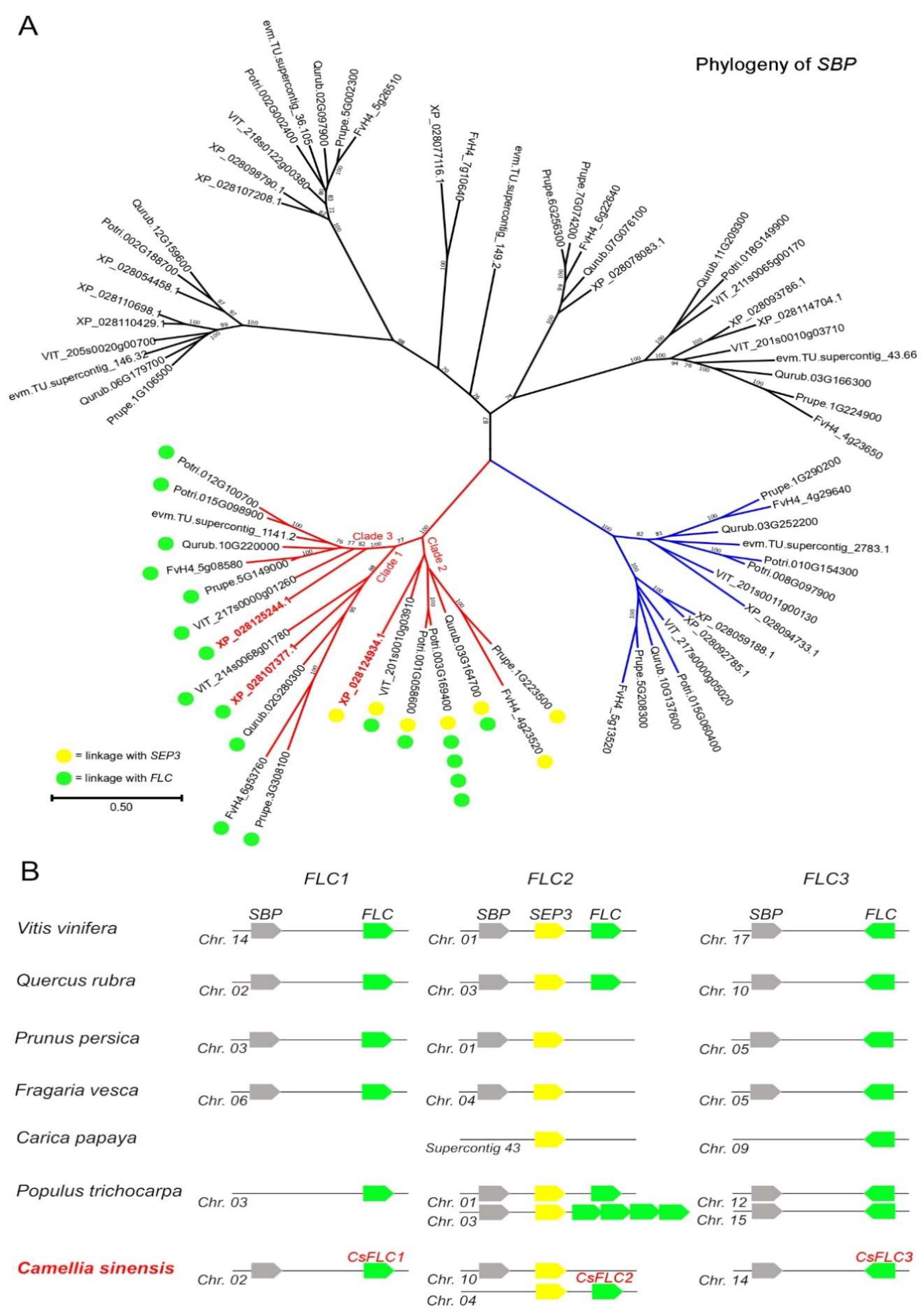
Figure 1. Identification and characterization of CsFLC genes. (A). As reported earlier by Ruelen et al. [30], SBP genes from a conserved monophyletic clade, shown in red, are closely linked to SEP3 and FLC, and they are triplicated in core eudicots (clades 1, 2 and 3). Two of the three SBP genes that we found in Camellia sinensis show this conserved linkage with FLC genes. The genes of sister clade shown in blue are instead linked to LOFSEP and SQUA MADS-box genes. Potri: Populus trichocarpa; VIT: Vitis vinifera; XP: Camellia sinensis; Prupe: Prunus persica; FvH4: Fragaria vesca; Qurub: Quercus rubra; evm: Carica papaya. The tree was generated using protein sequences. Bootstrap values lower than 70 are not shown, and the scale bar indicates the number of amino acid sequence substitutions per site. (B). The three SBP-(SEP3)-FLC paralogous tandems of core eudicots show different patterns of conservation and duplications among species. In Camellia sinensis, which has an ancient tetraploid genome [32], six regions are expected; however, only duplicated copies of SEP3 were retained. The results of this analysis suggest the presence of only three FLC genes in tea plant, which we refer to as CsFLC1, CsFLC2 and CsFLC3.
Ijms 23 15711 g001
Figure 2. Subcellular location of CsFLC1 in Nicotiana Benthamiana. GFP: green fluorescent protein; RFP: red fluorescent protein; bar: 50 µm.
Ijms 23 15711 g002
Figure 3. Expression patterns of CsFLC1. (A) Expression of CsFLC1 in axillary or flower buds of tea plant throughout the year. (B) Different tissue expression of CsFLC in tea plant. (C) Various parts of flowers of tea plant. (D) Expression patterns of CsFLC1 in different parts of flowers in tea plant. (E) GUS staining of pCsFLC1::GUS transgenic Arabidopsis thaliana.
Ijms 23 15711 g003
Figure 4. Expression pattern of CsFLC1 under low-temperature treatment in tea plant. The t-test was applied for difference comparison between measurement data, and each was compared with CK; ** represented p < 0.01; * represented p < 0.05.
Ijms 23 15711 g004
Figure 5. CsFLC1 in response to low temperature and daylength. (AC) pCsFLC1::GUS response to low temperature (LT; 4 °C): (A) before LT treatment; (B) after LT treatment for 3 h; (C) and after LT treatment for 4 h. (DF) pCsFLC1::GUS staining in the light: (D) in plants under LDs; (E) in plants under MDs; and (F) in plants under SDs. (GI) pCsFLC1::GUS staining in the dark: (G) in plants under LDs; (H) in plants under MDs; (I) and in plants under SDs. Bar = 1 cm; LD: long days; MD: medium days; SD: short days.
Ijms 23 15711 g005
Figure 6. Phenotypes of 35S::CsFLC1 transgenic Arabidopsis thaliana. (A) Image of WT and OE-CsFLC1 lines. (B) CsFLC1 expression levels in different Arabidopsis thaliana lines. (C) Seed germination rates of different Arabidopsis thaliana lines. (D) Boxplot showing bolting and blooming times of different Arabidopsis thaliana lines: the bolting and blooming times were significant earlier in OE lines compared to WT, while there were no significant differences between OE lines.
Ijms 23 15711 g006
Figure 7. Transcriptome analysis of WT and OE-CsFLC1 lines. (A) Gene cluster and GO enrichment of upregulated genes in OE-CsFLC1 lines compared to WT. (B) Gene cluster and GO enrichment of downregulated genes in OE-CsFLC1 lines compared to WT. (C) KEGG enrichment of upregulated genes in OE-CsFLC1 lines compared to WT.
Ijms 23 15711 g007
Figure 8. Expression of genes related to phytohormones in WT and OE-CsFLC1 lines. (A) Expression of genes related to auxin in WT and OE-CsFLC1 lines. (B) Expression of genes related to SA in WT and OE-CsFLC1 lines. (C) Expression of genes related to JA in WT and OE-CsFLC1 lines. (D) Expression of genes related to ABA in WT and OE-CsFLC1 lines. IAA: indole-acetic acid; SA: salicylic acid; JA: jasmonic acid; ABA: abscisic acid. Different letters (a, b and c) on the column indicate significant difference between the data (p < 0.05).
Ijms 23 15711 g008
Figure 9. Expression patterns of Arabidopsis AtFLC target genes [33] in WT and OE-CsFLC1 transgenic Arabidopsis thaliana. The pink color represents upregulated genes, the blue color represents downregulated genes, and the heatmap was generated by TBtools.
Ijms 23 15711 g009
Figure 10. Variation trends of different phytohormone contents in tea plant during the whole year. (A) Content of IAA in tea plant throughout the year. (B) JA content of tea plant throughout the year. (C) Content of SA in tea plant throughout the year. The t-test was applied for difference comparison between measurement data, and each was compared with the previous one; ** represented p < 0.01; * represented p < 0.05. IAA: indole-3-acetic acid; JA: jasmonic acid; SA: salicylic acid.
Ijms 23 15711 g010
Publisher’s Note: MDPI stays neutral with regard to jurisdictional claims in published maps and institutional affiliations.