

Bioactive Carboxymethyl Cellulose (CMC)-Based Films Modified with Melanin and Silver Nanoparticles (AgNPs)—The Effect of the Degree of CMC Substitution on the In Situ Synthesis of AgNPs and Films’ Functional Properties
 , ,
, ,  ,
, 
Abstract
1. Introduction
2. Results and Discussion
2.1. Thickness and Mechanical Properties
2.2. UV-Barrier Properties
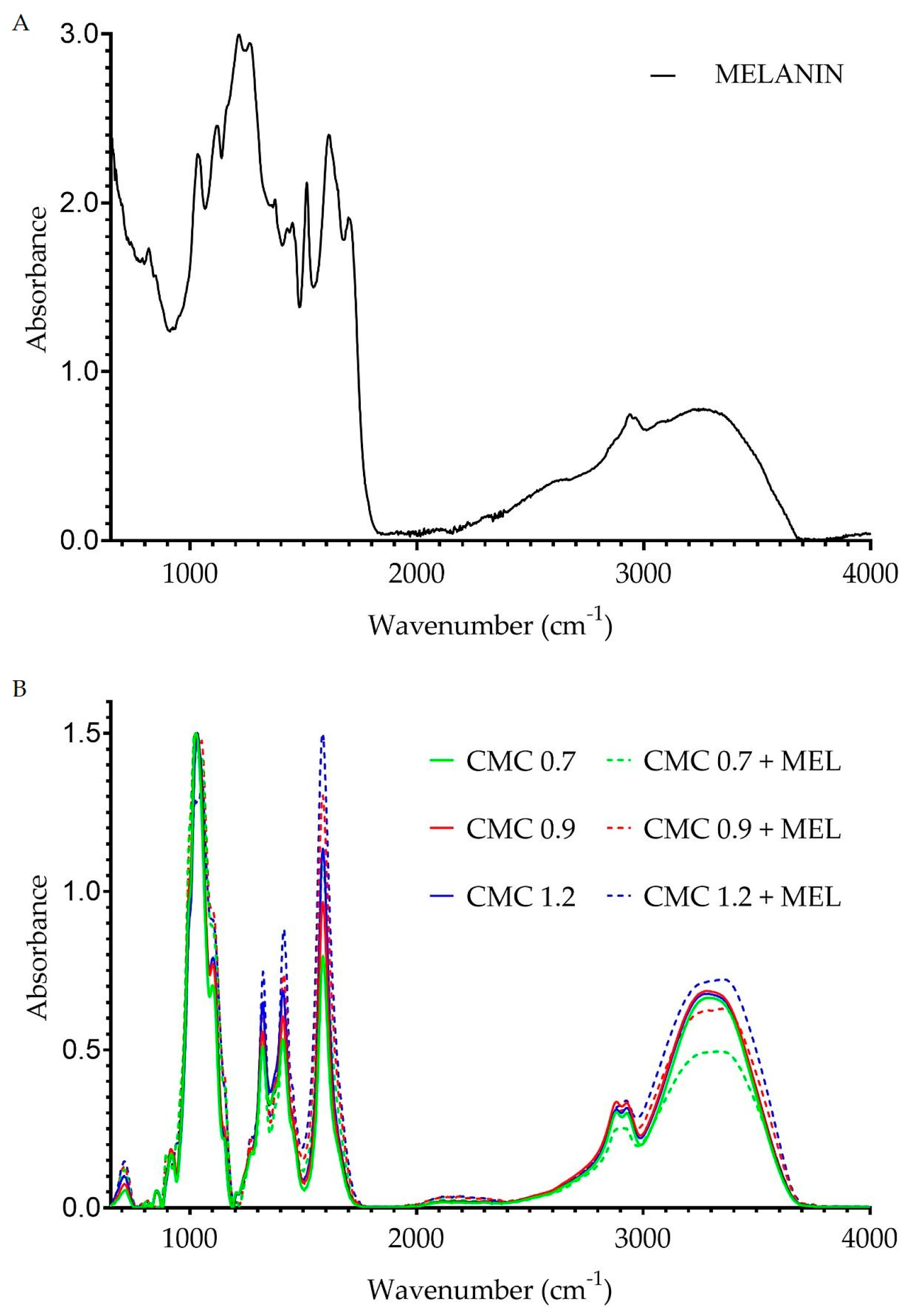
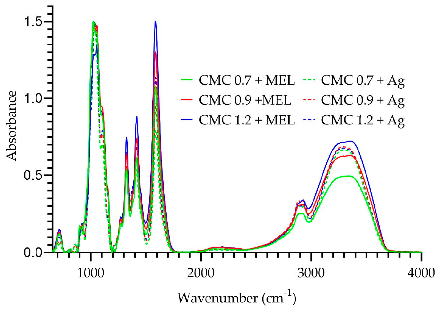
2.3. FT-IR Analysis
2.4. SEM Analysis
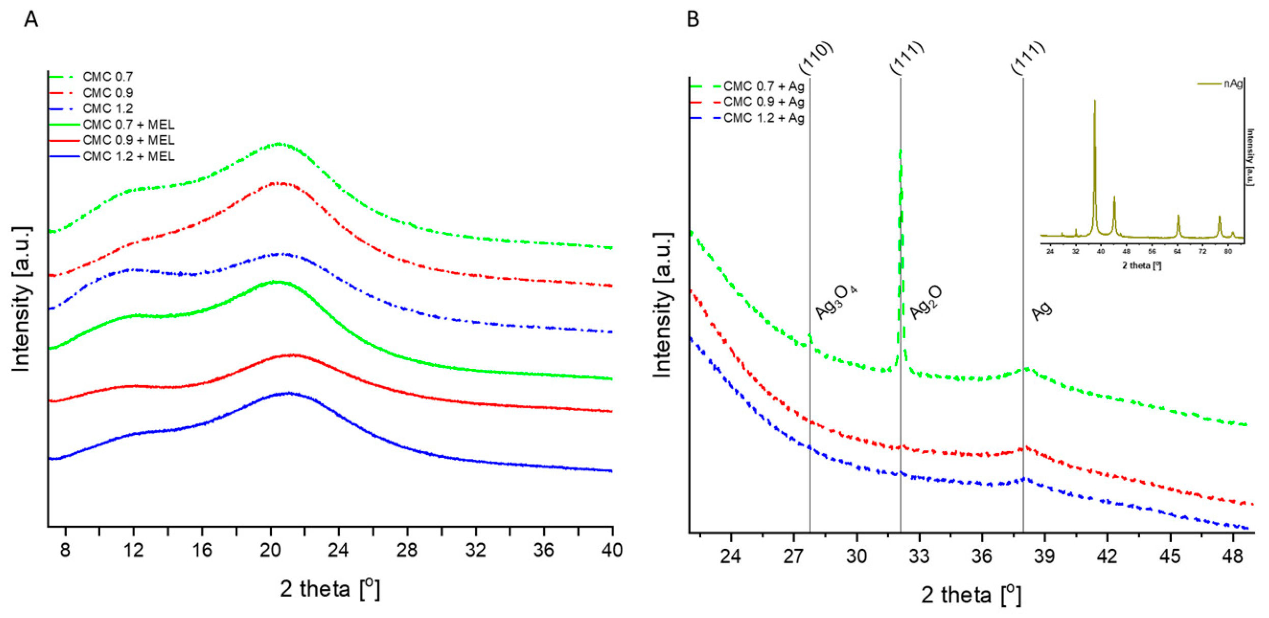
2.5. XRD Analysis
2.6. Color
2.7. Antioxidant Properties
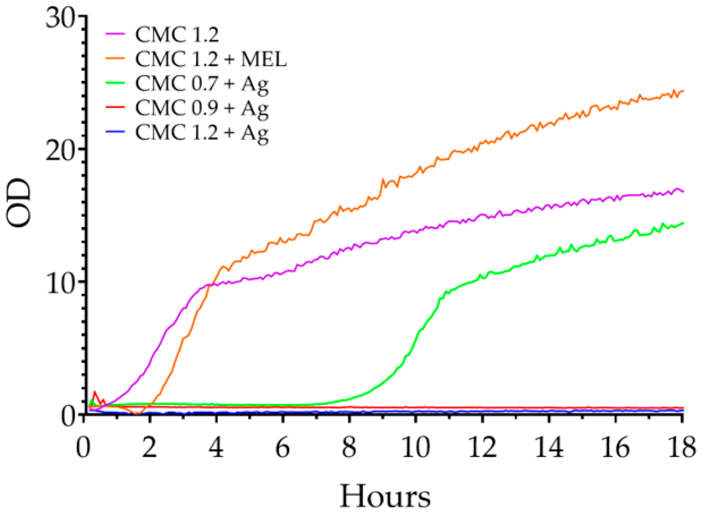
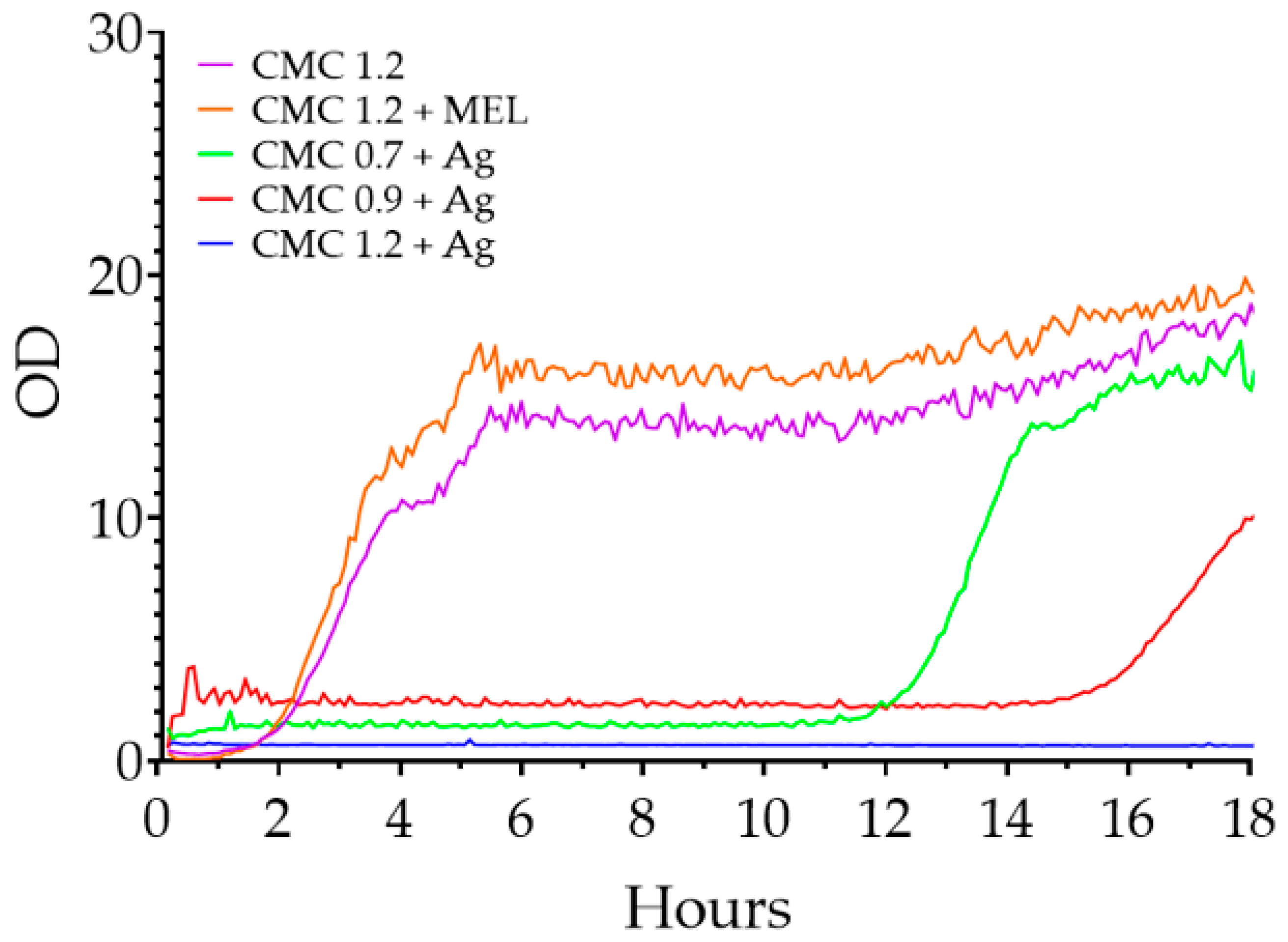
2.8. Antimicrobial Properties
3. Materials and Methods
3.1. Materials and Reagents
3.2. Preparation of Films
3.3. Preparation of Nanoparticles
3.4. Characterization of Biocomposite Films
3.4.1. Thickness and Mechanical Properties
3.4.2. Spectral Analysis of Films
3.4.3. Structure and Morphology Analysis of Films
3.4.4. Color Analysis of Films
3.4.5. Antioxidant Properties of Films
3.4.6. Antimicrobial Properties
3.5. Statistical Analysis
4. Conclusions
Supplementary Materials
Author Contributions
Funding
Institutional Review Board Statement
Informed Consent Statement
Data Availability Statement
Conflicts of Interest
References
- Marsh, K.; Bugusu, B. Food Packaging—Roles, Materials, and Environmental Issues: Scientific Status Summary. J. Food Sci. 2007, 72, R39–R55. [Google Scholar] [CrossRef] [PubMed]
- Trang, P.T.T.; Dong, H.Q.; Toan, D.Q.; Hanh, N.T.X.; Thu, N.T. The Effects of Socio-Economic Factors on Household Solid Waste Generation and Composition: A Case Study in Thu Dau Mot, Vietnam. Energy Procedia 2017, 107, 253–258. [Google Scholar] [CrossRef]
- Giordano, C.; Falasconi, L.; Cicatiello, C.; Pancino, B. The Role of Food Waste Hierarchy in Addressing Policy and Research: A Comparative Analysis. J. Clean. Prod. 2020, 252, 119617. [Google Scholar] [CrossRef]
- Priefer, C.; Jörissen, J.; Bräutigam, K.R. Food Waste Prevention in Europe—A Cause-Driven Approach to Identify the Most Relevant Leverage Points for Action. Resour. Conserv. Recycl. 2016, 109, 155–165. [Google Scholar] [CrossRef]
- Duenas, M.; Garciá-Estévez, I. Agricultural and Food Waste: Analysis, Characterization and Extraction of Bioactive Compounds and Their Possible Utilization. Foods 2020, 9, 817. [Google Scholar] [CrossRef]
- Paletta, A.; Leal Filho, W.; Balogun, A.L.; Foschi, E.; Bonoli, A. Barriers and Challenges to Plastics Valorisation in the Context of a Circular Economy: Case Studies from Italy. J. Clean. Prod. 2019, 241, 118149. [Google Scholar] [CrossRef]
- Gedde, U.W.; Hedenqvist, M.S.; Hakkarainen, M.; Nilsson, F.; Das, O. Applied Polymer Science; Springer: Cham, Switzerland, 2021; ISBN 978-3-030-68471-6. [Google Scholar]
- Geyer, R.; Jambeck, J.R.; Law, K.L. Production, Use, and Fate of All Plastics Ever Made. Sci. Adv. 2017, 3. [Google Scholar] [CrossRef]
- Stafford, R.; Jones, P.J.S. Viewpoint—Ocean Plastic Pollution: A Convenient but Distracting Truth? Mar. Policy 2019, 103, 187–191. [Google Scholar] [CrossRef]
- Thompson, R.C.; Moore, C.J.; Saal, F.S.V.; Swan, S.H. Plastics, the Environment and Human Health: Current Consensus and Future Trends. Philos. Trans. R. Soc. B Biol. Sci. 2009, 364, 2153–2166. [Google Scholar] [CrossRef]
- Jain, R.; Tiwari, A. Biosynthesis of Planet Friendly Bioplastics Using Renewable Carbon Source. J. Environ. Health Sci. Eng. 2015, 13, 1–5. [Google Scholar] [CrossRef]
- Abral, H.; Ariksa, J.; Mahardika, M.; Handayani, D.; Aminah, I.; Sandrawati, N.; Pratama, A.B.; Fajri, N.; Sapuan, S.M.; Ilyas, R.A. Transparent and Antimicrobial Cellulose Film from Ginger Nanofiber. Food Hydrocoll. 2020, 98, 105266. [Google Scholar] [CrossRef]
- Yang, M.; Zhang, X.; Guan, S.; Dou, Y.; Gao, X. Preparation of Lignin Containing Cellulose Nanofibers and Its Application in PVA Nanocomposite Films. Int. J. Biol. Macromol. 2020, 158, 1259–1267. [Google Scholar] [CrossRef]
- Hoque, M.E.; Rayhan, A.M.; Shaily, S.I. Natural Fiber-Based Green Composites: Processing, Properties and Biomedical Applications. Appl. Sci. Eng. Prog. 2021, 14, 689–718. [Google Scholar] [CrossRef]
- Abdollahi, M.; Damirchi, S.; Shafafi, M.; Rezaei, M.; Ariaii, P. Carboxymethyl Cellulose-Agar Biocomposite Film Activated with Summer Savory Essential Oil as an Antimicrobial Agent. Int. J. Biol. Macromol. 2019, 126, 561–568. [Google Scholar] [CrossRef]
- Roy, S.; Rhim, J.W. Carboxymethyl Cellulose-Based Antioxidant and Antimicrobial Active Packaging Film Incorporated with Curcumin and Zinc Oxide. Int. J. Biol. Macromol. 2020, 148, 666–676. [Google Scholar] [CrossRef]
- Du, B.; Li, J.; Zhang, H.; Huang, L.; Chen, P.; Zhou, J. Influence of Molecular Weight and Degree of Substitution of Carboxymethylcellulose on the Stability of Acidified Milk Drinks. Food Hydrocoll. 2009, 23, 1420–1426. [Google Scholar] [CrossRef]
- Duan, H.; Wang, D.; Li, Y. Green Chemistry for Nanoparticle Synthesis. Chem. Soc. Rev. 2015, 44, 5778–5792. [Google Scholar] [CrossRef]
- Gour, A.; Jain, N.K. Advances in Green Synthesis of Nanoparticles. Artif. Cells Nanomed. Biotechnol. 2019, 47, 844–851. [Google Scholar] [CrossRef]
- Iravani, S. Green Synthesis of Metal Nanoparticles Using Plants. Green Chem. 2011, 13, 2638–2650. [Google Scholar] [CrossRef]
- Malik, P.; Shankar, R.; Malik, V.; Sharma, N.; Mukherjee, T.K. Green Chemistry Based Benign Routes for Nanoparticle Synthesis. J. Nanoparticles 2014, 2014, 1–14. [Google Scholar] [CrossRef]
- Salam, H.A.; Rajiv, P.; Kamaraj, M.; Jagadeeswaran, P.; Gunalan, S.; Sivaraj, R. Plants: Green Route for Nanoparticle Synthesis. Int. Res. J. Biol. Sci. 2012, 1, 85–90. [Google Scholar]
- Bang, Y.J.; Roy, S.; Rhim, J.W. A Facile In Situ Synthesis of Resorcinol-Mediated Silver Nanoparticles and the Fabrication of Agar-Based Functional Nanocomposite Films. J. Compos. Sci. 2022, 6, 124. [Google Scholar] [CrossRef]
- Shameem, M.M.; Sasikanth, S.M.; Annamalai, R.; Raman, R.G. A Brief Review on Polymer Nanocomposites and Its Applications. Mater. Today Proc. 2021, 45, 2536–2539. [Google Scholar] [CrossRef]
- Zare, Y.; Shabani, I. Polymer/Metal Nanocomposites for Biomedical Applications. Mater. Sci. Eng. C 2016, 60, 195–203. [Google Scholar] [CrossRef] [PubMed]
- Alavi, M.; Rai, M. Recent Advances in Antibacterial Applications of Metal Nanoparticles (MNPs) and Metal Nanocomposites (MNCs) against Multidrug-Resistant (MDR) Bacteria. Expert Rev. Anti-Infect. Ther. 2019, 17, 419–428. [Google Scholar] [CrossRef]
- Garcia, C.V.; Shin, G.H.; Kim, J.T. Metal Oxide-Based Nanocomposites in Food Packaging: Applications, Migration, and Regulations. Trends Food Sci. Technol. 2018, 82, 21–31. [Google Scholar] [CrossRef]
- Roy, S.; Rhim, J. New Insight into Melanin for Food Packaging and Biotechnology Applications New Insight into Melanin for Food Packaging and Biotechnology Applications. Crit. Rev. Food Sci. Nutr. 2021, 62, 1–27. [Google Scholar] [CrossRef]
- Łopusiewicz, Ł.; Macieja, S.; Śliwiński, M.; Bartkowiak, A.; Roy, S.; Sobolewski, P. Alginate Biofunctional Films Modified with Melanin from Watermelon Seeds and Zinc Oxide/Silver Nanoparticles. Materials 2022, 15, 2381. [Google Scholar] [CrossRef]
- Rao, S.Q.; Zhang, R.Y.; Chen, R.; Gao, Y.J.; Gao, L.; Yang, Z. quan Nanoarchitectonics for Enhanced Antibacterial Activity with Lactobacillus Buchneri S-Layer Proteins-Coated Silver Nanoparticles. J. Hazard. Mater. 2022, 426, 128029. [Google Scholar] [CrossRef]
- Klasen, H.J. Historical Review of the Use of Silver in the Treatment of Burns. I. Early Uses. Burns 2000, 26, 117–130. [Google Scholar] [CrossRef]
- Dong, F.; Li, S. Wound Dressings Based on Chitosan-Dialdehyde Cellulose Nanocrystals-Silver Nanoparticles: Mechanical Strength, Antibacterial Activity and Cytotoxicity. Polymers 2018, 10, 673. [Google Scholar] [CrossRef]
- Hsueh, Y.H.; Lin, K.S.; Ke, W.J.; Hsieh, C.T.; Chiang, C.L.; Tzou, D.Y.; Liu, S.T. The Antimicrobial Properties of Silver Nanoparticles in Bacillus Subtilis Are Mediated by Released Ag+ Ions. PLoS ONE 2015, 10, 1–17. [Google Scholar] [CrossRef]
- Peiris, M.M.K.; Fernando, S.S.N.; Jayaweera, P.M.; Arachchi, N.D.H.; Guansekara, T.D.C.P. Comparison of Antimicrobial Properties of Silver Nanoparticles Synthesized from Selected Bacteria. Indian J. Microbiol. 2018, 58, 301–311. [Google Scholar] [CrossRef]
- Yunusov, K.E.; Sarymsakov, A.A.; Jalilov, J.Z.O.; Atakhanov, A.A.O. Physicochemical Properties and Antimicrobial Activity of Nanocomposite Films Based on Carboxymethylcellulose and Silver Nanoparticles. Polym. Adv. Technol. 2021, 32, 1822–1830. [Google Scholar] [CrossRef]
- Capanema, N.S.V.; Mansur, A.A.P.; Carvalho, S.M.; Mansur, L.L.; Ramos, C.P.; Lage, A.P.; Mansur, H.S. Physicochemical Properties and Antimicrobial Activity of Biocompatible Carboxymethylcellulose-Silver Nanoparticle Hybrids for Wound Dressing and Epidermal Repair. J. Appl. Polym. Sci. 2018, 135, 45812. [Google Scholar] [CrossRef]
- Hebeish, A.A.; El-Rafie, M.H.; Abdel-Mohdy, F.A.; Abdel-Halim, E.S.; Emam, H.E. Carboxymethyl Cellulose for Green Synthesis and Stabilization of Silver Nanoparticles. Carbohydr. Polym. 2010, 82, 933–941. [Google Scholar] [CrossRef]
- Łopusiewicz, Ł.; Kwiatkowski, P.; Drozłowska, E.; Trocer, P.; Kostek, M.; Śliwiński, M.; Polak-Śliwińska, M.; Kowalczyk, E.; Sienkiewicz, M. Preparation and Characterization of Carboxymethyl Cellulose-Based Bioactive Composite Films Modified with Fungal Melanin and Carvacrol. Polymer 2021, 13, 499. [Google Scholar] [CrossRef]
- Łopusiewicz, Ł.; Drozłowska, E.; Trocer, P.; Kostek, M.; Śliwiński, M.; Henriques, M.H.F.; Bartkowiak, A.; Sobolewski, P. Whey Protein Concentrate/Isolate Biofunctional Films Modified with Melanin from Watermelon (Citrullus Lanatus) Seeds. Materials 2020, 13, 3876. [Google Scholar] [CrossRef]
- Roy, S.; Rhim, J.W. Agar-Based Antioxidant Composite Films Incorporated with Melanin Nanoparticles. Food Hydrocoll. 2019, 94, 391–398. [Google Scholar] [CrossRef]
- Shankar, S.; Wang, L.F.; Rhim, J.W. Effect of Melanin Nanoparticles on the Mechanical, Water Vapor Barrier, and Antioxidant Properties of Gelatin-Based Films for Food Packaging Application. Food Packag. Shelf Life 2019, 21, 100363. [Google Scholar] [CrossRef]
- Roy, S.; Rhim, J.W. Preparation of Carrageenan-Based Functional Nanocomposite Films Incorporated with Melanin Nanoparticles. Colloids Surf. B Biointerfaces 2019, 176, 317–324. [Google Scholar] [CrossRef] [PubMed]
- Bang, Y.J.; Shankar, S.; Rhim, J.W. Preparation of Polypropylene/Poly (Butylene Adipate-Co-Terephthalate) Composite Films Incorporated with Melanin for Prevention of Greening of Potatoes. Packag. Technol. Sci. 2020, 33, 433–441. [Google Scholar] [CrossRef]
- Shankar, S.; Rhim, J.W. Amino Acid Mediated Synthesis of Silver Nanoparticles and Preparation of Antimicrobial Agar/Silver Nanoparticles Composite Films. Carbohydr. Polym. 2015, 130, 353–363. [Google Scholar] [CrossRef] [PubMed]
- Chu, Z.; Zhao, T.; Li, L.; Fan, J.; Qin, Y. Characterization of Antimicrobial Poly (Lactic Acid)/Nano-Composite Films with Silver and Zinc Oxide Nanoparticles. Materials 2017, 10, 659. [Google Scholar] [CrossRef] [PubMed]
- Fortunati, E.; Armentano, I.; Zhou, Q.; Iannoni, A.; Saino, E.; Visai, L.; Berglund, L.A.; Kenny, J.M. Multifunctional Bionanocomposite Films of Poly(Lactic Acid), Cellulose Nanocrystals and Silver Nanoparticles. Carbohydr. Polym. 2012, 87, 1596–1605. [Google Scholar] [CrossRef]
- Roy, S.; Kim, H.C.; Kim, J.W.; Zhai, L.; Zhu, Q.Y.; Kim, J. Incorporation of Melanin Nanoparticles Improves UV-Shielding, Mechanical and Antioxidant Properties of Cellulose Nanofiber Based Nanocomposite Films. Mater. Today Commun. 2020, 24, 100984. [Google Scholar] [CrossRef]
- Rhim, J.W.; Wang, L.F.; Hong, S.I. Preparation and Characterization of Agar/Silver Nanoparticles Composite Films with Antimicrobial Activity. Food Hydrocoll. 2013, 33, 327–335. [Google Scholar] [CrossRef]
- Cao, W.; Zhou, X.; McCallum, N.C.; Hu, Z.; Ni, Q.Z.; Kapoor, U.; Heil, C.M.; Cay, K.S.; Zand, T.; Mantanona, A.J.; et al. Unraveling the Structure and Function of Melanin through Synthesis. J. Am. Chem. Soc. 2021, 143, 2622–2637. [Google Scholar] [CrossRef]
- Roy, S.; van Hai, L.; Kim, H.C.; Zhai, L.; Kim, J. Preparation and Characterization of Synthetic Melanin-like Nanoparticles Reinforced Chitosan Nanocomposite Films. Carbohydr. Polym. 2020, 231, 115729. [Google Scholar] [CrossRef]
- Vimala, K.; Mohan, Y.M.; Sivudu, K.S.; Varaprasad, K.; Ravindra, S.; Reddy, N.N.; Padma, Y.; Sreedhar, B.; MohanaRaju, K. Fabrication of Porous Chitosan Films Impregnated with Silver Nanoparticles: A Facile Approach for Superior Antibacterial Application. Colloids Surf. B Biointerfaces 2010, 76, 248–258. [Google Scholar] [CrossRef]
- Mondal, M.I.H.; Yeasmin, M.S.; Rahman, M.S. Preparation of Food Grade Carboxymethyl Cellulose from Corn Husk Agrowaste. Int. J. Biol. Macromol. 2015, 79, 144–150. [Google Scholar] [CrossRef]
- Voo, W.P.; Lee, B.B.; Idris, A.; Islam, A.; Tey, B.T.; Chan, E.S. Production of Ultra-High Concentration Calcium Alginate Beads with Prolonged Dissolution Profile. RSC Adv. 2015, 5, 36687–36695. [Google Scholar] [CrossRef]
- Lawrie, G.; Keen, I.; Drew, B.; Chandler-Temple, A.; Rintoul, L.; Fredericks, P.; Grøndahl, L. Interactions between Alginate and Chitosan Biopolymers Characterized Using FTIR and XPS. Biomacromolecules 2007, 8, 2533–2541. [Google Scholar] [CrossRef]
- Fertah, M.; Belfkira, A.; Dahmane, E.M.; Taourirte, M.; Brouillette, F. Extraction and Characterization of Sodium Alginate from Moroccan Laminaria Digitata Brown Seaweed. Arab. J. Chem. 2017, 10, S3707–S3714. [Google Scholar] [CrossRef]
- Viera, R.G.P.; Filho, G.R.; de Assunção, R.M.N.; da Carla, C.; Vieira, J.G.; de Oliveira, G.S. Synthesis and Characterization of Methylcellulose from Sugar Cane Bagasse Cellulose. Carbohydr. Polym. 2007, 67, 182–189. [Google Scholar] [CrossRef]
- Yang, M.; Li, L.; Yu, S.; Liu, J.; Shi, J. High Performance of Alginate/Polyvinyl Alcohol Composite Film Based on Natural Original Melanin Nanoparticles Used as Food Thermal Insulating and UV–Vis Block. Carbohydr. Polym. 2020, 233, 115884. [Google Scholar] [CrossRef]
- Yi, M.; Yeom, D.; Lee, W.; Jang, S.; Cho, G. Scalability on Roll-to-Roll Gravure Printed Dielectric Layers for Printed Thin Film Transistors. J. Nanosci. Nanotechnol. 2013, 13, 5360–5364. [Google Scholar] [CrossRef]
- Abdulhameed, A.; Mbuvi, H.M.; Changamu, E.O.; Maingi, F.M. Microwave Synthesis of Carboxymethylcellulose (CMC) from Rice Husk. IOSR J. Appl. Chem. (IOSR-JAC) 2019, 12, 33–42. [Google Scholar] [CrossRef]
- Buljan, I.; Kosanović, C.; Subotić, B.; Tušar, N.N.; Ristić, A.; Gabrovšek, R.; Kaučič, V.; Radić, T.M. Kinetic Analysis of Isothermal Crystallization of Potassium Aluminosilicate Ceramics (Leucite and Kalsilite) from Amorphous Potassium Aluminosilicate Precursors. Cryst. Growth Des. 2010, 10, 838–844. [Google Scholar] [CrossRef]
- Meng, Y. A Sustainable Approach to Fabricating Ag Nanoparticles/PVA Hybrid Nanofiber and Its Catalytic Activity. Nanomaterials 2015, 5, 1124–1135. [Google Scholar] [CrossRef]
- Koohpeima, F.; Mokhtari, M.J.; Khalafi, S. The Effect of Silver Nanoparticles on Composite Shear Bond Strength to Dentin with Different Adhesion Protocols. J. Appl. Oral Sci. 2017, 25, 367–373. [Google Scholar] [CrossRef]
- Singh, R.K.; Khatri, O.P. A scanning electron microscope based new method for determining degree of substitution of sodium carboxymethyl cellulose. J. Microsc. 2012, 246, 43–52. [Google Scholar] [CrossRef] [PubMed]
- Łopusiewicz, Ł.; Jędra, F.; Mizielińska, M. New Poly(Lactic Acid) Active Packaging Composite Films Incorporated with Fungal Melanin. Polymers 2018, 10, 386. [Google Scholar] [CrossRef] [PubMed]
- Rodriguez-Garraus, A.; Azqueta, A.; Vettorazzi, A.; de Cerain, A.L. Genotoxicity of Silver Nanoparticles. Nanomaterials 2020, 10, 251. [Google Scholar] [CrossRef]
- Liao, C.; Li, Y.; Tjong, S.C. Bactericidal and Cytotoxic Properties of Silver Nanoparticles. Int. J. Mol. Sci. 2019, 20, 449. [Google Scholar] [CrossRef]
- Hegazy, M.A.; Borham, E. Preparation and Characterization of Silver Nanoparticles Homogenous Thin Films. NRIAG J. Astron. Geophys. 2018, 7, 27–30. [Google Scholar] [CrossRef]


| Sample | Tensile Strength (MPa) | Elongation at Break (%) | Thickness (mm) |
|---|---|---|---|
| CMC 0.7 | 34.6 ± 17.1 a,b,c,d | 5.4 ± 3.4 a | 0.064 ± 0.017 a,b |
| CMC 0.9 | 34.9 ± 10.4 b,c,d | 6.6 ± 5.0 a | 0.067 ± 0.013 a,b |
| CMC 1.2 | 25.2 ± 11.1 a,b,c | 7.2 ± 6.1 a | 0.081 ± 0.026 a |
| CMC 0.7 + MEL | 45.8 ± 17.0 d | 3.5 ± 1.3 a | 0.048 ± 0.016 b |
| CMC 0.9 + MEL | 47.9 ± 14.4 d | 4.4 ± 1.4 a | 0.055 ± 0.012 a,b |
| CMC 1.2 + MEL | 39.4 ± 9.52 c,d | 5.0 ± 2.7 a | 0.053 ± 0.018 a,b |
| CMC 0.7 + Ag | 18.8 ± 5.46 a,b | 30.6 ± 13.0 b | 0.079 ± 0.014 a |
| CMC 0.9 + Ag | 15.5 ± 3.05 a | 31.3 ± 13.8 b | 0.061 ± 0.019 a,b |
| CMC 1.2 + Ag | 24.6 ± 8.41 a,b,c | 30.8 ± 12.9 b | 0.058 ± 0.013 a,b |
| Sample | L* | a* | b* | ΔE | YI | Opacity | T280 (%) | T660 (%) |
|---|---|---|---|---|---|---|---|---|
| CMC 0.7 | 78.30 ± 7.01 b | −0.57 ± 0.07 a | 13.32 ± 0.77 a | Used as standard | 23.89 ± 1.02 a | 7.56 ± 0.16 a | 82.54 ± 1.31 b | 91.39 ± 1.34 a |
| CMC 0.9 | 77.82 ± 1.48 b | −0.52 ± 0.14 a | 12.55 ± 1.72 a | Used as standard | 25.23 ± 2.08 a | 7.52 ± 0.08 a | 89.75 ± 1.29 d | 88.41 ± 1.64 a |
| CMC 1.2 | 88.52 ± 2.04 e | −0.69 ± 0.03 a | 14.35 ± 0.43 a | Used as standard | 23.22 ± 0.17 a | 7.50 ± 0.16 a | 90.61 ± 0.94 d | 91.75 ± 1.75 a |
| CMC 0.7 + MEL | 70.57 ± 5.24 a | −0.33 ± 0.05 a | 14.69 ± 0.63 a | 9.22 ± 1.50 d | 29.43 ± 1.06 a | 7.49 ± 0.11 a | 79.53 ± 1.73 b,c | 88.87 ± 1.48 a |
| CMC 0.9 + MEL | 73.72 ± 1.62 a | −0.34 ± 0.03 a | 14.64 ± 0.16 a | 4.95 ± 1.20 c | 28.33 ± 0.34 a | 7.42 ± 0.11 a | 81.20 ± 1.63 b | 89.23 ± 1.45 a |
| CMC 1.2 + MEL | 71.08 ± 2.12 a,b | −0.33 ± 0.02 a | 14.50 ± 0.29 a | 17.45 ± 2.13 e | 29.25 ± 0.31 a | 7.41 ± 0.07 a | 76.44 ± 1.27 c | 91.56 ± 1.39 a |
| CMC 0.7 + Ag | 2.87 ± 0.76 c | 7.07 ± 2.24 b | 3.79 ± 1.08 b | 76.42 ± 0.59 a,b | 185.86 ± 11.27 b | 3.96 ± 0.15 b,c | 4.51 ± 0.58 a | 12.85 ± 1.82 c |
| CMC 0.9 + Ag | 5.59 ± 1.30 c | 16.16 ± 3.14 c | 8.54 ± 2.27 c | 74.94 ± 0.74 a | 218.07 ± 7.33 c | 3.89 ± 0.11 b | 7.65 ± 0.84 a | 32.67 ± 1.13 b |
| CMC 1.2 + Ag | 13.45 ± 1.11 d | 18.51 ± 0.80 d | 21.49 ± 2.18 d | 77.85 ± 0.66 b | 227.99 ± 4.36 d | 4.14 ± 0.10 c | 7.98 ± 0.75 a | 28.31 ± 1.48 b |
| Sample | DPPH 20 min (%) | DPPH 24 h (%) | ABTS (%) |
|---|---|---|---|
| CMC 0.7 | 14.17 ± 0.14 b,c | 19.29 ± 0.24 a | 24.74 ± 0.56 a |
| CMC 0.9 | 14.44 ± 0.23 c | 20.52 ± 0.95 a | 25.72 ± 0.93 a |
| CMC 1.2 | 14.03 ± 0.19 b | 19.64 ± 0.67 a | 24.49 ± 0.49 a |
| CMC 0.7 + MEL | 23.54 ± 0.22 e | 54.47 ± 0.53 c | 81.28 ± 1.07 c |
| CMC 0.9 + MEL | 24.32 ± 0.41 d | 53.85 ± 0.80 c | 84.73 ± 1.28 d |
| CMC 1.2 + MEL | 26.04 ± 0.36 f | 54.41 ± 0.48 c | 88.94 ± 1.14 e |
| CMC 0.7 + Ag | 8.87 ± 0.30 a | 12.02 ± 0.47 b | 34.76 ± 1.47 b |
| CMC 0.9 + Ag | 8.73 ± 0.24 a | 12.47 ± 0.71 b | 35.53 ± 1.73 b |
| CMC 1.2 + Ag | 8.92 ± 0.29 a | 12.67 ± 0.59 b | 34.94 ± 1.82 b |
| Sample | CMC Degree of Substitution | Presence of Melanin | Presence of AgNPs |
|---|---|---|---|
| CMC 0.7 | 0.7 | No | No |
| CMC 0.9 | 0.9 | No | No |
| CMC 1.2 | 1.2 | No | No |
| CMC 0.7 + MEL | 0.7 | Yes | No |
| CMC 0.9 + MEL | 0.9 | Yes | No |
| CMC 1.2 + MEL | 1.2 | Yes | No |
| CMC 0.7 + Ag | 0.7 | Yes | Yes |
| CMC 0.9 + Ag | 0.9 | Yes | Yes |
| CMC 1.2 + Ag | 1.2 | Yes | Yes |
Publisher’s Note: MDPI stays neutral with regard to jurisdictional claims in published maps and institutional affiliations. |
© 2022 by the authors. Licensee MDPI, Basel, Switzerland. This article is an open access article distributed under the terms and conditions of the Creative Commons Attribution (CC BY) license (https://creativecommons.org/licenses/by/4.0/).
Share and Cite
Macieja, S.; Środa, B.; Zielińska, B.; Roy, S.; Bartkowiak, A.; Łopusiewicz, Ł. Bioactive Carboxymethyl Cellulose (CMC)-Based Films Modified with Melanin and Silver Nanoparticles (AgNPs)—The Effect of the Degree of CMC Substitution on the In Situ Synthesis of AgNPs and Films’ Functional Properties. Int. J. Mol. Sci. 2022, 23, 15560. https://doi.org/10.3390/ijms232415560
Macieja S, Środa B, Zielińska B, Roy S, Bartkowiak A, Łopusiewicz Ł. Bioactive Carboxymethyl Cellulose (CMC)-Based Films Modified with Melanin and Silver Nanoparticles (AgNPs)—The Effect of the Degree of CMC Substitution on the In Situ Synthesis of AgNPs and Films’ Functional Properties. International Journal of Molecular Sciences. 2022; 23(24):15560. https://doi.org/10.3390/ijms232415560
Chicago/Turabian StyleMacieja, Szymon, Bartosz Środa, Beata Zielińska, Swarup Roy, Artur Bartkowiak, and Łukasz Łopusiewicz. 2022. "Bioactive Carboxymethyl Cellulose (CMC)-Based Films Modified with Melanin and Silver Nanoparticles (AgNPs)—The Effect of the Degree of CMC Substitution on the In Situ Synthesis of AgNPs and Films’ Functional Properties" International Journal of Molecular Sciences 23, no. 24: 15560. https://doi.org/10.3390/ijms232415560
APA StyleMacieja, S., Środa, B., Zielińska, B., Roy, S., Bartkowiak, A., & Łopusiewicz, Ł. (2022). Bioactive Carboxymethyl Cellulose (CMC)-Based Films Modified with Melanin and Silver Nanoparticles (AgNPs)—The Effect of the Degree of CMC Substitution on the In Situ Synthesis of AgNPs and Films’ Functional Properties. International Journal of Molecular Sciences, 23(24), 15560. https://doi.org/10.3390/ijms232415560