Article

Design, Synthesis and Antimicrobial Properties of New Tetracyclic Quinobenzothiazine Derivatives

1
Department of Organic Chemistry, Faculty of Pharmaceutical Sciences in Sosnowiec, Medical University of Silesia, Jagiellońska 4, 41-200 Sosnowiec, Poland
2
Department of Analytical Chemistry, Faculty of Natural Sciences, Comenius University, Ilkovicova 6, 842 15 Bratislava, Slovakia
3
Department of Cell Biology, Faculty of Pharmaceutical Sciences in Sosnowiec, Medical University of Silesia, Jedności 9, 41-200 Sosnowiec, Poland
4
Institute of Chemistry, University of Silesia, Szkolna 9, 40-007 Katowice, Poland
5
Faculty of Mathematics and Natural Sciences, Cardinal Stefan Wyszyński University, K. Woycickiego 1/3, 01-938 Warszawa, Poland
6
Center for Translational Research and Molecular Biology of Cancer, Maria Skłodowska-Curie National Research Institute of Oncology, Wybrzeże AK 15, 44-101 Gliwice, Poland
7
Department of Infectious Diseases and Microbiology, Faculty of Veterinary Medicine, University of Veterinary Sciences Brno, Palackeho 1946/1, 612 42 Brno, Czech Republic
8
Department of Chemical Biology, Faculty of Science, Palacky University Olomouc, Slechtitelu 27, 783 71 Olomouc, Czech Republic
*
Author to whom correspondence should be addressed.
Int. J. Mol. Sci. 2022, 23(23), 15078; https://doi.org/10.3390/ijms232315078
Submission received: 4 November 2022 / Revised: 28 November 2022 / Accepted: 28 November 2022 / Published: 1 December 2022
(This article belongs to the Section Biochemistry)

Abstract

:
A new method for modifying the structure of tetracyclic quinobenzothiazinium derivatives has been developed, allowing introduction of various substituents at different positions of the benzene ring. The method consists of reacting appropriate aniline derivatives with 5,12-(dimethyl)thioquinantrenediinium bis-chloride. A series of new quinobenzothiazine derivatives was obtained with propyl, allyl, propargyl and benzyl substituents in 9, 10 and 11 positions, respectively. The structure of the obtained compounds was analyzed by 1H and 13C NMR (HSQC, HMBC) and X-ray analysis. All the compounds were tested against reference strains Staphylococcus aureus ATCC 29213 and Enterococcus faecalis ATCC 29212, and representatives of multidrug-resistant clinical isolates of methicillin-resistant S. aureus (MRSA) and vancomycin-resistant E. faecalis (VRE). In addition, all the compounds were evaluated in vitro against Mycobacterium smegmatis ATCC 700084 and M. marinum CAMP 5644. 9-Benzyloxy-5-methyl-12H-quino [3,4-b][1,4]benzothiazinium chloride (6j), 9-propoxy-5-methyl-12H-quino[3,4-b][1,4]benzothiazinium chloride (6a) and 9-allyloxy-5-methyl-12H-quino[3,4-b][1,4]benzothiazinium chloride (6d) demonstrated high activity against the entire tested microbial spectrum. The activities of the compounds were comparable with oxacillin, tetracycline and ciprofloxacinagainst staphylococcal strains and with rifampicin against both mycobacterial strains. Compound 6j had a significant effect on the inhibition of bacterial respiration as demonstrated by the MTT assay. The compounds showed not only bacteriostatic activity, but also bactericidal activity. Preliminary in vitro cytotoxicity screening of the compounds performed using normal human dermal fibroblasts (NHDF) proved that the tested compounds showed an insignificant cytotoxic effect on human cells (IC50 > 37 µM), making these compounds interesting for further investigation. Moreover, the intermolecular similarity of novel compounds was analyzed in the multidimensional space (mDS) of the structure/property-related in silico descriptors by means of principal component analysis (PCA) and hierarchical clustering analysis (HCA), respectively. The distance-oriented structure/property distribution was related with the experimental lipophilic data.

1. Introduction

Phenothiazine is a tricyclic heterocyclic compound with a 1,4-thiazine system. This compound was first obtained by Bernthsen in 1883 and has been an object of interest for researchers since, because the compound exhibits very interesting chemical and biological properties [1]. Phenothiazines, substituted at position 10 with alkylaminoalkyl groups, constitute the first group of effective neuroleptics. They have also been used as antiemetic, antihistamine, antipruritic, analgesic and anthelmintic drugs [2,3]. Increasingly, the design and production of new drugs is based on modifications of the basic structural fragments of known and already used drugs. The phenothiazine system is an example of how slight structural changes can affect the strength and the direction of drugs’ interaction. Thus, the phenothiazine skeleton has become a platform for the design and synthesis of biologically active compounds [4,5,6,7]. Some sources claim that over 6000 new phenothiazine derivatives have been reported to date [8]. Recent reports concern, inter alia, antitumor, antibacterial, antiviral, anti-inflammatory and antioxidant activities, reversal of multi-drug resistance and potential treatment of Alzheimer’s and Creutzfeldt-Jakob disease [9,10,11,12,13,14]. The influence of various types of pharmacophore groups on the direction of biological interaction has been extensively analysed. However, there are no reports on the influence of the location of these groups in the phenothiazine or azaphenothiazine system on the interaction of these compounds. The phenothiazine system is usually obtained by four basic methods: (i) thionation of diphenylamines with elemental sulphide, (ii) cyclization of diphenyl sulphides proceeding directly as Ullmann cyclization or with Smiles rearrangement, (iii) reactions of ortho-aminobenzenethiols with ortho-disubstituted benzenes, (iv) addition of the benzene ring to the benzene moiety 1,4-thiazines [15]. The substitution of one or both of the benzene rings with heteroaromatic azine rings leads to the production of nitrogen analogues of phenothiazines (azaphenothiazines). The structural modifications of phenothiazines and azaphenothiazines described so far mainly involved the introduction of various types of substituents to the thiazine nitrogen atom. In fact, the introduction of substituents to the carbon atoms of the benzene or azine rings is difficult; therefore, there are few published papers on the synthesis of these types of derivatives [16,17,18]. Halogen atoms or small functional groups were most often introduced into benzene rings. So far, the influence of various types of substituents at the thiazine nitrogen atom on the biological activity of phenothiazine derivatives and their nitrogen analogues has been studied. The effect that the nature of the substituents and their position on the benzene rings will exert on the biological activity of phenothiazine derivatives is of particular interest.
As a matter of fact, similarity-based property profiling is still regarded as the core of many SAR-guided procedures which assume that small modifications of molecular structures correspond to small variations of biological activities, and vice versa [19].Despite some shortcomings, the distance-related similarity scrutiny of structurally alike compounds is common practice that contributes noticeably to quantitative and/or qualitative on-target (receptor-dependent) and off-target (receptor-independent) SAR mapping [20]. In practice, chemical composition (topology and/or topography) can be structurally coded by calculated multi-dimensional (mD) descriptors and/or represented by experimental property data. Accordingly, SAR-related exploration of the descriptor-based feature/structural chemical space (CS) seems crucial to quantitative potency modelling and ADMET-tailored property prediction or production. It is expected that in silico mapping of molecular descriptors to targeted functionalities can support the synthetic efforts at the decision-making phases of hit→lead→seed→drug design [21].
In this work, we present a new method of synthesis of phenothiazine derivatives that allows the introduction of substituents at different positions in the phenothiazine system. We analyse the dependence of the antibacterial/antimycobacterial and cytotoxic properties of the obtained quinobenzothiazine derivatives on both the nature of the introduced substituents and their position in the quinobenzothiazine system. Following the common practice, the intermolecular similarity of novel derivatives was estimated in the multidimensional space (mDS) of the structure/property-related in silico descriptors using principal component analysis (PCA) and hierarchical clustering analysis (HCA), respectively. Moreover, the distance-oriented property distribution for new series of compounds was correlated with the experimental TLC lipophilic data. Finally, the antibacterial profile of newly synthesised molecules was investigated.

2. Results and Discussion

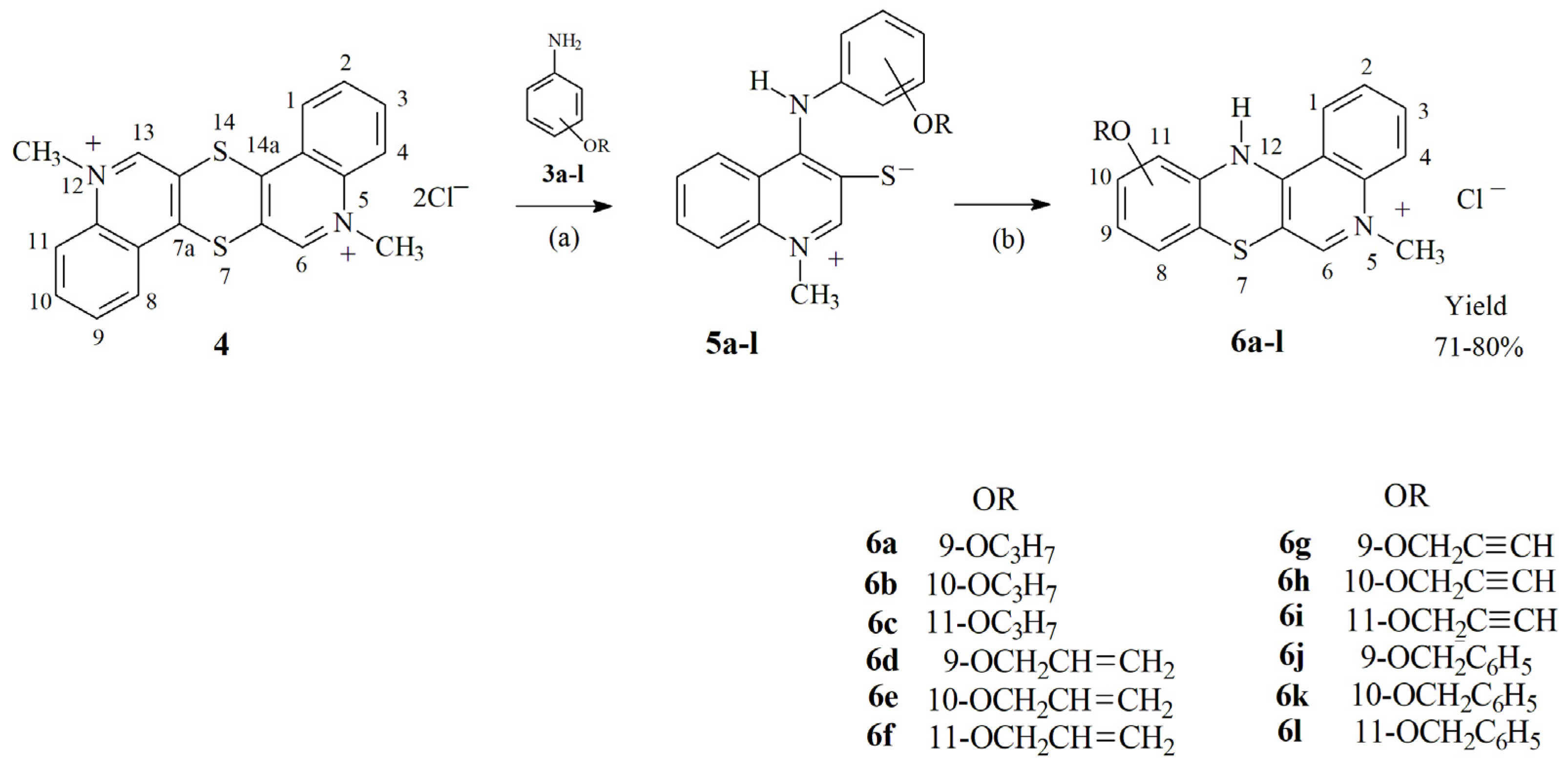
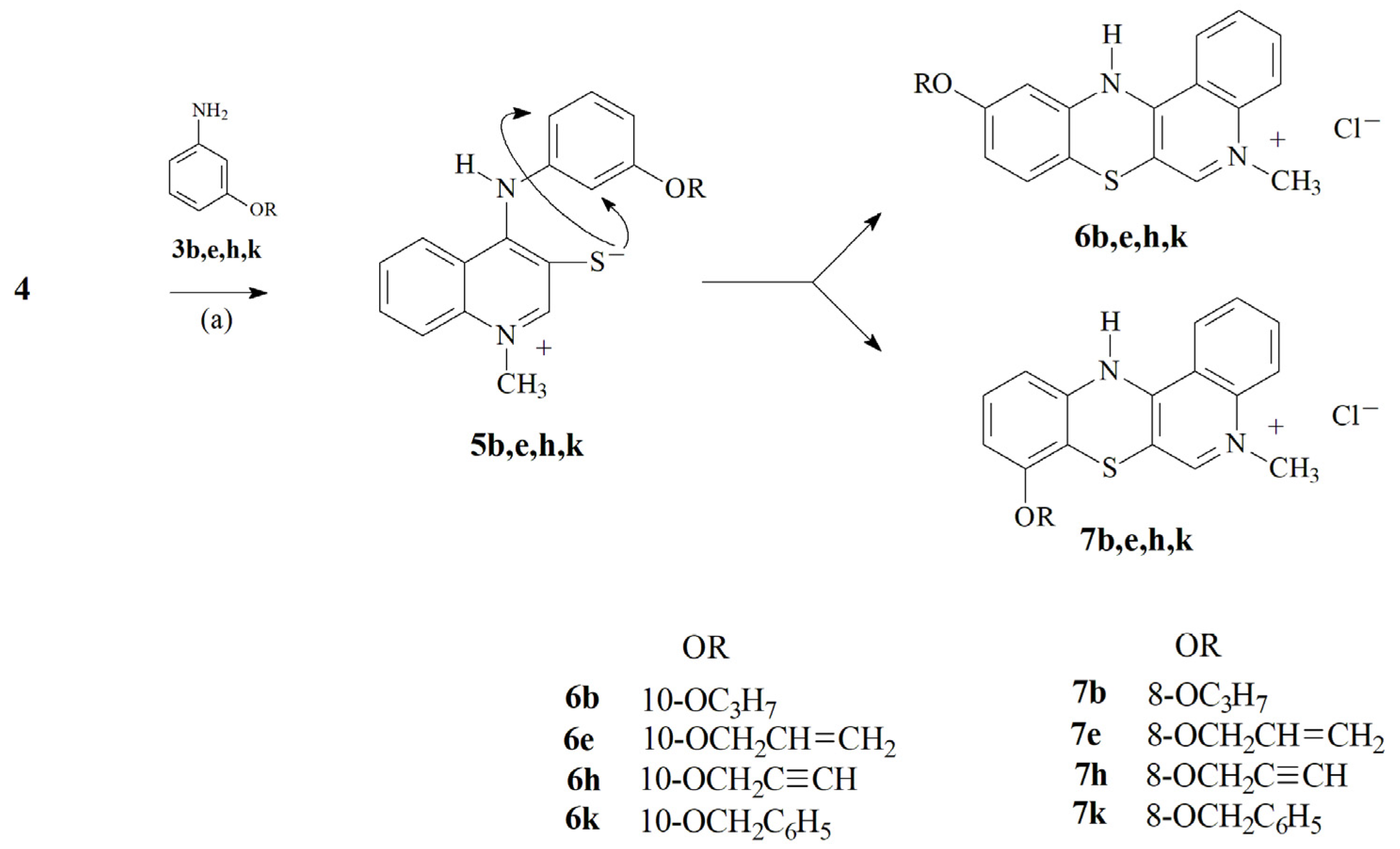
2.1. Chemistry—Design and Synthesis

In our previous reports, we described the method for the synthesis of tetracylic quinobenzothiazinium derivatives, in which the intermediate products are 4-aminoquinoline betaine systems containing a negatively charged sulphur atom in the 3-quinoline position. In the presence of hydrogen chloride and oxidant (atmospheric oxygen) these compounds undergo cyclization, consisting of oxidative nucleophilic substitution of the hydrogen atom with a thialate sulphur atom [22,23]. Using this method, we obtained a number of tetracyclic azaphenothiazine and diazaphenothiazine derivatives containing simple substituents in the benzene ring, such as: halogen atoms, amino, hydroxyl or methoxy groups. We observed a significant influence of these substituents on the antibacterial and antitumor activity of this group of compounds [24,25]. As we reported earlier, there are few examples of modifications of the phenothiazine system structure consisting of introducing substituents to benzene rings. We undertook such attempts earlier by functionalising the hydroxyl group in the quinobenzothiazinium system [16]. Using this method, we introduced, among other things, aminoalkyl groups at position 10. However, such reactions could not be performed at other positions of the quinobenzothiazinium system. Therefore, in this study we present a new method of introducing substituents at various positions of the quinobenzothiazinium system by modifying aniline derivatives, the substrates in reactions leading to this group of compounds.
For this purpose, we performed alkylation reactions of isomeric hydroxyacetamides 1ac with propyl, allyl, propagyl and benzyl bromides. The reaction was carried out in anhydrous DMF in the presence of a stoichiometric amount of sodium hydride. The corresponding alkoxy acetanilide derivatives 2al were obtained in good yields. The crude compounds 2 were identified immediately after receipt by 1H, 13C NMR and HR-MS and processed further without further purification; therefore, we do not report their melting points. Compounds 2al were converted to the corresponding alkoxy aniline derivatives 3al by hydrolysis in a mixture of hydrochloric acid and ethanol as shown in Scheme 1. The structure of compounds 3 was confirmed by 1H and 13C NMR spectroscopy as well as HR-MS spectrometry.
Next we conducted the reaction of 5,12-(dimethyl)thiquinanthrenediinium bis-chloride 4 with the corresponding aniline derivatives 3al. The reactions were carried out in anhydrous pyridine using a 2.5-fold molar excess of compounds 3 with respect to the bis-chloride 4.
As in the case of the previously described reactions of bis-chloride 4 with aniline derivatives, the reactions with compounds 3 proceeded in two stages. First, there was a nucleophilic attack of the amine in positions 7a and 14a, which led to the cleavage of the dithiine ring and the formation of two intermediate molecules with the structure of 1-methyl-4-(arylamino)quinolinio-3-thiolates 5al. In the second step, compounds 5al were cyclised to quinobenzothiazinium derivatives 6al (see Scheme 2). Cyclization of the intermediates 5 proceeds as a nucleophilic oxidative substitution of a hydrogen atom by a thiolate sulphur atom. Such a reaction requires the presence of atmospheric oxygen as an oxidant in the reaction medium.
In the case of quinolinio-3-thiolanes formed by the reaction of bis-salt 4 with 2- or 4-alkoxy aniline derivatives, the cyclization reaction proceeded selectively to the corresponding compounds 6. In the case of quinolinio-3-thiolanes formed by the reaction of bis-chloride 4 of 3-alkoxy aniline derivatives, the cyclization can take place at the 2 or 4 position of the arylamino ring of the compounds 5 (see Scheme 3). When the reactions were carried out at 80 °C, it resulted in a mixture of products 6 and 7 in a molar ratio close to 3:1. The composition of the mixtures was determined on the basis of signal integration in the 1H NMR spectra. These compounds have very similar physicochemical properties and it was not possible to separate them by chromatographic methods and recrystallization. When the reaction of bis-chloride 4 with 3-alkoxy aniline derivatives was carried out at 20 °C, it mainly led to compounds 6 having alkoxy substituents at the 10 position of the quinobenzothiazinium system. Only traces of compounds 7 were found in the crude products. Pure compounds 6 were obtained from the crude products. It was not possible to isolate and characterise compounds 7 in pure form from post-reaction mixtures.

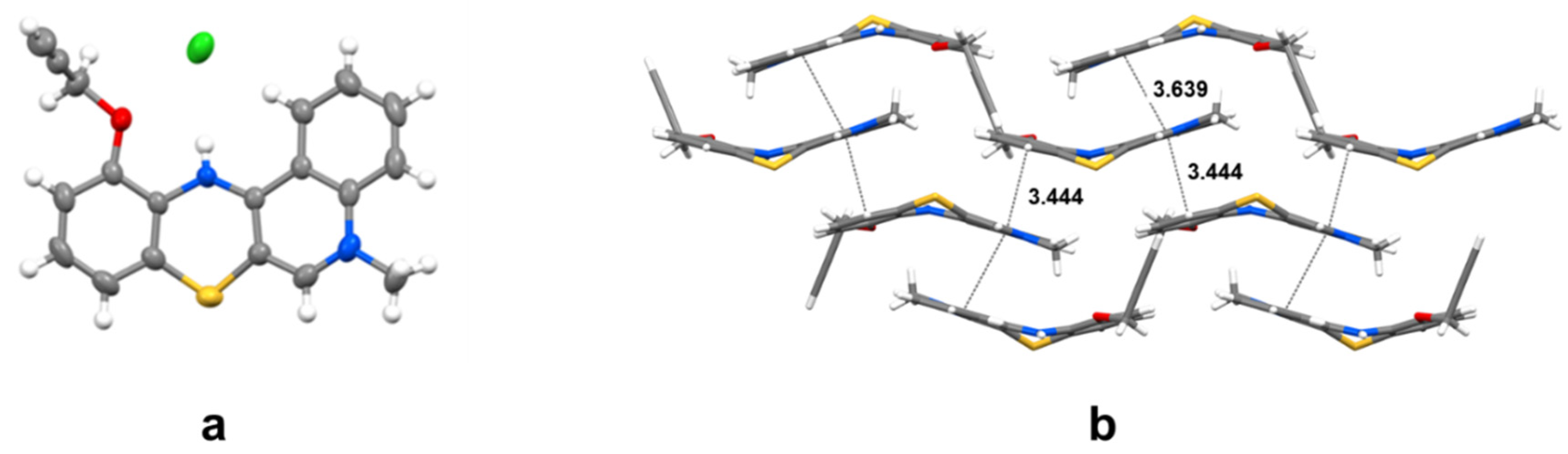
2.2. Structural Analysis

The structure of all compounds obtained was confirmed by 1H and 13C NMR spectroscopy using two-dimensional HSQC and HMBC techniques. Additionally, the structure was confirmed by the HR-MS method using the ESI ionization method. For all the compounds, the experimentally determined molecular weights differed from the theoretically calculated values only in the third decimal place, which fully confirms the elemental composition of these compounds and their structure.
X-ray analysis not only confirmed the structure of the final products determined by spectroscopic methods, but also showed their spatial structure in the solid phase. By crystallization from ethanol, it was possible to obtain a single crystal of the derivative 6i. The X-ray structure of the derivative 6i is shown in Figure 1. The molecule of 11-propargyloxy-5-methyl-12H-quino[3,4-b][1,4]benzothiazinium chloride 6i is bent along the axis defined by the nitrogen and sulphur atom of the thiazine ring under the angle of 146.5°. The angle formed by the C6a-S7-C7a atoms in the thiazine ring is 97.9°, while that between the C11a-N12-C12a atoms is 122.3°. Most of the structural parameters of the molecule are similar (or correspond well) to the parameters of the previously reported unsubstituted tetracyclic structure of 5-methyl-12H-quino[3,4-b][1,4]benzothiazinium chloride [22].
Each molecule interacts with adjacent molecules with three π–π stacking interactions, shown in Figure 1 with grey broken lines.

2.3. Biological Screening

2.3.1. In Vitro Antimicrobial Activity

Some of the previously obtained quinobenzothiazinium derivatives showed interesting antimicrobial properties [10]. All the investigated compounds were tested for in vitro antibacterial and antimycobacterial activity against a battery of microbial pathogens. The selection of the studied bacterial strains was adopted following the CLSI (National Committee for Clinical Laboratory Standards) international reference methodologies [26], i.e., standardization. For this purpose, universally sensitive collection strains from ATCC (Staphylococcus aureus ATCC 29213 and Enterococcus faecalis ATCC 29212) were selected. The second aspect of strain selection was the current state of occurrence of strains with an epidemiologically significant type of resistance, represented by clinical isolates of human and veterinary origin, i.e., different sequence types limited to human and animal populations, e.g., methicillin-resistant Staphylococcus aureus (MRSA) SA 3202, SA 630 and 63718 isolates carrying the mecA gene [27]. In the case of vancomycin-resistant E. faecalis (VRE) 342B, 368 and 725B isolates carrying the vanA gene [28], these were isolates from wild birds that were colonised from US hospital wastewater, as confirmed. Therefore, it can be concluded that the tested strains differed in the spectrum of antibiotic resistance, genetic make-up and, probably, accessory genome. In addition, all the compounds were evaluated in vitro against Mycobacterium smegmatis ATCC 700084 and M. marinum CAMP 5644 as a safe alternative to M. tuberculosis. The genus Mycobacterium is a closely related group of fast- and slow-growing species. Alternative model pathogens for M. tuberculosis can be used in laboratory studies to reduce risks and facilitate laboratory handling. M. smegmatis is an ideal representative of a fast-growing non-pathogenic microorganism particularly useful in the study of basic cellular processes of particular importance for pathogenic mycobacteria [29]. M. marinum is very closely related to M. tuberculosis and is the cause of TB-like infections in poikilothermic organisms, especially frogs and fish. M. marinum is a good model for study mainly due to the lower risk for laboratory workers, and its genetic relatedness and similar pathology to human TB [30]. Activities are expressed as the minimum inhibitory concentrations (MICs) and the minimum bactericidal concentrations (MBCs) as reported in Table 1. To establish that a compound demonstrates a bactericidal effect against a particular tested strain, it must meet the condition MIC/MBC ≤ 4 [27,31]. MBC values that fulfil this requirement, i.e., the compound is bactericidal, are indicated in bold in Table 1.
9-Benzyloxy-5-methyl-12H-quino[3,4-b][1,4]benzothiazinium chloride (6j) demonstrated the highest activity against the entire tested microbial spectrum. Furthermore, 9-propoxy-5-methyl-12H-quino[3,4-b][1,4]benzothiazinium chloride (6a) and 9-allyloxy-5-methyl-12H-quino[3,4-b][1,4]benzothiazinium chloride (6d) also showed high activity. Compounds 6b, 6e, 6k and 6l expressed antistaphylococcal activity, while compound 6h had only a moderate effect against enterococci. The efficacy of the active agents was comparable to the antibiotics clinically used against staphylococcal strains, especially against MRSA isolates. It should be noted that compounds showed comparable antistaphylococcal activities, both against methicillin-susceptible S. aureus and MRSA isolates; therefore, it can be assumed that the presence of the mecA gene (which encodes an alternative transpeptidase and causes methicillin resistance [27,32]) in MRSA does not affect the activity of these compounds. Thus, the above can be speculated concerning the specific activity against Staphylococcus sp. Similarly, the close activity of the compounds against both E. faecalis and VRE indicates a mechanism of action unrelated to vancomycin resistance [28]. It is important to mention that all compounds demonstrated bactericidal activity (see bolded MBC values in Table 1).
In addition to 6j, 6a and 6d, the compounds 6k, 6b, 6e, 6g and 6h proved to be active against fast-growing M. smegnatis, and compounds 6l, 6e, 6j and 6g were effective against slow-growing M. marinum. All the mentioned compounds were more effective than isoniazid, and their activities were comparable with rifampicin against both mycobacterial strains.
If these compounds are compared with recently described ones [25], it can be stated that there was a significant increase in antibacterial activity. The absence of heteroatoms in positions 8 or 10, and the replacement of halogens by alkoxy substituents on C(9) resulted in significant progress in the design of quino[3,4-b][1,4]benzothiazines as antimicrobial agents [25]. Based on the data presented in Table 1, it can be summarised that the substitution by the benzyloxy or propoxy chain appears to be the most advantageous, specifically in the 9 position, followed by the 10 position. The position 11 is completely disadvantageous in terms of antimicrobial activity. This empirical reasoning is supported by a multidimensional-based study of structure–property and structure–activity relationships (see Section 2.4). In addition, similar observations were found in the previous studies, where the highest antistaphylococcal and antimycobacterial activities were shown by derivatives substituted in the 9 or 10 position with branched alkoxy tails [33,34] or with a phenylalkyl chain [35,36,37], which mimics the benzyloxy substituent used here.
Since the compounds act more on aerobic staphylococci than on facultatively anaerobic enterococci [38], known among other things for high resistance to disinfection procedures and antibiotics [39,40,41], it was hypothesised that a possible mechanism of action could be inhibition of respiration and, therefore, a standard MTT test was performed with the most active derivatives. The MTT assay can be used to assess cell growth by measuring respiration. Respiratory activity of bacterial cells (which is finally reflected in their viability) of less than 70% after exposure to the MIC values for each tested compound is considered as a positive result of this assay. This low level of cell oxidative metabolism indicates inhibition of cell growth by inhibition of respiration [42,43]. The lowest multiples of the MIC values by which inhibition of S. aureus ATCC 29213 viability (%) greater than 70% was achieved are given in Table 2. It can be concluded that evaluated compounds 6a, 6e, 6g and 6k did not show a decrease in viability < 70% at its MIC value, which suggests that the main mechanism of action is not inhibition of the respiratory chain, although they are able to significantly affect it, compared to, e.g., ciprofloxacin. On the other hand, compound 6j (R = 9-OCH2C6H5) demonstrated strong inhibition of the respiratory chain (0.5×MIC/0.5×MBC).
Phenothiazines are also known for their ability to interact with and damage the cell wall, which has fatal consequences for the microorganism [44,45]. Therefore, the bacterial suspensions were treated by compounds 6a, 6e, 6g, 6j and 6k (4× MIC) for 1 h. The uptake of crystal violet was expressed as a percentage compared to the original crystal violet solution. The results are shown in Figure 2. The test compounds did not affect plasma membrane permeability of S. aureus, because the percentage absorption of crystal violet was comparable to the growth control and disproportionately lower than the 1% Tween 20 solution used as the positive control. Based on these results, it could be assumed that an increase in the membrane permeability was not the mechanism of action of these compounds.
As the antibacterial activity of the discussed compounds cannot be explained solely on the basis of inhibition of respiration, it is also very difficult to reach a conclusion regarding the actual mechanism of action of the antimycobacterial activity. Phenothiazines, like quinolines, are multi-target agents, i.e., they have the ability to affect several different targets, leading to the death of treated microorganisms. Quinolines are known to be able to inhibit, for example, the respiratory chain (possessing a similar mechanism of action to bedaquiline [46,47,48,49]), sulphur metabolic pathways [50] or mycobacterial FtsZ protein [51]; phenothiazines, in addition to the aforementioned ability to interact with the bacterial membrane, have the ability to inhibit the respiratory chain, dissipate the membrane potential, reduce the level of ATP, increase oxidative stress or increase the level of intracellular ions [44,45,52,53].
Therefore, the results obtained in this study will need to be supported by functional genomic and proteomic studies to reveal the mechanisms of actions of these 5-methyl-12H-quino[3,4-b][1,4]benzothiazinium chlorides on staphylococci and mycobacteria.

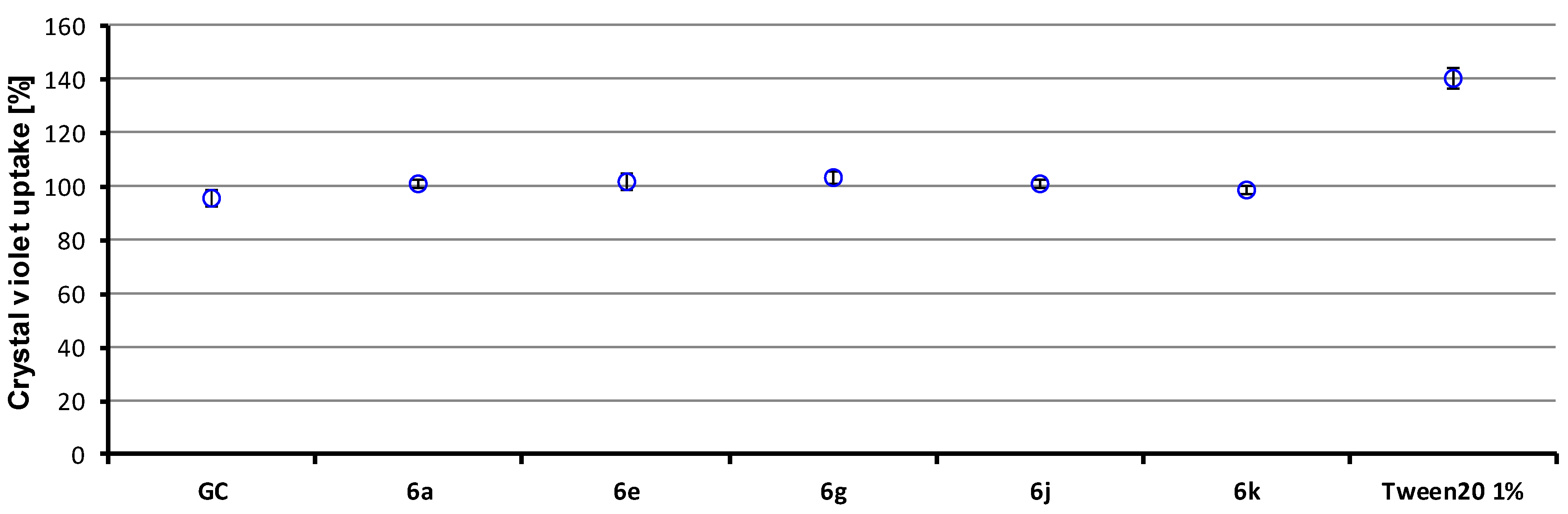
2.3.2. In Vitro Cell Viability

A preliminary in vitro cytotoxicity screening of target compounds 6al was performed using normal human dermal fibroblasts (NHDF) and was expressed as IC50 values (see Table 1). A compound is considered cytotoxic if it demonstrates a toxic effect on cells at a concentration up to 10 µM [54]. For example, the IC50 for doxorubicin was found to be 5.5 µM in this assay. The lowest concentration that affected human non-tumour cells was 37.7 µM for compound 6j, i.e., more than three times the value at which the bioactive agent is considered cytotoxic. Even this fact does not prevent active substances from being considered interesting for further investigation and from being generally considered as insignificantly cytotoxic.

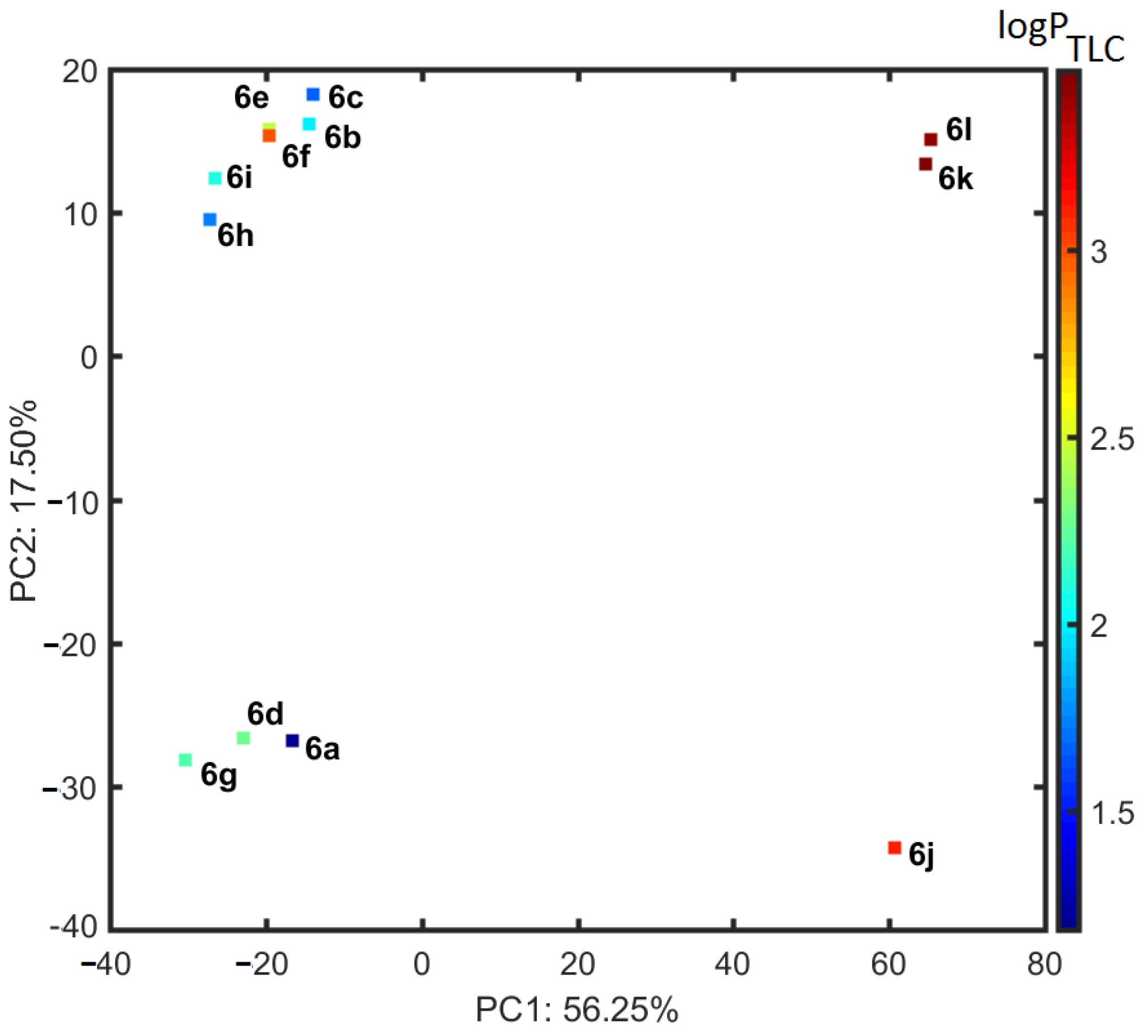
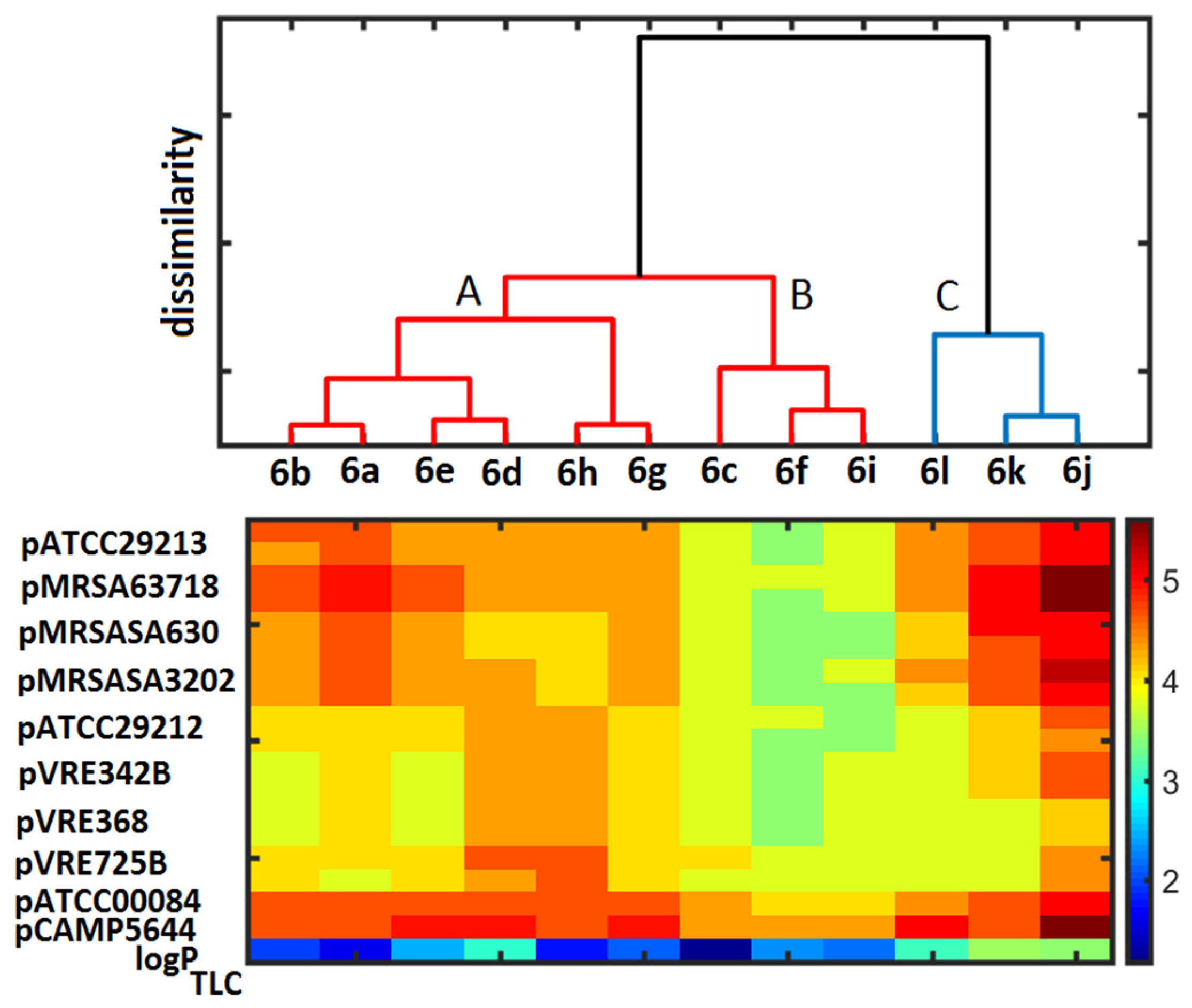
2.4. Similarity-Mediated Property Mapping

Many factors and parameters play an important role in the design and subsequent development of bioactive agents [55]. One of them is lipophilicity, which is among the most important of all investigated physicochemical properties, as it affects not only the ligand–target binding interaction, but also solubility and subsequent absorption (biological availability), binding to transporters, metabolism and excretion [56]. The lipophilicity of the studied compounds was determined using TLC as logPTLC parameter (see Table 1).
In practice, the clustering tendency of the descriptor-driven data can be investigated by tracing the (dis)similarities between objects in the multidimensional variable space; therefore, the distance-related property mapping was conducted using principal component analysis (PCA) and hierarchical clustering analysis (HCA) on the pool of 2666 descriptors produced by Dragon 6.0 software. The calculated data were organised into matrix X12×2666 with rows representing objects (compounds 6al) and columns representing in silico parameters (descriptors). The resulting matrix was centred and standardised, because the numerical parameters varied noticeably. The percentage of the modelled data variance was taken into account in order to specify the number of relevant principal components (PCs). The first three PCs describe 87.75% of the total data variance, while the first two PCs account for 73.75%, respectively. The corresponding projection (scoreplot) of molecules 6al on the plane defined by PC1 vs. PC2, and additionally colour-coded according to the empirical lipophilicity (logPTLC), is shown in Figure 3.
Not surprisingly, the benzyl-substituted isomers (6jl) are located separately (PC1 > 0) from the aliphatic derivatives 6ai, which are characterised by the negative values of the first component (PC1 < 0) and relatively lower lipophilic characteristics (logPTLC < 3). The interesting distribution of analogues 6al is observed according to the second principal component (PC2), where isomers 6a, d, g and j are grouped together along the negative values of PC2. Bafflingly, derivatives with substituents in positions 10 and 11 are clustered together on the positive part of PC2, as illustrated in Figure 3.
In an attempt to relate the (dis)similarity between objects (molecules) in the multidimensional descriptor-based space to the molecular property profile (e.g., biological or lipophilic compound characteristics), hierarchical clustering analysis (HCA) was combined with a colour-coded map of the empirical dataset [25,57]. On the whole, the exploratory HCA approach produces the sub-optimal clustering pattern of objects that is largely dependent on the clusters’ linkage procedure engaged; therefore, the similarity measure (e.g., Euclidean distance) as well as the manner of resulting sub-clusters’ linkage (e.g., Ward’s algorithm) need to be chosen a priori [58]. A dendrogram in Figure 4, conjugated with the colourful display of experimental data (e.g., biological activities and lipophilic characteristics) allows for the direct interpretation of the produced clusters in terms of the original parameters, where OX illustrates the order of objects and the OY axis presents the dissimilarity, respectively. The exploratory HCA procedure generated the clustering pattern of objects that confirms our previous PCA findings (see Figure 3). Likewise, the benzyl-containing molecules 6jl vary noticeably (cluster C) from the remaining compounds (cluster A and B) of the dataset. Roughly speaking, molecules 6c, f and i are grouped together in cluster B, which is characterised by noticeably lower values of antibacterial potency and lipophilic values, as shown in colour-coded vectors in Figure 4. An inverted SAR trend is observed for molecules 6a, b, d, e, g and h (cluster A and C), which are generally marked by higher values of biological activities. On the other hand, there is no evident SPR (structure–property) relationship with logPTLC in the 6a, b, d, e, g and h subunit, because these isomers in cluster C are described by higher lipophilic values.

3. Materials and Methods

3.1. Chemisty

Melting points are uncorrected. NMR spectra were recorded using a Bruker Ascend 600 spectrometer (Bruker, Billerica, MA, USA). To assign the structures, the following 2D experiments were employed: 1H-13C gradient selected HSQC and HMBC sequences. Standard experimental conditions and standard Bruker programs were used. The 1H NMR and 13C NMR spectral data are provided relative to the TMS signal at 0.0 ppm. HR mass spectra were recorded with Bruker Impact II (Bruker, Billerica, MA, USA).

3.1.1. Synthesis of Acetanilide Derivatives 2al

A solution of 10 mmol of the appropriate hydroxyacetanilide (1) in 10 mL of anhydrous DMF was added portionwise to a suspension of 10 mmol NaH (60% suspension in mineral oil) in 10 mL dry DMF with stirring. To the reaction mixture obtained, 12 mmol of the alkylating agent were added in portions while stirring. After 12 h the mixture was poured into 100 mL of water and extracted with chloroform (3 × 15 mL). The combined extracts were dried over anhydrous sodium sulphate. After evaporation of the solvent in vacuo, the dry residue was purified by aluminium oxide column chromatography, eluting with chloroform: ethanol v/v 10:1.
2-(propoxy)acetanilide (2a): yield: 92%; 1H NMR (CDCl3, 600 MHz), δ(ppm): 1.02–1.10 (t, J = 7.2 Hz, 3H,CH2-CH3), 1.75–1.83 (m, 2H, CH2CH2CH3), 2.21 (s, 3H, COCH3) 3.97–4.07 (t, J = 6.6 Hz, 2H, CH2CH2CH3), 6.83–6.85 (m, 1H, Harom), 6.85–6.97 (m, 1H, Harom), 6.99–7.05 (m, 1H, Harom), 7.82 (s, 1H, NH), 6.83–6.88 (m, 1H, Harom); 13C NMR, (CDCl3, 150.9 MHz) δ (ppm): 9.93, 21.48, 23.89, 69.08, 109.92, 118.73, 1189.86, 122.56, 126.81, 148.06, 168.14; ESI-HRMS Calcd for C11H16NO2 ([M + H]+): 194.1181, found: 194.1177.
3-(propoxy)acetanilide (2b): yield: 95%; 1H NMR (CDCl3, 600 MHz), δ (ppm): 0.96–1.07 (t, J = 7.2 Hz, 3H,CH2-CH3), 1.73–1.83 (m, 2H, CH2CH2CH3), 2.15 (s, 3H, COCH3) 3.82–3.89 (t, J = 6.6 Hz, 2H, CH2CH2CH3), 6.60–6.73 (m, 1H, Harom), 6.95–6.99 (m, 1H, Harom), 7.05–7.15 (m, 1H, Harom), 7.23–7.30 (m, 1H, Harom), 8.53 (s, 1H, NH), 13C NMR (CDCl3, 150.9 MHz), δ (ppm): 9.46, 21.50, 23.37, 68.43 105.38, 109.45, 111.04, 128.44, 138.39, 158.56, 167.10; ESI-HRMS Calcd for C11H16NO2 ([M + H]+): 194.1181, found: 194.1175.
4-(propoxy)acetanilide (2c): yield: 96%; 1H NMR (DMSO, 600 MHz), δ (ppm): 0.92–1.00 (t, J = 7.2 Hz, 3H, CH2-CH3), 1.64–1.73 (m, 2H, CH2CH2CH3), 2.03 (s, 3H, COCH3), 3.80–3.90 (t, J = 6.6 Hz, 2H, CH2CH2CH3), 6.80–6.87 (m, 2H, Harom), 7.42–7.50 (m, 2H, Harom), 9.77 (s, 1H, NH); 13C NMR(DMSO, 150.9 MHz), δ (ppm): 10.99 (CH3), 22.55 (CH2), 24.28 (COCH3), 69.46 (OCH2), 114.79, 114.39, 120.94, 132.90, 154.88, 168.14; ESI-HRMS Calcd for C11H16NO2 ([M + H]+): 194.1181, found: 194.1172.
2-(allyloxy)acetanilide (2d): yield: 94%: 1H NMR (CDCl3, 600 MHz), δ (ppm): 2.19 (s, 3H, CH3), 4.53–4.65 (m, 2H, CH2-CH=CH2), 5.30–5.38 (m, 1H, CH2-CH=CH2), 5.38–5.45 (m, 1H, CH2-CH=CH2), 5.98–6.15 (m, 1H, CH2-CH=CH2), 6.80–6.89 (m, 1H, Harom), 6.89–7.05 (m, 1H, Harom), 7.78–7.89 (m, 1H, Harom), 8.30–8.40 (m, 1H, Harom); 13C NMR (CDCl3, 150.9 MHz), δ (ppm): 24.28 (CH3), 69.79 (CH2-CH), 115.23, 120.93, 128.14, 128.23, 128.86, 133.22, 137.68, 154.52, 168.20 (C=O); 13C NMR (CDCl3, 150.9 MHz), δ (ppm): 24,93 (CH3), 69.48 (CH2), 111,37, 118.21, 119.97, 121.26, 123.57, 127.93, 132.84, 146.72, 168.21; ESI-HRMS Calcd for C11H14NO2 ([M + H]+): 192.1024, found: 192.1018.
3-(allyloxy)acetanilide (2e): yield: 94%: 1H NMR (CDCl3, 600 MHz), δ (ppm): 2.06 (s, 3H, CH3), 4.35–4.48 (m, 2H, CH2-CH=CH2), 5.05–5.20 (m, 1H, CH2-CH=CH2), 5.20–5.37 (m, 1H, CH2-CH=CH2), 5.80–98 (m, 1H, CH2-CH=CH2), 6.50–6.58 (m, 1H, Harom), 6.90–6.98 (m, 1H, Harom), 7.03–7.10 (m,1H, Harom), 7.25–7.30 (m, 1H, Harom), 8.55 (s, 1H, NH); 13C NMR (CDCl3, 150.9 MHz), δ (ppm): 24.41 (CH3), 68.76 (CH2-CH), 106.60, 110.61, 112.40, 117.63, 129.52, 133.17, 133.17, 139.51, 159.01, 169.22 (C=O); ESI-HRMS Calcd for C11H14NO2 ([M + H]+): 192.1024, found: 192.1012.
4-(allyloxy)acetanilide (2f): yield: 90%: 1H NMR (CDCl3, 600 MHz), δ (ppm): 2.15 (s, 3H, CH3), 4.48–4.48 (m, 2H, CH2-CH=CH2), 5.20–5.28 (m, 1H, CH2-CH=CH2), 5.35–5.42 (m, 1H, CH2-CH=CH2), 5.98–6.10 (m, 1H, CH2-CH=CH2), 6.82–6.88 (m, 2H, Harom), 7.35–7.42 (m, 2H, Harom), 7.90 (s, 1H, NH); 13C NMR (CDCl3, 150.9 MHz), δ (ppm): 23.09 (CH3), 68.04 (CH2-CH), 113.95, 116.67 (CH=CH2), 120.93, 130.17 (CH=CH2), 132.20 154.40, 167.69 (C=O); ESI-HRMS Calcd for C11H14NO2 ([M + H]+): 192.1024, found: 192.32.
2-(propargyloxy)acetanilide (2g): yield: 87%: 1H NMR (CDCl3, 600 MHz), δ (ppm): 2.02 (s, 3H, CH3), 2.50–2.52 (t, J = 2.4 Hz, 1H, CH), 4.53–4.59 (d, J = 2.4 Hz, 2H, CH2), 6.75–6.90 (m, 3H, Harom), 7.93 (s, 1H, NH), 8.11–8.16 (m, 1H, Harom); 13C NMR (CDCl3, 150.9 MHz), δ (ppm): 24.57, 56.37, 76.28, 73.03, 111.87, 120.44, 121.71, 123.49, 128.10, 146.06, 168.40; ESI-HRMS Calcd for C11H12NO2 ([M + H]+): 190.0868, found: 190.0863.
3-(propargyloxy)acetanilide (2h): yield: 87%: 1H NMR (CDCl3, 600 MHz), δ (ppm): 2.06 (s, 3H, CH3), 2.42–2.51 (t, J = 2.4 Hz, 1H, CH), 4.49–4.55 (d, J = 2.4 Hz, 2H, CH2), 6.56–6.61 (m, 1H, Harom), 7.05–7.12 (m, 2H, Harom), 7.25–7.31 (m, 1H, Harom), 9.26 (s, 1H, NH); 13C NMR (CDCl3, 150.9 MHz), δ (ppm): 24.26, 55.71, 75.71, 78.55, 110.30, 113.21, 129.50, 139.80, 157.84, 169.58; ESI-HRMS Calcd for C11H12NO2 ([M + H]+): 190.0868, found: 190.0865.
4-(propargyloxy)acetanilide (2i): yield: 92%: 1H NMR (CDCl3, 600 MHz), δ (ppm): 2.06 (s, 3H, CH3), 2.42–2.52 (t, J = 2.4 Hz, 1H, CH), 4.55–4.62 (d, J = 2.4 Hz, CH2), 6.80–6.85 (d, J = 9 Hz, 2H, Harom), 7.38–7.45 (d, J = 9 Hz, 2H, Harom), 8.88 (s, 1H, NH); 13C NMR (CDCl3, 150.9 MHz), δ (ppm): 24.01, 56.07, 75.57, 78.60, 115.07, 121.78, 132,56, 153,91, 168.98; ESI-HRMS Calcd for C11H12NO2 ([M + H]+): 190.0868, found: 190.0857.
2-(benzyloxy)acetanilide (2j): yield: 86%: 1H NMR (DMSO, 600 MHz), δ (ppm): 2.11 (s, 3H, CH3); 5.21 (s, 2H, CH2); 6.83–6.92 (m, 1H, Harom), 6.98–7.10 (m, 2H, Harom), 7.28–7.35 (m, 1H, Harom), 7.35–7.42 (m, 2H, Harom), 7.47–7.54 (m, 1H, Harom), 7.81–7.90 (m, 1H, Harom), 9.17 (s, 1H, NH); 13C NMR (DMSO, 150.9 MHz), δ (ppm): 24.28 (CH3), 70.02 (CH2), 113.31, 120.90, 123.33, 124.83, 127.75, 128.17, 128.22, 128.85, 137.58, 149.31, 168.97; ESI-HRMS Calcd for C15H16NO2 ([M + H]+): 242.1181, found: 242.1176.
3-(benzyloxy)acetanilide (2k): yield: 89%: 1H NMR (CDCl3, 600 MHz), δ (ppm): 2.19 (s, 3H, CH3), 5.07 (s, 2H, CH2), 6.71–6.78 (m, 1H, Harom), 6.96–7.03 (m, 1H, Harom), 7.15–7.25 (m, 1H, Harom), 7.29–7.38 (m, 1H, Harom), 7.39–7.48 (m, 5H, Harom); 13C NMR (CDCl3, 150.9 MHz), δ (ppm): 24.70 (CH3), 70.01 (CH2), 106.55, 110.99, 112.22, 127.54, 127.98, 128.58, 129.71, 136.87, 139.11, 159.33, 168.46; ESI-HRMS Calcd for C15H16NO2 ([M + H]+): 242.1181, found: 242.1173.
4-(benzyloxy)acetanilide (2l): yield: 93%: 1H NMR (CDCl3, 600 MHz), δ (ppm): 2.16 (s, 3H, CH3), 5.05 (s, 2H, CH2), 6.90–6.97 (m, 2H, Harom), 7.37–7.49 (m, 7H, Harom), 7.79 (s, 1H, NH); 13C NMR (CDCl3, 150.9 MHz), δ (ppm): 24.15 (CH3), 70.29 (CH2), 115.15, 122.07, 127.50, 128.00, 128.60, 131,19, 136,93, 155.69, 168.77; ESI-HRMS Calcd for C15H16NO2 ([M + H]+): 242.1181, found: 242.1183.

3.1.2. Synthesis of Aniline Derivatives 3al

To a solution of 10 mmol of the appropriate acetanilide (2) in 30 mL of ethanol, 30 mL of 36% hydrochloric acid was added. The resulting mixture was heated (while stirring) at 60 °C for 24 h. After cooling to room temperature, the mixture was poured into 50 mL of water and extracted with chloroform (3 × 15 mL). The combined extracts were dried over anhydrous sodium sulphate. After evaporation of the solvent in vacuo, the dry residue was purified by aluminium oxide column chromatography, eluting with chloroform: ethanol v/v 10:1.
2-(propoxy)aniline (3a): oil; yield: 84%; 1H NMR (DMSO, 600 MHz) δ (ppm): 0.95–1.05 (t, J = 7.2 Hz, 3H, CH2-CH3), 1.70–1.79 (m, 2H, CH2CH2CH3), 3.82–3.93 (t, J = 6.3 Hz, 2H, CH2CH2CH3), 4.64 (s, 2H, NH2), 6.48–6.56 (m, 1H, Harom), 6.65–6.72 (m, 2H, Harom), 6.75–6.79 (m, 1H, Harom); 13C NMR (CDCl3, 150.9 MHz), δ (ppm): 10.99 (CH3), 22.73 (CH2), 69.59 (OCH2), 112.03, 114.39, 116.73, 121.23, 138.19, 146.13; ESI-HRMS Calcd for C9H14NO ([M + H]+): 152.1075, found: 152.1067.
3-(propoxy)aniline (3b): oil; yield: 92%; 1H NMR (DMSO, 600 MHz) δ (ppm): 0.90–1.02 (t, J = 7.4 Hz, 3H, CH2-CH3), 1.62–1.71 (m, 2H, CH2CH2CH3), 3.73–3.82 (t, J = 6.6 Hz, 2H, CH2CH2CH3), 5.02 (s, 2H, NH2), 6.02–6.10 (m,1H, Harom), 6.12–6.20 (m, 2H, Harom), 6.85–6.92 (m, 1H, Harom); 13C NMR (CDCl3, 150.9 MHz), δ (ppm):): 10.89 (CH3), 22.64 (CH2), 68.84 (OCH2), 100.48, 102.53, 107.20, 129.94, 150.38, 160.16; ESI-HRMS Calcd for C9H14NO ([M + H]+): 152.1075, found: 152.1085.
4-(propoxy)aniline (3c): oil; yield: 85%; 1H NMR (DMSO, 600 MHz) δ (ppm): 0.91–0.98 (t, J = 7.2 Hz, 3H, CH2-CH3), 1.60–1.69 (m, 2H, CH2CH2CH3), 3.73–3.78 (t, J = 6.6 Hz, 2H, CH2CH2CH3), 4.59 (s, 2H, NH2), 6.46–6.53 (m, 2H, Harom), 6.60–6.65 (m, 2H, Harom); 13C NMR (DMSO, 150.9 MHz), δ (ppm): 10.94 (CH3), 22.72 (CH2), 69.88 (OCH2), 115.41, 115.77, 142.74, 150.47; ESI-HRMS Calcd for C9H14NO ([M + H]+): 152.1075, found: 152.1077.
2-(allyloxy)aniline (3d): oil; yield: 82%; 1H NMR (CDCl3, 600 MHz), δ (ppm): 3.73 (s, 2H, NH2), 4.52–4.62 (d, J = 4.8 Hz, 2H, CH2-CH=CH2), 5.25–5.32 (m, 1H, CH2-CH=CH2), 5.38–5.48 (m, 1H, CH2-CH=CH2), 6.02–6.12 (m, 1H, CH2-CH=CH2), 6.70–6.78 (m, 2H, Harom), 6.80–6.90 (m, 2H, Harom); 13C NMR (CDCl3 150.9 MHz), δ (ppm): 69.16 (OCH2), 112.00, 115.20, 117.41, 118.30, 121.37, 133.58, 136.52, 146.21; ESI-HRMS Calcd for C9H12NO ([M + H]+): 150.0918, found: 150.0915.
3-(allyloxy)aniline (3e): oil; yield: 89%; 1H NMR (CDCl3, 600 MHz), δ (ppm): 3.56 (s, 2H, NH2), 4.52–4.62 (d, J = 3.6 Hz, 2H, CH2-CH=CH2), 5.25–5.32 (m, 1H, CH2-CH=CH2), 5.38–5.48 (m, 1H, CH2-CH=CH2), 5.98–6.12 (m, 1H, CH2-CH=CH2), 6.32–6.40 (m, 3H, Harom), 6.98–7.10 (m, 1H, Harom); 13C NMR (CDCl3 150.9 MHz), δ (ppm): 68.63 (OCH2), 101.85, 104.68, 108.09, 117.49, 130.08, 133.48, 147.84, 159.75; ESI-HRMS Calcd for C9H12NO ([M + H]+): 150.0918, found: 150.0909.
4-(allyloxy)aniline (3f): oil; yield: 80%; 1H NMR (CDCl3, 600 MHz), δ (ppm): 3.48 (s, 2H, NH2), 4.42–4.50 (d, J = 4.8 Hz, 2H, CH2-CH=CH2), 5.22–5.31 (m, 1H, CH2-CH=CH2), 5.35–5.45 (m, 1H, CH2-CH=CH2), 6.02–6.12 (m, 1H, CH2-CH=CH2), 6.60–6.69 (d, J = 8.4 Hz, 2H, Harom), 6.72–6.80 (d, J = 8.4 Hz, 2H, Harom); 13C NMR (CDCl3 150.9 MHz), δ (ppm): 69.59 (OCH2), 115.93, 116.38, 117.39, 133.84, 140.26, 151.72; ESI-HRMS Calcd for C9H12NO ([M + H]+): 150.0918, found: 150.0911.
2-(propargyloxy)aniline (3g): oil; yield: 85%; 1H NMR (CDCl3, 600 MHz), δ (ppm): 2.50–2.60 (m, 1H, CH), 3.76 (s, 2H, NH2), 4.70–4.78 (m, 2H, CH2), 6.20–6.30 (m, 2H, Harom), 6.85–6.90 (m, 1H, Harom), 7.92–6.97 (m, 1H, Harom);13C NMR (CDCl3, 150.9 MHz), δ (ppm): 56.37 (CH2), 75.48 (CH), 78.84 (CH2-CCH), 112.62, 115.55, 118.35, 122.29, 136.68, 145.30; ESI-HRMS Calcd for C9H10NO ([M + H]+): 148.0765, found: 148.0746.
3-(propargyloxy)aniline (3h): oil; yield: 88%; 1H NMR (CDCl3 600 MHz), δ (ppm): 2.38–2.45 (m, 1H, CH), 3.37 (s, 2H, NH2), 4.57–4.66 (m, 2H, CH2), 6.28–6.36 (m, 2H, Harom), 6.36–6.43 (m, 1H, Harom), 7.02–7.12 (m, 1H, Harom); 13C NMR (CDCl3, 150.9 MHz), δ (ppm): 56.21 (CH2), 75.41 (CH), 78.82 (CH2-CCH), 101.97, 104.63, 108.77, 130.14, 147.85, 158.73; ESI-HRMS Calcd for C9H10NO ([M + H]+): 148.0765, found: 148.0758.
4-(propargyloxy)aniline (3i): oil; yield: 92%; 1H NMR (CDCl3, 600 MHz), δ (ppm): 2.48–2.55 (m, 1H, CH), 3.52 (s, 2H, NH2), 4.60–2.65 (m, 2H, CH2), 6.62–6.70 (m, 2H, Harom), 6.80–6.88 (m, 2H, Harom), 13C NMR (CDCl3, 150.9 MHz), δ (ppm): 56.73 (CH2), 75.18 (CH), 79.12 (CH2-CCH), 116.27, 116.38, 149.86, 150.70; ESI-HRMS Calcd. for C9H10NO ([M + H]+): 148.0765, found: 148.0761.
2-(benzyloxy)aniline (3j): oil; yield: 83%; 1H NMR (CDCl3, 600 MHz), δ (ppm): 4.76 (s, 2H, NH2), 5.09 (s, 2H, CH2), 6.48–6.56 (m, 1H, Harom), 6.68–6.74 (m, 2H, Harom), 6.87–6.90 (m, 1H, Harom), 7.30–7.37 (m, 1H, Harom), 7.37–7.42 (m, 2H, Harom), 7.48–7.53 (m, 2H, Harom); 13C NMR (CDCl3, 150.9 MHz), δ (ppm):69.77 (CH2), 112.66, 114.65, 116.68, 121.67, 127.87, 128.11, 128.84, 138.02, 138.47, 145.77; ESI-HRMS Calcd for C13H14NO ([M + H]+): 200.1075, found: 200.1076.
3-(benzyloxy)aniline (3k): oil; yield: 85%; 1H NMR (CDCl3, 600 MHz), δ (ppm): 3.78 (s, 2H, NH2), 5.04 (s, 2H, CH2), 6.39–6.38 (m, 2H, Harom), 6.41–6.47 (m, 1H, Harom), 7.03–7.11 (m, 1H, Harom), 7.30–7.47 (m, 5H, Harom); 13C NMR (CDCl3, 150.9 MHz), δ (ppm): 69.81 (CH2), 102.09, 104.95, 108.31, 127.50, 127.90, 128.58, 130.15, 137.23, 147.66, 159.99; ESI-HRMS Calcd for C13H14NO ([M + H]+): 200.1075, found: 200.1070.
4-(benzyloxy)aniline (3l): oil; yield: 80%; 1H NMR (CDCl3, 600 MHz), δ (ppm): 3.76 (s, 2H, NH2), 5.02 (s, 2H, CH2), 6.68–6.73 (m, 2H, Harom), 6.82–6.88 (m, 2H, Harom), 7.30–7.49 (m, 5H, Harom); 13C NMR (CDCl3, 150.9 MHz), δ (ppm): 70.74 (CH2), 116.05, 116.72, 127.52, 127.84, 128.53, 137.45, 152.29, 162.58; ESI-HRMS Calcd for C13H14NO ([M + H]+): 200.1075, found: 200.1077.

3.1.3. Synthesis of 5-Methyl-12H-Quino[3,4-b][1,4]Benzothiazine Chloride 6al

Procedure A. 2.5 mmol of the appropriate aniline (3) was added to a suspension of 1 mmol (0.419 g) of 5,12-(dimethyl)thioquinantrenediinium bis-chloride (4) in 5 mL of anhydrous pyridine. The resulting reaction mixture was heated at 80 °C with vigorous (continuous) stirring to allow the introduction of atmospheric oxygen. After 24 h, the mixture was cooled to room temperature and the resulting solid was filtered off and washed with anhydrous ether (3 × 5 mL). The crude product was purified on an alumina chromatography column, eluting with chloroform: ethanol v/v 10:1.
Procedure B. 2.5 mmol of the appropriate aniline (3) was added to a suspension of 1 mmol (0.419 g) of 5,12-(dimethyl)thioquinantrenediinium bis-chloride (4) in 5 mL of anhydrous pyridine. The resulting reaction mixture was heated at 20 °C with vigorous (continuous) stirring to allow the introduction of atmospheric oxygen. After 7 days, the resulting solid was filtered off and washed with anhydrous ether (3 × 5 mL). The crude product was purified on an alumina chromatography column, eluting with chloroform: ethanol v/v 10:1.
9-propoxy-5-methyl-12H-quino[3,4-b][1,4]benzothiazinium chloride (6a): Procedure A. Yield: 82%; 1H NMR (DMSO, 600 MHz), δ (ppm): 0.90–1.00 (t, J = 7.5 Hz, 3H, CH2CH3), 1.62–1.75 (m, 2H, CH2CH2CH3), 3.80–3.89 (t, J = 6.3 Hz, 2H, CH2CH2CH3), 4.08 (s, 3H, NCH3), 6.58–6.63 (m, 1H, Harom), 6.63–6.70 (m, 1H, Harom), 7.54–7.61 (m, 1H, Harom), 7.92–8.00 (m, 2H, Harom), 8.50–8.60 (m, 2H, Harom), 9.02–9.12 (m, 1H, Harom), 11.28 (s, 1H, NH); 13C NMR (DMSO, 150.9 MHz), δ (ppm): 10.80 (CH3), 22.39 (CH2), 42.75 (NCH3), 69.88 (OCH2), 104.98, 113.04, 114.07, 118.52, 118.86, 120.50, 124.42, 124.82, 134.65, 136.71, 139.08, 142.84, 150.00, 151.00, 158.03; ESI-HRMS Calcd for C19H19N2OS ([M]+): 323.1218, found: 323.1217.
10-propoxy-5-methyl-12H-quino[3,4-b][1,4]benzothiazinium chloride (6b): Procedure B. Yield: 80%; 1H NMR (DMSO, 600 MHz), δ (ppm): 0.88–1.03 (t, J = 7.2 Hz, 3H, CH2CH3), 1.65–1.80 (m, 2H, CH2CH2CH3), 3.80–3.90 (t, J = 6.3 Hz, 2H, CH2CH2CH3), 4.114 (s, 3H, NCH3), 6.62–6.67 (m, 1H, Harom), 6.89–6.94 (m, 1H, Harom), 7.38–7.45 (m, 1H, Harom), 7.75–7.84 (m, 1H, Harom), 7.95–8.09 (m, 2H, Harom), 8.67 (s, 1H, H6), 9.12–9.20 (m, 1H, Harom), 11.26 (s, 1H, NH); 13C NMR (DMSO, 150.9 MHz), δ (ppm): 10.80 (CH3), 22.34 (CH2), 43.08 (NCH3), 69.76 (OCH2), 106.48, 106.75, 106.99, 113.20, 116.09, 119.12, 124.76, 127.83, 128.17, 134.78, 137.91, 139.04, 143.58, 151.76, 159.32; ESI-HRMS Calcd for C19H19N2OS ([M]+): 323.1218, found: 323.1256.
11-propoxy-5-methyl-12H-quino[3,4-b][1,4]benzothiazinium chloride (6c): Procedure A. Yield: 76%; 1H NMR (DMSO, 600 MHz), δ (ppm): 0.99-1.05 (t, J = 7.5 Hz, 3H, CH2CH3), 1.80–1.90 (m, 2H, CH2CH2CH3), 4.02–4.12 (t, J = 6.3 Hz, 2H, CH2CH2CH3), 4.19 (s, 3H, NCH3), 6.70–6.74 (m, 1H, Harom), 697–7.02 (m, 1H, Harom), 7.08–7.13 (m, 1H, Harom), 7.86–7.92 (m, 1H, Harom), 8.06–8.10 (m, 1H, Harom), 8.10–8.14 (m, 1H, Harom), 8.40–8.45 (m, 1H, Harom), 8.84 (s, 1H, H6), 9.84 (s, 1H, NH); 13C NMR (DMSO, 150.9 MHz), δ (ppm): 10.84 (CH3), 22.30 (CH2), 43.34 (NCH3), 71.01 (OCH2), 107.77, 113.18, 116.30, 118.89, 119.32, 119.41, 123.50, 125.57, 128.11, 128.56, 135.01, 139.14, 144.31, 148.25, 152.02; ESI-HRMS Calcd for C19H19N2OS ([M]+): 323.1218, found: 323.1215.
9-allyloxy-5-methyl-12H-quino[3,4-b][1,4]benzothiazinium chloride (6d): Procedure A. Yield: 78%; 1H NMR (DMSOd-6, 600 MHz), δ(ppm): 4.09 (s, 3H, CH3), 4.49–4.55 (d, J = 4.8 Hz, 2H, CH2-CH=CH2), 5.25–5.30 (m, 1H, CH=CH2), 5.35–5.45 (m, 1H, CH=CH2), 5.95–6.10 (m, 1H, CH=CH2), 6.63–6.73 (m, 2H, Harom), 7.50–7.58 (m, 1H, Harom), 7.72–7.80 (m, 1H, Harom), 7.95–8.05 (m, 2H, Harom), 8.57 (s, 1H, H6), 8.98–9.05 (m, 1H, Harom), 11.20 (s, 1H, NH); 13C NMR (DMSO, 150.9 MHz), δ (ppm): 42.80 (CH3), 69.04 (CH2), 105.02, 113.38, 114.39, 115.83, 118.27, 118.51, 118.95, 120.39, 124.71, 127.88, 129.59, 133.75, 134.72, 139.09, 142.99, 151.08, 157.50; ESI-HRMS Calcd for C19H17N2OS ([M]+): 321.1062, found: 321.1056.
10-allyloxy-5-methyl-12H-quino[3,4-b][1,4]benzothiazinium chloride (6e): Procedure B. Yield: 78%; 1H NMR (DMSO, 600 MHz), δ (ppm): 4.14 (s, 3H, CH3), 4.48–4.54 (d, J = 4.8 Hz, 2H, CH2-CH=CH2), 5.28–5.32 (m, 1H, CH=CH2), 5.38–5.45 (m, 1H, CH=CH2), 5.95–6.10 (m, 1H, CH=CH2), 6.60–6.68 (m, 1H, Harom), 6.90–6.95 (m, 1H, Harom), 7.40–7.49 (m, 1H, Harom), 7.73–7.80 (m, 1H, Harom), 7.95–8.18 (m, 2H, Harom), 8.67 (s, 1H, H6), 9.10–9.19 (m, 1H,Harom), 11.27 (s, 1H, NH); 13C NMR (DMSO, 150.9 MHz), δ (ppm): 4.10 (CH3), 68.90 (CH2), 106.75, 106.98, 107.15, 113.33, 116.11, 118.46, 119.14, 124.74, 127.86, 128.20, 133.63, 134.80, 137.92, 139.05, 143.62, 151.76, 158.80; ESI-HRMS Calcd for C19H17N2OS ([M]+): 321.1062, found: 321.1063.
11-allyloxy-5-methyl-12H-quino[3,4-b][1,4]benzothiazinium chloride (6f): yield: Procedure A. 72%; 1H NMR (DMSO, 600 MHz), δ (ppm): 4.19 (s, 3H, CH3), 4.70–4.78 (d, J = 4.8 Hz, 2H, CH2-CH=CH2), 5.30–5.36 (m, 1H, CH=CH2), 5.45–5.55 (m, 1H, CH2-CH=CH2), 6.09–6.19 (m, 1H, CH=CH2), 6.70–6.75 (m, 1H, Harom), 6.95–7.0 (m, 1H, Harom), 7.05–7.13 (m, 1H, Harom), 7.85–7.92 (m, 1H, Harom), 8.05–8.15 (m, 2H, Harom), 8.45–8.52 (m, 1H, Harom), 8.85 (s, 1H, H6), 9.92 (s, 1H, NH); 13C NMR (DMSO, 150.9 MHz), δ (ppm): 43.32 (CH3), 70.17 (CH2), 107.71, 113.70, 116.30, 118.53, 119.09, 119.35, 119.58, 123.71, 125.70, 127.98, 128.52, 133.70, 135.01, 139.12, 144.27, 147.81, 152.02; ESI-HRMS Calcd for C19H17N2OS ([M]+): 321.1062, found: 321.1062.
9-propargyloxy-5-methyl-12H-quino[3,4-b][1,4]benzothiazinium chloride (6g): yield: Procedure A. 75%; 1H NMR (DMSO, 600 MHz), δ (ppm): 3.62–3.67 (m, 1H, CH), 4.09 (s, 3H, CH3), 4.76–4.97 (m, 2H, CH2), 6.70–6.82 (m, 2H, Harom), 7.45–7.58 (m, 1H, Harom), 7.73–7.82 (m, 1H, Harom), 7.94–8.08 (m, 2H, Harom), 8.59 (s, 1H, H6), 8.90–9.04 (m, 1H, Harom), 11.15 (s, 1H, NH); 13C NMR (DMSO, 150.9 MHz), δ (ppm): 42.86 (CH3), 56.32 (CH2), 79.15, 79.28, 105.07, 113.72, 114.79, 115.89, 118.54, 119.03, 120.25, 124.56, 127.98, 130.21, 134.78, 139.13, 143.15, 151.23, 156.48; ESI-HRMS Calcd for C19H15N2OS ([M]+): 319.0905, found: 319.0900.
10-propargyloxy-5-methyl-12H-quino[3,4-b][1,4]benzothiazinium chloride (6h): Procedure B. Yield: 71%; 1H NMR (DMSO, 600 MHz), δ (ppm): 3.64–3.68 (m, 1H, CH), 4.15 (s, 3H, CH3), 4.77–4.92 (m, 2H, CH2), 6.70–6.75 (m, 1H, Harom), 6.97–7.05 (m, 1H, Harom), 7.70–7.77 (m, 1H, Harom), 7.80–7.90 (m, 1H, Harom), 8.02–8.14 (m, 2H, Harom), 8.69 (s, 1H, H6), 8.88–8.97 (m, 1H, Harom), 10.98 (s, 1H, NH); 13C NMR (DMSO, 150.9 MHz), δ (ppm): 43.16 (CH3), 56.17 (CH2), 79.24, 79.55, 106.91, 107.07, 108.06, 113.34, 116.12, 119.28, 124.26, 127.98, 128.34, 14.87, 137.90, 139.07, 143.77, 151.74, 157.91; ESI-HRMS Calcd for C19H15N2OS ([M]+): 319.0905, found: 319.0903.
11-propargyloxy-5-methyl-12H-quino[3,4-b][1,4]benzothiazinium chloride (6i): Procedure A. Yield: 71%; 1H NMR (DMSO, 600 MHz), δ (ppm): 3.71 (t, J = 2.4 Hz, 1H, CH), 4.19 (s, 3H, CH3), 5.03 (d, J = 2.4 Hz, 2H, CH2), 6.78–6.83 (m, 1H, Harom), 7.03–7.18 (m, 2H, Harom), 7.82–7.90 (m, 1H, Harom), 8.02–8.14 (m, 2H, Harom), 8.52–8.58 (m,1H, Harom), 8.83 (s, 1H, H6), 9.96 (s. 1H, NH); 13C NMR (DMSO, 150.9 MHz), δ (ppm): 43.39 (CH3), 57.43 (CH2), 79.13, 79.82, 107.77, 114.15, 116.35, 119.24, 119.34, 120.29, 123.87, 126.19, 127.85, 128.52, 135.06, 139.11, 144.32, 146.82, 152.12; ESI-HRMS Calcd for C19H15N2OS ([M]+): 319.0905, found: 319.0901.
9-benzyloxy-5-methyl-12H-quino[3,4-b][1,4]benzothiazinium chloride (6j): Procedure A. Yield: 74%; 1H NMR (DMSO, 600 MHz), δ (ppm): 4.09 (s, 3H, CH3), 5.08 (s, 2H, CH2), 6.78–6.84 (m, 2H, Harom), 7.40–7.51 (m, 7H, Harom), 7.95–8.10 (m, 2H, Harom), 8.58 (s, 1H, H6), 8.87–8.99 (m, 1H, Harom), 11.08 (s, 1H, NH); 13C NMR (DMSO, 150.9 MHz), δ (ppm): 42.84 (CH3), 70.10 (CH2), 105.10, 113.66, 114.66, 118.56, 119.04, 120.30, 124.41, 124.44, 127.99, 128.27, 128.48, 128.95, 134.78, 136.76, 137.03, 139.13, 149.99, 151.12, 157.71; ESI-HRMS Calcd for C23H19N2OS ([M]+): 371.1218, found: 371.1226.
10-benzyloxy-5-methyl-12H-quino[3,4-b][1,4]benzothiazinium chloride (6k): yield: Procedure B. 71%; 1H NMR (DMSO, 600 MHz), δ (ppm): 4.14 (s, 3H, CH3), 5.08 (s, 2H, CH2), 6.70–6.82 (m, 1H, Harom), 6.95–7.04 (m, 1H, Harom), 7.34–7.38 (m, 1H, Harom), 7.38–7.51 (m, 5H, Harom), 7.79–7.88 (m, 1H, Harom), 7.96–8.14 (m, 2H, Harom), 8.68 (s, 1H, Harom), 9.05–9.12 (m, 1H, Harom), 11.18 (s, 1H, NH); 13C NMR (DMSO, 150.9 MHz), δ (ppm): 43.11 (CH3), 69.93 (CH2), 106.81, 107.04, 107.33, 113.45, 116.13, 119.18, 124.59, 127.94, 128.49, 128.55, 128.97, 129.00, 134.82, 136.97, 137.96, 139.06, 143.68, 151.77, 158.99; ESI-HRMS Calcd for C23H19N2OS ([M]+): 371.1218, found: 371.1217.
11-benzyloxy-5-methyl-12H-quino[3,4-b][1,4]benzothiazinium chloride (6l): Procedure A. Yield: 76%; 1H NMR (DMSO, 600 MHz), δ (ppm): 4.20 (s, 3H, CH3), 5.33 (s, 2H, CH2), 6.69–6.74 (m, 1H, Harom), 6.95–7.09 (m, 1H, Harom), 7.06–7.09 (m, 1H, Harom), 7.30–7.38 (m, 1H, Harom), 7.39–7.44 (m, 2H, Harom), 7.53–7.59 (m, 2H, Harom), 7.82–7.90 (m, 1H, Harom), 8.05–8.10 (m, 1H, Harom), 8.11–8.17 (m, 1H, Harom), 8.42–8.49 (m, 1H, Harom), 8.89 (s, 1H, H6), 9.97 (m, 1H, NH); 13C NMR (DMSO, 150.9 MHz), δ (ppm): 43.32 (CH3), 70.98 (CH2), 107.81, 114.11, 116.33, 119.13, 119.38, 119.72, 123.70, 125.15, 126.02, 127.92, 128.46, 128.48, 128.96, 135.02, 137.01, 139.11, 144.30, 147.83, 152.03; ESI-HRMS Calcd for C23H19N2OS ([M]+): 371.1218, found: 371.1225.
1HNMR and 13CNMR spectrum data are reported in Supplementary Materials.

3.2. X-ray Structural Analysis

C19H15ClN2OS, Mr = 354.85, red-brown block, 0.081 × 0.132 × 0.259 mm, monoclinic, space group P21/c, a = 9.445(4), b = 13.847(5),c = 12.819(5) Å, β = 100.51(1)°, V = 1648(1) Å3, Z = 2, Dc = 1.430 g/cm3, F000 = 736, Bruker AXS D8 VENTURE, Mo radiation, λ = 0.71073 Å, T = 293(2) K, 2θmax = 55.06°, 40,268 reflections collected, 3711 unique (Rint = 0.047). The structure was solved and refined using the programs XT, VERSION 2018/2 [59] and SHELXL-2019/1 [60], respectively. Final GooF = 1.09, R = 0.05, wR = 0.125, R indices based on 3119 reflections with I > 2σ(I) (refinement on F2), 211 parameters, 0 restraints. Lp and absorption corrections applied, μ = 0.366 mm−1. CCDC deposition number 2178985.

3.3. Thin-Layer Chromatography

The lipophilicity parameters were determined experimentally using reverse phase thin-layer chromatography (RP-TLC). Chromatograms were prepared on RP-18F254s plates (1.05559.0001, Merck, Germany) precoated with nonpolar silicone oil. Plates were developed in glass chromatography chambers previously saturated with vapor of mobile phase. The mobile phase was composed of 0.2M Tris buffer (pH = 7.4) and acetone in different concentrations, i.e., 50%, 60%, 70%, 80 and 90%. The chromatograms were visualised in UV light (λ = 254 nm). Determination of the RF coefficient was carried out twice for all compounds; in all cases, acetone concentrations used. The final value of RF is the mean of the two measurements. The obtained RF coefficient was used to calculate the value of the RM parameter according to the equation: RM = log [(1/RF) − 1]. By extrapolating RM values to zero acetone concentration, a relative RM0 lipophilicity parameter was obtained, according to the equation: RM = RM0 + bC, where b is slope and C is concentration of acetone in mobile phase. Based on RM0 and the calibration curve, the values of logPTLC for compounds 6al were determined. The calibration curve was calculated using the relationship between RM0 of the standard molecules (acetanilide, p-kresol, p-bromoacetophenone, benzophenone, anthracene) and the corresponding logP parameters specified by the extraction method, respectively. RM0 parameters of the standard molecules and quinobenzothiazine derivatives 6al were determined in the same empirical conditions. Experimental lipophilic values of the individual compounds in the logarithmic scale are reported in Table 1.

3.4. Biological Evaluation

3.4.1. In Vitro Antibacterial Evaluation

In vitro antibacterial activity of the synthesised compounds was evaluated against representatives of multidrug-resistant bacteria, three clinical isolates of methicillin-resistant S. aureus: clinical isolate of animal origin, MRSA 63718 (Department of Infectious Diseases and Microbiology, Faculty of Veterinary Medicine, University of Veterinary Sciences, Brno, Czech Republic), carrying the mecA gene [32]; and MRSA SA 630 and MRSA SA 3202 [27] (National Institute of Public Health, Prague, Czech Republic), both of human origin. These three clinical isolates were classified as vancomycin-susceptible (but with higher MIC of vancomycin equal to 2μg/mL (VA2-MRSA), within the susceptible range for MRSA 63718) methicillin-resistant S. aureus (VS-MRSA) [27]. Vancomycin- and methicillin-susceptible S. aureus ATCC 29213 and vancomycin-susceptible Enterococcus faecalis ATCC 29212, obtained from the American Type Culture Collection, were used as the reference and quality control strains. Three vanA gene-carrying vancomycin-resistant isolates of E. faecalis (VRE 342B, VRE 368, VRE 725B) were provided by Oravcova et al. [28].
The minimum inhibitory concentrations (MICs) were evaluated by the microtitration broth method according to the CLSI, with some modifications [61,62]. The compounds were dissolved in DMSO (Sigma, St. Louis, MO, USA) to obtain concentration 10 µg/mL and diluted in a microtitration plate in an appropriate medium, i.e., Cation Adjusted Mueller–Hinton Broth (CaMH, Oxoid, Basingstoke, UK) for staphylococci, and Brain Heart Infusion Broth (BHI, Oxoid) for enterococci to reach the final concentration of 256–0.125 µg/mL. Microtiter plates were inoculated with test microorganisms so that the final concentration of bacterial cells was 105. Ampicillin, oxacillin, tetracycline and ciprofloxacin (Sigma) were used as reference drugs. A drug-free control and a sterility control were included. The plates were incubated for 24 h at 37 °C for staphylococci and enterococci. After static incubation in the darkness in an aerobic atmosphere, the MIC was visually evaluated as the lowest concentration of the tested compound, which completely inhibited the growth of the microorganism. The experiments were repeated three times. The results are summarised in Table 1.

3.4.2. Determination of Minimum Bactericidal Concentrations

For the above-mentioned strains/isolates, the agar aliquot subculture method was used as a test for bactericidal agents [63,64]. After the MIC value determination, the inoculum was transferred to CaMH (Oxoid) for staphylococci, and BHI (Oxoid) for enterococci medium using a multipoint inoculator. The plates were incubated in a thermostat at 37 °C for 24 h. The lowest concentration of test compound at which ≤5 colonies were obtained was then evaluated as MBC, corresponding to a 99.9% decrease in CFU relative to the original inoculum.

3.4.3. MTT Assay

Compounds were prepared as previously stated and diluted in CaMH broth for S.aureus to achieve the desired final concentrations. S. aureus bacterial suspension in sterile distilled water at 0.5 McFarland was diluted 1:3. Inocula were added to each well by multi-inoculator. Diluted mycobacteria in broth free from inhibiting compounds were used as the growth control. All compounds were prepared in duplicate. Plates were incubated at 37 °C for 24 h for S. aureus. After the incubation period, 10% well volume of MTT (3-(4,5-dimethylthiazol-2-yl)-2,5-diphenyltetrazolium bromide) reagent (Sigma) was mixed into each well and incubated at 37 °C 1 h for S. aureus. Then, 100 µL of 17% sodium dodecyl sulphate in 40% dimethylformamide was added to each well. The plates were read at 570 nm. The absorbance readings from the cells grown in the presence of the tested compounds were compared with uninhibited cell growth to determine the relative percent inhibition. The percent inhibition was determined through MTT assay. The percent viability is calculated through the comparison of a measured value and that of the uninhibited control: % viability = OD570E/OD570P × 100, where OD570E is the reading from the compound-exposed cells, while OD570P is the reading from the uninhibited cells (positive control). Cytotoxic potential is determined by a percent viability of <70% [42,43]. The results are summarised in Table 2.

3.4.4. In Vitro Antimycobacterial Evaluation

The evaluation of in vitro antimycobacterial activity of the compounds was performed against Mycobacterium marinum CAMP 5644 and M. smegmatis ATCC 700084. The broth dilution micro-method in Middlebrook 7H9 medium (Difco, Lawrence, KS, USA) supplemented with ADC Enrichment (Difco) was used to determine the minimum inhibitory concentration (MIC), as previously described [62]. The compounds were dissolved in DMSO (Sigma), and the final concentration of DMSO did not exceed 2.5% of the total solution composition. The final concentrations of the evaluated compounds, ranging from 256 μg/mL to 0.125 μg/mL, were obtained by twofold serial dilution of the stock solution in a microtiter plate with sterile medium. Isoniazid and rifampicin (Sigma) were used as reference antibacterial drugs. Bacterial inocula were prepared by transferring colonies from culture to sterile water. The cell density was adjusted to 0.5 McFarland units using a densitometer (Densi-La-Meter, LIAP, Riga, Latvia). The final inoculum was made by 1:1000 dilution of the suspension with sterile water. Drug-free controls, sterility controls and controls consisting of medium and DMSO alone were included. The determination of results was performed visually after 3 days of static incubation in the darkness at 37 °C in an aerobic atmosphere for M. smegmatis and after 21 days of static incubation in the darkness at 28 °C in an aerobic atmosphere for M. marinum. The minimum inhibitory concentrations (MICs) were defined as the lowest concentration of the compound at which no visible bacterial growth was observed. The MIC value is routinely and widely used in bacterial assays and is a standard detection limit according to the CLSI [61]. The results are summarised in Table 1.

3.4.5. In Vitro Cell Viability Analysis

Compounds were evaluated for their antiproliferative activity using NHDF (normal human dermal fibroblasts, ATCC, Manassas, VA, USA). The cultured cells were kept at 37 °C and 5% CO2. The cells were seeded (1 × 10⁴ cells/well/100 μL DMEM supplemented with 10% FCS and streptomycin/penicillin) into 96-well plates (Corning Inc., Corning, NY, USA). Cells were counted using a hemocytometer (Burker chamber) and phase contrast Olympus IX50 microscope equipped with Sony SSC-DC58 AP camera and Olympus DP10 digital camera. The cell viability of the compounds was determined using the Cell Proliferation Reagent WST-1 assay (Roche Molecular Biochemicals, Mannheim, Germany). The examined cells were exposed to the tested compounds (1 mg/mL DMSO stock) for 72 h at various concentrations (0.1–100 μg/mL). The control was included in order to eliminate the DMSO effect at the concentration used. Cell cultures were incubated with WST-1 (10 μL) for 1 h. The absorbance of the samples was measured against a background control at 450 nm using a microplate reader with a reference wavelength at 600 nm. The obtained results are expressed as means of at least two independent experiments performed in triplicate. The values of IC50 (compound concentration required to cause 50% inhibition) were calculated from the dose–response relationship with respect to control.

3.5. Principal Component and Hierarchical Clustering Analysis

The visual distribution of molecules in the experimental (FCS) and virtual (VCS) molecular 2D/3D space can be inspected using principal component analysis (PCA) and hierarchical clustering analysis (HCA). Briefly, PCA reduces the space dimensionality by transforming descriptor-based multi-dimensional data (mD) into 2D/3D space (scores and loadings) with a relatively small number of so-called principal components (PCs) in order to maximise the description of variance within the input data. The PCA model with f principal components for a data matrix X can be calculated according to simple formula:
X = TPT + E
where X is a data matrix with m objects and n variables, T is the score matrix with dimensions (m × f), PT is a transposed matrix of loadings with dimensions (f × n) and E is a matrix of the residual variance (m × n) that is not explained by the first f principal components. Generally, the first few principal components usually describe data variance sufficiently.
In an attempt to investigate the (dis)similarities between objects in the descriptor-based mD space, hierarchical clustering analysis (HCA) can be combined with colour-coded vectors of empirical datasets. Practically, the similarity measure (e.g., Euclidean distance) as well as the manner of resulting sub-clusters’ linkage (e.g., Ward’s algorithm) should be specified a priori. A dendrogram augmented with visual map of experimental data allows to roughly examine structure–activity and structure–property relationships.

4. Conclusions

A new method for the preparation of quinobenzothiazine derivatives on the benzene ring has been developed. The method is based on the structural modification of isomeric hydroxylanilines (aminophenols) leading to aniline derivatives being substrates in the reaction of obtaining tetracyclic quinobenzothiazinium derivatives. The method allows for the introduction of various types of substituents in the 9, 10 and 11 positions of the quinobenzothiazine system. Compounds with such a structure have not been synthesised by other methods to date. Using this method of synthesis, a number of propoxy, allyloxy, propargyloxy and benzyloxy derivatives were obtained. The structure of the products was confirmed by NMR spectroscopy, HR-MS spectrometry and X-ray analysis. The developed method creates a wide range of possibilities for further modification of the structure of quinobenzothiazines by introducing further pharmacophore groups and creating hybrid systems with other structural systems that are important in the medical chemistry. It can be very helpful for finding structures with biological properties. 9-Benzyloxy-5-methyl-12H-quino[3,4-b][1,4]benzothiazinium chloride (6j) demonstrated the highest activity against the entire tested microbial spectrum. Furthermore, 9-propoxy-5-methyl-12H-quino[3,4-b][1,4]benzothiazinium chloride (6a) and 9-allyloxy-5-methyl-12H-quino[3,4-b][1,4]benzothiazinium chloride (6d) also showed high activity. The activity of the compounds were comparable with oxacillin, tetracycline and ciprofloxacin against staphylococcal strains and with rifampicin against both mycobacterial strains. Compound 6j had a significant effect on the inhibition of bacterial respiration, as demonstrated by the MTT assay. Although none of the discussed compounds damaged the bacterial wall, the effective compounds demonstrated bactericidal activity. Preliminary in vitro cytotoxicity screening of the compounds performed using normal human dermal fibroblasts (NHDF) proved that the tested compounds showed an insignificant cytotoxic effect on human cells (IC50 > 37 µM).
Moreover, the intermolecular similarity of novel derivatives was analysed in the multidimensional space of the structure/property-related in silico descriptors using principal component analysis and hierarchical clustering analysis, respectively. Hence, the distance-oriented property distribution for newly synthesised series of isomers substituted on C(9)/C(10)/C(11) was correlated with the experimental TLC lipophilic profile. Not surprisingly, the benzyl-containing molecules are located separately from the aliphatic derivatives, which are marked by relatively lower lipophilic values (logPTLC < 3). Moreover, 6c, f and i isomers are grouped together along the second principal component. In fact, the HCA findings confirmed the PCA results, where isomers 6c, f and i compose cluster B, which is characterised by noticeably lower values of antibacterial potency and lipophilic logPTLC values. The reverse SAR trend is observed for molecules 6a, b, d, e, g and h (cluster A and C), which are generally marked by higher values of biological activities. Conversely, there is no evident structure–lipophilicity relationship in molecules 6a, b, d, e, g and h, because isomers of cluster C are described by higher lipophilic values.
The antimicrobial activities of the discussed compounds and their negligible cytotoxicity make these multi-target compounds interesting candidates for further research. However, further molecular-biological studies at the genome and proteome level will be necessary to reveal the mechanisms of action.

Supplementary Materials

The following are available online at https://www.mdpi.com/article/10.3390/ijms232315078/s1.

Author Contributions

A.Z. developed the concept of the work. E.K.-N. and A.Z. carried out the synthetic work. D.P., A.C. and J.J. conducted a study of the antimicrobial activity. M.L. conducted a study of the cytotoxic analysis. E.K.-N., A.B. and V.K. conducted a similarity analysis. K.S. performed an X-ray analysis. A.Z., J.J., A.B. and A.S. analysed the data and wrote the paper. All authors have read and agreed to the published version of the manuscript.

Funding

This work was supported by the Medical University of Silesia in Katowice, Poland (Grant No. PCN-1-039/K/2/F, PCN-2-006/N/1/F, PCN-1-060/K/0/F), and by the projects APVV-17-0373, APVV-17-0318 and VEGA 1/0116/22.

Informed Consent Statement

Not applicable.

Data Availability Statement

Crystal data for structure (6i) are available in the Cambridge Crystallographic Data Centre. CCDC deposition number 2178985. https://ccdc.cam.ac.uk/ (accessed on 14 August 2022).

Conflicts of Interest

The authors declare no conflict of interest.

References

  1. Berntsen, A. Uber das Methylenblau, U. Ber. Dtsch. Chem. Ges. 1883, 16, 2896–2904. [Google Scholar]
  2. Gupta, R.R.; Kumar, M. Synthesis, properties and reactions of phenothiazines. In Phenothiazine and 1,4-Benzothiazines—Chemical and Biological Aspect; Gupta, R.R., Ed.; Elsevier: Amsterdam, The Netherlands, 1988; pp. 1–161. [Google Scholar]
  3. Silberg, I.A.; Cormos, G.; Oniciu, D.C. Retrosynthetic approach to the synthesis of phenothiazines. In Advances in Heterocyclic Chemistry; Katritzky, A.R., Ed.; Elsevier: New York, NY, USA, 2006; pp. 205–237. [Google Scholar]
  4. Mosnaim, A.D.; Ranade, V.V.; Wolf, M.E.; Puente, J.; Valenzuela, M.A. Phenothiazine molecule provides the basic chemical structure for various classes of pharmacotherapeutic agents. Am. J. Therapeut. 2006, 13, 261–273. [Google Scholar] [CrossRef] [PubMed]
  5. Pluta, K.; Morak-Młodawska, B.; Jeleń, M. Recent progress in biological activities of synthesized phenothiazines. Eur. J. Med. Chem. 2011, 46, 3179–3189. [Google Scholar] [CrossRef]
  6. Aszczyszyn, A.J.; Gąsiorowski, K.; Świątek, P.; Malinka, W.; Cieślik-Boczula, K.; Petrus, J.; Matusewicz-Czarnik, B. Chemical structure of phenothiazines and their biological activity. Pharm. Rep. 2012, 64, 16–23. [Google Scholar] [CrossRef] [PubMed]
  7. Aaron, J.J.; Gaye Seye, M.D.; Trajkovska, S.; Motohashi, N. Bioactive phenothiazines and benzo[a]phenothiazines: Spectroscopic studies and biological and biomedical properties and applications. In Topics in Heterocyclic Chemistry; Springer: Berlin, Germany, 2009; Volume 16, pp. 153–231. [Google Scholar]
  8. Pluta, K.; Jeleń, M.; Morak-Młodawska, B.; Zimecki, M.; Artym, J.; Kocięba, M.; Zaczyńska, E. Azaphenothiazines—Promising phenothiazine derivatives. An insight into nomenclature, synthesis, structure elucidation and biological properties. Eur. J. Med. Chem. 2017, 138, 774–806. [Google Scholar] [CrossRef]
  9. Zięba, A.; Latocha, M.; Sochanik, A. Synthesis and in vitro antiproliferative activity of novel 12(H)-quino [3,4-b][1,4]benzothiazine derivatives. Med. Chem. Res. 2013, 22, 4158–4163. [Google Scholar] [CrossRef] [Green Version]
  10. Zięba, A.; Czuba, Z.P.; Król, W. In vitro antimicrobial activity of novel azaphenothiazine derivatives. Acta Pol. Pharm. 2012, 69, 1149–1152. [Google Scholar]
  11. Barazarte, A.; Lobo, G.; Gamboa, N.; Rodrigues, J.R.; Capparelli, M.V.; Alvarez-Larena, A.; Lopez, S.E.; Charris, J.E. Synthesis and antimalarial activity of pyrazolo and pyrimido benzothiazine dioxide derivatives. Eur. J. Med. Chem. 2009, 44, 1303–1310. [Google Scholar] [CrossRef] [PubMed]
  12. Matralis, A.N.; Kourounakis, A.P. Design of novel potent antihyperlipidemic agents with antioxidant/anti-inflammatory properties exploiting phenothiazine’s strong antioxidant activity. J. Med. Chem. 2014, 57, 2568–2581. [Google Scholar] [CrossRef]
  13. Wesołowska, O. Interaction of phenothiazines, stilbenes and flavonoids with multidrug resistance-associated transporters, P-glycoprotein and MRP1. Acta Biochim. Pol. 2011, 58, 433–448. [Google Scholar] [CrossRef]
  14. Gonzalez-Munoz, G.C.; Arce, M.P.; Lopez, B.; Perez, C.; Romero, A.; del Barrio, L.; Martín-de-Saavedra, M.D.; Egea, J.; Leon, R.; Villarroya, M.; et al. Old phenothiazine and dibenzothiadiazepine derivatives for tomorrow’s neuroprotective therapies against neurodegenerative diseases. Eur. J. Med. Chem. 2010, 45, 6152–6158. [Google Scholar] [CrossRef]
  15. Pluta, K.; Morak-Młodawska, B.; Jeleń, M. Synthesis and properties of diaza-, triaza- and tetraazaphenothiazines. J. Heterocycl. Chem. 2009, 45, 355–391. [Google Scholar] [CrossRef]
  16. Zięba, A.; Latocha, M.; Sochanik, A.; Nycz, A.; Kuśmierz, D. Synthesis and in vitro antiproliferative activity of novel phenyl ring-substituted 5-alkyl-12(H)quino [3,4-b][1,4]benzothiazine derivatives. Molecules 2016, 21, 1455. [Google Scholar] [CrossRef] [Green Version]
  17. Kaneko, T.; Clark, R.; Ohi, N.; Kawahara, T.; Akamatsu, H.; Ozaki, F.; Kamada, A.; Okano, K.; Yokohama, H.; Muramoto, K.; et al. Inhibitors of adhesion molecules expression; the synthesis and pharmacological properties of 10H-pyrazino [2,3-b][1,4]benzothiazine derivatives. Chem. Pharm. Bull. 2002, 50, 922–929. [Google Scholar] [CrossRef] [PubMed] [Green Version]
  18. Kaneko, T.; Clark, R.; Ohi, N.; Ozaki, F.; Kawahara, T.; Kamada, A.; Okano, K.; Yokohama, H.; Ohkuro, M.; Muramoto, K.; et al. Piperidine carboxylic acid derivatives of 10H-pyrazino [2,3-b][1,4]benzothiazine as orally-active adhesion molecule inhibitors. Chem. Pharm. Bull. 2004, 52, 675–687. [Google Scholar] [CrossRef] [PubMed] [Green Version]
  19. Peltason, L.; Bajorath, J. Systematic computational analysis of structure-activity relationships: Concepts, challenges and recent advances. Future Med. Chem. 2009, 1, 451–466. [Google Scholar] [CrossRef] [PubMed]
  20. Holliday, J.D.; Salim, N.; Whittle, M.; Willett, P. Analysis and display of the size dependence of chemical similarity coefficients. J. Chem. Inf. Comput. Sci. 2003, 43, 819–828. [Google Scholar] [CrossRef]
  21. Lopez-Lopez, E.; Prieto-Martínez, F.D.; Medina-Franco, J.L. Activity landscape and molecular modeling to explore the SAR of dual epigenetic inhibitors: A focus on G9a and DNMT1. Molecules 2018, 23, 3282. [Google Scholar] [CrossRef] [Green Version]
  22. Zięba, A.; Maślankiewicz, A.; Suwińska, K. Azinyl Sulfides, Part LXIII, 1-Alkyl-4-(arylamino)quinolinium-3-thiolates and 7-Alkyl-12H-quino [3,4-b]-1,4-benzothiazinium salts. Eur. J. Org. Chem. 2000, 16, 2947–2953. [Google Scholar] [CrossRef]
  23. Zięba, A.; Suwińska, K. 1-Alkyl-4-(3-pyridinylamino)quinolinium-3-thiolates and their transformation into new diazaphenothiazine derivatives. Heterocycles 2006, 68, 495–503. [Google Scholar] [CrossRef]
  24. Zięba, A.; Sochanik, A.; Szurko, A.; Rams, M.; Mrozek, A.; Cmoch, P. Synthesis and in vitro antiproliferative activity of 5-alkyl-12(H)-quino [3,4-b][1,4]benzothiazinium salts. Eur. J. Med. Chem. 2010, 45, 4733–4739. [Google Scholar] [CrossRef] [PubMed]
  25. Empel, A.; Bak, A.; Kozik, V.; Latocha, M.; Cizek, A.; Jampilek, J.; Suwinska, K.; Sochanik, A.; Zieba, A. Towards property profiling: Synthesis and SAR probing of new tetracyclic diazaphenothiazine analogues. Int. J. Mol. Sci. 2021, 22, 12826. [Google Scholar] [CrossRef] [PubMed]
  26. Global Laboratory Standards for a Healthier World. Available online: https://clsi.org/ (accessed on 15 November 2022).
  27. Zadrazilova, I.; Pospisilova, S.; Pauk, K.; Imramovsky, A.; Vinsova, J.; Cizek, A.; Jampilek, J. In vitro bactericidal activity of 4- and 5-chloro-2-hydroxy-N-[1-oxo-1-(phenylamino)alkan-2-yl]benzamides against MRSA. BioMed Res. Int. 2015, 2015, 349534. [Google Scholar] [CrossRef] [PubMed] [Green Version]
  28. Oravcova, V.; Zurek, L.; Townsend, A.; Clark, A.B.; Ellis, J.C.; Cizek, A. American crows as carriers of vancomycin-resistant enterococci with van A gene. Environ. Microbiol. 2014, 16, 939–949. [Google Scholar] [CrossRef]
  29. Sundarsingh, J.A.T.; Ranjitha, J.; Rajan, A.; Shankar, V. Features of the biochemistry of Mycobacterium smegmatis, as a possible model for Mycobacterium tuberculosis. J. Inf. Public Health 2020, 13, 1255–1264. [Google Scholar]
  30. Luukinen, H.; Hammaren, M.M.; Vanha-Aho, L.M.; Parikka, M. Modeling tuberculosis in Mycobacterium marinum infected adult Zebrafish. J. Vis. Exp. 2018, 140, 58299. [Google Scholar] [CrossRef] [PubMed] [Green Version]
  31. Pankey, G.A.; Sabath, L.D. Clinical relevance of bacteriostatic versus bactericidal mechanisms of action in the treatment of Gram-positive bacterial infections. Clin. Infect. Dis. 2004, 38, 864–870. [Google Scholar] [CrossRef] [Green Version]
  32. Nubel, U.; Dordel, J.; Kurt, K.; Strommenger, B.; Westh, H.; Shukla, S.K.; Zemlickova, H.; Leblois, R.; Wirth, T.; Jombart, T.; et al. A timescale for evolution, population expansion, and spatial spread of an emerging clone of methicillin-resistant Staphylococcus aureus. PLoS Pathog. 2010, 6, e1000855. [Google Scholar] [CrossRef] [PubMed] [Green Version]
  33. Gonec, T.; Zadrazilova, I.; Nevin, E.; Kauerova, T.; Pesko, M.; Kos, J.; Oravec, M.; Kollar, P.; Coffey, A.; O’Mahony, J.; et al. Synthesis and biological evaluation of N-alkoxyphenyl-3-hydroxynaphthalene-2-carboxanilides. Molecules 2015, 20, 9767–9787. [Google Scholar] [CrossRef] [Green Version]
  34. Gonec, T.; Pospisilova, T.S.; Kauerova, T.; Kos, J.; Dohanosova, J.; Oravec, M.; Kollar, P.; Coffey, A.; Liptaj, T.; Cizek, A.; et al. N-alkoxyphenylhydroxynaphthalenecarboxamides and their antimycobacterial activity. Molecules 2016, 21, 1068. [Google Scholar] [CrossRef] [Green Version]
  35. Imramovsky, A.; Pesko, M.; Kralova, K.; Vejsova, M.; Stolarikova, J.; Vinsova, J.; Jampilek, J. Investigating spectrum of biological activity of 4- and 5-chloro-2-hydroxy-N-[2-(arylamino)-1-alkyl-2-oxoethyl]benzamides. Molecules 2011, 16, 2414–2430. [Google Scholar] [CrossRef] [PubMed] [Green Version]
  36. Pauk, K.; Zadrazilova, I.; Imramovsky, A.; Vinsova, J.; Pokorna, M.; Masarikova, M.; Cizek, A.; Jampilek, J. New derivatives of salicylamides: Preparation and antimicrobial activity against various bacterial species. Bioorg. Med. Chem. 2013, 21, 6574–6581. [Google Scholar] [CrossRef] [PubMed]
  37. Pindjakova, D.; Pilarova, E.; Pauk, K.; Michnova, H.; Hosek, J.; Magar, P.; Cizek, A.; Imramovsky, A.; Jampilek, J. Study of biological activities and ADMET-related properties of salicylanilide-based peptidomimetics. Int. J. Mol. Sci. 2022, 23, 11648. [Google Scholar] [CrossRef] [PubMed]
  38. Portela, C.A.; Smart, K.F.; Tumanov, S.; Cook, G.M.; Villas-Boas, S.G. Global metabolic response of Enterococcus faecalis to oxygen. J. Bacteriol. 2014, 196, 2012–2022. [Google Scholar] [CrossRef] [Green Version]
  39. Gilmore, M.S.; Clewell, D.B.; Ike, Y.; Shankar, N. Enterococci: From Commensals to Leading Causes of Drug Resistant Infection; Massachusetts Eye and Ear Infirmary: Boston, MA, USA, 2014. Available online: https://www.ncbi.nlm.nih.gov/books/NBK190432/ (accessed on 15 September 2022).
  40. Ramos, S.; Silva, V.; Dapkevicius, M.d.L.E.; Igrejas, G.; Poeta, P. Enterococci, from harmless bacteria to a pathogen. Microorganisms 2020, 8, 1118. [Google Scholar] [CrossRef]
  41. Gilmore, M.S.; Salamzade, R.; Selleck, E.; Bryan, N.; Mello, S.S.; Manson, A.L.; Earl, A.M. Genes contributing to the unique biology and intrinsic antibiotic resistance of Enterococcus faecalis. mBio 2020, 11, e02962-20. [Google Scholar] [CrossRef]
  42. Measuring Cell Viability/Cytotoxicity. Dojindo EU GmbH, Munich, Germany. Available online: https://www.dojindo.eu.com/Protocol/Dojindo-Cell-Proliferation-Protocol.pdf (accessed on 15 September 2022).
  43. Grela, E.; Kozłowska, J.; Grabowiecka, A. Current methodology of MTT assay in bacteria—A review. Acta Histochem. 2018, 120, 303–311. [Google Scholar] [CrossRef]
  44. Jampilek, J. Drug repurposing to overcome microbial resistance. Drug Discov. Today 2022, 27, 2028–2041. [Google Scholar] [CrossRef]
  45. Jampilek, J. Novel avenues for identification of new antifungal drugs and current challenges. Expert Opin. Drug Discov. 2022, 17, 949–968. [Google Scholar] [CrossRef]
  46. Bedaquilin. DrugBank. Available online: https://go.drugbank.com/drugs/DB08903 (accessed on 14 February 2022).
  47. Khan, S.R.; Singh, S.; Roy, K.K.; Akhtar, M.S.; Saxena, A.K.; Krishnan, M.Y. Biological evaluation of novel substituted chloroquinolines targeting mycobacterial ATP synthase. Int. J. Antimicrob. Agents 2013, 41, 41–46. [Google Scholar] [CrossRef]
  48. Kos, J.; Zadrazilova, I.; Nevin, E.; Soral, M.; Gonec, T.; Kollar, P.; Oravec, M.; Coffey, A.; O’Mahony, J.; Liptaj, T.; et al. Ring-substituted 8-hydroxyquinoline-2-carboxanilides as potential antimycobacterial agents. Bioorg. Med. Chem. 2015, 23, 4188–4196. [Google Scholar] [CrossRef] [PubMed]
  49. Mapari, M.; Bhole, R.P.; Khedekar, P.B.; Chikhale, R.V. Challenges in targeting mycobacterial ATP synthase: The known and beyond. J. Mol. Struct. 2022, 1247, 131331. [Google Scholar] [CrossRef]
  50. Paritala, H.; Carroll, K.S. New targets and inhibitors of Mycobacterial sulfur metabolism. Infect. Disord. Drug Targets 2013, 13, 85–115. [Google Scholar] [CrossRef] [PubMed]
  51. Mathew, B.; Ross, L.; Reynolds, R.C. A novel quinoline derivative that inhibits mycobacterial FtsZ. Tuberculosis 2013, 93, 398–400. [Google Scholar] [CrossRef] [Green Version]
  52. Warman, A.J.; Rito, T.S.; Fisher, N.E.; Moss, D.M.; Berry, N.G.; O’Neill, P.M.; Ward, S.A.; Biagini, G.A. Antitubercular pharmacodynamics of phenothiazines. J. Antimicrob. Chemother. 2013, 68, 869–880. [Google Scholar] [CrossRef] [Green Version]
  53. Kristiansen, J.E.; Dastidar, S.G.; Palchoudhuri, S.; Roy, D.S.; Das, S.; Hendricks, O.; Christensen, J.B. Phenothiazines as a solution for multidrug resistant tuberculosis: From the origin to present. Int. Microbiol. 2015, 18, 1–12. [Google Scholar]
  54. Suffness, M.; Douros, J. Current status of the NCI plant and animal product program. J. Nat. Prod. 1982, 45, 1–14. [Google Scholar] [CrossRef]
  55. Kruger, A.; Maltarollo, V.G.; Wrenger, C.; Kronenberger, T. ADME profiling in drug discovery and a new path paved on silica. In Drug Discovery and Development—New Advances; Gaitonde, V., Karmakar, P., Trivedi, A., Eds.; IntechOpen: Rijeka, Croatia, 2019; Available online: https://www.intechopen.com/chapters/66969 (accessed on 4 October 2022).
  56. Kerns, E.H.; Di, L. Drug-Like Properties: Concepts. Structure Design and Methods: From ADME to Toxicity Optimization; Academic Press: San Diego, CA, USA, 2008. [Google Scholar]
  57. Bak, A.; Polanski, J. Modeling robust QSAR 3: SOM-4D-QSAR with iterative variable elimination IVE-PLS: Application to steroid, azo dye, and benzoic acid series. J. Chem. Inf. Model. 2007, 47, 1469–1480. [Google Scholar] [CrossRef]
  58. Kos, J.; Kozik, V.; Pindjakova, D.; Jankech, T.; Smolinski, A.; Stepankova, S.; Hosek, J.; Oravec, M.; Jampilek, J.; Bak, A. Synthesis and hybrid SAR property modeling of novel cholinesterase inhibitors. Int. J. Mol. Sci. 2021, 22, 3444. [Google Scholar] [CrossRef]
  59. Sheldrick, G.M. Crystal structure refinement with SHELXL. Acta Crystallogr. Sect. C Struct. Chem. 2015, 71, 3–8. [Google Scholar] [CrossRef] [Green Version]
  60. Sheldrick, G.M. SHELXT-integrated space-group and crystal-structure determination. Acta Cryst. 2015, 71, 3–8. [Google Scholar] [CrossRef] [PubMed] [Green Version]
  61. National Committee for Clinical Laboratory Standards. Methods for Dilution Antimicrobial Susceptibility Tests for Bacteria That Grow Aerobically, 11th ed.; M07; NCCLS: Wayne, PA, USA, 2018. [Google Scholar]
  62. Schwalbe, R.; Steele-Moore, L.; Goodwin, A.C. Antimicrobial Susceptibility Testing Protocols; CRC Press: Boca Raton, FL, USA, 2007. [Google Scholar]
  63. Scandorieiro, S.; de Camargo, L.C.; Lancheros, C.A.; Yamada-Ogatta, S.F.; Nakamura, C.V.; de Oliveira, A.G.; Andrade, C.G.; Duran, N.; Nakazato, G.; Kobayashi, R.K. Synergistic and additive effect of oregano essential oil and biological silver nanoparticles against multidrug-resistant bacterial strains. Front. Microbiol. 2016, 7, 760. [Google Scholar] [CrossRef] [PubMed]
  64. Guimaraes, A.C.; Meireles, L.M.; Lemos, M.F.; Guimaraes, M.C.C.; Endringer, D.C.; Fronza, M.; Scherer, R. Antibacterial activity of terpenes and terpenoids present in essential oils. Molecules 2019, 24, 2471. [Google Scholar] [CrossRef] [PubMed]
Scheme 1. Preparation of the alkoxy aniline derivatives 2al and 3al. Reagents and conditions: (a) DMF, NaH, RBr, room temp. (b) 36% HCl aq, EtOH, 60 °C.
Ijms 23 15078 sch001
Scheme 2. Preparation of alkoxy quinobenzothiazinium derivatives 6al. Reagents and conditions: (a) Procedure A: pyridine, 80 °C; Procedure B: pyridine, 20 °C; (b) pyridine, HCl, O2.
Ijms 23 15078 sch002
Scheme 3. Influence of the structure of intermediate products 5 on the direction of the cyclization reaction. (a) Procedure A: pyridine, 80 °C; Procedure B: pyridine, 20 °C.
Ijms 23 15078 sch003
Figure 1. (a) Molecular structure of 11-propargyloxy-5-methyl-12H-quinolino[3,4-b][1,4]benzo thiazinium chloride (6i) (displacement ellipsoids for non-hydrogen atoms are drawn at a 50% probability level). (b) π–π stacking interactions in the crystal.
Ijms 23 15078 g001
Figure 2. Uptake of crystal violet by S. aureus (GC = growth control).
Ijms 23 15078 g002
Figure 3. Projection of molecules 6al on plane defined by PC1 and PC2 with corresponding experimental lipophilicity in logarithmic scale. Colours code logPTLC values.
Ijms 23 15078 g003
Figure 4. Dendrogram of molecules 6al in descriptor-based space with colour-coded map of biological and lipophilic data in logarithmic scale.
Ijms 23 15078 g004
Table 1. In vitro antistaphylococcal, antienterococcal activities (MIC/MBC [μM]) compared to ampicillin (AMP), oxacillin (OXA), tetracycline (TTC) and ciprofloxacin (CPX). Antimycobacterial activities (MIC [μM]) compared to isoniazid (INH) and rifampicin (RIF), and in vitro cell viability on normal human dermal fibroblasts (NHDF). Experimental lipophilicity expressed as logPTLC.
No.MIC [μM]
MBC [μM]
IC50 [µM] NHDFlogPTLC
SAMRSA1MRSA2MRSA3EFVRE1VRE2VRE3MSMM
6a22.3
22.3
22.3
22.3
22.3
22.3
11.1
11.1
89.1
89.1
89.1
89.1
89.1
89.1
89.1
178
22.3
22.3
>1001.65
6b22.3
44.6
44.6
44.6
44.6
44.6
22.3
22.3
89.1
89.1
178
178
178
178
89.1
89.1
22.3
22.3
65.51.99
6c178
178
178
178
178
178
178
178
178
178
178
178
178
178
89.1
178
44.6
44.6
>1001.18
6d44.8
44.8
44.8
44.8
89.6
89.6
44.8
44.8
44.8
44.8
44.8
44.8
44.8
44.8
22.4
44.8
22.4
11.2
>1002.98
6e44.8
44.8
44.8
44.8
44.8
44.8
22.4
22.4
89.6
89.6
179
179
179
179
89.6
89.6
22.4
11.2
>1002.47
6f359
359
359
359
359
359
179
359
179
359
359
359
359
359
179
179
89.6
44.8
>1002.29
6g45.1
45.1
45.1
45.1
45.1
45.1
45.1
45.1
90.1
90.1
90.1
90.1
90.1
90.1
90.1
90.1
22.5
11.3
>1002.11
6h45.1
45.1
90.1
90.1
90.1
90.1
45.1
45.1
45.1
45.1
45.1
45.1
45.1
45.1
22.5
22.5
22.5
22.5
>1001.74
6i180
180
180
361
361
361
180
180
361
361
180
180
180
180
180
180
90.1
45.1
47.52.20
6j9.83
9.83
4.91
9.83
9.83
9.83
2.46
2.46
19.7
39.3
19.7
19.7
78.6
78.6
39.3
39.3
9.83
2.50
37.73.41
6k19.7
19.7
19.7
19.7
9.83
19.7
9.83
9.83
78.6
78.6
78.6
78.6
157
157
157
157
19.7
19.7
>1003.48
6l39.3
39.3
39.3
78.6
78.6
78.6
39.3
39.3
157
157
157
157
157
157
157
157
39.3
9.83
>1003.10
AMP5.72
5.72
2.81
2.81
11.5
11.5
11.5
11.5
11.5
11.5
OXA1.25
1.25
79.8
79.8
29.7
29.7
9.9
9.9
TTC4.52
NT
36.1
NT
36.1
NT
>72.1
NT
CPX1.51
NT
48.3
NT
24.2
NT
24.2
NT
INH117
467
RIF19.4
2.43
SA = Staphylococcus aureus ATCC 29213; MRSA1–3 = clinical isolates of methicillin-resistant S. aureusSA 3202, SA 630 (National Institute of Public Health, Prague, Czech Republic) and 63718 (Department of Infectious Diseases and Microbiology, Faculty of Veterinary Medicine, University of Veterinary Sciences Brno, Czech Republic); EF = Enterococcus faecalis ATCC 29213 and vancomycin-resistant enterococci VRE1–3 = VRE 342B, VRE 368 and VRE 725B; MS = M. smegmatis ATCC 700084; MM = Mycobacterium marinum CAMP 5644. NT = not tested. The real bactericidal values required by the MBC/MIC ≤ 4 rule are in bold.
Table 2. Lowest MIC values with at least 70% inhibition of S. aureus ATCC 29213 respiratory activity.
No.Conc.S. aureus Respiration Inhibition [%]
6a2× MIC (2× MBC)97.7
6e2× MIC (2× MBC)97.4
6g2× MIC (2× MBC)96.8
6j0.5× MIC (0.5× MBC)91.3
6k2× MIC (2× MBC)97.6
APM16× MIC (>16× MBC)81.9
CPX32× MIC (32× MBC)96.1
Publisher’s Note: MDPI stays neutral with regard to jurisdictional claims in published maps and institutional affiliations.

