Article

Hepatic Oleate Regulates Insulin-like Growth Factor-Binding Protein 1 Partially through the mTORC1-FGF21 Axis during High-Carbohydrate Feeding

1
Department of Biochemistry, University of Wisconsin-Madison, 433 Babcock Drive, Madison, WI 53706, USA
2
College of Science and Health Professions, King Saud Bin Abdulaziz University for Health Sciences, Riyadh 11564, Saudi Arabia
3
King Abdullah International Medical Research Center (KAIMRC), Riyadh 11564, Saudi Arabia
4
Department of Nutritional Sciences, University of Wisconsin-Madison, 1415 Linden Drive, Madison, WI 53706, USA
*
Author to whom correspondence should be addressed.
Int. J. Mol. Sci. 2022, 23(23), 14671; https://doi.org/10.3390/ijms232314671
Submission received: 21 October 2022 / Revised: 13 November 2022 / Accepted: 14 November 2022 / Published: 24 November 2022
(This article belongs to the Special Issue New Advances in Metabolic Syndrome)

Abstract

:
Stearoyl-CoA desaturase-1 (SCD1) catalyzes the rate-liming step of monounsaturated fatty acid biosynthesis and is a key regulator of systemic glucose metabolism. Mice harboring either a global (GKO) or liver-specific deletion (LKO) of Scd1 display enhanced insulin signaling and whole-body glucose uptake. Additionally, GKO and LKO mice are protected from high-carbohydrate diet-induced obesity. Given that high-carbohydrate diets can lead to chronic metabolic diseases such as obesity, diabetes, and hepatic steatosis, it is critical to understand how Scd1 deficiency confers metabolically beneficial phenotypes. Here we show that insulin-like growth factor-binding protein 1 (IGFBP1), a hepatokine that has been reported to enhance insulin signaling, is significantly elevated in the liver and plasma of GKO and LKO mice fed a low-fat high-carbohydrate diet. We also observed that the expression of hepatic Igfbp1 is regulated by oleic acid (18:1n9), a product of SCD1, through the mTORC1-FGF21 axis both in vivo and in vitro.

1. Introduction

Obesity is prevalent in society and is a known predisposition for several diseases, including type 2 diabetes, cardiovascular disease, hypertension, and cancer [1]. Obesity is influenced by a combination of genetic, dietary, and environmental factors [1,2]. Currently, it is estimated that more than 70% of adults are overweight or obese in the United States of America (U.S.) and by 2030, more than half of the U.S. population will have obesity [3,4]. Consumption of excess carbohydrates increases de novo lipogenesis and is one aspect that can lead to obesity [5]. One factor that regulates weight gain in response to excess dietary carbohydrates is stearoyl-CoA desaturase-1 (SCD1). Remarkably, targeted disruption and pharmacological inhibition of SCD1 have been shown to prevent obesity in mice consuming a low-fat high-carbohydrate diet (HCD), but the mechanism of protection has not been fully elucidated [6,7].
SCD1 is an endoplasmic reticulum (ER) trans-membrane enzyme that catalyzes the rate-limiting step of monounsaturated fatty acid (MUFA) biosynthesis [8]. SCD1 mainly converts the saturated fatty acids (SFA) palmitic acid (16:0) and stearic acid (18:0) into the MUFAs palmitoleic acid (16:1n7) and oleic acid (18:1n9), respectively [9]. Palmitoleic acid and oleic acid act as signaling molecules, post-translational modifications, sources of energy, and are major components of various lipid classes such as triglycerides, cholesterol esters, and phospholipids [10,11,12,13,14]. Mice globally deficient for Scd1 (GKO) are protected against obesity in response to HCD, high-fat diet (HFD), and genetically induced adiposity [15,16]. Additionally, mice harboring a liver-specific deletion of Scd1 (LKO) recapitulate protection from HCD-induced obesity, whereas mice harboring a skin-specific deletion of Scd1 recapitulate protection from HFD-induced obesity [6,17]. This indicates that the liver is a key organ that regulates whole-body metabolism under high-carbohydrate feeding conditions. Therefore, it is important to understand how hepatic and systemic metabolic pathways are altered by Scd1 deficiency to produce metabolically favorable phenotypes in both the liver and other organs such as adipose and muscle.
Previously, a microarray analysis of hepatic gene expression was performed on GKO and wild-type mice (WT) fed HCD to determine which genes were differentially expressed. One of the most elevated genes in GKO mice compared to WT mice was insulin-like growth factor-binding protein 1 (Igfbp1) [18]. IGFBP1 is a hepatokine that has been reported to enhance β-cell proliferation, insulin signaling, and glucose uptake [19,20,21]. Therefore, IGFBP1 could be contributing to the beneficial effects seen in GKO and LKO mice.
The mechanism leading to IGFBP1 induction in GKO and LKO mice fed HCD is currently unknown, but we hypothesize that fibroblast growth factor 21 (FGF21) signaling is involved. Similar to IGFBP1, FGF21 is a hepatokine that has been shown to increase insulin sensitivity and glucose uptake in muscle and adipose tissue [22,23,24,25]. Moreover, FGF21 signaling can lead to the transcriptional activation of Igfbp1 [26,27]. In GKO and LKO mice fed HCD, FGF21 expression is significantly elevated, which suggests that FGF21 signaling may be partly responsible for up-regulating IGFBP1. In LKO mice fed HCD, we showed that expression of FGF21 is mediated by peroxisome proliferator-activated receptor-gamma coactivator-1 alpha (PGC-1α) and that PGC-1α expression is dependent upon mTORC1 activation [28,29]. Therefore, we hypothesized that IGFBP1 is regulated by this mTORC1-FGF21 axis.
Global and hepatic Scd1 deficiency coupled with HCD decreases the ratio MUFAs to SFAs, which leads to the activation of mTORC1 [28]. Specifically, increasing the MUFA levels in GKO mice fed HCD by restoring hepatic oleic acid but not palmitoleic acid biosynthesis suppressed mTORC1 activation [28]. Similarly, supplementing dietary oleic acid, in the form of triolein, to HCD, was shown to suppress FGF21 induction in LKO mice [29]. Lastly, treating LKO primary hepatocytes with oleic acid but not palmitoleic acid suppressed the induction of Fgf21 [29]. This suggests that if IGFBP1 is regulated by the mTORC1-FGF21 axis then it would also be modulated by the ratio of MUFAs to SFAs.
In this study, we show that IGFBP1 is elevated by global and liver-specific deletion of Scd1 in mice, as well as by treating a human hepatocyte cell line, with an SCD inhibitor. Moreover, we show that Igfbp1 induction is blocked by hepatic Fgf21 deletion, supplementing HCD with triolein, or intraperitoneal injections of mTORC1 inhibitor. These findings were recapitulated in vitro in hepatocytes treated with an SCD inhibitor, mTORC1 inhibitor, shRNA targeting FGF21, and oleic acid. These in vitro treatments blocked IGFBP1 induction. Taken together, we report that hepatic IGFBP1 is regulated by oleic acid through the mTORC1-FGF21 axis and along with FGF21 could be responsible for enhanced systemic glucose uptake and insulin sensitivity in Scd1 deficient mice.

2. Results

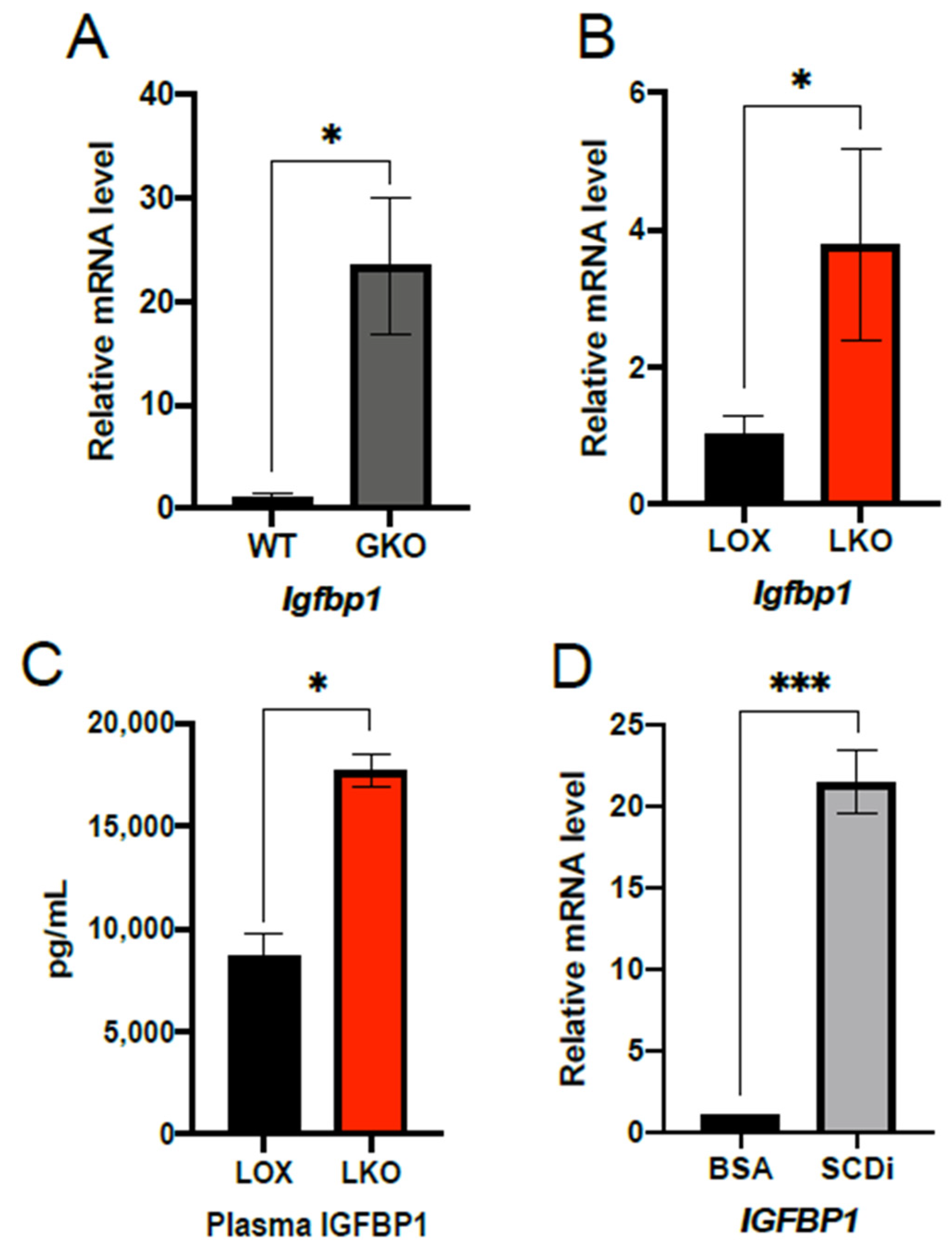
2.1. IGFBP1 Expression Is Enhanced by SCD1 Deletion and Inhibition of Enzyme Activity

Previous microarray analysis revealed that Igfbp1 mRNA was significantly increased in GKO mice fed HCD relative to WT controls [18]. Because GKO mice are protected from HCD-induced obesity, we wanted to assess hepatic Igfbp1 expression in HCD feeding for 10 days. As expected, Igfbp1 mRNA transcript levels were significantly elevated in GKO mice compared to WT (Figure 1A). We next assessed if Igfbp1 was also increased in LKO mice fed HCD. To do this, LKO and LOX control mice were subjected to 7–10 days of HCD, and Igfbp1 was measured in the liver and plasma. Similar to GKO mice, LKO mice exhibited a significant increase in hepatic Igfbp1 transcript (Figure 1B). Additionally, LKO mice had significantly elevated plasma protein levels of IGFBP1 (Figure 1C). Lastly, to further support that hepatic deficiency of Scd1 leads to increased expression of IGFBP1, we assessed IGFBP1 in HepG2 cells treated with an SCD inhibitor. Consistent with our in vivo results, treating HepG2 cells for 24 h with an SCD inhibitor significantly increased IGFBP1 mRNA levels (Figure 1D).

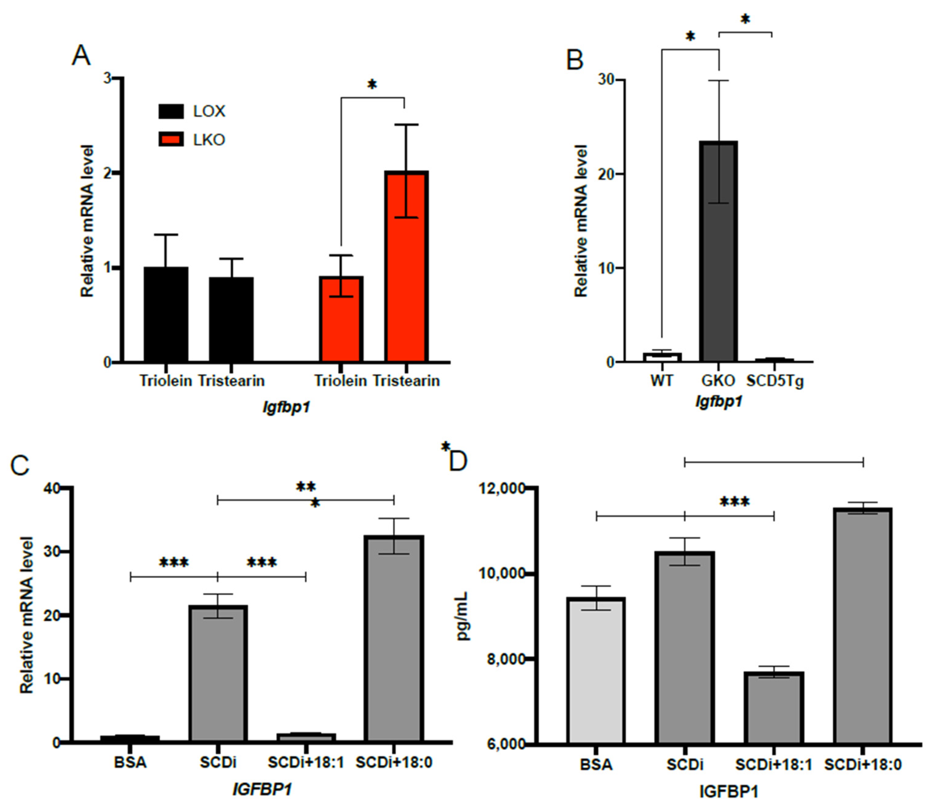
2.2. Oleic Acid Suppresses IGFBP1 Expression under HCD Feeding Conditions

To determine if IGFBP1 expression is regulated by the SCD1 product, oleic acid under HCD feeding conditions, we used a combination of dietary, genetic, and cell culture approaches. First, we sought to determine if adding oleic acid in the form of triolein to HCD would be sufficient to blunt the induction of Igfbp1 in LKO mice. To do this, we fed LKO and LOX mice HCD or HCD supplemented with either triolein or tristearin for 10 days. The LKO mice fed HCD supplemented with triolein exhibited similar Igfbp1 mRNA levels to LOX mice fed either triolein or tristearin (Figure 2A). In contrast, LKO mice fed HCD supplemented with tristearin exhibited significantly higher mRNA levels of Igfbp1 compared to LKO mice fed HCD supplemented with triolein or LOX mice fed HCD supplemented with either triolein or tristearin (Figure 2A). This indicates that hepatic oleic acid suppresses the induction of Igbp1 in LKO mice fed HCD.
Next, we assessed if oleic acid synthesized de novo could also blunt Igfbp1 induction in LKO mice fed HCD. To restore hepatic de novo synthesis of oleic acid, we used a previously developed transgenic GKO mouse model that expresses human stearoyl-CoA desaturase-5 specifically in the liver (SCD5Tg) [30]. The preferred substrate of hSCD5 is stearic acid, and thus, oleic acid is its predominant product [9]. We subjected WT, GKO, and SCD5Tg mice to 10 days of HCD feeding and measured hepatic Igfbp1 mRNA levels. Transgenic expression of hSCD5 was able to significantly suppress Igfbp1 mRNA compared to GKO mice (Figure 2B).
Lastly, to provide further evidence that oleic acid is regulating IGFBP1, we assessed whether oleic acid could suppress the induction of Igfbp1 in HepG2 cells treated with an SCD inhibitor. Similar to our in vivo results, HepG2 cells treated with an SCD inhibitor and oleic acid, but not steric acid, showed significantly decreased Igfbp1 mRNA (Figure 2C). In addition, HepG2 cells treated with an SCD inhibitor and oleic acid, but not steric acid, showed significantly decreased secreted IGFBP1 protein levels in the culture media (Figure 2D).

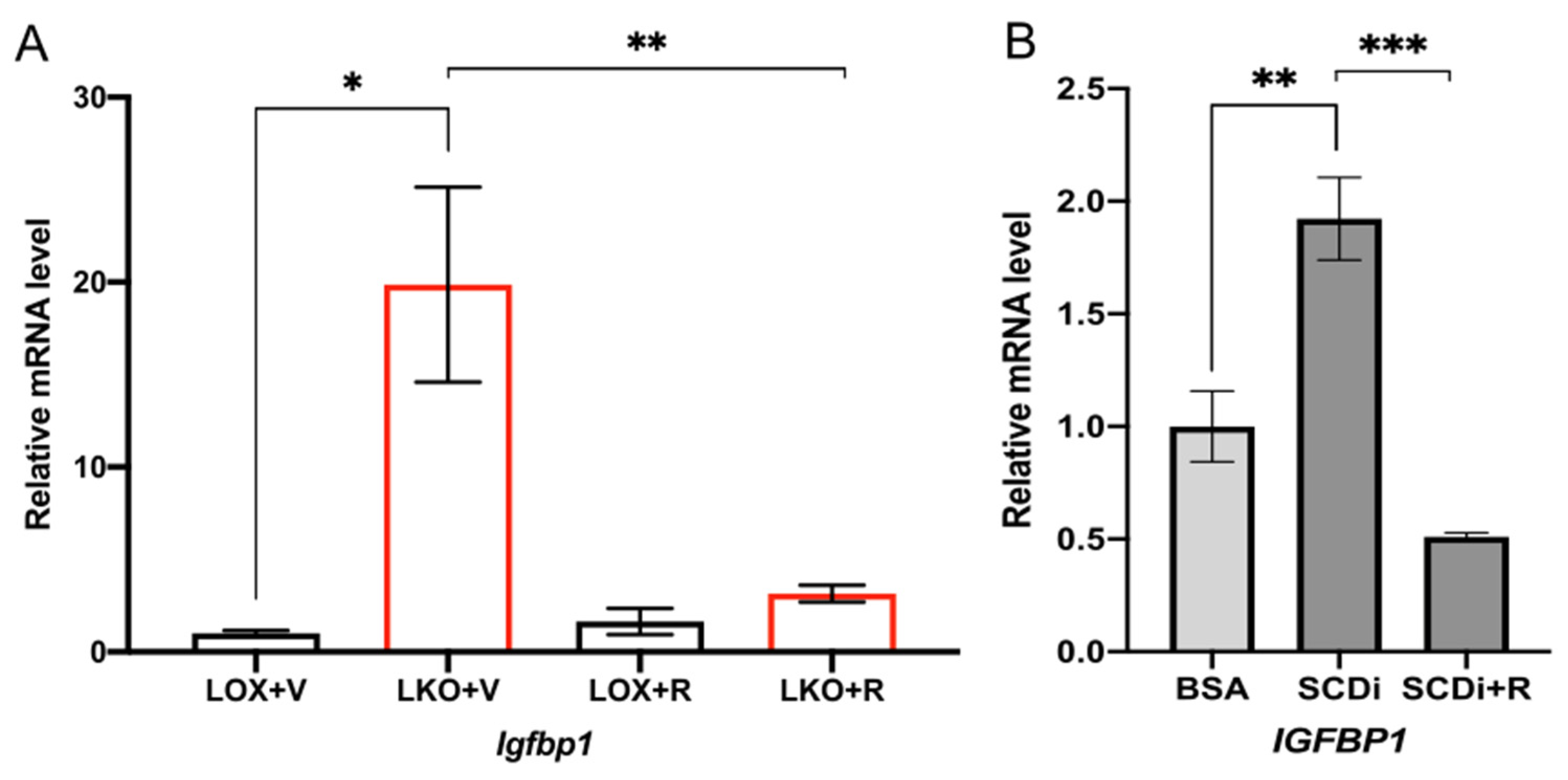
2.3. SCD1 Deficiency Enhances IGFBP1 through mTORC1

We have previously reported that in mice fed HCD, hepatic deletion of Scd1 leads to activation of the nutrient-sensing kinase mTORC1 and its downstream targets, such as S6 kinase [28]. To determine if IGFBP1 activation by Scd1 deletion is mTORC1 dependent, we treated LKO mice and SCD1-inhibited HepG2 cells with an mTORC1 inhibitor, rapamycin. In LKO mice fed HCD for 10 days, rapamycin treatment significantly reduced Igfbp1 expression compared to vehicle-treated controls (Figure 3A). Similarly, in HepG2 cells treated with SCD inhibitor, rapamycin supplementation also significantly reduced IGFBP1 mRNA levels compared to HepG2 cells treated with SCD inhibitor alone (Figure 3B). This suggests that Igfbp1 induction by HCD and low levels of oleic acid is mediated by mTORC1.

2.4. Oleic Acid Regulates IGFBP1 through the mTORC1-FGF21 Axis

To test whether IGFBP1 is downstream of FGF21 in the mTORC1 axis and if this axis mediates the induction of IGFBP1, we specifically deleted both Scd1 and Fgf21 in the livers of mice (DKO) (Figure 4A). In the DKO mice fed HCD for 10 days, there was no significant difference in Igfbp1 mRNA levels compared to Scd1lox/lox; Fgf21loxl/lox control mice (Figure 4B). To further support these results, we assessed this mechanism in vitro. Using HepG2 cells, we first showed that Fgf21 is significantly induced by the treatment of SCD inhibitor and that inhibiting mTORC1 with rapamycin blunts Fgf21 induction (Figure 4C). Similarly, supplementing oleic acid but not steric acid blunts Fgf21 induction (Figure 4C). Next, we assessed whether the knockdown of Fgf21 could suppress the induction of Igfbp1 by SCD inhibition. To do this, HepG2 cells were treated with either lentiviral control particles or shRNA lentiviral particles targeting Fgf21, in the presence and absence of an SCD inhibitor. As expected, both Fgf21 and Igfbp1 were significantly induced in cells treated with control lentiviral particles and SCD inhibitor compared to cells treated with control lentiviral particles alone (Figure 4D,E). Lastly, treating HepG2 cells with SCD inhibitor and Fgf21 shRNA lentiviral particles blunted Fgf21 and Igfbp1 induction compared to cells treated with control lentiviral particles and SCD inhibitor (Figure 4D,E). Taken together, this suggests oleic acid regulates IGFBP1 through the mTORC1-FGF21 axis.

3. Discussion

Global deletion of Scd1 protects against diet-induced and genetically induced obesity and improves glucose homeostasis [15,16]. Moreover, hepatic deletion of Scd1 recapitulates protection against HCD-induced obesity [6]. When fed an HCD LKO mice remain lean, insulin-sensitive, and have enhanced glucose uptake in adipose tissue, brown adipose tissue, and liver [29]. The exact mechanism by which SCD1 mediates these beneficial phenotypes remains unclear; however, evidence suggests that hepatokines such as FGF21 and IGFBP1 are contributing factors [20,25,29]. Microarray analysis revealed that Igfbp1 is one of the most differentially expressed hepatic genes between GKO and WT mice fed HCD, and here we showed that the induction of IGFBP1 is also exhibited in HCD-fed LKO mice. IGFBP1 is a hepatokine that increases β-cell proliferation and enhances insulin signaling [19,20,21]. In humans, a strong positive correlation between circulating IGFBP1 concentrations and insulin sensitivity has been established [31,32,33,34,35,36,37,38]. Likewise, low levels of IGFBP1 have been reported to predict the development of type 2 diabetes and IGFBP1 has been proposed as a potential marker of insulin sensitivity [39,40].
In the present study, we sought to determine the mechanism by which Scd1 deficiency induces Igfbp1 expression during high-carbohydrate feeding. Previously, our HCD studies revealed that Scd1 deficiency induces ER stress through SFA activation of the mTORC1-PGC-1α axis [28,41]. Since Fgf21 and Igfbp1 are ER stress-inducible genes, their up-regulation is likely in response to ER stress produced by a high ratio of SFA to MUFA levels [42,43]. Increasing the levels of MUFAs relative to SFAs by dietary supplementation or endogenous synthesis of oleic acid inhibits mTORC1 activation and subsequently decreases expression of PGC-1α, FGF21, and ER stress markers [28,29]. Here we provided evidence that IGFBP1 is also regulated by modulating the ratio of MUFAs to SFAs, as well as inhibition of mTORC1 and decreased Fgf21 expression. Taken together, we show that Igfbp1 is induced through the mTORC1-FGF21 axis (Figure 5).
Specifically, we showed that Igfbp1 expression by Scd1 deficiency is suppressed by dietary supplementation with triolein and endogenously synthesized oleic acid by transgenic expression of hSCD5. Additionally, in cultured HepG2 cells treated with SCD inhibitor, the addition of oleic acid reduces IGFBP1 expression compared to cells treated with SCD inhibitor alone or stearic acid. Moreover, we showed that Igfbp1 expression is dependent on mTORC1 activation because Igfbp1 is suppressed by rapamycin treatment of LKO mice or SCD-inhibited HepG2 cells. Lastly, we showed that induction of Igfbp1 is dependent on FGF21 both in vivo and in vitro. Mice harboring a combined liver-specific deletion of Scd1 and Fgf21 fail to significantly induce Igfbp1 compared to LOX/LOX controls when fed an HCD for 10 days. Similarly, the knockdown of Fgf21 in SCD-inhibited HepG2 cells blunts the induction of Igfbp1 compared to cells treated with SCD inhibitor alone.
In conclusion, we report that the hepatokine IGFBP1 is regulated by oleic acid partly through the mTORC1-FGF21 axis. The increased production of IGFBP1 due to hepatic Scd1 deficiency could be contributing to increased insulin sensitivity and glucose tolerance in Scd1 deficient mice. More research is needed to determine the effects of IGFBP1 because previous studies have shown conflicting results [44]. If IGFBP1 is acting beneficially, then by reducing the levels of hepatic oleic acid, either by dietary or pharmacological intervention, may be an effective form of treating HCD-induced obesity and the associated metabolic dysfunction.

4. Materials and Methods

4.1. Animals and Diets

All animal studies were approved by and conducted following the Institutional Animal Care and Use Committee guidelines, University of Wisconsin-Madison, protocol #A005125. Mice were maintained at the University of Wisconsin-Madison animal care facility on regular 12  h light/dark cycles with free access to food and water. All mice used in this study were in the C57BL/6 genetic background and were fed a standard rodent chow diet (Purina 5008; Harlan Teklad, Madison, WI, USA) until being subjected to dietary treatments. All studies were carried out using 8 to 14 weeks old mice. The process of generating Scd1−/− (GKO) mice, Scd1−/−; SCD5Tg + (SCD5Tg), Scd1lox/lox (LOX), Scd1lox/lox; Albumin Cre/+, has been described previously [6,15,30]. To generate Scd1 and Fgf21 liver-specific double knockout mice, Scd1lox/lox; Albumin Cre/+ were crossed to Fgf21lox/lox mice obtained from The Jackson Laboratory (Fgf21tm1.2Djm, JAX stock #022361) to yield Scd1lox/+; Fgf21lox/+; Albumin Cre/+ and Scd1lox/+; Fgf21lox/+. These mice were subsequently crossed to yield Scd1lox/lox; Fgf21loxl/lox; Albumin Cre/+. For dietary studies, mice were fed a very-low-fat high-carbohydrate diet (HCD; Harlan Teklad, Madison, WI, USA TD.03045) for a period of 7–14 days. Triolein or tristearin-supplemented HCDs were prepared by supplementing the fat-free basal mix (TD150776.PWD; Harlan Teklad, Madison, WI, USA) with 15% by weight of tristearin (T5016; Sigma, St. Louis, MO, USA) or triolein (99% purity, T7140; Sigma). For rapamycin studies, mice were treated intraperitoneally with 2  mg/kg/day of rapamycin (from LC laboratories, Woburn, MA, USA) dissolved in saline and 2% ethanol for 10 days. All mice were fasted for 4  h before being euthanized by isoflurane overdose. Collected tissues and plasma were frozen in liquid nitrogen and stored at −80 °C for future analysis. Plasma IGFBP1 protein levels were measured using the Mouse IGFBP-1 ELISA Kit PicoKine™ (Boster Biological Technology, Pleasanton, CA, USA, Catalog #EK0383).

4.2. Quantitative Real-Time PCR

Total RNA was isolated using the RNeasy Lipid Tissue Mini Kit (Qiagen) for animal tissues and the Direct-zol RNA microprep kit (Zymo Research, Irvine, CA, USA) for cell culture samples. cDNA was synthesized from RNA using the ABI High Capacity cDNA Reverse Transcription Kit (Life Technologies/Invitrogen, Carlsbad, CA, USA). qPCR was performed on an ABI7500 instrument using the ABI Fast SYBR Green Master Mix (Life Technologies/Invitrogen, Carlsbad, CA, USA) for animal tissues and 2x SYBR Green qPCR Master Mix (Bimake, Houston, TX, USA) for cell culture samples. Relative mRNA abundance was calculated as relative Ct value and normalized to 18 s for animal tissues and GAPDH for cell culture samples by the ΔΔCt method. Primer sequences are available upon request.

4.3. Cell Culture and Treatments

HepG2 cells were cultured in Dulbeccos’s Modified Eagle Medium (DMEM) supplemented with 10% fetal bovine serum (FBS), penicillin (100 U/mL), and streptomycin (100 μg/mL). Upon confluence, the HepG2 cells were serum-starved for 24 h before various treatments. To model SCD1 deficiency, HepG2 cells were treated with 1 or 4 μM of SCD inhibitor A939572 (Cayman Chemical, MI, USA) for 24 h. To inhibit mTORC1, HepG2 cells were treated with 100 nM of rapamycin (APExBIO, Houston, TX, USA) for 24 h.
Oleic acid and stearic acid were conjugated to fatty acid free bovine serum albumin to create 100 or 500 mM stock solutions, respectively. These stock solutions were subsequently diluted to 100 or 500 μM with DMEM without FBS, penicillin, and streptomycin, and added directly to the HepG2 cells for 24 h. Fatty acid treatment between 50–500 μM is within the physiologically relevant range of human and mouse models [45,46]. A 0.17 mM solution of fatty acid free BSA and PBS was prepared by first warming PBS to 37 °C on a heated stir plate. Next, fatty acid free BSA was slowly added to PBS under gentle stirring until it was completely dissolved. To create the 100 and 500 mM stock solutions, fatty acid was weighed out and dissolved in hexane. Next, the dissolved fatty acid was transferred into a glass vial and dried under a stream of nitrogen gas. The fatty acid free BSA solution was added to the vial and gently stirred for 4 h at 37 °C. This was created either 1 mM fatty acid to 0.17 mM BSA solution with a 6:1 molar ratio of fatty acid to BSA or 5 mM fatty acid to 0.017 mM BSA with a 30:1 molar ratio of fatty acid to BSA.
Treatment of HepG2 cells with FGF-21 shRNA lentiviral particles (Santa Cruz Biotechnology, Dallas, TX, USA; sc-39484-V) and copGFP control lentiviral particles (Santa Cruz Biotechnology, Dallas, TX, USA; sc-108084) was performed according to the manufacturer’s instructions. Briefly, cells were grown to 50% confluence in 6-well plates and treated with 20 μL of lentiviral particles and 2 μg/mL polybrene for 24 h. After 24 h, the media was removed and replaced with complete media with and without SCD inhibitor. Lastly, secreted IGFBP1 protein was measured using the Human IGFBP-1 ELISA Kit PicoKine™ (Boster Biological Technology, Pleasanton, CA, USA, Catalog #EK0382).

Author Contributions

Conceptualization, L.M.O. and J.M.N.; investigation, L.M.O., Y.X.P., Z.L., S.A.L., A.A., A.M. and G.W.; writing—original draft preparation, L.M.O. and J.M.N.; writing—review and editing, L.M.O., A.M., M.K., J.S. and J.M.N. All authors have read and agreed to the published version of the manuscript.

Funding

This work was supported by National Institutes of Health (NIH) Grant R01 DK118093 (to J.M.N); Glenn Glenn Foundation and American Federation for Aging Research (A22068 to JS); Hatch Grant (WIS04000-1024796 to JS); JDRF (JDRF201309442 to JS); an R01 through NIDDK (R01DK133479 to JS); NIH National Research Service Award T32 GM07215 (to L.M.O.); and Advanced Opportunity Fellowship through SciMed Graduate Research Scholars at the University of Wisconsin-Madison (to L.M.O.).

Institutional Review Board Statement

The animal study protocol was approved by the Institutional Animal Care and Use Committee guidelines, University of Wisconsin-Madison, protocol #A005125.

Informed Consent Statement

Not applicable.

Acknowledgments

We thank Laura Vanderploeg (University of Wisconsin-Madison Biochemistry Department Media Center) for her help with the design and production of the figures.

Conflicts of Interest

The authors do not have any conflict of interest to declare.

References

  1. Ravussin, E.; Ryan, D.H. Three New Perspectives on the Perfect Storm: What’s Behind the Obesity Epidemic? Obesity 2018, 26, 9–10. [Google Scholar] [CrossRef]
  2. Slyper, A.H. The pediatric obesity epidemic: Causes and controversies. J. Clin. Endocrinol. Metab. 2004, 89, 2540–2547. [Google Scholar] [CrossRef] [PubMed]
  3. Flegal, K.M.; Carroll, M.D.; Kuczmarski, R.J.; Johnson, C.L. Overweight and obesity in the United States: Prevalence and trends, 1960-1994. Int. J. Obes. Relat. Metab. Disord. 1998, 22, 39–47. [Google Scholar] [CrossRef] [PubMed] [Green Version]
  4. Ng, M.; Fleming, T.; Robinson, M.; Thomson, B.; Graetz, N.; Margono, C.; Mullany, E.C.; Biryukov, S.; Abbafati, C.; Abera, S.F.; et al. Global, regional, and national prevalence of overweight and obesity in children and adults during 1980-2013: A systematic analysis for the Global Burden of Disease Study 2013. Lancet 2014, 384, 766–781. [Google Scholar] [CrossRef] [Green Version]
  5. Flowers, M.T.; Ntambi, J.M. Stearoyl-CoA desaturase and its relation to high-carbohydrate diets and obesity. Biochim. Biophys. Acta 2009, 1791, 85–91. [Google Scholar] [CrossRef] [Green Version]
  6. Miyazaki, M.; Flowers, M.T.; Sampath, H.; Chu, K.; Otzelberger, C.; Liu, X.; Ntambi, J.M. Hepatic stearoyl-CoA desaturase-1 deficiency protects mice from carbohydrate-induced adiposity and hepatic steatosis. Cell Metab. 2007, 6, 484–496. [Google Scholar] [CrossRef] [Green Version]
  7. Oballa, R.M.; Belair, L.; Black, W.C.; Bleasby, K.; Chan, C.C.; Desroches, C.; Du, X.; Gordon, R.; Guay, J.; Guiral, S.; et al. Development of a liver-targeted stearoyl-CoA desaturase (SCD) inhibitor (MK-8245) to establish a therapeutic window for the treatment of diabetes and dyslipidemia. J. Med. Chem. 2011, 54, 5082–5096. [Google Scholar] [CrossRef] [PubMed]
  8. Man, W.C.; Miyazaki, M.; Chu, K.; Ntambi, J.M. Membrane topology of mouse stearoyl-CoA desaturase 1. J. Biol. Chem. 2006, 281, 1251–1260. [Google Scholar] [CrossRef] [PubMed] [Green Version]
  9. Miyazaki, M.; Bruggink, S.M.; Ntambi, J.M. Identification of mouse palmitoyl-coenzyme A Delta9-desaturase. J. Lipid Res. 2006, 47, 700–704. [Google Scholar] [CrossRef] [PubMed] [Green Version]
  10. Miyazaki, M.; Kim, Y.C.; Ntambi, J.M. A lipogenic diet in mice with a disruption of the stearoyl-CoA desaturase 1 gene reveals a stringent requirement of endogenous monounsaturated fatty acids for triglyceride synthesis. J. Lipid Res. 2001, 42, 1018–1024. [Google Scholar] [CrossRef] [PubMed]
  11. Tocher, D.R.; Leaver, M.J.; Hodgson, P.A. Recent advances in the biochemistry and molecular biology of fatty acyl desaturases. Prog. Lipid. Res. 1998, 37, 73–117. [Google Scholar] [CrossRef] [PubMed]
  12. Cao, H.; Gerhold, K.; Mayers, J.R.; Wiest, M.M.; Watkins, S.M.; Hotamisligil, G.S. Identification of a lipokine, a lipid hormone linking adipose tissue to systemic metabolism. Cell 2008, 134, 933–944. [Google Scholar] [CrossRef] [Green Version]
  13. Lounis, M.A.; Bergeron, K.F.; Burhans, M.S.; Ntambi, J.M.; Mounier, C. Oleate activates SREBP-1 signaling activity in. Am. J. Physiol. Endocrinol. Metab. 2017, 313, E710–E720. [Google Scholar] [CrossRef] [PubMed]
  14. Takada, R.; Satomi, Y.; Kurata, T.; Ueno, N.; Norioka, S.; Kondoh, H.; Takao, T.; Takada, S. Monounsaturated Fatty Acid Modification of Wnt Protein: Its Role in Wnt Secretion. Dev. Cell 2006, 11, 791–801. [Google Scholar] [CrossRef] [Green Version]
  15. Ntambi, J.M.; Miyazaki, M.; Stoehr, J.P.; Lan, H.; Kendziorski, C.M.; Yandell, B.S.; Song, Y.; Cohen, P.; Friedman, J.M.; Attie, A.D. Loss of stearoyl-CoA desaturase-1 function protects mice against adiposity. Proc. Natl. Acad. Sci. USA 2002, 99, 11482–11486. [Google Scholar] [CrossRef] [PubMed] [Green Version]
  16. Miyazaki, M.; Sampath, H.; Liu, X.; Flowers, M.T.; Chu, K.; Dobrzyn, A.; Ntambi, J.M. Stearoyl-CoA desaturase-1 deficiency attenuates obesity and insulin resistance in leptin-resistant obese mice. Biochem. Biophys. Res. Commun. 2009, 380, 818–822. [Google Scholar] [CrossRef] [PubMed] [Green Version]
  17. Sampath, H.; Flowers, M.T.; Liu, X.; Paton, C.M.; Sullivan, R.; Chu, K.; Zhao, M.; Ntambi, J.M. Skin-specific deletion of stearoyl-CoA desaturase-1 alters skin lipid composition and protects mice from high fat diet-induced obesity. J. Biol. Chem. 2009, 284, 19961–19973. [Google Scholar] [CrossRef] [Green Version]
  18. Flowers, M.T.; Keller, M.P.; Choi, Y.; Lan, H.; Kendziorski, C.; Ntambi, J.M.; Attie, A.D. Liver gene expression analysis reveals endoplasmic reticulum stress and metabolic dysfunction in SCD1-deficient mice fed a very low-fat diet. Physiol. Genom. 2008, 33, 361–372. [Google Scholar] [CrossRef] [PubMed] [Green Version]
  19. Lu, J.; Liu, K.-C.; Schulz, N.; Karampelias, C.; Charbord, J.; Hilding, A.; Rautio, L.; Bertolino, P.; Östenson, C.-G.; Brismar, K.; et al. IGFBP1 increases β-cell regeneration by promoting α- to β-cell transdifferentiation. EMBO J. 2016, 35, 2026–2044. [Google Scholar] [CrossRef]
  20. Haywood, N.J.; Cordell, P.A.; Tang, K.Y.; Makova, N.; Yuldasheva, N.Y.; Imrie, H.; Viswambharan, H.; Bruns, A.F.; Cubbon, R.M.; Kearney, M.T.; et al. Insulin-Like Growth Factor Binding Protein 1 Could Improve Glucose Regulation and Insulin Sensitivity Through Its RGD Domain. Diabetes 2017, 66, 287–299. [Google Scholar] [CrossRef] [PubMed]
  21. Rajwani, A.; Ezzat, V.; Smith, J.; Yuldasheva, N.Y.; Duncan, E.R.; Gage, M.; Cubbon, R.M.; Kahn, M.B.; Imrie, H.; Abbas, A.; et al. Increasing Circulating IGFBP1 Levels Improves Insulin Sensitivity, Promotes Nitric Oxide Production, Lowers Blood Pressure, and Protects Against Atherosclerosis. Diabetes 2012, 61, 915–924. [Google Scholar] [CrossRef] [Green Version]
  22. Mashili, F.L.; Austin, R.L.; Deshmukh, A.S.; Fritz, T.; Caidahl, K.; Bergdahl, K.; Zierath, J.R.; Chibalin, A.V.; Moller, D.E.; Kharitonenkov, A.; et al. Direct effects of FGF21 on glucose uptake in human skeletal muscle: Implications for type 2 diabetes and obesity. Diabetes Metab. Res. Rev. 2011, 27, 286–297. [Google Scholar] [CrossRef]
  23. Markan, K.R.; Naber, M.C.; Ameka, M.K.; Anderegg, M.D.; Mangelsdorf, D.J.; Kliewer, S.A.; Mohammadi, M.; Potthoff, M.J. Circulating FGF21 Is Liver Derived and Enhances Glucose Uptake During Refeeding and Overfeeding. Diabetes 2014, 63, 4057–4063. [Google Scholar] [CrossRef] [PubMed] [Green Version]
  24. Xu, J.; Stanislaus, S.; Chinookoswong, N.; Lau, Y.Y.; Hager, T.; Patel, J.; Ge, H.; Weiszmann, J.; Lu, S.-C.; Graham, M.; et al. Acute glucose-lowering and insulin-sensitizing action of FGF21 in insulin-resistant mouse models—Association with liver and adipose tissue effects. Am. J. Physiol. Metab. 2009, 297, E1105–E1114. [Google Scholar] [CrossRef] [PubMed] [Green Version]
  25. Liu, M.; Cao, H.; Hou, Y.; Sun, G.; Li, D.; Wang, W. Liver Plays a Major Role in FGF-21 Mediated Glucose Homeostasis. Cell. Physiol. Biochem. 2018, 45, 1423–1433. [Google Scholar] [CrossRef] [PubMed]
  26. Wang, X.; Wei, W.; Krzeszinski, J.Y.; Wang, Y.; Wan, Y. A Liver-Bone Endocrine Relay by IGFBP1 Promotes Osteoclastogenesis and Mediates FGF21-Induced Bone Resorption. Cell Metab. 2015, 22, 811–824. [Google Scholar] [CrossRef] [PubMed] [Green Version]
  27. Inagaki, T.; Lin, V.Y.; Goetz, R.; Mohammadi, M.; Mangelsdorf, D.; Kliewer, S.A. Inhibition of Growth Hormone Signaling by the Fasting-Induced Hormone FGF21. Cell Metab. 2008, 8, 77–83. [Google Scholar] [CrossRef] [PubMed] [Green Version]
  28. Aljohani, A.; Khan, M.I.; Syed, D.N.; Abram, B.; Lewis, S.; Neill, L.O.; Mukhtar, H.; Ntambi, J.M. Hepatic Stearoyl-CoA desaturase-1 deficiency-mediated activation of mTORC1- PGC-1α axis regulates ER stress during high-carbohydrate feeding. Sci. Rep. 2019, 9, 15761. [Google Scholar] [CrossRef] [Green Version]
  29. Aljohani, A.; Khan, M.I.; Bonneville, A.; Guo, C.; Jeffery, J.; O’Neill, L.; Syed, D.N.; Lewis, S.; Burhans, M.; Mukhtar, H.; et al. Hepatic stearoyl CoA desaturase 1 deficiency increases glucose uptake in adipose tissue partially through the PGC-1α–FGF21 axis in mice. J. Biol. Chem. 2019, 294, 19475–19485. [Google Scholar] [CrossRef]
  30. Burhans, M.S.; Flowers, M.T.; Harrington, K.; Bond, L.M.; Guo, C.-A.; Anderson, R.M.; Ntambi, J.M. Hepatic oleate regulates adipose tissue lipogenesis and fatty acid oxidation. J. Lipid Res. 2015, 56, 304–318. [Google Scholar] [CrossRef] [PubMed]
  31. Heald, A.H.; Cruickshank, J.K.; Riste, L.K.; Cade, J.E.; Anderson, S.; Greenhalgh, A.; Sampayo, J.; Taylor, W.; Fraser, W.; White, A.; et al. Close relation of fasting insulin-like growth factor binding protein-1 (IGFBP-1) with glucose tolerance and cardiovascular risk in two populations. Diabetologia 2001, 44, 333–339. [Google Scholar] [CrossRef] [Green Version]
  32. Gokulakrishnan, K.; Velmurugan, K.; Ganesan, S.; Mohan, V. Circulating levels of insulin-like growth factor binding protein-1 in relation to insulin resistance, type 2 diabetes mellitus, and metabolic syndrome (Chennai Urban Rural Epidemiology Study 118). Metabolism 2012, 61, 43–46. [Google Scholar] [CrossRef]
  33. Liew, C.F.; Wise, S.D.; Yeo, K.P.; Lee, K.-O. Insulin-like growth factor binding protein-1 is independently affected by ethnicity, insulin sensitivity, and leptin in healthy, glucose-tolerant young men. J. Clin. Endocrinol. Metab. 2005, 90, 1483–1488. [Google Scholar] [CrossRef] [Green Version]
  34. Rajpathak, S.N.; McGinn, A.P.; Strickler, H.D.; Rohan, T.E.; Pollak, M.; Cappola, A.R.; Kuller, L.; Xue, X.; Newman, A.B.; Strotmeyer, E.S.; et al. Insulin-like growth factor-(IGF)-axis, inflammation, and glucose intolerance among older adults. Growth Horm. IGF Res. 2008, 18, 166–173. [Google Scholar] [CrossRef] [PubMed] [Green Version]
  35. Mogul, H.R.; Marshall, M.; Frey, M.; Burke, H.B.; Wynn, P.S.; Wilker, S.; Southern, A.L.; Gambert, S.R. Insulin like growth factor-binding protein-1 as a marker for hyperinsulinemia in obese menopausal women. J. Clin. Endocrinol. Metab. 1996, 81, 4492–4495. [Google Scholar]
  36. Mohamed-Ali, V.; Pinkney, J.H.; Panahloo, A.; Cwyfan-Hughes, S.; Holly, J.M.; Yudkin, J.S. Insulin-like growth factor binding protein-1 in NIDDM: Relationship with the insulin resistance syndrome. Clin. Endocrinol. 1999, 50, 221–228. [Google Scholar] [CrossRef]
  37. Travers, S.H.; Labarta, J.I.; Gargosky, S.E.; Rosenfeld, R.G.; Jeffers, B.W.; Eckel, R.H. Insulin-Like Growth Factor Binding Protein-I Levels Are Strongly Associated with Insulin Sensitivity and Obesity in Early Pubertal Children1. J. Clin. Endocrinol. Metab. 1998, 83, 1935–1939. [Google Scholar] [CrossRef] [PubMed] [Green Version]
  38. Saitoh, H.; Kamoda, T.; Nakahara, S.; Hirano, T.; Nakamura, N. Serum concentrations of insulin, insulin-like growth factor(IGF)-I, IGF binding protein (IGFBP)-1 and -3 and growth hormone binding protein in obese children: Fasting IGFBP-1 is suppressed in normoinsulinaemic obese children. Clin. Endocrinol. 1998, 48, 487–492. [Google Scholar] [CrossRef] [PubMed]
  39. Borai, A.; Livingstone, C.; Zarif, H.; Ferns, G. Serum insulin-like growth factor binding protein-1: An improvement over other simple indices of insulin sensitivity in the assessment of subjects with normal glucose tolerance. Ann. Clin. Biochem. Int. J. Lab. Med. 2009, 46, 109–113. [Google Scholar] [CrossRef] [PubMed]
  40. Petersson, U.; Östgren, C.; Brudin, L.; Brismar, K.; Nilsson, P. Low levels of insulin-like growth-factor-binding protein-1 (IGFBP-1) are prospectively associated with the incidence of type 2 diabetes and impaired glucose tolerance (IGT): The Söderåkra Cardiovascular Risk Factor Study. Diabetes Metab. 2009, 35, 198–205. [Google Scholar] [CrossRef] [PubMed]
  41. Liu, X.; Burhans, M.S.; Flowers, M.T.; Ntambi, J.M. Hepatic oleate regulates liver stress response partially through PGC-1α during high-carbohydrate feeding. J. Hepatol. 2016, 65, 103–112. [Google Scholar] [CrossRef]
  42. Schaap, F.G.; Kremer, A.E.; Lamers, W.H.; Jansen, P.L.; Gaemers, I.C. Fibroblast growth factor 21 is induced by endoplasmic reticulum stress. Biochimie 2013, 95, 692–699. [Google Scholar] [CrossRef] [PubMed]
  43. Marchand, A.; Tomkiewicz, C.; Magne, L.; Barouki, R.; Garlatti, M. Endoplasmic Reticulum Stress Induction of Insulin-like Growth Factor-binding Protein-1 Involves ATF4. J. Biol. Chem. 2006, 281, 19124–19133. [Google Scholar] [CrossRef] [PubMed] [Green Version]
  44. Haywood, N.J.; Slater, T.A.; Matthews, C.J.; Wheatcroft, S.B. The insulin like growth factor and binding protein family: Novel therapeutic targets in obesity & diabetes. Mol. Metab. 2019, 19, 86–96. [Google Scholar] [PubMed]
  45. Zou, Y.; Wang, Y.-N.; Ma, H.; He, Z.-H.; Tang, Y.; Guo, L.; Liu, Y.; Ding, M.; Qian, S.-W.; Tang, Q.-Q. SCD1 promotes lipid mobilization in subcutaneous white adipose tissue. J. Lipid Res. 2020, 61, 1589–1604. [Google Scholar] [CrossRef] [PubMed]
  46. Sera, R.K.; McBride, J.H.; Higgins, S.A.; Rodgerson, D.O. Evaluation of reference ranges for fatty acids in serum. J. Clin. Lab. Anal. 1994, 8, 81–85. [Google Scholar] [CrossRef]
Figure 1. Hepatic SCD1 deficiency up-regulates IGFBP1. (A) Relative hepatic Igfbp1 mRNA levels in 8–10 weeks old male WT (n = 4) and GKO (n = 6) mice fed HCD for 10 days. (B) Relative hepatic Igfbp1 mRNA levels in 8–13 weeks old male LOX (n = 13) and LKO (n = 12) mice fed HCD for 7–10 days. (C) Plasma IGFBP1 protein concentration in 8–10 weeks old male LOX (n = 7) and LKO (n = 7) mice fed HCD for 7–10 days. (D) Relative IGFBP1 mRNA levels in HepG2 cells treated for 24 h with BSA (n = 10) or BSA plus 4 μM (n = 12) of SCD inhibitor. Values are mean ± SEM; * p < 0.05 vs. WT, LOX, or BSA, *** p < 0.005 vs. WT, LOX or BSA by Student’s two-tailed t-test.
Ijms 23 14671 g001
Figure 2. Oleic acid blunts IGFBP1 up-regulation in vivo and in vitro. (A) Relative hepatic Igfbp1 mRNA levels in 12–14 weeks old male LOX and LKO mice fed either HCD supplemented with 15% by weight tristearin (n = 4 LOX and n = 4 LKO) or 15% by weight triolein (n = 5 LOX; 7 LKO) for 10 days. (B) Relative hepatic Igfbp1 mRNA levels in 8–10 weeks old male WT (n = 4), GKO (n = 6), and SCD5tg (n = 5) mice fed HCD for 10 days. (C) Relative Igfbp1 mRNA level in HepG2 cells treated for 24 h with either BSA (n = 10), BSA plus 4 μM SCD inhibitor (n = 12), BSA plus 4μM SCD inhibitor, and 500 μM oleic acid (n = 12), or BSA plus SCD inhibitor and 500 μM steric acid (n = 12). (D) Secreted IGFBP1 protein concentration in HepG2 cells treated for 24 h with either BSA (n = 7), BSA plus 4 μM SCD inhibitor (n = 8), BSA plus 4 μM SCD inhibitor and 500 μM oleic acid (n = 7), or BSA plus SCD inhibitor and 500 μM steric acid (n = 8). Values are mean ± SEM; * p < 0.05 vs. WT, LOX, or BSA; ** p < 0.01 vs. WT, LOX, or BSA; *** p < 0.005 vs. WT, LOX triolein or BSA by Student′s two-tailed t-test.
Ijms 23 14671 g002
Figure 3. Inhibition of mTORC1 by rapamycin suppresses IGFBP1 induction. (A) Relative hepatic Igfbp1 mRNA levels in 10–12 weeks old male LOX and LKO mice fed HCD diet for 10 days treated daily with either vehicle (n = 3 LOX; 3 LKO) or rapamycin (n = 5 LOX + R; 5 LKO + R). (B) Relative Igfbp1 mRNA levels of HepG2 cells treated with either BSA (n = 5), BSA plus 1μM SCD inhibitor (n = 5), or BSA plus 4 μM SCD inhibitor and rapamycin (n = 5). Values are mean ± SEM; * p < 0.05 vs. LOX or BSA; ** p < 0.01 vs. LOX+V or BSA; *** p < 0.005 vs. LOX or BSA by Student′s two-tailed t-test.
Ijms 23 14671 g003
Figure 4. IGFBP1 is regulated by SCD1 through the mTORC1-FGF21 axis. (A) Relative hepatic Scd1 and Fgf21 mRNA levels in LOX/LOX (n = 6, 3 male; 3 female) and DLKO (n = 6, 3 male; 3 female) mice fed HCD for 10 days. (B) Relative hepatic Igfbp1 mRNA level in LOX/LOX (n = 6, 3 male; 3 female) and DLKO mice fed HCD (n = 6, 3 male; 3 female) for 10 days. (C) Relative Fgf21 mRNA level in HepG2 cells treated with BSA (n = 6), BSA plus 1 μM SCD inhibitor (n = 6), BSA plus 1 μM SCD inhibitor and 100 μM oleic acid (n = 5), BSA plus 1 μM SCD inhibitor and 100 μM steric acid (n = 5), BSA plus 1 μM SCD inhibitor and rapamycin (n = 5). (D) Relative FGF21 mRNA level in HepG2 cells treated with control lentiviral particles (n = 6), control lentiviral particles plus SCD inhibitor (n = 6), or shFGF21 lentiviral particles plus SCD inhibitor (n = 6). (E) Relative IGFBP1 mRNA level in HepG2 cells treated with control lentiviral particles (n = 6), control lentiviral particles plus SCD inhibitor (n = 5), or shFGF21 lentiviral particles plus SCD inhibitor (n = 5). Values are mean ± SEM; ns = not significant; * p < 0.05 vs. LOX/LOX, BSA, or control; ** p < 0.01 vs. LOX/LOX, BSA, or control; *** p < 0.005 vs. LOX/LOX, BSA, or control by Student′s two-tailed t-test.
Ijms 23 14671 g004
Figure 5. Proposed model of IGFBP1 induction through the mTORC1-FGF21 axis.
Ijms 23 14671 g005
Publisher’s Note: MDPI stays neutral with regard to jurisdictional claims in published maps and institutional affiliations.

