Review

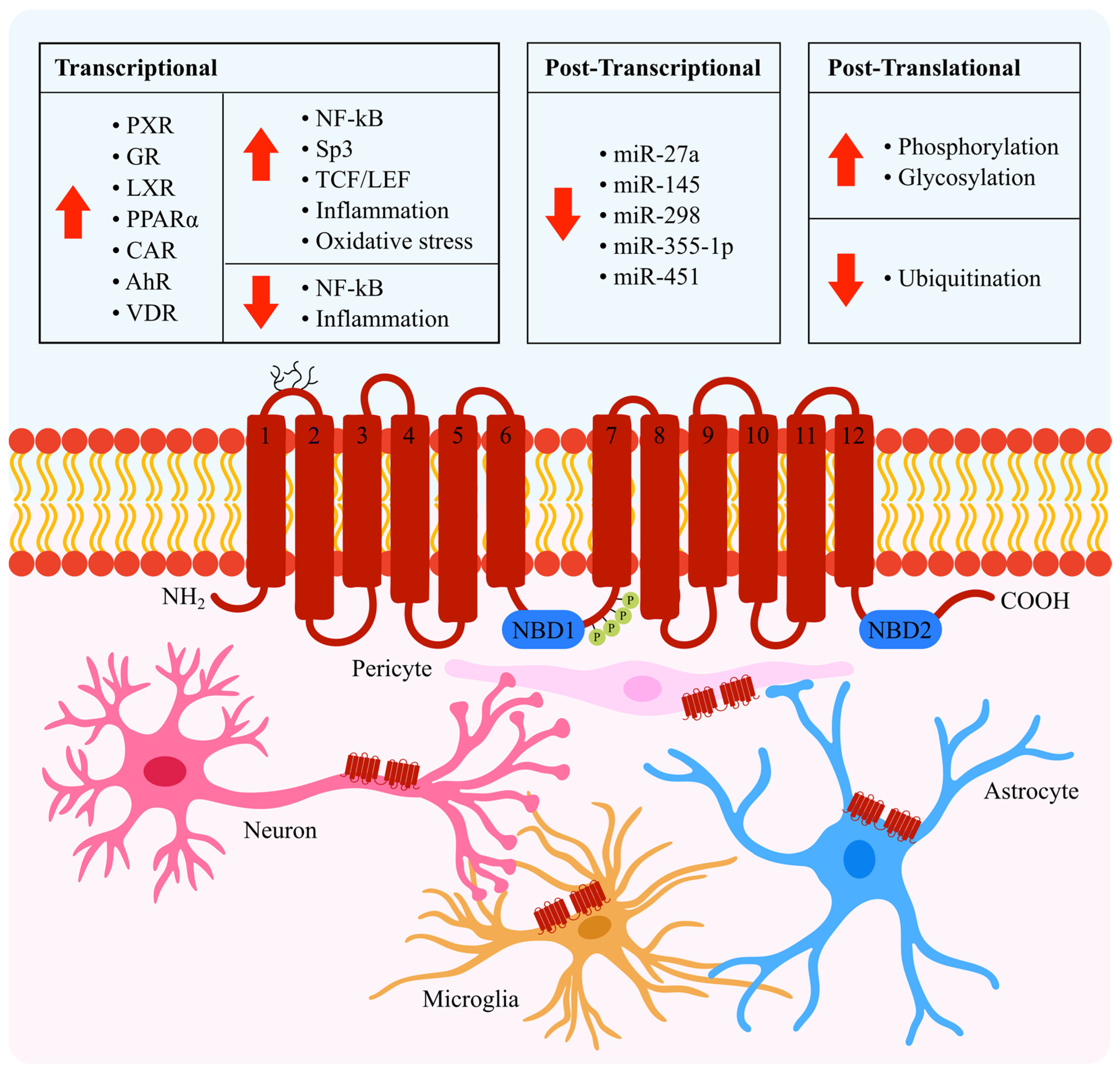
Regulation of P-Glycoprotein in the Brain

by
Amanda B. Chai
1,
Richard Callaghan
2 and
Ingrid C. Gelissen
1,*
1
School of Pharmacy, Faculty of Medicine and Health, University of Sydney, Sydney, NSW 2006, Australia
2
School of Biomedical Sciences, Faculty of Biological Sciences, University of Leeds, Leeds LS2 9JT, UK
*
Author to whom correspondence should be addressed.
Int. J. Mol. Sci. 2022, 23(23), 14667; https://doi.org/10.3390/ijms232314667
Submission received: 20 October 2022 / Revised: 18 November 2022 / Accepted: 22 November 2022 / Published: 24 November 2022
(This article belongs to the Special Issue ABC Transporters: Where Are We 45 Years On?)

Abstract

:
Maintenance of the tightly regulated homeostatic environment of the brain is facilitated by the blood–brain barrier (BBB). P-glycoprotein (P-gp), an ATP-binding cassette transporter, is expressed on the luminal surface of the endothelial cells in the BBB, and actively exports a wide variety of substrates to limit exposure of the vulnerable brain environment to waste buildup and neurotoxic compounds. Downregulation of P-gp expression and activity at the BBB have been reported with ageing and in neurodegenerative diseases. Upregulation of P-gp at the BBB contributes to poor therapeutic outcomes due to altered pharmacokinetics of CNS-acting drugs. The regulation of P-gp is highly complex, but unravelling the mechanisms involved may help the development of novel and nuanced strategies to modulate P-gp expression for therapeutic benefit. This review summarises the current understanding of P-gp regulation in the brain, encompassing the transcriptional, post-transcriptional and post-translational mechanisms that have been identified to affect P-gp expression and transport activity.

1. Introduction

The blood–brain barrier (BBB) serves as a dynamic and selective interface separating the central nervous system (CNS) from the periphery. Homeostasis within the CNS is maintained via coordination of physical, metabolic and transport-mediated mechanisms that carefully control the counter-directional transport of nutrients and waste products from the brain [1]. One of the key transport proteins expressed on the luminal membrane of the capillary endothelial cells that constitute the BBB is P-glycoprotein (P-gp), also referred to as ATP-binding cassette subfamily B member 1 (ABCB1) or multi-drug resistance protein 1 (MDR1) [2]. P-gp serves a protective function, utilising the energy from ATP hydrolysis to actively export a wide array of structurally diverse endogenous and exogenous substrates into the periphery [2]. P-gp contributes to BBB integrity, however, its involvement in neurological, neuroinflammatory and neurodegenerative conditions including Alzheimer’s disease and epilepsy, is growing in recognition [3]. Accordingly, increasing attention has been cast on the potential for modulating P-gp expression and/or activity for therapeutic benefit [4]. Hence, unravelling the regulatory mechanisms of P-gp has the potential to enhance our understanding of disease mechanisms and facilitate the discovery of new therapeutic targets.
P-gp is encoded by the ABCB1 (MDR1) gene in humans and the Abcb1a and Abcb1b gene isoforms in rodents. The regulation of P-gp is highly complex, affecting mRNA and protein levels as well as transporter activity. Although P-gp regulation is well studied in multi-drug resistance in cancer owing to its overexpression in many tumour cell lines [5], in this review we will highlight mechanisms relevant to P-gp regulation at the BBB (summarized in Figure 1). We furthermore address the viability of targeting P-gp regulation as a therapeutic strategy, by discussing the attempts that have been trialled thus far, and the potential complications that may arise from such approaches.

2. Transcriptional Regulation

Transcriptional regulation of the human ABCB1 gene is complex, involving numerous signalling pathways and transcription factors that influence the conversion of DNA to mRNA. The transcription factors that have been implicated in the regulation of brain-expressed P-gp, as well as two key stimuli for altered P-gp transcription, namely inflammation and oxidative stress, are discussed below.

2.1. NF-κB

The nuclear factor kappa-light-chain-enhancer of activated B cells (NF-κB) family of transcription factors serve a critical role in mediating immune responses by inducing the expression of pro-inflammatory cytokines and chemokines [11,12]. An NF-κB element has been identified within the promoter region of the human ABCB1 gene, and accordingly, NF-κB activation has been shown to upregulate ABCB1 transcription and thus P-gp expression. In fact, it is considered a “master regulator” of ABCB1 transcription due to its involvement as a downstream mediator of an array of stress-induced signals [13], including inflammation [14], oxidative stress [15], and epileptic seizures [16]. One such pathway involves pro-inflammatory cytokine-induced activation of protein kinase C (PKC). Briefly, tumour necrosis factor α (TNFα) exposure can lead to activation of nitric oxide synthase (NOS) and PKC isoform β2, which ultimately activates NF-κB to enhance ABCB1 transcription at the BBB [14]. In another pathway, oxidative stress induced by exposure to sulforaphane was shown to promote the expression and transport activity of P-gp in rat brain capillaries via activation of the neuroprotective molecular sensor of oxidative stress, nuclear factor erythroid-derived 2-like 2 (Nrf2). Although Nrf2 itself is a ligand-activated transcription factor, it has been suggested that Nrf2-mediated P-gp upregulation occurs via an indirect manner, instead involving activation of NF-κB via p53 and p38 signalling [17]. Indeed, administration of Nrf2 ligands in murine models of traumatic brain injury is neuroprotective and helps to preserve BBB integrity [18]. A third pathway involves the upregulation of P-gp expression and activity at the BBB in response to epileptic seizures [19,20]. Excess release of the excitatory neurotransmitter, glutamate, signals through the ionotropic N-methyl-D-aspartate (NMDA) receptor to activate phospholipase A2, which releases arachidonic acid. Arachidonic acid is converted to prostaglandin E2 via cyclooxygenase-2 (COX-2), which then signals through a receptor, likely EP-1, to activate NF-κB and thereby increase P-gp expression [13,21]. Seizure-induced upregulation of P-gp can be attenuated by the COX-2-selective inhibitors, indomethacin and celecoxib [19,20].
NF-κB has also been shown to indirectly repress ABCB1 transcription [22]. Amyloid-β (Aβ) peptides are the key constituent of amyloid plaques that accumulate in the brain in Alzheimer’s disease. P-gp is involved in the clearance of Aβ from the brain by actively exporting the peptides across the BBB [23,24]. Intriguingly, Aβ peptides have been reported to compromise P-gp expression both in vivo (in mice expressing five familial Alzheimer’s mutations, FXFAD) and in vitro (in murine brain endothelial cells, bEnd.3) [12], thus impeding its own clearance from the brain [25]. Park et al. suggested this could occur via receptor for advanced glycation end products (RAGE)-NF-κB-dependent signalling. Inhibition of the NF-κB pathway in bEnd.3 cells using BAY-11-7082, which blocks phosphorylation of the inhibitor protein IkBα, attenuated Aβ42-induced decrease in P-gp expression and luciferase activity. Furthermore, treatment of these cells with a neutralising antibody against RAGE also prevented the reduction in P-gp expression [12]. In a subsequent study, Chen et al. revealed the nuclear receptor, peroxisome proliferator activated receptor γ (PPARγ) (discussed below) as a downstream mediator of this pathway [26]. Together, these findings suggest that Aβ binds and activates RAGE, resulting in enhanced NF-κB-mediated downregulation of PPARγ, which thereby represses ABCB1 gene transcription and P-gp expression [12,26].

2.2. Sp3

Specificity protein 3 (Sp3), a member of the Sp family of transcription factors, has been identified in human brain endothelial cells (hCMEC/D3). Gromnicova et al. demonstrated that Sp3 associates with the GC-box within the ABCB1 promoter region [27]. Interestingly, it was noted that the interaction between Sp transcription factors and the ABCB1 promoter region varied depending on cell type. Although both Sp1 and Sp3 are expressed in brain endothelium, Sp3 exhibited greater association with the ABCB1 promoter in these cells. Contrastingly, Sp1 preferentially associated with the promoter region in human colon-derived epithelial cells (Caco-2). These results suggest the potential for selective/cell-specific regulation of P-gp expression [27]. Considering P-gp is constitutively expressed in a variety of tissues in the body, including the intestines, liver, placenta and kidney [28], this would be desirable for therapeutic purposes to develop approaches to target specific organs and reduce collateral effects. However, further studies are still required to confirm whether enhancing or inhibiting the binding of Sp3 to the ABCB1 promoter results in changes to P-gp mRNA and protein levels.

2.3. TCF/LEF

Activation of the T-cell factor/lymphoid enhancer factor (TCF/LEF) transcription factor induces ABCB1 expression [5]. TCF/LEF is a downstream target of the Wnt/β-catenin canonical signalling pathway, which serves important functions in the development and maintenance of the BBB [29,30]. Binding of Wnt ligands to the Frizzled receptor and LRP5/LRP6 co-receptors leads to inactivation of the glycogen synthase kinase-3 (GSK3) enzyme. This protects cytoplasmic β-catenin from destruction, enabling its accumulation and translocation from the cytosol to the nucleus where it binds to the TCF/LEF transcription factor to induce target gene transcription, among which includes ABCB1 [30]. Several studies have demonstrated that modulating Wnt/β-catenin signalling can lead to up- or downregulation of P-gp mRNA (and hence protein) expression.
Activation of Wnt signalling using a pan-Wnt agonist, Wnt3a (Wnt ligand), or GSK3 inhibitors (lithium chloride, 6-bromoindirubin-3′-oxime) increased P-gp mRNA, protein and efflux activity in hCMEC/D3 cells. These effects were abolished upon treatment with ICRT-3, a small molecule inhibitor that disrupts the binding of β-catenin to the TCF-4 transcription factor. Similarly, treatment with Dickkopf-1 (Dkk-1), a naturally occurring peptide inhibitor of the Wnt/β-catenin pathway, or quercetin which inhibits TCF transcriptional activity, significantly decreased P-gp mRNA and protein expression [29,31]. Temozolomide, an anti-cancer drug used for the treatment of glioblastoma, was shown to downregulate P-gp mRNA and protein expression in hCMEC/D3 cells via disruption of Wnt/β-catenin signalling. This downregulation was accompanied by increased BBB permeability of P-gp substrates (rhodamine 123, doxorubicin and vinblastine) resulting from impaired P-gp transport activity. Mechanistically, temozolomide is able to methylate the promoter of the Wnt3 gene, thus reducing Wnt3 expression, leading to reduced β-catenin transcriptional activity [32].

2.4. Nuclear Receptors

Nuclear receptors are a family of ligand-activated transcription factors that bind directly to DNA to regulate the expression of target genes. A number of nuclear receptors have been shown to affect ABCB1 transcription, as detailed below [33]. The associated ligands are wide-ranging, encompassing steroid hormones, oxysterols, vitamins, therapeutic drugs, and environmental toxins [3]. These ligands are able to cross the plasma membrane to directly bind with the cytosolic nuclear receptor, without first needing to interact with cell-surface receptors [34].

2.4.1. Pregnane X Receptor (PXR) and Glucocorticoid Receptor (GR)

PXR is activated by a number of endogenous and xenobiotic ligands, including steroids, glucocorticoids, and various therapeutic drugs [4]. ABCB1 transcription can be induced by PXR activation. Using DNA binding assays and transfections, Geick et al. identified an enhancer element within the ABCB1 upstream region containing a cluster of DR4 response elements to which PXR, heterodimerised with RXRα, can bind to mediate ABCB1 induction [35]. In vitro and ex vivo studies have corroborated these findings. Exposure of the PXR ligands, rifampin and hyperforin, was associated with significantly elevated P-gp mRNA, protein expression and transport activity in porcine brain capillary endothelial cells [36] and in brain capillaries isolated from transgenic mice expressing human PXR (hPXR) [37].
Several drugs and phytochemical compounds, including rifampicin, phenytoin, carbamazepine, doxycycline, hyperforin and curcumin, have been shown to induce P-gp expression via activation of PXR [38]. Some of these have been investigated for their potential clinical use. For example, in a mouse model of Alzheimer’s disease, St John’s wort extract containing 5% hyperforin increased cerebrovascular expression of P-gp protein and significantly reduced soluble Aβ40 and Aβ42 peptide concentrations and plaque load in the brain [39]. Correspondingly, administration of the endogenous steroid hormone pregnenolone, which is also a PXR ligand, into Alzheimer’s model mice expressing lower levels of P-gp than wild-type mice, restored BBB P-gp protein expression and transport activity, resulting in significantly reduced capillary membrane deposition of Aβ40 and Aβ42 peptides [23]. However, daily administration of rifampicin and/or doxycycline for 12 months did not yield any symptomatic improvements in human subjects with mild-to-moderate Alzheimer’s disease [40]. This was likely due to greater induction of P-gp expression in the periphery rather than at the BBB [41,42].
Many studies have also explored the effects of the synthetic glucocorticoid, dexamethasone, on P-gp expression. Treatment of brain endothelial cells from rats [43] or foetal guinea pigs [44] with dexamethasone dose-dependently and reversibly induced P-gp mRNA expression, protein expression, and transport activity. Dexamethasone, a known PXR ligand, was shown to upregulate PXR expression [43]. However, it was noted the aforementioned effects on P-gp upregulation were also significantly impaired (but not entirely inhibited) by RU486, a GR antagonist. Thus, dexamethasone appears to upregulate P-gp expression and function by acting via both PXR and GR [13,43]. Indeed, the presence of a glucocorticoid-responsive element has been identified in the murine Abcb1b promoter region [45].

2.4.2. Liver X Receptor (LXR)

The liver X receptors LXRα and LXRβ play important roles in transcriptional regulation of genes implicated in lipid metabolism and cholesterol homeostasis. LXR is activated endogenously by oxysterols, which leads to the formation of a heterodimer with RXR and subsequent binding to LXR response elements within the promoter region of target genes to activate transcription [46]. Activation of LXR using the synthetic agonist T0901317 has been shown to enhance the expression of P-gp protein in mouse brain capillaries [47]. This finding was corroborated in an in vitro BBB model utilising bovine brain capillary endothelial cells (BCECs), wherein 24 h treatment with LXR ligands (the oxysterols 24S-hydroxycholesterol and 27-hydroxycholesterol, and T0901317) significantly upregulated the mRNA and protein expression of P-gp. This was accompanied by reduced cellular influx of the P-gp substrates [3H]-colchicine and soluble Aβ40 peptides. Thus, oxysterols appear to induce P-gp expression and activity via LXR activation, resulting in increased restriction of the influx of P-gp substrates across BCECs [48].
LXR activation not only increases P-gp expression, but is also separately involved in the regulation of apolipoprotein E expression (which affects several aspects of Aβ metabolism), neuroinflammation, and maintenance of BBB endothelial integrity [47,49,50]. Therefore, activation of the LXR pathway presents attractive potential as a therapeutic strategy in Alzheimer’s disease for enhancing P-gp activity, and alleviating the Aβ burden and its associated neurodegenerative effects [51,52], as well as in ischaemic strokes to help restore the BBB following hypoxia-induced BBB breakdown [47,53]. Nonetheless, an aspect requiring further consideration is that many LXR agonists, such as T0901317 and GW3965, activate both the LXRα and LXRβ isoforms to a comparable extent [54]. The two isoforms exhibit different expression patterns depending on cell type and serve differential physiological functions [55]. One study showed that LXRα, but not LXRβ, is critical for the maintenance of BBB integrity [55]. However, activation of LXRα is associated with increased hepatic lipogenesis which can increase susceptibility to atherosclerosis, leading to increased preference for the development of LXRβ-specific agonists [54,56,57].

2.4.3. Peroxisome Proliferator Activated Receptor (PPAR)

PPARα, a master regulator of lipid metabolism that is activated by dietary lipids and xenobiotics, is also implicated in P-gp regulation. In vitro exposure of rat brain capillaries, as well as in vivo exposure of mouse brain to PPARα ligands (linoleic acid, clofibrate, or perfluor-oalkyl fire-fighting foam components) increased P-gp protein expression and efflux capacity at the BBB. These effects were abolished by the administration of GW6471, a specific PPARα antagonist [58].

2.4.4. Constitutive Androstane Receptor (CAR)

P-gp expression is also positively regulated by CAR. Treatment of mouse brain capillaries with TCPOBOP (a mouse-specific CAR ligand), or rat brain capillaries with phenobarbital (CAR activator) significantly increased P-gp protein expression and activity, the latter of which was evidenced by enhanced luminal accumulation of the P-gp substrate, NBD-CSA. Activation of CAR requires dephosphorylation by protein phosphatase 2A (PPA2); accordingly, the effects of phenobarbital on P-gp were abolished by treatment with a PP2A inhibitor. Nevertheless, it remains to be confirmed whether CAR-mediated upregulation of P-gp occurs via direct transcriptional activation, or indirectly via CAR-induced signalling mechanisms [59].

2.4.5. Aryl Hydrocarbon Receptor (AhR)

AhR ligands consist mainly of environmental pollutants such as polycyclic aromatic hydrocarbons, halogenated aromatic hydrocarbons and dioxins [60]. Treatment of rat brain capillaries with the AhR receptor ligands, TCDD and β-napthoflavone, enhanced P-gp protein expression and transport activity, and the effects of TCDD were counteracted by the AhR antagonists, α-napthoflavone and resveratrol, demonstrating that AhR activation positively regulates P-gp [61].

2.4.6. Vitamin D Receptor (VDR)

Activation of VDR with the physiological ligand, 1α,25-dihydroxyvitamin D3, has been shown to upregulate P-gp expression and increase efflux of the P-gp substrate digoxin at the BBB in mice [62]. In subsequent studies by the same group, VDR activation-mediated induction of P-gp not only reversed accumulation of Aβ peptides in rat brain endothelial and human hCMEC/D3 cells [63], but also decreased cerebral Aβ deposition and improved conditioned fear memory in transgenic AD mice [64]. The VDR pathway has also been implicated in P-gp downregulation in Parkinson’s disease. In two separate mice models of Parkinson’s disease (6-hydroxydopamine-induced model, and α-synuclein preformed fibril injection model), transcriptional repression of the VDR gene and its downstream target gene, MDR1a, were restored upon treatment with 1α,25-dihydroxyvitamin D3. Treatment with this VDR ligand furthermore restored brain vascular endothelial expression of P-gp [65].

2.5. Inflammation

The effects of inflammation on P-gp are intricate and multifarious. Some studies have suggested inflammation increases P-gp expression and/or activity, whereas other studies have suggested the opposite (reviewed in [66]). For instance, treatment of RBE4 cerebral endothelial cells with TNFα induced P-gp mRNA (at 2–24 h) and protein expression (after 6–24 h of incubation), resulting in reduced cellular uptake of the P-gp substrate vinblastine [67]. Prolonged treatment of hCMEC/D3 cells with TNFα for 72 h similarly increased P-gp mRNA and protein expression, although P-gp-mediated transport of rhodamine 123 remained unchanged. In these cells, other pro-inflammatory cytokines, namely interleukin (IL)-1β and IL-6, slightly reduced P-gp mRNA, but had no effect on protein or activity levels [68]. Contrastingly, human U373MG glioblastoma cells transfected to express TNFα exhibited reduced P-gp protein expression, in a manner inversely proportional to the amount of TNFα secreted, which was accompanied by reductions in rhodamine 123 transport and increased chemosensitivity to vincristine and doxorubicin [69]. In guinea pig-derived brain endothelial cells, treatment with IL-1β, IL-6 and TNFα for 24 h reduced P-gp mRNA levels and transport function [70]. In vivo injection of lipopolysaccharide (LPS), which can induce the release of IL-1β, IL-2, IL-6, TNFα and interferon (IFN)-γ [71], into rat brain rapidly and significantly reduced P-gp mRNA levels and transport activity between 6–24 h [66].
While differences in the specific cytokines, concentrations of those cytokines, and cell lines employed across these experiments likely contributed to the varying effects observed, it appears that time of exposure to inflammatory mediators also has significant repercussions for P-gp [4]. In this regard, short-term exposure of isolated rat brain capillaries to TNFα rapidly and reversibly reduced the luminal accumulation of NBD-CSA (a fluorescent P-gp substrate). The inhibitory effect of TNFα was achieved within minutes and persisted for up to four hours, and was of comparable magnitude to that of PSC833, a potent P-gp inhibitor. The authors proposed the following mechanistic pathway: TNFα acts through the TNF-R1 receptor to release endothelin (ET)-1, which activates ETB receptors, resulting in activation of NOS and then PKC isoform β1, which subsequently reduces P-gp transport activity without affecting its expression (further discussed in “Indirect Regulation”) [72,73]. The same group later demonstrated that a longer-term exposure to TNFα of 6 h increased both P-gp protein expression and transport activity in rat brain capillaries. Similar to before, TNFα activates TNF-R1, releasing ET-1, which binds ETB to activate NOS. However, the longer-term exposure subsequently activates PKC isoform β2, which activates the NF-κB transcription factor to enhance P-gp levels [13,14]. It could be hypothesized that this upregulation of P-gp serves as a compensatory strategy to protect the CNS when exposed to prolonged inflammation [14,74], but further investigations are needed before any conclusions can be made.
Neuroinflammation is associated with many neurological and neurodegenerative conditions, including Alzheimer’s disease, Parkinson’s disease, HIV-associated dementia, multiple sclerosis and amyotrophic lateral sclerosis (ALS), in which it is considered to be both a consequence as well as driver of pathology [75,76,77]. Studies that have thus far examined the effects of inflammatory mediators on P-gp expression in the brain have largely been limited to in vitro and animal models with relatively short periods of exposure to inflammation (<72 h). It thus remains uncertain how P-gp in the brain may be affected in situations of persistent long-term exposure to inflammatory mediators that occur in chronic neuroinflammatory conditions.

2.6. Oxidative Stress

Glutathione is an important and ubiquitous tripeptide that functions as an antioxidant to protect cells from oxidative stress. Glutathione depletion, which has been implicated in neurological disease, leaves cells more vulnerable to oxidative stress, and has been demonstrated to upregulate P-gp mRNA and protein expression as well as transport activity in vivo [78] and in vitro [79] at the rat BBB. These changes were reversed upon treatment with the reactive oxygen species (ROS) scavenger, N-acetylcysteine, thus implicating oxidative stress in the regulation of P-gp [78,79]. Hydrogen peroxide-induced oxidative stress has similarly been shown to invoke concentration-dependent increases in P-gp expression and activity in vitro via induction of NF-κB signalling and transcription [15,80]. Air pollution, including diesel exhaust particles, can promote oxidative stress by activating NADPH oxidase, thus increasing the production of the pro-inflammatory cytokine, TNFα. Correspondingly, exposure of isolated rat brain capillaries to such pollutant particles for 6 h induces P-gp expression and activity. However, unlike the LPS-induced pro-inflammatory context [14], this occurs via a mechanism independent of NF-κB activation. Instead, oxidative stress-induced TNFα production activates the c-Jun amino-terminal kinase (JNK) pathway of mitogen-activated protein kinase (MAPK) signalling, to activate the activator protein 1 (AP-1) transcription factor, to promote ABCB1 transcription [81]. Based on the experimental data available, which reflect exposure periods up to 24 h, P-gp at the BBB is upregulated, likely as a compensatory protective measure, in response to oxidative stress. However, the effect of chronic exposure to oxidative stress, which may bear greater relevance in neurological disease states, on P-gp expression remains unknown.

3. Post-Transcriptional Regulation

MicroRNAs (miRs) are small single-stranded non-coding RNA molecules ranging from 19–25 nucleotides in length that can post-transcriptionally regulate mRNA expression. Several miRs have been shown to bind to the 3′ untranslated region (3′UTR) of ABCB1 mRNA, resulting in suppression of protein expression via mRNA degradation or inhibition of translation [82]. For instance, miR-298 binds directly to ABCB1 to inhibit P-gp expression, leading to reduced efflux of anti-epileptic drugs from drug-resistant human brain microvascular endothelial cells and U87-MG glioblastoma cells [83]. Similarly, miR-451, miR-331-5p, miR-298, miR-145 and miR-27a have been shown to decrease P-gp expression, thus facilitating the reversal of MDR in cancer cell lines [5,82]. MiRs, including miR-146a-5p and miR-138, can also regulate P-gp expression indirectly, by suppressing the expression of transcription factors such as NF-κB/p65, that control ABCB1 transcription [84,85]. Thus far, such studies investigating the regulation of P-gp by miRs have only been conducted in vitro, hence their physiological relevance remain to be examined in the in vivo setting.

4. Post-Translational Regulation

The addition (or removal) of modifying groups to a protein following its biosynthesis augments the complexity and diversity of protein expression and function beyond what is encoded by the genome. Post-translational modifications to P-gp include phosphorylation, glycosylation, and ubiquitination, which have been shown to affect the stability and activity of the protein.

4.1. Phosphorylation and Glycosylation

Phosphorylation and glycosylation are both major types of post-translational modifications. Although they are not unique to brain-expressed P-gp, they are nevertheless included here for completeness.
Human P-gp is phosphorylated by protein kinases A (PKA) and C (PKC) (Figure 1), contributing to the regulation of transport activity [72,73,86,87]. P-gp also contains a consensus sequence for phosphorylation by the serine/threonine protein kinase, Pim-1. Pim-1-mediated phosphorylation protects under-glycosylated P-gp protein from degradation by proteases in the endoplasmic reticulum, before it undergoes further glycosylation within the Golgi [88].
In the Golgi, N-linked glycosylation of P-gp occurs at three asparagine residues located on the first extracellular loop of the protein (Figure 1). This yields a mature fully glycosylated species with an apparent molecular weight of 170 kDa [6]. Glycosylation is important for stability and trafficking of the protein to the plasma membrane, however it is not essential for P-gp transport function [89,90].

4.2. Ubiquitination

Ubiquitination refers to the ligation of ubiquitin, a 76 amino acid polypeptide, to a target protein, via a process involving the sequential actions of three enzymes—ubiquitin-activating enzyme (E1), ubiquitin conjugating enzyme (E2), and ubiquitin ligase (E3) [91]. Ubiquitinated proteins may be targeted for degradation via the 26S proteasome (ubiquitin-proteasome system; UPS) or via the autophagy-lysosome pathway, or may undergo non-proteolytic processes such as endocytosis or recycling which influence the level of active protein available [92].
Ubiquitinated P-gp has been demonstrated to undergo degradation via the UPS. Immunoprecipitation experiments indicate the existence of ubiquitinated P-gp in various drug-resistant and ABCB1-transfected cell lines, and transfection of the latter with ubiquitin significantly increased the levels of ubiquitinated P-gp. This concomitantly reduced the overall levels of P-gp protein expression, resulting in enhanced intracellular accumulation of the P-gp substrate doxorubicin [90]. In mice, chemical inhibition of the E1 enzyme by PYR-41 has been shown to prevent ubiquitination of P-gp, thereby preventing the degradation of P-gp protein and preserving its transport activity at the BBB [93,94]. Inhibition of proteasomal activity using MG-132, lactacystin and bortezomib rapidly increases the accumulation of ubiquitinated P-gp, indicating that degradation of P-gp occurs in the proteasome [90,94,95]. Inhibiting UPS-mediated degradation of P-gp could thus serve to restrain the downregulation of P-gp activity observed in ageing and disease. Compounds targeting different components of the ubiquitin-proteasome pathway, including proteasome inhibitors, E1/E2/E3 ligase modulators, and deubiquitinase inhibitors, are already employed in the oncology setting [96,97]. However, targeting of the E3 ligase may be favourable due to is greater substrate specificity as compared with E1 and E2 ligases and proteasomes [92]. Although several E3 ligases have been identified that either recognise P-gp as a substrate [95] or indirectly regulate [98,99,100] P-gp in the context of cancer, only one E3 ligase, namely neural precursor cell-expressed developmentally downregulated protein 4-1 (NEDD4-1), has thus far been investigated with respect to P-gp expression in the brain. In brain capillaries isolated from mice, P-gp protein expression is inversely correlated with protein expression of Nedd4 (the rodent homologue of NEDD4-1) [101]. In vitro experiments demonstrate that NEDD4-1 recognises P-gp as a substrate, leading to its ubiquitination and internalisation from the cell surface [101]. Correspondingly, knockdown of Nedd4 using siRNA increases P-gp protein expression and transport activity [102]. Interestingly, exposure of mouse brain capillaries to human Aβ40 peptides enhances Nedd4 protein expression and concomitantly decreases P-gp protein expression [101], thus paving the potential for modulation of NEDD4-1 as a strategy to restore P-gp expression and activity in Alzheimer’s disease.

5. Indirect Regulation

P-gp can also be indirectly regulated via mechanisms that influence the activity of the protein in an acute and reversible manner without impacting its expression levels. For instance, many pharmacological compounds can inhibit P-gp activity by acting as competitive substrates for transport [103,104]. Other mechanisms identified involve sphingolipid signalling, vascular endothelial growth factor (VEGF) signalling, trafficking and internalisation, and alterations to the membrane lipid environment.
Sphingolipid signalling can differentially modulate P-gp transport activity, without affecting protein expression. On one hand, sphingosine and fingolimod (a sphinogsine-1-phosphate analogue) were found to rapidly and reversibly reduce P-gp export activity in isolated rat brain capillaries [105]. Short-term exposure of these brain capillaries to TNFα led to downstream activation of PKCβ1, and sphingosine signalling through sphingosine-1-phosphate receptor 1 (S1PR1) was found to be responsible for mediating the effect of PKCβ1 activation on inhibiting P-gp activity [105]. On the other hand, a different sphingolipid, ceramide 1-phosphate (C1P), was found to increase P-gp transport activity at the BBB. This occurred via a separate pathway implicating the COX-2/prostaglandin E2 signalling cascade, which was suggested to promote the trafficking of intracellularly located P-gp to the plasma membrane surface to enhance activity without altering overall protein expression levels [106].
Vascular endothelial growth factor (VEGF) is a signalling protein that stimulates vasculo- and angio-genesis. Its overexpression in the brain has been associated with neurological disease, brain injury and BBB dysfunction [107]. In vitro experiments utilising isolated rat brain capillaries, as well as in situ rat brain perfusion studies, collectively demonstrate that exposure to VEGF leads to rapid and reversible reductions in P-gp transport activity, without affecting protein expression [107]. VEGF signals through flk-1 and Src kinases to move P-gp protein from the plasma membrane to a sub-apical or vesicular compartments, rendering it unable to contribute to membrane efflux activity. Furthermore, VEGF increases Tyr-14 phosphorylation of the caveolae protein, caveolin-1, by Src kinase, which has been demonstrated to reduce P-gp transport activity in rat brain endothelial cells, providing an additional mechanism for the downregulation of P-gp function [107,108].
Considering that P-gp function is dependent upon the expression of the protein on the cell surface, preventing its internalisation can enhance its activity. Nocodazole is a microtubule inhibitor that disrupts the intracellular trafficking and blocks internalisation of membrane proteins. Treatment of a mouse model of Alzheimer’s disease with nocodazole not only restored P-gp protein expression levels to that of wild-type mice and enhanced P-gp transport activity, but also lowered overall human Aβ40 and Aβ42 levels in the brain [109].
Finally, the membrane environment, including characteristics relating to lipid composition and fluidity, within which P-gp is localized can also affect its function. Lipid rafts refer to membrane microdomains that are more highly enriched with cholesterol and glycosphingolipids, display greater order, and are more tightly packed than the surrounding bulk membrane [110]. P-gp exists in both raft and non-raft membrane domains, and this may consequently affect its stability, ATPase activity, substrate binding and transport function [111]. For instance, cholesterol depletion of cell membranes has been reported to decrease substrate binding and P-gp transport function, whereas presence of cholesterol stimulates basal ATPase activity of P-gp [111,112]. Furthermore, the effect of cholesterol on substrate-dependent P-gp ATPase activity has been suggested to vary depending on the size of the transported substrate, with greater stimulation associated with smaller substrates [113].

6. Significance and Implications of Modulating P-gp

As a “gatekeeper” of the BBB, P-gp serves a crucial role in protecting the CNS by mediating selective extrusion from, and impeding entry of substrates into the brain [2]. Disruptions to BBB function resulting from increased barrier permeability and transport protein dysfunction expose the vulnerable environment of the CNS to environmental toxins and build-up of metabolic waste which can drive pathology [114]. Indeed, reduced P-gp expression and compromised P-gp function at the BBB have been implicated in the pathogenesis of neurodegenerative conditions including Parkinson’s disease [65,115,116], Alzheimer’s disease [25,117] and multiple sclerosis [118], as well as with ageing [119,120,121]. Upregulating P-gp activity could be a viable strategy for restoring P-gp function that has been impaired as a result of disease and/or ageing. As discussed in this review, strategies such as increasing protein transcription, enhancing protein trafficking and stability, and curtailing protein degradation, have been provisionally investigated with respect to their therapeutic potential. However, while these strategies may indeed enhance P-gp expression and transport activity, further investigations are warranted and important caveats must be taken into consideration when assessing their clinical viability.
In addition to the brain, P-gp is expressed throughout the periphery [28]. Consequently, it is important to consider the potential impact of modulating P-gp activity on the normal physiological functions of the transporter at these sites. Reduced P-gp activity is anticipated to elicit greater vulnerability to toxic substances, whereas increased P-gp activity may impart greater protection. However, considering the large and diverse array of substances known to be transported by P-gp [104], the latter may occur at the expense of impaired tissue penetration of therapeutic drugs [122]. In CNS conditions such as epilepsy [117,123,124], ALS [125], and brain tumours [3], P-gp expression and activity in the brain have been found to be upregulated as a consequence of disease, thereby contributing to pharmacotherapeutic failure since many drugs used to treat these conditions are substrates of P-gp. On the other hand, intentional manipulation of P-gp activity has been investigated as a therapeutic approach to improve drug delivery to the brain, and indeed has shown promise in in vitro and animal models [105,126,127,128]. However, these strategies have not been clinically adopted as of yet [129]. Ultimately, care must be exercised in individuals experiencing polypharmacy and multi-morbidities with respect to the potential for drug–drug and drug-disease interactions involving P-gp.
Several avenues of research are deserving of further investigation. Firstly, although the present review has predominantly focused on P-gp expressed at the BBB, expression of P-gp has additionally been reported in other brain cells including pericytes, astrocytes [7,8], neurons [7,9] and microglia [10] (Figure 1). The regulation of P-gp in these cells is presently unknown, but could bear pathophysiological significance considering that dysfunction of these cells has been implicated in neurodegenerative diseases [130,131,132]. Secondly, there remains the possibility that P-gp could be differentially regulated depending on its localisation, for example in the CNS versus the periphery [27]. Lastly, while there are many effective strategies to upregulate P-gp activity, in vivo studies are warranted to establish whether these translate into improved clinical outcomes.
Elucidating the mechanisms by which P-gp is regulated not only helps us understand its functional role in health and disease, but may also facilitate the development of novel and nuanced strategies to manipulate P-gp for therapeutic benefit. Rather than utilising a global/non-specific approach to modulating P-gp activity, targeting mechanisms of P-gp regulation that are specifically implicated in disease pathophysiology can offer a more effective approach while also limiting the potential for adverse collateral effects.

Author Contributions

Conceptualisation, A.B.C., R.C. and I.C.G.; original draft preparation, A.B.C.; review and editing, A.B.C., R.C. and I.C.G. All authors have read and agreed to the published version of the manuscript.

Funding

This research received no external funding.

Data Availability Statement

Not applicable.

Conflicts of Interest

The authors declare no conflict of interest.

References

  1. Daneman, R.; Prat, A. The blood-brain barrier. Cold Spring Harb. Perspect. Biol. 2015, 7, a020412. [Google Scholar] [CrossRef] [PubMed] [Green Version]
  2. Schinkel, A.H. P-Glycoprotein, a gatekeeper in the blood–brain barrier. Adv. Drug Deliv. Rev. 1999, 36, 179–194. [Google Scholar] [CrossRef] [PubMed]
  3. Qosa, H.; Miller, D.S.; Pasinelli, P.; Trotti, D. Regulation of ABC efflux transporters at blood-brain barrier in health and neurological disorders. Brain Res. 2015, 1628 Pt B, 298–316. [Google Scholar] [CrossRef] [Green Version]
  4. Miller, D.S.; Bauer, B.; Hartz, A.M. Modulation of P-glycoprotein at the blood-brain barrier: Opportunities to improve central nervous system pharmacotherapy. Pharmacol. Rev. 2008, 60, 196–209. [Google Scholar] [CrossRef] [PubMed] [Green Version]
  5. Katayama, K.; Noguchi, K.; Sugimoto, Y. Regulations of P-Glycoprotein/ABCB1/MDR1 in Human Cancer Cells. New J. Sci. 2014, 2014, 476974. [Google Scholar] [CrossRef] [Green Version]
  6. Ambudkar, S.V.; Kimchi-Sarfaty, C.; Sauna, Z.E.; Gottesman, M.M. P-glycoprotein: From genomics to mechanism. Oncogene 2003, 22, 7468–7485. [Google Scholar] [CrossRef] [Green Version]
  7. Bernstein, H.-G.; Hölzl, G.; Dobrowolny, H.; Hildebrandt, J.; Trübner, K.; Krohn, M.; Bogerts, B.; Pahnke, J. Vascular and extravascular distribution of the ATP-binding cassette transporters ABCB1 and ABCC1 in aged human brain and pituitary. Mech. Ageing Dev. 2014, 141–142, 12–21. [Google Scholar] [CrossRef] [Green Version]
  8. Bendayan, R.; Ronaldson, P.T.; Gingras, D.; Bendayan, M. In situ localization of P-glycoprotein (ABCB1) in human and rat brain. J. Histochem. Cytochem. 2006, 54, 1159–1167. [Google Scholar] [CrossRef] [Green Version]
  9. Chai, A.B.; Hartz, A.M.S.; Gao, X.; Yang, A.; Callaghan, R.; Gelissen, I.C. New Evidence for P-gp-Mediated Export of Amyloid-β Peptides in Molecular, Blood-Brain Barrier and Neuronal Models. Int. J. Mol. Sci. 2020, 22, 246. [Google Scholar] [CrossRef]
  10. Lee, G.; Schlichter, L.; Bendayan, M.; Bendayan, R. Functional expression of P-glycoprotein in rat brain microglia. J. Pharmacol. Exp. Ther. 2001, 299, 204–212. [Google Scholar]
  11. Liu, T.; Zhang, L.; Joo, D.; Sun, S.-C. NF-κB signaling in inflammation. Signal Transduct. Target. Ther. 2017, 2, 17023. [Google Scholar] [CrossRef]
  12. Park, R.; Kook, S.Y.; Park, J.C.; Mook-Jung, I. Aβ1-42 reduces P-glycoprotein in the blood-brain barrier through RAGE-NF-κB signaling. Cell Death Dis. 2014, 5, e1299. [Google Scholar] [CrossRef] [Green Version]
  13. Miller, D.S. Regulation of ABC transporters at the blood–brain barrier. Clin. Pharmacol. Ther. 2015, 97, 395–403. [Google Scholar] [CrossRef]
  14. Bauer, B.; Hartz, A.M.; Miller, D.S. Tumor necrosis factor alpha and endothelin-1 increase P-glycoprotein expression and transport activity at the blood-brain barrier. Mol. Pharmacol. 2007, 71, 667–675. [Google Scholar] [CrossRef] [PubMed] [Green Version]
  15. Nwaozuzu, O.M.; Sellers, L.A.; Barrand, M.A. Signalling pathways influencing basal and H2O2-induced P-glycoprotein expression in endothelial cells derived from the blood-brain barrier. J. Neurochem. 2003, 87, 1043–1051. [Google Scholar] [CrossRef] [Green Version]
  16. Yu, N.; Di, Q.; Liu, H.; Hu, Y.; Jiang, Y.; Yan, Y.K.; Zhang, Y.F.; Zhang, Y.D. Nuclear factor-kappa B activity regulates brain expression of P-glycoprotein in the kainic acid-induced seizure rats. Mediat. Inflamm. 2011, 2011, 670613. [Google Scholar] [CrossRef] [Green Version]
  17. Wang, X.; Campos, C.R.; Peart, J.C.; Smith, L.K.; Boni, J.L.; Cannon, R.E.; Miller, D.S. Nrf2 upregulates ATP binding cassette transporter expression and activity at the blood-brain and blood-spinal cord barriers. J. Neurosci. 2014, 34, 8585–8593. [Google Scholar] [CrossRef] [Green Version]
  18. Zhao, J.; Moore, A.N.; Redell, J.B.; Dash, P.K. Enhancing expression of Nrf2-driven genes protects the blood brain barrier after brain injury. J. Neurosci. 2007, 27, 10240–10248. [Google Scholar] [CrossRef] [Green Version]
  19. Bauer, B.; Hartz, A.M.; Pekcec, A.; Toellner, K.; Miller, D.S.; Potschka, H. Seizure-induced up-regulation of P-glycoprotein at the blood-brain barrier through glutamate and cyclooxygenase-2 signaling. Mol. Pharmacol. 2008, 73, 1444–1453. [Google Scholar] [CrossRef] [Green Version]
  20. Zibell, G.; Unkrüer, B.; Pekcec, A.; Hartz, A.M.; Bauer, B.; Miller, D.S.; Potschka, H. Prevention of seizure-induced up-regulation of endothelial P-glycoprotein by COX-2 inhibition. Neuropharmacology 2009, 56, 849–855. [Google Scholar] [CrossRef]
  21. Potschka, H. Modulating P-glycoprotein regulation: Future perspectives for pharmacoresistant epilepsies? Epilepsia 2010, 51, 1333–1347. [Google Scholar] [CrossRef] [PubMed]
  22. Sui, H.; Fan, Z.Z.; Li, Q. Signal transduction pathways and transcriptional mechanisms of ABCB1/Pgp-mediated multiple drug resistance in human cancer cells. J. Int. Med. Res. 2012, 40, 426–435. [Google Scholar] [CrossRef] [PubMed] [Green Version]
  23. Hartz, A.M.; Miller, D.S.; Bauer, B. Restoring blood-brain barrier P-glycoprotein reduces brain amyloid-beta in a mouse model of Alzheimer’s disease. Mol. Pharmacol. 2010, 77, 715–723. [Google Scholar] [CrossRef] [PubMed] [Green Version]
  24. Lam, F.C.; Liu, R.; Lu, P.; Shapiro, A.B.; Renoir, J.-M.; Sharom, F.J.; Reiner, P.B. β-Amyloid efflux mediated by p-glycoprotein. J. Neurochem. 2001, 76, 1121–1128. [Google Scholar] [CrossRef] [PubMed]
  25. Chai, A.B.; Leung, G.K.F.; Callaghan, R.; Gelissen, I.C. P-glycoprotein: A role in the export of amyloid-β in Alzheimer’s disease? FEBS J. 2020, 287, 612–625. [Google Scholar] [CrossRef] [Green Version]
  26. Chen, F.; Ghosh, A.; Hu, M.; Long, Y.; Sun, H.; Kong, L.; Hong, H.; Tang, S. RAGE-NF-κB-PPARγ Signaling is Involved in AGEs-Induced Upregulation of Amyloid-β Influx Transport in an In Vitro BBB Model. Neurotox. Res. 2018, 33, 284–299. [Google Scholar] [CrossRef]
  27. Gromnicova, R.; Romero, I.; Male, D. Transcriptional Control of the Multi-Drug Transporter ABCB1 by Transcription Factor Sp3 in Different Human Tissues. PLoS ONE 2012, 7, e48189. [Google Scholar] [CrossRef]
  28. Staud, F.; Ceckova, M.; Micuda, S.; Pavek, P. Expression and function of p-glycoprotein in normal tissues: Effect on pharmacokinetics. Methods Mol. Biol. 2010, 596, 199–222. [Google Scholar]
  29. Laksitorini, M.D.; Yathindranath, V.; Xiong, W.; Hombach-Klonisch, S.; Miller, D.W. Modulation of Wnt/β-catenin signaling promotes blood-brain barrier phenotype in cultured brain endothelial cells. Sci. Rep. 2019, 9, 19718. [Google Scholar] [CrossRef] [Green Version]
  30. Liu, L.; Wan, W.; Xia, S.; Kalionis, B.; Li, Y. Dysfunctional Wnt/β-catenin signaling contributes to blood-brain barrier breakdown in Alzheimer’s disease. Neurochem. Int. 2014, 75, 19–25. [Google Scholar] [CrossRef]
  31. Lim, J.C.; Kania, K.D.; Wijesuriya, H.; Chawla, S.; Sethi, J.K.; Pulaski, L.; Romero, I.A.; Couraud, P.O.; Weksler, B.B.; Hladky, S.B.; et al. Activation of beta-catenin signalling by GSK-3 inhibition increases p-glycoprotein expression in brain endothelial cells. J. Neurochem. 2008, 106, 1855–1865. [Google Scholar]
  32. Riganti, C.; Salaroglio, I.C.; Pinzòn-Daza, M.L.; Caldera, V.; Campia, I.; Kopecka, J.; Mellai, M.; Annovazzi, L.; Couraud, P.O.; Bosia, A.; et al. Temozolomide down-regulates P-glycoprotein in human blood-brain barrier cells by disrupting Wnt3 signaling. Cell Mol. Life Sci. 2014, 71, 499–516. [Google Scholar] [CrossRef]
  33. Tirona, R.G.; Kim, R.B. Nuclear receptors and drug disposition gene regulation. J. Pharm. Sci. 2005, 94, 1169–1186. [Google Scholar] [CrossRef]
  34. Sever, R.; Glass, C.K. Signaling by nuclear receptors. Cold Spring Harb. Perspect. Biol. 2013, 5, a016709. [Google Scholar] [CrossRef] [Green Version]
  35. Geick, A.; Eichelbaum, M.; Burk, O. Nuclear receptor response elements mediate induction of intestinal MDR1 by rifampin. J. Biol. Chem. 2001, 276, 14581–14587. [Google Scholar] [CrossRef] [Green Version]
  36. Lemmen, J.; Tozakidis, I.E.; Galla, H.J. Pregnane X receptor upregulates ABC-transporter Abcg2 and Abcb1 at the blood-brain barrier. Brain Res. 2013, 1491, 1–13. [Google Scholar] [CrossRef]
  37. Bauer, B.; Yang, X.; Hartz, A.M.; Olson, E.R.; Zhao, R.; Kalvass, J.C.; Pollack, G.M.; Miller, D.S. In vivo activation of human pregnane X receptor tightens the blood-brain barrier to methadone through P-glycoprotein up-regulation. Mol. Pharmacol. 2006, 70, 1212–1219. [Google Scholar] [CrossRef] [Green Version]
  38. Elmeliegy, M.; Vourvahis, M.; Guo, C.; Wang, D.D. Effect of P-glycoprotein (P-gp) Inducers on Exposure of P-gp Substrates: Review of Clinical Drug-Drug Interaction Studies. Clin. Pharmacokinet. 2020, 59, 699–714. [Google Scholar] [CrossRef] [Green Version]
  39. Brenn, A.; Grube, M.; Jedlitschky, G.; Fischer, A.; Strohmeier, B.; Eiden, M.; Keller, M.; Groschup, M.H.; Vogelgesang, S. St. John’s Wort reduces beta-amyloid accumulation in a double transgenic Alzheimer’s disease mouse model-role of P-glycoprotein. Brain Pathol. 2014, 24, 18–24. [Google Scholar] [CrossRef]
  40. Molloy, D.W.; Standish, T.I.; Zhou, Q.; Guyatt, G. A multicenter, blinded, randomized, factorial controlled trial of doxycycline and rifampin for treatment of Alzheimer’s disease: The DARAD trial. Int. J. Geriatr. Psychiatry 2013, 28, 463–470. [Google Scholar] [CrossRef]
  41. Liu, L.; Collier, A.C.; Link, J.M.; Domino, K.B.; Mankoff, D.A.; Eary, J.F.; Spiekerman, C.F.; Hsiao, P.; Deo, A.K.; Unadkat, J.D. Modulation of P-glycoprotein at the Human Blood-Brain Barrier by Quinidine or Rifampin Treatment: A Positron Emission Tomography Imaging Study. Drug Metab. Dispos. 2015, 43, 1795–1804. [Google Scholar] [CrossRef] [PubMed] [Green Version]
  42. Mealey, K.L.; Barhoumi, R.; Burghardt, R.C.; Safe, S.; Kochevar, D.T. Doxycycline induces expression of P glycoprotein in MCF-7 breast carcinoma cells. Antimicrob. Agents Chemother. 2002, 46, 755–761. [Google Scholar] [CrossRef] [PubMed] [Green Version]
  43. Narang, V.S.; Fraga, C.; Kumar, N.; Shen, J.; Throm, S.; Stewart, C.F.; Waters, C.M. Dexamethasone increases expression and activity of multidrug resistance transporters at the rat blood-brain barrier. Am. J. Physiol. Cell Physiol. 2008, 295, C440–C450. [Google Scholar] [CrossRef] [PubMed]
  44. Iqbal, M.; Baello, S.; Javam, M.; Audette, M.C.; Gibb, W.; Matthews, S.G. Regulation of Multidrug Resistance P-Glycoprotein in the Developing Blood-Brain Barrier: Interplay between Glucocorticoids and Cytokines. J. Neuroendocrinol. 2016, 28, 12360. [Google Scholar] [CrossRef] [PubMed]
  45. Cohen, D.; Piekarz, R.L.; Hsu, S.I.; DePinho, R.A.; Carrasco, N.; Horwitz, S.B. Structural and functional analysis of the mouse mdr1b gene promoter. J. Biol. Chem. 1991, 266, 2239–2244. [Google Scholar] [CrossRef]
  46. Baranowski, M. Biological role of liver X receptors. J. Physiol. Pharmacol. 2008, 59 (Suppl. 7), 31–55. [Google Scholar]
  47. ElAli, A.; Hermann, D.M. Liver X receptor activation enhances blood-brain barrier integrity in the ischemic brain and increases the abundance of ATP-binding cassette transporters ABCB1 and ABCC1 on brain capillary cells. Brain Pathol. 2012, 22, 175–187. [Google Scholar] [CrossRef]
  48. Saint-Pol, J.; Candela, P.; Boucau, M.C.; Fenart, L.; Gosselet, F. Oxysterols decrease apical-to-basolateral transport of Aß peptides via an ABCB1-mediated process in an in vitro Blood-brain barrier model constituted of bovine brain capillary endothelial cells. Brain Res. 2013, 1517, 1–15. [Google Scholar] [CrossRef]
  49. Zelcer, N.; Khanlou, N.; Clare, R.; Jiang, Q.; Reed-Geaghan, E.G.; Landreth, G.E.; Vinters, H.V.; Tontonoz, P. Attenuation of neuroinflammation and Alzheimer’s disease pathology by liver x receptors. Proc. Natl. Acad. Sci. USA 2007, 104, 10601. [Google Scholar] [CrossRef] [Green Version]
  50. Chai, A.B.; Lam, H.H.J.; Kockx, M.; Gelissen, I.C. Apolipoprotein E isoform-dependent effects on the processing of Alzheimer’s amyloid-β. Biochim. Biophys. Acta Mol. Cell Biol. Lipids 2021, 1866, 158980. [Google Scholar] [CrossRef]
  51. Riddell, D.R.; Zhou, H.; Comery, T.A.; Kouranova, E.; Lo, C.F.; Warwick, H.K.; Ring, R.H.; Kirksey, Y.; Aschmies, S.; Xu, J.; et al. The LXR agonist TO901317 selectively lowers hippocampal Abeta42 and improves memory in the Tg2576 mouse model of Alzheimer’s disease. Mol. Cell Neurosci. 2007, 34, 621–628. [Google Scholar] [CrossRef]
  52. Sodhi, R.K.; Singh, N. Liver X receptors: Emerging therapeutic targets for Alzheimer’s disease. Pharmacol. Res. 2013, 72, 45–51. [Google Scholar] [CrossRef]
  53. Bernardo-Castro, S.; Sousa, J.A.; Brás, A.; Cecília, C.; Rodrigues, B.; Almendra, L.; Machado, C.; Santo, G.; Silva, F.; Ferreira, L.; et al. Pathophysiology of Blood-Brain Barrier Permeability Throughout the Different Stages of Ischemic Stroke and Its Implication on Hemorrhagic Transformation and Recovery. Front. Neurol. 2020, 11, 594672. [Google Scholar] [CrossRef]
  54. Lund, E.G.; Peterson, L.B.; Adams, A.D.; Lam, M.-H.N.; Burton, C.A.; Chin, J.; Guo, Q.; Huang, S.; Latham, M.; Lopez, J.C.; et al. Different roles of liver X receptor α and β in lipid metabolism: Effects of an α-selective and a dual agonist in mice deficient in each subtype. Biochem. Pharmacol. 2006, 71, 453–463. [Google Scholar] [CrossRef]
  55. Wouters, E.; de Wit, N.M.; Vanmol, J.; van der Pol, S.M.A.; van Het Hof, B.; Sommer, D.; Loix, M.; Geerts, D.; Gustafsson, J.A.; Steffensen, K.R.; et al. Liver X Receptor Alpha Is Important in Maintaining Blood-Brain Barrier Function. Front. Immunol. 2019, 10, 1811. [Google Scholar] [CrossRef] [Green Version]
  56. Bischoff, E.D.; Daige, C.L.; Petrowski, M.; Dedman, H.; Pattison, J.; Juliano, J.; Li, A.C.; Schulman, I.G. Non-redundant roles for LXRalpha and LXRbeta in atherosclerosis susceptibility in low density lipoprotein receptor knockout mice. J. Lipid Res. 2010, 51, 900–906. [Google Scholar]
  57. Bradley, M.N.; Hong, C.; Chen, M.; Joseph, S.B.; Wilpitz, D.C.; Wang, X.; Lusis, A.J.; Collins, A.; Hseuh, W.A.; Collins, J.L.; et al. Ligand activation of LXRβ reverses atherosclerosis and cellular cholesterol overload in mice lacking LXRα and apoE. J. Clin. Investig. 2007, 117, 2337–2346. [Google Scholar] [CrossRef]
  58. More, V.R.; Campos, C.R.; Evans, R.A.; Oliver, K.D.; Chan, G.N.; Miller, D.S.; Cannon, R.E. PPAR-α, a lipid-sensing transcription factor, regulates blood-brain barrier efflux transporter expression. J. Cereb. Blood Flow Metab. 2017, 37, 1199–1212. [Google Scholar] [CrossRef] [Green Version]
  59. Wang, X.; Sykes, D.B.; Miller, D.S. Constitutive androstane receptor-mediated up-regulation of ATP-driven xenobiotic efflux transporters at the blood-brain barrier. Mol. Pharmacol. 2010, 78, 376–383. [Google Scholar] [CrossRef] [Green Version]
  60. Baldwin, W.S. Phase 0 of the Xenobiotic Response: Nuclear Receptors and Other Transcription Factors as a First Step in Protection from Xenobiotics. Nucl. Recept. Res. 2019, 6, 101447. [Google Scholar] [CrossRef]
  61. Wang, X.; Hawkins, B.T.; Miller, D.S. Aryl hydrocarbon receptor-mediated up-regulation of ATP-driven xenobiotic efflux transporters at the blood-brain barrier. FASEB J. 2011, 25, 644–652. [Google Scholar] [CrossRef] [PubMed] [Green Version]
  62. Chow, E.C.; Durk, M.R.; Cummins, C.L.; Pang, K.S. 1Alpha,25-dihydroxyvitamin D3 up-regulates P-glycoprotein via the vitamin D receptor and not farnesoid X receptor in both fxr(−/−) and fxr(+/+) mice and increased renal and brain efflux of digoxin in mice in vivo. J. Pharmacol. Exp. Ther. 2011, 337, 846–859. [Google Scholar] [CrossRef] [PubMed] [Green Version]
  63. Durk, M.R.; Chan, G.N.; Campos, C.R.; Peart, J.C.; Chow, E.C.; Lee, E.; Cannon, R.E.; Bendayan, R.; Miller, D.S.; Pang, K.S. 1α,25-Dihydroxyvitamin D3-liganded vitamin D receptor increases expression and transport activity of P-glycoprotein in isolated rat brain capillaries and human and rat brain microvessel endothelial cells. J. Neurochem. 2012, 123, 944–953. [Google Scholar] [CrossRef] [PubMed] [Green Version]
  64. Durk, M.R.; Han, K.; Chow, E.C.; Ahrens, R.; Henderson, J.T.; Fraser, P.E.; Pang, K.S. 1α,25-Dihydroxyvitamin D3 reduces cerebral amyloid-β accumulation and improves cognition in mouse models of Alzheimer’s disease. J. Neurosci. 2014, 34, 7091–7101. [Google Scholar] [CrossRef] [PubMed] [Green Version]
  65. Kim, H.; Shin, J.Y.; Lee, Y.S.; Yun, S.P.; Maeng, H.J.; Lee, Y. Brain Endothelial P-Glycoprotein Level Is Reduced in Parkinson’s Disease via a Vitamin D Receptor-Dependent Pathway. Int. J. Mol. Sci. 2020, 21, 8538. [Google Scholar] [CrossRef]
  66. Roberts, D.J.; Goralski, K.B. A critical overview of the influence of inflammation and infection on P-glycoprotein expression and activity in the brain. Expert Opin. Drug Metab. Toxicol. 2008, 4, 1245–1264. [Google Scholar] [CrossRef]
  67. Yu, C.; Kastin, A.J.; Tu, H.; Waters, S.; Pan, W. TNF activates P-glycoprotein in cerebral microvascular endothelial cells. Cell Physiol. Biochem. 2007, 20, 853–858. [Google Scholar] [CrossRef]
  68. Poller, B.; Drewe, J.; Krähenbühl, S.; Huwyler, J.; Gutmann, H. Regulation of BCRP (ABCG2) and P-glycoprotein (ABCB1) by cytokines in a model of the human blood-brain barrier. Cell. Mol. Neurobiol. 2010, 30, 63–70. [Google Scholar] [CrossRef]
  69. Walther, W.; Stein, U.; Pfeil, D. Gene transfer of human TNFα into glioblastoma cells permits modulation of mdr1 expression and potentiation of chemosensitivity. Int. J. Cancer 1995, 61, 832–839. [Google Scholar] [CrossRef]
  70. Iqbal, M.; Ho, H.L.; Petropoulos, S.; Moisiadis, V.G.; Gibb, W.; Matthews, S.G. Pro-inflammatory cytokine regulation of P-glycoprotein in the developing blood-brain barrier. PLoS ONE 2012, 7, e43022. [Google Scholar] [CrossRef] [Green Version]
  71. Fernandez, C.; Buyse, M.; German-Fattal, M.; Gimenez, F. Influence of the pro-inflammatory cytokines on P-glycoprotein expression and functionality. J. Pharm. Pharm. Sci. 2004, 7, 359–371. [Google Scholar]
  72. Hartz, A.M.; Bauer, B.; Fricker, G.; Miller, D.S. Rapid regulation of P-glycoprotein at the blood-brain barrier by endothelin-1. Mol. Pharmacol. 2004, 66, 387–394. [Google Scholar] [CrossRef] [Green Version]
  73. Hartz, A.M.; Bauer, B.; Fricker, G.; Miller, D.S. Rapid modulation of P-glycoprotein-mediated transport at the blood-brain barrier by tumor necrosis factor-alpha and lipopolysaccharide. Mol. Pharmacol. 2006, 69, 462–470. [Google Scholar] [CrossRef]
  74. Liu, J.; Zhou, F.; Chen, Q.; Kang, A.; Lu, M.; Liu, W.; Zang, X.; Wang, G.; Zhang, J. Chronic inflammation up-regulates P-gp in peripheral mononuclear blood cells via the STAT3/Nf-κb pathway in 2,4,6-trinitrobenzene sulfonic acid-induced colitis mice. Sci. Rep. 2015, 5, 13558. [Google Scholar] [CrossRef] [Green Version]
  75. Alasmari, F.; Ashby, C.R., Jr.; Hall, F.S.; Sari, Y.; Tiwari, A.K. Modulation of the ATP-Binding Cassette B1 Transporter by Neuro-Inflammatory Cytokines: Role in the Pathogenesis of Alzheimer’s Disease. Front. Pharmacol. 2018, 9, 658. [Google Scholar] [CrossRef]
  76. Tansey, M.G.; Goldberg, M.S. Neuroinflammation in Parkinson’s disease: Its role in neuronal death and implications for therapeutic intervention. Neurobiol. Dis. 2010, 37, 510–518. [Google Scholar] [CrossRef]
  77. Amor, S.; Puentes, F.; Baker, D.; van der Valk, P. Inflammation in neurodegenerative diseases. Immunology 2010, 129, 154–169. [Google Scholar] [CrossRef]
  78. Wu, J.; Hong, H.; Ji, H.; Wang, Y.Y.; Wang, Y.; Li, Y.Q.; Li, W.G.; Long, Y.; Xia, Y.Z. Glutathione depletion upregulates P-glycoprotein expression at the blood-brain barrier in rats. J. Pharm. Pharmacol. 2009, 61, 819–824. [Google Scholar] [CrossRef]
  79. Hong, H.; Lu, Y.; Ji, Z.N.; Liu, G.Q. Up-regulation of P-glycoprotein expression by glutathione depletion-induced oxidative stress in rat brain microvessel endothelial cells. J. Neurochem. 2006, 98, 1465–1473. [Google Scholar] [CrossRef]
  80. Felix, R.A.; Barrand, M.A. P-glycoprotein expression in rat brain endothelial cells: Evidence for regulation by transient oxidative stress. J. Neurochem. 2002, 80, 64–72. [Google Scholar] [CrossRef] [Green Version]
  81. Hartz, A.M.; Bauer, B.; Block, M.L.; Hong, J.S.; Miller, D.S. Diesel exhaust particles induce oxidative stress, proinflammatory signaling, and P-glycoprotein up-regulation at the blood-brain barrier. FASEB J. 2008, 22, 2723–2733. [Google Scholar] [CrossRef] [PubMed] [Green Version]
  82. Lopes-Rodrigues, V.; Seca, H.; Sousa, D.; Sousa, E.; Lima, R.T.; Vasconcelos, M.H. The network of P-glycoprotein and microRNAs interactions. Int. J. Cancer 2014, 135, 253–263. [Google Scholar] [CrossRef]
  83. Xie, Y.; Shao, Y.; Deng, X.; Wang, M.; Chen, Y. MicroRNA-298 Reverses Multidrug Resistance to Antiepileptic Drugs by Suppressing MDR1/P-gp Expression in vitro. Front. Neurosci. 2018, 12, 602. [Google Scholar] [CrossRef] [PubMed]
  84. Deng, X.; Shao, Y.; Xie, Y.; Feng, Y.; Wu, M.; Wang, M.; Chen, Y. MicroRNA-146a-5p Downregulates the Expression of P-Glycoprotein in Rats with Lithium-Pilocarpine-Induced Status Epilepticus. Biol. Pharm. Bull. 2019, 42, 744–750. [Google Scholar] [CrossRef] [PubMed] [Green Version]
  85. Requenez-Contreras, J.L.; López-Castillejos, E.S.; Hernández-Flores, R.; Moreno-Eutimio, M.A.; Granados-Riveron, J.T.; Martinez-Ruiz, G.U.; Aquino-Jarquin, G. MiR-138 indirectly regulates the MDR1 promoter by NF-κB/p65 silencing. Biochem. Biophys. Res. Commun. 2017, 484, 648–655. [Google Scholar] [CrossRef]
  86. Chambers, T.C.; Pohl, J.; Raynor, R.L.; Kuo, J.F. Identification of specific sites in human P-glycoprotein phosphorylated by protein kinase C. J. Biol. Chem. 1993, 268, 4592–4595. [Google Scholar] [CrossRef]
  87. Vanoye, C.G.; Castro, A.F.; Pourcher, T.; Reuss, L.; Altenberg, G.A. Phosphorylation of P-glycoprotein by PKA and PKC modulates swelling-activated Cl- currents. Am. J. Physiol. 1999, 276, C370–C378. [Google Scholar] [CrossRef]
  88. Xie, Y.; Burcu, M.; Linn, D.E.; Qiu, Y.; Baer, M.R. Pim-1 kinase protects P-glycoprotein from degradation and enables its glycosylation and cell surface expression. Mol. Pharmacol. 2010, 78, 310–318. [Google Scholar] [CrossRef] [Green Version]
  89. Gribar, J.J.; Ramachandra, M.; Hrycyna, C.A.; Dey, S.; Ambudkar, S.V. Functional characterization of glycosylation-deficient human P-glycoprotein using a vaccinia virus expression system. J. Membr. Biol. 2000, 173, 203–214. [Google Scholar] [CrossRef]
  90. Zhang, Z.; Wu, J.Y.; Hait, W.N.; Yang, J.M. Regulation of the stability of P-glycoprotein by ubiquitination. Mol. Pharmacol. 2004, 66, 395–403. [Google Scholar] [CrossRef] [Green Version]
  91. Foot, N.; Henshall, T.; Kumar, S. Ubiquitination and the Regulation of Membrane Proteins. Physiol. Rev. 2016, 97, 253–281. [Google Scholar] [CrossRef] [Green Version]
  92. Komander, D.; Rape, M. The ubiquitin code. Annu. Rev. Biochem. 2012, 81, 203–229. [Google Scholar] [CrossRef] [Green Version]
  93. Hartz, A.M.S.; Zhong, Y.; Shen, A.N.; Abner, E.L.; Bauer, B. Preventing P-gp Ubiquitination Lowers Aβ Brain Levels in an Alzheimer’s Disease Mouse Model. Front. Aging Neurosci. 2018, 10, 186. [Google Scholar] [CrossRef] [Green Version]
  94. Hartz, A.M.S.; Zhong, Y.; Wolf, A.; LeVine, H.; Miller, D.S.; Bauer, B. Aβ40 Reduces P-Glycoprotein at the Blood–Brain Barrier through the Ubiquitin–Proteasome Pathway. J. Neurosci. 2016, 36, 1930. [Google Scholar] [CrossRef] [Green Version]
  95. Katayama, K.; Noguchi, K.; Sugimoto, Y. FBXO15 regulates P-glycoprotein/ABCB1 expression through the ubiquitin--proteasome pathway in cancer cells. Cancer Sci. 2013, 104, 694–702. [Google Scholar] [CrossRef]
  96. Wertz, I.E.; Wang, X. From Discovery to Bedside: Targeting the Ubiquitin System. Cell Chem. Biol. 2019, 26, 156–177. [Google Scholar] [CrossRef]
  97. Zhang, X.; Linder, S.; Bazzaro, M. Drug Development Targeting the Ubiquitin-Proteasome System (UPS) for the Treatment of Human Cancers. Cancers 2020, 12, 902. [Google Scholar] [CrossRef] [Green Version]
  98. Liu, M.; Aneja, R.; Wang, H.; Sun, L.; Dong, X.; Huo, L.; Joshi, H.; Zhou, J. Modulation of multidrug resistance in cancer cells by the E3 ubiquitin ligase seven-in-absentia homologue 1. J. Pathol. 2008, 214, 508–514. [Google Scholar] [CrossRef]
  99. Zhang, Y.; Qu, X.; Hu, X.; Yang, X.; Hou, K.; Teng, Y.; Zhang, J.; Sada, K.; Liu, Y. Reversal of P-glycoprotein-mediated multi-drug resistance by the E3 ubiquitin ligase Cbl-b in human gastric adenocarcinoma cells. J. Pathol. 2009, 218, 248–255. [Google Scholar] [CrossRef]
  100. Zhang, Y.; Qu, X.; Teng, Y.; Li, Z.; Xu, L.; Liu, J.; Ma, Y.; Fan, Y.; Li, C.; Liu, S.; et al. Cbl-b inhibits P-gp transporter function by preventing its translocation into caveolae in multiple drug-resistant gastric and breast cancers. Oncotarget 2015, 6, 6737–6748. [Google Scholar] [CrossRef] [Green Version]
  101. Akkaya, B.G.; Zolnerciks, J.K.; Ritchie, T.K.; Bauer, B.; Hartz, A.M.; Sullivan, J.A.; Linton, K.J. The multidrug resistance pump ABCB1 is a substrate for the ubiquitin ligase NEDD4-1. Mol. Membr. Biol. 2015, 32, 39–45. [Google Scholar] [CrossRef] [Green Version]
  102. Chai, A.B.; Callaghan, R.; Gelissen, I.C. The Ubiquitin E3 Ligase Nedd4 Regulates the Expression and Amyloid-β Peptide Export Activity of P-Glycoprotein. Int. J. Mol. Sci. 2022, 23, 1019. [Google Scholar] [CrossRef] [PubMed]
  103. Amin, M.L. P-glycoprotein Inhibition for Optimal Drug Delivery. Drug Target Insights 2013, 7, 27–34. [Google Scholar] [CrossRef] [PubMed]
  104. Kim, R.B. Drugs as P-glycoprotein substrates, inhibitors, and inducers. Drug Metab. Rev. 2002, 34, 47–54. [Google Scholar] [CrossRef] [PubMed]
  105. Cannon, R.E.; Peart, J.C.; Hawkins, B.T.; Campos, C.R.; Miller, D.S. Targeting blood-brain barrier sphingolipid signaling reduces basal P-glycoprotein activity and improves drug delivery to the brain. Proc. Natl. Acad. Sci. USA 2012, 109, 15930–15935. [Google Scholar] [CrossRef] [PubMed] [Green Version]
  106. Mesev, E.V.; Miller, D.S.; Cannon, R.E. Ceramide 1-Phosphate Increases P-Glycoprotein Transport Activity at the Blood-Brain Barrier via Prostaglandin E2 Signaling. Mol. Pharmacol. 2017, 91, 373–382. [Google Scholar] [CrossRef]
  107. Hawkins, B.T.; Sykes, D.B.; Miller, D.S. Rapid, reversible modulation of blood-brain barrier P-glycoprotein transport activity by vascular endothelial growth factor. J. Neurosci. 2010, 30, 1417–1425. [Google Scholar] [CrossRef] [Green Version]
  108. Barakat, S.; Demeule, M.; Pilorget, A.; Régina, A.; Gingras, D.; Baggetto, L.G.; Béliveau, R. Modulation of p-glycoprotein function by caveolin-1 phosphorylation. J. Neurochem. 2007, 101, 1–8. [Google Scholar] [CrossRef]
  109. Ding, Y.; Zhong, Y.; Baldeshwiler, A.; Abner, E.L.; Bauer, B.; Hartz, A.M.S. Protecting P-glycoprotein at the blood-brain barrier from degradation in an Alzheimer’s disease mouse model. Fluids Barriers CNS 2021, 18, 10. [Google Scholar] [CrossRef]
  110. Sharom, F.J. Complex Interplay between the P-Glycoprotein Multidrug Efflux Pump and the Membrane: Its Role in Modulating Protein Function. Front. Oncol. 2014, 4, 41. [Google Scholar] [CrossRef] [Green Version]
  111. Orlowski, S.; Martin, S.; Escargueil, A. P-glycoprotein and ‘lipid rafts’: Some ambiguous mutual relationships (floating on them, building them or meeting them by chance?). Cell. Mol. Life Sci. 2006, 63, 1038–1059. [Google Scholar] [CrossRef] [PubMed]
  112. Hegedüs, C.; Telbisz, Á.; Hegedűs, T.; Sarkadi, B.; Özvegy-Laczka, C. Lipid Regulation of the ABCB1 and ABCG2 Multidrug Transporters. Adv. Cancer Res. 2015, 125, 97–137. [Google Scholar] [PubMed] [Green Version]
  113. Kimura, Y.; Kodan, A.; Matsuo, M.; Ueda, K. Cholesterol fill-in model: Mechanism for substrate recognition by ABC proteins. J. Bioenerg. Biomembr. 2007, 39, 447–452. [Google Scholar] [CrossRef] [PubMed]
  114. Archie, S.R.; Al Shoyaib, A.; Cucullo, L. Blood-Brain Barrier Dysfunction in CNS Disorders and Putative Therapeutic Targets: An Overview. Pharmaceutics 2021, 13, 1779. [Google Scholar] [CrossRef] [PubMed]
  115. Bartels, A.L.; Willemsen, A.T.; Kortekaas, R.; de Jong, B.M.; de Vries, R.; de Klerk, O.; van Oostrom, J.C.; Portman, A.; Leenders, K.L. Decreased blood-brain barrier P-glycoprotein function in the progression of Parkinson’s disease, PSP and MSA. J. Neural Transm. 2008, 115, 1001–1009. [Google Scholar] [CrossRef] [Green Version]
  116. Kortekaas, R.; Leenders, K.L.; van Oostrom, J.C.; Vaalburg, W.; Bart, J.; Willemsen, A.T.; Hendrikse, N.H. Blood-brain barrier dysfunction in parkinsonian midbrain in vivo. Ann. Neurol. 2005, 57, 176–179. [Google Scholar] [CrossRef]
  117. Taggi, V.; Riera Romo, M.; Piquette-Miller, M.; Meyer zu Schwabedissen, H.E.; Neuhoff, S. Transporter Regulation in Critical Protective Barriers: Focus on Brain and Placenta. Pharmaceutics 2022, 14, 1376. [Google Scholar] [CrossRef]
  118. Kooij, G.; van Horssen, J.; de Lange, E.C.; Reijerkerk, A.; van der Pol, S.M.; van Het Hof, B.; Drexhage, J.; Vennegoor, A.; Killestein, J.; Scheffer, G.; et al. T lymphocytes impair P-glycoprotein function during neuroinflammation. J. Autoimmun. 2010, 34, 416–425. [Google Scholar] [CrossRef]
  119. Van Assema, D.M.; Lubberink, M.; Boellaard, R.; Schuit, R.C.; Windhorst, A.D.; Scheltens, P.; Lammertsma, A.A.; van Berckel, B.N. P-glycoprotein function at the blood-brain barrier: Effects of age and gender. Mol. Imaging Biol. 2012, 14, 771–776. [Google Scholar] [CrossRef] [Green Version]
  120. Toornvliet, R.; van Berckel, B.N.; Luurtsema, G.; Lubberink, M.; Geldof, A.A.; Bosch, T.M.; Oerlemans, R.; Lammertsma, A.A.; Franssen, E.J. Effect of age on functional P-glycoprotein in the blood-brain barrier measured by use of (R)-[(11)C]verapamil and positron emission tomography. Clin. Pharmacol. Ther. 2006, 79, 540–548. [Google Scholar] [CrossRef]
  121. Bartels, A.L.; Kortekaas, R.; Bart, J.; Willemsen, A.T.; de Klerk, O.L.; de Vries, J.J.; van Oostrom, J.C.; Leenders, K.L. Blood-brain barrier P-glycoprotein function decreases in specific brain regions with aging: A possible role in progressive neurodegeneration. Neurobiol. Aging 2009, 30, 1818–1824. [Google Scholar] [CrossRef]
  122. Abdullahi, W.; Davis, T.P.; Ronaldson, P.T. Functional Expression of P-glycoprotein and Organic Anion Transporting Polypeptides at the Blood-Brain Barrier: Understanding Transport Mechanisms for Improved CNS Drug Delivery? AAPS J. 2017, 19, 931–939. [Google Scholar] [CrossRef] [Green Version]
  123. Feldmann, M.; Asselin, M.C.; Liu, J.; Wang, S.; McMahon, A.; Anton-Rodriguez, J.; Walker, M.; Symms, M.; Brown, G.; Hinz, R.; et al. P-glycoprotein expression and function in patients with temporal lobe epilepsy: A case-control study. Lancet Neurol. 2013, 12, 777–785. [Google Scholar] [CrossRef]
  124. Hartz, A.M.S.; Pekcec, A.; Soldner, E.L.B.; Zhong, Y.; Schlichtiger, J.; Bauer, B. P-gp Protein Expression and Transport Activity in Rodent Seizure Models and Human Epilepsy. Mol. Pharm. 2017, 14, 999–1011. [Google Scholar] [CrossRef] [Green Version]
  125. Jablonski, M.R.; Markandaiah, S.S.; Jacob, D.; Meng, N.J.; Li, K.; Gennaro, V.; Lepore, A.C.; Trotti, D.; Pasinelli, P. Inhibiting drug efflux transporters improves efficacy of ALS therapeutics. Ann. Clin. Transl. Neurol. 2014, 1, 996–1005. [Google Scholar] [CrossRef] [Green Version]
  126. O’Brien, F.E.; O’Connor, R.M.; Clarke, G.; Dinan, T.G.; Griffin, B.T.; Cryan, J.F. P-glycoprotein Inhibition Increases the Brain Distribution and Antidepressant-Like Activity of Escitalopram in Rodents. Neuropsychopharmacology 2013, 38, 2209–2219. [Google Scholar] [CrossRef]
  127. De Gooijer, M.C.; de Vries, N.A.; Buckle, T.; Buil, L.C.M.; Beijnen, J.H.; Boogerd, W.; van Tellingen, O. Improved Brain Penetration and Antitumor Efficacy of Temozolomide by Inhibition of ABCB1 and ABCG2. Neoplasia 2018, 20, 710–720. [Google Scholar] [CrossRef]
  128. Kemper, E.M.; van Zandbergen, A.E.; Cleypool, C.; Mos, H.A.; Boogerd, W.; Beijnen, J.H.; van Tellingen, O. Increased penetration of paclitaxel into the brain by inhibition of P-Glycoprotein. Clin. Cancer Res. 2003, 9, 2849–2855. [Google Scholar]
  129. Li, J.; Zheng, M.; Shimoni, O.; Banks, W.A.; Bush, A.I.; Gamble, J.R.; Shi, B. Development of Novel Therapeutics Targeting the Blood–Brain Barrier: From Barrier to Carrier. Adv. Sci. 2021, 8, 2101090. [Google Scholar] [CrossRef]
  130. Phatnani, H.; Maniatis, T. Astrocytes in neurodegenerative disease. Cold Spring Harb. Perspect. Biol. 2015, 7, a020628. [Google Scholar] [CrossRef] [Green Version]
  131. Kam, T.-I.; Hinkle, J.T.; Dawson, T.M.; Dawson, V.L. Microglia and astrocyte dysfunction in parkinson’s disease. Neurobiol. Dis. 2020, 144, 105028. [Google Scholar] [CrossRef] [PubMed]
  132. Fakhoury, M. Microglia and Astrocytes in Alzheimer’s Disease: Implications for Therapy. Curr. Neuropharmacol. 2018, 16, 508–518. [Google Scholar] [CrossRef] [PubMed]
Figure 1. Structure, location and regulation of P-gp in the brain. The P-gp protein consists of two transmembrane domains, each comprising six membrane-spanning helices, and two intracellular ATP-binding regions (nucleotide-binding domains; NBDs). Phosphorylation sites (serine residues S661, S667, S671 and S683 [6]), depicted by green circles, are located within the linker region connecting the two domains. Glycosylation of P-gp occurs at the asparagine residues N91, N94 and N99 [6], depicted by branched lines between transmembrane helices 1 and 2. In the brain, P-gp is expressed on the luminal surface of BBB endothelial cells. Here, its mRNA and protein expression and activity levels are up- or downregulated by a range of transcription factors, miRNAs and post-translational mechanisms. The expression of P-gp protein has additionally been identified in pericytes, astrocytes [7,8], neurons [7,9] and microglia [10] of the brain.
Ijms 23 14667 g001
Publisher’s Note: MDPI stays neutral with regard to jurisdictional claims in published maps and institutional affiliations.

