Review

Antiarrhythmic and Inotropic Effects of Selective Na+/Ca2+ Exchanger Inhibition: What Can We Learn from the Pharmacological Studies?

1
ELKH-SZTE Research Group of Cardiovascular Pharmacology, 6720 Szeged, Hungary
2
Department of Pharmacology and Pharmacotherapy, Albert Szent-Györgyi Medical School, University of Szeged, 6720 Szeged, Hungary
3
Department of Physiology, Faculty of Medicine, University of Debrecen, 4032 Debrecen, Hungary
4
Department of Dental Physiology and Pharmacology, Faculty of Dentistry, University of Debrecen, 4032 Debrecen, Hungary
*
Author to whom correspondence should be addressed.
Int. J. Mol. Sci. 2022, 23(23), 14651; https://doi.org/10.3390/ijms232314651
Submission received: 12 September 2022 / Revised: 15 November 2022 / Accepted: 16 November 2022 / Published: 24 November 2022
(This article belongs to the Special Issue STIMulating Ca2+ Homeostasis 2.0)

Abstract

:
Life-long stable heart function requires a critical balance of intracellular Ca2+. Several ion channels and pumps cooperate in a complex machinery that controls the influx, release, and efflux of Ca2+. Probably one of the most interesting and most complex players of this crosstalk is the Na+/Ca2+ exchanger, which represents the main Ca2+ efflux mechanism; however, under some circumstances, it can also bring Ca2+ into the cell. Therefore, the inhibition of the Na+/Ca2+ exchanger has emerged as one of the most promising possible pharmacological targets to increase Ca2+ levels, to decrease arrhythmogenic depolarizations, and to reduce excessive Ca2+ influx. In line with this, as a response to increasing demand, several more or less selective Na+/Ca2+ exchanger inhibitor compounds have been developed. In the past 20 years, several results have been published regarding the effect of Na+/Ca2+ exchanger inhibition under various circumstances, e.g., species, inhibitor compounds, and experimental conditions; however, the results are often controversial. Does selective Na+/Ca2+ exchanger inhibition have any future in clinical pharmacological practice? In this review, the experimental results of Na+/Ca2+ exchanger inhibition are summarized focusing on the data obtained by novel highly selective inhibitors.

1. Introduction

Calcium ions (Ca2+) play a pivotal role in several aspects of cardiac function. They contribute to the long-lasting plateau phase of the ventricular action potential, trigger the opening of the ryanodine receptors (RyRs), contribute to the feed-back regulation of Ca2+-influx, induce the activation of the contractile machinery of myocytes, and, finally, are involved in the regulation of many cardiac signal transduction pathways. Under physiological conditions, cardiac cells are in equilibrium, i.e., the entry and efflux of Ca2+ are equal on a long time scale. This is also the case in the diseased heart; however, equilibrium can be reached only at a higher cytosolic Ca2+ level. This leads to cardiac Ca2+ overload, which may be considered as adaptation to the compromised contractility, but also results in the impaired relaxation and development of life-threatening cardiac arrhythmias [1,2,3].
There are two major strategies applied to treat insufficient cardiac contractility. The first approach is to increase the Ca2+ content of the myocytes. This classical strategy is primarily based on the elevation of the cytosolic Na+ concentration—either by a partial suppression of the Na+/K+ pump (cardiac glycosides, e.g., digoxin) or by increasing the amplitude of the late Na+ current. In both cases, the increased intracellular Na+ activity results in elevation of the intracellular Ca2+ concentration via the Na+/Ca2+ exchanger (NCX). Unfortunately, both strategies may also carry serious proarrhythmic risk. This is especially true for cardiac glycosides, which can be applied in clinical practice only cautiously due to their very narrow therapeutic window. The use of digoxin is well-explained in heart failure (HF) patients with reduced ejection fraction; however, the supporting evidence is conflicting [4]. A recent systematic review and meta-analysis reported that digoxin has no effect on the all-cause mortality and hospitalization in heart failure with preserved ejection fraction patients [5]. Furthermore, a recent study showed poor outcomes in atrial fibrillation patients when digoxin was used instead of beta-blockers [6]. Regarding the late Na+ current, it is a prospective target of suppression rather than stimulation due to its strong proarrhythmic activity [7,8].
An alternative possibility to augment the developed force without increasing the Ca2+ load in cardiac cells is the application of calcium sensitizers. The most commonly used of these drugs is levosimendan, which also increases the cAMP level in cardiac cells because of its moderate phosphodiesterase inhibitory effect [9,10,11]. However, this seems to be a useful combination since both actions are known to increase contractility. A recently-developed agent, omecamtiv mecarbil, acts exclusively on the contractile machinery by increasing its Ca2+-sensitivity [12,13]. It is important to bear in mind that shifting the contractile threshold to lower cytosolic Ca2+ concentrations may cause difficulties in relaxation (i.e., diastolic stiffness).

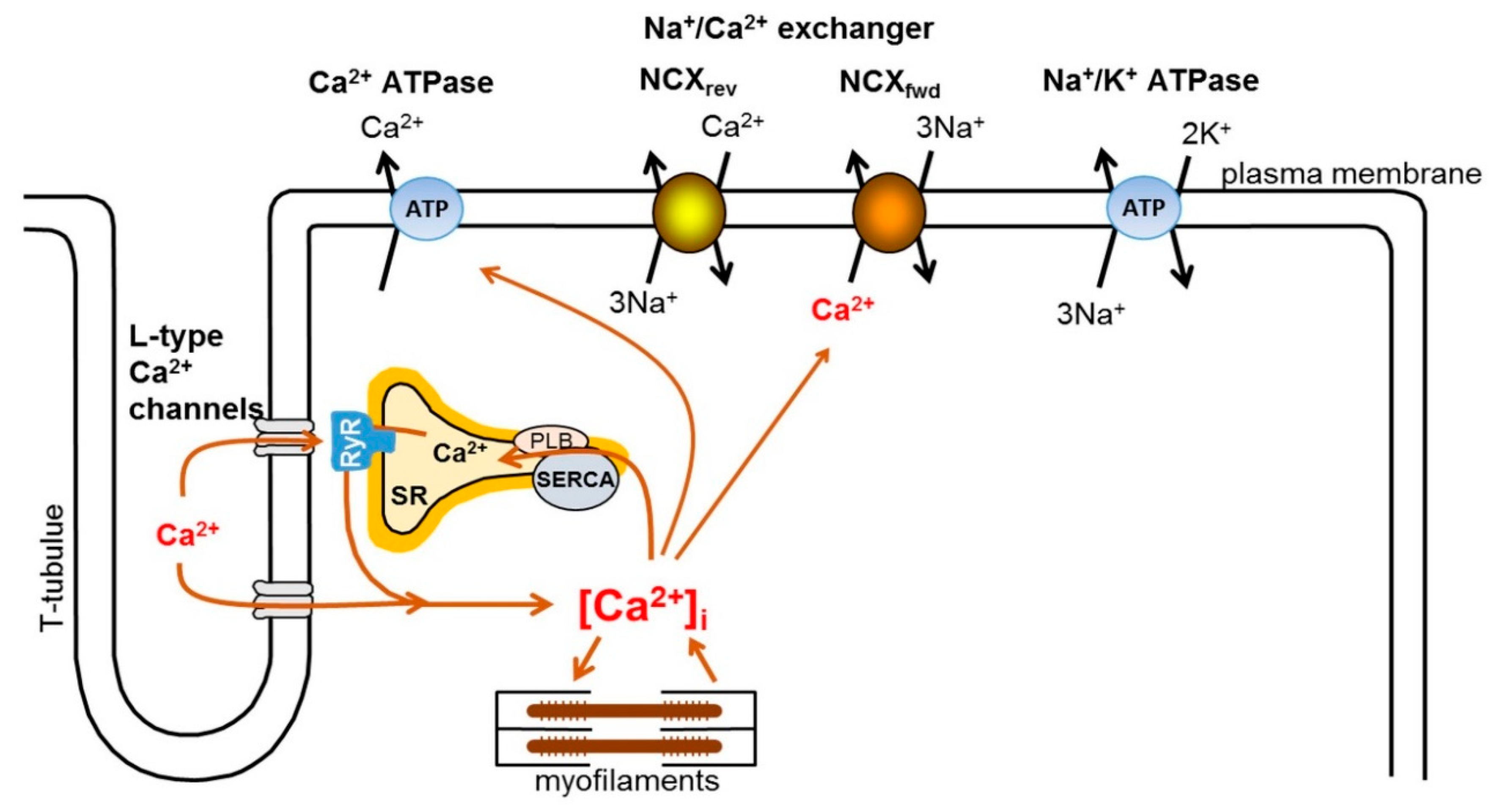
2. Intracellular Ca2+ Handling in the Heart

In cardiac myocytes, the intracellular Ca2+ level is tightly controlled by several mechanisms, including excitation–contraction coupling (ECC). The main function of the coupling is to mediate between the membrane potential change and cardiac contraction. The E–C coupling is initiated by the opening of L-type Ca2+ channels, activated in the initial phase of the action potential, and its open state is long enough—mainly during the plateau phase of the action potential—to provide sufficient Ca2+ influx to the cell [1]. L-type Ca2+ channels are expressed abundantly in the membrane invaginations, called T-tubules, being closely associated with the RyRs located in the membrane of the sarcoplasmic reticulum (SR). It is important to note that there is no direct protein–protein interaction between L-type Ca-channels and RyRs; a narrow gap exists between these structures, called as “fuzzy-space”, where the intracellular Ca2+ concentration may reach a considerably higher level than in the bulk space of the cytosol [14]. Ca2+, entering through L-type Ca2+ channels, open RyRs, leading to the release of large amounts of the stored Ca2+ from the SR and ultimately resulting in the development of the intracellular Ca2+-transient. The released Ca2+ binds to the myofilaments, specifically to troponin-C, leading to cardiac contraction. After contraction, the relaxation process requires sequestration of the released Ca2+ back into the SR. This is achieved by the SR Ca-ATPase (SERCA), while a smaller fraction is extruded from the cell by the Na+/Ca2+ exchanger (NCX), located in the sarcolemma. The activity of the SERCA is controlled by phospholamban (PLB), which inhibits the SERCA when tightly associated. Phosphorylation of PLB, e.g., as a result of beta-adrenergic activation, leads to dissociation of PLB with the concomitant improvement of SERCA activity [1] (Figure 1).
Under normal conditions, the Ca2+ released from the intracellular store is initiated by Ca2+ ions that trigger the ryanodine receptors. The source of the trigger Ca2+ could be mainly the L-type Ca2+ channels; however, further Ca2+-sources (such as reverse mode of the NCX) could also contribute [1]. It must be noted, however, that trigger Ca2+ contributes also directly to the activation of myofilaments in a species-dependent manner. The ratio of Ca2+ influx and the amount of released Ca2+ gives the amplification of Ca2+ release, called as the gain of E–C coupling.
The ryanodine receptors are organized in clusters and an elementary Ca2+ release from these clusters is the so-called Ca2+ spark. The spontaneous Ca2+ sparks do not depend on extracellular Ca2+ entry through the L-type Ca2+ channels [15,16]: spontaneous sparks were found in the absence of extracellular Ca2+, during pharmacological block of the ICaL, and even in saponin-permeabilized cells [15,16,17,18]. It is feasible that spontaneous Ca2+ sparks are influenced by intracellular Ca2+ and intra-luminal Ca2+ level. The evoked sparks are ignited by Ca2+-induced Ca2+ release via L-type Ca2+ channels, and it seems that their kinetics, amplitude, and spatial properties are similar to the spontaneous sparks.
It was demonstrated that the evoked sparks can be attributable to the Ca2+ flux [19]. The discovery of Ca2+ sparks intimately changed the understanding of the nature of Ca2+ transients: the Ca2+ release from the SR consists of “discrete units,” rather than being a continuum [20] that provides graded control of Ca2+ release, i.e., E–C coupling can be changed by altering the number of recruited release units [15,21].

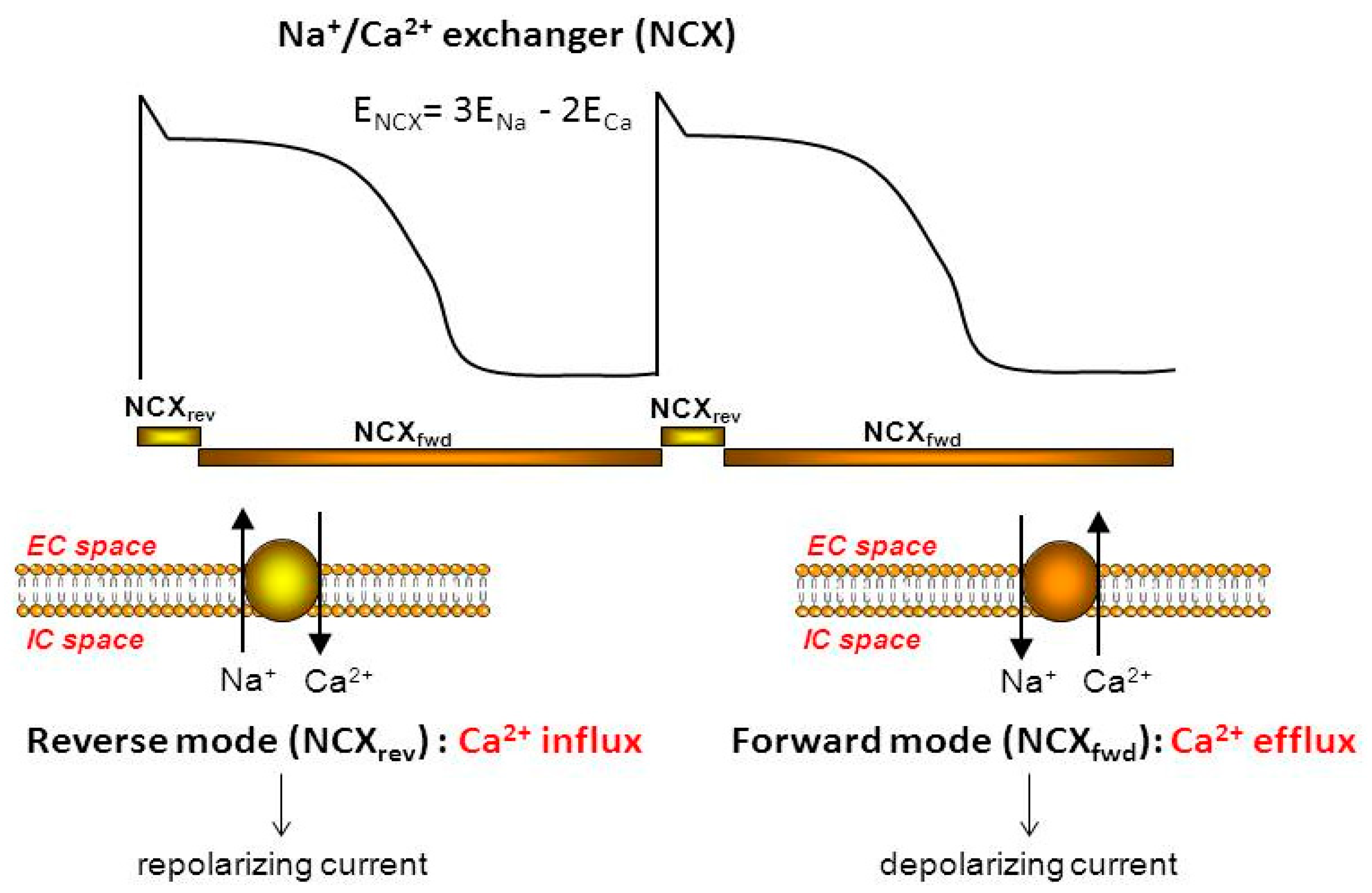
3. Physiology and Pharmacology of the Exchanger

NCX belongs to the Ca2+/cation antiporter superfamily, having the isoforms NCX1, NCX2, and NCX3 encoded by genes of SLC8A1-SLC8A3, where the NCX1 splice variants represent the cardiac isoform of NCX (for detailed molecular biological description see reference [22]).
Normally, NCX extrudes 3 Na+ for 1 Ca2+ (forward mode operation), generating electrogenic transport. In this case, inward current is generated while outward current is generated during the reverse mode function, when Ca2+ entry is combined with Na+ efflux (Figure 2).
The direction of the NCX current is regulated by intracellular and extracellular Na+ and Ca2+ levels as well as the actual transmembrane potential. Among these, the level of the intracellular Ca2+ and membrane voltage are altered within a wide range from beat to beat, causing fluctuation of the reversal potential of NCX in each cycle. Reverse mode activity of NCX develops when the reversal potential of NCX is more positive than the membrane potential, while forward mode operation is anticipated if the reversal potential is more negative than the membrane potential [23].
Since the estimated reversal potential of NCX ranges from −30 to −40 mV in the presence of a diastolic Ca2+ level, both reverse and forward mode functions are feasible during each action potential [1]. However, there is no agreement regarding the timing of the reversal point or the ratio between the two operational modes, neither in modelling studies nor in experimental results. The direction of the transport has crucial importance regarding the effect of NCX inhibition.

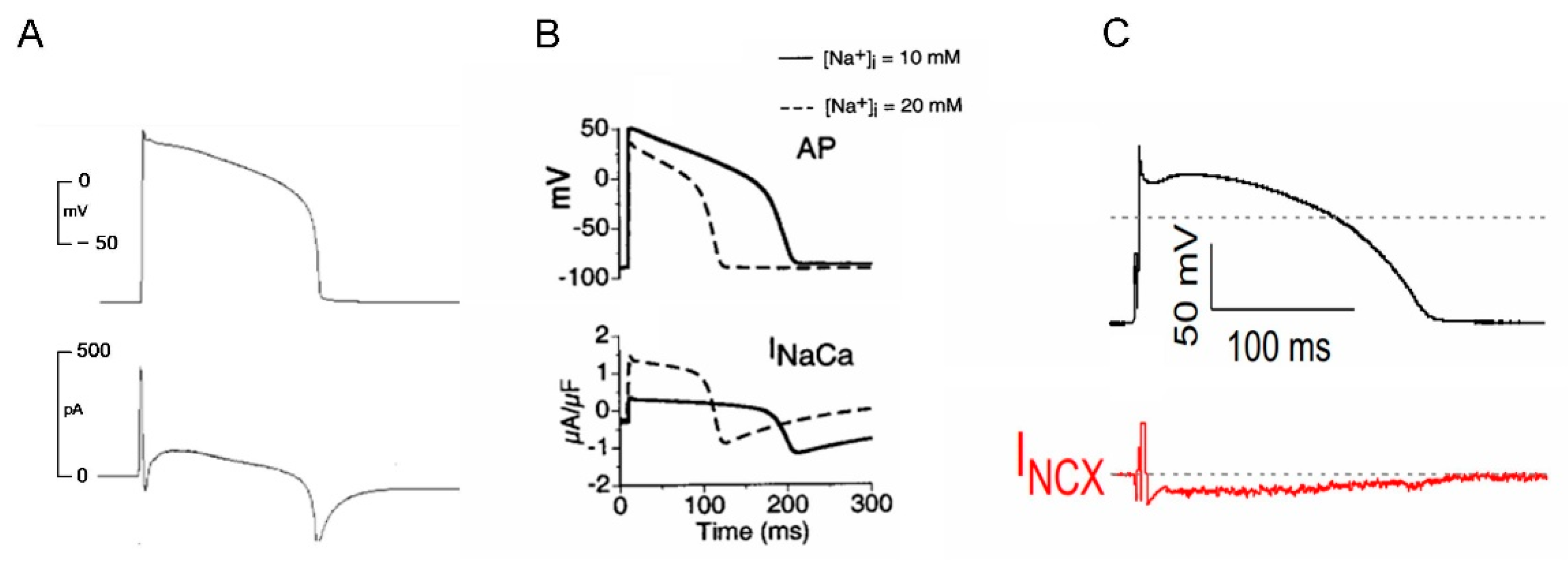
3.1. NCX Current during an Action Potential

Egan and Twist (1989) described NCX current as an inward current during the action potential, calculated from experimental results [24]. In line with this, duBell et al. [25] observed inward NCX current which prolonged action potential duration in rats. Noble et al., in 1991, predicted that NCX current is inward during the action potential, with a very brief reverse mode following the action potential upstroke [26]. The Luo–Rudy model (1994) predicted that NCX current operates in reverse mode in the first 100 ms of the guinea pig action potential then turns to forward mode, reaching its peak during terminal repolarization [27]. In this study, however, the authors calculated only with a peak free [Ca2+]i of 1 µmol/L. Experimental data from Grantham and Cannell (1996) obtained in guinea pig myocytes showed that NCX current is mainly reversed during the action potential and reaches the reversal point shortly after complete repolarization [28]. Using an improved model, Noble et al., in 1998, observed that the time of NCX reversal is approximately 200 ms after the upstroke in guinea pigs [29]. Faber and Rudy, in 2000, described a small outward current during the plateau phase of the action potential which turned to forward immediately after terminal repolarization. Furthermore, they pointed out that NCX was very sensitive to increases in [Na+]i, causing considerable changes in both the reverse and forward modes, and the former may contribute to frequency-dependent shortening of the action potential [30]. The Bers group found that NCX current is mainly forward during the action potential in rabbits, with an estimated reversal point of less than 30 ms following the upstroke because of the very high Ca2+ level in the subsarcolemmal space [31]. This sensitivity of NCX current to membrane potential changes was, later, further supported by Armoundas et al. in 2003 [32].
The first experimental evidence applying a relatively selective NCX inhibitor SEA-0400 (3 µM) was published by Bányász et al. in 2012 in guinea pig ventricular myocytes [33]. They found NCX current to be completely inward during the action potential, reaching its peak at the end of the plateau phase. Later, Nagy et al. (2014) reported similar results by applying ORM-10103, a more selective inhibitor without an effect on ICaL. The authors reported a small and brief outward current corresponding to phase 1 repolarization and a small inward current in the entire action potential. Both the reverse and forward mode operation of NCX increased when intracellular Na+ concentration was increased by activation of late sodium current [34]. Horvath et al. demonstrated an entirely inward NCX current, defined as an ORM-10962 sensitive current using the so-called “onion-peeling technique” in canine ventricular myocytes [35].
However, the time course of NCX current during the action potential is not completely consistent, it seems feasible that experimental data and modelling predictions suggestonly marginal reverse NCX current during the action potential under normal conditions. This means that selective inhibition may influence mainly forward mode operation of NCX, resulting in reduced Ca2+ extrusion from the intracellular space with concomitant suppression of the NCX-induced inward current during the action potential, which might tend to shorten the action potential (Figure 3).

3.2. Pharmacology of Novel NCX Inhibitors

As previous NCX inhibitors (such as KB-R7943) exerted non-selective behaviour, the interpretation of the results was difficult [36]. Especially, the results were contaminated by the parallel ICaL block, since it can markedly influence the Ca2+ homeostasis, distorting the effect of selective NCX inhibition. Furthermore, it may decrease the intracellular Ca2+ content that counteracts the possible positive inotropic effect of the selective NCX inhibition. Estimation of the time course of NCX current is also difficult, as ICaL overlaps the reverse NCX current during an action potential. Therefore, it was a large demand from the researchers to drug companies to develop new, completely selective NCX inhibitor compounds.
SEA-0400 (2-(4-(2,5-difluorobenzyloxy)phenoxy)-5-ethoxyaniline) was the first NCX inhibitor which was shown to exert improved selectivity. The EC50 of the NCX block was 3.35 ± 0.82 µM, for forward mode, and 4.74 ± 0.69 µM for reverse mode operation [36]. SEA-0400 did not inhibit K+ or Na+ currents; however, it decreased ICaL with EC50 of 3.6 µM. This means that the typically used 1 µM concentration causes approximately a 20% reduction in ICaL amplitude. Still, the submicromolar concentrations of SEA-0400 could be considered relatively selective for NCX [36].
ORM-10103, a novel compound with improved selectivity, was investigated in detail in 2013 [37]. The major benefit of ORM-10103 is the lack of effect on ICaL; however, a 20% blocking effect on IKr was identified. ORM-10103 equally inhibited both the reverse (EC50: 960 nM) and the forward (EC50: 780 nM) modes of NCX. Importantly, IK1, IKs, Ito, and Na/K pump were not influenced by 3 µM ORM-100103.
The next compound, ORM-10962, was further improved, displaying complete selectivity to NCX. ORM-10962 equally inhibited both the reverse and forward mode NCX currents with considerably lower EC50 values (outward: 67 nM, inward: 55 nM) [38].

3.3. Potential Therapeutic Benefit of Pharmacological NCX Inhibition

During ECC, a stable Ca2+ balance is crucial for the Ca2+ handling integrity; therefore, the Ca2+ influx and Ca2+ efflux must be equal, causing no Ca2+ gain or loss within beats [2,39].
In several diseases, the intracellular Ca2+ largely increases and considerably facilitates the activity of the forward NCX [40,41]. The Ca2+ removal by the NCX generates inward Na+ current that could lead to membrane depolarization causing triggered activity either during phase 2 and phase 3 of repolarization (early afterdepolarization, EAD) or at resting membrane potential (delayed afterdepolarization, DEAD). These triggered activities can lead to the development of extrasystole and—in the presence of arrhythmogenic substrate—may cause life threatening arrhythmias. Additionally, the actual intracellular Na+ level also significantly alters the function of NCX. Intracellular Na+ concentration can be increased due to suppressed Na+/K+ ATPase activity, increased Na+/H+ function, or via enhanced late Na+ current during the action potential plateau. The increased Na+ level activates the reverse mode of NCX, causing net Ca2+ entry, which, in turn, facilitates the activity of the forward mode leading to development of triggered activity [40,41]. Therefore, theoretically, both the inhibition of the forward mode (due to suppressed depolarizing inward current) and inhibition of the reverse mode (due to decreased Ca2+ entry that causes secondary reduction in the forward mode) could be antiarrhythmic [42].
Another aspect of the selective NCX inhibition is the proposed positive inotropic effect. Considering that the contribution of the reverse mode during the action potential is restricted, NCX inhibition mainly prevailed on the forward mode. The decreased Ca2+-extrusion could increase the intracellular Ca2+ level, which is able to accelerate the Ca2+-dependent inactivation of the L-type Ca2+ channels to match the Ca2+-influx to the Ca2+ efflux. Therefore, presumably, NCX inhibition is able to set a new Ca2+ balance where NCX activity is reduced and intracellular Ca2+ is elevated, leading to a consequent increase in contractility.

4. Effect of Novel NCX Inhibitors on Cellular Arrhythmogenic Mechanisms

In most cases, cardiac arrhythmias are complex phenomena involving several cellular mechanisms at the same time, such as EAD, increased repolarization dispersion, Ca2+ overload and DAD, alternans, depolarized membrane potential, shift in the refractory period, etc. Furthermore, arrhythmia experiments could also differ in experimental conditions. Accordingly, Ca2+ overload-related arrhythmias could be caused by Na+-induced Ca2+ overload following Na/K ATPase blockade, or may be due to increased sympathetic tone, hypokalaemia, etc. In this part of the review, the effects of selective NCX inhibition on elementary arrhythmogenic events are summarized. Since, among NCX inhibitors, only the SEA-0400 and ORM-compounds can be considered more or less selective, the next sections are restricted to summarizing the results obtained with these inhibitors.

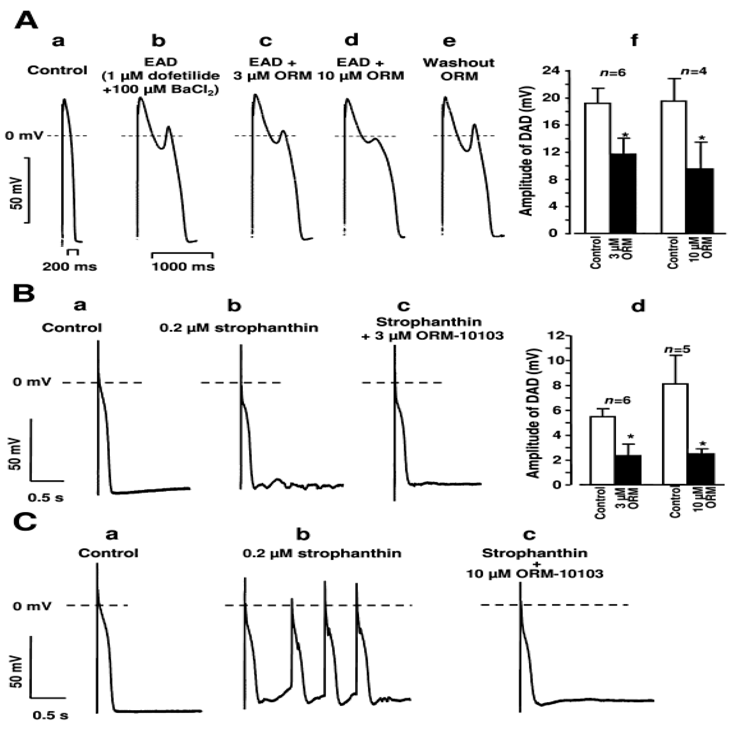
4.1. DAD and Ca2+ Waves

The first observation of DAD-related events was reported by Segers in 1940 and 1947. In frog, turtle, and rabbit hearts, spontaneous activity was observed which could be facilitated by external Ba2+ or increased external Ca2+. The phenomenon was termed as “potentiels tardifs,” or late potentials. The first direct measurements of DADs were carried out in 1977 by Crenfield et al. [43]. It was found that intracellular Ca2+ content can be increased via increased sympathetic tone, or can develop as a consequence of several diseases, such as catecholaminergic polymorph tachycardia, hyperthyreosis, etc., when the cells are overloaded with Ca2+. Ca2+ overload increases Ca2+ reuptake to the SR leading to spontaneous diastolic Ca2+ releases. These events drive the forward mode of the Na+/Ca2+ exchanger, causing spontaneous depolarizations [44].
Since NCX function is considered to be a crucial player of the DAD development, selective NCX inhibition was a promising candidate to suppress arrhythmias related to DADs [42]. SEA-0400 was reported to effectively suppress arrhythmogenic afterdepolarizations under pathophysiological conditions associated with Na+- and the consequent Ca2+ overload [45,46,47]. In these studies, intracellular Na+ gain was induced by oubain/strophantine administration in guinea pigs and dogs.
Fujiwara et al. showed a close link between spontaneous Ca2+ releases, DADs, and extra beats following rapid pacing [48]. In this study, SEA-0400 suppressed the triggered activity by attenuating DADs but could not prevent the spontaneous Ca2+ waves.
Among new NCX inhibitors, the effect of ORM-10103 was tested against DAD development [37]. DAD was evoked by 40 consecutive stimuli, having a cycle length of 400 ms on canine right ventricular Purkinje fibres in the presence of 0.2 µM strophantin. Furthermore, 3 and 10 µM ORM-10103 decreased the amplitude of DADs in a concentration-dependent manner.
In a further study, strophantin-induced spontaneous Ca2+ releases were investigated in isolated dog ventricular myocytes during rest after a rapid pacing period. In the pacing-free period, several spontaneous Ca2+ releases were observed which were significantly suppressed by 10 µM ORM-10103. In this paper, the effect of strophantin on steady-state Ca2+ transients was also investigated: 1 µM strophantin markedly increased the amplitude of Ca2+ transients; however, when the cells were pre-treated with 10 µM ORM-10103, strophantin failed to increase their amplitude [34].
Similar results were obtained by using ORM-10962 [38]. Dog right ventricular Purkinje fibres were treated with 150 nM digoxin and were stimulated at a 400 ms pacing cycle length. It was observed that 1 µM ORM-10962 significantly suppressed the amplitude of DADs. When the reverse mode NCX was facilitated by using 70 mM external NaCl, the subsequently applied 1 µM ORM-10962 reduced the amplitude of the Ca2+ transient [38,49]. The application of reduced external Na+ shifted the NCX reversal potential toward more negative values and increased the time for reverse mode operation.
These results suggest that selective NCX inhibition effectively suppresses the occurrence of the DADs. Regarding the mechanism, it is feasible that DAD can be suppressed either directly, i.e., by inhibition of the forward mode, or indirectly via reduction of reverse mode leading to decreased intracellular Ca2+ concentration and reduced forward mode NCX activity. Since, in these experiments, the DADs were evoked by Na/KATPase inhibition, it is possible that the increased intracellular Na+ facilitated the reverse mode activity of NCX, resulting in increasedCa2+ entry. This scenario is further supported by the fact that NCX inhibition prevented Ca2+ accumulation caused by strophantin, while the magnitude of the Ca2+ transient was unchanged [34]. Therefore, it seems feasible that ORM-compounds suppress DADs and spontaneous Ca2+ releases via reverse mode NCX inhibition during Na+-induced Ca2+ overload.
Ca2+ waves induced by rapid pacing in dog atrium were found to activate the forward mode of NCX and markedly depolarized the membrane. The application of SEA-0400 or ORM-10103 effectively abolished the Ca2+ waves [50].
It appears that selective NCX inhibition displays a beneficial effect against DAD formation. However, it is uncertain whether the reverse or the forward mode inhibition dominates the effect. Data show that, when DADs are the consequence of Na+-induced Ca2+ overload, it seems feasible that NCX inhibition reduces DAD development, which is presumably the consequence of reverse mode block, and the concomitant decreased Ca2+ indirectly reduces the forward mode NCX. This effect resulted in decreased intracellular Ca2+ and reduced depolarizing events.

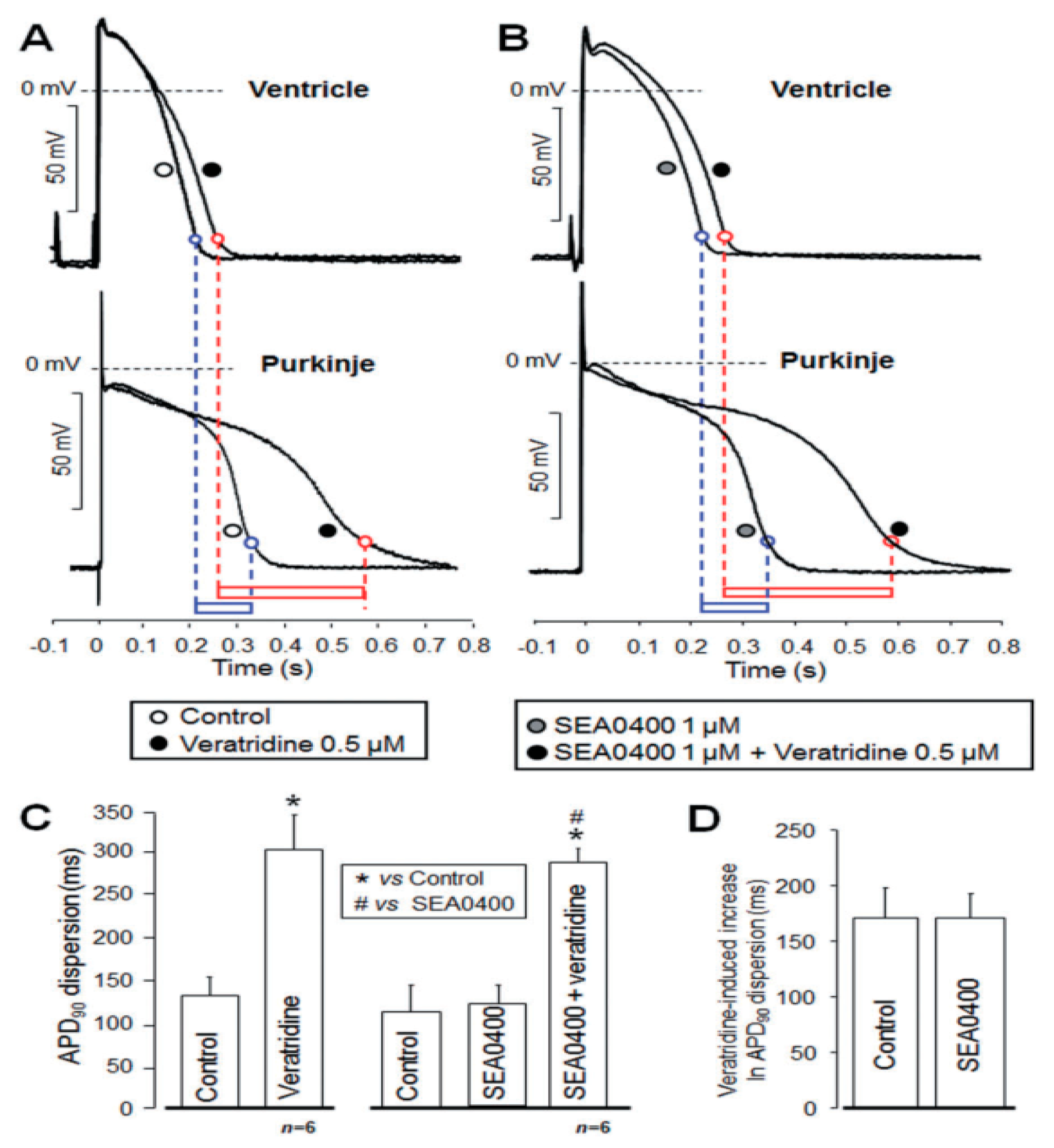
4.2. EAD, LQT and TdP

The term EAD indicates a spontaneous depolarizing event that occurs before the completion of repolarization. The classical proposed mechanism for EAD development is the reactivation of the L-type Ca2+ current during a long-lasting action potential [51]; however, the roles of the late Na+ current [52,53], the NCX [37], CaMKII, and PKA [54] were also suggested as underlying mechanisms. EADs may play an essential role in the arrhythmogenesis by providing arrhythmia triggers that promote re-entry [55]. EADs typically appear when the action potential is excessively long, often due to K+ channel inhibition and/or during slow pacing rate. Also, several diseases, such as chronic heart failure or long QT-syndrome, are also reported to be associated with the development of EADs [56]. Torsades de pointes (Tdp) is polymorphic tachycardia which can lead to fibrillation and sudden cardiac death. It is widely accepted that Tdp is initiated by a trigger (EAD or DAD) and maintained by an arrhythmogenic substrate (repolarization dispersion) [57].
In a guinea pig model, LQT3 was pharmacologically mimicked by the enhancement of late Na+ current using aconitine, where SEA-0400 was unable to suppress ventricular tachycardia and triggered activity [58]. In another study, SEA-0400 effectively decreased dofetilide-induced TdP arrhythmias in anesthetized dogs in a dose-dependent manner [59]. A lower (0.4 mg/kg) dose of SEA-0400 partially suppressed TdPs, while a higher dose (0.8 mg/kg) completely terminated TdPs; however, the dofetilide-induced QT prolongation remained unchanged [59].
In Langendorff-perfused rabbit hearts with atrioventricular block, NCX inhibition by SEA-0400 did not suppress the incidence of dofetilide-mediated TdP [60].
In contrast, in Langendorff-perfused rabbit hearts, LQT2 and LQT3 were pharmacologically evoked by sotalol or veratridine, respectively [61]. The hearts responded with marked prolongation of monophasic action potential duration, increased dispersion, and, in some cases, TdPs were observed. The administration of SEA-0400 significantly reduced action potential duration as well as the dispersion and TdP incidence. In dog subendocardial tissue, 1 µM SEA-0400 markedly decreased the EAD amplitude [47].
The effect of NCX inhibition on EAD incidence was investigated also in canine right ventricular papillary muscle (Figure 4). EADs were evoked by slow pacing (at 5 s cycle length) and parallel application of 1 µM dofetilide and 100 µM BaCl2. It was found that 3 and 10 µM ORM-10103 decreased the amplitude of EADs in a dose-dependent manner, indicating the role of forward mode NCX in the development of EAD. A further (or additional) possibility for the mechanism of action is that ORM increased the intracellular Ca2+ level, inducing faster inactivation of Ca2+ channels. In this case, if EADs were mediated by Ca2+ channels, reopening of the channel performing window Ca2+ current—as it was claimed previously in [51]—NCX inhibition might indirectly reduce the amplitude of EADs [37]. In rabbit hearts, it was found that SEA-0400 eliminated the H2O2-induced EADs [62].
Chang et al. investigated the antiarrhythmic effect of ORM-10103 in normal and failing rabbit hearts [63]. QT-interval prolongation in the ECG curve was stimulated by IKr inhibition, leading to enhanced EAD development and Tdp inducibility in both control and failing hearts. When ORM-10103 was applied, the number of premature beats was reduced; however, Tdp inducibility remained unchanged. It was found that ORM-10103 increased action potential duration and inhomogeneity in failing hearts. These findings are consistent with previous results by Farkas et al., where SEA-0400 failed to reduce Tdp incidence in rabbit hearts [60]. It is important to note that ORM-10103 is not highly selective: it exerted mild IKr inhibitory action [37], which could be augmented when repolarization reserve was attenuated, e.g., in the case of heart failure. Therefore, the observed prolongation action potential duration and its increased dispersion is not necessarily related to the NCX blockade.
It seems possible that selective NCX inhibition has a limited effect against EAD development. However, this is probably not surprising since changes in several ion currents, such as ICaL, INaL, and INCX are known to contribute to EAD formation. It is conceivable that the participation of these currents in the EAD formation highly influences the effect of NCX inhibition. Conditions enabling a larger contribution of NCX to EAD development, such as higher cytosolic Ca2+ concentration, predict a more effective beneficial influence of selective NCX inhibition on EAD incidence.

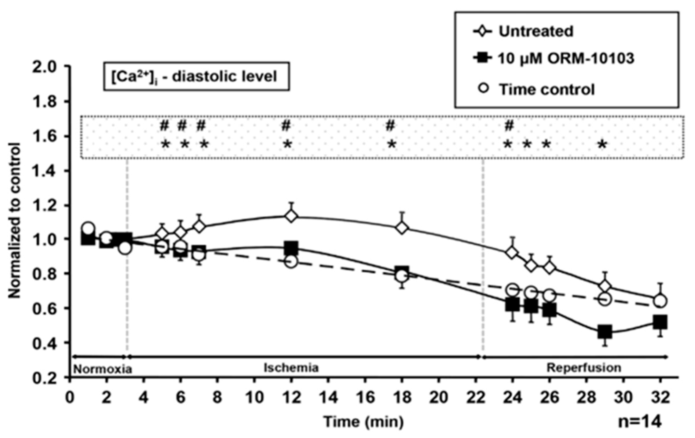
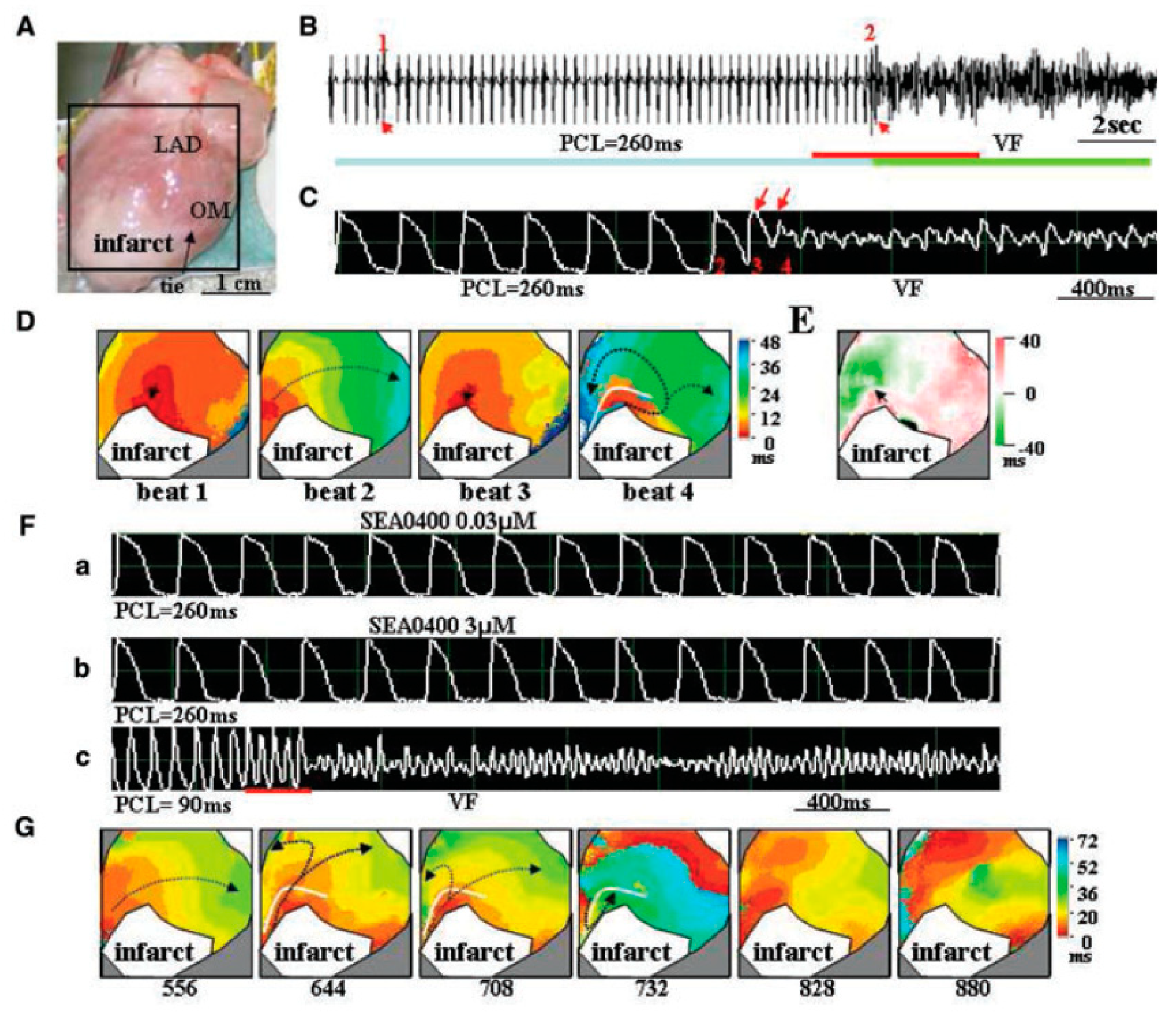
4.3. Ischemia-Reperfusion

The aerobic metabolism of the heart is seriously compromised during ischemia, which occurs typically after regional coronary artery occlusion. The concomitant decline in intracellular ATP level, intracellular acidosis, deliberation of reactive species, etc., may influence the vast majority of ion channels and transporters [64], leading to marked changes in the intracellular milieu of the cells, action potential configuration, and impulse propagation, ultimately largely increasing the arrhythmia propensity of the heart [65,66,67].
It Is well-known that NCX has a key role in arrhythmogenesis related to ischemia/reperfusion. The rise of the intracellular Na+ concentration due to suppression of the Na+/K+ pump is a consequence of ATP depletion. This may facilitate the reverse mode operation of NCX, leading to net Ca2+ gain [68,69,70]. Therefore, it seems feasible that the prognosis of ischemia worsens due to high NCX activity which is supported by murine experiments with heterozygous over-expression of NCX, where increased susceptibility to ischemia/reperfusion injury was observed [71,72].
In vivo studies on ischemia-/reperfusion-related arrhythmias obtained with NCX blockers yielded controversial results, since the majority of experiments failed to show any antiarrhythmic effect of SEA-0400 [73,74,75]. In a one-month myocardial infarction rabbit model, SEA-0400 reduced the number of pacing-induced ventricular premature beats but was also found to be proarrhythmic at the same time due to modifying restitution of action potential duration and enhancing the development of discordant cardiac alternans [76]. Ventricular fibrillation incidence was decreased by NCX inhibition only in the study of Takahashi et al., when studied using a model of coronary occlusion in rats [77].
In Langendorff-perfused rat hearts, SEA-0400 improved the cardiac function and energy metabolism [78] but failed to provide protection against ischemia-/reperfusion-induced ventricular arrhythmias [78,79]. In another study, SEA-0400 was protective against glycolytic inhibition-induced triggered activity in rat hearts; however, this beneficial effect disappeared when the heart rate decreased [80].
The effect of ORM compounds in ischemia/reperfusion injury was investigated on both isolated cells and Langendorff-perfused hearts. Simulated ischemia was induced by a modified external solution containing unusually high K+ and lactate concentrations, but no glucose, combined with acidic pH and hypoxia. Intracellular Ca2+ level, action potentials, and cell viability were recorded in these experiments.
For single cell investigation, canine left ventricular myocytes were used [81]. Application of the ischemic solution largely increased the diastolic Ca2+ level, decreased the amplitude of the Ca2+ transient, caused depolarization, shortening, and triangulation of action potentials, and ultimately cell death. An amount of 10 µM ORM-10103 prevented the rise of diastolic Ca2+ during both ischaemia and reperfusion (Figure 5), and increased cell survival, while the ischemia-induced changes in action potential morphology were not restored. The marked attenuation of the ischemia-induced rise in diastolic Ca2+ suggests that the reverse mode NCX inhibition could be primarily attributable to the observed protective effect. Presumably, the ATP depletion reduced the Na/K ATPase activity and increased intracellular Na+ concentration, which drove the reverse NCX activity to overload the cells with Ca2+. In line with this, it was also reported that selective NCX inhibition is able to hinder the Na+-induced Ca2+ gain after strophantin application [34].
Szepesi et al. investigated the possible protective effect of selective NCX inhibition against coronary ligation-induced ischemia in Langendorff-perfused rat hearts, where 10 min of ischemia was followed by 30 min of reperfusion [79]. Extrasystoles, ventricular tachycardia, and ventricular fibrillation were observed during reperfusion. The application of 10 µM ORM-10103 increased the arrhythmia-free period and decreased the incidence of the extrasystoles, while it had no effect on the incidence and duration of ventricular fibrillation.
In contrast, in the case of ischemia-induced by coronary ligation in anaesthetized rats, 1 µM ORM-10962 failed to exert any antiarrhythmic effect [38]. During ischaemia, arrhythmias were not detected, while a large number of life-threatening arrhythmias appeared during reperfusion. In fact, 33% of the animals died by irreversible ventricular fibrillation. Other types of arrhythmias, such as reversible fibrillation or tachycardia, were often observed among survivor animals; however, ORM-10962 failed to cause a change either in the type or the time of onset of the developing arrhythmia.
Similar results were obtained from experiments with zero-flow ischemia in Langendorff-perfused guinea pig hearts. Reperfusion after the no-flow ischemia induced similar ventricular fibrillation in both groups (i.e., control and ORM-10962-treated) without any statistical difference between the results [38].
Although selective NCX inhibition clearly exerts beneficial effects against some pathophysiological components of the ischemia/reperfusion injury, such as the Na+-induced Ca2+ load, it has a limited protective effect against other symptoms of ischemia/reperfusion injury. It is likely, therefore, that the antiarrhythmic effect observed against the Na+-induced Ca2+-overload may be attributable mainly to the reverse mode blockade of NCX. However, in a whole heart aspect, several other factors are present that could be unaffected by the NCX inhibitors, such as changes in repolarization, reduction in impulse propagation, depolarization of the resting membrane potential, etc.

4.4. Action Potential Duration, Refractory Period, and APD-Dispersion

The equilibrium potential of the NCX predicts that both outward and inward current could be developed during an action potential. However, under normal conditions, the reverse mode is suggested to be restricted in the first section of the action potential [31]; therefore, mainly inward current is expected to be developed. It is predicted that the exchanger activity may lengthen the actions potential duration, thus its inhibition should shorten the action potential and the QT-interval.
Milberg et al. recorded monophasic action potentials from Langendorff-perfused rabbit hearts. When SEA-0400 was applied alone, the drug shortened action potentials within the range of 0.8 and 1.6 µM. Similarly, the QT interval was also shortened in this concentration range [61]. SEA-0400 effectively prevented the action potential lengthening and QT interval prolongation caused by free artemether, an antimalaria agent [82]. In this study, 1 µM SEA-0400 exerted a tendency to shorten action potential in single cells isolated from mice. SEA-0400 also effectively attenuated the repolarization prolongation in a murine model of Chagas disease without shortening it under control conditions [83].
Tadros and Cheung reported no change in the duration of rat action potentials after the down-regulation of NCX [84]. Namekata found unaltered action potential morphology following SEA-0400 treatment [46], similarly to Kormos et al., who also failed to demonstrate any effect of ORM-10103 in canine single cell action potentials [81]. In contrast, Tanaka reported action potential shortening after SEA-0400 in isolated murine myocytes [85]. Nagy et al. also found no change in the action potential configuration, neither in the presence of SEA-0400 nor after ORM-10103 administration. However, it was also reported that SEA-0400 reduced the action potential duration in rabbit isolated cells [62].
In the study where ORM-10962 was used for NCX inhibition, the action potential duration was unaltered after administration of 1 µM of the compound [38]. Slight changes were observed after the manipulation of NCX transport direction: when reverse mode was augmented, action potentials marginally lengthened; when forward mode was facilitated, the plateau potential was slightly depressed.
An important additional effect of selective NCX inhibition is that it increased the effective refractory period [86]. This effect may also contribute to the stabilization of the membrane potential. Action potential dispersion means that differences between the action potentials originated from different regions of the heart. Marked dispersion of action potential duration can be observed between the midmyocardial layer and the endocardial or epicardial myocardium. Similarly, the heterogeneity between the ventricular myocardium and Purkinje fibres is also significant. The excess of dispersion has a crucial role in arrhythmogenesis, since increasing dispersion causes asynchronous refractory periods between different regions, providing a vulnerable window for arrhythmia triggers [87,88]. The effect of NCX-inhibition on dispersion was also tested in some studies.
In the study of Nagy et al., the Purkinje fibre and ventricular muscle action potentials were simultaneously recorded from a Purkinje-ventricular preparation using two microelectrodes, where the two preparations were connected (Figure 6). The application of 10 µM ORM-10103 did not change the dispersion obtained between Purkinje fibre and ventricular action potentials [34].
Milberg et al. demonstrated that the sotalol- or varatridine-induced dispersion between epicardial and endocardial monophasic action potentials was significantly reduced by the application of 1 µM SEA-0400 [89]. This effect was achieved by asymmetrical action potential shortening caused by NCX inhibition.
Human atrial APD90 was unchanged when SAR296968, a novel, selective NCX inhibitor compound, was used. Similarly, action potential amplitude and resting potential remained unaltered after selective NCX inhibition (Figure 7) [90].
In summary, the results obtained with NCX inhibition on the action potential duration are controversial. Data obtained from the SEA-0400 experiments showed that it exerted moderate action potential shortening or no effect; however, it cannot be ignored that SEA-0400 also suppressed L-type Ca2+ current even at a concentration of 1 µM [36], which could amplify the effect of a potential NCX-inhibition-induced action potential shortening. When using ORM compounds, no effect on action potential duration has been described so far.

4.5. Action Potential and Ca2+ Transient Alternans

Cardiac alternans refer to a beat-to-beat oscillation of the action potential as well as the intracellular Ca2+-transient, appearing typically at high pacing rates. Cardiac alternans is considered a reliable predictor of serious life-threatening arrhythmias such as ventricular fibrillation [91,92]. The underlying mechanisms of alternans are not fully understood. Briefly, there are two leading concepts for the ionic mechanism of alternans: (I.) the voltage-driven theory claims that alternans is expected when the slope of the restitution curve of the action potential duration (i.e., the relation of action potential duration and the preceding diastolic interval) is larger than 1 [93]. (II.) The Ca2+-driven theory implies that alternans develops when the beat-to-beat balance of Ca2+ handling is compromised [94,95,96]. Since NCX is an important player in intracellular Ca2+ handling and may also influence the membrane potential, its inhibition has emerged as a potential strategy to suppress or prevent alternans.
In Langendorff-perfused rat hearts, glycolytic inhibition-related ventricular fibrillation was prevented by 2 µM SEA-0400 in three of six hearts [80]. Simultaneous Ca2+ and Vm mapping in control rabbits and with one-month myocardial infarct revealed that SEA-0400 suppressed the pacing-induced ventricular premature beats. SEA-0400 steepened the slope of the restitution curve and enhanced spatially discordant alternans. It was concluded that, in one-month infarct hearts, SEA-0400 suppresses pacing-induced ventricular extrasytoles but promotes the development of discordant alternans (Figure 8) [76].
Action potential and Ca2+-transients were simultaneously measured in isolated guinea pig ventricular cells. Under control condition positive Ca2+-to-Vm coupling was found, i.e., large Ca2+-transients were associated to long action potentials, indicating the predominance of the inward current generated by NCX, which lengthened the action potentials during Ca2+ extrusion. The application of SEA-0400 did not attenuate alternans, but changed the alternans to negative Ca2+-to-Vm coupling, indicating that L-type Ca2+ current became the dominant charge carrier after NCX inhibition. The positive coupling under control conditions suggests the predominance of NCX during action potential alternans [97].
In 2021, the effects of selective NCX inhibition on action potential duration and Ca2+-transient alternans were investigated in canine ventricular tissue and isolated myocytes. Both action potential duration and Ca2+-transient alternans were evoked by rapid pacing (ranging from 250 to 170 ms cycle length). It was found that 1 µM ORM-10962 significantly decreased the amplitude of both types of alternans, while the slope of the restitution curve remained unaltered. Furthermore, the ORM-10962 increased the refractory period of the action potential. The underlying mechanism of the “anti-alternans” effect was supposed to be a consequence of establishing a new balance in the Ca2+ homeostasis when SR Ca2+ loading is increased and the SR release refractoriness is reduced. These data also suggest that the main driver of the alternans is the intracellular Ca2+ rather than the slope of the restitution curve.

5. Inotropic Effects of Novel NCX Inhibitors

NCX has a crucial role in Ca2+ extrusion from the cells in each cycle; therefore, its selective inhibition was a promising novel therapeutic tool to modify intracellular Ca2+ handling and improve cardiac performance, especially in heart failure. The seminal work of Hobai et al. [98] demonstrated that XIP, a peptide inhibitor of the NCX, was able to restore the reduced Ca2+ transient magnitude near to the normal level in canine failing hearts. However, the application of XIP has several limitations. Subsequently, several experimental approaches were performed by using novel inhibitors to address this issue.
Regarding the inotropic action of NCX inhibition, a species-dependent effect seems to be delineated: the results obtained from rat or mice are different from those recorded from larger mammals developing longer, plateau-forming action potentials. In order to better illustrate this discrepancy, the results have to be discussed according to the experimental animal applied.

5.1. Selective NCX Inhibition Results in Positive Inotropy in Rat and Mouse and Human Atrium

In 2005, Tanaka et al. investigated the effects of 1 and 10 µM SEA-0400 in rats. An amount of 1 µM SEA-0400 caused about 25% inotropy in tissue preparations, and a similar increase was found in cell shortening and Ca2+ transients [85]. Single cell action potential measurements exerted considerable shortening after the application of 1 µM SEA-0400, indicating inward NCX current inhibition; however, this result could be augmented by an additional reduction in L-type Ca2+ current.
In 2007, Acsai et al. investigated the effect of selective NCX inhibition in isolated rat myocytes by using 0.3 µM SEA-0400. Ca2+ transients and cell shortening were recorded in the absence and presence of SEA-0400 [99]. It was found that both cell shortening (by ~40%) and Ca2+ transient amplitude (by ~30%) were increased after selective NCX inhibition. Parallel with increasing the amplitude of the Ca2+ transient, the magnitude of L-type Ca2+ current decreased. This change could be the consequence of the Ca2+ current-inhibitory side effect of SEA-0400; however, additionally, some indirect effect of the increased intracellular Ca2+ also could reduce Ca2+ current by a Ca2+-dependent inactivation process [100].
Similar results were obtained in 2008 by Farkas et al. [101]. In this study, 1 µM SEA-0400 was investigated in Langendorff-perfused rat hearts. Left ventricular performance was tracked by the measurement of developed pressure via non-elastic balloon placed in the ventricle. The application of 1 µM SEA-0400 increased the maximal systolic pressure by 12%. Also, in 2008, Szentandrássy et al. investigated the effect of 0.3 and 1 µM SEA-0400 in Langendorff-perfused rat hearts, recording Ca2+ transients [102]. The authors found that both concentrations of SEA-0400 increased the developed pressure and the amplitude of the Ca2+ transient. Ozdemir et al. (2008) reported that 1 µM SEA-0400 facilitated cell shortening and decreased the rate of relaxation in murine myocytes [103]. A contradictory result, published in 2015 by Szepesi et al. [79], was obtained from Langendorff-perfused rat hearts, where 1 µM SEA-0400 failed to influence left ventricular systolic pressure.
In 2019, Primessnig et al. investigated the effect of ORM-11035 on heart failure with preserved ejection fraction in rats. It was found that chronic application of the compound markedly attenuated the cardiac remodeling and diastolic dysfunction [104].
In 2022, Hegner et al. used a novel, selective NCX inhibitor SAR296968 on human atrial myocytes [90]. It was found that 3 µM SAR296968 increased the SR Ca2+ content and, in 300 nM and in 3 µM, increased the developed tension in human atrial trabeculae.

5.2. Selective NCX Inhibition Failed to Exert Positive Inotropy in Guinea Pigs, Rabbits and Dogs

Despite the mostly positive results regarding the inotropic effect of NCX inhibition in rats, studies using guinea pigs, rabbits, or dogs mainly reported that NCX inhibitors failed to influence contractility.
In 2008, Farkas et al. reported no effect of 1 µM SEA-0400 on the maximal systolic pressure of the rabbit heart measured in a Langendorff-perfusion system [101]. This study also reported a positive inotropic effect of SEA-0400 in rat hears; therefore, a direct comparison between the two species was possible, where the experimental conditions were completely identical. Also, in 2008, Birinyi et al. studied the effect of 0.1, 0.3, and 1 µM SEA-0400 on Ca2+ transients, cell shortening, and SR Ca2+ content in canine myocytes [105] (Figure 9).
In spite of the results of current measurements, indicating that SEA-0400 effectively inhibited the NCX current, no change was found in the transient and cell shortening irrespective of the applied concentration. The results were explained by a Ca2+-dependent effect of SEA-0400, suggesting that higher intracellular Ca2+ decreases the effect of NCX-inhibition. Therefore, current measurements (where intracellular Ca2+ is buffered to a low level) could exert a relatively high (~80%) effect; however, this could be markedly decreased under normal Ca2+ handling. Importantly, the relaxation of caffeine-induced Ca2+ transient was delayed by 1 µM SEA-0400, suggesting that NCX inhibition could be more pronounced when NCX operates with nearly its full capacity. In line with these results, Szentandrassy et al. (2008) also reported no alteration in the systolic and diastolic pressure and intracellular Ca2+ concentration in Langendorff-perfused guinea pig hearts after the application of 0.3 and 1 µM SEA-0400 [102].
A novel NCX inhibitor, ORM-10103, was also tested in canine cardiomyocytes. Under normal conditions, ORM-10103 failed to change the amplitude of the Ca2+-transient or the rate of cell shortening. When cells were pre-treated by 2 nM ATX-II (activator of late Na+ current), the magnitude of the Ca2+-transient was reduced by ORM-10103. Similar results were obtained when the Na/K pump was inhibited by strophantin prior to the application of ORM-10103 [34].
Kohajda et al. studied an improved NCX inhibitor, ORM-10962 (1 µM), in canine ventricular myocytes. In spite of the approximately 80% NCX inhibition in both the forward and reverse modes, ORM-10962 failed to influence the Ca2+ transient and cell shortening under normal conditions. In contrast, a positive inotropic effect was observed when intracellular Ca2+ was increased by forskolin prior to ORM administration [38]. This result also suggests that, in the case of intact Ca2+ handling, the effect of NCX-inhibition may be markedly less than 80%. In order to overcome this, NCX must be considerably burdened by Ca2+. In contrast, when reverse NCX was facilitated by a low (70 mM) external NaCl concentration, ORM-10962 reduced the amplitude of Ca2+ transients, indicating a reverse NCX-mediated Ca2+ loss.
Similar results were obtained in a study by Oravecz et al., where 1 µM ORM-10962 failed to influence the Ca2+ transient and cell shortening in canine ventricular myocytes under normal conditions. Augmentation of the reverse mode NCX activity by low external Na+ decreased, while facilitation of the forward mode activity increased the effect of NCX inhibition on the amplitude of Ca2+ transients [49].
Jin et al. used 3 µM ORM-10103 in porcine atrial myocytes, where 11-deoxycorticosterone acetate was used to induce hypertension. In these cells, it was found that NCX inhibition increased the amplitude of Ca2+ transient and also the Ca2+ content of the SR in left atrial myocytes but failed to alter the parameters of the Ca2+ transient and SR Ca2+ content in right atrial cells. This discrepancy could be attributable to the shift of NCX toward reverse mode in the case of right atrial cells. The NCX blockade ratio was found to be approximately 60% in both the left and right atriums [106].
Otsomaa et al. used a brand new ORM compound, ORM-11372, which exerted extremely low EC50 values (5 and 6 nM for reverse and forward mode, respectively) for NCX [107]. It was found that ORM-11372 elicited positive inotropy in anaesthetized rabbit left ventricular contractility. Other parameters, such as heart rate or systolic blood pressure, were not influenced.
Pelat et al., in 2021, by using a novel inhibitor, SAR340835, found that selective NCX inhibition improved the systolic function and sympathovagal balance in a dog heart failure model [108].
Taking together inotropic data obtained from SEA-0400, ORM-10103/10962/11372, and SAR296968/340835 experiments, it seems likely that selective NCX inhibition shows a species-dependent effect. In experiments performed in rats (or in human atrial myocytes that exerted very short APD90 (~50 ms)), primarily performed using SEA-0400, mainly positive inotropic effects were found. In contrast, when SEA-0400 or ORM compounds were applied in animal models having long lasting action potentials with a pronounced plateau, the vast majority of the results reported no effect of NCX inhibition.
The most prominent difference between rat and dog/rabbit ventricular cells is probably that the rat action potential is about 3–4 times shorter, lacking the plateau phase. The baseline frequency of the rat heart is also considerably higher than that in dogs or rabbits. The short action potential and the consequent fast decline of membrane potential suggests minimal reverse mode NCX activity during the action potential, since reverse mode exchanger activity is primarily expected at depolarized membrane potential levels. Therefore, it is feasible that, in rats, NCX activity quickly changes to the forward mode when intracellular Ca2+ is near to its peak or it is still high; thus, the Ca2+ extrusion function is boosted. In this case, NCX inhibition decreases the portion of active exchanger molecules, leading to exhaustion of the Ca2+ extrusion capacity of the exchanger that could be manifested in intracellular Ca2+ gain. In contrast, in dogs or rabbits, the plateau phase maintains the membrane potential at a depolarized level for a relatively long period of time where the speed of Ca2+ extrusion by the NCX is restricted. When the membrane potential drops to more negative values, and forward NCX is boosted, the intracellular Ca2+ is in strong decline due to SERCA function. Therefore, the lower intracellular Ca2+ may exhaust the NCX capacity less; thus, the NCX-inhibition may be “better tolerated” and unable to shift the Ca2+ balance toward higher cytosolic Ca2+ levels. However, this assumption could stand for only the compounds that have a limited NCX-inhibitory effect under normal Ca2+ cycling (~ 50% [49]). It is feasible that novel compounds having higher potency to inhibit a larger fraction of the NCX could cause positive inotropy in these cases; however, this requires further experiments on large animal models.

6. Conclusions

The data suggest that selective NCX inhibition could be a promising novel therapeutic tool against triggered arrhythmias, especially during Na+-induced Ca2+-overload. In this case, it is feasible that the antiarrhythmic effect is driven by the reverse mode inhibition, mediated by a reduction in cytosolic Ca2+ concentration which suppresses triggered potentials induced by the forward mode activity of NCX. The inhibition of NCX could also be promising to attenuate action potential duration and Ca2+-transient alternans. However, it is important to note that these ‘beneficial’ effects of NCX inhibition could have important side effects. For instance, reverse mode inhibition could lead to decreased Ca2+I; however, it may have a detrimental effect to the contraction. Action potential shortening and shifts in the Ca2+ handling attenuate alternans; however, they could facilitate the development of atrial fibrillation and increase the propensity of the heart to the development of triggered arrhythmias. These potential unwanted effects could limit the possible future clinical application of NCX inhibitors and require further studies on large animals.
In contrast, selective NCX inhibition seems to be ineffective to induce significant, clinically useful inotropy in ventricular myocardium of larger mammals, including humans. The lack of effect could be based on the peculiar time course of NCX current during the action potential and the relatively low degree of NCX blockade that leaves enough capacity for NCX to maintain normal Ca2+ extrusion.

Author Contributions

Conceptualization, N.N. and P.P.N.; writing—original draft preparation, N.N. and N.T.; writing—review and editing, P.P.N.; funding acquisition, N.N. All authors have read and agreed to the published version of the manuscript.

Funding

This research was funded by National Research Development and Innovation Office grant number: FK-129117.

Institutional Review Board Statement

Not applicable.

Informed Consent Statement

Not applicable.

Data Availability Statement

Not applicable.

Conflicts of Interest

The authors declare no conflict of interest.

References

  1. Bers, D.M. Cardiac excitation-contraction coupling. Nature 2002, 415, 198–205. [Google Scholar] [CrossRef] [PubMed]
  2. Eisner, D.A.; Choi, H.S.; Díaz, M.E.; O’Neill, S.C.; Trafford, A. Integrative Analysis of Calcium Cycling in Cardiac Muscle. Circ. Res. 2000, 87, 1087–1094. [Google Scholar] [CrossRef] [PubMed]
  3. Eisner, D.; Trafford, A.; Dñaz, M.; Overend, C.; O’Neill, S. The control of Ca release from the cardiac sarcoplasmic reticulum: Regulation versus autoregulation. Cardiovasc. Res. 1998, 38, 589–604. [Google Scholar] [CrossRef] [PubMed] [Green Version]
  4. Parikh, R.R.; Patel, K.R.; Pergolizzi, J.V., Jr.; Breve, F.; Magnusson, P. Effects of Digoxin in Heart Failure (HF) With Reduced Ejection Fraction (EF). Cureus 2022, 14, e22778. [Google Scholar] [CrossRef] [PubMed]
  5. Hashemi-Shahri, S.H.; Aghajanloo, A.; Ghavami, V.; Arasteh, O.; Mohammadpour, A.H.; Reiner, Ž.; Sahebkar, A. Digoxin and Outcomes in Patients with Heart Failure and Preserved Ejection Fraction [HFpEF] Patients: A Systematic Review and Meta-analysis. Curr. Drug Targets 2022. [Google Scholar] [CrossRef]
  6. Ding, W.Y.; Boriani, G.; Marin, F.; Blomstrom-Lundqvist, C.; Potpara, T.S.; Fauchier, L.; Lip, G.Y.H.; ESC–EHRA EORP AF Long-Term General Registry Investigators. Outcomes of digoxin vs. beta blocker in atrial fibrillation: Report from ESC-EHRA EORP AF Long-Term General Registry. Eur. Heart J. Cardiovasc. Pharmacother. 2022, 8, 372–382. [Google Scholar] [CrossRef]
  7. Horváth, B.; Hézső, T.; Kiss, D.; Kistamas, K.; Magyar, J.; Nánási, P.P.; Bányász, T. Late Sodium Current Inhibitors as Potential Antiarrhythmic Agents. Front. Pharmacol. 2020, 11, 413. [Google Scholar] [CrossRef] [PubMed] [Green Version]
  8. Soliman, D.; Wang, L.; Hamming, K.S.; Yang, W.; Fatehi, M.; Carter, C.C.; Clanachan, A.S.; Light, P.E. Late sodium current inhibition alone with ranolazine is sufficient to reduce ischemia- and cardiac glycoside-induced calcium overload and contractile dysfunction mediated by reverse-mode sodium/calcium exchange. J. Pharmacol. Exp. Ther. 2012, 343, 325–332. [Google Scholar] [CrossRef] [PubMed]
  9. Udvary, É.; Papp, J.G.; Végh, Á. Cardiovascular effects of the calcium sensitizer, levosimendan, in heart failure induced by rapid pacing in the presence of aortic constriction. Br. J. Pharmacol. 1995, 114, 656–661. [Google Scholar] [CrossRef] [PubMed] [Green Version]
  10. Virág, L.; Hála, O.; Marton, A.; Varró, A.; Papp, J.G. Cardiac electrophysiological effects of levosimendan, a new calcium sensitizer. Gen. Pharmacol. Vasc. Syst. 1996, 27, 551–556. [Google Scholar] [CrossRef]
  11. Papp, Z.; Csapó, K.; Pollesello, P.; Haikala, H.; Édes, I. Pharmacological Mechanisms Contributing to the Clinical Efficacy of Levosimendan. Cardiovasc. Drug Rev. 2005, 23, 71–98. [Google Scholar] [CrossRef] [PubMed] [Green Version]
  12. Horváth, B.; Szentandrássy, N.; Veress, R.; Almássy, J.; Magyar, J.; Bányász, T.; Tóth, A.; Papp, Z.; Nánási, P.P. Frequency-dependent effects of omecamtiv mecarbil on cell shortening of isolated canine ventricular cardiomyocytes. Naunyn-Schmiedeberg’s Arch. Pharmacol. 2017, 390, 1239–1246. [Google Scholar] [CrossRef] [PubMed]
  13. Fülöp, G.; Oláh, A.; Csipo, T.; Kovács, Á.; Pórszász, R.; Veress, R.; Horváth, B.; Nagy, L.; Bódi, B.; Fagyas, M.; et al. Omecamtiv mecarbil evokes diastolic dysfunction and leads to periodic electromechanical alternans. Basic Res. Cardiol. 2021, 116, 24. [Google Scholar] [CrossRef] [PubMed]
  14. Acsai, K.; Antoons, G.; Livshitz, L.; Rudy, Y.; Sipido, K.R. Microdomain [Ca2+] near ryanodine receptors as reported by L-type Ca2+ and Na+/Ca2+ exchange currents. J. Physiol. 2011, 589, 2569–2583. [Google Scholar] [CrossRef]
  15. Cannell, M.; Cheng, H.; Lederer, W. Spatial non-uniformities in [Ca2+]i during excitation-contraction coupling in cardiac myocytes. Biophys. J. 1994, 67, 1942–1956. [Google Scholar] [CrossRef] [Green Version]
  16. Cheng, H.; Lederer, W.J.; Cannell, M.B. Calcium sparks: Elementary events underlying excitation-contraction coupling in heart muscle. Science 1993, 262, 740–744. [Google Scholar] [CrossRef]
  17. Lukyanenko, V.; Gyorke, S. Ca2+ sparks and Ca2+ waves in saponin-permeabilized rat ventricular myocytes. J. Physiol. 1999, 521 Pt 3, 575–585. [Google Scholar] [CrossRef]
  18. Luo, D.; Yang, D.; Lan, X.; Li, K.; Li, X.; Chen, J.; Zhang, Y.; Xiao, R.-P.; Han, Q.; Cheng, H. Nuclear Ca2+ sparks and waves mediated by inositol 1,4,5-trisphosphate receptors in neonatal rat cardiomyocytes. Cell Calcium 2008, 43, 165–174. [Google Scholar] [CrossRef] [Green Version]
  19. Song, L.-S.; Sham, J.S.; Stern, M.D.; Lakatta, E.G.; Cheng, H. Direct measurement of SR release flux by tracking ‘Ca2+ spikes’ in rat cardiac myocytes. J. Physiol. 1998, 512, 677–691. [Google Scholar] [CrossRef]
  20. Cheng, H.; Lederer, W.J. Calcium sparks. Physiol. Rev. 2008, 88, 1491–1545. [Google Scholar] [CrossRef]
  21. Franzini-Armstrong, C.; Protasi, F.; Ramesh, V. Shape, Size, and Distribution of Ca2+ Release Units and Couplons in Skeletal and Cardiac Muscles. Biophys. J. 1999, 77, 1528–1539. [Google Scholar] [CrossRef] [PubMed] [Green Version]
  22. Lytton, J. Na+/Ca2+ exchangers: Three mammalian gene families control Ca2+ transport. Biochem. J. 2007, 406, 365–382. [Google Scholar] [CrossRef]
  23. Bers, D.M.; Weber, C.R. Na/Ca Exchange Function in Intact Ventricular Myocytes. Ann. N. Y. Acad. Sci. 2006, 976, 500–512. [Google Scholar] [CrossRef]
  24. Egan, T.; Noble, D.; Noble, S.J.; Powell, T.; Spindler, A.J.; Twist, V.W. Sodium-calcium exchange during the action potential in guinea-pig ventricular cells. J. Physiol. 1989, 411, 639–661. [Google Scholar] [CrossRef] [PubMed]
  25. Dubell, W.H.; Boyett, M.R.; Spurgeon, H.A.; Talo, A.; Stern, M.; Lakatta, E. The cytosolic calcium transient modulates the action potential of rat ventricular myocytes. J. Physiol. 1991, 436, 347–369. [Google Scholar] [CrossRef] [PubMed]
  26. Noble, D.; Noble, S.J.; Bett, G.C.L.; Earm, Y.E.; Ho, W.K.; So, I.K. The Role of Sodium–Calcium Exchange during the Cardiac Action Potential. Ann. N. Y. Acad. Sci. 1991, 639, 334–353. [Google Scholar] [CrossRef]
  27. Luo, C.H.; Rudy, Y. A dynamic model of the cardiac ventricular action potential. I. Simulations of ionic currents and concentration changes. Circ. Res. 1994, 74, 1071–1096. [Google Scholar] [CrossRef] [Green Version]
  28. Grantham, C.; Cannell, M. Ca2+ Influx During the Cardiac Action Potential in Guinea Pig Ventricular Myocytes. Circ. Res. 1996, 79, 194–200. [Google Scholar] [CrossRef]
  29. Noble, D.; Varghese, A.; Kohl, P.; Noble, P. Improved guinea-pig ventricular cell model incorporating a diadic space, IKr and IKs, and length- and tension-dependent processes. Can. J. Cardiol. 1998, 14, 123–134. [Google Scholar]
  30. Faber, G.M.; Rudy, Y. Action Potential and Contractility Changes in [Na+]i Overloaded Cardiac Myocytes: A Simulation Study. Biophys. J. 2000, 78, 2392–2404. [Google Scholar] [CrossRef] [Green Version]
  31. Weber, C.R.; Piacentino, V., III; Ginsburg, K.S.; Houser, S.R.; Bers, D.M. Na+-Ca2+ exchange current and submembrane [Ca2+] during the cardiac action potential. Circ. Res. 2002, 90, 182–189. [Google Scholar] [CrossRef] [PubMed]
  32. Armoundas, A.A.; Hobai, I.A.; Tomaselli, G.F.; Winslow, R.L.; O’Rourke, B. Role of Sodium-Calcium Exchanger in Modulating the Action Potential of Ventricular Myocytes from Normal and Failing Hearts. Circ. Res. 2003, 93, 46–53. [Google Scholar] [CrossRef] [PubMed] [Green Version]
  33. Banyasz, T.; Horvath, B.; Jian, Z.; Izu, L.T.; Chen-Izu, Y. Profile of L-type Ca2+ current and Na+/Ca2+ exchange current during cardiac action potential in ventricular myocytes. Heart Rhythm 2012, 9, 134–142. [Google Scholar] [CrossRef] [PubMed] [Green Version]
  34. Nagy, N.; Kormos, A.; Kohajda, Z.; Szebeni, A.; Szepesi, J.; Pollesello, P.; Levijoki, J.; Acsai, K.; Virag, L.; Nanasi, P.P.; et al. Selective Na+/Ca2+ exchanger inhibition prevents Ca2+ overload-induced triggered arrhythmias. Br. J. Pharmacol. 2014, 171, 5665–5681. [Google Scholar] [CrossRef] [Green Version]
  35. Horváth, B.; Kiss, D.; Dienes, C.; Hézső, T.; Kovács, Z.; Szentandrássy, N.; Almássy, J.; Magyar, J.; Bányász, T.; Nánási, P.P. Ion current profiles in canine ventricular myocytes obtained by the “onion peeling” technique. J. Mol. Cell. Cardiol. 2021, 158, 153–162. [Google Scholar] [CrossRef]
  36. Birinyi, P.; Acsai, K.; Banyasz, T.; Toth, A.; Horvath, B.; Virag, L.; Szentandrassy, N.; Magyar, J.; Varro, A.; Fulop, F.; et al. Effects of SEA0400 and KB-R7943 on Na+/Ca2+ exchange current and L-type Ca2+ current in canine ventricular cardiomyocytes. Naunyn-Schmiedeberg’s Arch. Pharmacol. 2005, 372, 63–70. [Google Scholar] [CrossRef]
  37. Jost, N.; Nagy, N.; Corici, C.; Kohajda, Z.; Horváth, A.; Acsai, K.; Biliczki, P.; Levijoki, J.; Pollesello, P.; Koskelainen, T.; et al. ORM-10103, a novel specific inhibitor of the Na+/Ca2+ exchanger, decreases early and delayed afterdepolarizations in the canine heart. Br. J. Pharmacol. 2013, 170, 768–778. [Google Scholar] [CrossRef] [Green Version]
  38. Kohajda, Z.; Farkas-Morvay, N.; Jost, N.; Nagy, N.; Geramipour, A.; Horváth, A.; Varga, R.S.; Hornyik, T.; Corici, C.; Acsai, K.; et al. The Effect of a Novel Highly Selective Inhibitor of the Sodium/Calcium Exchanger (NCX) on Cardiac Arrhythmias in In Vitro and In Vivo Experiments. PLoS ONE 2016, 11, e0166041. [Google Scholar] [CrossRef] [Green Version]
  39. Eisner, D.; Bode, E.; Venetucci, L.; Trafford, A. Calcium flux balance in the heart. J. Mol. Cell. Cardiol. 2013, 58, 110–117. [Google Scholar] [CrossRef]
  40. Despa, S.; Bers, D.M. Na+ transport in the normal and failing heart—Remember the balance. J. Mol. Cell. Cardiol. 2013, 61, 2–10. [Google Scholar] [CrossRef] [Green Version]
  41. Venetucci, L.A.; Trafford, A.; O’Neill, S.C.; Eisner, D.A. Na/Ca Exchange: Regulator of Intracellular Calcium and Source of Arrhythmias in the Heart. Ann. N. Y. Acad. Sci. 2007, 1099, 315–325. [Google Scholar] [CrossRef] [PubMed]
  42. Antoons, G.; Willems, R.; Sipido, K.R. Alternative strategies in arrhythmia therapy: Evaluation of Na/Ca exchange as an anti-arrhythmic target. Pharmacol. Ther. 2012, 134, 26–42. [Google Scholar] [CrossRef] [PubMed]
  43. Cranefield, P.F. Action potentials, afterpotentials, and arrhythmias. Circ. Res. 1977, 41, 415–423. [Google Scholar] [CrossRef] [PubMed] [Green Version]
  44. Wit, A.L. Afterdepolarizations and triggered activity as a mechanism for clinical arrhythmias. Pacing Clin. Electrophysiol. 2018, 41, 883–896. [Google Scholar] [CrossRef] [PubMed]
  45. Tanaka, H.; Shimada, H.; Namekata, I.; Kawanishi, T.; Iida-Tanaka, N.; Shigenobu, K. Involvement of the Na+/Ca2+ exchanger in ouabain-induced inotropy and arrhythmogenesis in guinea-pig myocardium as revealed by SEA0400. J. Pharmacol. Sci. 2007, 103, 241–246. [Google Scholar] [CrossRef] [Green Version]
  46. Namekata, I.; Tsuneoka, Y.; Takahara, A.; Shimada, H.; Sugimoto, T.; Takeda, K.; Nagaharu, M.; Shigenobu, K.; Kawanishi, T.; Tanaka, H. Involvement of the Na+/Ca2+ exchanger in the automaticity of guinea-pig pulmonary vein myocardium as revealed by SEA0400. J. Pharmacol. Sci. 2009, 110, 111–116. [Google Scholar] [CrossRef] [Green Version]
  47. Nagy, Z.A.; Virag, L.; Toth, A.; Biliczki, P.; Acsai, K.; Banyasz, T.; Nanasi, P.; Papp, J.G.; Varro, A. Selective inhibition of sodium-calcium exchanger by SEA-0400 decreases early and delayed after depolarization in canine heart. Br. J. Pharmacol. 2009, 143, 827–831. [Google Scholar] [CrossRef] [Green Version]
  48. Fujiwara, K.; Tanaka, H.; Mani, H.; Nakagami, T.; Takamatsu, T. Burst emergence of intracellular Ca2+ waves evokes arrhythmogenic oscillatory depolarization via the Na+-Ca2+ exchanger: Simultaneous confocal recording of membrane potential and intracellular Ca2+ in the heart. Circ. Res. 2008, 103, 509–518. [Google Scholar] [CrossRef] [Green Version]
  49. Oravecz, K.; Kormos, A.; Gruber, A.; Márton, Z.; Kohajda, Z.; Mirzaei, L.; Jost, N.; Levijoki, J.; Pollesello, P.; Koskelainen, T.; et al. Inotropic effect of NCX inhibition depends on the relative activity of the reverse NCX assessed by a novel inhibitor ORM-10962 on canine ventricular myocytes. Eur. J. Pharmacol. 2018, 818, 278–286. [Google Scholar] [CrossRef]
  50. Gussak, G.; Marszalec, W.; Yoo, S.; Modi, R.; O’Callaghan, C.; Aistrup, G.L.; Cordeiro, J.M.; Goodrow, R.; Kanaporis, G.; Blatter, L.A.; et al. Triggered Ca2+ Waves Induce Depolarization of Maximum Diastolic Potential and Action Potential Prolongation in Dog Atrial Myocytes. Circ. Arrhythmia Electrophysiol. 2020, 13, e008179. [Google Scholar] [CrossRef]
  51. January, C.T.; Riddle, J.M. Early afterdepolarizations: Mechanism of induction and block. A role for L-type Ca2+ current. Circ. Res. 1989, 64, 977–990. [Google Scholar] [CrossRef] [PubMed]
  52. Horvath, B.; Hezso, T.; Szentandrassy, N.; Kistamas, K.; Arpadffy-Lovas, T.; Varga, R.; Gazdag, P.; Veress, R.; Dienes, C.; Baranyai, D.; et al. Late sodium current in human, canine and guinea pig ventricular myocardium. J. Mol. Cell. Cardiol. 2020, 139, 14–23. [Google Scholar] [CrossRef] [PubMed] [Green Version]
  53. Horvath, B.; Banyasz, T.; Jian, Z.; Hegyi, B.; Kistamas, K.; Nanasi, P.P.; Izu, L.T.; Chen-Izu, Y. Dynamics of the late Na+ current during cardiac action potential and its contribution to afterdepolarizations. J. Mol. Cell. Cardiol. 2013, 64, 59–68. [Google Scholar] [CrossRef] [PubMed] [Green Version]
  54. Dai, L.; Zang, Y.; Zheng, D.; Xia, L.; Gong, Y. Role of CaMKII and PKA in Early Afterdepolarization of Human Ventricular Myocardium Cell: A Computational Model Study. Comput. Math. Methods Med. 2016, 2016, 4576313. [Google Scholar] [CrossRef] [PubMed] [Green Version]
  55. Dutta, S.; Mincholé, A.; Zacur, E.; Quinn, T.A.; Taggart, P.; Rodriguez, B. Early afterdepolarizations promote transmural reentry in ischemic human ventricles with reduced repolarization reserve. Prog. Biophys. Mol. Biol. 2016, 120, 236–248. [Google Scholar] [CrossRef] [PubMed] [Green Version]
  56. Weiss, J.N.; Garfinkel, A.; Karagueuzian, H.S.; Chen, P.-S.; Qu, Z. Early afterdepolarizations and cardiac arrhythmias. Heart Rhythm 2010, 7, 1891–1899. [Google Scholar] [CrossRef] [Green Version]
  57. Antzelevitch, C.; Oliva, A. Amplification of spatial dispersion of repolarization underlies sudden cardiac death associated with catecholaminergic polymorphic VT, long QT, short QT and Brugada syndromes. J. Intern. Med. 2006, 259, 48–58. [Google Scholar] [CrossRef] [Green Version]
  58. Amran, M.S.; Hashimoto, K.; Homma, N. Effects of sodium-calcium exchange inhibitors, KB-R7943 and SEA0400, on aconitine-induced arrhythmias in guinea pigs in vivo, in vitro, and in computer simulation studies. J. Pharmacol. Exp. Ther. 2004, 310, 83–89. [Google Scholar] [CrossRef] [Green Version]
  59. Bourgonje, V.J.; Vos, M.A.; Ozdemir, S.; Doisne, N.; Acsai, K.; Varro, A.; Sztojkov-Ivanov, A.; Zupko, I.; Rauch, E.; Kattner, L.; et al. Combined Na+/Ca2+ exchanger and L-type calcium channel block as a potential strategy to suppress arrhythmias and maintain ventricular function. Circ. Arrhythmia Electrophysiol. 2013, 6, 371–379. [Google Scholar] [CrossRef] [Green Version]
  60. Farkas, A.S.; Makra, P.; Csik, N.; Orosz, S.; Shattock, M.J.; Fulop, F.; Forster, T.; Csanady, M.; Papp, J.G.; Varro, A.; et al. The role of the Na+/Ca2+ exchanger, INa and ICaL in the genesis of dofetilide-induced torsades de pointes in isolated, AV-blocked rabbit hearts. Br. J. Pharmacol. 2009, 156, 920–932. [Google Scholar] [CrossRef] [Green Version]
  61. Milberg, P.; Pott, C.; Fink, M.; Frommeyer, G.; Matsuda, T.; Baba, A.; Osada, N.; Breithardt, G.; Noble, D.; Eckardt, L. Inhibition of the Na+/Ca2+ exchanger suppresses torsades de pointes in an intact heart model of long QT syndrome-2 and long QT syndrome-3. Heart Rhythm 2008, 5, 1444–1452. [Google Scholar] [CrossRef] [PubMed]
  62. Zhao, Z.; Wen, H.; Fefelova, N.; Allen, C.; Baba, A.; Matsuda, T.; Xie, L.-H. Revisiting the ionic mechanisms of early afterdepolarizations in cardiomyocytes: Predominant by Ca waves or Ca currents? Am. J. Physiol. Heart Circ. Physiol. 2012, 302, H1636–H1644. [Google Scholar] [CrossRef]
  63. Chang, P.-C.; Lu, Y.-Y.; Lee, H.-L.; Lin, S.-F.; Chu, Y.; Wen, M.-S.; Chou, C.-C. Paradoxical Effects of Sodium–Calcium Exchanger Inhibition on Torsade de Pointes and Early Afterdepolarization in a Heart Failure Rabbit Model. J. Cardiovasc. Pharmacol. 2018, 72, 97–105. [Google Scholar] [CrossRef] [PubMed]
  64. Carmeliet, E. Cardiac Ionic Currents and Acute Ischemia: From Channels to Arrhythmias. Physiol. Rev. 1999, 79, 917–1017. [Google Scholar] [CrossRef]
  65. Janse, M.J.; Kléber, A.G. Electrophysiological changes and ventricular arrhythmias in the early phase of regional myocardial ischemia. Circ. Res. 1981, 49, 1069–1081. [Google Scholar] [CrossRef] [Green Version]
  66. Janse, M.J.; Kleber, A.G.; Capucci, A.; Coronel, R.; Wilms-Schopman, F. Electrophysiological basis for arrhythmias caused by acute ischemia: Role of the subendocardium. J. Mol. Cell. Cardiol. 1986, 18, 339–355. [Google Scholar] [CrossRef] [PubMed]
  67. Pinto, J.M.; Boyden, P.A. Electrical remodeling in ischemia and infarction. Cardiovasc. Res. 1999, 42, 284–297. [Google Scholar] [CrossRef] [PubMed] [Green Version]
  68. Imahashi, K.; Kusuoka, H.; Hashimoto, K.; Yoshioka, J.; Yamaguchi, H.; Nishimura, T. Intracellular Sodium Accumulation During Ischemia as the Substrate for Reperfusion Injury. Circ. Res. 1999, 84, 1401–1406. [Google Scholar] [CrossRef] [PubMed] [Green Version]
  69. Ladilov, Y.; Haffner, S.; Balser-Schafer, C.; Maxeiner, H.; Piper, H.M. Cardioprotective effects of KB-R7943: A novel inhibitor of the reverse mode of Na+/Ca2+ exchanger. Am. J. Physiol. Heart Circ. Physiol. 1999, 276, H1868–H1876. [Google Scholar] [CrossRef] [PubMed]
  70. Murphy, E.; Cross, H.; Steenbergen, C. Sodium Regulation During Ischemia Versus Reperfusion and Its Role in Injury. Circ. Res. 1999, 84, 1469–1470. [Google Scholar] [CrossRef] [Green Version]
  71. Cross, H.R.; Lu, L.; Steenbergen, C.; Philipson, K.D.; Murphy, E. Overexpression of the cardiac Na+/Ca2+ exchanger increases susceptibility to ischemia/reperfusion injury in male, but not female, transgenic mice. Circ. Res. 1998, 83, 1215–1223. [Google Scholar] [CrossRef]
  72. Sugishita, K.; Su, Z.; Li, F.; Philipson, K.D.; Barry, W.H. Gender influences [Ca2+]i during metabolic inhibition in myocytes overexpressing the Na+-Ca2+ exchanger. Circulation 2001, 104, 2101–2106. [Google Scholar] [CrossRef] [PubMed] [Green Version]
  73. Nagasawa, Y.; Zhu, B.-M.; Chen, J.; Kamiya, K.; Miyamoto, S.; Hashimoto, K. Effects of SEA0400, a Na+/Ca2+ exchange inhibitor, on ventricular arrhythmias in the in vivo dogs. Eur. J. Pharmacol. 2005, 506, 249–255. [Google Scholar] [CrossRef] [PubMed]
  74. Lu, H.R.; Yang, P.; Remeysen, P.; Saels, A.; Dai, D.Z.; De Clerck, F. Ischemia/reperfusion-induced arrhythmias in anaesthetized rats: A role of Na+ and Ca2+ influx. Eur. J. Pharmacol. 1999, 365, 233–239. [Google Scholar] [CrossRef] [PubMed]
  75. Miyamoto, S.; Zhu, B.-M.; Kamiya, K.; Nagasawa, Y.; Hashimoto, K. KB-R7943, a Na+/Ca2+ Exchange Inhibitor, Does Not Suppress Ischemia/Reperfusion Arrhythmias nor Digitalis Arrhythmias in Dogs. Jpn. J. Pharmacol. 2002, 90, 229–235. [Google Scholar] [CrossRef] [Green Version]
  76. Chou, C.-C.; Chang, P.-C.; Wen, M.-S.; Lee, H.-L.; Chu, Y.; Baba, A.; Matsuda, T.; Yeh, S.-J.; Wu, D. Effects of SEA0400 on Arrhythmogenicity in a Langendorff-Perfused 1-Month Myocardial Infarction Rabbit Model. Pacing Clin. Electrophysiol. 2013, 36, 596–606. [Google Scholar] [CrossRef]
  77. Takahashi, K.; Takahashi, T.; Suzuki, T.; Onishi, M.; Tanaka, Y.; Hamano-Takahashi, A.; Ota, T.; Kameo, K.; Matsuda, T.; Baba, A. Protective effects of SEA0400, a novel and selective inhibitor of the Na+/Ca2+ exchanger, on myocardial ischemia-reperfusion injuries. Eur. J. Pharmacol. 2003, 458, 155–162. [Google Scholar] [CrossRef] [PubMed]
  78. Feng, N.C.; Satoh, H.; Urushida, T.; Katoh, H.; Terada, H.; Watanabe, Y.; Hayashi, H. A selective inhibitor of Na+/Ca2+ exchanger, SEA0400, preserves cardiac function and high-energy phosphates against ischemia/reperfusion injury. J. Cardiovasc. Pharmacol. 2006, 47, 263–270. [Google Scholar] [CrossRef] [PubMed]
  79. Szepesi, J.; Acsai, K.; Sebok, Z.; Prorok, J.; Pollesello, P.; Papp, J.G.; Varro, A.; Toth, A. Comparison of the efficiency of Na+/Ca2+ exchanger or Na+/H+ exchanger inhibition and their combination in reducing coronary reperfusion-induced arrhythmias. J. Physiol. Pharmacol. 2015, 66, 215–226. [Google Scholar] [PubMed]
  80. Morita, N.; Lee, J.-H.; Bapat, A.; Fishbein, M.C.; Mandel, W.J.; Chen, P.-S.; Weiss, J.N.; Karagueuzian, H.S. Glycolytic inhibition causes spontaneous ventricular fibrillation in aged hearts. Am. J. Physiol. Circ. Physiol. 2011, 301, H180–H191. [Google Scholar] [CrossRef] [PubMed] [Green Version]
  81. Kormos, A.; Nagy, N.; Acsai, K.; Váczi, K.; Ágoston, S.; Pollesello, P.; Levijoki, J.; Szentandrassy, N.; Papp, J.G.; Varro, A.; et al. Efficacy of selective NCX inhibition by ORM-10103 during simulated ischemia/reperfusion. Eur. J. Pharmacol. 2014, 740, 539–551. [Google Scholar] [CrossRef] [PubMed]
  82. Souza, A.C.M.; Grabe-Guimarães, A.; Cruz, J.D.S.; Santos-Miranda, A.; Farah, C.; Oliveira, L.T.; Lucas, A.; Aimond, F.; Sicard, P.; Mosqueira, V.C.F.; et al. Mechanisms of artemether toxicity on single cardiomyocytes and protective effect of nanoencapsulation. Br. J. Pharmacol. 2020, 177, 4448–4463. [Google Scholar] [CrossRef]
  83. Santos-Miranda, A.; Costa, A.D.; Joviano-Santos, J.V.; Rhana, P.; Bruno, A.S.; Rocha, P.; Cau, S.B.; Vieira, L.Q.; Cruz, J.S.; Roman-Campos, D. Inhibition of calcium/calmodulin (Ca2+/CaM)-Calcium/calmodulin-dependent protein kinase II (CaMKII) axis reduces in vitro and ex vivo arrhythmias in experimental Chagas disease. FASEB J. Off. Publ. Fed. Am. Soc. Exp. Biol. 2021, 35, e21901. [Google Scholar]
  84. Tadros, G.M.; Zhang, X.-Q.; Song, J.; Carl, L.L.; Rothblum, L.I.; Tian, Q.; Dunn, J.; Lytton, J.; Cheung, J.Y. Effects of Na+/Ca2+ exchanger downregulation on contractility and [Ca2+]i transients in adult rat myocytes. Am. J. Physiol. Heart Circ. Physiol. 2002, 283, H1616–H1626. [Google Scholar] [CrossRef] [PubMed] [Green Version]
  85. Tanaka, H.; Namekata, I.; Takeda, K.; Kazama, A.; Shimizu, Y.; Moriwaki, R.; Hirayama, W.; Sato, A.; Kawanishi, T.; Shigenobu, K. Unique excitation-contraction characteristics of mouse myocardium as revealed by SEA0400, a specific inhibitor of Na+-Ca2+ exchanger. Naunyn-Schmiedeberg’s Arch. Pharmacol. 2005, 371, 526–534. [Google Scholar] [CrossRef]
  86. Szlovák, J.; Tomek, J.; Zhou, X.; Tóth, N.; Veress, R.; Horváth, B.; Szentandrássy, N.; Levijoki, J.; Papp, J.G.; Herring, N.; et al. Blockade of sodium-calcium exchanger via ORM-10962 attenuates cardiac alternans. J. Mol. Cell. Cardiol. 2020, 153, 111–122. [Google Scholar] [CrossRef]
  87. Antzelevitch, C. Role of spatial dispersion of repolarization in inherited and acquired sudden cardiac death syndromes. Am. J. Physiol. Circ. Physiol. 2007, 293, H2024–H2038. [Google Scholar] [CrossRef] [PubMed] [Green Version]
  88. Coronel, R.; Wilms-Schopman, F.J.; Opthof, T.; Janse, M.J. Dispersion of repolarization and arrhythmogenesis. Heart Rhythm 2009, 6, 537–543. [Google Scholar] [CrossRef]
  89. Milberg, P.; Pott, C.; Frommeyer, G.; Fink, M.; Ruhe, M.; Matsuda, T.; Baba, A.; Klocke, R.; Quang, T.H.; Nikol, S.; et al. Acute inhibition of the Na+/Ca2+ exchanger reduces proarrhythmia in an experimental model of chronic heart failure. Heart Rhythm 2012, 9, 570–578. [Google Scholar] [CrossRef] [PubMed]
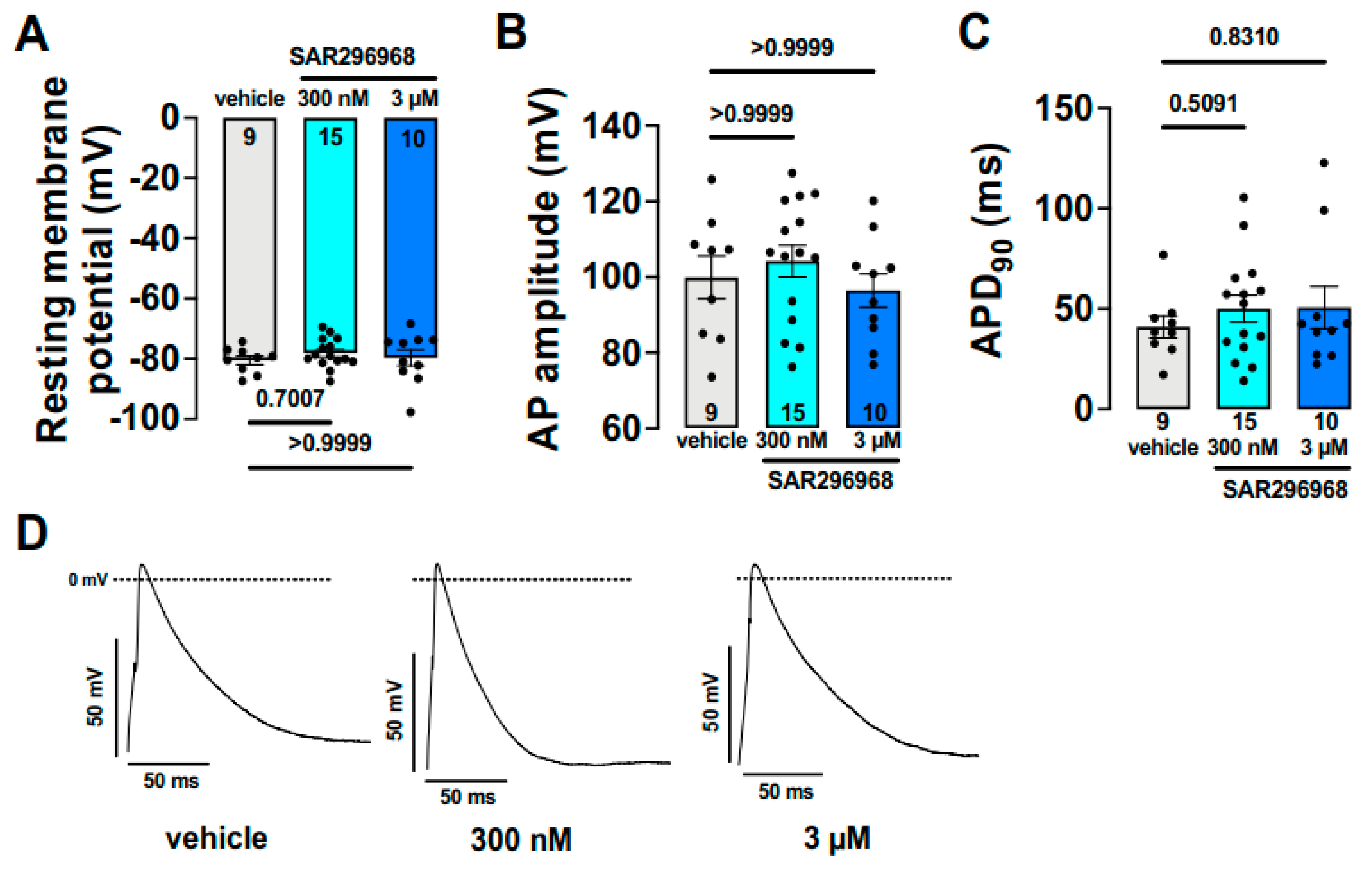
  90. Hegner, P.; Drzymalski, M.; Biedermann, A.; Memmel, B.; Durczok, M.; Wester, M.; Floerchinger, B.; Provaznik, Z.; Schmid, C.; Zausig, Y.; et al. SAR296968, a Novel Selective Na+/Ca2+ Exchanger Inhibitor, Improves Ca2+ Handling and Contractile Function in Human Atrial Cardiomyocytes. Biomedicines 2022, 10, 1932. [Google Scholar] [CrossRef] [PubMed]
  91. Wilson, L.D.; Rosenbaum, D.S. Mechanisms of arrythmogenic cardiac alternans. EP Eur. 2007, 9 Suppl. S6, vi77–vi82. [Google Scholar] [CrossRef] [PubMed]
  92. Narayan, S.M.; Franz, M.R.; Clopton, P.; Pruvot, E.J.; Krummen, D.E. Repolarization alternans reveals vulnerability to human atrial fibrillation. Circulation 2011, 123, 2922–2930. [Google Scholar] [CrossRef] [PubMed]
  93. Nolasco, J.B.; Dahlen, R.W. A graphic method for the study of alternation in cardiac action potentials. J. Appl. Physiol. 1968, 25, 191–196. [Google Scholar] [CrossRef] [PubMed]
  94. Díaz, M.E.; O’Neill, S.C.; Eisner, D.A. Sarcoplasmic Reticulum Calcium Content Fluctuation Is the Key to Cardiac Alternans. Circ. Res. 2004, 94, 650–656. [Google Scholar] [CrossRef] [PubMed] [Green Version]
  95. Fox, J.J.; McHarg, J.L.; Gilmour, R.F., Jr. Ionic mechanism of electrical alternans. Am. J. Physiol. Heart Circ. Physiol. 2002, 282, H516–H530. [Google Scholar] [CrossRef] [PubMed]
  96. Edwards, J.N.; Blatter, L.A. Cardiac alternans and intracellular calcium cycling. Clin. Exp. Pharmacol. Physiol. 2014, 41, 524–532. [Google Scholar] [CrossRef] [Green Version]
  97. Wan, X.; Cutler, M.; Song, Z.; Karma, A.; Matsuda, T.; Baba, A.; Rosenbaum, D.S. New experimental evidence for mechanism of arrhythmogenic membrane potential alternans based on balance of electrogenic INCX/ICa currents. Heart Rhythm 2012, 9, 1698–1705. [Google Scholar] [CrossRef] [Green Version]
  98. Hobai, I.A.; Maack, C.; O’Rourke, B. Partial Inhibition of Sodium/Calcium Exchange Restores Cellular Calcium Handling in Canine Heart Failure. Circ. Res. 2004, 95, 292–299. [Google Scholar] [CrossRef]
  99. Acsai, K.; Kun, A.; Farkas, A.S.; Fulop, F.; Nagy, N.; Balazs, M.; Szentandrassy, N.; Nanasi, P.P.; Papp, J.G.; Varro, A.; et al. Effect of partial blockade of the Na+/Ca2+-exchanger on Ca2+ handling in isolated rat ventricular myocytes. Eur. J. Pharmacol. 2007, 576, 1–6. [Google Scholar] [CrossRef] [PubMed]
  100. Grandi, E.; Morotti, S.; Ginsburg, K.S.; Severi, S.; Bers, D.M. Interplay of voltage and Ca-dependent inactivation of L-type Ca current. Prog. Biophys. Mol. Biol. 2010, 103, 44–50. [Google Scholar] [CrossRef] [PubMed] [Green Version]
  101. Farkas, A.S.; Acsai, K.; Nagy, N.; Toth, A.; Fulop, F.; Seprenyi, G.; Birinyi, P.; Nanasi, P.P.; Forster, T.; Csanady, M.; et al. Na+/Ca2+ exchanger inhibition exerts a positive inotropic effect in the rat heart, but fails to influence the contractility of the rabbit heart. Br. J. Pharmacol. 2009, 154, 93–104. [Google Scholar] [CrossRef] [PubMed] [Green Version]
  102. Szentandrassy, N.; Birinyi, P.; Szigeti, G.; Farkas, A.; Magyar, J.; Toth, A.; Csernoch, L.; Varro, A.; Nanasi, P.P. SEA0400 fails to alter the magnitude of intracellular Ca2+ transients and contractions in Langendorff-perfused guinea pig heart. Naunyn-Schmiedeberg’s Arch. Pharmacol. 2008, 378, 65–71. [Google Scholar] [CrossRef] [PubMed]
  103. Ozdemir, S.; Bito, V.; Holemans, P.; Vinet, L.; Mercadier, J.-J.; Varro, A.; Sipido, K.R. Pharmacological Inhibition of Na/Ca Exchange Results in Increased Cellular Ca2+ Load Attributable to the Predominance of Forward Mode Block. Circ. Res. 2008, 102, 1398–1405. [Google Scholar] [CrossRef] [PubMed] [Green Version]
  104. Primessnig, U.; Bracic, T.; Levijoki, J.; Otsomaa, L.; Pollesello, P.; Falcke, M.; Pieske, B.; Heinzel, F.R. Long-term effects of Na+/Ca2+ exchanger inhibition with ORM-11035 improves cardiac function and remodelling without lowering blood pressure in a model of heart failure with preserved ejection fraction. Eur. J. Heart Fail. 2019, 21, 1543–1552. [Google Scholar] [CrossRef] [Green Version]
  105. Birinyi, P.; Toth, A.; Jona, I.; Acsai, K.; Almassy, J.; Nagy, N.; Prorok, J.; Gherasim, I.; Papp, Z.; Hertelendi, Z.; et al. The Na+/Ca2+ exchange blocker SEA0400 fails to enhance cytosolic Ca2+ transient and contractility in canine ventricular cardiomyocytes. Cardiovasc. Res. 2008, 78, 476–484. [Google Scholar] [CrossRef] [Green Version]
  106. Jin, G.; Manninger, M.; Adelsmayr, G.; Schwarzl, M.; Alogna, A.; Schönleitner, P.; Zweiker, D.; Blaschke, F.; Sherif, M.; Radulovic, S.; et al. Cellular contribution to left and right atrial dysfunction in chronic arterial hypertension in pigs. ESC Heart Fail. 2021, 8, 151–161. [Google Scholar] [CrossRef]
  107. Otsomaa, L.; Levijoki, J.; Wohlfahrt, G.; Chapman, H.; Koivisto, A.P.; Syrjanen, K.; Koskelainen, T.; Peltokorpi, S.E.; Finckenberg, P.; Heikkila, A.; et al. Discovery and characterization of ORM-11372, a novel inhibitor of the sodium-calcium exchanger with positive inotropic activity. Br. J. Pharmacol. 2020, 177, 5534–5554. [Google Scholar] [CrossRef]
  108. Pelat, M.; Barbe, F.; Daveu, C.; Ly-Nguyen, L.; Lartigue, T.; Marque, S.; Tavares, G.; Ballet, V.; Guillon, J.-M.; Steinmeyer, K.; et al. SAR340835, a Novel Selective Na+/Ca2+ Exchanger Inhibitor, Improves Cardiac Function and Restores Sympathovagal Balance in Heart Failure. J. Pharmacol. Exp. Ther. 2021, 377, 293–304. [Google Scholar] [CrossRef]
Figure 1. Schematic diagram of intracellular Ca2+ handling in ventricular myocytes.
Ijms 23 14651 g001
Figure 2. Schematic illustration of the presumed NCX function during ventricular action potential. The reverse mode of the NCX (Ca2+ influx/outward current) is restricted to the beginning of the action potential. The forward mode (Ca2+ efflux/inward current) is suggested to be active during the latter part of the plateau.
Ijms 23 14651 g002
Figure 3. Estimation of NCX current during an action potential. (A): Noble et al. (reprinted with permission from Ref. [29]. 2022, Elsevier Science & Technology Journals), the lower panel indicates mainly outward NCX during an action potential. (B): Faber and Rudy action potential model demonstrates that NCX current depends on [Na+]i (reprinted with permission from Ref. [30]. 2022, Elsevier). (C): Horvath et al. defined NCX current during an action potential by using the “onion-peeling” technique in dog ventricular myocytes. (reprinted with permission from Ref. [35]. 2022, Elsevier).
Ijms 23 14651 g003
Figure 4. Effect of ORM-10103 against triggered activity. (A) ORM-10103 reduces the amplitude of EAD. (ae) indicates the subsequent sections of the experiment. Bar graphs show the effect of ORM on EAD amplitude in 3 and 10 µM (f). (B) ORM-10103 decreases the strophantin-induced membrane potential oscillations. (ac) indicates the subsequent sections of the experiment, and bar graphs show the numerical data of the result (d). (C) ORM-10103 reduces the incidence of DADs. (ac) indicates the subsequent sections of the experiment. * denotes p < 0.05. Reprinted with permission from Ref. [37]. 2022, Wiley.
Ijms 23 14651 g004
Figure 5. Selective NCX inhibition by 10 µM ORM-10103 avoids diastolic Ca2+ increase during simulated cellular ischemia in canine isolated ventricular myocytes. # and * indicate significant (p < 0.05) difference between time control vs. untreated and untreated vs. ORM-treated values, respectively. Reprinted with permission from Ref. [81]. 2022, Elsevier.
Ijms 23 14651 g005
Figure 6. Selective NCX inhibition does not influence the ventricle–Purkinje action potential dispersion. (A): after control recordings (open symbol) 0.5 µM veratridine (filled symbol) was applied. (B): Prior to verartidine application 1 µM SEA-0400 was employed. Bar graphs illustrate the APD dispersion (C) and the veratridine induced increase of APD dispersion under control condition and in the presence of SEA-0400 (D). Reprinted with permission from Ref. [34]. 2022, Wiley.
Ijms 23 14651 g006
Figure 7. Effect of SAR296968 on human atrial action potentials. Bar graphs (AC) and original recordings (D) indicate no effect of NCX inhibition on action potential duration.
Ijms 23 14651 g007
Figure 8. Effect of SEA-0400 on ventricular arrhythmias measured by optical mapping. (A): Mapped region; (B): ECG measurement. Red arrows indicate ventricular premature beats; Numbers indicate the number of beats (C): action potentials corresponding from the red bar in panel. Red arrows indicate ventricular premature beats; (D): isochronal map showing beat marked in (B) (beat 1) and in panel (C) (beat 2–4); (E): APD80 map with nodal line; (F): action potential tracing: a and b—no arrhythmia was observed in the presence of 0.03 and 3 µM SEA-0400 at 260 ms pacing cycle length c—at 90 ms pacing cycle length fibrillation was observed in the presence of 3 µM SEA-0400; (G): isochronal map corresponding to (Fc). Numbers indicate the time of data acquisition. Frames 708 and 732 illustrate formation of re-entrant wavefront, 828 and 880 depict multiple wavefronts (Reprinted with permission from Ref. [76]. 2022, Wiley).
Ijms 23 14651 g008
Figure 9. SEA-0400 failed to influence the Ca2+ transient (A) and cell shortening (B) amplitudes in canine isolated myocytes. Reprinted with permission from Ref. [105]. 2022, Oxford University Press).
Ijms 23 14651 g009
Publisher’s Note: MDPI stays neutral with regard to jurisdictional claims in published maps and institutional affiliations.

