

Review

Bioluminescent Dinoflagellates as a Bioassay for Toxicity Assessment

by
Luíza S. Perin
1,
Gabriela V. Moraes
1,
Gabriela A. Galeazzo
1 and
Anderson G. Oliveira
2,*
1
Department of Physical, Chemical and Geological Oceanography, Oceanographic Institute, University of São Paulo, São Paulo 05508-120, Brazil
2
Department of Chemistry and Biochemistry, Yeshiva University, 245 Lexington Avenue, New York, NY 10016, USA
*
Author to whom correspondence should be addressed.
Int. J. Mol. Sci. 2022, 23(21), 13012; https://doi.org/10.3390/ijms232113012
Submission received: 21 July 2022 / Revised: 19 September 2022 / Accepted: 24 September 2022 / Published: 27 October 2022

Abstract

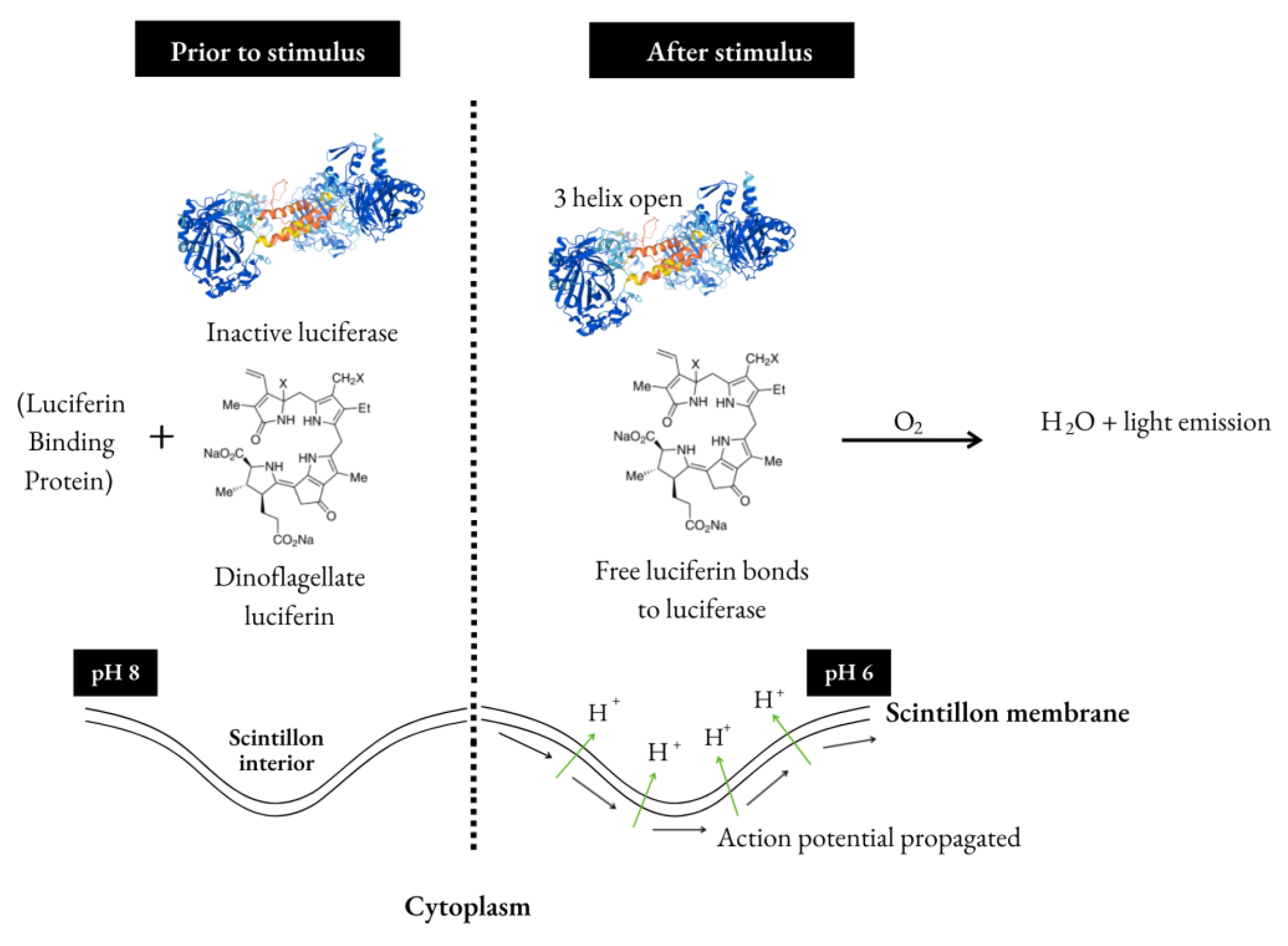
Dinoflagellates bioluminescence mechanism depends upon a luciferin–luciferase reaction that promotes blue light emission (480 nm) in specialized luminogenic organelles called scintillons. The scintillons contain luciferin, luciferase and, in some cases, a luciferin-binding protein (LBP), which prevents luciferin from non-enzymatic oxidation in vivo. Even though dinoflagellate bioluminescence has been studied since the 1950s, there is still a lack of mechanistic understanding on whether the light emission process involves a peroxidic intermediate or not. Still, bioassays employing luminous dinoflagellates, usually from Gonyaulax or Pyrocystis genus, can be used to assess the toxicity of metals or organic compounds. In these dinoflagellates, the response to toxicity is observed as a change in luminescence, which is linked to cellular respiration. As a result, these changes can be used to calculate a percentage of light inhibition that correlates directly with toxicity. This current approach, which lies in between fast bacterial assays and more complex toxicity tests involving vertebrates and invertebrates, can provide a valuable tool for detecting certain pollutants, e.g., metals, in marine sediment and seawater. Thus, the present review focuses on how the dinoflagellates bioluminescence can be applied to evaluate the risks caused by contaminants in the marine environment.
Keywords: circadian cycle; scintillons; biotechnological applications; ecotoxicological assays circadian cycle; scintillons; biotechnological applications; ecotoxicological assays


