Abstract
The roles of thioredoxin-interacting protein (TXNIP) and receptor for advanced glycation end-products (RAGE)-dependent mechanisms of NOD-like receptor family, pyrin domain containing 3 (NLRP3) inflammasome-driven macrophage activation during acute lung injury are underinvestigated. Cultured THP-1 macrophages were treated with a RAGE agonist (S100A12), with or without a RAGE antagonist; cytokine release and intracytoplasmic production of reactive oxygen species (ROS) were assessed in response to small interfering RNA knockdowns of TXNIP and NLRP3. Lung expressions of TXNIP and NLRP3 and alveolar levels of IL-1β and S100A12 were measured in mice after acid-induced lung injury, with or without administration of RAGE inhibitors. Alveolar macrophages from patients with acute respiratory distress syndrome and from mechanically ventilated controls were analyzed using fluorescence-activated cell sorting. In vitro, RAGE promoted cytokine release and ROS production in macrophages and upregulated NLRP3 and TXNIP mRNA expression in response to S100A12. TXNIP inhibition downregulated NLRP3 gene expression and RAGE-mediated release of IL-1β by macrophages in vitro. In vivo, RAGE, NLRP3 and TXNIP lung expressions were upregulated during experimental acute lung injury, a phenomenon being reversed by RAGE inhibition. The numbers of cells expressing RAGE, NLRP3 and TXNIP among a specific subpopulation of CD16+CD14+CD206- (“pro-inflammatory”) alveolar macrophages were higher in patients with lung injury. This study provides a novel proof-of-concept of complex RAGE–TXNIP–NLRP3 interactions during macrophage activation in acute lung injury.
1. Introduction
Acute respiratory distress syndrome (ARDS) is a heterogeneous syndrome of diffuse alveolar injury leading to increased permeability, pulmonary edema, alveolar filling and rapid onset of hypoxemic respiratory failure [1,2]. Despite advances in our understanding of the pathogenesis of ARDS, no drug has reduced mortality in ARDS, and the syndrome is still under-recognized, undertreated and associated with high mortality [3,4]. There is growing evidence supporting a pivotal role for the receptor for advanced glycation end-products (RAGE) in the pathogenesis of ARDS [5,6,7,8]. RAGE is a transmembrane protein of the immunoglobulin superfamily that amplifies and perpetuates the inflammatory response [9,10,11]. RAGE is abundantly expressed in lung alveolar type (AT) 1 epithelial cells, but also in various other cell types, e.g., monocytes/macrophages, AT2 and endothelial cells. RAGE is constitutively expressed abundantly in the lung under basal conditions and its expression is enhanced during inflammatory states such as ARDS [12,13]. Its main soluble form, sRAGE, is secreted into the alveolar space and is detectable in the serum, serving as a biomarker for the degree of lung injury [7,12,13,14,15,16,17,18] and acting as a decoy receptor to downregulate the injurious pulmonary inflammatory response [19]. RAGE interacts with multiple ligands, including advanced glycation end-products (AGEs), high-mobility group box 1 protein (HMGB1), calgranulins/S100 proteins, amyloid peptides and macrophage adhesion ligand-1 (MAC-1) [20,21,22]. RAGE controls a variety of cellular processes such as cell proliferation and migration, inflammation, autophagy or apoptosis. Recent studies also point out a critical role for RAGE in modulating inflammation in macrophages [23], as cAMP elevation has been demonstrated to be capable of suppressing ligand-induced inflammatory reactions, including secretion of monocyte chemotactic protein 1 and macrophage recruitment, by promoting the conversion of RAGE isoforms from membrane RAGE to sRAGE [24].
The NOD-like receptor family, pyrin domain containing 3 (NLRP3) inflammasome is a multi-protein complex of the innate immune system expressed in myeloid cells, consisting of the NOD-like receptor NLRP3, the adaptor protein ASC (apoptosis-associated speck-like protein containing a CARD) and caspase 1 [25]. Whereas activation of the NLRP3 inflammasome can help host defense against invading bacteria and pathogens, excessive activation of the inflammasome can lead to inflammation-associated lung tissue injury [26]. Emerging studies suggest that NLRP3 inflammasome-driven activation in alveolar macrophages plays a critical role during ARDS [26,27,28]. NLRP3 inflammasome formation and activation require a canonical two-step mechanism involving the sequential ligand-dependent activation of toll-like receptors (e.g., TLR4) or related receptors that, in turn, stimulate NF-κB-dependent transcription of NLRP3 and pro-IL-1β, leading to NLRP3 and pro-IL-1β protein synthesis (priming) [29]. This is followed by a second signal generated by ion flux, phagosomal destabilization, mitochondrial reactive oxygen species (ROS) or mitochondrial damage associated molecular patterns (DAMPs), that results in NLRP3 inflammasome assembly and activation [30,31]. Once activated, caspase 1 proteolytically cleaves the cytokine precursors pro-IL-1β and pro-IL-18, resulting in the maturation and release of IL-1β and IL-18 [29]. As a pattern-recognition receptor, RAGE shares similarities with TLR4, including downstream activation of NF-κB-dependent [32] and ROS generation [33], with subsequent upregulation of RAGE expression itself [34]. In addition, in vitro studies suggest a role of RAGE in upregulated synthesis of pro-IL-1β and pro-IL-18 in THP-1 macrophages [35].
Recent findings demonstrate a potential role for thioredoxin-interacting protein (TXNIP) in innate immunity as a link between oxidative stress and NLRP3 inflammasome activation [36,37]. TXNIP was initially identified as one of the proteins that interacts with thioredoxin (TRX) and reduces its function of ROS scavenger, thus promoting oxidative stress [38]. TXNIP mediates inflammasome activation and injury in podocytes during diabetic nephropathy [39]. Furthermore, HMGB1 stimulates ROS production through TLR4 and a synergistic collaboration with RAGE signaling, in a mouse model of hemorrhagic shock [40]. In turn, ROS promote the association of TXNIP with NLRP3 and subsequent inflammasome activation in lung endothelial cells [40]. However, the role of TXNIP and RAGE-dependent mechanisms of TXNIP–NLRP3-driven macrophage activation are underinvestigated in the setting of acute lung injury [41,42,43].
In this study, we first tested the hypothesis that RAGE activation would induce NLRP3 activation and ROS production in a TXNIP-dependent manner. To test this hypothesis, we investigated the effects of S100A12–RAGE interaction on the production of IL-1β and ROS in cultured THP-1 macrophages. Next, we examined in vitro the role of TXNIP on IL-1β secretion and ROS production using small interfering (si)RNA knockdown. We further tested in vivo the hypothesis of RAGE–TXNIP–NLRP3-dependent mechanisms of lung injury using a mouse model of acid-induced lung injury and fluorescence-activated cell sorting (FACS) analysis of alveolar macrophages from patients with ARDS.
2. Results
2.1. S100A12 Stimulates ROS Production and IL-1β Secretion in a RAGE-Dependent Manner in THP-1 Cells
After stimulation by S100A12, cultured THP-1 macrophages released higher levels of cytokines IL-1β in the medium, compared to controls. S100A12-induced release of cytokines was inhibited, at least partially, by FPS-ZM1 (Figure 1). The production of intracellular ROS was increased by S100A12, a phenomenon inhibited by RAGE antagonist FPS-ZM1 (Figure 2).
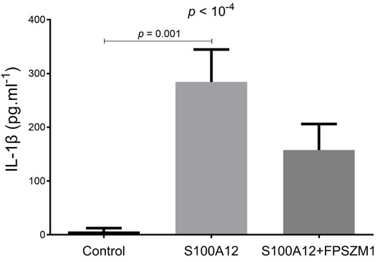
Figure 1.
Levels of IL-1β in the culture medium of THP-1 cells treated for 24 h in the absence/presence of RAGE agonist S100A12 (40 nM, MyBiosource) alone or in combination with RAGE antagonist FPS-ZM1 (1 µM, EMD Millipore). Supernatants were collected and stored at −20 °C before duplicate measurement of IL-1β (R&D Systems) using magnetic Luminex assay kits.
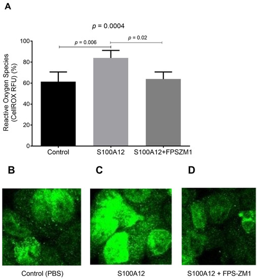
Figure 2.
(A) Quantitative analysis of ROS measurements using CellROX Deep Red Reagent kit (Molecular Probes) in THP-1 cells treated for 24 h in the absence/presence of RAGE agonist S100A12 (40 nM, MyBiosource) alone or in combination with RAGE antagonist FPS-ZM1 (1 µM, EMD Millipore). Data are expressed as relative fluorescent units (RFU, in %). Representative images of CellROX staining in THP-1 cells treated with (B) PBS (control), (C) S100A12 alone or (D) S100A12 in combination with FPS-ZM1. Images of ROS staining were photographed under a microscope (Zeiss Axiophot). Image processing was conducted by Image J software 1.49.
2.2. Effects of siRNA-Targeted TXNIP and NLRP3 Knockdown on S100A12-Induced IL-1β Secretion and ROS Production by THP-1 Cells
siRNA-targeted knockdown of NLRP3 inhibited the S100A12-induced upregulation of IL-1β secretion by macrophages (Figure 3A). In contrast, siRNA-targeted TXNIP knockdown was associated with increased S100A12-induced IL-1β secretion into the medium, compared to TXNIP siRNA-transfected cells that were not exposed to S100A12 (Figure 3A).
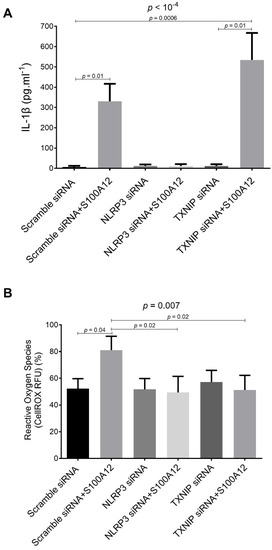
Figure 3.
(A) Levels of IL-1β in the culture medium of THP-1 cells treated for 24 h in the absence/presence of RAGE agonist S100A12 (40 nM, MyBiosource) alone or in combination with RAGE antagonist FPS-ZM1 (1 µM, EMD Millipore). Supernatants were collected and stored at −20 °C before duplicate measurement using magnetic Luminex assay kits (R&D Systems). (B) Quantitative analysis of intracellular ROS production using CellROX Deep Red Reagent kit (Molecular Probes) in THP-1 macrophages transfected with specific siRNA oligonucleotides for mouse TXNIP (siRNA TXNIP, Dharmacon), NLRP3 (siRNA NLRP3, Dharmacon) or with scrambled oligonucleotide (0.1 μM Scramble siRNA; Dharmacon). Efficiency of siRNA knockdown was verified with duplicate ELISA of TXNIP (CycLex Co) and NLRP3 (Antibodies-online GmbH). Cells were treated for 24 h in the absence/presence of RAGE agonist S100A12 (40 nM, MyBiosource) alone or in combination with RAGE antagonist FPS-ZM1 (1 µM, EMD Millipore). ROS production is expressed in relative fluorescent units (RFU, in %).
NLRP3 and TXNIP knockdown THP-1 cells did not increase ROS production after exposure to S100A12, as compared to scramble siRNA-transfected S100A12-treated cells (Figure 3B).
2.3. RAGE-Dependent Regulation of Gene Expression in S100A12-Activated THP-1 Cells
RAGE mRNA expression was upregulated by S100A12, and this upregulation was inhibited by FPS-ZM1. This phenomenon was notably absent after TXNIP knockdown (Figure S1, online supplement).
mRNA expression levels of NLRP3 and TXNIP were both upregulated by S100A12 in THP-1 cells, in a RAGE-dependent manner (Figure S2, online supplement).
2.4. Anti-RAGE mAb and Recombinant sRAGE Prevent HCl Injury-Induced Upregulation of Lung NLRP3 and TXNIP In Vivo
In lungs from mice with acid-induced lung injury, NLRP3 and TXNIP protein expression was significantly upregulated on days 1–2, compared to sham animals, whereas lung mRNA levels of NLRP3 and TXNIP rose earlier after injury (from day 0) (Figure 4). Upregulated lung expression of NLRP3 and TXNIP after lung injury was alleviated by treatment with anti-RAGE mAB or sRAGE (Figure 4).
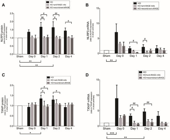
Figure 4.
Lung levels of NLRP3 (A) protein and (B) mRNA, TXNIP (C) protein and (D) mRNA in acid-injured mice (HCl group), acid-injured mice treated with anti-RAGE monoclonal antibody (HCl+anti-RAGE mAb group) or with recombinant sRAGE (HCl+sRAGE group) and in uninjured, untreated mice (Sham group) (n = 4–6 for each time point). Threshold levels of mRNA expression (∆∆Ct) were normalized to housekeeping genes. Protein and mRNA expression levels are expressed as ratios to those in sham animals. Values are reported as means ± SD and are analyzed with Kruskal–Wallis test (nonparametric data). * p < 0.05, ** p < 10−2, *** p < 10−3 versus sham animals. # p < 0.05, ## p < 10−2 versus acid-injured mice.
2.5. Effects of Acid-Induced Injury and RAGE Modulation on Alveolar Levels of IL-1β
Acid-induced injury significantly upregulated bronchoalveolar lavage (BAL) levels of IL-1β from day 0 to day 2, compared to sham animals, whereas treatment with sRAGE or anti-RAGE mAb significantly prevented the rise in BAL IL-1β on days 1–2 (Figure 5).
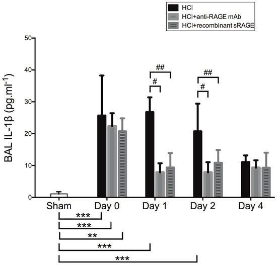
Figure 5.
Measurement of bronchoalveolar lavage (BAL) levels of interleukin (IL)-1β in acid-injured mice (HCl group), acid-injured mice treated with anti-RAGE monoclonal antibody (HCl+anti-RAGE mAb group) or with recombinant sRAGE (HCl+sRAGE group) and in uninjured, untreated mice (Sham group) (n = 4–6 for each time point). Values are reported as means ± SD and are analyzed with Kruskal–Wallis test (nonparametric data). ** p < 10−2, *** p < 10−3 versus sham animals. # p < 0.05, ## p < 10−2 versus acid-injured mice.
2.6. RAGE, TXNIP and NLRP3 Expression by Human Alveolar Macrophages during ARDS
The baseline characteristics and clinical outcomes of patients with or without ARDS are reported in the Table 1. Patients with ARDS had higher BAL levels of IL-1β and S100A12 than mechanically ventilated patients without ARDS (Figure 6).

Table 1.
Baseline Characteristics and Clinical Outcomes of Patients without or with acute respiratory distress syndrome (ARDS).
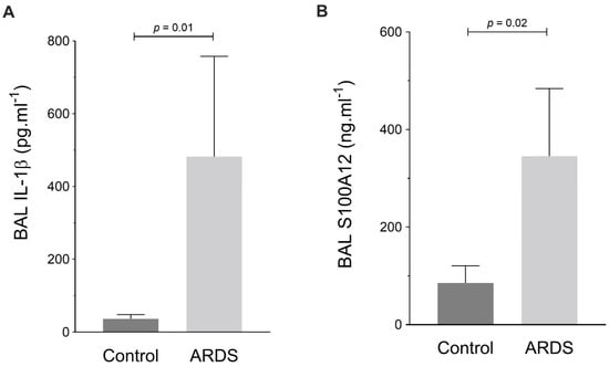
Figure 6.
Measurement of bronchoalveolar lavage (BAL) levels of (A) interleukin (IL)-1β (in pg·mL−1) and (B) S100A12 (in ng·mL−1) in patients with acute respiratory distress syndrome (ARDS) and in mechanically ventilated patients without ARDS (Control). Values are reported as medians ± interquartile ranges and are analyzed with the Mann–Whitney test (nonparametric data).
In FACS analysis, groups did not significantly differ with regards to: the proportions of CD16+CD14- or CD16+CD14+ cells within the CD45+ population; the percentages of CD163+ and CD206+ cells within the CD16+CD14- subpopulation; the percentages of CD163+ cells within the CD16+CD14+ subpopulation; or the percentages of RAGE+, NLRP3+ and TXNIP+ cells within these latter subpopulations (Figures S4–S6, online supplement). However, the percentage of CD206- cells within the CD16+CD14+ subpopulation was higher in patients with ARDS, compared to those without ARDS (Figure 7). In the CD16+CD14+CD206+ subpopulation, cells were more frequently RAGE+ in patients with ARDS than in those without (Figure S7, online supplement). In the CD16+CD14+CD206- subpopulation, there were more RAGE+ and NLRP3+ cells, but fewer TXNIP+ cells, in patients with ARDS than in those without ARDS (Figure 7).
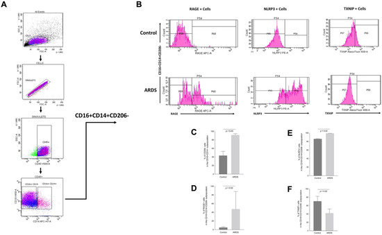
Figure 7.
(A) Flow cytometry gating strategy used to identify pro-inflammatory (“M1-like”) CD16+CD14+CD206- alveolar macrophages. (B) Representative FACS images for the cell-surface expression of RAGE, NLRP3 and TXNIP. C-F) Quantification of FACS images: percentages of CD206- cells within the CD16+CD14+ subpopulation (C), RAGE+ cells within the CD16+CD14+CD206- subpopulation (D), NLRP3+ cells within the CD16+CD14+CD206- subpopulation (E) and of TXNIP+ cells within the CD16+CD14+CD206- subpopulation (F) of alveolar macrophages from patients with acute respiratory distress syndrome (ARDS) and in mechanically ventilated patients without ARDS (Control). Values are reported as medians ± interquartile ranges and are analyzed with the Mann–Whitney test (nonparametric data).
3. Discussion
In this study, we found that: (1) S100A12–RAGE interaction mediates cytokine release and ROS production in THP-1 macrophages, through an upregulation of mRNA expression levels of NLRP3 and TXNIP; (2) TXNIP has a role in RAGE-mediated NLRP3 activation in vitro; (3) RAGE, NLRP3 and TXNIP expressions were increased after acute lung injury in a mouse model of ARDS, which was reversed by RAGE inhibition; (4) within pro-inflammatory (“M1-like”) CD16+CD14+CD206- alveolar macrophages, cells were more frequently RAGE+ and NLRP3+ cells, and less frequently TXNIP+, in patients with ARDS, compared to those without ARDS.
These results are in line with recent evidence of a prominent role of the NLRP3 inflammasome in ARDS [26,27,44], with increased BAL IL-1β and upregulated lung expression of NLRP3 in vivo after acid-induced lung injury. NLRP3-deficient (Nlrp3−/−) mice displayed increased mortality under hyperoxic conditions relative to wild-type and IL-1β−/− mice. Under hyperoxia, Nlrp3−/− mice displayed reduced lung inflammatory responses, including inflammatory cell infiltration and cytokine expression, but without differences in lung IL-1β production compared to wild-type mice [45]. Using a murine model of ventilator-induced lung injury (VILI), Dolinay et al. demonstrated that VILI upregulated several inflammasome-related genes including IL-1α, caspase-activator domain-10, IL-1 receptor and PYCARD (encoding for ASC protein, the inflammasome adaptor); in this model, genetic deletion of IL-18 or caspase 1 reduced lung injury, inflammation, alveolar-capillary barrier dysfunction and edema in response to injurious ventilation [46]. In human studies using plasma specimens from critically ill patients, the expression of inflammasome-related caspase 1, IL-18 and IL-1β mRNA transcripts was significantly higher in patients with sepsis or ARDS, compared with patients with systemic inflammatory response syndrome alone [46]. Importantly, circulating IL-18 and mitochondrial DNA, a DAMP associated with NLRP3 inflammasome activation, were significantly elevated in plasma from human patients critically ill with diseases such as sepsis and sepsis-induced ARDS, and significantly associated with ICU mortality [46,47]. Although previous studies underscored a role for inflammasome system and its regulated cytokines in the propagation of experimental ARDS, we report here for the first time that lung-injury-induced NLRP3 activation may be, at least in part, mediated by RAGE. Indeed, anti-RAGE therapy decreased BAL levels of IL-1β and prevented upregulation of mRNA and protein lung expression of NLRP3 in injured mice. Such findings are further supported by RAGE-dependent mechanisms of NLRP3 activation in cultured macrophages, as S100A12–RAGE interaction stimulates ROS production and IL-1β secretion in vitro, a phenomenon that was abrogated by NLRP3-targeted siRNA knockdown.
Our findings also provide novel insights into a role for TXNIP on RAGE-mediated NLRP3 activation in macrophages. Using specific siRNA knockdown in THP-1 cells, we could demonstrate that both TXNIP and NLRP3, which are upregulated in vivo during lung injury, seem mandatory for RAGE-induced upregulation of ROS production. The release of IL-1β by THP-1 macrophages necessitated NLRP3, but Il-1β levels were upregulated in the culture medium of TXNIP siRNA-transfected cells exposed to S100A12 in our study [48]. Such findings are surprising, because TXNIP has been previously described as a NLRP3 activator in the presence of ROS, with subsequent cytokine secretion [36,37,49,50]. Such a mechanism has been reported in podocytes and in Schwann cells, with downregulated IL-1β release after TXNIP knockdown [51,52]. Discrepancies with our findings may be explained by distinct responses in various cell types, various effects of distinct RAGE ligands (through RAGE itself, but also through other membrane receptors, including TLR4) or adaptive mechanisms that may have developed in macrophages from TXNIP knockout mice in previous studies. Nevertheless, because NLRP3 gene expression was increased in the absence of TXNIP in vitro, and because it was further increased by S100A12, we hypothesize that TXNIP may downregulate NLRP3 gene expression (even under basal culture conditions) and S100A12-RAGE-mediated release of IL-1β in macrophages. Interestingly, upregulated lung mRNA expression of TXNIP was, in turn, downregulated by treatment with anti-RAGE mAb or recombinant sRAGE in lung-injured mice, thus further supporting crosstalk or feedback mechanisms between TXNIP and RAGE pathways.
Our FACS analysis found significant differences in the number of cells expressing RAGE, NLRP3 and TXNIP among classically activated (M1)—or “proinflammatory”—human alveolar macrophages, but not among other tested subpopulations. Indeed, more CD16+CD14+CD206- cells expressed RAGE and NLRP3, and fewer CD16+CD14+CD206- cells expressed TXNIP, in patients with ARDS than in those without ARDS.
According to our findings from in vitro and animal studies, this could suggest that, at least in some M1-like macrophages, the activation of RAGE and NLRP3 pathways might be associated with a downregulation of TXNIP activity during ARDS. Whether interactions between RAGE, TXNIP and NLRP3 contribute to the presence and/or evolution of distinct alveolar macrophage subsets that regulate the inflammatory response and its resolution remains unknown [53]. Understanding the precise regulatory mechanisms for such a complex network deserves further investigation.
Our study has some limitations. First, the exploratory design of our study does not provide precise regulatory mechanisms linking RAGE, TXNIP and NLRP3 pathways. However, we report hypothesis-generating findings, and the first demonstration of a RAGE-mediated pathway of NLRP3 inflammasome activation in cultured macrophages and in an in vivo model of acute lung injury. In addition, we also explored for the first time the expressions of RAGE, NLRP3 and TXNIP in human alveolar macrophages, along with the in vivo effects of RAGE inhibition on TXNIP–NLRP3 activation during acute lung injury. Such novel findings have the potential to stimulate future research on precise intracellular pathways that interact upon RAGE, NLRP3 and TXNIP activation, along with their downstream targets, using additional siRNA targets, knockout animals and/or specific pharmacological antagonists of downstream effectors. Second, we only described the role of RAGE activation by its ligand S100A12 in THP-1 cells. As RAGE downstream pathways vary among cell types and depend on RAGE ligands, our results cannot be generalizable to other settings [9,33]. Third, we did not use primary cultures of macrophages in our study, and our findings may have been different in cells that may better reflect in vivo mechanisms. Fourth, the role of intercellular crosstalks within the lung (e.g., between macrophages and epithelial/endothelial cells), mitochondrial dysfunction and migratory properties of macrophages may significantly influence highly regulated cascades such as RAGE, TXNIP and NLRP3 pathways. Such explorations were out of the scope of our study but will be of great importance to better understand RAGE–NLRP3-driven alveolar inflammation and its role in ARDS. Finally, the choice of our ARDS animal model, based on direct lung injury by HCl, hampers extrapolability of our results to other causes of experimental ARDS.
4. Materials and Methods
4.1. Cell Experiments
Human THP-1 cells were obtained from the American Type Culture Collection and cultured in suspension in RPMI-Glutamax (Gibco) supplemented with streptomycin (100 μg·mL−1), penicillin G (100 UI·mL−1), amphotericin B (2.5 μg·mL−1) and 10% decomplemented fetal bovine serum (HyClone Standard, GE). THP-1 were differentiated into macrophages following treatment with 200 nM phorbol 12-myristate 13-acetate (Abcam) for 72 h [54]. Then, cells were treated for 24 h in the absence or presence of RAGE agonist S100A12 (40 nM, MyBiosource) [55,56], alone or in combination with RAGE antagonist FPS-ZM1 (1 µM, EMD Millipore) [57].
Supernatants were collected and stored at −20 °C before duplicate measurement of IL-1β (R&D Systems) using magnetic Luminex assay kits. Total RNA was extracted and gene expressions of RAGE (mRNA Refseq NM_001136), NLRP3 (NM_183395) and TXNIP (NM_006472) were assessed using semi-quantitative real-time polymerase chain reaction (human RT² Profiler™ PCR Array, Qiagen). For quantification of intracellular reactive oxygen species (ROS) production, the CellROX® Deep Red Reagent kit was applied (Molecular Probes®).
For siRNA knockdown experiments, THP-1 cells were grown in 6-well plates and transfected with specific siRNA oligonucleotides for mouse TXNIP, NLRP3 (Dharmacon) or or with scrambled oligonucleotide (0.1 μM; siGENOME non-targeting siRNA #1, Dharmacon) using Lipofectamine RNAiMax transfection reagent (Invitrogen). Cells were harvested 48 h after transfection.
4.2. Animal Studies
Mouse experiments were approved by the Animal Ethics Committee of the French Ministère de l’Education Nationale, de l’Enseignement Supérieur et de la Recherche (approval number CE 67–12; 4 December 2012) and performed in accordance with relevant guidelines and regulations for animal experimentation, including the 3R principles.
C57BL/6JRj mice were anesthetized prior to orotracheal instillation of hydrochloric acid [7,58]. After a recovery period under humidified oxygen, mice were transferred to stabulation. Injured and sham animals were evaluated at baseline and at specified time-points (1, 2, 4 days) after acid instillation in injured mice [7,58,59], using a modification of previous in situ models [7,58,60,61]. After 30-min ventilation (tidal volume = 8 mL·kg−1, positive end-expiratory pressure = 6 cmH2O, respiratory rate = 160 breaths·min−1 and FiO2 = 1), BAL was sampled as previously described [7] and levels of BAL IL-1 (R&D Systems) were measured in duplicate using mouse magnetic Luminex assay kits (eBioscience).
To examine the effect of RAGE inhibition on acid-induced lung injury, lung-injured mice were divided into three groups. Acid-injured mice (HCl group) received intratracheal instillation of HCl 0.1M pH 1 (75 μL/mouse). The mAb group was injected intravenously with anti-RAGE monoclonal antibody (15 mg·kg−1, R&D Systems) 30 minutes before the HCl instillation [62,63], and the sRAGE group was administered recombinant mouse sRAGE (100 μg/mouse, R&D Systems) intraperitoneally 1 h after the HCl instillation [63,64].
The animals were sacrificed after the ventilation period and subjected to lung sampling for assessment of protein and mRNA expression levels. Lung proteins were extracted from right lung tissue homogenates and further analyzed by enzyme-linked immunoassays using mouse NLRP3 and TXNIP (MyBiosource) kits. In parallel, total RNA was isolated from left lung and gene expressions of RAGE (mRNA Refseq NM_007425), NLRP3 (NM_145827) and TXNIP (NM_011324) were assessed using semi-quantitative real-time polymerase chain reaction (mouse RT² Profiler™ PCR Array, Qiagen).
4.3. Human Study
Protocols were approved by the Institutional Review Board of the University Hospital of Clermont-Ferrand, France (Comité de Protection des Personnes Sud Est VI, approval number AU1151). All participants, or their next-of-kin, provided written consent to participate. The study was registered on www.clinicaltrials.gov (ClinicalTrials.gov Identifier: NCT02545621, accessed on 30 September 2022).
Five adult patients admitted to Clermont-Ferrand university hospital intensive care unit (ICU) within the first 24 h after onset of moderate to severe ARDS according to the 2012 Berlin definition (ARDS group) [65] and five age- and sex-matched ICU patients without ARDS but under mechanical ventilation for less than 24 h (Control group) were included in this monocenter prospective observational study. Exclusion criteria included: pregnancy, acute exacerbation of diabetes (ketoacidosis, hyperosmolar hyperglycemic state), dialysis-dependent chronic renal failure, Alzheimer’s disease, amyloidosis, evolutive neoplastic lesion, chronic pulmonary disease requiring long-term oxygen therapy or mechanical ventilation, chemotherapy treatment in the last 30 days, severe neutropenia (<0.5 G/L).
At inclusion, fiberoptic-guided BAL (performed with 2 × 50 mL NaCl 0.9%) was analyzed using fluorescence-activated cell sorting (FACS). Alveolar macrophages were isolated and stained depending on their expression of membrane markers of M1-like (“pro-inflammatory”) or M2-like (“anti-inflammatory”) phenotypes [66], membrane RAGE and intracellular TXNIP and NLRP3. The following antibodies were used: rat anti-human NLRP3 monoclonal antibody (MAB7578, R&D Systems), goat anti-human C18-conjugated VDUP1/TXNIP polyclonal antibody (SC-33099, Santa Cruz), rabbit anti-human APC-conjugated anti-RAGER polyclonal antibody (LS-C212626, Lifespan Biosciences), APC-H7 mouse anti-human CD14, V450 mouse anti-human CD16, V500 mouse anti-human CD45, BV711 mouse anti-human CD163 and PE-CF594 mouse anti-human CD206 antibodies (BD Biosciences). Supernatants were analyzed for levels of S100A12 (CycLex Co) and IL-1β (R&D Systems) using duplicate ELISA.
4.4. Statistical Analysis
Categorical data were expressed as numbers and percentages, and quantitative data as mean ± standard deviation (SD) or median and interquartile range (IQR) according to statistical distribution. For studies of protein and mRNA expression, data are presented as ratio to expression in control cell conditions or in sham animals. Statistical analyses were performed using Kruskal–Wallis with Bonferroni tests for pairwise comparisons between each time-point and sham controls (represented as day 0), or between various cell culture conditions. A limited number of animals was used for baseline comparisons (n = 3–4), and 4–6 animals were used in each group on days 1, 2 and 4. Comparisons of patients characteristics between groups were performed using the chi-squared or Fisher’s exact tests for categorical variables, and Student t-test or Mann–Whitney test were used when assumption if t-test were not met (normality and homoscedasticity studied using Fisher–Snedecor test) for quantitative variables. Analyses were performed using Prism 6 (Graphpad Software, La Jolla, CA, USA) and Stata software (version 13, StataCorp, College Station, TX, USA). A p < 0.05 (two-sided) was considered significant.
5. Conclusions
In this study, S100A12–RAGE interaction mediated cytokine release and ROS production by macrophages in vitro. TXNIP had a crucial role in S100A12–RAGE-mediated NLRP3 activation and production of IL-1β and of intracellular ROS by macrophages (Figure 8). RAGE, NLRP3 and TXNIP expressions were increased in an animal model of acute lung injury, a phenomenon that was reversed by RAGE inhibition. Proinflammatory CD16+CD14+CD206- alveolar macrophages expressing RAGE and NLRP3 were more frequent, and those expressing TXNIP less frequent, in patients with ARDS than in those without ARDS, supporting a novel proof-of-concept of RAGE–TXNIP–NLRP3-driven mechanisms of macrophage activation during acute lung injury.
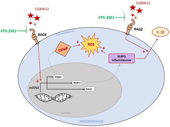
Figure 8.
RAGE ligand S100A12 stimulates IL-1β release, ROS production and increased TXNIP, NLRP3 and RAGE mRNA levels. These effects were inhibited by RAGE antagonist FPS-ZM1. We demonstrated that TXNIP has a crucial role in S100A12–RAGE-mediated NLRP3 activation and production of IL-1β and of intracellular ROS by macrophages. In addition, RAGE, NLRP3 and TXNIP were increased in an animal model of acute lung injury and in proinflammatory CD16+CD14+CD206- human alveolar macrophages.
Supplementary Materials
The following supporting information can be downloaded at: https://www.mdpi.com/article/10.3390/ijms231911659/s1.
Author Contributions
Conceptualization, M.J. and V.S.; Methodology, W.L.M.B., M.B., V.S. and M.J.; Formal Analysis, W.L.M.B., M.F., J.L., C.B. (Céline Bourgne) and M.J.; Investigations, W.L.M.B., R.Z., M.F., J.L., R.B., C.B. (Céline Bourgne), C.L., C.S.-B., C.T., C.B. (Corinne Belville), D.B., L.B. and M.J.; Writing—Original Draft Preparation, W.L.M.B. and M.J.; Writing—Review and Editing, all authors; Supervision, M.J. and V.S.; Project Administration, M.J. and V.S.; Funding Acquisition, M.J. All authors have read and agreed to the published version of the manuscript.
Funding
This work was supported by grants from the Auvergne Regional Council (“Programme Nouveau Chercheur de la Région Auvergne” 2013), the french Agence Nationale de la Recherche and the Direction Générale de l’Offre de Soins (“Programme de Recherche Translationnelle en Santé” ANR-13-PRTS-0010). The funders had no influence in the study design, conduct and analysis or in the preparation of this article.
Institutional Review Board Statement
The animal study protocol was approved by the Animal Ethics Committee of the French Ministère de l’Education Nationale, de l’Enseignement Supérieur et de la Recherche (approval number CE 67–12; 4 December 2012). All experiments were performed in accordance with relevant guidelines and regulations for animal experimentation. The clinical study was conducted in accordance with the Declaration of Helsinki, and approved by the Institutional Review Board of the University Hospital of Clermont-Ferrand, France (Comité de Protection des Personnes Sud Est VI; approval number AU1151; 4 December 2014).
Informed Consent Statement
All participants, or their next-of-kin, provided written consent to participate to the clinical study, which was registered on www.clinicaltrials.gov (ClinicalTrials.gov Identifier: NCT02545621, accessed on 30 September 2022).
Data Availability Statement
The data presented in this study will be available on request from the corresponding author after publication of this article. Data transfer agreements may be required.
Acknowledgments
The authors wish to thank the technicians and staff from the department of Medical Biochemistry and Molecular Genetics and from the departments of Hematology and Cytology at CHU Clermont-Ferrand, the technicians and staff from Université Clermont Auvergne.
Conflicts of Interest
The authors declare no conflict of interest.
References
- Thompson, B.T.; Chambers, R.C.; Liu, K.D. Acute Respiratory Distress Syndrome. N. Engl. J. Med. 2017, 377, 562–572. [Google Scholar] [CrossRef] [PubMed]
- Matthay, M.A.; Zemans, R.L.; Zimmerman, G.A.; Arabi, Y.M.; Beitler, J.R.; Mercat, A.; Herridge, M.; Randolph, A.G.; Calfee, C.S. Acute Respiratory Distress Syndrome. Nat. Rev. Dis. Primers 2019, 5, 18. [Google Scholar] [CrossRef] [PubMed]
- Bellani, G.; Laffey, J.G.; Pham, T.; Fan, E.; Brochard, L.; Esteban, A.; Gattinoni, L.; van Haren, F.; Larsson, A.; McAuley, D.F.; et al. Epidemiology, Patterns of Care, and Mortality for Patients With Acute Respiratory Distress Syndrome in Intensive Care Units in 50 Countries. JAMA 2016, 315, 788–800. [Google Scholar] [CrossRef] [PubMed]
- Williams, G.W.; Berg, N.K.; Reskallah, A.; Yuan, X.; Eltzschig, H.K. Acute Respiratory Distress Syndrome. Anesthesiology 2021, 134, 270–282. [Google Scholar] [CrossRef]
- Guo, W.A.; Knight, P.R.; Raghavendran, K. The Receptor for Advanced Glycation End Products and Acute Lung Injury/acute Respiratory Distress Syndrome. Intensive Care Med. 2012, 38, 1588–1598. [Google Scholar] [CrossRef]
- Li, J.; Wang, K.; Huang, B.; Li, R.; Wang, X.; Zhang, H.; Tang, H.; Chen, X. The Receptor for Advanced Glycation End Products Mediates Dysfunction of Airway Epithelial Barrier in a Lipopolysaccharides-Induced Murine Acute Lung Injury Model. Int. Immunopharmacol. 2021, 93, 107419. [Google Scholar] [CrossRef]
- Jabaudon, M.; Blondonnet, R.; Roszyk, L.; Bouvier, D.; Audard, J.; Clairefond, G.; Fournier, M.; Marceau, G.; Déchelotte, P.; Pereira, B.; et al. Soluble Receptor for Advanced Glycation End-Products Predicts Impaired Alveolar Fluid Clearance in Acute Respiratory Distress Syndrome. Am. J. Respir. Crit. Care Med. 2015, 192, 191–199. [Google Scholar] [CrossRef]
- Bos, L.D.J.; Laffey, J.G.; Ware, L.B.; Heijnen, N.F.L.; Sinha, P.; Patel, B.; Jabaudon, M.; Bastarache, J.A.; McAuley, D.F.; Summers, C.; et al. Towards a Biological Definition of ARDS: Are Treatable Traits the Solution? Intensive Care Med. Exp. 2022, 10, 8. [Google Scholar] [CrossRef]
- Schmidt, A.M.; Yan, S.D.; Yan, S.F.; Stern, D.M. The Biology of the Receptor for Advanced Glycation End Products and Its Ligands. Biochim. Biophys. Acta 2000, 1498, 99–111. [Google Scholar] [CrossRef]
- Schmidt, A.M.; Yan, S.D.; Yan, S.F.; Stern, D.M. The Multiligand Receptor RAGE as a Progression Factor Amplifying Immune and Inflammatory Responses. J. Clin. Investig. 2001, 108, 949–955. [Google Scholar] [CrossRef]
- Schmidt, A.M.; Stern, D.M. Receptor for Age (RAGE) Is a Gene within the Major Histocompatibility Class III Region: Implications for Host Response Mechanisms in Homeostasis and Chronic Disease. Front. Biosci. 2001, 6, D1151–D1160. [Google Scholar] [PubMed]
- Uchida, T.; Shirasawa, M.; Ware, L.B.; Kojima, K.; Hata, Y.; Makita, K.; Mednick, G.; Matthay, Z.A.; Matthay, M.A. Receptor for Advanced Glycation End-Products Is a Marker of Type I Cell Injury in Acute Lung Injury. Am. J. Respir. Crit. Care Med. 2006, 173, 1008–1015. [Google Scholar] [CrossRef] [PubMed]
- Jabaudon, M.; Blondonnet, R.; Roszyk, L.; Pereira, B.; Guérin, R.; Perbet, S.; Cayot, S.; Bouvier, D.; Blanchon, L.; Sapin, V.; et al. Soluble Forms and Ligands of the Receptor for Advanced Glycation End-Products in Patients with Acute Respiratory Distress Syndrome: An Observational Prospective Study. PLoS ONE 2015, 10, e0135857. [Google Scholar] [CrossRef] [PubMed]
- Jabaudon, M.; Futier, E.; Roszyk, L.; Chalus, E.; Guerin, R.; Petit, A.; Mrozek, S.; Perbet, S.; Cayot-Constantin, S.; Chartier, C.; et al. Soluble Form of the Receptor for Advanced Glycation End Products Is a Marker of Acute Lung Injury but Not of Severe Sepsis in Critically Ill Patients. Crit. Care Med. 2011, 39, 480–488. [Google Scholar] [CrossRef]
- Jabaudon, M.; Hamroun, N.; Roszyk, L.; Guérin, R.; Bazin, J.-E.; Sapin, V.; Pereira, B.; Constantin, J.-M. Effects of a Recruitment Maneuver on Plasma Levels of Soluble RAGE in Patients with Diffuse Acute Respiratory Distress Syndrome: A Prospective Randomized Crossover Study. Intensive Care Med. 2015, 41, 846–855. [Google Scholar] [CrossRef]
- Jabaudon, M.; Pereira, B.; Laroche, E.; Roszyk, L.; Blondonnet, R.; Audard, J.; Godet, T.; Futier, E.; Bazin, J.-E.; Sapin, V.; et al. Changes in Plasma Soluble Receptor for Advanced Glycation End-Products Are Associated with Survival in Patients with Acute Respiratory Distress Syndrome. J. Clin. Med. Res. 2021, 10, 2076. [Google Scholar] [CrossRef]
- Lim, A.; Radujkovic, A.; Weigand, M.A.; Merle, U. Soluble Receptor for Advanced Glycation End Products (sRAGE) as a Biomarker of COVID-19 Disease Severity and Indicator of the Need for Mechanical Ventilation, ARDS and Mortality. Ann. Intensive Care 2021, 11, 50. [Google Scholar] [CrossRef]
- Jabaudon, M.; Blondonnet, R.; Ware, L.B. Biomarkers in Acute Respiratory Distress Syndrome. Curr. Opin. Crit. Care 2021, 27, 46–54. [Google Scholar] [CrossRef]
- Zhang, L.; Bukulin, M.; Kojro, E.; Roth, A.; Metz, V.V.; Fahrenholz, F.; Nawroth, P.P.; Bierhaus, A.; Postina, R. Receptor for Advanced Glycation End Products Is Subjected to Protein Ectodomain Shedding by Metalloproteinases. J. Biol. Chem. 2008, 283, 35507–35516. [Google Scholar] [CrossRef]
- Herold, K.; Moser, B.; Chen, Y.; Zeng, S.; Yan, S.F.; Ramasamy, R.; Emond, J.; Clynes, R.; Schmidt, A.M. Receptor for Advanced Glycation End Products (RAGE) in a Dash to the Rescue: Inflammatory Signals Gone Awry in the Primal Response to Stress. J. Leukoc. Biol. 2007, 82, 204–212. [Google Scholar] [CrossRef]
- Hofmann, M.A.; Drury, S.; Fu, C.; Qu, W.; Taguchi, A.; Lu, Y.; Avila, C.; Kambham, N.; Bierhaus, A.; Nawroth, P.; et al. RAGE Mediates a Novel Proinflammatory Axis: A Central Cell Surface Receptor for S100/calgranulin Polypeptides. Cell 1999, 97, 889–901. [Google Scholar] [CrossRef]
- Abraham, E.; Arcaroli, J.; Carmody, A.; Wang, H.; Tracey, K.J. HMG-1 as a Mediator of Acute Lung Inflammation. J. Immunol. 2000, 165, 2950–2954. [Google Scholar] [CrossRef] [PubMed]
- Jin, X.; Yao, T.; Zhou, Z. ’e; Zhu, J.; Zhang, S.; Hu, W.; Shen, C. Advanced Glycation End Products Enhance Macrophages Polarization into M1 Phenotype through Activating RAGE/NF-κB Pathway. Biomed Res. Int. 2015, 2015, 732450. [Google Scholar] [CrossRef] [PubMed]
- Motoyoshi, S.; Yamamoto, Y.; Munesue, S.; Igawa, H.; Harashima, A.; Saito, H.; Han, D.; Watanabe, T.; Sato, H.; Yamamoto, H. cAMP Ameliorates Inflammation by Modulation of Macrophage Receptor for Advanced Glycation End-Products. Biochem. J 2014, 463, 75–82. [Google Scholar] [CrossRef]
- Levy, M.; Thaiss, C.A.; Elinav, E. Taming the Inflammasome. Nat. Med. 2015, 21, 213–215. [Google Scholar] [CrossRef]
- Lee, S.; Suh, G.-Y.; Ryter, S.W.; Choi, A.M.K. Regulation and Function of the Nucleotide Binding Domain Leucine-Rich Repeat-Containing Receptor, Pyrin Domain-Containing-3 Inflammasome in Lung Disease. Am. J. Respir. Cell Mol. Biol. 2016, 54, 151–160. [Google Scholar] [CrossRef]
- Grailer, J.J.; Canning, B.A.; Kalbitz, M.; Haggadone, M.D.; Dhond, R.M.; Andjelkovic, A.V.; Zetoune, F.S.; Ward, P.A. Critical Role for the NLRP3 Inflammasome during Acute Lung Injury. J. Immunol. 2014, 192, 5974–5983. [Google Scholar] [CrossRef]
- Hosseinian, N.; Cho, Y.; Lockey, R.F.; Kolliputi, N. The Role of the NLRP3 Inflammasome in Pulmonary Diseases. Ther. Adv. Respir. Dis. 2015, 4, 188–197. [Google Scholar] [CrossRef]
- Latz, E.; Xiao, T.S.; Stutz, A. Activation and Regulation of the Inflammasomes. Nat. Rev. Immunol. 2013, 13, 397–411. [Google Scholar] [CrossRef]
- Schroder, K.; Tschopp, J. The Inflammasomes. Cell 2010, 140, 821–832. [Google Scholar] [CrossRef]
- Lamkanfi, M.; Dixit, V.M. Mechanisms and Functions of Inflammasomes. Cell 2014, 157, 1013–1022. [Google Scholar] [CrossRef] [PubMed]
- van Zoelen, M.A.D.; Yang, H.; Florquin, S.; Meijers, J.C.M.; Akira, S.; Arnold, B.; Nawroth, P.P.; Bierhaus, A.; Tracey, K.J.; van der Poll, T. Role of Toll-like Receptors 2 and 4, and the Receptor for Advanced Glycation End Products in High-Mobility Group Box 1-Induced Inflammation in Vivo. Shock 2009, 31, 280–284. [Google Scholar] [CrossRef] [PubMed]
- Wautier, M.P.; Chappey, O.; Corda, S.; Stern, D.M.; Schmidt, A.M.; Wautier, J.L. Activation of NADPH Oxidase by AGE Links Oxidant Stress to Altered Gene Expression via RAGE. Am. J. Physiol. Endocrinol. Metab. 2001, 280, E685–E694. [Google Scholar] [CrossRef]
- Zhong, Y.; Cheng, C.-F.; Luo, Y.-Z.; Tian, C.-W.; Yang, H.; Liu, B.-R.; Chen, M.-S.; Chen, Y.-F.; Liu, S.-M. C-Reactive Protein Stimulates RAGE Expression in Human Coronary Artery Endothelial Cells in Vitro via ROS Generation and ERK/NF-κB Activation. Acta Pharmacol. Sin. 2015, 36, 440–447. [Google Scholar] [CrossRef]
- He, Q.; You, H.; Li, X.-M.; Liu, T.-H.; Wang, P.; Wang, B.-E. HMGB1 Promotes the Synthesis of pro-IL-1β and pro-IL-18 by Activation of p38 MAPK and NF-κB through Receptors for Advanced Glycation End-Products in Macrophages. Asian Pac. J. Cancer Prev. 2012, 13, 1365–1370. [Google Scholar] [CrossRef] [PubMed]
- Zhou, R.; Tardivel, A.; Thorens, B.; Choi, I.; Tschopp, J. Thioredoxin-Interacting Protein Links Oxidative Stress to Inflammasome Activation. Nat. Immunol. 2010, 11, 136–140. [Google Scholar] [CrossRef]
- Schroder, K.; Zhou, R.; Tschopp, J. The NLRP3 Inflammasome: A Sensor for Metabolic Danger? Science 2010, 327, 296–300. [Google Scholar] [CrossRef]
- Schulze, P.C.; Yoshioka, J.; Takahashi, T.; He, Z.; King, G.L.; Lee, R.T. Hyperglycemia Promotes Oxidative Stress through Inhibition of Thioredoxin Function by Thioredoxin-Interacting Protein. J. Biol. Chem. 2004, 279, 30369–30374. [Google Scholar] [CrossRef]
- Gao, P.; Meng, X.-F.; Su, H.; He, F.-F.; Chen, S.; Tang, H.; Tian, X.-J.; Fan, D.; Wang, Y.-M.; Liu, J.-S.; et al. Thioredoxin-Interacting Protein Mediates NALP3 Inflammasome Activation in Podocytes during Diabetic Nephropathy. Biochim. Biophys. Acta 2014, 1843, 2448–2460. [Google Scholar] [CrossRef]
- Xiang, M.; Shi, X.; Li, Y.; Xu, J.; Yin, L.; Xiao, G.; Scott, M.J.; Billiar, T.R.; Wilson, M.A.; Fan, J. Hemorrhagic Shock Activation of NLRP3 Inflammasome in Lung Endothelial Cells. J. Immunol. 2011, 187, 4809–4817. [Google Scholar] [CrossRef]
- Kelleher, Z.T.; Sha, Y.; Foster, M.W.; Foster, W.M.; Forrester, M.T.; Marshall, H.E. Thioredoxin-Mediated Denitrosylation Regulates Cytokine-Induced Nuclear Factor κB (NF-κB) Activation. J. Biol. Chem. 2013, 289, 3066–3072. [Google Scholar] [CrossRef] [PubMed]
- Tipple, T.E.; Welty, S.E.; Nelin, L.D.; Hansen, J.M.; Rogers, L.K. Alterations of the Thioredoxin System by Hyperoxia: Implications for Alveolar Development. Am. J. Respir. Cell Mol. Biol. 2009, 41, 612–619. [Google Scholar] [CrossRef] [PubMed]
- Chen, T.; Wang, R.; Jiang, W.; Wang, H.; Xu, A.; Lu, G.; Ren, Y.; Xu, Y.; Song, Y.; Yong, S.; et al. Protective Effect of Astragaloside IV Against Paraquat-Induced Lung Injury in Mice by Suppressing Rho Signaling. Inflammation 2016, 39, 483–492. [Google Scholar] [CrossRef]
- Han, S.; Cai, W.; Yang, X.; Jia, Y.; Zheng, Z.; Wang, H.; Li, J.; Li, Y.; Gao, J.; Fan, L.; et al. ROS-Mediated NLRP3 Inflammasome Activity Is Essential for Burn-Induced Acute Lung Injury. Mediators Inflamm. 2015, 2015, 720457. [Google Scholar] [CrossRef] [PubMed]
- Mizushina, Y.; Shirasuna, K.; Usui, F.; Karasawa, T.; Kawashima, A.; Kimura, H.; Kobayashi, M.; Komada, T.; Inoue, Y.; Mato, N.; et al. NLRP3 Protein Deficiency Exacerbates Hyperoxia-Induced Lethality through Stat3 Protein Signaling Independent of Interleukin-1β. J. Biol. Chem. 2015, 290, 5065–5077. [Google Scholar] [CrossRef]
- Dolinay, T.; Kim, Y.S.; Howrylak, J.; Hunninghake, G.M.; An, C.H.; Fredenburgh, L.; Massaro, A.F.; Rogers, A.; Gazourian, L.; Nakahira, K.; et al. Inflammasome-Regulated Cytokines Are Critical Mediators of Acute Lung Injury. Am. J. Respir. Crit. Care Med. 2012, 185, 1225–1234. [Google Scholar] [CrossRef]
- Nakahira, K.; Kyung, S.-Y.; Rogers, A.J.; Gazourian, L.; Youn, S.; Massaro, A.F.; Quintana, C.; Osorio, J.C.; Wang, Z.; Zhao, Y.; et al. Circulating Mitochondrial DNA in Patients in the ICU as a Marker of Mortality: Derivation and Validation. PLoS Med. 2013, 10, e1001577. [Google Scholar] [CrossRef]
- Nishiyama, A.; Matsui, M.; Iwata, S.; Hirota, K.; Masutani, H.; Nakamura, H.; Takagi, Y.; Sono, H.; Gon, Y.; Yodoi, J. Identification of Thioredoxin-Binding Protein-2/vitamin D(3) up-Regulated Protein 1 as a Negative Regulator of Thioredoxin Function and Expression. J. Biol. Chem. 1999, 274, 21645–21650. [Google Scholar] [CrossRef]
- Yoshihara, E.; Masaki, S.; Matsuo, Y.; Chen, Z.; Tian, H.; Yodoi, J. Thioredoxin/Txnip: Redoxisome, as a Redox Switch for the Pathogenesis of Diseases. Front. Immunol. 2014, 4, 514. [Google Scholar] [CrossRef]
- Mohamed, I.N.; Hafez, S.S.; Fairaq, A.; Ergul, A.; Imig, J.D.; El-Remessy, A.B. Thioredoxin-Interacting Protein Is Required for Endothelial NLRP3 Inflammasome Activation and Cell Death in a Rat Model of High-Fat Diet. Diabetologia 2014, 57, 413–423. [Google Scholar] [CrossRef]
- Abais, J.M.; Xia, M.; Li, G.; Chen, Y.; Conley, S.M.; Gehr, T.W.B.; Boini, K.M.; Li, P.-L. Nod-like Receptor Protein 3 (NLRP3) Inflammasome Activation and Podocyte Injury via Thioredoxin-Interacting Protein (TXNIP) during Hyperhomocysteinemia. J. Biol. Chem. 2014, 289, 27159–27168. [Google Scholar] [CrossRef] [PubMed]
- Sbai, O.; Devi, T.S.; Melone, M.A.B.; Feron, F.; Khrestchatisky, M.; Singh, L.P.; Perrone, L. RAGE-TXNIP Axis Is Required for S100B-Promoted Schwann Cell Migration, Fibronectin Expression and Cytokine Secretion. J. Cell Sci. 2010, 123, 4332–4339. [Google Scholar] [CrossRef]
- Johnston, L.K.; Rims, C.R.; Gill, S.E.; McGuire, J.K.; Manicone, A.M. Pulmonary Macrophage Subpopulations in the Induction and Resolution of Acute Lung Injury. Am. J. Respir. Cell Mol. Biol. 2012, 47, 417–426. [Google Scholar] [CrossRef] [PubMed]
- Park, E.K.; Jung, H.S.; Yang, H.I.; Yoo, M.C.; Kim, C.; Kim, K.S. Optimized THP-1 Differentiation Is Required for the Detection of Responses to Weak Stimuli. Inflamm. Res. 2007, 56, 45–50. [Google Scholar] [CrossRef] [PubMed]
- Kiryushko, D.; Novitskaya, V.; Soroka, V.; Klingelhofer, J.; Lukanidin, E.; Berezin, V.; Bock, E. Molecular Mechanisms of Ca(2+) Signaling in Neurons Induced by the S100A4 Protein. Mol. Cell. Biol. 2006, 26, 3625–3638. [Google Scholar] [CrossRef]
- Wittkowski, H.; Sturrock, A.; van Zoelen, M.A.D.; Viemann, D.; van der Poll, T.; Hoidal, J.R.; Roth, J.; Foell, D. Neutrophil-Derived S100A12 in Acute Lung Injury and Respiratory Distress Syndrome. Crit. Care Med. 2007, 35, 1369–1375. [Google Scholar] [CrossRef] [PubMed]
- Deane, R.; Singh, I.; Sagare, A.P.; Bell, R.D.; Ross, N.T.; LaRue, B.; Love, R.; Perry, S.; Paquette, N.; Deane, R.J.; et al. A Multimodal RAGE-Specific Inhibitor Reduces Amyloid β-Mediated Brain Disorder in a Mouse Model of Alzheimer Disease. J. Clin. Investig. 2012, 122, 1377–1392. [Google Scholar] [CrossRef]
- Patel, B.V.; Wilson, M.R.; Takata, M. Resolution of Acute Lung Injury and Inflammation: A Translational Mouse Model. Eur. Respir. J. 2012, 39, 1162–1170. [Google Scholar] [CrossRef]
- Matute-Bello, G.; Downey, G.; Moore, B.B.; Groshong, S.D.; Matthay, M.A.; Slutsky, A.S.; Kuebler, W.M. Acute Lung Injury in Animals Study Group An Official American Thoracic Society Workshop Report: Features and Measurements of Experimental Acute Lung Injury in Animals. Am. J. Respir. Cell Mol. Biol. 2011, 44, 725–738. [Google Scholar] [CrossRef]
- Aeffner, F.; Traylor, Z.P.; Yu, E.N.Z.; Davis, I.C. Double-Stranded RNA Induces Similar Pulmonary Dysfunction to Respiratory Syncytial Virus in BALB/c Mice. Am. J. Physiol. Lung Cell. Mol. Physiol. 2011, 301, L99–L109. [Google Scholar] [CrossRef]
- Wolk, K.E.; Lazarowski, E.R.; Traylor, Z.P.; Yu, E.N.Z.; Jewell, N.A.; Durbin, R.K.; Durbin, J.E.; Davis, I.C. Influenza A Virus Inhibits Alveolar Fluid Clearance in BALB/c Mice. Am. J. Respir. Crit. Care Med. 2008, 178, 969–976. [Google Scholar] [CrossRef] [PubMed]
- Lutterloh, E.C.; Opal, S.M.; Pittman, D.D.; Keith, J.C., Jr.; Tan, X.-Y.; Clancy, B.M.; Palmer, H.; Milarski, K.; Sun, Y.; Palardy, J.E.; et al. Inhibition of the RAGE Products Increases Survival in Experimental Models of Severe Sepsis and Systemic Infection. Crit. Care 2007, 11, R122. [Google Scholar] [CrossRef] [PubMed]
- Blondonnet, R.; Audard, J.; Belville, C.; Clairefond, G.; Lutz, J.; Bouvier, D.; Roszyk, L.; Gross, C.; Lavergne, M.; Fournet, M.; et al. RAGE Inhibition Reduces Acute Lung Injury in Mice. Sci. Rep. 2017, 7, 7208. [Google Scholar] [CrossRef] [PubMed]
- Zhang, H.; Tasaka, S.; Shiraishi, Y.; Fukunaga, K.; Yamada, W.; Seki, H.; Ogawa, Y.; Miyamoto, K.; Nakano, Y.; Hasegawa, N.; et al. Role of Soluble Receptor for Advanced Glycation End Products on Endotoxin-Induced Lung Injury. Am. J. Respir. Crit. Care Med. 2008, 178, 356–362. [Google Scholar] [CrossRef]
- ARDS Definition Task Force; Ranieri, V.M.; Rubenfeld, G.D.; Thompson, B.T.; Ferguson, N.D.; Caldwell, E.; Fan, E.; Camporota, L.; Slutsky, A.S. Acute Respiratory Distress Syndrome: The Berlin Definition. JAMA 2012, 307, 2526–2533. [Google Scholar]
- Sica, A.; Mantovani, A. Macrophage Plasticity and Polarization: In Vivo Veritas. J. Clin. Investig. 2012, 122, 787–795. [Google Scholar] [CrossRef]
Publisher’s Note: MDPI stays neutral with regard to jurisdictional claims in published maps and institutional affiliations. |
© 2022 by the authors. Licensee MDPI, Basel, Switzerland. This article is an open access article distributed under the terms and conditions of the Creative Commons Attribution (CC BY) license (https://creativecommons.org/licenses/by/4.0/).