

Practical Significance of Biomarkers in Axial Spondyloarthritis: Updates on Diagnosis, Disease Activity, and Prognosis

 ,
,  ,
,
Abstract
1. Introduction
1.1. Axial Spondyloarthritis: Past, Present, and Future
1.2. AxSpA and Biomarkers
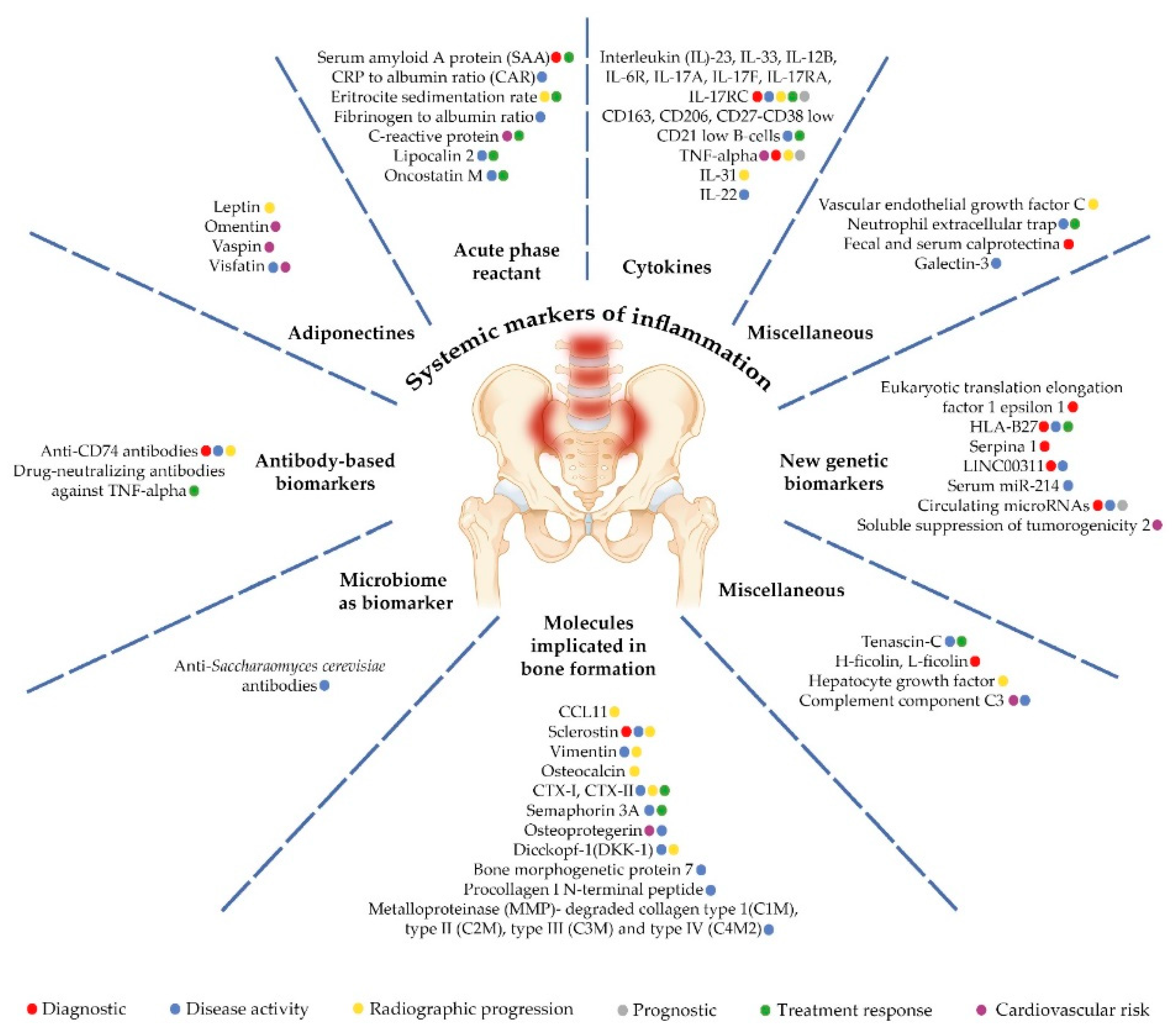
2. Systemic Markers of Inflammation
2.1. Acute-Phase Proteins
2.2. Adipokines
2.3. Cytokines
2.4. Miscellaneous
3. Molecules Involved in Bone Homeostasis
4. Antibody-Based Biomarkers
5. Human Leukocyte Antigen B27 (HLA-B27) and Newer Genetic Biomarkers
6. The Microbiome as a Biomarker
7. Miscellaneous Biomarkers
8. Conclusions
Author Contributions
Funding
Data Availability Statement
Acknowledgments
Conflicts of Interest
References
- Firestein, G.S.; Budd, R.C.; Koretzky, G.; Gabriel, S.E.; McInnes, I.B.; O’Dell, J.R. Firestein & Kelley’s Textbook of Rheumatology, 11th ed.; Elsevier: Amsterdam, The Netherlands, 2020; Volume 2, p. 1307. [Google Scholar]
- Cardelli, C.; Monti, S.; Terenzi, R.; Carli, L. One year in review 2021: Axial spondyloarthritis. Clin. Exp. Rheumatol. 2021, 39, 1272–1281. [Google Scholar] [CrossRef] [PubMed]
- Walsh, J.A.; Magrey, M. Clinical Manifestations and Diagnosis of Axial Spondyloarthritis. J. Clin. Rheumatol. 2021, 27, e547–e560. [Google Scholar] [CrossRef] [PubMed]
- Ancuta, C.; Pomirleanu, C. Principii de Diagnostic si Tratament in Reumatologie, 2nd ed.; Universitatea de Medicină și Farmacie „Grigore T. Popa” din Iași: Iasi, Romania, 2019; p. 177. [Google Scholar]
- Califf, R.M. Biomarker definitions and their applications. Exp. Biol. Med. 2018, 243, 213–221. [Google Scholar] [CrossRef] [PubMed]
- Abdal, S.J.; Yesmin, S.; Shazzad, M.N.; Azad, M.A.K.; Shahin, M.A.; Choudhury, M.R.; Islam, M.N.; Haq, S.A. Development of a Bangla version of the Bath Ankylosing Spondylitis Disease Activity Index (BASDAI) and the Bath Ankylosing Spondylitis Functional Index (BASFI). Int. J. Rheum. Dis. 2021, 24, 74–80. [Google Scholar] [CrossRef]
- Abdelaziz, M.M.; Gamal, R.M.; Ismail, N.M.; Lafy, R.A.; Hetta, H.F. Diagnostic value of anti-CD74 antibodies in early and late axial spondyloarthritis and its relationship to disease activity. Rheumatology 2021, 60, 263–268. [Google Scholar] [CrossRef]
- Do, L.; Granåsen, G.; Hellman, U.; Lejon, K.; Geijer, M.; Baraliakos, X.; Witte, T.; Forsblad-d’Elia, H. Anti-CD74 IgA autoantibodies in radiographic axial spondyloarthritis: A longitudinal Swedish study. Rheumatology 2021, 60, 4085–4093. [Google Scholar] [CrossRef]
- Ziade, N.R.; Mallak, I.; Merheb, G.; Ghorra, P.; Baerlecken, N.; Witte, T.; Baraliakos, X. Added Value of Anti-CD74 Autoantibodies in Axial SpondyloArthritis in a Population with Low HLA-B27 Prevalence. Front. Immunol. 2019, 10, 574. [Google Scholar] [CrossRef]
- de Winter, J.J.; van de Sande, M.G.; Baerlecken, N.; Berg, I.; Ramonda, R.; van der Heijde, D.; van Gaalen, F.A.; Witte, T.; Baeten, D.L. Anti-CD74 antibodies have no diagnostic value in early axial spondyloarthritis: Data from the spondyloarthritis caught early (SPACE) cohort. Arthritis Res. Ther. 2018, 20, 38. [Google Scholar] [CrossRef]
- Hu, C.J.; Li, M.T.; Li, X.; Peng, L.Y.; Zhang, S.Z.; Leng, X.M.; Su, J.M.; Zeng, X.F. CD74 auto-antibodies display little clinical value in Chinese Han population with axial spondyloarthritis. Medicine 2020, 99, 23433. [Google Scholar] [CrossRef]
- Descamps, E.; Molto, A.; Borderie, D.; Lories, R.; Richard, C.M.; Pons, M.; Roux, C.; Briot, K. Changes in bone formation regulator biomarkers in early axial spondyloarthritis. Rheumatology 2021, 60, 1185–1194. [Google Scholar] [CrossRef]
- Navarini, L.; Currado, D.; Marino, A.; Di Donato, S.; Biaggi, A.; Caso, F.; Costa, L.; Tasso, M.; Ruscitti, P.; Pavlych, V.; et al. Persistence of C-reactive protein increased levels and high disease activity are predictors of cardiovascular disease in patients with axial spondyloarthritis. Sci. Rep. 2022, 12, 7498. [Google Scholar] [CrossRef] [PubMed]
- Baraliakos, X.; Szumski, A.; Koenig, A.S.; Jones, H. The role of C-reactive protein as a predictor of treatment response in patients with ankylosing spondylitis. Semin. Arthritis Rheum. 2019, 48, 997–1004. [Google Scholar] [CrossRef] [PubMed]
- Xu, Y.; Jiang, W.; Zhang, H. Association between C-reactive protein gene variant and treatment efficacy of etanercept in ankylosing spondylitis patients receiving hip arthroplasty. J. Clin. Lab. Anal. 2020, 34, e23343. [Google Scholar] [CrossRef] [PubMed]
- Sundström, B.; Ljung, L.; Wållberg-Jonsson, S. Exercise habits and C-reactive protein may predict development of spinal immobility in patients with ankylosing spondylitis. Clin. Rheumatol. 2018, 37, 2881–2885. [Google Scholar] [CrossRef] [PubMed]
- Sohn, D.H.; Jeong, H.; Roh, J.S.; Lee, H.N.; Kim, E.; Koh, J.H.; Lee, S.G. Serum CCL11 level is associated with radiographic spinal damage in patients with ankylosing spondylitis. Rheumatol. Int. 2018, 38, 1455–1464. [Google Scholar] [CrossRef]
- Heftdal, L.D.; Loft, A.G.; Hendricks, O.; Ashouri Christiansen, A.; Schiøttz-Christensen, B.; Arnbak, B.; Jurik, A.G.; Østgård, R.; Winding Deleuran, B.; Møller, H.J.; et al. Divergent effects on macrophage biomarkers soluble CD163 and CD206 in axial spondyloarthritis. Scand. J. Clin. Lab. Investig. 2018, 78, 483–489. [Google Scholar] [CrossRef]
- Wilbrink, R.; Spoorenberg, A.; Arends, S.; van der Geest, K.S.M.; Brouwer, E.; Bootsma, H.; Kroese, F.G.M.; Verstappen, G.M. CD27-CD38lowCD21low B-Cells Are Increased in Axial Spondyloarthritis. Front. Immunol. 2021, 12, 686273. [Google Scholar] [CrossRef]
- Luo, Q.; Fu, B.; Zhang, L.; Guo, Y.; Huang, Z.; Li, J. Expression and clinical significance of circular RNA hsa_circ_0079787 in the peripheral blood of patients with axial spondyloarthritis. Mol. Med. Rep. 2020, 22, 4197–4206. [Google Scholar] [CrossRef]
- Perez-Sanchez, C.; Font-Ugalde, P.; Ruiz-Limon, P.; Lopez-Pedrera, C.; Castro-Villegas, M.C.; Abalos-Aguilera, M.C.; Barbarroja, N.; Arias-de la Rosa, I.; Lopez-Montilla, M.D.; Escudero-Contreras, A.; et al. Circulating microRNAs as potential biomarkers of disease activity and structural damage in ankylosing spondylitis patients. Hum. Mol. Genet. 2018, 27, 875–890. [Google Scholar] [CrossRef]
- Arias de la Rosa, I.; Font, P.; Escudero-Contreras, A.; López-Montilla, M.D.; Pérez-Sánchez, C.; Ábalos-Aguilera, M.C.; Ladehesa-Pineda, L.; Ibáñez-Costa, A.; Torres-Granados, C.; Jimenez-Gomez, Y.; et al. Complement component 3 as biomarker of disease activity and cardiometabolic risk factor in rheumatoid arthritis and spondyloarthritis. Ther. Adv. Chronic. Dis. 2020, 11, 2040622320965067. [Google Scholar] [CrossRef]
- Zhong, Z.; Huang, Y.; Liu, Y.; Chen, J.; Liu, M.; Huang, Q.; Zheng, S.; Guo, X.; Deng, W.; Li, T. Correlation between C-Reactive Protein to Albumin Ratio and Disease Activity in Patients with Axial Spondyloarthritis. Dis. Markers 2021, 2021, 6642486. [Google Scholar] [CrossRef] [PubMed]
- Pamukcu, M.; Duran, T.I. Could C-Reactive Protein/Albumin Ratio be an Indicator of Activation in Axial Spondyloarthritis? J. Coll. Physicians Surg. Pak. 2021, 30, 537–541. [Google Scholar] [PubMed]
- Zhao, Z.; Wang, G.; Wang, Y.; Yang, J.; Wang, Y.; Zhu, J.; Huang, F. Correlation between magnetic resonance imaging (MRI) findings and the new bone formation factor Dkk-1 in patients with spondyloarthritis. Clin. Rheumatol. 2019, 38, 465–475. [Google Scholar] [CrossRef] [PubMed]
- Liao, H.T.; Lin, Y.F.; Tsai, C.Y.; Chou, T.C. Bone morphogenetic proteins and Dickkopf-1 in ankylosing spondylitis. Scand. J. Rheumatol. 2018, 47, 56–61. [Google Scholar] [CrossRef]
- Kraev, K.; Geneva-Popova, M.; Popova, V.; Popova, S.; Maneva, A.; Batalov, A.; Stankova, T.; Delcheva, G.; Stefanova, K. Drug-neutralizing Antibodies against TNF-α blockers as Biomarkers of Therapy Effect Evaluation. Folia Med. 2020, 62, 282–289. [Google Scholar] [CrossRef]
- Wiśniewski, A.; Kasprzyk, S.; Majorczyk, E.; Nowak, I.; Wilczyńska, K.; Chlebicki, A.; Zoń-Giebel, A.; Kuśnierczyk, P. ERAP1-ERAP2 haplotypes are associated with ankylosing spondylitis in Polish patients. Hum. Immunol. 2019, 80, 339–343. [Google Scholar] [CrossRef]
- Kang, K.Y.; Chung, M.K.; Kim, H.N.; Hong, Y.S.; Ju, J.H.; Park, S.H. Severity of Sacroiliitis and Erythrocyte Sedimentation Rate are Associated with a Low Trabecular Bone Score in Young Male Patients with Ankylosing Spondylitis. J. Rheumatol. 2018, 45, 349–356. [Google Scholar] [CrossRef]
- Dong, Y.; Guo, J.; Bi, L. Baseline Interleukin-6 and Erythrocyte Sedimentation Rate Can Predict Clinical Response of TNF Inhibitor Treatment in Patients with Ankylosing Spondylitis. Ann. Clin. Lab. Sci. 2019, 49, 611–618. [Google Scholar]
- Fan, X.; Qi, B.; Ma, L.; Ma, F. Screening of underlying genetic biomarkers for ankylosing spondylitis. Mol. Med. Rep. 2019, 19, 5263–5274. [Google Scholar] [CrossRef]
- Olofsson, T.; Lindqvist, E.; Mogard, E.; Andréasson, K.; Marsal, J.; Geijer, M.; Kristensen, L.E.; Wallman, J.K. Elevated faecal calprotectin is linked to worse disease status in axial spondyloarthritis: Results from the SPARTAKUS cohort. Rheumatology 2019, 58, 1176–1187. [Google Scholar] [CrossRef]
- Liu, M.; Huang, Y.; Huang, Z.; Zhong, Z.; Deng, W.; Huang, Z.; Huang, Q.; Li, T. The role of fibrinogen to albumin ratio in ankylosing spondylitis: Correlation with disease activity. Clin. Chim. Acta 2020, 505, 136–140. [Google Scholar] [CrossRef]
- Cao, M.Y.; Wang, J.; Gao, X.L.; Hu, Y.B. Serum galectin-3 concentrations in patients with ankylosing spondylitis. J. Clin. Lab. Anal. 2019, 33, e22914. [Google Scholar] [CrossRef] [PubMed]
- Ozkaramanli Gur, D.; Ozaltun, D.N.; Guzel, S.; Sarifakioglu, B.; Akyuz, A.; Alpsoy, S.; Aycicek, O.; Baykiz, D. Novel imaging modalities in detection of cardiovascular involvement in ankylosing spondylitis. Scand. Cardiovasc. J. 2018, 52, 320–327. [Google Scholar] [CrossRef] [PubMed]
- Troldborg, A.; Thiel, S.; Mistegaard, C.E.; Hansen, A.; Korsholm, T.L.; Stengaard-Pedersen, K.; Loft, A.G. Plasma levels of H- and L-ficolin are increased in axial spondyloarthritis: Improvement of disease identification. Clin. Exp. Immunol. 2020, 199, 79–87. [Google Scholar] [CrossRef]
- Deminger, A.; Klingberg, E.; Nurkkala, M.; Geijer, M.; Carlsten, H.; Jacobsson, L.T.H.; Forsblad-d’Elia, H. Elevated serum level of hepatocyte growth factor predicts development of new syndesmophytes in men with ankylosing spondylitis. Rheumatology 2021, 60, 1804–1813. [Google Scholar] [CrossRef] [PubMed]
- Torres, L.; Klingberg, E.; Nurkkala, M.; Carlsten, H.; Forsblad-d’Elia, H. Hepatocyte growth factor is a potential biomarker for osteoproliferation and osteoporosis in ankylosing spondylitis. Osteoporos. Int. 2019, 30, 441–449. [Google Scholar] [CrossRef] [PubMed]
- Ziade, N.; Abi Karam, G.; Merheb, G.; Mallak, I.; Irani, L.; Alam, E.; Messaykeh, J.; Menassa, J.; Mroue’, K.; Uthman, I.; et al. HLA-B27 prevalence in axial spondyloarthritis patients and in blood donors in a Lebanese population: Results from a nationwide study. Int. J. Rheum. Dis. 2019, 22, 708–714. [Google Scholar] [CrossRef]
- Lim, C.S.E.; Sengupta, R.; Gaffney, K. The clinical utility of human leucocyte antigen B27 in axial spondyloarthritis. Rheumatology 2018, 57, 959–968. [Google Scholar] [CrossRef]
- de Jong, H.M.Y.; de Winter, J.J.H.; van der Horst-Bruinsma, I.E.; van Schaardenburg, D.J.; van Gaalen, F.A.; van Tubergen, A.M.; Weel, A.E.A.M.; Landewé, R.B.M.; Baeten, D.L.P.; van de Sande, M.G.H. Progression from subclinical inflammation to overt SpA in first degree relatives of SpA patients is associated with HLA-B27: The Pre-SpA cohort. Arthritis Care Res. 2021, 0, 1–9. [Google Scholar]
- Rosine, N.; Etcheto, A.; Hendel-Chavez, H.; Seror, R.; Briot, K.; Molto, A.; Chanson, P.; Taoufik, Y.; Wendling, D.; Lories, R.; et al. Increase In Il-31 Serum Levels Is Associated With Reduced Structural Damage In Early Axial Spondyloarthritis. Sci. Rep. 2018, 8, 7731. [Google Scholar] [CrossRef]
- Iwaszko, M.; Wielińska, J.; Świerkot, J.; Kolossa, K.; Sokolik, R.; Bugaj, B.; Chaszczewska-Markowska, M.; Jeka, S.; Bogunia-Kubik, K. IL-33 Gene Polymorphisms as Potential Biomarkers of Disease Susceptibility and Response to TNF Inhibitors in Rheumatoid Arthritis, Ankylosing Spondylitis, and Psoriatic Arthritis Patients. Front. Immunol. 2021, 12, 631603. [Google Scholar] [CrossRef] [PubMed]
- Ruan, W.F.; Xie, J.T.; Jin, Q.; Wang, W.D.; Ping, A.S. The Diagnostic and Prognostic Role of Interleukin 12B and Interleukin 6R Gene Polymorphism in Patients With Ankylosing Spondylitis. J. Clin. Rheumatol. 2018, 24, 18–24. [Google Scholar] [CrossRef] [PubMed]
- Sagiv, M.; Adawi, M.; Awisat, A.; Shouval, A.; Peri, R.; Sabbah, F.; Rosner, I.; Kessel, A.; Slobodin, G. The association between elevated serum interleukin-22 and the clinical diagnosis of axial spondyloarthritis: A retrospective study. Int. J. Rheum. Dis. 2022, 25, 56–60. [Google Scholar] [CrossRef] [PubMed]
- Saif, D.S.; El Tabl, M.A.; Afifi, N.; Abdallah, M.S.; El Hefnawy, S.M.; Hassanein, S.A. Interleukin-17A biomarker as a predictor for detection of early axial spondyloarthritis changes in patients with psoriasis. Int. J. Rheum. Dis. 2020, 23, 1664–1669. [Google Scholar] [CrossRef]
- Sode, J.; Bank, S.; Vogel, U.; Andersen, P.S.; Sørensen, S.B.; Bojesen, A.B.; Andersen, M.R.; Brandslund, I.; Dessau, R.B.; Hoffmann, H.J.; et al. Genetically determined high activities of the TNF-alpha, IL23/IL17, and NFkB pathways were associated with increased risk of ankylosing spondylitis. BMC Med. Genet. 2018, 19, 165. [Google Scholar] [CrossRef]
- Park, J.H.; Lee, S.G.; Jeon, Y.K.; Park, E.K.; Suh, Y.S.; Kim, H.O. Relationship between serum adipokine levels and radiographic progression in patients with ankylosing spondylitis: A preliminary 2-year longitudinal study. Medicine 2017, 96, e7854. [Google Scholar] [CrossRef]
- Rademacher, J.; Tietz, L.M. Added value of biomarkers compared with clinical parameters for the prediction of radiographic spinal progression in axial spondyloarthritis. Rheumatology 2019, 58, 1556–1564. [Google Scholar] [CrossRef]
- Zhong, H.; Zhong, M. LINC00311 is overexpressed in ankylosing spondylitis and predict treatment outcomes and recurrence. BMC Musculoskelet. Disord. 2019, 20, 278. [Google Scholar] [CrossRef]
- Tsui, F.W.L.; Lin, A.; Sari, I.; Zhang, Z.; Tsui, H.W.; Inman, R.D. Serial Lipocalin 2 and Oncostatin M levels reflect inflammation status and treatment response in axial spondyloarthritis. Arthritis Res. Ther. 2021, 23, 141. [Google Scholar] [CrossRef]
- Hušáková, M.; Bay-Jensen, A.C.; Forejtová, Š.; Zegzulková, K.; Tomčík, M.; Gregová, M.; Bubová, K.; Hořínková, J.; Gatterová, J.; Pavelka, K.; et al. Metabolites of type I, II, III, and IV collagen may serve as markers of disease activity in axial spondyloarthritis. Sci. Rep. 2019, 9, 11218. [Google Scholar] [CrossRef]
- Ruiz-Limon, P.; Ladehesa-Pineda, M.L.; Castro-Villegas, M.D.C.; Abalos-Aguilera, M.D.C.; Lopez-Medina, C.; Lopez-Pedrera, C.; Barbarroja, N.; Espejo-Peralbo, D.; Gonzalez-Reyes, J.A.; Villalba, J.M.; et al. Enhanced NETosis generation in radiographic axial spondyloarthritis: Utility as biomarker for disease activity and anti-TNF-α therapy effectiveness. J. Biomed. Sci. 2020, 27, 54. [Google Scholar] [CrossRef]
- Genre, F.; Rueda-Gotor, J.; Remuzgo-Martínez, S.; Pulito-Cueto, V.; Corrales, A.; Mijares, V.; Lera-Gómez, L.; Portilla, V.; Expósito, R.; Mata, C.; et al. Omentin: A biomarker of cardiovascular risk in individuals with axial spondyloarthritis. Sci. Rep. 2020, 10, 9636. [Google Scholar] [CrossRef] [PubMed]
- Genre, F.; Rueda-Gotor, J.; Remuzgo-Martínez, S.; Corrales, A.; Ubilla, B.; Mijares, V.; Fernández-Díaz, C.; Portilla, V.; Blanco, R.; Hernández, J.L.; et al. Implication of osteoprotegerin and sclerostin in axial spondyloarthritis cardiovascular disease: Study of 163 Spanish patients. Clin. Exp. Rheumatol. 2018, 36, 302–309. [Google Scholar] [PubMed]
- Nisihara, R.; Skare, T.L.; Zeni, J.O.; Rasera, H.; Lidani, K.; Messias-Reason, I. Plasma levels of pentraxin 3 in patients with spondyloarthritis. Biomarkers 2018, 23, 14–17. [Google Scholar] [CrossRef] [PubMed]
- Li, X.; Liang, A.; Chen, Y.; Lam, N.S.; Long, X.; Xu, X.; Zhong, S. Procollagen I N-terminal peptide correlates with inflammation on sacroiliac joint magnetic resonance imaging in ankylosing spondylitis but not in non-radiographic axial spondyloarthritis: A cross-sectional study. Mod. Rheumatol. 2022, 32, 770–775. [Google Scholar] [CrossRef]
- Luchetti, M.M.; Ciccia, F.; Avellini, C.; Benfaremo, D.; Guggino, G.; Farinelli, A.; Ciferri, M.; Rossini, M.; Svegliati, S.; Spadoni, T.; et al. Sclerostin and Antisclerostin Antibody Serum Levels Predict the Presence of Axial Spondyloarthritis in Patients with Inflammatory Bowel Disease. J. Rheumatol. 2018, 45, 630–637. [Google Scholar] [CrossRef]
- Perrotta, F.M.; Ceccarelli, F.; Barbati, C.; Colasanti, T.; De Socio, A.; Scriffignano, S.; Alessandri, C.; Lubrano, E. Serum Sclerostin as a Possible Biomarker in Ankylosing Spondylitis: A Case-Control Study. J. Immunol. Res. 2018, 2018, 9101964. [Google Scholar] [CrossRef]
- Liao, H.T.; Lin, Y.F.; Chou, C.T.; Tsai, C.Y. Semaphorin 3A in Ankylosing Spondylitis. J. Microbiol. Immunol. Infect. 2019, 52, 151–157. [Google Scholar] [CrossRef]
- Perrotta, F.M.; Ceccarelli, F.; Barbati, C.; Colasanti, T.; Montepaone, M.; Alessandri, C.; Valesini, G.; Lubrano, E. Assessment of semaphorin 3A and its role in bone remodelling in a group of ankylosing spondylitis patients. Clin. Exp. Rheumatol. 2017, 35, 313–316. [Google Scholar]
- Liu, S.; Ji, W.; Lu, J.; Tang, X.; Guo, Y.; Ji, M.; Xu, T.; Gu, W.; Kong, D.; Shen, Q. Discovery of Potential Serum Protein Biomarkers in Ankylosing Spondylitis Using Tandem Mass Tag-Based Quantitative Proteomics. J. Proteome Res. 2020, 19, 864–872. [Google Scholar] [CrossRef]
- Jarlborg, M.; Courvoisier, D.S.; Lamacchia, C.; Martinez Prat, L.; Mahler, M.; Bentow, C.; Finckh, A.; Gabay, C.; Nissen, M.J. Serum calprotectin: A promising biomarker in rheumatoid arthritis and axial spondyloarthritis. Arthritis Res. Ther. 2020, 22, 105. [Google Scholar] [CrossRef] [PubMed]
- Hu, H.; Du, F.; Zhang, S.; Zhang, W. Serum calprotectin correlates with risk and disease severity of ankylosing spondylitis and its change during first month might predict favorable response to treatment. Mod. Rheumatol. 2019, 29, 836–842. [Google Scholar] [CrossRef] [PubMed]
- Genre, F.; Rueda-Gotor, J.; Remuzgo-Martínez, S.; Corrales, A.; Mijares, V.; Expósito, R.; Mata, C.; Portilla, V.; Blanco, R.; Hernández, J.L.; et al. Association of circulating calprotectin with lipid profile in axial spondyloarthritis. Sci. Rep. 2018, 8, 13728. [Google Scholar] [CrossRef] [PubMed]
- Rademacher, J.; Siderius, M.; Gellert, L.; Wink, F.R.; Verba, M.; Maas, F.; Tietz, L.M.; Poddubnyy, D.; Spoorenberg, A.; Arends, S. Baseline serum biomarkers of inflammation, bone turnover and adipokines predict spinal radiographic progression in ankylosing spondylitis patients on TNF inhibitor therapy. Semin. Arthritis Rheum. 2022, 53, 151974. [Google Scholar] [CrossRef] [PubMed]
- Kook, H.Y.; Jin, S.H.; Park, P.R.; Lee, S.J.; Shin, H.J.; Kim, T.J. Serum miR-214 as a novel biomarker for ankylosing spondylitis. Int. J. Rheum. Dis. 2019, 22, 1196–1201. [Google Scholar] [CrossRef]
- Bubová, K.; Prajzlerová, K.; Hulejová, H.; Gregová, M.; Mintálová, K.; Hušáková, M.; Forejtová, Š.; Filková, M.; Tomčík, M.; Vencovský, J.; et al. Elevated Tenascin-C Serum Levels in Patients with Axial Spondyloarthritis. Physiol. Res. 2020, 69, 653–660. [Google Scholar] [CrossRef]
- Gupta, L.; Bhattacharya, S.; Aggarwal, A. Tenascin-C, a biomarker of disease activity in early ankylosing spondylitis. Clin. Rheumatol. 2018, 37, 1401–1405. [Google Scholar] [CrossRef]
- Rueda-Gotor, J.; López-Mejías, R.; Remuzgo-Martínez, S.; Pulito-Cueto, V.; Corrales, A.; Lera-Gómez, L.; Portilla, V.; González-Mazón, Í.; Blanco, R.; Expósito, R.; et al. Vaspin in atherosclerotic disease and cardiovascular risk in axial spondyloarthritis: A genetic and serological study. Arthritis Res. Ther. 2021, 23, 111. [Google Scholar] [CrossRef]
- Siebuhr, A.S.; Hušaková, M.; Forejtová, S.; Zegzulková, K.; Tomčik, M.; Urbanová, M.; Grobelná, K.; Gatterová, J.; Bay-Jensen, A.C.; Pavelka, K. Metabolites of C-reactive protein and vimentin are associated with disease activity of axial spondyloarthritis. Clin. Exp. Rheumatol. 2019, 37, 358–366. [Google Scholar]
- Baykara, A.R.; Küçük, A.; Tuzcu, A.; Tuzcu, G.; Cüre, E.; Uslu, A.U.; Omma, A. The relationship of serum visfatin levels with clinical parameters, flow-mediated dilation, and carotid intima-media thickness in patients with ankylosing spondylitis. Turk. J. Med. Sci. 2021, 51, 1865–1874. [Google Scholar] [CrossRef]
- Proft, F.; Muche, B.; Schmidt, M.; Spiller, L.; Rodriguez, V.R.; Rademacher, J.; Weber, A.-K.; Lüders, S.; Protopopov, M.; Spiller, I.; et al. FRI0194 Ankylosing spondylitis disease activity score (ASDAS) based on a quick quantitative crp assay performs similarly well to asdas based on conventional crp in patients with axial spondyloarthritis. Ann. Rheum. Dis. 2018, 77, 637–638. [Google Scholar]
- Smoldovskaya, O.V.; Voloshin, S.A.; Novikov, A.A.; Aleksandrova, E.N.; Feyzkhanova, G.U.; Rubina, A.Y. Adaptation of Microarray Assay for Serum Amyloid A Analysis in Human Serum. Mol. Biol. 2022, 56, 336–342. [Google Scholar] [CrossRef] [PubMed]
- Sorić Hosman, I.; Kos, I.; Lamot, L. Serum Amyloid A in Inflammatory Rheumatic Diseases: A Compendious Review of a Renowned Biomarker. Front. Immunol. 2021, 11, 631299. [Google Scholar] [CrossRef] [PubMed]
- Lorenzin, M.; Ometto, F.; Ortolan, A.; Felicetti, M.; Favero, M.; Doria, A.; Ramonda, R. An update on serum biomarkers to assess axial spondyloarthritis and to guide treatment decision. Ther. Adv. Musculoskelet. Dis. 2020, 12, 1759720X20934277. [Google Scholar] [CrossRef]
- Inoue, K.; Kodama, T.; Daida, H. Pentraxin 3: A novel biomarker for inflammatory cardiovascular disease. Int. J. Vasc. Med. 2012, 2012, 657025. [Google Scholar] [CrossRef]
- Özdemirel, A.E.; Güven, S.C.; Sunar, İ.; Sari Sürmeli, Z.; Doğanci, A.; Tutkak, H.; Yalçin Sayin, A.P.; Ataman, Ş. The Relationship between Plasma Pentraxin 3 and Serum Amyloid P Levels and Disease Activity in Ankylosing Spondylitis. Int. J. Clin. Pract. 2022, 2022, 7243399. [Google Scholar] [CrossRef]
- Qi, Q.; Geng, Y.; Sun, M.; Chen, H.; Wang, P.; Chen, Z. Hyperfibrinogen Is Associated with the Systemic Inflammatory Response and Predicts Poor Prognosis in Advanced Pancreatic Cancer. Pancreas 2015, 44, 977–982. [Google Scholar] [CrossRef]
- Keller, U. Nutritional Laboratory Markers in Malnutrition. J. Clin. Med. 2019, 8, 775. [Google Scholar] [CrossRef]
- Singh, V.; Yeoh, B.S.; Chassaing, B.; Zhang, B.; Saha, P.; Xiao, X.; Awasthi, D.; Shashidharamurthy, R.; Dikshit, M.; Gewirtz, A.; et al. Microbiota-inducible Innate Immune, Siderophore Binding Protein Lipocalin 2 is Critical for Intestinal Homeostasis. Cell. Mol. Gastroenterol. Hepatol. 2016, 2, 482–498. [Google Scholar] [CrossRef]
- West, N.R.; Hegazy, A.N.; Owens, B.M.J.; Bullers, S.J.; Linggi, B.; Buonocore, S.; Coccia, M.; Görtz, D.; This, S.; Stockenhuber, K.; et al. Oncostatin M drives intestinal inflammation and predicts response to tumor necrosis factor-neutralizing therapy in patients with inflammatory bowel disease. Nat. Med. 2017, 23, 579–589. [Google Scholar] [CrossRef]
- Moschen, A.R.; Adolph, T.E.; Gerner, R.R.; Wieser, V.; Tilg, H. Lipocalin-2: A Master Mediator of Intestinal and Metabolic Inflammation. Trends Endocrinol. Metab. 2017, 28, 388–397. [Google Scholar] [CrossRef] [PubMed]
- Mancuso, P. The role of adipokines in chronic inflammation. Immunotargets Ther. 2016, 5, 47–56. [Google Scholar] [CrossRef] [PubMed]
- Saddi-Rosa, P.; Oliveira, C.S.; Giuffrida, F.M.; Reis, A.F. Visfatin, glucose metabolism and vascular disease: A review of evidence. Diabetol. Metab. Syndr. 2010, 2, 21. [Google Scholar] [CrossRef]
- Watanabe, T.; Watanabe-Kominato, K.; Takahashi, Y.; Kojima, M.; Watanabe, R. Adipose Tissue-Derived Omentin-1 Function and Regulation. Compr. Physiol. 2017, 7, 765–781. [Google Scholar] [PubMed]
- Rao, S.S.; Hu, Y.; Xie, P.L.; Cao, J.; Wang, Z.X.; Liu, J.H.; Yin, H.; Huang, J.; Tan, Y.J.; Luo, J.; et al. Omentin-1 prevents inflammation-induced osteoporosis by downregulating the pro-inflammatory cytokines. Bone Res. 2018, 6, 9. [Google Scholar] [CrossRef]
- Zhang, J.M.; An, J. Cytokines, inflammation, and pain. Int. Anesthesiol. Clin. 2007, 45, 27–37. [Google Scholar] [CrossRef]
- Furst, D.E.; Louie, J.S. Targeting inflammatory pathways in axial spondyloarthritis. Arthritis Res. Ther. 2019, 21, 135. [Google Scholar] [CrossRef]
- Jethwa, H.; Bowness, P. The interleukin (IL)-23/IL-17 axis in ankylosing spondylitis: New advances and potentials for treatment. Clin. Exp. Immunol. 2016, 183, 30–36. [Google Scholar] [CrossRef]
- Walsham, N.E.; Sherwood, R.A. Fecal calprotectin in inflammatory bowel disease. Clin. Exp. Gastroenterol. 2016, 9, 21–29. [Google Scholar]
- Romand, X.; Bernardy, C.; Nguyen, M.V.C.; Courtier, A.; Trocme, C.; Clapasson, M.; Paclet, M.H.; Toussaint, B.; Gaudin, P. Systemic calprotectin and chronic inflammatory rheumatic diseases. Jt. Bone Spine 2019, 86, 691–698. [Google Scholar] [CrossRef]
- Turina, M.C.; Sieper, J.; Yeremenko, N.; Conrad, K.; Haibel, H.; Rudwaleit, M.; Baeten, D.; Poddubnyy, D. Calprotectin serum level is an independent marker for radiographic spinal progression in axial spondyloarthritis. Ann. Rheum. Dis. 2014, 73, 1746–1748. [Google Scholar] [CrossRef] [PubMed]
- Duran, A.; Kobak, S.; Sen, N.; Aktakka, S.; Atabay, T.; Orman, M. Fecal calprotectin is associated with disease activity in patients with ankylosing spondylitis. Bosn. J. Basic Med. Sci. 2016, 16, 71–74. [Google Scholar] [CrossRef] [PubMed]
- Li, T.; Zhang, Z.; Li, X.; Dong, G.; Zhang, M.; Xu, Z.; Yang, J. Neutrophil Extracellular Traps: Signaling Properties and Disease Relevance. Mediat. Inflamm. 2020, 2020, 9254087. [Google Scholar] [CrossRef]
- Dong, R.; Zhang, M.; Hu, Q.; Zheng, S.; Soh, A.; Zheng, Y.; Yuan, H. Galectin-3 as a novel biomarker for disease diagnosis and a target for therapy (Review). Int. J. Mol. Med. 2018, 41, 599–614. [Google Scholar] [CrossRef] [PubMed]
- Omran, A.; Atanasova, D.; Landgren, F.; Magnusson, P. Sclerostin: From Molecule to Clinical Biomarker. Int. J. Mol. Sci. 2022, 23, 4751. [Google Scholar] [CrossRef]
- Baron, R.; Rawadi, G. Targeting the Wnt/beta-catenin pathway to regulate bone formation in the adult skeleton. Endocrinology 2007, 148, 2635–2643. [Google Scholar] [CrossRef]
- Ito, N.; Prideaux, M.; Wijenayaka, A.R.; Yang, D.; Ormsby, R.T.; Bonewald, L.F.; Atkins, G.J. Sclerostin Directly Stimulates Osteocyte Synthesis of Fibroblast Growth Factor-23. Calcif. Tissue Int. 2021, 109, 66–76. [Google Scholar] [CrossRef]
- Appel, H.; Ruiz-Heiland, G.; Listing, J.; Zwerina, J.; Herrmann, M.; Mueller, R.; Haibel, H.; Baraliakos, X.; Hempfing, A.; Rudwaleit, M.; et al. Altered skeletal expression of sclerostin and its link to radiographic progression in ankylosing spondylitis. Arthritis Rheum. 2009, 60, 3257–3262. [Google Scholar] [CrossRef]
- Mihara, A.; Yukata, K.; Seki, T.; Iwanaga, R.; Nishida, N.; Fujii, K.; Nagao, Y.; Sakai, T. Effects of sclerostin antibody on bone healing. World J. Orthop. 2021, 12, 651–659. [Google Scholar] [CrossRef]
- Aluganti Narasimhulu, C.; Singla, D.K. The Role of Bone Morphogenetic Protein 7 (BMP-7) in Inflammation in Heart Diseases. Cells 2020, 9, 280. [Google Scholar] [CrossRef]
- Mattey, D.L.; Packham, J.C.; Nixon, N.B.; Coates, L.; Creamer, P.; Hailwood, S.; Taylor, G.J.; Bhalla, A.K. Association of cytokine and matrix metalloproteinase profiles with disease activity and function in ankylosing spondylitis. Arthritis Res. Ther. 2012, 14, R127. [Google Scholar] [CrossRef] [PubMed]
- Siebuhr, A.S.; van der Heijde, D.; Bay-Jensen, A.C.; Karsdal, M.A.; Landewé, R.; van Tubergen, A.; Ramiro, S. Is radiographic progression in radiographic axial spondyloarthritis related to matrix metalloproteinase degradation of extracellular matrix? RMD Open 2018, 4, 000648. [Google Scholar] [CrossRef] [PubMed]
- Kučukalić-Selimović, E.; Valjevac, A.; Hadžović-Džuvo, A. The utility of procollagen type 1 N-terminal propeptide for the bone status assessment in postmenopausal women. Bosn. J. Basic Med. Sci. 2013, 13, 259–265. [Google Scholar] [CrossRef] [PubMed]
- Koivula, M.K.; Risteli, L.; Risteli, J. Measurement of aminoterminal propeptide of type I procollagen (PINP) in serum. Clin. Biochem. 2012, 45, 920–927. [Google Scholar] [CrossRef] [PubMed]
- Ivaska, J.; Pallari, H.M.; Nevo, J.; Eriksson, J.E. Novel functions of vimentin in cell adhesion, migration, and signaling. Exp. Cell Res. 2007, 313, 2050–2062. [Google Scholar] [CrossRef]
- Bay-Jensen, A.C.; Karsdal, M.A.; Vassiliadis, E.; Wichuk, S.; Marcher-Mikkelsen, K.; Lories, R.; Christiansen, C.; Maksymowych, W.P. Circulating citrullinated vimentin fragments reflect disease burden in ankylosing spondylitis and have prognostic capacity for radiographic progression. Arthritis Rheum. 2013, 65, 972–980. [Google Scholar] [CrossRef]
- Klingberg, E.; Nurkkala, M.; Carlsten, H.; Forsblad-d’Elia, H. Biomarkers of bone metabolism in ankylosing spondylitis in relation to osteoproliferation and osteoporosis. J. Rheumatol. 2014, 41, 1349–1356. [Google Scholar] [CrossRef]
- Yilmaz, Y.; Kurt, R.; Eren, F.; Imeryuz, N. Serum osteocalcin levels in patients with nonalcoholic fatty liver disease: Association with ballooning degeneration. Scand. J. Clin. Lab. Investig. 2011, 71, 631–636. [Google Scholar] [CrossRef]
- Kwon, S.R.; Lim, M.J.; Suh, C.H.; Park, S.G.; Hong, Y.S.; Yoon, B.Y.; Kim, H.A.; Choi, H.J.; Park, W. Dickkopf-1 level is lower in patients with ankylosing spondylitis than in healthy people and is not influenced by anti-tumor necrosis factor therapy. Rheumatol. Int. 2012, 32, 2523–2527. [Google Scholar] [CrossRef]
- Pavliuk, O.; Shevchuk, S. Levels of osteocalcin and procollagen i n-terminal propeptide (pinp) in men suffering from ankylosing spondylitis. Wiad. Lek. 2021, 74, 2384–2391. [Google Scholar] [CrossRef]
- Wu, M.; Chen, M.; Ma, Y.; Yang, J.; Han, R.; Yuan, Y.; Hu, X.; Wang, M.; Zhang, X.; Xu, S.; et al. Dickkopf-1 in ankylosing spondylitis: Review and meta-analysis. Clin. Chim. Acta 2018, 481, 177–183. [Google Scholar] [CrossRef] [PubMed]
- Garcia, S. Role of Semaphorins in Immunopathologies and Rheumatic Diseases. Int. J. Mol. Sci. 2019, 20, 374. [Google Scholar] [CrossRef] [PubMed]
- McGovern, K.E.; Wilson, E.H. Role of Chemokines and Trafficking of Immune Cells in Parasitic Infections. Curr. Immunol. Rev. 2013, 9, 157–168. [Google Scholar] [CrossRef] [PubMed]
- Ivanovska, M.; Abdi, Z.; Murdjeva, M.; Macedo, D.; Maes, A.; Maes, M. CCL-11 or Eotaxin-1: An Immune Marker for Ageing and Accelerated Ageing in Neuro-Psychiatric Disorders. Pharmaceuticals 2020, 13, 230. [Google Scholar] [CrossRef] [PubMed]
- Kindstedt, E.; Holm, C.K.; Sulniute, R.; Martinez-Carrasco, I.; Lundmark, R.; Lundberg, P. CCL11, a novel mediator of inflammatory bone resorption. Sci. Rep. 2017, 7, 5334. [Google Scholar] [CrossRef]
- Kalayci, M.; Gul, E. Eotaxin-1 Levels in Patients with Myocardial Infarction. Clin. Lab. 2022, 68, 3. [Google Scholar] [CrossRef]
- Komsalova, L.Y.; Martínez Salinas, M.P.; Jiménez, J.F.G. Predictive values of inflammatory back pain, positive HLA B27 antigen and acute and chronic magnetic resonance changes in early diagnosis of Spondyloarthritis. A study of 133 patients. PLoS ONE 2020, 15, e0244184. [Google Scholar] [CrossRef]
- Rudwaleit, M.; Claudepierre, P.; Wordsworth, P.; Cortina, E.L.; Sieper, J.; Kron, M.; Carcereri-De-Prati, R.; Kupper, H.; Kary, S. Effectiveness, safety, and predictors of good clinical response in 1250 patients treated with adalimumab for active ankylosing spondylitis. J. Rheumatol. 2009, 36, 801–808. [Google Scholar] [CrossRef]
- Robinson, P.C.; Claushuis, T.A.; Cortes, A.; Martin, T.M.; Evans, D.M.; Leo, P.; Mukhopadhyay, P.; Bradbury, L.A.; Cremin, K.; Harris, J.; et al. Genetic dissection of acute anterior uveitis reveals similarities and differences in associations observed with ankylosing spondylitis. Arthritis Rheumatol. 2015, 67, 140–151. [Google Scholar] [CrossRef]
- Lin, H.; Gong, Y.Z. Reply to “Association of HLA-B27 and its subtypes with ankylosing spondylitis and clinical manifestations of ankylosing spondylitis in different HLA-B27 subtypes: Comment on the article by Lin et al.”. Rheumatol. Int. 2017, 37, 1685. [Google Scholar] [CrossRef][Green Version]
- Li, M.Y.; Yao, Z.Q.; Liu, X.Y. Advance of research on HLA-B27 and the immunological mechanism of ankylosing spondylitis. Sheng Li Ke Xue Jin Zhan 2011, 42, 16–20. [Google Scholar] [PubMed]
- Caba, L.; Florea, L.; Gug, C.; Dimitriu, D.C.; Gorduza, E.V. Circular RNA-Is the Circle Perfect? Biomolecules 2021, 11, 1755. [Google Scholar] [CrossRef] [PubMed]
- Kumar, L.; Shamsuzzama; Jadiya, P.; Haque, R.; Shukla, S.; Nazir, A. Functional Characterization of Novel Circular RNA Molecule, circzip-2 and Its Synthesizing Gene zip-2 in C. elegans Model of Parkinson’s Disease. Mol. Neurobiol. 2018, 55, 6914–6926. [Google Scholar] [CrossRef] [PubMed]
- Qu, S.; Yang, X.; Li, X.; Wang, J.; Gao, Y.; Shang, R.; Sun, W.; Dou, K.; Li, H. Circular RNA: A new star of noncoding RNAs. Cancer Lett. 2015, 365, 141–148. [Google Scholar] [CrossRef]
- Wang, L.; Long, H.; Zheng, Q.; Bo, X.; Xiao, X.; Li, B. Circular RNA circRHOT1 promotes hepatocellular carcinoma progression by initiation of NR2F6 expression. Mol. Cancer 2019, 18, 119. [Google Scholar] [CrossRef]
- Ouyang, Q.; Wu, J.; Jiang, Z.; Zhao, J.; Wang, R.; Lou, A.; Zhu, D.; Shi, G.P.; Yang, M. Microarray Expression Profile of Circular RNAs in Peripheral Blood Mononuclear Cells from Rheumatoid Arthritis Patients. Cell. Physiol. Biochem. 2017, 42, 651–659. [Google Scholar] [CrossRef]
- Luo, Q.; Zhang, L.; Li, X.; Fu, B.; Deng, Z.; Qing, C.; Su, R.; Xu, J.; Guo, Y.; Huang, Z.; et al. Identification of circular RNAs hsa_circ_0044235 in peripheral blood as novel biomarkers for rheumatoid arthritis. Clin. Exp. Immunol. 2018, 194, 118–124. [Google Scholar] [CrossRef]
- Luo, Q.; Li, X.; Fu, B.; Zhang, L.; Fang, L.; Qing, C.; Guo, Y.; Huang, Z.; Li, J. Expression profile and diagnostic value of circRNAs in peripheral blood from patients with systemic lupus erythematosus. Mol. Med. Rep. 2021, 23, 1. [Google Scholar] [CrossRef]
- Zhang, Y.; Li, Q.; Liu, C.; Gao, S.; Ping, H.; Wang, J.; Wang, P. MiR-214-3p attenuates cognition defects via the inhibition of autophagy in SAMP8 mouse model of sporadic Alzheimer’s disease. Neurotoxicology 2016, 56, 139–149. [Google Scholar] [CrossRef]
- Li, D.; Liu, J.; Guo, B.; Liang, C.; Dang, L.; Lu, C.; He, X.; Cheung, H.Y.; Xu, L.; Lu, C. Osteoclast-derived exosomal miR-214-3p inhibits osteoblastic bone formation. Nat. Commun. 2016, 7, 10872. [Google Scholar] [CrossRef]
- Yan, J.; Parekh, V.V.; Mendez-Fernandez, Y.; Olivares-Villagómez, D.; Dragovic, S.; Hill, T.; Roopenian, D.C.; Joyce, S.; Van Kaer, L. In vivo role of ER-associated peptidase activity in tailoring peptides for presentation by MHC class Ia and class Ib molecules. J. Exp. Med. 2006, 203, 647–659. [Google Scholar] [CrossRef] [PubMed]
- Haroon, N.; Tsui, F.W.; Chiu, B.; Tsui, H.W.; Inman, R.D. Serum cytokine receptors in ankylosing spondylitis: Relationship to inflammatory markers and endoplasmic reticulum aminopeptidase polymorphisms. J. Rheumatol. 2010, 37, 1907–1910. [Google Scholar] [CrossRef] [PubMed]
- Li, L.; Batliwala, M.; Bouvier, M. ERAP1 enzyme-mediated trimming and structural analyses of MHC I-bound precursor peptides yield novel insights into antigen processing and presentation. J. Biol. Chem. 2019, 294, 18534–18544. [Google Scholar] [CrossRef] [PubMed]
- Martín-Esteban, A.; Guasp, P.; Barnea, E.; Admon, A.; López de Castro, J.A. Functional Interaction of the Ankylosing Spondylitis-Associated Endoplasmic Reticulum Aminopeptidase 2 With the HLA-B*27 Peptidome in Human Cells. Arthritis Rheumatol. 2016, 68, 2466–2475. [Google Scholar] [CrossRef] [PubMed]
- Lalevée, S.; Feil, R. Long noncoding RNAs in human disease: Emerging mechanisms and therapeutic strategies. Epigenomics 2015, 7, 877–879. [Google Scholar] [CrossRef] [PubMed]
- Wang, Y.; Luo, T.B.; Liu, L.; Cui, Z.Q. LncRNA LINC00311 Promotes the Proliferation and Differentiation of Osteoclasts in Osteoporotic Rats Through the Notch Signaling Pathway by Targeting DLL3. Cell. Physiol. Biochem. 2018, 47, 2291–2306. [Google Scholar] [CrossRef] [PubMed]
- Hassan, M.K.; Kumar, D.; Naik, M.; Dixit, M. The expression profile and prognostic significance of eukaryotic translation elongation factors in different cancers. PLoS ONE 2018, 13, e0191377. [Google Scholar] [CrossRef] [PubMed]
- Yu, X.; Zheng, H.; Chan, M.T.; Wu, W.K. HULC: An oncogenic long non-coding RNA in human cancer. J. Cell. Mol. Med. 2017, 21, 410–417. [Google Scholar] [CrossRef]
- Ozgocmen, S.; Godekmerdan, A.; Ozkurt-Zengin, F. Acute-phase response, clinical measures and disease activity in ankylosing spondylitis. Jt. Bone Spine 2007, 74, 249–253. [Google Scholar] [CrossRef]
- Aimo, A.; Vergaro, G.; Passino, C.; Ripoli, A.; Ky, B.; Miller, W.L.; Bayes-Genis, A.; Anand, I.; Januzzi, J.L.; Emdin, M. Prognostic Value of Soluble Suppression of Tumorigenicity-2 in Chronic Heart Failure: A Meta-Analysis. JACC Heart Fail. 2017, 5, 280–286. [Google Scholar] [CrossRef]
- Demyanets, S.; Kaun, C.; Kaider, A.; Speidl, W.; Prager, M.; Oravec, S.; Hohensinner, P.; Wojta, J.; Rega-Kaun, G. The pro-inflammatory marker soluble suppression of tumorigenicity-2 (ST2) is reduced especially in diabetic morbidly obese patients undergoing bariatric surgery. Cardiovasc. Diabetol. 2020, 19, 26. [Google Scholar] [CrossRef] [PubMed]
- De Boer, R.A.; Daniels, L.B.; Maisel, A.S.; Januzzi, J.L., Jr. State of the Art: Newer biomarkers in heart failure. Eur. J. Heart Fail. 2015, 17, 559–569. [Google Scholar] [CrossRef] [PubMed]
- Roldan, C.A. Valvular and coronary heart disease in systemic inflammatory diseases: Systemic Disorders in heart disease. Heart 2008, 94, 1089–1101. [Google Scholar] [CrossRef] [PubMed]
- Noer, H.R. An “experimental” epidemic of Reiter’s syndrome. JAMA 1966, 198, 693–698. [Google Scholar] [CrossRef]
- Gill, T.; Asquith, M.; Rosenbaum, J.T.; Colbert, R.A. The intestinal microbiome in spondyloarthritis. Curr. Opin. Rheumatol. 2015, 27, 319–325. [Google Scholar] [CrossRef]
- Tiffany, C.R.; Bäumler, A.J. Dysbiosis: From fiction to function. Am. J. Physiol. Gastrointest. Liver Physiol. 2019, 317, 602–608. [Google Scholar] [CrossRef]
- Ciccia, F.; Ferrante, A.; Triolo, G. Intestinal dysbiosis and innate immune responses in axial spondyloarthritis. Curr. Opin. Rheumatol. 2016, 28, 352–358. [Google Scholar] [CrossRef]
- Sagard, J.; Olofsson, T.; Mogard, E.; Marsal, J.; Andréasson, K.; Geijer, M.; Kristensen, L.E.; Lindqvist, E.; Wallman, J.K. Gut dysbiosis associated with worse disease activity and physical function in axial spondyloarthritis. Arthritis Res. Ther. 2022, 24, 42. [Google Scholar] [CrossRef]
- Gill, T.; Brooks, S.R.; Rosenbaum, J.T.; Asquith, M.; Colbert, R.A. Novel Inter-omic Analysis Reveals Relationships Between Diverse Gut Microbiota and Host Immune Dysregulation in HLA-B27-Induced Experimental Spondyloarthritis. Arthritis Rheumatol. 2019, 71, 1849–1857. [Google Scholar] [CrossRef]
- Oikonomopoulou, K.; Ricklin, D.; Ward, P.A.; Lambris, J.D. Interactions between coagulation and complement--their role in inflammation. Semin. Immunopathol. 2012, 34, 151–165. [Google Scholar] [CrossRef]
- Phillips, C.M.; Kesse-Guyot, E.; Ahluwalia, N.; McManus, R.; Hercberg, S.; Lairon, D.; Planells, R.; Roche, H.M. Dietary fat, abdominal obesity and smoking modulate the relationship between plasma complement component 3 concentrations and metabolic syndrome risk. Atherosclerosis 2012, 220, 513–519. [Google Scholar] [CrossRef] [PubMed]
- Barbu, A.; Hamad, O.A.; Lind, L.; Ekdahl, K.N.; Nilsson, B. The role of complement factor C3 in lipid metabolism. Mol. Immunol. 2015, 67, 101–107. [Google Scholar] [PubMed]
- Midwood, K.S.; Orend, G. The role of tenascin-C in tissue injury and tumorigenesis. J. Cell Commun. Signal. 2009, 3, 287–310. [Google Scholar] [CrossRef] [PubMed]
- Petrini, I. Biology of MET: A double life between normal tissue repair and tumor progression. Ann. Transl. Med. 2015, 3, 82. [Google Scholar] [PubMed]
- Nakamura, T.; Sakai, K.; Nakamura, T.; Matsumoto, K. Hepatocyte growth factor twenty years on: Much more than a growth factor. J. Gastroenterol. Hepatol. 2011, 26, 188–202. [Google Scholar] [CrossRef]
- Grano, M.; Galimi, F.; Zambonin, G.; Colucci, S.; Cottone, E.; Zallone, A.Z.; Comoglio, P.M. Hepatocyte growth factor is a coupling factor for osteoclasts and osteoblasts in vitro. Proc. Natl. Acad. Sci. USA 1996, 93, 7644–7648. [Google Scholar] [CrossRef] [PubMed]
- Kuraya, M.; Ming, Z.; Liu, X.; Matsushita, M.; Fujita, T. Specific binding of L-ficolin and H-ficolin to apoptotic cells leads to complement activation. Immunobiology 2005, 209, 689–697. [Google Scholar] [CrossRef] [PubMed]


| Biomarker | Predictability | Study type | Reference |
|---|---|---|---|
| Anti-CD74 antibodies | Positive association with diagnosis and disease activity. Positive association with radiographic progression. Positive association with diagnosis. Negative association with diagnosis. Positive association with diagnosis. | Cross-sectional study Prospective study Prospective study Prospective study Prospective study | 2021, Marwa Mahmoud Abdelaziz [7] 2021, Lan Do [8] 2019, Nelly R. Ziade [9] 2018, Janneke J. de Winter [10] 2020, Chao-Jun Hu [11] |
| Bone morphogenetic protein 7 (BMP-7), sclerostin, dicckopf-1 (DKK-1) | Positive association with disease association | Prospective multicenter study | 2021, Elise Descamps [12] |
| C-reactive protein (CRP) | Positive association with cardiovascular risk Positive association with treatment response Positive association with treatment response Positive association with treatment response | Retrospective analysis Prospective study Case-control study Cross-sectional study | 2022, Luca Navarini [13] 2019, Xenofon Baraliakos [14] 2020, Xu Yuansheng [15] 2018, Björn Sundström [16] |
| C-C motif chemokine ligand 11 (CCL11) | Positive association with radiographic progression | Cross-sectional study | 2018, Dong Hyun Sohn [17] |
| CD163, CD206 | Positive association with disease activity, treatment response | Prospective study | 2018, Line Dam Heftdal [18] |
| CD27-CD38lowCD21low B cells | Positive association with disease activity | Cross-sectional study | 2021, Rick Wilbrink [19] |
| circRNA hsa_circ_0079787 | Positive association with diagnosis, disease activity | Cross-sectional study | 2020, Qing Luo [20] |
| Circulating microRNAs | Positive association with disease activity Positive association with diagnosis and disease activity | Prospective study Cross-sectional study | 2020, Qing Luo [20] 2018, Carlos Perez-Sanchez [21] |
| Complement component C3 | Positive association with disease activity and cardiovascular risk | Cross-sectional study | 2020, Ivan Arias de la Rosa [22] |
| CRP to albumin ratio (CAR) | Positive association with disease activity Positive association with disease activity | Retrospective study Cross-sectional study | 2021, Zheng Zhong [23] 2021, Melih Pamukcu [24] |
| Dickkopf homologue-1 (Dkk-1) | Positive association with radiographic progression | Prospective study | 2019, Zhonghai Zhao [25] |
| Bone morphogenetic proteins (BMPs) and Dkk-1 | Positive association with disease activity | Prospective study | 2018, Hsien-Tzung Liao [26] |
| Drug-neutralizing Antibodies against TNF-α | Positive association with treatment response | Cross-sectional study | 2020, Krasimir Kraev [27] |
| ERAP1 (rs2287987, rs30187, rs27044), ERAP2 (rs2248374) | Positive association with diagnosis | Case-control study | 2019, Andrzej Wiśniewski [28] |
| Erythrocyte sedimentation rate (ESR) ESR + Interleukin (IL)-6 | Positive association with radiographic progression Positive association with treatment response | Retrospective study Prospective study | 2018, Kwi Young Kang [29] 2019, Yidian Dong [30] |
| Eukaryotic translation elongation factor 1 epsilon 1 (EEF1E1) | Positive association with diagnosis | Prospective study | 2019, Xutao Fan [31] |
| Fecal calprotectina and anti-Saccharaomyces cerevisiae antibodies (ASCA) | Positive association with disease activity | Cross-sectional study | 2019, Tor Olofsson [32] |
| Fibrinogen-to-albumin ratio (FAR) | Positive association with disease activity | Retrospective study | 2020, Meng Liu [33] |
| Galectin-3 | Positive association with disease activity | Prospective study | 2019, Ming-Yu Cao [34] |
| Galectin-3, soluble suppression-of-tumorogenity-2 (sST2) (cardiac fibrosis), hsCRP | Positive association with cardiovascular risk | Prospective study | 2018, Demet Ozkaramanli Gur [35] |
| H-ficolin, L-ficolin | Positive association with diagnosis | Cross-sectional cohort | 2020, Anne Troldborg [36] |
| Hepatocyte growth factor (s-HGF) | Positive association with radiographic progression Positive association with radiographic progression | Prospective cohort study Prospective study | 2021, Anna Deminger [37] 2019, Liset Torres [38] |
| HLA B-27 antigen | Positive association with diagnosis Positive association with diagnosis Positive association with diagnosis | Prospective study Prospective study Prospective study | 2019, Nelly Ziade [39] 2018, Chong Seng Edwin Lim [40] 2021, Henriëtte M Y de Jong [41] |
| Interleukin (IL)-31 | Positive association with radiographic progression | Longitudinal prospective cohort study | 2018, Nicolas Rosine [42] |
| IL-33 | Positive association with disease activity and treatment response | Prospective study | 2021, Milena Iwaszko [43] |
| IL-12B and IL-6R | Positive association with diagnosis and prognosis | Prospective cohort study | 2018, Wen-Feng Ruan [44] |
| IL-22 | Positive association with diagnosis | Retrospective study | 2022, Michal Sagiv [45] |
| IL-17 IL-23 and IL-17 | Positive association with diagnosis Positive association with diagnosis | Cross-sectional study Case-control study | 2020, Dalia S. Saif [46] 2018, Jacob Sode [47] |
| Leptin | Positive association with radiographic progression Positive association with radiographic progression | Longitudinal prospective study Prospective study | 2017, Ji-Heg Park [48] 2019, Judith Rademacher [49] |
| Leptin + high molecular weight adiponectin + VEGF | Positive association with radiographic progression | Cross-sectional study | 2019, Judith Rademacher [49] |
| LINC00311 | Positive association with diagnosis and disease activity | Prospective study | 2019, Hongfa Zhong [50] |
| Lipocalin 2 (LCN 2), Oncostatin M (OSM) | Positive association with disease activity and treatment response | Longitudinal observational study | 2021, Florence W.L Tsui [51] |
| Metalloproteinase (MMP)-degraded collagen type I (C1M), type II (C2M), type III (C3M), and type IV (C4M2) | Positive association with disease activity | Prospective cohort study | 2019, Markéta Husšáková [52] |
| Neutrophil extracellular trap (NET) | Positive association with disease activity and treatment response | Cross-sectional study, case report | 2020, Patricia Ruiz-Limon [53] |
| Omentin-1 | Positive association with cardiovascular risk | Prospective study | 2020, Fernanda Genre [54] |
| Osteoprotegerin (OPG) and sclerostin (SCL) | Positive association with cardiovascular risk | Prospective study | 2018, Fernanda Genre [55] |
| Pentraxin 3 | Positive association with disease activity | Cross-sectional study | 2018, Renato Nisihara [56] |
| Procollagen I N-terminal peptide | Positive association with disease activity | Cross-sectional study | 2022, Xuegang Li [57] |
| Sclerostin and antisclerostin antibody | Positive association with gastrointestinal risk Positive association with diagnosis Positive association with radiographic progression | Prospective study Case-control study Prospective study | 2018, Michele Maria Luchetti [58] 2018, Perrota Fabio Massimo [59] 2019, Judith Rademacher [49] |
| Semaphorin 3A (Sema 3A) | Positive association with disease activity Positive association with treatment response | Prospective study Prospective study | 2019, Hsien-Tzung Liao [60] 2017, Fabio Massimo Perrotta [61] |
| Serum amyloid A1 (SAA1) | Positive association with diagnosis | Prospective study | 2020, Shijia Liu [62] |
| Serum calprotectin | Positive association with disease activity Positive association with treatment response Positive association with cardiovascular risk Positive association with radiographic progression | Prospective study Prospective study Prospective study Prospective study | 2020, Matthias Jarlborg [63] 2019, Hua Hu [64] 2018, Fernanda Genre [65] 2022, Judith Rademacher [66] |
| Serum miR-214 | Positive association with disease activity | Prospective study | 2019, Hyun Yi Kook [67] |
| Tenascin-C (TNC) | Positive association with disease activity Positive association with disease activity | Prospective study Prospective study | 2020, Kristyna Bubova [68] 2018, Latika Gupta [69] |
| Vaspin | Positive association with cardiovascular risk | Prospective study | 2021, Javier Rueda-Gotor [70] |
| Vimentin | Positive association with radiographic progression | Prospective study | 2019, Anne Sofie Siebuhr [71] |
| Visfatin | Positive association with cardiovascular risk Positive association with disease activity | Prospective study Prospective study | 2021, Rabia Aydogan Baykara [72] 2022, Judith Rademacher [66] |
Publisher’s Note: MDPI stays neutral with regard to jurisdictional claims in published maps and institutional affiliations. |
© 2022 by the authors. Licensee MDPI, Basel, Switzerland. This article is an open access article distributed under the terms and conditions of the Creative Commons Attribution (CC BY) license (https://creativecommons.org/licenses/by/4.0/).
Share and Cite
Diaconu, A.-D.; Ceasovschih, A.; Șorodoc, V.; Pomîrleanu, C.; Lionte, C.; Șorodoc, L.; Ancuța, C. Practical Significance of Biomarkers in Axial Spondyloarthritis: Updates on Diagnosis, Disease Activity, and Prognosis. Int. J. Mol. Sci. 2022, 23, 11561. https://doi.org/10.3390/ijms231911561
Diaconu A-D, Ceasovschih A, Șorodoc V, Pomîrleanu C, Lionte C, Șorodoc L, Ancuța C. Practical Significance of Biomarkers in Axial Spondyloarthritis: Updates on Diagnosis, Disease Activity, and Prognosis. International Journal of Molecular Sciences. 2022; 23(19):11561. https://doi.org/10.3390/ijms231911561
Chicago/Turabian StyleDiaconu, Alexandra-Diana, Alexandr Ceasovschih, Victorița Șorodoc, Cristina Pomîrleanu, Cătălina Lionte, Laurențiu Șorodoc, and Codrina Ancuța. 2022. "Practical Significance of Biomarkers in Axial Spondyloarthritis: Updates on Diagnosis, Disease Activity, and Prognosis" International Journal of Molecular Sciences 23, no. 19: 11561. https://doi.org/10.3390/ijms231911561
APA StyleDiaconu, A.-D., Ceasovschih, A., Șorodoc, V., Pomîrleanu, C., Lionte, C., Șorodoc, L., & Ancuța, C. (2022). Practical Significance of Biomarkers in Axial Spondyloarthritis: Updates on Diagnosis, Disease Activity, and Prognosis. International Journal of Molecular Sciences, 23(19), 11561. https://doi.org/10.3390/ijms231911561