Review

Female Germ Cell Development in Chickens and Humans: The Chicken Oocyte Enriched Genes Convergent and Divergent with the Human Oocyte

Department of Agricultural Biotechnology, Research Institute of Agriculture and Life Sciences, Seoul National University, 1 Gwanak-ro, Gwanak-gu, Seoul 08826, Korea
*
Author to whom correspondence should be addressed.
Int. J. Mol. Sci. 2022, 23(19), 11412; https://doi.org/10.3390/ijms231911412
Submission received: 17 August 2022 / Revised: 17 September 2022 / Accepted: 21 September 2022 / Published: 27 September 2022
(This article belongs to the Special Issue Germ Cells Molecular Research and Application)

Abstract

:
The development of germ cells and other physiological events in the differentiated ovary of humans are highly conserved with several mammalian species, except for the differences in timing. However, comparative knowledge on this topic is very scarce with respect to humans and lower vertebrates, such as chickens. In chickens, female germ cells enter into meiosis around embryonic day (E) 15.5 and are arrested in meiotic prophase I as primary oocytes. The oocytes arrested in meiosis I are accumulated in germ-cell cysts; shortly after hatching, they are enclosed by flattened granulosa cells in order to form primordial follicles. In humans, the process of meiotic recombination in female germ cells begins in the 10–11th week of gestation, and primordial follicles are formed at around week 20. In this review, we comprehensively elucidate both the conservation and the species-specific differences between chickens and humans with respect to germ cell, oocyte, and follicle development. Importantly, we provide functional insights into a set of chicken oocyte enriched genes (from E16 to 1 week post-hatch) that show convergent and divergent expression patterns with respect to the human oocyte (from week 11 to 26).

1. Introduction

The principal female germ cells, oocytes, are developed from primordial germ cells (PGCs) that are usually specified in an extragonadal region during early embryogenesis. The specification of PGCs was reported to take place via either an epigenesis mode (particularly in Mammalia and Caudata) or an inherited mode (in Aves, Anura, Teleostei, and Insecta) in animals [1]. Irrespective of these specification modes, the specified PGCs migrate to the gonads and simultaneously undergo epigenetic reprogramming, including genome-wide DNA demethylation and dynamic changes in histone modifications [2,3,4]. Fertility functions, including germ cell development in females and males, are essential for producing healthy offspring [5]. Particularly in the female sex, late gonadal PGCs, or oogonia, are properly entered into meiosis and arrested in meiotic prophase I as oocytes; the oocytes are also surrounded by the granulosa and theca cell layers during ovarian follicular development [6,7,8,9]. In addition, the oocytes store several RNA transcripts, proteins, and cytoplasmic organelles that are passed to offspring and play a critical role during zygotic genome activation [10,11]. Moreover, it was reported that a large number of genes are differentially expressed and play a critical role during oocyte meiosis [12,13,14]. The development of PGCs, oocytes, and ovarian follicles involves highly conserved processes among mammalian species and non-mammalian vertebrates. Considering the greater evolutionary distance between chickens and humans compared to the differences between other mammals and humans, a comparative study at the transcriptome level related to the differences in the development of female germ cells between these species is necessary. Such a study is helpful for determining whether or not the regulatory system of germ cells is preserved between species, which is a question that has great significance in the field of germ cell research. Therefore, we address two main issues in this review: the first is the transcriptome-based conservation of female germ cell development in chickens and humans; the second involves functional insights with respect to chicken oocyte enriched genes that are convergent and divergent with the human oocyte.

2. Development of Germ Cells, Oocytes, and Ovarian Follicles in Chickens

In chickens (Figure 1), the precursor cells of oocytes, which are called PGCs, are specified by the inherited mode (also referred to as preformation mode). In this mode, a set of RNAs, proteins, and selected energy-rich mitochondria is stored in the germ plasm of mature oocytes and are exclusively allocated to prospective PGCs when the zygote undergoes cleavage and cellularization [1,15]. After specification, the PGCs are mainly located in the central region of the embryo during chicken intrauterine embryonic development. During in ovo embryonic development, PGCs polarize first and move passively toward the anterior region by the morphogenetic movement of the embryo. In the anterior part, PGCs are incorporated into the semi-circular-shaped germinal crescent region. At about embryonic day 2 (E2.0), PGCs enter into the lumina of blood vessels and remain in blood circulation. The circulating PGCs enter into the genital ridge (i.e., the future gonadal region) at around E3.0–E3.5 [15,16,17,18]. Furthermore, it was reported that the genital ridge cells secrete a chemoattractant stromal cell-derived factor 1 (SDF1), which is received by PGC G protein-coupled receptor C-X-C motif chemokine receptor 4 (CXCR4) during the migration of PGCs toward the genital ridge [19]. Additionally, the migrating PGCs undergo genome-wide DNA demethylation, and de novo DNA methylation is then established in PGCs entering the gonads [2,4].
In chicken gonads, PGCs undergo dynamic proliferation and differentiate into oogonia (at about E8.0) shortly after the female sex of the gonads is determined, and the oogonia continue dynamic proliferation until the first meiotic arrest occurs in the female [20,21,22]. Asymmetric gonadal development, in which only the left gonad of the female becomes a functional ovary, is very common in chickens and other avian species. In contrast, the right gonad undergoes degeneration [23]. It was reported in chickens that the retinoic acid (RA) produced by the mesonephros promotes meiosis initiation in oocytes by triggering the expression of the pre-meiotic marker that is stimulated by retinoic acid 8 (STRA8), as well as the subsequent expression of synaptonemal complex proteins (SYCPs), such as SYCP1, SYCP2, and SYCP3 [20,24]. The cells that entered into meiosis are arrested in meiotic prophase I as oocytes (termed primary oocytes) from E15.5, with the subsequent upregulation of meiotic prophase I markers, such as SPO11, initiator of meiotic double-stranded breaks (SPO11); RAD21 cohesin complex component like 1 (RAD21L1); and piwi like RNA-mediated gene silencing 1 (PIWIL1) [20,21,25]. From E17, the chicken ovaries show a composite population of oocytes at different stages of meiotic prophase I, and the maximum amount of pachytene stage oocytes was, in a previous work, found around the time of hatching [26]. Although the process is not clear in chickens, meiotic-arrested oocytes accumulated in germ-cell cysts have been reported at least 1 day post-hatch and 4 weeks post-hatch in the ovaries [2,21,27].
Primordial follicles are first-stage follicles formed in the ovary, and these follicles remain in a dormant state until the ovarian cyclic activity induces the growth of selected follicles [7]. Along with the sharp increase in follicle-stimulating hormone receptors (FSHRs), the primordial follicles start to develop within 4 days of hatching by the breakdown of germ-cell cysts and the enclosure of oocytes by a single layer of flattened pre-granulosa cells [28]. In the primary follicle, the oocyte is enclosed by differentiated cuboidal granulosa cells. In contrast, in the secondary follicle, the oocyte is enclosed by granulosa cells and theca cells [6,7]. The growing follicles appear from 6 days after hatching [28]. At this time, oocytes store all of the components (such as RNAs, proteins, and cytoplasmic organelles) that are later required for zygotic genome activation [11]. The growing follicles, which are notably without antrum or follicular fluid, naturally protrude on the surface of the ovary and are classified based on their size and developmental stage, including prehierarchical growing follicles (small white follicles (SWF, 1–4 mm), large white follicles (LWF, 4–6 mm), and small yellow follicles (SYF, 6–8 mm)) and preovulatory hierarchical follicles (large yellow follicles (LYF, 9–40 mm)) [6,7,29,30]. A mature oocyte from the largest yolk-filled hierarchical follicle is released into the infundibulum of the chicken oviduct. Here, the ovulated ovum, or egg, is surrounded only by the perivitelline layer (equivalent to the mammalian zona pellucida) and not by the granulosa cell layer [8,31]. During follicular development, the oocytes are continuously arrested in meiotic prophase I; they resume from meiosis I when oocyte growth is fully achieved (i.e., a few hours before ovulation) but are again arrested in the metaphase of meiosis II until ovulation, which is a phenomenon similar to what occurs in mammalian oocytes [8,9,11].

3. Development of Germ Cells, Oocytes, and Ovarian Follicles in Humans

In humans (Figure 1), the PGCs are specified by epigenesis mode (also referred to as induction mode). In this mode, PGCs are specified in the early post-implantation embryo by bone morphogenetic protein 4 (BMP4) and Wnt family member 3 (WNT3) signaling from extra-embryonic tissues, which is the same as what occurs in mice [32]. However, the core regulatory network induced by these signals in a few pluripotent epiblast cells for germ cell fate determination differs between mice and humans. The core regulatory network in humans includes four transcription factors, namely SRY-box transcription factor 17 (SOX17), PR/SET domain 1 (PRDM1), PRDM14, and transcription factor AP-2 gamma (TFAP2C), that are necessary for upregulating germ cells and pluripotency genes while repressing WNT signaling and somatic markers [32,33,34,35]. The human PGCs are established around the time of gastrulation (week 2–3), and the specified PGCs are localized near the yolk sac wall, close to the allantois, at week 4. Then, PGCs migrate through the hindgut and colonize the developing genital ridge by early week 6. In humans, the migrating PGCs also exhibit hallmark properties, including genome-wide DNA demethylation, dynamic changes in histone modifications, and imprint erasure, as in mice PGCs [32]. Additionally, the loss of DNA methylation and repressive H3K27me3 at the inactivated X chromosome leads to X chromosome reactivation in female PGCs, ensuring that each oocyte contains an active X chromosome [32,36]. Gonadal PGCs remain proliferative and rapid, particularly in female gonads, until around week 9. This week is a critical time point in human female germ cell development because the cells are considered late PGCs or oogonia; however, the oogonia remain hypomethylated until birth [3,32,33].
The initiation of oocyte meiosis by the mesonephros secreting RA that triggers the expression of STRA8, as described above, is the same process that takes place in the human fetal ovary. In addition, processes related to oocyte/follicular development in humans are highly conserved with mice, which is a well-studied animal in this regard, and with a few other mammalian species, such as cows and pigs, with some differences involving timing and genes/proteins [37]. Specifically, the process of meiotic recombination in human female germ cells begins asynchronously in week 10–11, and PRDM9 has been shown to be a major determinant of meiotic recombination hotspots [38,39]. The cells entered into meiosis are arrested in meiotic prophase I as primary oocytes with decondensed chromatin (also called dictyate-stage arrest) in week 14–15 [38]. The Balbiani body (Bb) is a prominent feature in the oocytes of many diverse organisms. It is an aggregate of proteins, germ plasm, mRNA, membrane-bound organelles, and mitochondria at one pole of the oocyte nucleus [1]. It was observed that the Bb of human primary oocytes contains more enriched mitochondria than that of mouse primary oocytes, which contain no or only a few mitochondria [40,41].
The primordial follicles start to develop around week 20 by the breakdown of germ-cell cysts and the enclosure of oocytes by flattened pre-granulosa cells [3]. Furthermore, transcriptional activity in the oocyte appears discontinuous throughout folliculogenesis in humans, which is similar to what occurs in mice, cows, and pigs, and transcription is hardly detectable in the oocyte of primordial follicles [37]. These dormant primordial follicles, termed the ovarian reserve, are the principal marker of female fertility. Among several hundred thousand primordial follicles present in the fetal ovary, only a small number of primordial follicles are activated shortly before birth to develop and enter the growing pool of follicles; these include primary follicles, secondary follicles (preantral stages), antral follicles, and preovulatory follicles [42,43]. Moreover, excessive activation of primordial follicles causes the development of premature ovarian insufficiency, characterized by the loss of ovarian follicles and menopause before 40 years old [43,44]. In preantral follicle development, the oocytes grow in size and reach their maximum size when the diameter of secondary follicles reaches about 0.2 mm; however, the weight of the thecal tissue, the number of granulosa cells, and the volume of the antral fluid increase with respect to the diameter of healthy antral follicles (0.4–16 mm) and preovulatory follicles (17–25 mm) [45,46]. When menstrual cycles begin after sexual maturity in response to gonadotropin stimulation, fully grown primary oocytes resume meiosis I cell division, extrude first polar bodies, and form secondary oocytes. The ovulated secondary oocytes (referred to as the egg–cumulus complex leaving the mature follicle) complete meiosis II cell division only after fertilization, which is a phenomenon that is similar in humans and most mammalian species [3,47].

4. Ovarian Follicles at Young and Old Ages

The ovary is the principal female reproductive organ, in which follicles develop and the secretion of female sex hormones (estrogen and progesterone) takes place. In chickens, studies on the ovaries of both young and old birds are more limited than those studying birds of adult age. A recent study analyzed the ovaries of post-hatch chickens at ages ranging from 1 to 16 weeks old and revealed that ovary features are closely related to follicular developmental stage. Notably, follicles are in a very slow-growth phase from 1 to 3 weeks of age, entering a fast-growth phase from 4 to 16 weeks and bulging the ovarian tissue. In addition, atretic follicles at various regression stages are observable from 3 weeks of age [48]. The onset of egg-laying time in chickens occurs at about 20–21 weeks. The egg production rate increases 6 to 8 weeks after the onset of egg laying, and peak rate is maintained for a few months. The egg production rate normally decreases in older chickens (150–200 weeks), and they lay fewer but larger eggs than younger chickens (30–40 weeks). This phenomenon in older chickens is connected with a reduced rate of follicle recruitment for rapid growth and a lengthy period of follicular growth and development [49]. Several factors, including the environmental conditions and nutrition supply, influence the decrease in egg production in laying chickens [50]. However, oocyte apoptosis, a failure to maintain the ovarian reserve due to the excessive activation of primordial follicles for rapid growth, elevated levels of reactive oxygen species (an oxidative stress condition), the formation of a high number of atretic follicles, and a decline in liver function are factors primarily associated with the gradual decrease in, and even cessation of, egg production in older chickens [51,52,53].
In humans, preantral ovarian follicles enter the early antral stage and undergo subsequent stages of development via the accumulation of antral fluid and the proliferation of granulosa cells and theca interna cells [54]. A significant proportion of follicles present at the time of birth become atretic, with fewer than 1% undergoing ovulation [46]. Atresia represents the breakdown of ovarian follicles, and it is necessary for it to occur throughout life for women to maintain a healthy reproductive system [55]. It was demonstrated that collagen content increased, while levels of hyaluronan decreased, in the ovaries of humans between the ages of 11–20 and older than 51. These age-dependent changes were associated with ovarian stiffness and may impact follicle development and oocyte quality [56]. The onset of ovulation time in humans is about 13 years old; in contrast to chickens, humans usually ovulate one egg per menstrual cycle (which includes the follicular phase, the ovulation phase, and the luteal phase) from a fully mature follicle. Furthermore, the number of selectable follicles per ovary and the number of all classes of growing follicles decreases significantly in humans over the age of 40 when compared to younger women aged 19–30 [54]. In fact, a woman’s capacity to reproduce decreases, after a peak of efficiency in her 20s, due to various age-dependent cellular and molecular changes in the ovarian follicles [57]. The age-related decrease in the number of follicles dictates the onset of cycle irregularity and the final cessation of the menstrual cycle [58]. The final menstrual period (so-called natural menopause) occurs at a mean age of 51 in women, when the decrease in follicles reaches about 1000 from a peak of two million at birth; however, the age range for the onset of menopause varies between 40 and 60 in some women [42,58,59].

5. Transcriptome-Based Conservation of Female Germ Cell Development in Chickens and Humans

The conservation of germ cell development, including oocyte development and folliculogenesis, in humans and several other mammalian species, such as mice, rats, cows, pigs, and monkeys, has been reported in various earlier studies [3,37,60,61]. Despite some differences in timing and the mechanism of gene and protein expression, the conservation of germ cell development between those species is quite reasonable because they are all from the same vertebrate class. Studies elucidating the conservation of germ cell development between humans and chickens, which are a lower vertebrate, are very rare and disconnected [62]. In a recent paper, chicken germ cell dynamics were determined using single-cell RNA sequencing (scRNA-seq), and the development of chicken and human germ cells during embryogenesis was compared [25]. As a result of analyzing the chicken female germ cells from E2.5 to 1 week post-hatch, five stages (female stage 1 (fS1)—female stage 5 (fS5)) were defined for the chicken female germ cells on the basis of cluster-specific signature scores for the Kyoto encyclopedia of genes and genomes (KEGG) pathways [25]. Additionally, based on the activated signature scores and the associated developmental time points for each stage, the chicken female germ cell stages were annotated as follows: migration to differentiating PGCs (fS1, enriched for cells at E2.5, E6, and E8); mitotic to RA-responsive oogonia (fS2, enriched for cells at E12); RA-responsive to meiotic-arrest oocytes (fS3, enriched for cells at E16 and at hatch); meiotic-arrest to primordial follicular oocytes (fS4, enriched for cells at hatch and at 1 week); and primordial follicular to growing follicular oocytes/apoptotic oocytes (fS5, enriched for cells at 1 week) [20,22,24,25,28].
Based on the available scRNA-seq data for human fetal germ cells (FGCs) and marker gene expression, human female FGCs from week 5 to week 26 after fertilization were divided into four stages, including the mitotic stage (fS1, week 5–26), the RA-responsive stage (fS2, week 11–26), the meiotic stage (fS3, week 14–26), and the oogenesis stage (fS4, week 18–26) [25,63]. Obviously, the length of time required for embryonic development in humans is much longer (268 days median time from ovulation to birth) [64] than the length of time required for embryonic development in chickens (22 days from ovulation to hatching) [65,66]. In addition, due to asynchronous and heterogeneous types of germ cell development in both species, their germ cells were mapped to a wide range of developmental stages: i.e., human fS1 to chicken fS1; human fS2 to chicken fS2–fS4; human fS3–fS4 to chicken fS4 [25].

6. Chicken Oocyte Expressed Genes Convergent with the Human Oocyte

An earlier study compared the developmental trajectories of human FGCs with chicken germ cells by inspecting expression dynamics in each species. As a result, the authors identified 119 orthologs dynamically expressed along the human (week 5 to 26) or chicken (E2.5 to 1 week) developmental trajectories in females. Of these orthologs, 83 were convergent genes (evolutionarily conserved), and 36 were divergent (species-specific) genes [25].
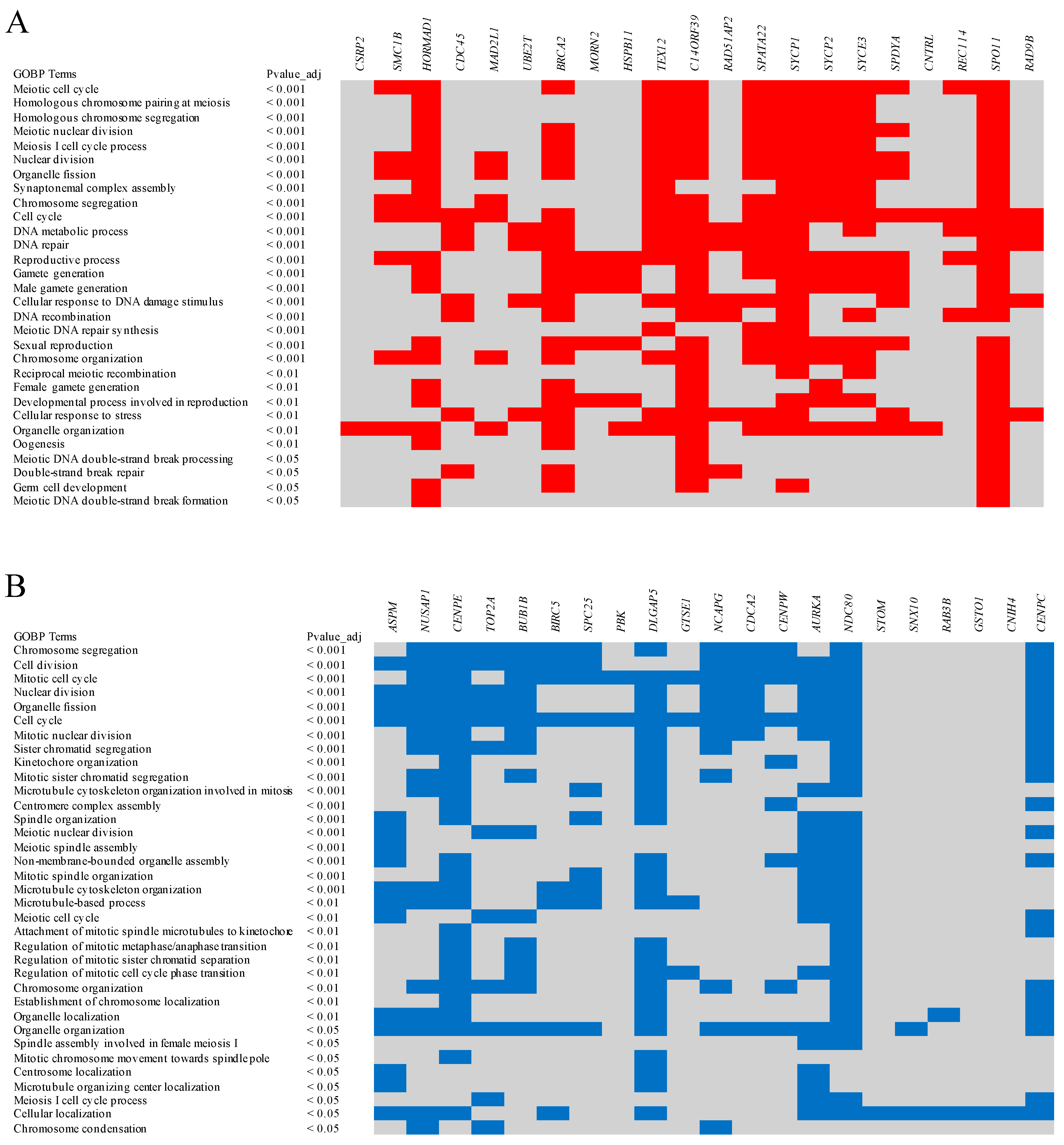
Hereafter, we study, in particular, the genes enriched in the chicken female germ cells of stages fS3-fS4 (at E16—1 week) because the primary oocytes arrested in meiotic prophase I and accumulated in germ-cell cysts or enclosed by flattened granulosa cells were identified at these time points [20,27,28]. As shown in an earlier study [25], about 29 genes were enriched in the chicken oocytes at E16—1 week and showed convergent expression patterns with human female FGCs (RA-responsive, meiotic, and oogenesis stage cells at week 11–26) (Table 1). The gene ontology biological processes (GOBPs) of all convergent genes were observed using g:Profiler with a g:SCS threshold of 0.05 [67]. Among the convergent genes, at least 21 genes, namely CSRP2, SMC1B, HORMAD1, CDC45, MAD2L1, UBE2T, BRCA2, MORN2, HSPB11, TEX12, C14ORF39, RAD51AP2, SPATA22, SYCP1, SYCP2, SYCE3, SPDYA, CNTRL, REC114, SPO11, and RAD9B, were repeatedly identified in several biological processes related to germ cell development, oocyte development, and meiosis (Figure 2A). Most of the top biological processes were related to meiotic events, such as the meiotic cell cycle, homologous chromosome pairing, homologous chromosome segregation, meiotic nuclear division, and synaptonemal complex assembly.
Male and female germ cells undergo one round of meiotic cell division during their development to reduce the number of complete sets of chromosomes (ploidy), thereby maintaining the ploidy of the species after fertilization [12]. However, the female germ cells are arrested for a longer time in meiotic prophase I as they enter into meiosis much earlier than the male germ cells. Several genes and non-coding RNA transcripts are exclusively expressed and/or developmentally regulated in order to play a critical role during oocyte meiosis [12,13,14]. For instance, with respect to the above convergent genes, CDC45 plays a critical role in DNA replication to ensure that chromosomal DNA is replicated only once per cell cycle [68]. DNA replication in the S phase is important before the cells enter meiotic prophase I. SPDYA is also a cell cycle gene preferentially expressed in the G1/S phase and plays a role in cell cycle progression, including oocyte maturation [69]. The SPO11-dependent introduction of DNA double-strand breaks across the genome is necessary to initiate meiotic recombination at the beginning of meiotic prophase I [70,71]. REC114 is also involved in the formation of DNA double-strand breaks that initiate meiotic recombination, and mutations in REC114 are responsible for human multiple pronuclei formation and early embryonic arrest, indicating the gene’s critical role in oocyte meiosis and female fertility [72].
BRCA2 is the principal DNA double-strand break repair gene for homologous recombination. Its lower expression in humans is significantly associated with primary ovarian insufficiency, characterized by the loss of ovarian follicles and oocytes [44]. The protein complex of SPATA22 and meiosis specific with OB-fold (MEIOB) interacts with replication protein A (RPA) to form a highly compacted protein-coated single-stranded DNA during meiotic homologous recombination [73]. A homozygous variant of SPATA22 is also associated with primary ovarian insufficiency [74]. Together with other cohesins, SMC1B participates in sister chromatid cohesion throughout the whole meiotic process [75]. TEX12, SYCP1, SYCP2, and SYCE3 are the components of the synaptonemal complex, which is an evolutionarily conserved structure that holds homologous chromosomes together during the pachytene stage of meiotic prophase I, providing the structural framework for meiotic recombination and crossover formation [20,76,77]. HORMAD1 is located in abundance on the synaptonemal complex in the unsynapsed region; thus, it potentially functions in the synapsis surveillance system [78]. MAD2L1 is a spindle checkpoint component and is crucial for the commitment of chromosome segregation in both mitotic and meiotic cells [79]. CNTRL is required for cell cycle progression and cytokinesis in mitotic cells; however, in the oocytes, it is localized to meiotic spindles and concentrated at the spindle poles and midbody, in order to play a role in regulating the asymmetric division of meiotic oocytes [80,81].
Moreover, the biological processes of eight convergent genes, namely RBM46, SLBP, DEPDC1, STK31, GLCCI1, ZMAT1, CCDC73, and C18ORF63, are not clearly understood in the germ cells/oocytes of chickens or humans; however, they may be well-studied in other species. Rbm46 is involved in the posttranscriptional regulation of genes essential for germ cell development in zebrafishes; also, an impairment in spermatogenesis and meiosis was observed in mutant zebrafishes [82]. SLBP protein regulates the translation of several histone mRNAs in both immature and maturing oocytes of mice [83]. Stk31 expression was detected in the embryonic gonocytes of both sexes in mice; however, the gene is dispensable for mice reproduction [84].
In order to investigate the interacting networks of the convergent genes, we used the Search Tool for the Retrieval of Interacting Genes/Proteins (STRING, Version 11.5) database [85]. According to a medium confidence search (score 0.4) and K-means clustering, all the convergent genes were clustered into three groups. Specifically, genes related to the biological processes of homologous chromosome pairing/segregation and synaptonemal complex assembly were found in cluster 1. Genes related to the biological processes of the meiotic cell cycle and meiotic nuclear division were found in cluster 2. Genes related to the biological processes of DNA replication and DNA double-strand break repair were found in cluster 3 (Figure 3A).

7. Chicken Oocyte Expressed Genes Divergent from the Human Oocyte

As shown in an earlier study [25], about 27 genes were enriched in chicken oocytes at E16—1 week but showed divergent expression patterns with human oocytes (Table 1). Here, the human cells showing enriched expression of those 27 genes are considered mitotic stage female germ cells, described beginning in week 5 by Li et al. [63]. Similar to the procedure described above, the biological processes of all the divergent genes were observed using g:Profiler. Among the divergent genes, at least 21 genes, namely ASPM, NUSAP1, CENPE, TOP2A, BUB1B, BIRC5, SPC25, PBK, DLGAP5, GTSE1, NCAPG, CDCA2, CENPW, AURKA, NDC80, STOM, SNX10, RAB3B, GSTO1, CNIH4, and CENPC, were repeatedly identified in several mitosis-related biological processes, as well as a few meiosis-related biological processes (Figure 2B). Most of the top biological processes were related to mitotic events, such as the mitotic cell cycle, chromosome segregation, cell division, organelle fission, and mitotic nuclear division.
Before entering into meiosis, male and female germ cells undergo several rounds of mitotic cell division to increase their cell population because the initially specified cells are only a few in number. Moreover, maintaining an adequate number of germ cells is necessary for successful reproductive life in both males and females because apoptosis is a common phenomenon in germ cells. Apoptosis typically occurs in male and female germ cells that show abnormal migration to the gonads and defects during the meiotic cell cycle [86,87]. Additionally, a large proportion of oocytes undergo apoptosis during the breakdown of germ-cell cysts and the formation of primordial follicles [88]. With respect to the above divergent genes, ASPM encodes a mitotic spindle pole-associated protein and participates in spindle organization, spindle positioning, and cytokinesis in all dividing cells [89]. NUSAP1 is involved in spindle microtubule organization, with selective expression in proliferating cells, and it peaks at the transition of G2 to mitosis [90]. DLGAP5 is part of a multicomponent complex that affects the growth or stability of spindle microtubules and is required for spindle microtubule organization [91]. As promising biomarkers, both NUSAP1 and DLGAP5 are highly expressed and involved in the proliferation, migration, and invasion of multiple cancer cells, including ovarian cancer cells [92,93]. GTSE1 tunes microtubule stability in mitosis to ensure chromosome alignment and segregation by suppressing MCAK (mitotic centromere-associated kinesin) microtubule depolymerase activity [94].
CENPE is one of the mitotic spindle assembly checkpoint components localized to kinetochores. In addition, it is required for the prevention of premature advancement to the anaphase in the presence of unattached kinetochores [95]. BUB1B is also a mitotic spindle assembly checkpoint component; however, it plays an important role in the kinetochore association of other spindle checkpoint proteins [96]. To form a kinetochore, which is essential for proper chromosome segregation during mitosis, constitutive centromere-associated network (CCAN) proteins are assembled on the centromere chromatin that comprises the centromere-specific histone CENPA [97]. CENPC is a CCAN protein and directly interacts with CENPA to nucleate the kinetochore structure [97]. Furthermore, CENPW, an interacting partner of CENPT, is also a component of the centromeric complex required for proper chromosome segregation during mitosis [98]. The NDC80 complex, which consists of NDC80, SPC25, and two other members, contributes to the microtubule–kinetochore attachment and spindle assembly checkpoint in mitosis. SPC25 is additionally required for chromosome alignment, spindle formation, and proper spindle checkpoint signaling during oocyte meiosis [99]. AURKA is a member of the aurora family of kinases, which consists of AURKA, AURKB, and AURKC. They exhibit different subcellular localization; however, they work together to execute cell division successfully [100]. AURKA is mainly involved in bipolar spindle formation and chromosome alignment in mitosis and meiosis. AURKB is a chromosome-localized protein in mitosis and meiosis and is involved in multiple processes during cell division. AURKC is a chromosome- and spindle pole-localized protein expressed exclusively in meiosis [101].
NCAPG is one of the non-SMC (structural maintenance of chromosomes) subunits of condensin I, which play an important role in the condensation and segregation of chromosomes during mitosis [102]. Protein phosphatase 1 (PP1) is a major serine/threonine protein phosphatase involved in a wide range of cellular processes, including the condensation and segregation of chromosomes during mitosis, and it was reported that CDCA2 selectively recruits PP1γ isoform onto mitotic chromatin in the anaphase and into the following interphase [103,104]. PBK is a mitotic serine/threonine protein kinase overexpressed in various actively proliferative normal cells as well as malignant tumor cells; thus, it may be a potential therapeutic target in various malignant tumors [105]. The chromatin remodeling-related protein-coding gene TOP2A was reported to be a major factor in the reprogramming properties of oocytes and human embryonic stem cells (ESCs) [106]. BIRC5 regulates cell division, inhibits apoptosis in most cancer cells, and plays a role in normal physiological events, including oogenesis and embryogenesis [107]. STOM is a ubiquitously expressed gene, and its product is an oligomeric, monotopic membrane protein associated with cholesterol-rich membranes [108]. SNX10 is a member of the sorting nexin protein families that possess a PX (phox homology) domain; it is responsible for membrane attachment to organelles of the secretory and endocytic systems via the binding of phosphoinositide lipids. Particularly, SNX10 enhances the fusion of Golgi-derived vesicles with endosomes [109]. CNIH4 is an endoplasmic reticulum (ER)/ER–Golgi intermediate complex (ER/ERGIC) localized protein, which promotes the exit of G protein-coupled receptors (GPCRs) from the early secretory pathway, most likely through interactions with the COPII component proteins [110]. RAB3B is one of the members of the Rab family. It exhibits GTPase activities and acts as a central regulator of vesicular traffic [111,112]. RAB3B expression has been associated with several cancer cells, where the silencing of RAB3B significantly inhibits cell proliferation and promotes apoptosis [112,113]. GSTO2 was detected ubiquitously; however, it was localized in the cytoplasm only or both the cytoplasm and the nucleus in a cell-specific manner. GSTO2 plays an important role in cellular signaling [114].
Moreover, the divergent genes for which the germ cell/oocyte-related biological processes are not clearly known in chickens or humans are SAT1, CKB, MGST3, COA7, LITAF, and SERPINI1. The role of sat1 as a membrane transporter (sulphate transporter) was reported in the oocytes of amphibians [115]. CKB, which encodes the enzyme creatine kinase, was implicated in sperm quality and maturity in men with oligospermia [116]. The expression level of LITAF may control sperm survival in hen oviducts as higher levels of it in the vagina lead to sperm degradation and elimination, while lower levels in the utero–vaginal junction permit sperm survival in sperm storage tubules [117].
The interacting networks of the divergent genes were also investigated using the STRING database. According to a medium confidence search (score 0.4) and K-means clustering, most of the genes related to the biological processes of the mitotic cell cycle and chromosome segregation were strongly interactive and found in cluster 1. In contrast, most of the divergent genes related to the biological processes of organelle organization and intracellular trafficking, which were found in cluster 2, were not interactive. Similarly, the divergent genes related to the biological processes of organelle localization and cellular signaling, which were found in cluster 3, were poorly interactive (Figure 3B).

8. Conclusions

This review reports that germ cell specification and migration before settlement in the gonads and ovarian follicular development and maturation after sexual maturity exhibit species-specific events when comparing chickens and humans. Furthermore, the middle stages of development, including the differentiation of oogonia into primary oocytes and the formation of primary and secondary follicles, are essentially similar between chickens and humans. Therefore, genes critically involved in oocyte meiosis, such as those involved in homologous chromosome pairing and segregation, synaptonemal complex assembly, DNA replication, double-strand break repair, meiotic nuclear division, etc., are convergent between chickens and humans. Germ cell specification and the migratory route of the early mitotic germ cells in humans differ from what is observed in chickens. This may be a reason for a set of germ-cell-specific genes that shows divergent expression and functions between humans and chickens.

Author Contributions

D.R. and J.Y.H. designed the study. D.R. reviewed the literature and wrote the manuscript. J.Y.H. critically checked the content of the manuscript. All authors have read and agreed to the published version of the manuscript.

Funding

This work was supported by the National Research Foundation of Korea (NRF) grant funded by the Korea government (MSIP) (No. NRF-2015R1A3A2033826).

Institutional Review Board Statement

All experimental procedures and care of chickens were approved by the Institute of Laboratory Animal Resources, Seoul National University, Korea, and all methods were carried out in accordance with ARRIVE (Animal Research: Reporting of In Vivo Experiments) guidelines and approved by the Institutional Animal Care and Use Committee (IACUC, SNU-190401-1-1) of Seoul National University, Korea.

Informed Consent Statement

Not applicable.

Data Availability Statement

The single-cell RNA sequencing data of chicken oocytes are available in the SRA database under the accession code PRJNA761874. The transcripts per kilobase million count tables of the human female FGCs were downloaded from the NCBI GEO database (GSE86146) and reanalyzed for the present paper.

Conflicts of Interest

The authors declare no conflict of interest.

References

  1. Jamieson-Lucy, A.; Mullins, M.C. The vertebrate Balbiani body, germ plasm, and oocyte polarity. Curr. Top. Dev. Biol. 2019, 135, 1–34. [Google Scholar] [PubMed]
  2. Rengaraj, D.; Lee, B.R.; Lee, S.I.; Seo, H.W.; Han, J.Y. Expression patterns and miRNA regulation of DNA methyltransferases in chicken primordial germ cells. PLoS ONE 2011, 6, e19524. [Google Scholar] [CrossRef] [PubMed]
  3. Saitou, M.; Miyauchi, H. Gametogenesis from pluripotent stem cells. Cell Stem Cell 2016, 18, 721–735. [Google Scholar] [CrossRef]
  4. Choi, H.J.; Jung, K.M.; Rengaraj, D.; Lee, K.Y.; Yoo, E.; Kim, T.H.; Han, J.Y. Single-cell RNA sequencing of mitotic-arrested prospermatogonia with DAZL::GFP chickens and revealing unique epigenetic reprogramming of chickens. J. Anim. Sci. Biotechnol. 2022, 13, 64. [Google Scholar] [CrossRef]
  5. Rengaraj, D.; Hong, Y.H. Effects of dietary vitamin E on fertility functions in poultry species. Int. J. Mol. Sci. 2015, 16, 9910–9921. [Google Scholar] [CrossRef]
  6. Nitta, H.; Mason, J.I.; Bahr, J.M. Localization of 3 beta-hydroxysteroid dehydrogenase in the chicken ovarian follicle shifts from the theca layer to granulosa layer with follicular maturation. Biol. Reprod. 1993, 48, 110–116. [Google Scholar] [CrossRef]
  7. Lovell, T.M.; Gladwell, R.T.; Groome, N.P.; Knight, P.G. Ovarian follicle development in the laying hen is accompanied by divergent changes in inhibin A, inhibin B, activin A and follistatin production in granulosa and theca layers. J. Endocrinol. 2003, 177, 45–55. [Google Scholar] [CrossRef]
  8. Okumura, H. Avian egg and egg coat. Adv. Exp. Med. Biol. 2017, 1001, 75–90. [Google Scholar]
  9. Hamazaki, N.; Kyogoku, H.; Araki, H.; Miura, F.; Horikawa, C.; Hamada, N.; Shimamoto, S.; Hikabe, O.; Nakashima, K.; Kitajima, T.S.; et al. Reconstitution of the oocyte transcriptional network with transcription factors. Nature 2021, 589, 264–269. [Google Scholar] [CrossRef]
  10. Jukam, D.; Shariati, S.A.M.; Skotheim, J.M. Zygotic genome activation in vertebrates. Dev. Cell 2017, 42, 316–332. [Google Scholar] [CrossRef]
  11. Rengaraj, D.; Hwang, Y.S.; Lee, H.C.; Han, J.Y. Zygotic genome activation in the chicken: A comparative review. Cell. Mol. Life Sci. 2020, 77, 1879–1891. [Google Scholar] [CrossRef] [PubMed]
  12. MacLennan, M.; Crichton, J.H.; Playfoot, C.J.; Adams, I.R. Oocyte development, meiosis and aneuploidy. Semin. Cell Dev. Biol. 2015, 45, 68–76. [Google Scholar] [CrossRef] [PubMed]
  13. Peng, Y.; Chang, L.; Wang, Y.; Wang, R.; Hu, L.; Zhao, Z.; Geng, L.; Liu, Z.; Gong, Y.; Li, J.; et al. Genome-wide differential expression of long noncoding RNAs and mRNAs in ovarian follicles of two different chicken breeds. Genomics 2019, 111, 1395–1403. [Google Scholar] [CrossRef]
  14. Li, Q.; Li, J.; Li, C.; Wu, X.; Si, S.J.; Yang, P.K.; Li, W.T.; Han, R.L.; Li, G.X.; Liu, X.J.; et al. Transcriptome identification and characterization of long non-coding RNAs in the ovary of hens at four stages. Anim. Biotechnol. 2022, 1–12, Online ahead of print. [Google Scholar] [CrossRef] [PubMed]
  15. Tsunekawa, N.; Naito, M.; Sakai, Y.; Nishida, T.; Noce, T. Isolation of chicken vasa homolog gene and tracing the origin of primordial germ cells. Development 2000, 127, 2741–2750. [Google Scholar] [CrossRef]
  16. Kang, K.S.; Lee, H.C.; Kim, H.J.; Lee, H.G.; Kim, Y.M.; Lee, H.J.; Park, Y.H.; Yang, S.Y.; Rengaraj, D.; Park, T.S.; et al. Spatial and temporal action of chicken primordial germ cells during initial migration. Reproduction 2015, 149, 179–187. [Google Scholar] [CrossRef]
  17. Lee, H.C.; Choi, H.J.; Lee, H.G.; Lim, J.M.; Ono, T.; Han, J.Y. DAZL expression explains origin and central formation of primordial germ cells in chickens. Stem Cells Dev. 2016, 25, 68–79. [Google Scholar] [CrossRef]
  18. Kim, Y.M.; Han, J.Y. The early development of germ cells in chicken. Int. J. Dev. Biol. 2018, 62, 145–152. [Google Scholar] [CrossRef]
  19. Stebler, J.; Spieler, D.; Slanchev, K.; Molyneaux, K.A.; Richter, U.; Cojocaru, V.; Tarabykin, V.; Wylie, C.; Kessel, M.; Raz, E. Primordial germ cell migration in the chick and mouse embryo: The role of the chemokine SDF-1/CXCL12. Dev. Biol. 2004, 272, 351–361. [Google Scholar] [CrossRef]
  20. Zheng, Y.H.; Rengaraj, D.; Choi, J.W.; Park, K.J.; Lee, S.I.; Han, J.Y. Expression pattern of meiosis associated SYCP family members during germline development in chickens. Reproduction 2009, 138, 483–492. [Google Scholar] [CrossRef]
  21. Rengaraj, D.; Lee, B.R.; Park, K.J.; Lee, S.I.; Kang, K.S.; Choi, J.W.; Kang, S.J.; Song, G.; Han, J.Y. The distribution of neuron-specific gene family member 1 in brain and germ cells: Implications for the regulation of germ-line development by brain. Dev. Dyn. 2011, 240, 850–861. [Google Scholar] [CrossRef] [PubMed]
  22. Yang, S.Y.; Lee, H.J.; Lee, H.C.; Hwang, Y.S.; Park, Y.H.; Ono, T.; Han, J.Y. The dynamic development of germ cells during chicken embryogenesis. Poult. Sci. 2018, 97, 650–657. [Google Scholar] [CrossRef] [PubMed]
  23. Yu, J.N.; Yan, L.Y.; Chen, Z.; Li, H.; Ying, S.J.; Zhu, H.X.; Shi, Z.D. Investigating right ovary degeneration in chick embryos by transcriptome sequencing. J. Reprod. Dev. 2017, 63, 295–303. [Google Scholar] [CrossRef] [PubMed]
  24. Smith, C.A.; Roeszler, K.N.; Bowles, J.; Koopman, P.; Sinclair, A.H. Onset of meiosis in the chicken embryo; evidence of a role for retinoic acid. BMC Dev. Biol. 2008, 8, 85. [Google Scholar] [CrossRef]
  25. Rengaraj, D.; Cha, D.G.; Lee, H.J.; Lee, K.Y.; Ha Choi, Y.; Jung, K.M.; Kim, Y.M.; Choi, H.J.; Choi, H.J.; Yoo, E.; et al. Dissecting chicken germ cell dynamics by combining a germ cell tracing transgenic chicken model with single-cell RNA sequencing. Comput. Struct. Biotecnol. J. 2022, 20, 1654–1669. [Google Scholar] [CrossRef] [PubMed]
  26. Del Priore, L.; Pigozzi, M.I. Chromosomal axis formation and meiotic progression in chicken oocytes: A quantitative analysis. Cytogenet. Genome Res. 2012, 137, 15–21. [Google Scholar] [CrossRef]
  27. Rengaraj, D.; Lee, S.I.; Yoo, M.; Kim, T.H.; Song, G.; Han, J.Y. Expression and knockdown analysis of glucose phosphate isomerase in chicken primordial germ cells. Biol. Reprod. 2012, 87, 57. [Google Scholar] [CrossRef]
  28. Guo, C.Q.; Liu, G.; Zhao, D.; Mi, Y.L.; Zhang, C.Q.; Li, J. Interaction of follicle-stimulating hormone and stem cell factor to promote primordial follicle assembly in the chicken. Front. Endocrinol. 2019, 10, 91. [Google Scholar] [CrossRef]
  29. Johnson, P.A. Follicle selection in the avian ovary. Reprod. Domest. Anim. 2012, 47, 283–287. [Google Scholar] [CrossRef]
  30. Nie, R.X.; Zheng, X.T.; Zhang, W.H.; Zhang, B.; Ling, Y.; Zhang, H.; Wu, C.X. Morphological characteristics and transcriptome landscapes of chicken follicles during selective development. Animals 2022, 12, 713. [Google Scholar] [CrossRef]
  31. Perry, M.M. Nuclear events from fertilization to the early cleavage stages in the domestic fowl (Gallus domesticus). J. Anat. 1987, 150, 99–109. [Google Scholar] [PubMed]
  32. Tang, W.W.C.; Kobayashi, T.; Irie, N.; Dietmann, S.; Surani, M.A. Specification and epigenetic programming of the human germ line. Nat. Rev. Genet. 2016, 17, 585–600. [Google Scholar] [CrossRef] [PubMed]
  33. Canovas, S.; Campos, R.; Aguilar, E.; Cibelli, J.B. Progress towards human primordial germ cell specification in vitro. Mol. Hum. Reprod. 2017, 23, 4–15. [Google Scholar] [CrossRef] [PubMed]
  34. Chen, D.; Sun, N.; Hou, L.; Kim, R.; Faith, J.; Aslanyan, M.; Tao, Y.; Zheng, Y.; Fu, J.; Liu, W.; et al. Human primordial germ cells are specified from lineage-primed progenitors. Cell Rep. 2019, 29, 4568–4582.e5. [Google Scholar] [CrossRef] [PubMed]
  35. Sybirna, A.; Tang, W.W.C.; Smela, M.P.; Dietmann, S.; Gruhn, W.H.; Brosh, R.; Surani, M.A. A critical role of PRDM14 in human primordial germ cell fate revealed by inducible degrons. Nat. Commun. 2020, 11, 1282. [Google Scholar] [CrossRef]
  36. Singersam, J.; Goldstein, L.; Dai, A.H.; Gartler, S.M.; Riggs, A.D. A potentially critical Hpa-II site of the X-chromosome-linked PGK1 gene is unmethylated prior to the onset of meiosis of human oogenic cells. Proc. Natl. Acad. Sci. USA 1992, 89, 1413–1417. [Google Scholar] [CrossRef]
  37. Dalbies-Tran, R.; Cadoret, V.; Desmarchais, A.; Elis, S.; Maillard, V.; Monget, P.; Monniaux, D.; Reynaud, K.; Saint-Dizier, M.; Uzbekova, S. A comparative analysis of oocyte development in mammals. Cells 2020, 9, 1002. [Google Scholar] [CrossRef]
  38. Jones, K.T. Meiosis in oocytes: Predisposition to aneuploidy and its increased incidence with age. Hum. Reprod. Update 2008, 14, 143–158. [Google Scholar] [CrossRef]
  39. Baudat, F.; Buard, J.; Grey, C.; Fledel-Alon, A.; Ober, C.; Przeworski, M.; Coop, G.; de Massy, B. PRDM9 is a major determinant of meiotic recombination hotspots in humans and mice. Science 2010, 327, 836–840. [Google Scholar] [CrossRef]
  40. Hertig, A.T. The primary human oocyte: Some observations on the fine structure of Balbiani’s vitelline body and the origin of the annulate lamellae. Am. J. Anat. 1968, 122, 107–137. [Google Scholar] [CrossRef]
  41. Pepling, M.E.; Wilhelm, J.E.; O’Hara, A.L.; Gephardt, G.W.; Spradling, A.C. Mouse oocytes within germ cell cysts and primordial follicles contain a Balbiani body. Proc. Natl. Acad. Sci. USA 2007, 104, 187–192. [Google Scholar] [CrossRef] [PubMed] [Green Version]
  42. Faddy, M.J.; Gosden, R.G.; Gougeon, A.; Richardson, S.J.; Nelson, J.F. Accelerated disappearance of ovarian follicles in mid-life: Implications for forecasting menopause. Hum. Reprod. 1992, 7, 1342–1346. [Google Scholar] [CrossRef]
  43. Ford, E.A.; Beckett, E.L.; Roman, S.D.; McLaughlin, E.A.; Sutherland, J.M. Advances in human primordial follicle activation and premature ovarian insufficiency. Reproduction 2020, 159, R15–R29. [Google Scholar] [CrossRef] [PubMed]
  44. Miao, Y.L.; Wang, P.; Xie, B.T.; Yang, M.; Li, S.; Cui, Z.K.; Fan, Y.; Li, M.; Xiong, B. BRCA2 deficiency is a potential driver for human primary ovarian insufficiency. Cell Death Dis. 2019, 10, 474. [Google Scholar] [CrossRef] [PubMed]
  45. Mcnatty, K.P. Hormonal correlates of follicular development in the human-ovary. Aust. J. Biol. Sci. 1981, 34, 249–268. [Google Scholar] [CrossRef] [PubMed]
  46. Gougeon, A. Regulation of ovarian follicular development in primates: Facts and hypotheses. Endocr. Rev. 1996, 17, 121–155. [Google Scholar] [CrossRef]
  47. Simon, L.E.; Kumar, T.R.; Duncan, F.E. In vitro ovarian follicle growth: A comprehensive analysis of key protocol variables. Biol. Reprod. 2020, 103, 455–470. [Google Scholar] [CrossRef]
  48. Mfoundou, J.D.L.; Guo, Y.J.; Liu, M.M.; Ran, X.R.; Fu, D.H.; Yan, Z.Q.; Li, M.N.; Wang, X.R. The morphological and histological study of chicken left ovary during growth and development among Hy-line brown layers of different ages. Poult. Sci. 2021, 100, 101191. [Google Scholar] [CrossRef]
  49. Joyner, C.J.; Peddie, M.J.; Taylor, T.G. The effect of age on egg-production in the domestic hen. Gen. Comp. Endocr. 1987, 65, 331–336. [Google Scholar] [CrossRef]
  50. Cheng, S.B.; Li, X.Q.; Wang, J.X.; Wu, Y.; Li, P.; Pi, J.S. The effect of light on follicular development in laying hens. Anim. Biosci. 2021, 34, 1766–1775. [Google Scholar] [CrossRef]
  51. Mohammadi, H.; Ansari-Pirsaraei, Z. Follicle Diameters, Egg weight, and egg production performance in old laying hens injected with growth hormone and testosterone. J. Agr. Sci. Tech. 2016, 18, 949–959. [Google Scholar]
  52. Amevor, F.K.; Cui, Z.F.; Du, X.X.; Ning, Z.F.; Shu, G.; Jin, N.N.; Deng, X.; Tian, Y.F.; Zhang, Z.C.; Kang, X.C.; et al. Combination of quercetin and vitamin E supplementation promotes yolk precursor synthesis and follicle development in aging breeder hens via liver-blood-ovary signal axis. Animals 2021, 11, 1915. [Google Scholar] [CrossRef] [PubMed]
  53. Wang, J.P.; Jia, R.; Gong, H.J.; Celi, P.; Zhuo, Y.; Ding, X.M.; Bai, S.P.; Zeng, Q.F.; Yin, H.D.; Xu, S.Y.; et al. The effect of oxidative stress on the chicken ovary: Involvement of microbiota and melatonin interventions. Antioxidants 2021, 10, 1422. [Google Scholar] [CrossRef] [PubMed]
  54. Gougeon, A. Ovarian follicular growth in humans: Ovarian ageing and population of growing follicles. Maturitas 1998, 30, 137–142. [Google Scholar] [CrossRef]
  55. Zhou, J.W.; Peng, X.W.; Mei, S.Q. Autophagy in ovarian follicular development and atresia. Int. J. Biol. Sci. 2019, 15, 726–737. [Google Scholar] [CrossRef] [PubMed]
  56. Amargant, F.; Manuel, S.L.; Tu, Q.; Parkes, W.S.; Rivas, F.; Zhou, L.T.; Rowley, J.E.; Villanueva, C.E.; Hornick, J.E.; Shekhawat, G.S.; et al. Ovarian stiffness increases with age in the mammalian ovary and depends on collagen and hyaluronan matrices. Aging Cell 2020, 19, e13259. [Google Scholar] [CrossRef] [PubMed]
  57. Tatone, C.; Amicarelli, F.; Carbone, M.C.; Monteleone, P.; Caserta, D.; Marci, R.; Artini, P.G.; Piomboni, P.; Focarelli, R. Cellular and molecular aspects of ovarian follicle ageing. Hum. Reprod. Update 2008, 14, 131–142. [Google Scholar] [CrossRef]
  58. Broekmans, F.J.; Soules, M.R.; Fauser, B.C. Ovarian aging: Mechanisms and clinical consequences. Endocr. Rev. 2009, 30, 465–493. [Google Scholar] [CrossRef]
  59. Gold, E.B. The timing of the age at which natural menopause occurs. Obstet. Gynecol. Clin. N. Am. 2011, 38, 425–440. [Google Scholar] [CrossRef]
  60. Bukovsky, A.; Caudle, M.R.; Svetlikova, M.; Wimalasena, J.; Ayala, M.E.; Dominguez, R. Oogenesis in adult mammals, including humans: A review. Endocrine 2005, 26, 301–316. [Google Scholar] [CrossRef]
  61. Sasaki, K.; Nakamura, T.; Okamoto, K.; Yabuta, Y.; Iwatani, C.; Tsuchiya, H.; Seita, Y.; Nakamura, S.; Shiraki, N.; Takakuwa, T.; et al. The germ cell fate of cynomolgus monkeys is specified in the nascent amnion. Dev. Cell 2016, 39, 169–185. [Google Scholar] [CrossRef] [PubMed]
  62. Hwang, Y.S.; Seo, M.; Lee, B.R.; Lee, H.J.; Park, Y.H.; Kim, S.K.; Lee, H.C.; Choi, H.J.; Yoon, J.; Kim, H.; et al. The transcriptome of early chicken embryos reveals signaling pathways governing rapid asymmetric cellularization and lineage segregation. Development 2018, 145, dev157453. [Google Scholar] [CrossRef] [PubMed] [Green Version]
  63. Li, L.; Dong, J.; Yan, L.; Yong, J.; Liu, X.; Hu, Y.; Fan, X.; Wu, X.; Guo, H.; Wang, X.; et al. Single-cell RNA-seq analysis maps development of human germline cells and gonadal niche interactions. Cell Stem Cell 2017, 20, 858–873. [Google Scholar] [CrossRef] [PubMed]
  64. Jukic, A.M.; Baird, D.D.; Weinberg, C.R.; McConnaughey, D.R.; Wilcox, A.J. Length of human pregnancy and contributors to its natural variation. Hum. Reprod. 2013, 28, 2848–2855. [Google Scholar] [CrossRef]
  65. Hamburger, V.; Hamilton, H.L. A series of normal stages in the development of the chick embryo. J. Morphol. 1951, 88, 49–92. [Google Scholar] [CrossRef]
  66. Sheng, G.J. Day-1 chick development. Dev. Dyn. 2014, 243, 357–367. [Google Scholar] [CrossRef]
  67. Raudvere, U.; Kolberg, L.; Kuzmin, I.; Arak, T.; Adler, P.; Peterson, H.; Vilo, J. g:Profiler: A web server for functional enrichment analysis and conversions of gene lists (2019 update). Nucleic Acids Res. 2019, 47, W191–W198. [Google Scholar] [CrossRef]
  68. Pollok, S.; Bauerschmidt, C.; Sanger, J.; Nasheuer, H.P.; Grosse, F. Human Cdc45 is a proliferation-associated antigen. FEBS J. 2007, 274, 3669–3684. [Google Scholar] [CrossRef]
  69. Porter, L.A.; Dellinger, R.W.; Tynan, J.A.; Barnes, E.A.; Kong, M.; Lenormand, J.L.; Donoghue, D.J. Human Speedy: A novel cell cycle regulator that enhances proliferation through activation of Cdk2. J. Cell Biol. 2002, 157, 357–366. [Google Scholar] [CrossRef]
  70. Romanienko, P.J.; Camerini-Otero, R.D. Cloning, characterization, and localization of mouse and human SPO11. Genomics 1999, 61, 156–169. [Google Scholar] [CrossRef]
  71. Bellani, M.A.; Boateng, K.A.; McLeod, D.; Camerini-Otero, R.D. The expression profile of the major mouse SPO11 isoforms indicates that SPO11 beta introduces double strand breaks and suggests that SPO11 alpha has an additional role in prophase in both spermatocytes and oocytes. Mol. Cell Biol. 2010, 30, 4391–4403. [Google Scholar] [CrossRef] [PubMed]
  72. Wang, W.; Dong, J.; Chen, B.; Du, J.; Kuang, Y.; Sun, X.; Fu, J.; Li, B.; Mu, J.; Zhang, Z.; et al. Homozygous mutations in REC114 cause female infertility characterised by multiple pronuclei formation and early embryonic arrest. J. Med. Genet. 2020, 57, 187–194. [Google Scholar] [CrossRef]
  73. Ribeiro, J.; Dupaigne, P.; Petrillo, C.; Duquenne, C.; Veaute, X.; Saintome, C.; Busso, D.; Guerois, R.; Martini, E.; Livera, G. The meiosis-specific MEIOB-SPATA22 complex cooperates with RPA to form a compacted mixed MEIOB/SPATA22/RPA/ssDNA complex. DNA Repair 2021, 102, 103097. [Google Scholar] [CrossRef] [PubMed]
  74. Yao, C.C.; Hou, D.; Ji, Z.Y.; Pang, D.M.; Li, P.; Tian, R.H.; Zhang, Y.X.; Ou, N.J.; Bai, H.W.; Zhi, E.L.; et al. Bi-allelic SPATA22 variants cause premature ovarian insufficiency and nonobstructive azoospermia due to meiotic arrest. Clin. Genet. 2022, 101, 507–516. [Google Scholar] [CrossRef] [PubMed]
  75. Garcia-Cruz, R.; Brieno, M.A.; Roig, I.; Grossmann, M.; Velilla, E.; Pujol, A.; Cabero, L.; Pessarrodona, A.; Barbero, J.L.; Garcia Caldes, M. Dynamics of cohesin proteins REC8, STAG3, SMC1 beta and SMC3 are consistent with a role in sister chromatid cohesion during meiosis in human oocytes. Hum. Reprod. 2010, 25, 2316–2327. [Google Scholar] [CrossRef] [PubMed]
  76. Davies, O.R.; Maman, J.D.; Pellegrini, L. Structural analysis of the human SYCE2-TEX12 complex provides molecular insights into synaptonemal complex assembly. Open Biol. 2012, 2, 120099. [Google Scholar] [CrossRef] [PubMed]
  77. Sciurano, R.B.; Pigozzi, M.I.; Benavente, R. Disassembly of the synaptonemal complex in chicken oocytes analyzed by super-resolution microscopy. Chromosoma 2019, 128, 443–451. [Google Scholar] [CrossRef]
  78. Kurahashi, H.; Kogo, H.; Tsutsumi, M.; Inagaki, H.; Ohye, T. Failure of homologous synapsis and sex-specific reproduction problems. Front. Genet. 2012, 3, 112. [Google Scholar] [CrossRef]
  79. Homer, H.A.; McDougall, A.; Levasseur, M.; Murdoch, A.P.; Herbert, M. RNA interference in meiosis I human oocytes: Towards an understanding of human aneuploidy. Mol. Hum. Reprod. 2005, 11, 397–404. [Google Scholar] [CrossRef]
  80. Gromley, A.; Jurczyk, A.; Sillibourne, J.; Halilovic, E.; Mogensen, M.; Groisman, I.; Blomberg, M.; Doxsey, S. A novel human protein of the maternal centriole is required for the final stages of cytokinesis and entry into S phase. J. Cell Biol. 2003, 161, 535–545. [Google Scholar] [CrossRef]
  81. Sun, T.Y.; Wang, H.Y.; Kwon, J.W.; Yuan, B.; Lee, I.W.; Cui, X.S.; Kim, N.H. Centriolin, a centriole-appendage protein, regulates peripheral spindle migration and asymmetric division in mouse meiotic oocytes. Cell Cycle 2017, 16, 1774–1780. [Google Scholar] [CrossRef] [PubMed]
  82. Dai, X.; Cheng, X.; Huang, J.; Gao, Y.; Wang, D.; Feng, Z.; Zhai, G.; Lou, Q.; He, J.; Wang, Z.; et al. Rbm46, a novel germ cell-specific factor, modulates meiotic progression and spermatogenesis. Biol. Reprod. 2021, 104, 1139–1153. [Google Scholar] [CrossRef] [PubMed]
  83. Allard, P.; Yang, Q.; Marzluff, W.F.; Clarke, H.J. The stem-loop binding protein regulates translation of histone mRNA during mammalian oogenesis. Dev. Biol. 2005, 286, 195–206. [Google Scholar] [CrossRef] [PubMed]
  84. Zhou, J.; Leu, N.A.; Eckardt, S.; McLaughlin, K.J.; Wang, P.J. STK31/TDRD8, a germ cell-specific factor, is dispensable for reproduction in mice. PLoS ONE 2014, 9, e89471. [Google Scholar] [CrossRef]
  85. Szklarczyk, D.; Gable, A.L.; Nastou, K.C.; Lyon, D.; Kirsch, R.; Pyysalo, S.; Doncheva, N.T.; Legeay, M.; Fang, T.; Bork, P.; et al. The STRING database in 2021: Customizable protein-protein networks, and functional characterization of user-uploaded gene/measurement sets. Nucleic Acids Res. 2021, 49, D605–D612. [Google Scholar] [CrossRef] [PubMed]
  86. Ghafari, F.; Gutierrez, C.G.; Hartshorne, G.M. Apoptosis in mouse fetal and neonatal oocytes during meiotic prophase one. BMC Dev. Biol. 2007, 7, 87. [Google Scholar] [CrossRef]
  87. Shaha, C.; Tripathi, R.; Mishra, D.P. Male germ cell apoptosis: Regulation and biology. Philos. Trans. R. Soc. Lond. B Biol. Sci. 2010, 365, 1501–1515. [Google Scholar] [CrossRef]
  88. Pepling, M.E.; Spradling, A.C. Mouse ovarian germ cell cysts undergo programmed breakdown to form primordial follicles. Dev. Biol. 2001, 234, 339–351. [Google Scholar] [CrossRef]
  89. Higgins, J.; Midgley, C.; Bergh, A.M.; Bell, S.M.; Askham, J.M.; Roberts, E.; Binns, R.K.; Sharif, S.M.; Bennett, C.; Glover, D.M.; et al. Human ASPM participates in spindle organisation, spindle orientation and cytokinesis. BMC Cell Biol. 2010, 11, 85. [Google Scholar] [CrossRef]
  90. Raemaekers, T.; Ribbeck, K.; Beaudouin, J.; Annaert, W.; Van Camp, M.; Stockmans, I.; Smets, N.; Bouillon, R.; Ellenberg, J.; Carmeliet, G. NuSAP, a novel microtubule-associated protein involved in mitotic spindle organization. J. Cell Biol. 2003, 162, 1017–1029. [Google Scholar] [CrossRef]
  91. Koffa, M.D.; Casanova, C.M.; Santarella, R.; Kocher, T.; Wilm, M.; Mattaj, I.W. HURP is part of a ran-dependent complex involved in spindle formation. Curr. Biol. 2006, 16, 743–754. [Google Scholar] [CrossRef] [PubMed]
  92. Shen, J.Y.; Yu, S.Q.; Sun, X.W.; Yin, M.C.; Fei, J.; Zhou, J.W. Identification of key biomarkers associated with development and prognosis in patients with ovarian carcinoma: Evidence from bioinformatic analysis. J. Ovarian Res. 2019, 12, 110. [Google Scholar] [CrossRef] [PubMed] [Green Version]
  93. Tang, N.; Dou, X.L.; You, X.; Shi, Q.M.; Ke, M.J.; Liu, G.D. Pan-cancer analysis of the oncogenic role of discs large homolog associated protein 5 (DLGAP5) in human tumors. Cancer Cell Int. 2021, 21, 457. [Google Scholar] [CrossRef] [PubMed]
  94. Bendre, S.; Rondelet, A.; Hall, C.; Schmidt, N.; Lin, Y.C.; Brouhard, G.J.; Bird, A.W. GTSE1 tunes microtubule stability for chromosome alignment and segregation by inhibiting the microtubule depolymerase MCAK. J. Cell Biol. 2016, 215, 631–647. [Google Scholar] [CrossRef] [PubMed]
  95. Mao, Y.H.; Desai, A.; Cleveland, D.W. Microtubule capture by CENP-E silences BubR1-dependent mitotic checkpoint signaling. J. Cell Biol. 2005, 170, 873–880. [Google Scholar] [CrossRef]
  96. Chen, R.H. BubR1 is essential for kinetochore localization of other spindle checkpoint proteins and its phosphorylation requires Mad1. J. Cell Biol. 2002, 158, 487–496. [Google Scholar] [CrossRef]
  97. Watanabe, R.; Hara, M.; Okumura, E.I.; Herve, S.; Fachinetti, D.; Ariyoshi, M.; Fukagawa, T. CDK1-mediated CENP-C phosphorylation modulates CENP-A binding and mitotic kinetochore localization. J. Cell Biol. 2019, 218, 4042–4062. [Google Scholar] [CrossRef]
  98. Kim, H.; Lee, M.; Lee, S.; Park, B.; Koh, W.; Lee, D.J.; Lim, D.S.; Lee, S. Cancer-upregulated gene 2 (CUG2), a new component of centromere complex, is required for kinetochore function. Mol. Cells 2009, 27, 697–701. [Google Scholar] [CrossRef]
  99. Sun, S.C.; Lee, S.E.; Xu, Y.N.; Kim, N.H. Perturbation of Spc25 expression affects meiotic spindle organization, chromosome alignment and spindle assembly checkpoint in mouse oocytes. Cell Cycle 2010, 9, 4552–4559. [Google Scholar] [CrossRef]
  100. Crane, R.; Gadea, B.; Littlepage, L.; Wu, H.; Ruderman, J.V. Aurora A, meiosis and mitosis. Biol. Cell 2004, 96, 215–229. [Google Scholar] [CrossRef]
  101. Nguyen, A.L.; Schindler, K. Specialize and divide (twice): Functions of three aurora kinase homologs in mammalian oocyte meiotic maturation. Trends Genet. 2017, 33, 349–363. [Google Scholar] [CrossRef]
  102. Murphy, L.A.; Sarge, K.D. Phosphorylation of CAP-G is required for its chromosomal DNA localization during mitosis. Biochem. Biophys. Res. Commun. 2008, 377, 1007–1011. [Google Scholar] [CrossRef] [PubMed] [Green Version]
  103. Trinkle-Mulcahy, L.; Andersen, J.; Lam, Y.W.; Moorhead, G.; Mann, M.; Lamond, A.I. Repo-Man recruits PP1 gamma to chromatin and is essential for cell viability. J. Cell Biol. 2006, 172, 679–692. [Google Scholar] [CrossRef] [PubMed]
  104. Vagnarelli, P.; Hudson, D.F.; Ribeiro, S.A.; Trinkle-Mulcahy, L.; Spence, J.M.; Lai, F.; Farr, C.J.; Lamond, A.I.; Earnshaw, W.C. Condensin and Repo-Man-PP1 co-operate in the regulation of chromosome architecture during mitosis. Nat. Cell Biol. 2006, 8, 1133–1142. [Google Scholar] [CrossRef] [PubMed]
  105. Han, Z.; Li, L.; Huang, Y.; Zhao, H.; Luo, Y. PBK/TOPK: A therapeutic target worthy of attention. Cells 2021, 10, 371. [Google Scholar] [CrossRef]
  106. Assou, S.; Cerecedo, D.; Tondeur, S.; Pantesco, V.; Hovatta, O.; Klein, B.; Hamamah, S.; De Vos, J. A gene expression signature shared by human mature oocytes and embryonic stem cells. BMC Genomics 2009, 10, 10. [Google Scholar] [CrossRef] [PubMed]
  107. Balakier, H.; Xiao, R.; Zhao, J.; Zaver, S.; Dziak, E.; Szczepanska, K.; Opas, M.; Yie, S.; Librach, C. Expression of survivin in human oocytes and preimplantation embryos. Fertil. Steril. 2013, 99, 518–525. [Google Scholar] [CrossRef] [PubMed]
  108. Rungaldier, S.; Umlauf, E.; Mairhofer, M.; Salzer, U.; Thiele, C.; Prohaska, R. Structure-function analysis of human stomatin: A mutation study. PLoS ONE 2017, 12, e0178646. [Google Scholar] [CrossRef]
  109. Teasdale, R.D.; Collins, B.M. Insights into the PX (phox-homology) domain and SNX (sorting nexin) protein families: Structures, functions and roles in disease. Biochem. J. 2012, 441, 39–59. [Google Scholar] [CrossRef]
  110. Sauvageau, E.; Rochdi, M.D.; Oueslati, M.; Hamdan, F.F.; Percherancier, Y.; Simpson, J.C.; Pepperkok, R.; Bouvier, M. CNIH4 interacts with newly synthesized GPCR and controls their export from the endoplasmic reticulum. Traffic 2014, 15, 383–400. [Google Scholar] [CrossRef] [PubMed]
  111. Zahraoui, A.; Touchot, N.; Chardin, P.; Tavitian, A. The human Rab genes encode a family of GTP-binding proteins related to yeast Ypt1 and Sec4 products involved in secretion. J. Biol. Chem. 1989, 264, 12394–12401. [Google Scholar] [CrossRef]
  112. Tsunedomi, R.; Yoshimura, K.; Kimura, Y.; Nishiyama, M.; Fujiwara, N.; Matsukuma, S.; Kanekiyo, S.; Matsui, H.; Shindo, Y.; Watanabe, Y.; et al. Elevated expression of RAB3B plays important roles in chemoresistance and metastatic potential of hepatoma cells. BMC Cancer 2022, 22, 260. [Google Scholar] [CrossRef] [PubMed]
  113. Luo, Q.L.; Liu, Y.P.; Yuan, Z.L.; Huang, L.S.; Diao, B. Expression of Rab3b in human glioma: Influence on cell proliferation and apoptosis. Curr. Pharm. Design 2021, 27, 989–995. [Google Scholar] [CrossRef] [PubMed]
  114. Wang, L.; Xu, J.; Ji, C.; Gu, S.; Lv, Y.; Li, S.; Xu, Y.; Xie, Y.; Mao, Y. Cloning, expression and characterization of human glutathione S-transferase Omega 2. Int. J. Mol. Med. 2005, 16, 19–27. [Google Scholar] [CrossRef]
  115. Markovich, D.; Regeer, R.R. Expression of membrane transporters in cane toad Bufo marinus oocytes. J. Exp. Biol. 1999, 202, 2217–2223. [Google Scholar] [CrossRef]
  116. Hallak, J.; Sharma, R.K.; Pasqualotto, F.F.; Ranganathan, P.; Thomas, A.J.; Agarwal, A. Creatine kinase as an indicator of sperm quality and maturity in men with oligospermia. Urology 2001, 58, 446–451. [Google Scholar] [CrossRef]
  117. Das, S.C.; Isobe, N.; Yoshimura, Y. Changes in the expression of interleukin-1beta and lipopolysaccharide-induced TNF factor in the oviduct of laying hens in response to artificial insemination. Reproduction 2009, 137, 527–536. [Google Scholar] [CrossRef] [Green Version]
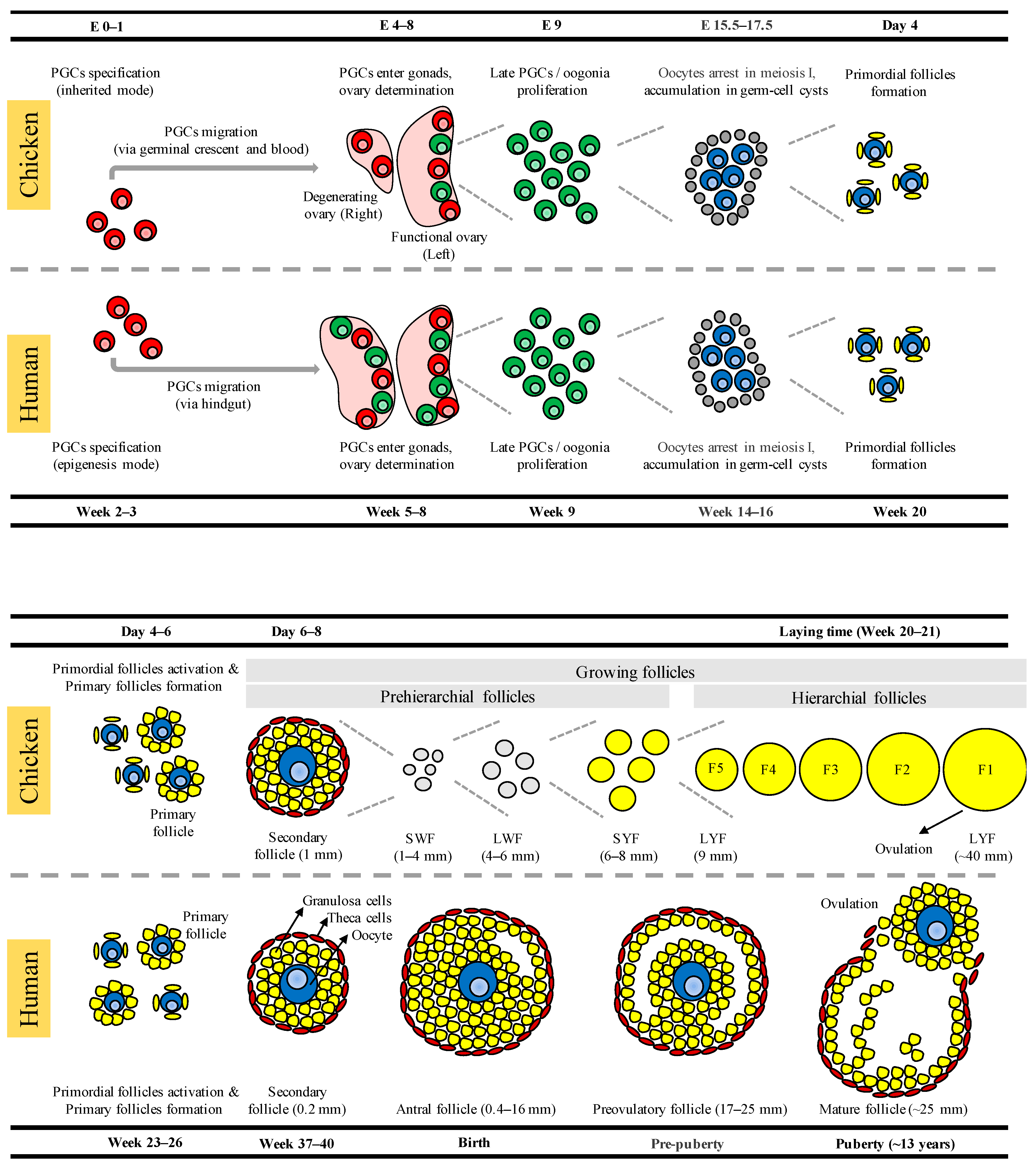
Figure 1. Schematic comparison of developing germ cells, oocytes, and ovarian follicles in chickens and humans. The primordial germ cells (PGCs) are specified by the inherited mode in chickens and by the epigenesis mode in humans. After specification, PGCs migrate to the gonads; this occurs via the germinal crescent and blood circulation in chickens and via the hindgut in humans. Further physiological events in the differentiated ovary, including oogonia proliferation and entry into meiosis, oocyte arrest in meiotic prophase I, the accumulation of oocytes in germ-cell cysts, and the formation of early follicles, are all essentially the same in both chickens and humans, except the differences in timing. In contrast, late folliculogenesis is meaningfully different between chickens and humans. SWF: small white follicles; LWF: large white follicles; SYF: small yellow follicles; LYF: large yellow follicles.
Ijms 23 11412 g001
Figure 2. Biological processes of the genes reviewed in this article. (A) Biological processes of the chicken oocyte enriched genes that are convergent with the human oocyte. (B) Biological processes of the chicken oocyte enriched genes that are divergent from the human oocyte. The involvement of convergent and divergent genes in the biological processes is emphasized in red and blue, respectively.
Ijms 23 11412 g002
Figure 3. Interacting networks of the genes reviewed in this article. (A) Interacting networks of the chicken oocyte enriched genes that are convergent with the human oocyte. (B) Interacting networks of the chicken oocyte enriched genes that are divergent from the human oocyte. Interacting genes found in clusters 1, 2, and 3 are emphasized with red, green, and blue, respectively.
Ijms 23 11412 g003
Table 1. Chicken oocyte enriched genes that showed convergent and divergent expression patterns with human female fetal germ cells (FGCs).
GeneDescriptionEnriched in ChickenEnriched in HumanChromosome in ChickenChromosome in Human
Convergent genes
CSRP2Cysteine and glycine rich protein 2OocytesOocytesChr.1Chr.12
SMC1BStructural maintenance of chromosomes 1BOocytesOocytesChr.1Chr.22
HORMAD1HORMA domain containing 1OocytesOocytesChr.25Chr.1
RBM46RNA binding motif protein 46OocytesOocytesChr.4Chr.4
SLBPStem-loop binding proteinOocytesOocytesChr.4Chr.4
DEPDC1DEP domain containing 1OocytesOocytesChr.8Chr.1
CDC45Cell division cycle 45OocytesOocytesChr.15Chr.22
MAD2L1Mitotic arrest deficient 2 like 1OocytesOocytesChr.4Chr.4
UBE2TUbiquitin conjugating enzyme E2 TOocytesOocytesChr.26Chr.1
BRCA2BRCA2, DNA repair associatedOocytesOocytesChr.1Chr.13
STK31Serine/threonine kinase 31OocytesOocytesChr.2Chr.7
GLCCI1Glucocorticoid induced 1OocytesOocytesChr.2Chr.7
ZMAT1Zinc finger matrin-type 1OocytesOocytesChr.4Chr.X
MORN2MORN repeat containing 2OocytesOocytesChr.3Chr.2
HSPB11Heat shock protein family B (small) member 11OocytesOocytesChr.8Chr.1
TEX12Testis expressed 12OocytesOocytesChr.24Chr.11
C14ORF39Chromosome 14 open reading frame 39OocytesOocytesChr.5Chr.14
RAD51AP2RAD51 associated protein 2OocytesOocytesChr.3Chr.2
SPATA22Spermatogenesis associated 22OocytesOocytesChr.19Chr.17
CCDC73Coiled-coil domain containing 73OocytesOocytesChr.5Chr.11
SYCP1Synaptonemal complex protein 1OocytesOocytesChr.26Chr.1
SYCP2Synaptonemal complex protein 2OocytesOocytesChr.20Chr.20
SYCE3Synaptonemal complex central element protein 3OocytesOocytesChr.1Chr.22
C18ORF63Chromosome 18 open reading frame 63OocytesOocytesChr.2Chr.18
SPDYASpeedy/RINGO cell cycle regulator family member AOocytesOocytesChr.3Chr.2
CNTRLCentriolinOocytesOocytesChr.17Chr.9
REC114REC114 meiotic recombination proteinOocytesOocytesChr.10Chr.15
SPO11SPO11, initiator of meiotic double stranded breaksOocytesOocytesChr.20Chr.20
RAD9BRAD9 checkpoint clamp component BOocytesOocytesChr.15Chr.12
Divergent genes
SAT1Spermidine/spermine N1-acetyltransferase 1OocytesMitotic GCsChr.1Chr.X
CKBCreatine kinase BOocytesMitotic GCsChr.5Chr.14
ASPMAbnormal spindle microtubule assemblyOocytesMitotic GCsChr.8Chr.1
NUSAP1Nucleolar and spindle associated protein 1OocytesMitotic GCsChr.5Chr.15
CENPECentromere protein EOocytesMitotic GCsChr.4Chr.4
TOP2ATopoisomerase (DNA) II alphaOocytesMitotic GCsChr.27Chr.17
BUB1BBUB1 mitotic checkpoint serine/threonine kinase BOocytesMitotic GCsChr.5Chr.15
BIRC5Baculoviral IAP repeat containing 5OocytesMitotic GCsChr.3Chr.17
SPC25SPC25, NDC80 kinetochore complex componentOocytesMitotic GCsChr.7Chr.2
PBKPDZ binding kinaseOocytesMitotic GCsChr.3Chr.8
DLGAP5DLG associated proteinOocytesMitotic GCsChr.5Chr.14
GTSE1G2 and S-phase expressed 1OocytesMitotic GCsChr.1Chr.22
NCAPGNon-SMC condensin I complex subunit GOocytesMitotic GCsChr.4Chr.4
CDCA2Cell division cycle associated 2OocytesMitotic GCsChr.22Chr.8
CENPWCentromere protein WOocytesMitotic GCsChr.3Chr.6
AURKAAurora kinase AOocytesMitotic GCsChr.20Chr.20
NDC80NDC80, kinetochore complex componentOocytesMitotic GCsChr.2Chr.18
STOMStomatinOocytesMitotic GCsChr.17Chr.9
MGST3Microsomal glutathione S-transferase 3OocytesMitotic GCsChr.8Chr.1
SNX10Sorting nexin 10OocytesMitotic GCsChr.2Chr.7
COA7Cytochrome c oxidase assembly factor 7OocytesMitotic GCsChr.8Chr.1
RAB3BRAB3B, member RAS oncogene familyOocytesMitotic GCsChr.8Chr.1
LITAFLipopolysaccharide induced TNF factorOocytesMitotic GCsChr.14Chr.16
GSTO1Glutathione S-transferase omega 1OocytesMitotic GCsChr.6Chr.10
SERPINI1Serpin family I member 1OocytesMitotic GCsChr.9Chr.3
CNIH4Cornichon family AMPA receptor auxiliary protein 4OocytesMitotic GCsChr.3Chr.1
CENPCCentromere protein COocytesMitotic GCsChr.4Chr.4
Publisher’s Note: MDPI stays neutral with regard to jurisdictional claims in published maps and institutional affiliations.