Motor Behavioral Deficits in the Cuprizone Model: Validity of the Rotarod Test Paradigm
Abstract
1. Introduction
2. Preclinical MS Models
3. General Aspects of the Assessment of Motor Behavior in Rodents
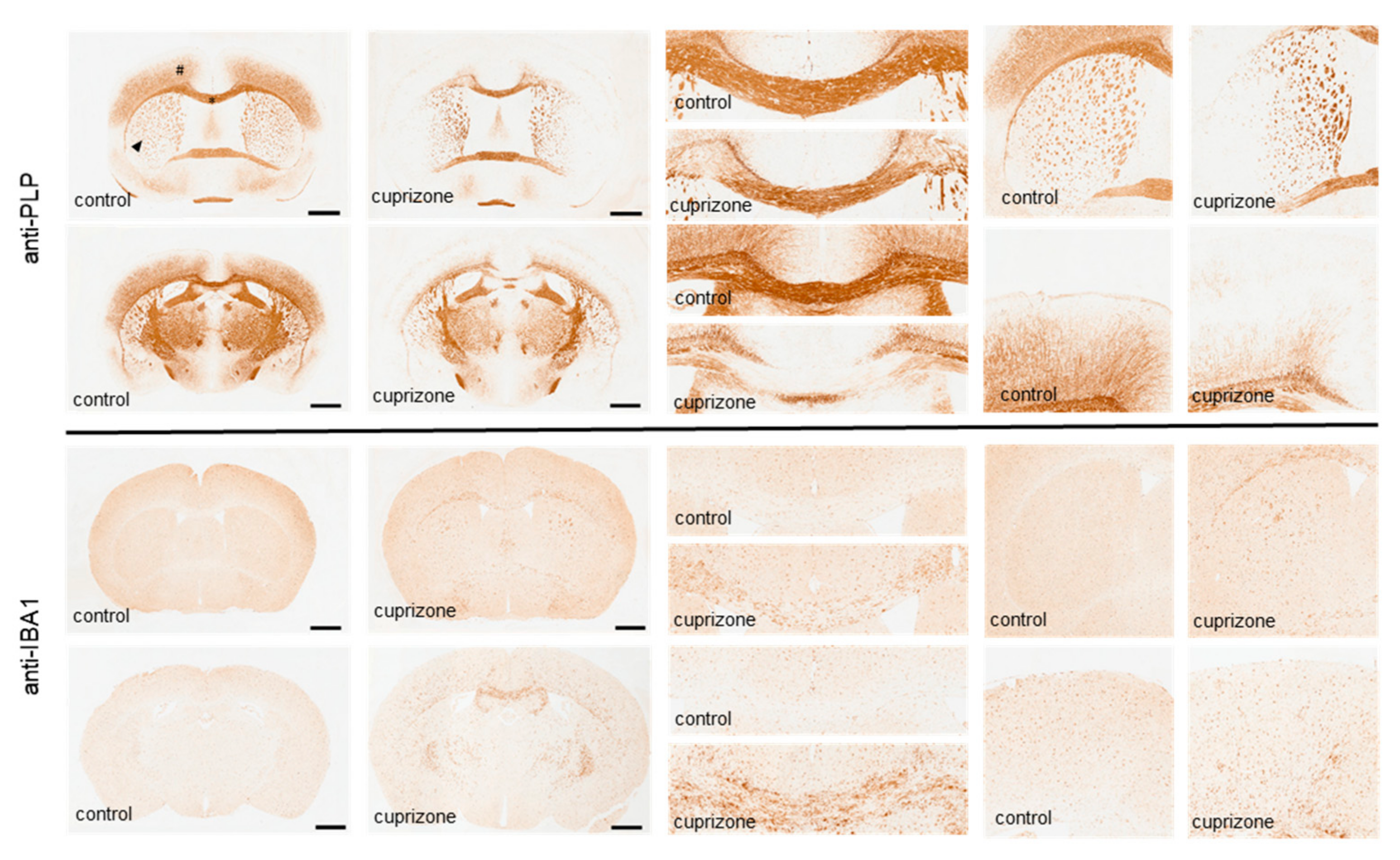
4. The Cuprizone-Induced Histopathological Changes
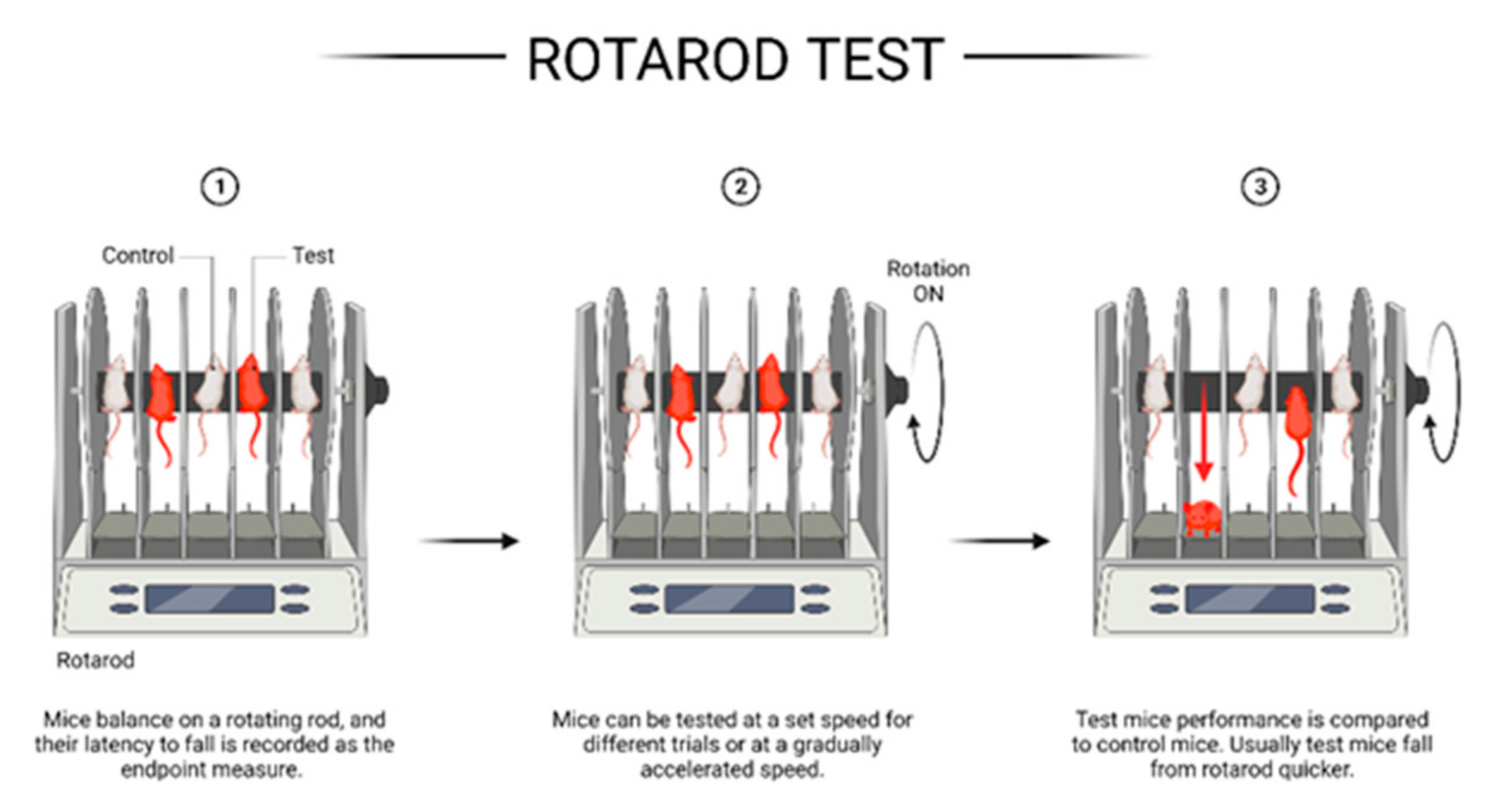
5. Value of the Rotarod Apparatus in Measuring Motor Deficits in the Cuprizone Model
6. Summary and Conclusions
Author Contributions
Funding
Conflicts of Interest
References
- Tobin, W.O.; Kalinowska-Lyszczarz, A.; Weigand, S.D.; Guo, Y.; Tosakulwong, N.; Parisi, J.E.E.; Metz, I.; Frischer, J.M.; Lassmann, H.; Brück, W.; et al. Clinical Correlation of Multiple Sclerosis Immunopathologic Subtypes. Neurology 2021, 97, e1906–e1913. [Google Scholar] [CrossRef] [PubMed]
- Cree, B.A.C.; Arnold, D.L.; Chataway, J.; Chitnis, T.; Fox, R.J.; Pozo Ramajo, A.; Murphy, N.; Lassmann, H. Secondary Progressive Multiple Sclerosis: New Insights. Neurology 2021, 97, 378–388. [Google Scholar] [CrossRef] [PubMed]
- Benkert, P.; Meier, S.; Schaedelin, S.; Manouchehrinia, A.; Yaldizli, Ö.; Maceski, A.; Oechtering, J.; Achtnichts, L.; Conen, D.; Derfuss, T.; et al. Serum neurofilament light chain for individual prognostication of disease activity in people with multiple sclerosis: A retrospective modelling and validation study. Lancet. Neurol. 2022, 21, 246–257. [Google Scholar] [CrossRef]
- Lie, I.A.; Weeda, M.M.; Mattiesing, R.M.; Mol, M.A.E.; Pouwels, P.J.W.; Barkhof, F.; Torkildsen, Ø.; Bø, L.; Myhr, K.M.; Vrenken, H. Relationship Between White Matter Lesions and Gray Matter Atrophy in Multiple Sclerosis: A Systematic Review. Neurology 2022, 98, e1562–e1573. [Google Scholar] [CrossRef] [PubMed]
- Healy, L.M.; Stratton, J.A.; Kuhlmann, T.; Antel, J. The role of glial cells in multiple sclerosis disease progression. Nat. Rev. Neurol. 2022, 18, 237–248. [Google Scholar] [CrossRef]
- Gorter, R.P.; Baron, W. Recent insights into astrocytes as therapeutic targets for demyelinating diseases. Curr. Opin. Pharmacol. 2022, 65, 102261. [Google Scholar] [CrossRef]
- Manouchehri, N.; Salinas, V.H.; Rabi Yeganeh, N.; Pitt, D.; Hussain, R.Z.; Stuve, O. Efficacy of Disease Modifying Therapies in Progressive MS and How Immune Senescence May Explain Their Failure. Front. Neurol. 2022, 13, 854390. [Google Scholar] [CrossRef]
- Giovannoni, G.; Knappertz, V.; Steinerman, J.R.; Tansy, A.P.; Li, T.; Krieger, S.; Uccelli, A.; Uitdehaag, B.M.; Montalban, X.; Hartung, H.-P.; et al. A randomized, placebo-controlled, phase 2 trial of laquinimod in primary progressive multiple sclerosis. Neurology 2020, 95, e1027–e1040. [Google Scholar] [CrossRef]
- Rojas, J.I.; Romano, M.; Ciapponi, A.; Patrucco, L.; Cristiano, E. Interferon Beta for primary progressive multiple sclerosis. Cochrane Database Syst. Rev. 2010, 1. [Google Scholar] [CrossRef]
- Nave, K.-A. Myelination and the trophic support of long axons. Nat. Rev. Neurosci. 2010, 11, 275–283. [Google Scholar] [CrossRef]
- Kipp, M.; Nyamoya, S.; Hochstrasser, T.; Amor, S. Multiple sclerosis animal models: A clinical and histopathological perspective. Brain Pathol. 2017, 27, 123–137. [Google Scholar] [CrossRef] [PubMed]
- Dubé, J.Y.; McIntosh, F.; Zarruk, J.G.; David, S.; Nigou, J.; Behr, M.A. Synthetic mycobacterial molecular patterns partially complete Freund’s adjuvant. Sci. Rep. 2020, 10, 5874. [Google Scholar] [CrossRef]
- Kipp, M.; van der Star, B.; Vogel, D.Y.; Puentes, F.; van der Valk, P.; Baker, D.; Amor, S. Experimental in vivo and in vitro models of multiple sclerosis: EAE and beyond. Mult. Scler. Relat. Disord. 2012, 1, 15–28. [Google Scholar] [CrossRef] [PubMed]
- Dalenogare, D.P.; Theisen, M.C.; Peres, D.S.; Fialho, M.F.; Andrighetto, N.; Barros, L.; Landini, L.; Titiz, M.; De Logu, F.; Oliveira, S.M.; et al. Transient receptor potential ankyrin 1 mediates headache-related cephalic allodynia in a mouse model of relapsing–remitting multiple sclerosis. Pain 2021, 163, 1346–1355. [Google Scholar] [CrossRef]
- Dworsky-Fried, Z.; Faig, C.A.; Vogel, H.A.; Kerr, B.J.; Taylor, A.M.W. Central amygdala inflammation drives pain hypersensitivity and attenuates morphine analgesia in experimental autoimmune encephalomyelitis. Pain 2022, 163, e49–e61. [Google Scholar] [CrossRef] [PubMed]
- Pender, M.P. Ascending impairment of nociception in rats with experimental allergic encephalomyelitis. J. Neurol. Sci. 1986, 75, 317–328. [Google Scholar] [CrossRef]
- Di Filippo, M.; Mancini, A.; Bellingacci, L.; Gaetani, L.; Mazzocchetti, P.; Zelante, T.; La Barbera, L.; De Luca, A.; Tantucci, M.; Tozzi, A.; et al. Interleukin-17 affects synaptic plasticity and cognition in an experimental model of multiple sclerosis. Cell Rep. 2021, 37, 110094. [Google Scholar] [CrossRef]
- Planche, V.; Panatier, A.; Hiba, B.; Ducourneau, E.-G.; Raffard, G.; Dubourdieu, N.; Maitre, M.; Lesté-Lasserre, T.; Brochet, B.; Dousset, V.; et al. Selective dentate gyrus disruption causes memory impairment at the early stage of experimental multiple sclerosis. Brain Behav. Immun. 2017, 60, 240–254. [Google Scholar] [CrossRef]
- Tu, J.L.; Zhao, C.B.; Vollmer, T.; Coons, S.; Lin, H.J.; Marsh, S.; Treiman, D.M.; Shi, J. APOE 4 polymorphism results in early cognitive deficits in an EAE model. Biochem. Biophys. Res. Commun. 2009, 384, 466–470. [Google Scholar] [CrossRef]
- Kocovski, P.; Tabassum-Sheikh, N.; Marinis, S.; Dang, P.T.; Hale, M.W.; Orian, J.M. Immunomodulation Eliminates Inflammation in the Hippocampus in Experimental Autoimmune Encephalomyelitis, but Does Not Ameliorate Anxiety-Like Behavior. Front. Immunol. 2021, 12, 639650. [Google Scholar] [CrossRef]
- Kocovski, P.; Jiang, X.; D’Souza, C.S.; Li, Z.; Dang, P.T.; Wang, X.; Chen, W.; Peter, K.; Hale, M.W.; Orian, J.M. Platelet Depletion is Effective in Ameliorating Anxiety-Like Behavior and Reducing the Pro-Inflammatory Environment in the Hippocampus in Murine Experimental Autoimmune Encephalomyelitis. J. Clin. Med. 2019, 8, 162. [Google Scholar] [CrossRef] [PubMed]
- Plemel, J.R.; Michaels, N.J.; Weishaupt, N.; Caprariello, A.V.; Keough, M.B.; Rogers, J.A.; Yukseloglu, A.; Lim, J.; Patel, V.V.; Rawji, K.S.; et al. Mechanisms of lysophosphatidylcholine-induced demyelination: A primary lipid disrupting myelinopathy. Glia 2017, 66, 327–347. [Google Scholar] [CrossRef] [PubMed]
- Pette, E. Demyelination As A Membrane Problem. Z. Fur Immun.-Und Allerg. 1964, 126, 108–109. [Google Scholar]
- Hall, S.M. The effect of injections of lysophosphatidyl choline into white matter of the adult mouse spinal cord. J. Cell Sci. 1972, 10, 535–546. [Google Scholar] [CrossRef]
- Waxman, S.G.; Kocsis, J.D.; Nitta, K.C. Lysophosphatidyl choline-induced focal demyelination in the rabbit corpus callosum. Light-microscopic observations. J. Neurol. Sci. 1979, 44, 45–53. [Google Scholar] [CrossRef]
- Allt, G.; Ghabriel, M.N.; Sikri, K. Lysophosphatidyl choline-induced demyelination. A freeze-fracture study. Acta Neuropathol. 1988, 75, 456–464. [Google Scholar] [CrossRef]
- Harrison, B. Schwann cell and oligodendrocyte remyelination in lysolecithin-induced lesions in irradiated rat spinal cord. J. Neurol. Sci. 1985, 67, 143–159. [Google Scholar] [CrossRef]
- Fancy, S.P.J.; Harrington, E.P.; Yuen, T.J.; Silbereis, J.C.; Zhao, C.; E Baranzini, S.; Bruce, C.C.; Otero, J.J.; Huang, E.; Nusse, R.; et al. Axin2 as regulatory and therapeutic target in newborn brain injury and remyelination. Nat. Neurosci. 2011, 14, 1009–1016. [Google Scholar] [CrossRef]
- Kipp, M.; Clarner, T.; Dang, J.; Copray, S.; Beyer, C. The cuprizone animal model: New insights into an old story. Acta Neuropathol. 2009, 118, 723–736. [Google Scholar] [CrossRef]
- Jhelum, P.; Santos-Nogueira, E.; Teo, W.; Haumont, A.; Lenoël, I.; Stys, P.K.; David, S. Ferroptosis Mediates Cuprizone-Induced Loss of Oligodendrocytes and Demyelination. J. Neurosci. 2020, 40, 9327–9341. [Google Scholar] [CrossRef]
- Mason, J.L.; Ye, P.; Suzuki, K.; D’Ercole, A.J.; Matsushima, G.K. Insulin-Like Growth Factor-1 Inhibits Mature Oligodendrocyte Apoptosis during Primary Demyelination. J. Neurosci. 2000, 20, 5703–5708. [Google Scholar] [CrossRef] [PubMed]
- Fischbach, F.; Nedelcu, J.; Leopold, P.; Zhan, J.; Clarner, T.; Nellessen, L.; Beißel, C.; van Heuvel, Y.; Goswami, A.; Weis, J.; et al. Cuprizone-induced graded oligodendrocyte vulnerability is regulated by the transcription factor DNA damage-inducible transcript 3. Glia 2018, 67, 263–276. [Google Scholar] [CrossRef] [PubMed]
- Kaddatz, H.; Joost, S.; Nedelcu, J.; Chrzanowski, U.; Schmitz, C.; Gingele, S.; Gudi, V.; Stangel, M.; Zhan, J.; Santrau, E.; et al. Cuprizone-induced demyelination triggers a CD8-pronounced T cell recruitment. Glia 2021, 69, 925–942. [Google Scholar] [CrossRef] [PubMed]
- Slowik, A.; Schmidt, T.; Beyer, C.; Amor, S.; Clarner, T.; Kipp, M. The sphingosine 1-phosphate receptor agonist FTY720 is neuroprotective after cuprizone-induced CNS demyelination. Br. J. Pharmacol. 2015, 172, 80–92. [Google Scholar] [CrossRef]
- Kipp, M.; Gingele, S.; Pott, F.; Clarner, T.; van der Valk, P.; Denecke, B.; Gan, L.; Siffrin, V.; Zipp, F.; Dreher, W.; et al. BLBP-expression in astrocytes during experimental demyelination and in human multiple sclerosis lesions. Brain Behav. Immun. 2011, 25, 1554–1568. [Google Scholar] [CrossRef]
- Wang, X.; Chang, L.; Wan, X.; Tan, Y.; Qu, Y.; Shan, J.; Yang, Y.; Ma, L.; Hashimoto, K. (R)-ketamine ameliorates demyelination and facilitates remyelination in cuprizone-treated mice: A role of gut-microbiota-brain axis. Neurobiol. Dis. 2022, 165, 105635. [Google Scholar] [CrossRef]
- Gudi, V.; Schäfer, N.; Gingele, S.; Stangel, M.; Skripuletz, T. Regenerative Effects of CDP-Choline: A Dose-Dependent Study in the Toxic Cuprizone Model of De- and Remyelination. Pharmaceuticals 2021, 14, 1156. [Google Scholar] [CrossRef]
- Ding, S.; Guo, Y.; Chen, X.; Du, S.; Han, Y.; Yan, Z.; Zhu, Q.; Li, Y. Demyelination and remyelination detected in an alternative cuprizone mouse model of multiple sclerosis with 7.0 T multiparameter magnetic resonance imaging. Sci. Rep. 2021, 11, 11060. [Google Scholar] [CrossRef]
- Albert, M.; Antel, J.; Brück, W.; Stadelmann, C. Extensive cortical remyelination in patients with chronic multiple sclerosis. Brain Pathol. 2007, 17, 129–138. [Google Scholar] [CrossRef]
- Kerbrat, A.; Gros, C.; Badji, A.; Bannier, E.; Galassi, F.; Combès, B.; Chouteau, R.; Labauge, P.; Ayrignac, X.; Carra-Dalliere, C.; et al. Multiple sclerosis lesions in motor tracts from brain to cervical cord: Spatial distribution and correlation with disability. Brain A. J. Neurol. 2020, 143, 2089–2105. [Google Scholar] [CrossRef]
- Healy, B.C.; Buckle, G.J.; Ali, E.N.; Egorova, S.; Khalid, F.; Tauhid, S.; Glanz, B.I.; Chitnis, T.; Guttmann, C.; Weiner, H.L.; et al. Characterizing Clinical and MRI Dissociation in Patients with Multiple Sclerosis. J. Neuroimaging 2017, 27, 481–485. [Google Scholar] [CrossRef] [PubMed]
- Barkhof, F. The clinico-radiological paradox in multiple sclerosis revisited. Curr. Opin. Neurol. 2002, 15, 239–245. [Google Scholar] [CrossRef] [PubMed]
- Scheld, M.; Rüther, B.J.; Grosse-Veldmann, R.; Ohl, K.; Tenbrock, K.; Dreymüller, D.; Fallier-Becker, P.; Zendedel, A.; Beyer, C.; Clarner, T.; et al. Neurodegeneration Triggers Peripheral Immune Cell Recruitment into the Forebrain. J. Neurosci. 2016, 36, 1410–1415. [Google Scholar] [CrossRef] [PubMed]
- Rüther, B.J.; Scheld, M.; Dreymueller, D.; Clarner, T.; Kress, E.; Brandenburg, L.-O.; Swartenbroekx, T.; Hoornaert, C.; Ponsaerts, P.; Fallier-Becker, P.; et al. Combination of cuprizone and experimental autoimmune encephalomyelitis to study inflammatory brain lesion formation and progression. Glia 2017, 65, 1900–1913. [Google Scholar] [CrossRef]
- Sen, M.K.; Mahns, D.A.; Coorssen, J.R.; Shortland, P.J. Behavioural phenotypes in the cuprizone model of central nervous system demyelination. Neurosci. Biobehav. Rev. 2019, 107, 23–46. [Google Scholar] [CrossRef]
- Zhu, X.; Yao, Y.; Hu, Y.; Yang, J.; Zhang, C.; He, Y.; Zhang, A.; Liu, X.; Zhang, C.; Gan, G. Valproic acid suppresses cuprizone-induced hippocampal demyelination and anxiety-like behavior by promoting cholesterol biosynthesis. Neurobiol. Dis. 2021, 158, 105489. [Google Scholar] [CrossRef]
- Zhang, H.; Zhang, Y.; Xu, H.; Wang, L.; Zhao, J.; Wang, J.; Zhang, Z.; Tan, Q.; Kong, J.; Huang, Q.; et al. Locomotor activity and anxiety status, but not spatial working memory, are affected in mice after brief exposure to cuprizone. Neurosci. Bull. 2013, 29, 633–641. [Google Scholar] [CrossRef][Green Version]
- Yan, G.; Xuan, Y.; Dai, Z.; Shen, Z.; Zhang, G.; Xu, H.; Wu, R. Brain metabolite changes in subcortical regions after exposure to cuprizone for 6 weeks: Potential implications for schizophrenia. Neurochem. Res. 2015, 40, 49–58. [Google Scholar] [CrossRef]
- Pollak, Y.; Ovadia, H.; Goshen, I.; Gurevich, R.; Monsa, K.; Avitsur, R.; Yirmiya, R. Behavioral aspects of experimental autoimmune encephalomyelitis. J. Neuroimmunol. 2000, 104, 31–36. [Google Scholar] [CrossRef]
- Herder, V.; Hansmann, F.; Stangel, M.; Skripuletz, T.; Baumgärtner, W.; Beineke, A. Lack of cuprizone-induced demyelination in the murine spinal cord despite oligodendroglial alterations substantiates the concept of site-specific susceptibilities of the central nervous system. Neuropathol. Appl. Neurobiol. 2011, 37, 676–684. [Google Scholar] [CrossRef]
- Liebetanz, D.; Merkler, D. Effects of commissural de- and remyelination on motor skill behaviour in the cuprizone mouse model of multiple sclerosis. Exp. Neurol. 2006, 202, 217–224. [Google Scholar] [CrossRef] [PubMed]
- Zhan, J.; Fegg, F.N.; Kaddatz, H.; Rühling, S.; Frenz, J.; Denecke, B.; Amor, S.; Ponsaerts, P.; Hochstrasser, T.; Kipp, M. Focal white matter lesions induce long-lasting axonal degeneration, neuroinflammation and behavioral deficits. Neurobiol. Dis. 2021, 155, 105371. [Google Scholar] [CrossRef] [PubMed]
- Zhu, K.; Sun, J.; Kang, Z.; Zou, Z.; Wu, G.; Wang, J. Electroacupuncture Promotes Remyelination after Cuprizone Treatment by Enhancing Myelin Debris Clearance. Front. Neurosci. 2016, 10, 613. [Google Scholar] [CrossRef] [PubMed]
- Wang, Q.; Wang, J.; Yang, Z.; Sui, R.; Miao, Q.; Li, Y.; Yu, J.; Liu, C.; Zhang, G.; Xiao, B.; et al. Therapeutic effect of oligomeric proanthocyanidin in cuprizone-induced demyelination. Exp. Physiol. 2019, 104, 876–886. [Google Scholar] [CrossRef]
- Khaledi, E.; Noori, T.; Mohammadi-Farani, A.; Sureda, A.; Dehpour, A.R.; Yousefi-Manesh, H.; Sobarzo-Sanchez, E.; Shirooie, S. Trifluoperazine reduces cuprizone-induced demyelination via targeting Nrf2 and IKB in mice. Eur. J. Pharmacol. 2021, 909, 174432. [Google Scholar] [CrossRef]
- Skripuletz, T.; Miller, E.; Moharregh-Khiabani, D.; Blank, A.; Pul, R.; Gudi, V.; Trebst, C.; Stangel, M. Beneficial effects of minocycline on cuprizone induced cortical demyelination. Neurochem. Res. 2010, 35, 1422–1433. [Google Scholar] [CrossRef]
- Sen, M.K.; Almuslehi, M.S.M.; Coorssen, J.R.; Mahns, D.A.; Shortland, P.J. Behavioural and histological changes in cuprizone-fed mice. Brain Behav. Immun. 2020, 87, 508–523. [Google Scholar] [CrossRef]
- Yamazaki, R.; Ohno, N.; Huang, J.K. Acute motor deficit and subsequent remyelination-associated recovery following internal capsule demyelination in mice. J. Neurochem. 2021, 156, 917–928. [Google Scholar] [CrossRef]
- Günaydın, C.; Önger, M.E.; Avcı, B.; Bozkurt, A.; Terzi, M.; Bilge, S.S. Tofacitinib enhances remyelination and improves myelin integrity in cuprizone-induced mice. Immunopharmacol. Immunotoxicol. 2021, 43, 790–798. [Google Scholar] [CrossRef]
- Brooks, S.P.; Dunnett, S.B. Tests to assess motor phenotype in mice: A user’s guide. Nat. Rev. Neurosci. 2009, 10, 519–529. [Google Scholar] [CrossRef]
- Antipova, V.A.; Holzmann, C.; Schmitt, O.; Wree, A.; Hawlitschka, A. Botulinum Neurotoxin A Injected Ipsilaterally or Contralaterally into the Striatum in the Rat 6-OHDA Model of Unilateral Parkinson’s Disease Differently Affects Behavior. Front. Behav. Neurosci. 2017, 11, 119. [Google Scholar] [CrossRef]
- Zhan, J.; Yakimov, V.; Rühling, S.; Fischbach, F.; Nikolova, E.; Joost, S.; Kaddatz, H.; Greiner, T.; Frenz, J.; Holzmann, C.; et al. High Speed Ventral Plane Videography as a Convenient Tool to Quantify Motor Deficits during Pre-Clinical Experimental Autoimmune Encephalomyelitis. Cells 2019, 8, 1439. [Google Scholar] [CrossRef] [PubMed]
- Dunham, N.; Miya, T. A Note on a Simple Apparatus for Detecting Neurological Deficit in Rats and Mice**College of Pharmacy, University of Nebraska, Lincoln 8. J. Am. Pharm. Assoc. 1957, 46, 208–209. [Google Scholar] [CrossRef] [PubMed]
- Kinnard, W.J., Jr.; Carr, C.J. A preliminary procedure for the evaluation of central nervous system depressants. J. Pharmacol. Exp. Ther. 1957, 121, 354–361. [Google Scholar] [PubMed]
- Weaver, J.E.; Miya, T.S. Effects of certain ataraxic agents on mice activity. J. Pharm. Sci. 1961, 50, 910–912. [Google Scholar] [CrossRef]
- Doeppner, T.R.; Kaltwasser, B.; Bähr, M.; Hermann, D.M. Effects of neural progenitor cells on post-stroke neurological impairment-a detailed and comprehensive analysis of behavioral tests. Front. Cell. Neurosci. 2014, 8, 338. [Google Scholar] [CrossRef]
- Wu, Y.; Pang, J.; Peng, J.; Cao, F.; Vitek, M.P.; Li, F.; Jiang, Y.; Sun, X. An apoE-derived mimic peptide, COG1410, alleviates early brain injury via reducing apoptosis and neuroinflammation in a mouse model of subarachnoid hemorrhage. Neurosci. Lett. 2016, 627, 92–99. [Google Scholar] [CrossRef]
- Tanji, J.; Okano, K.; Sato, K.C. Neuronal activity in cortical motor areas related to ipsilateral, contralateral, and bilateral digit movements of the monkey. J. Neurophysiol. 1988, 60, 325–343. [Google Scholar] [CrossRef]
- Preilowski, B.F. Possible contribution of the anterior forebrain commissures to bilateral motor coordination. Neuropsychologia 1972, 10, 267–277. [Google Scholar] [CrossRef]
- Franz, E.A.; Eliassen, J.C.; Ivry, R.B.; Gazzaniga, M.S. Dissociation of Spatial and Temporal Coupling in the Bimanual Movements of Callosotomy Patients. Psychol. Sci. 1996, 7, 306–310. [Google Scholar] [CrossRef]
- Eliassen, J.C.; Baynes, K.; Gazzaniga, M.S. Anterior and posterior callosal contributions to simultaneous bimanual movements of the hands and fingers. Brain 2000, 123, 2501–2511. [Google Scholar] [CrossRef]
- Kennerley, S.W.; Diedrichsen, J.; Hazeltine, E.; Semjen, A.; Ivry, R.B. Callosotomy patients exhibit temporal uncoupling during continuous bimanual movements. Nat. Neurosci. 2002, 5, 376–381. [Google Scholar] [CrossRef]
- Johansen-Berg, H.; Della-Maggiore, V.; Behrens, T.E.; Smith, S.M.; Paus, T. Integrity of white matter in the corpus callosum correlates with bimanual co-ordination skills. NeuroImage 2007, 36, T16–T21. [Google Scholar] [CrossRef]
- Muetzel, R.L.; Collins, P.F.; Mueller, B.A.; Schissel, A.M.; Lim, K.O.; Luciana, M. The development of corpus callosum microstructure and associations with bimanual task performance in healthy adolescents. NeuroImage 2008, 39, 1918–1925. [Google Scholar] [CrossRef]
- Bonzano, L.; Tacchino, A.; Roccatagliata, L.; Mancardi, G.L.; Abbruzzese, G.; Bove, M. Structural integrity of callosal midbody influences intermanual transfer in a motor reaction-time task. Hum. Brain Mapp. 2011, 32, 218–228. [Google Scholar] [CrossRef]
- Caeyenberghs, K.; Leemans, A.; Coxon, J.; Leunissen, I.; Drijkoningen, D.; Geurts, M.; Gooijers, J.; Michiels, K.; Sunaert, S.; Swinnen, S.P. Bimanual coordination and corpus callosum microstructure in young adults with traumatic brain injury: A diffusion tensor imaging study. J. Neurotrauma 2011, 28, 897–913. [Google Scholar] [CrossRef]
- Gooijers, J.; Caeyenberghs, K.; Sisti, H.M.; Geurts, M.; Heitger, M.H.; Leemans, A.; Swinnen, S.P. Diffusion tensor imaging metrics of the corpus callosum in relation to bimanual coordination: Effect of task complexity and sensory feedback. Hum. Brain Mapp. 2013, 34, 241–252. [Google Scholar] [CrossRef]
- Gooijers, J.; Swinnen, S.P. Interactions between brain structure and behavior: The corpus callosum and bimanual coordination. Neurosci. Biobehav. Rev. 2014, 43, 1–19. [Google Scholar] [CrossRef]
- Kobayashi, S.; Hirose, M.; Akutsu, Y.; Hirayama, K.; Ishida, Y.; Ugawa, Y. Disconnected Motor Intention and Spatial Attention in a Case of Probable Marchiafava-Bignami Disease. Cogn. Behav. Neurol. 2021, 34, 226–232. [Google Scholar] [CrossRef]
- Goldberg, J.; Clarner, T.; Beyer, C.; Kipp, M. Anatomical Distribution of Cuprizone-Induced Lesions in C57BL6 Mice. J. Mol. Neurosci. 2015, 57, 166–175. [Google Scholar] [CrossRef]
- Vega-Riquer, J.M.; Campos-Ordonez, T.; Galvez-Contreras, A.Y.; Gonzalez-Castañeda, R.E.; Gonzalez-Perez, O. Phenytoin promotes the proliferation of oligodendrocytes and enhances the expression of myelin basic protein in the corpus callosum of mice demyelinated by cuprizone. Exp. Brain Res. 2022, 240, 1617–1627. [Google Scholar] [CrossRef]
- Deacon, R.M. Measuring Motor Coordination in Mice. J. Vis. Exp. 2013, 75, e2609. [Google Scholar] [CrossRef]
- Robinson, A.P.; Zhang, J.Z.; Titus, H.E.; Karl, M.; Merzliakov, M.; Dorfman, A.R.; Karlik, S.; Stewart, M.G.; Watt, R.K.; Facer, B.D.; et al. Nanocatalytic activity of clean-surfaced, faceted nanocrystalline gold enhances remyelination in animal models of multiple sclerosis. Sci. Rep. 2020, 10, 1–16. [Google Scholar] [CrossRef]
- Schalomon, P.M.; Wahlsten, D. Wheel running behavior is impaired by both surgical section and genetic absence of the mouse corpus callosum. Brain Res. Bull. 2002, 57, 27–33. [Google Scholar] [CrossRef]
- Sullivan, G.M.; Knutsen, A.K.; Peruzzotti-Jametti, L.; Korotcov, A.; Bosomtwi, A.; Dardzinski, B.J.; Bernstock, J.D.; Rizzi, S.; Edenhofer, F.; Pluchino, S.; et al. Transplantation of induced neural stem cells (iNSCs) into chronically demyelinated corpus callosum ameliorates motor deficits. Acta Neuropathol. Commun. 2020, 8, 84. [Google Scholar] [CrossRef]
- Faizi, M.; Salimi, A.; Seydi, E.; Naserzadeh, P.; Kouhnavard, M.; Rahimi, A.; Pourahmad, J. Toxicity of cuprizone a Cu2+ chelating agent on isolated mouse brain mitochondria: A justification for demyelination and subsequent behavioral dysfunction. Toxicol. Mech. Methods 2016, 26, 276–283. [Google Scholar] [CrossRef]
- Yamamoto, S.; Gotoh, M.; Kawamura, Y.; Yamashina, K.; Yagishita, S.; Awaji, T.; Tanaka, M.; Maruyama, K.; Murakami-Murofushi, K.; Yoshikawa, K. Cyclic phosphatidic acid treatment suppress cuprizone-induced demyelination and motor dysfunction in mice. Eur. J. Pharmacol. 2014, 741, 17–24. [Google Scholar] [CrossRef]
- Kumar, P.; Sharma, G.; Gupta, V.; Kaur, R.; Thakur, K.; Malik, R.; Kumar, A.; Kaushal, N.; Raza, K. Preclinical Explorative Assessment of Dimethyl Fumarate-Based Biocompatible Nanolipoidal Carriers for the Management of Multiple Sclerosis. ACS Chem. Neurosci. 2018, 9, 1152–1158. [Google Scholar] [CrossRef]
- Elbaz, E.M.; Senousy, M.A.; El-Tanbouly, D.M.; Sayed, R.H. Neuroprotective effect of linagliptin against cuprizone-induced demyelination and behavioural dysfunction in mice: A pivotal role of AMPK/SIRT1 and JAK2/STAT3/NF-κB signalling pathway modulation. Toxicol. Appl. Pharmacol. 2018, 352, 153–161. [Google Scholar] [CrossRef]
- Bölcskei, K.; Kriszta, G.; Sághy, É.; Payrits, M.; Sipos, É.; Vranesics, A.; Berente, Z.; Ábrahám, H.; Ács, P.; Komoly, S.; et al. Behavioural alterations and morphological changes are attenuated by the lack of TRPA1 receptors in the cuprizone-induced demyelination model in mice. J. Neuroimmunol. 2018, 320, 1–10. [Google Scholar] [CrossRef]
- Polyák, H.; Cseh, E.K.; Bohár, Z.; Rajda, C.; Zádori, D.; Klivényi, P.; Toldi, J.; Vécsei, L. Cuprizone markedly decreases kynurenic acid levels in the rodent brain tissue and plasma. Heliyon 2021, 7, e06124. [Google Scholar] [CrossRef]
- Chang, H.; Liu, J.; Zhang, Y.; Wang, F.; Wu, Y.; Zhang, L.; Ai, H.; Chen, G.; Yin, L. Increased central dopaminergic activity might be involved in the behavioral abnormality of cuprizone exposure mice. Behav. Brain Res. 2017, 331, 143–150. [Google Scholar] [CrossRef]
- Ye, J.N.; Chen, X.S.; Su, L.; Liu, Y.L.; Cai, Q.Y.; Zhan, X.L.; Xu, Y.; Zhao, S.F.; Yao, Z.X. Progesterone alleviates neural behavioral deficits and demyelination with reduced degeneration of oligodendroglial cells in cuprizone-induced mice. PLoS ONE 2013, 8, e54590. [Google Scholar] [CrossRef]
- Liu, C.; Zhang, N.; Zhang, R.; Jin, L.; Petridis, A.K.; Loers, G.; Zheng, X.; Wang, Z.; Siebert, H.C. Cuprizone-Induced Demyelination in Mouse Hippocampus Is Alleviated by Ketogenic Diet. J. Agric. Food Chem. 2020, 68, 11215–11228. [Google Scholar] [CrossRef]
- Madadi, S.; Pasbakhsh, P.; Tahmasebi, F.; Mortezaee, K.; Khanehzad, M.; Boroujeni, F.B.; Noorzehi, G.; Kashani, I.R. Astrocyte ablation induced by La-aminoadipate (L-AAA) potentiates remyelination in a cuprizone demyelinating mouse model. Metab. Brain Dis. 2019, 34, 593–603. [Google Scholar] [CrossRef]
- Barati, S.; Kashani, I.R.; Tahmasebi, F. The effects of mesenchymal stem cells transplantation on A1 neurotoxic reactive astrocyte and demyelination in the cuprizone model. Histochem. J. 2022, 53, 333–346. [Google Scholar] [CrossRef]
- Abakumova, T.O.; Kuz’kina, A.A.; Zharova, M.E.; Pozdeeva, D.A.; Gubskii, I.L.; Shepeleva, I.I.; Antonova, O.M.; Nukolova, N.V.; Kekelidze, Z.I.; Chekhonin, V.P. Cuprizone Model as a Tool for Preclinical Studies of the Efficacy of Multiple Sclerosis Diagnosis and Therapy. Bull. Exp. Biol. Med. 2015, 159, 111–115. [Google Scholar] [CrossRef]
- Franco-Pons, N.; Torrente, M.; Colomina, M.T.; Vilella, E. Behavioral deficits in the cuprizone-induced murine model of demyelination/remyelination. Toxicol. Lett. 2007, 169, 205–213. [Google Scholar] [CrossRef]
- Hagemeyer, N.; Boretius, S.; Ott, C.; Von Streitberg, A.; Welpinghus, H.; Sperling, S.; Frahm, J.; Simons, M.; Ghezzi, P.; Ehrenreich, H. Erythropoietin attenuates neurological and histological consequences of toxic demyelination in mice. Mol. Med. 2012, 18, 628–635. [Google Scholar] [CrossRef]
- Wang, W.-W.; Lu, L.; Bao, T.-H.; Zhang, H.-M.; Yuan, J.; Miao, W.; Wang, S.-F.; Xiao, Z.-C. Scutellarin Alleviates Behavioral Deficits in a Mouse Model of Multiple Sclerosis, Possibly Through Protecting Neural Stem Cells. J. Mol. Neurosci. 2015, 58, 210–220. [Google Scholar] [CrossRef]
- Han, S.R.; Kang, Y.H.; Jeon, H.; Lee, S.; Park, S.J.; Song, D.Y.; Min, S.S.; Yoo, S.M.; Lee, M.S.; Lee, S.H. Differential Expression of miRNAs and Behavioral Change in the Cuprizone-Induced Demyelination Mouse Model. Int. J. Mol. Sci. 2020, 21, 646. [Google Scholar] [CrossRef]
- Lira-Diaz, E.; Monroy-Rodriguez, J.; Gonzalez-Pedroza, M.G.; Morales-Luckie, R.A.; Castro-Sánchez, L.; Gonzalez-Perez, O. EGF-Coupled Gold Nanoparticles Increase the Expression of CNPase and the Myelin-Associated Proteins MAG, MOG, and MBP in the Septal Nucleus Demyelinated by Cuprizone. Life 2022, 12, 333. [Google Scholar] [CrossRef]
- Tanaka, A.; Anada, K.; Yasue, M.; Honda, T.; Nakamura, H.; Murayama, T. Ceramide kinase knockout ameliorates multiple sclerosis-like behaviors and demyelination in cuprizone-treated mice. Life Sci. 2022, 296, 120446. [Google Scholar] [CrossRef]
- Hashimoto, M.; Yamamoto, S.; Iwasa, K.; Yamashina, K.; Ishikawa, M.; Maruyama, K.; Bosetti, F.; Yoshikawa, K. The flavonoid Baicalein attenuates cuprizone-induced demyelination via suppression of neuroinflammation. Brain Res. Bull. 2017, 135, 47–52. [Google Scholar] [CrossRef]
- Iwasa, K.; Yamamoto, S.; Takahashi, M.; Suzuki, S.; Yagishita, S.; Awaji, T.; Maruyama, K.; Yoshikawa, K. Prostaglandin F2α FP receptor inhibitor reduces demyelination and motor dysfunction in a cuprizone-induced multiple sclerosis mouse model. Prostaglandins, Leukot. Essent. Fat. Acids 2014, 91, 175–182. [Google Scholar] [CrossRef]
- Kondo, M.A.; Fukudome, D.; Smith, D.R.; Gallagher, M.; Kamiya, A.; Sawa, A. Dimensional assessment of behavioral changes in the cuprizone short-term exposure model for psychosis. Neurosci. Res. 2016, 107, 70–74. [Google Scholar] [CrossRef][Green Version]
- Sanadgol, N.; Golab, F.; Mostafaie, A.; Mehdizadeh, M.; Khalseh, R.; Mahmoudi, M.; Abdollahi, M.; Vakilzadeh, G.; Taghizadeh, G.; Sharifzadeh, M. Low, but not high, dose triptolide controls neuroinflammation and improves behavioral deficits in toxic model of multiple sclerosis by dampening of NF-κB activation and acceleration of intrinsic myelin repair. Toxicol. Appl. Pharmacol. 2018, 342, 86–98. [Google Scholar] [CrossRef]
- Yamamoto, S.; Yamashina, K.; Ishikawa, M.; Gotoh, M.; Yagishita, S.; Iwasa, K.; Maruyama, K.; Murakami-Murofushi, K.; Yoshikawa, K. Protective and therapeutic role of 2-carba-cyclic phosphatidic acid in demyelinating disease. J. Neuroinflammation 2017, 14, 142. [Google Scholar] [CrossRef]
- Yoshikawa, K.; Palumbo, S.; Toscano, C.; Bosetti, F. Inhibition of 5-lipoxygenase activity in mice during cuprizone-induced demyelination attenuates neuroinflammation, motor dysfunction and axonal damage. Prostaglandins Leukot. Essent. Fat. Acids 2011, 85, 43–52. [Google Scholar] [CrossRef]
- Zhang, Q.; Li, Z.; Wu, S.; Li, X.; Sang, Y.; Li, J.; Niu, Y.; Ding, H. Myricetin alleviates cuprizone-induced behavioral dysfunction and demyelination in mice by Nrf2 pathway. Food Funct. 2016, 7, 4332–4342. [Google Scholar] [CrossRef]
- Tian, H.; Sun, W.; Zhang, Q.; Li, X.; Sang, Y.; Li, J.; Niu, Y.; Ding, H. Procyanidin B2 mitigates behavioral impairment and protects myelin integrity in cuprizone-induced schizophrenia in mice. RSC Adv. 2018, 8, 23835–23846. [Google Scholar] [CrossRef]
- Mohamed, A.; Al-Kafaji, G.; Almahroos, A.; Almosawi, Z.; Alalwan, H.; Abdulla, R.; Alammadi, F.; Almubarak, A.; Al-Mahrezi, A.; Kamal, A. Effects of enhanced environment and induced depression on cuprizone mouse model of demyelination. Exp. Med. 2019, 18, 566–572. [Google Scholar] [CrossRef] [PubMed]
- Templeton, N.; Kivell, B.; McCaughey-Chapman, A.; Connor, B.; La Flamme, A.C. Clozapine administration enhanced functional recovery after cuprizone demyelination. PLoS ONE 2019, 14, e0216113. [Google Scholar] [CrossRef] [PubMed]
- Yamamoto, S.; Sakemoto, C.; Iwasa, K.; Maruyama, K.; Shimizu, K.; Yoshikawa, K. Ursolic acid treatment suppresses cuprizone-induced demyelination and motor dysfunction via upregulation of IGF-1. J. Pharmacol. Sci. 2020, 144, 119–122. [Google Scholar] [CrossRef] [PubMed]
- Buitrago, M.M.; Schulz, J.B.; Dichgans, J.; Luft, A.R. Short and long-term motor skill learning in an accelerated rotarod training paradigm. Neurobiol. Learn. Mem. 2004, 81, 211–216. [Google Scholar] [CrossRef]
- Bacmeister, C.M.; Barr, H.J.; McClain, C.R.; Thornton, M.A.; Nettles, D.; Welle, C.G.; Hughes, E.G. Motor learning promotes remyelination via new and surviving oligodendrocytes. Nat. Neurosci. 2020, 23, 819–831. [Google Scholar] [CrossRef]
- Hoyos, N.M.; Jürgens, T.; Grønborg, M.; Kreutzfeldt, M.; Schedensack, M.; Kuhlmann, T.; Schrick, C.; Brück, W.; Urlaub, H.; Simons, M.; et al. Late motor decline after accomplished remyelination: Impact for progressive multiple sclerosis. Ann. Neurol. 2011, 71, 227–244. [Google Scholar] [CrossRef] [PubMed]
- Carter, R.J.; Lione, L.A.; Humby, T.; Mangiarini, L.; Mahal, A.; Bates, G.; Dunnett, S.; Morton, A.J. Characterization of Progressive Motor Deficits in Mice Transgenic for the Human Huntington’s Disease Mutation. J. Neurosci. 1999, 19, 3248–3257. [Google Scholar] [CrossRef]
- Mendes, C.S.; Bartos, I.; Márka, Z.; Akay, T.; Márka, S.; Mann, R.S. Quantification of gait parameters in freely walking rodents. BMC Biol. 2015, 13, 50. [Google Scholar] [CrossRef]


| Cit | Duration | Dose | Weight | Age | Confirmed | Timepoint(s) | Setup | Readout | Training | Main Outcome |
|---|---|---|---|---|---|---|---|---|---|---|
| [97] | 4 wks | 0.6% | 16–18 g | 8-wks | no | week 4, week 4 + 2 | 21 rpm | latency | no | decreased latency |
| [98] | 3–6 wks | 0.2% | not given | 8-wks | yes | week 3–6, week 6 + 6 | 16, 24, or 32 rpm | number of falls | no | increased number of falls |
| [99] | 6 wks | 0.2% | not given | 8-wks | no | week 6, week 6 + 3 | 4 to 40 rpm in 5 min | latency | no | no change |
| [100] | 6 wks | 0.2% | not given | 8-wks | yes | week 6 + 11 days | 30 rpm, max 200 s | latency | no | decreased latency |
| [101] | 6 wks | 0.2% | not given | 7-wks | yes | week 6 + 2, 4, 6 | 15–16 rpm, max 60 s | number of falls | no | increased number of falls |
| [91] | 5 wks | 0.2% | 20–25 g | 8-wks | yes | week 3, 4, and 5, week 5 + 3, and 5 + 4 | 5 to 40 rpm in 300 s | latency | yes | no change |
| [95] | 12 wks | 0.2% | not given | 7-wks | yes | week 12 + 2 | 4 and 40 rpm in 3 min | latency | yes | decreased latency |
| [102] | 8 wks | 0.4% | 15–18 g | 60 d | no | week 8 + 3 | 8, 15, 30, and 35 rpm, max 60 s | normalized latency | yes | decreased latency |
| [96] | 12 wks | 0.2% | 20–25 g | 8-wks | yes | week 12 + 2 | 4 to 35 rpm in 3 min | latency | yes | decreased latency |
| [103] | 8 wks | 0.2% | not given | 8-wks | yes | week 8 | 28 rpm, max 300 s | latency | yes | decreased latency |
| [90] | 6 wks | 0.2% | not given | 8–9 wks | yes | week 1–6 | 4 to 40 rpm in 5 min | latency | yes | no change |
| [92] | 3 wks | 0.4% | 17–20 g | 6-wks | yes | day 16 | 4 to 35 rpm in 3 min | latency | yes | no change |
| [89] | 4 wks | 0.7–0.2% | 20–25 g | 6-wks | yes | week 4 | 25 rpm | latency | yes | decreased latency |
| [86] | 5 wks | 0.2% | 20 g | 8–10 wks | yes | week 5 | 6 rpm, max 120 s | latency | no | decreased latency |
| [104] | 6 wks | 0.2% | not given | 10-wks | yes | week 6 | 20 rpm, max 300 s | latency | yes | decreased latency |
| [105] | 5 wks | 0.2% | not given | 10-wks | yes | week 5 | 16 rpm, max 600 s | latency, number of falls | no | decreased latency, increased number of falls |
| [106] | 1 wk | 0.2% | not given | 8-wks | no | week 1 | 4 to 40 rpm in 2 min | latency | no | no change |
| [88] | 30 days | 6 mg/kg | 25–50 g | not given | yes | day 30 | 25 rpm, max 120 s | latency | yes | decreased latency |
| [107] | 6 wks | 0.2% | 18–20 g | 7–8-wks | yes | days 40, 41, and 42 | 4 to 40 rpm in 120 s | number of falls | no | increased number of falls |
| [87] | 5 wks | 0.2% | not given | 10-wks | yes | week 5 | 28 rpm, max 300 s | latency, number of falls and flips | no | decreased latency, increased number of falls and flips |
| [108] | 10 wks | 0.2% | not given | 10-wks | yes | week 5 and 10 | 20 or 28 rpm, max 300 s | latency, number of falls and flips | no | decreased latency, increased number of falls |
| [93] | 5 wks | 0.2% | 15–17 g | 6-wks | yes | week 5 | 5 to 40 rpm | latency | yes | decreased latency |
| [109] | 5 wks | 0.2% | not given | 8–10-wks | yes | week 5 | 32 rpm, max 300 s | latency, number of falls | yes | decreased latency, increased number of falls |
| [110] | 48 days | 0.2% | 18–22 g | 8–9-wks | yes | between day 34 and 48 | 28 rpm, max 300 s | latency, number of falls | yes | decreased latency, increased number of falls |
| [111] | 45 days | 0.2% | 18–20 g | 7-wks | yes | week 5 | 30 rpm, max 300 s | latency, number of falls | yes | decreased latency, increased number of falls |
| [94] | 5 wks | 0.2% | not given | 7-wks | yes | week 5 | 4 to 40 rpm | latency | no | decreased latency |
| [112] | 6 wks | 0.2% | 22 g | 6-wks | yes | week 6 | 4.5 m/min | latency | yes | no change |
| [113] | 6 wks | 0.3% | not given | 6–8-wks | yes | week 6 | 28 rpm, max 120 s | latency | no | decreased latency |
| [55] | 6 wks | 0.2% | 20–25 g | 8-wks | yes | week 6 | 25 rpm, max 300 s | latency | no | decreased latency |
| [114] | 5 wks | 0.2% | not given | 10-wks | yes | week 5 | 28 rpm, max 300 s | latency, number of falls and flips | no | decreased latency, increased number of falls and flips |
Publisher’s Note: MDPI stays neutral with regard to jurisdictional claims in published maps and institutional affiliations. |
© 2022 by the authors. Licensee MDPI, Basel, Switzerland. This article is an open access article distributed under the terms and conditions of the Creative Commons Attribution (CC BY) license (https://creativecommons.org/licenses/by/4.0/).
Share and Cite
Lubrich, C.; Giesler, P.; Kipp, M. Motor Behavioral Deficits in the Cuprizone Model: Validity of the Rotarod Test Paradigm. Int. J. Mol. Sci. 2022, 23, 11342. https://doi.org/10.3390/ijms231911342
Lubrich C, Giesler P, Kipp M. Motor Behavioral Deficits in the Cuprizone Model: Validity of the Rotarod Test Paradigm. International Journal of Molecular Sciences. 2022; 23(19):11342. https://doi.org/10.3390/ijms231911342
Chicago/Turabian StyleLubrich, Concordia, Paula Giesler, and Markus Kipp. 2022. "Motor Behavioral Deficits in the Cuprizone Model: Validity of the Rotarod Test Paradigm" International Journal of Molecular Sciences 23, no. 19: 11342. https://doi.org/10.3390/ijms231911342
APA StyleLubrich, C., Giesler, P., & Kipp, M. (2022). Motor Behavioral Deficits in the Cuprizone Model: Validity of the Rotarod Test Paradigm. International Journal of Molecular Sciences, 23(19), 11342. https://doi.org/10.3390/ijms231911342


