


New Route to Glycosylated Porphyrins via Aromatic Nucleophilic Substitution (SNAr)—Synthesis and Cellular Uptake Studies


Abstract
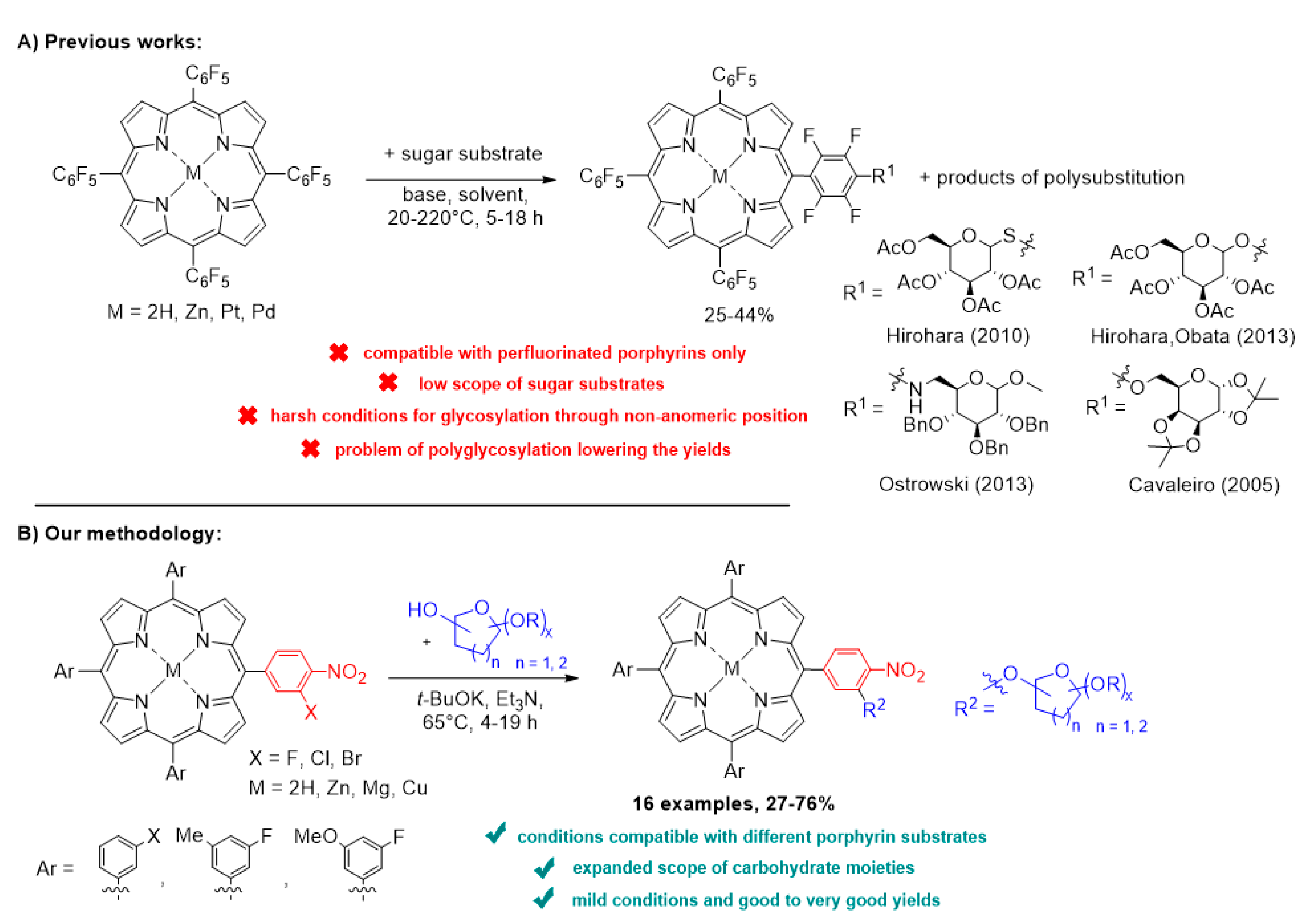
:1. Introduction
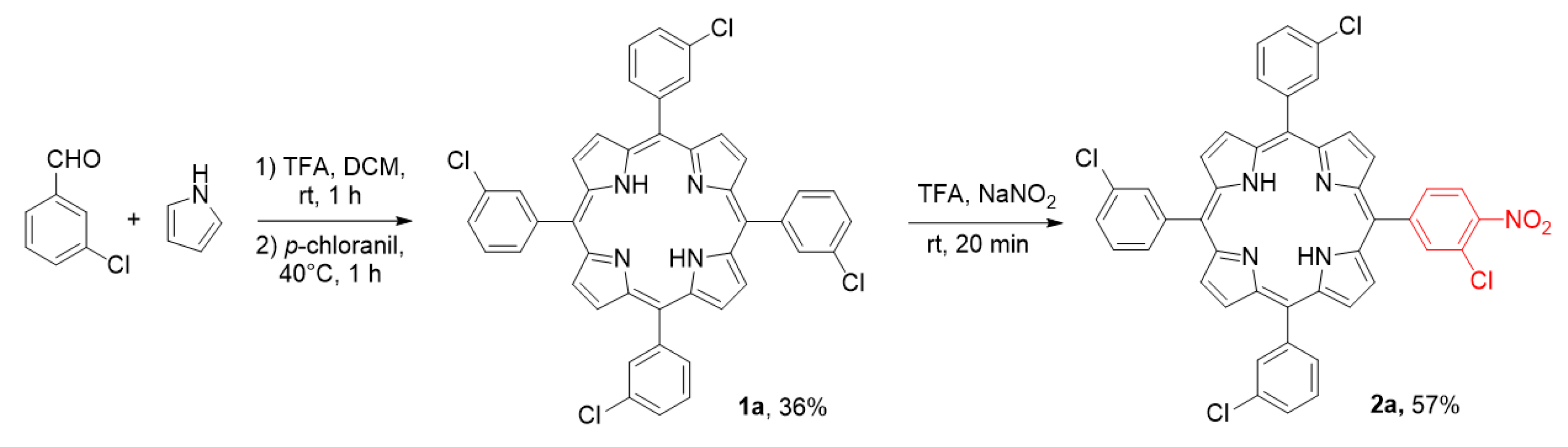
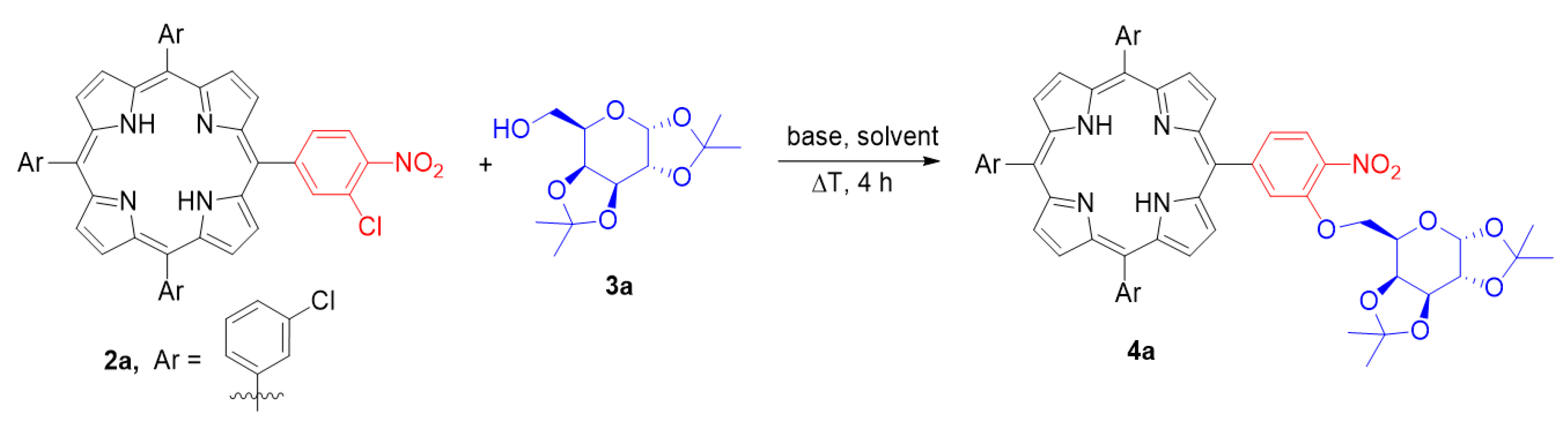
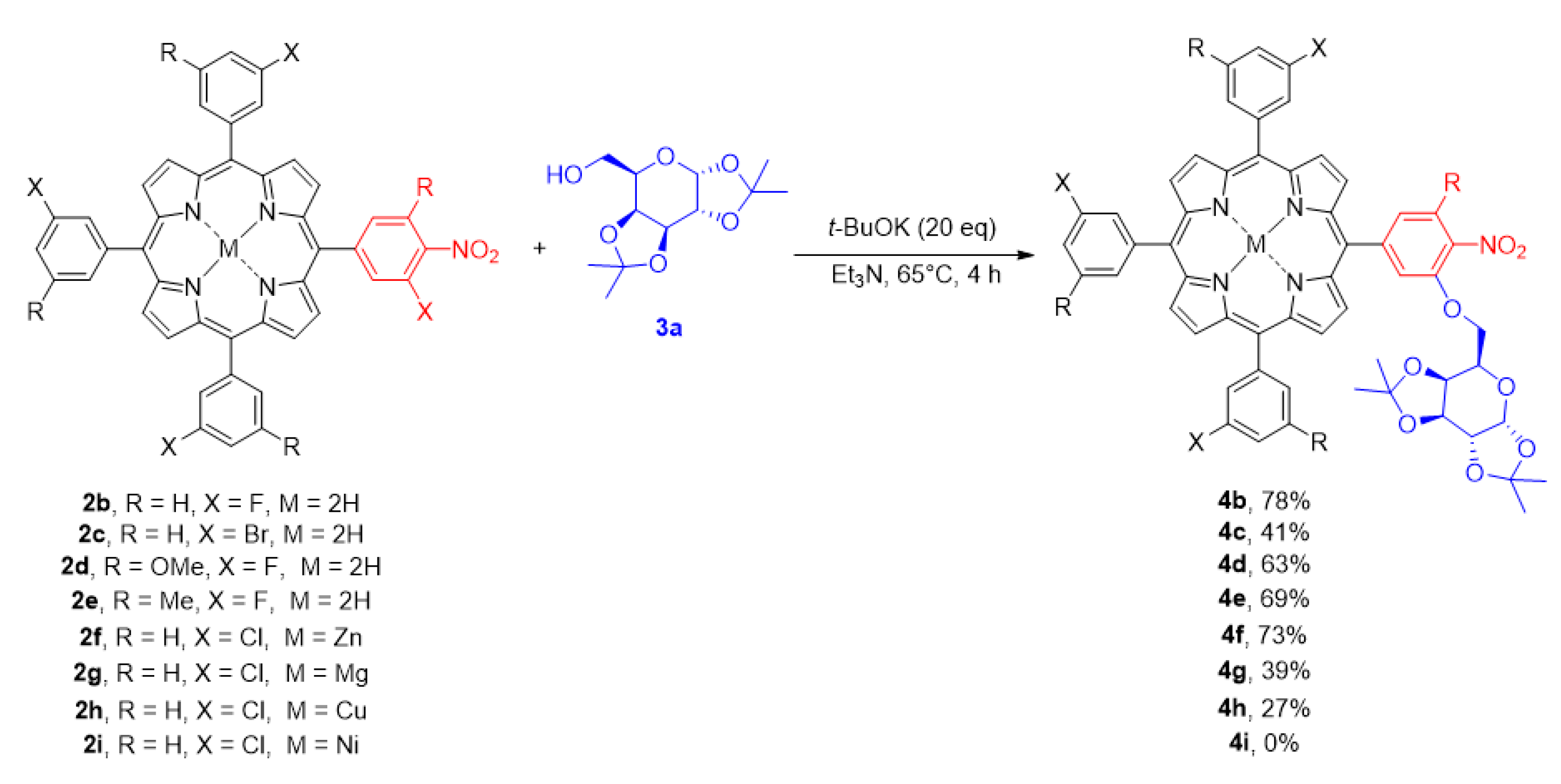
2. Results and Discussion
2.1. Initial Studies and Optimisation of the SNAr Process
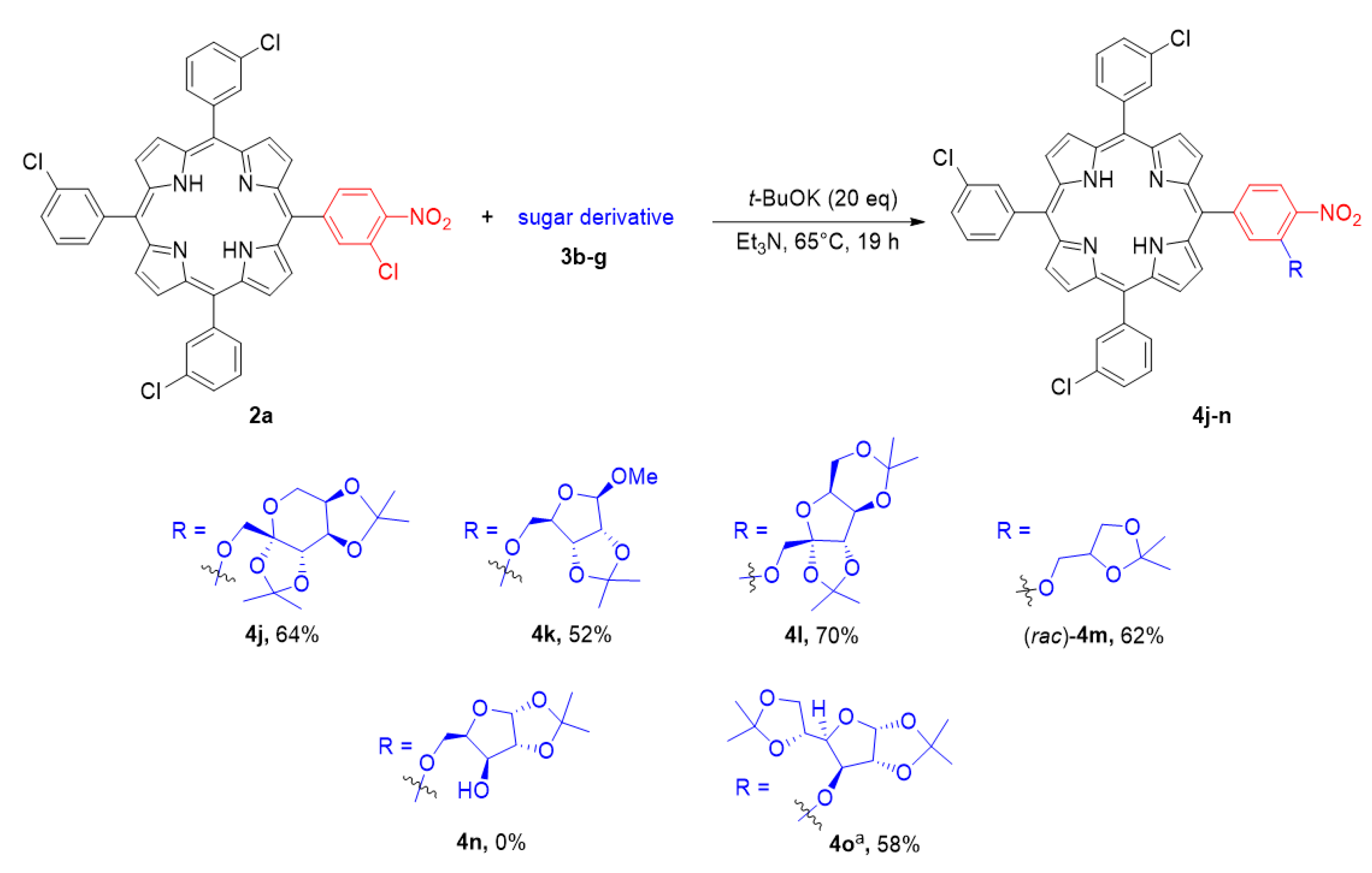
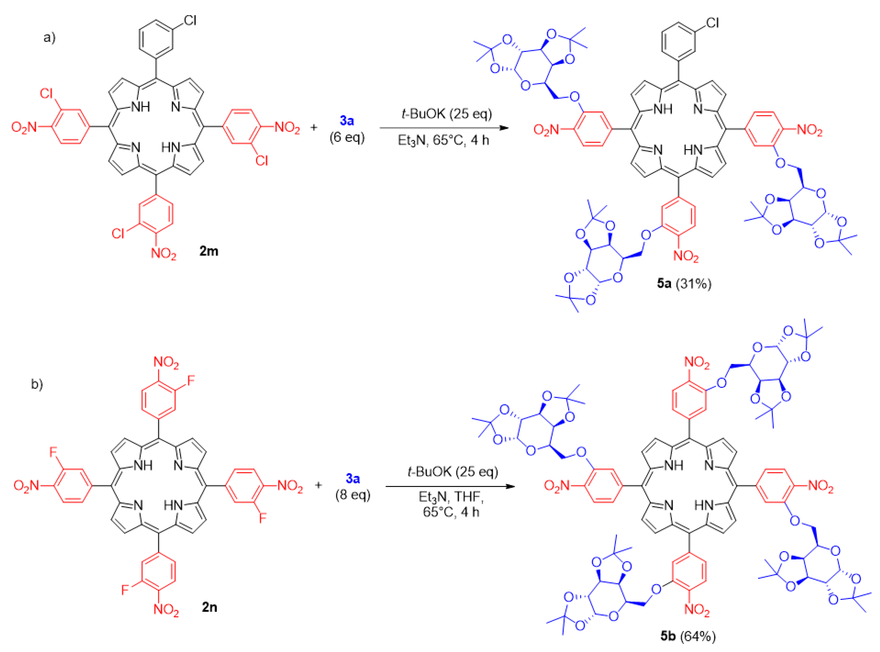
2.2. Investigation on the Reaction Scope
2.3. Biological Studies
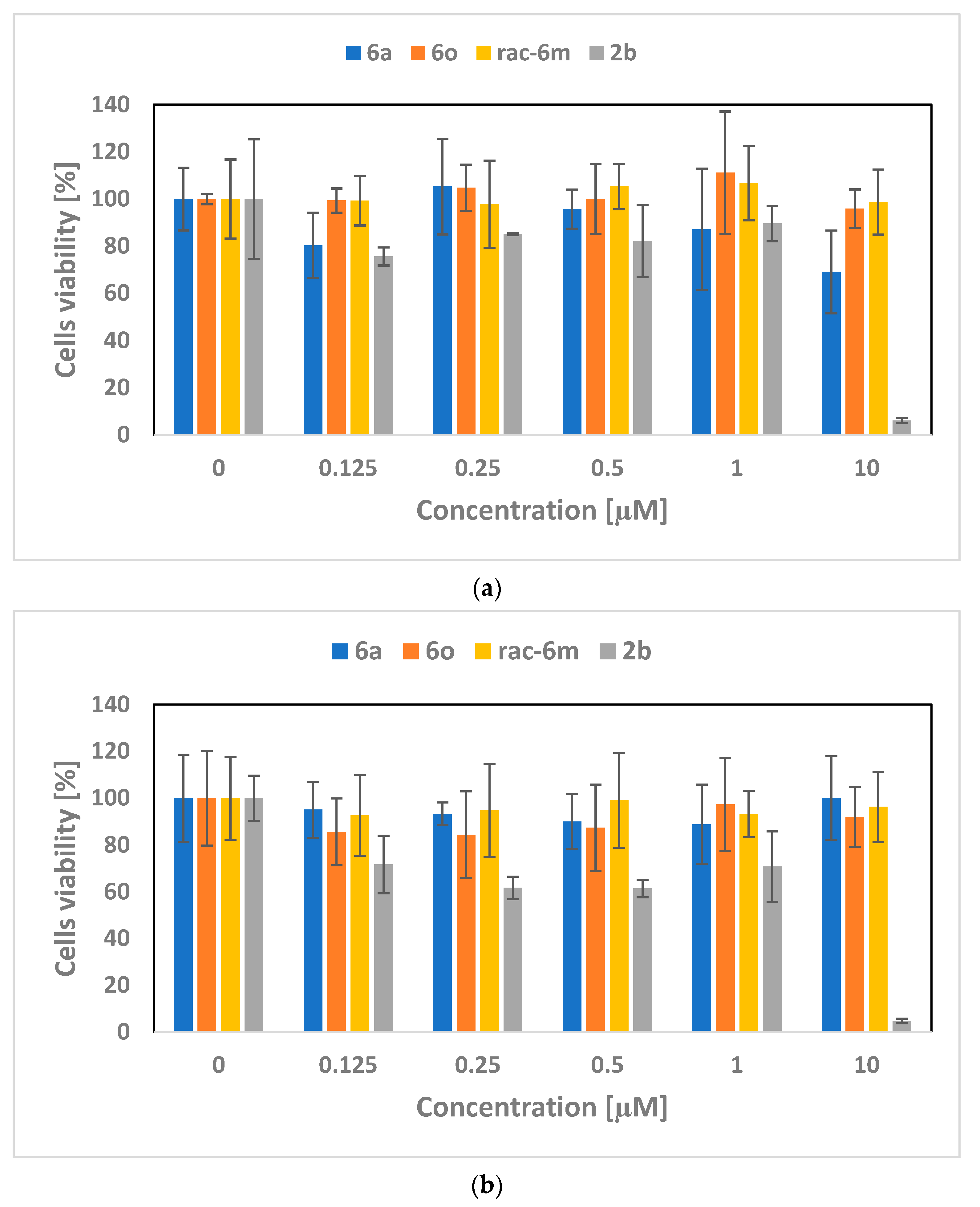
2.3.1. Cytotoxicity Assessment
2.3.2. Cellular Uptake
3. Methods and Materials
3.1. General Procedure for Synthesis of Porphyrin-Sugar Hybrids
3.2. Cell Cultures
3.3. Cytotoxicity Test
3.4. Ability of Compounds to Penetrate Cells
4. Conclusions
Supplementary Materials
Author Contributions
Funding
Institutional Review Board Statement
Informed Consent Statement
Data Availability Statement
Acknowledgments
Conflicts of Interest
References
- Terrier, F. Modern Nucleophilic Aromatic Substitution; Wiley-VCH: Weinheim, Germany, 2013; ISBN 9783527333912. [Google Scholar]
- Bunnett, J.F.; Zahler, R.E. Aromatic Nucleophilic Substitution Reactions. Chem. Rev. 1951, 49, 273–412. [Google Scholar] [CrossRef]
- Vlasov, V.M. Nucleophilic substitution of the nitro group, fluorine and chlorine in aromatic compounds. Russ. Chem. Rev. 2003, 72, 681–703. [Google Scholar] [CrossRef]
- Heravi, M.M.; Kheilkordi, Z.; Zadsirjan, V.; Heydari, M.; Malmir, M. Buchwald-Hartwig reaction: An overview. J. Organomet. Chem. 2018, 861, 17–104. [Google Scholar] [CrossRef]
- Ruiz-Castillo, P.; Buchwald, S.L. Applications of Palladium-Catalyzed C–N Cross-Coupling Reactions. Chem. Rev. 2016, 116, 12564–12649. [Google Scholar] [CrossRef]
- Dorel, R.; Grugel, C.P.; Haydl, A.M. The Buchwald–Hartwig Amination after 25 Years. Angew. Chem. Int. Ed. 2019, 58, 17118–17129. [Google Scholar] [CrossRef]
- Monnier, F.; Taillefer, M. Catalytic C-C, C-N, and C-O Ullmann-Type Coupling Reactions. Angew. Chem. Int. Ed. 2009, 48, 6954–6971. [Google Scholar] [CrossRef]
- Brown, D.G.; Boström, J. Analysis of Past and Present Synthetic Methodologies on Medicinal Chemistry: Where Have All the New Reactions Gone? J. Med. Chem. 2016, 59, 4443–4458. [Google Scholar] [CrossRef]
- Dimakos, V.; Taylor, M.S. Recent advances in the direct O-arylation of carbohydrates. Org. Biomol. Chem. 2021, 19, 514–524. [Google Scholar] [CrossRef]
- Jensen, K.J. O-Glycosylations under neutral or basic conditions. J. Chem. Soc. Perkin Trans. 1 2002, 20, 2219–2233. [Google Scholar] [CrossRef]
- Jacobsson, M.; Malmberg, J.; Ellervik, U. Aromatic O-glycosylation. Carbohydr. Res. 2006, 341, 1266–1281. [Google Scholar] [CrossRef]
- Henderson, A.S.; Medina, S.; Bower, J.F.; Galan, M.C. Nucleophilic Aromatic Substitution (SNAr) as an Approach to Challenging Carbohydrate–Aryl Ethers. Org. Lett. 2015, 17, 4846–4849. [Google Scholar] [CrossRef] [PubMed]
- Haines, A.H.; Symes, K.C. Syntheses of some polyfluoro-aromatic derivatives of carbohydrates and polyols. J. Chem. Soc. Perkin Trans. 1 1973, 53–56. [Google Scholar] [CrossRef]
- Hamashima, Y.; Kanai, M.; Shibasaki, M. Catalytic Enantioselective Cyanosilylation of Ketones. J. Am. Chem. Soc. 2000, 122, 7412–7413. [Google Scholar] [CrossRef]
- Hamashima, Y.; Kanai, M.; Shibasaki, M. Catalytic enantioselective cyanosilylation of ketones: Improvement of enantioselectivity and catalyst turn-over by ligand tuning. Tetrahedron Lett. 2001, 42, 691–694. [Google Scholar] [CrossRef]
- Sample, H.C.; Senge, M.O. Nucleophilic Aromatic Substitution (SNAr) and Related Reactions of Porphyrinoids: Mechanistic and Regiochemical Aspects. Eur. J. Org. Chem. 2021, 2021, 7–42. [Google Scholar] [CrossRef]
- Santos, F.D.C.; Cunha, A.C.; de Souza, M.C.B.; Tomé, A.C.; Neves, M.G.; Ferreira, V.F.; Cavaleiro, J.A. Synthesis of porphyrin–quinolone conjugates. Tetrahedron Lett. 2008, 49, 7268–7270. [Google Scholar] [CrossRef]
- Pedrosa, L.F.; De Souza, M.C.; Faustino, M.A.F.; Neves, M.G.P.M.S.; Silva, A.M.S.; Tomé, A.C.; Ferreira, V.F.; Cavaleiro, J.A.S. Porphyrin—Phosphoramidate Conjugates: Synthesis, Photostability and Singlet Oxygen Generation. Aust. J. Chem. 2011, 64, 939–944. [Google Scholar] [CrossRef]
- Vinagreiro, C.S.; Gonçalves, N.P.F.; Calvete, M.J.F.; Schaberle, F.A.; Arnaut, L.G.; Pereira, M.M. Synthesis and characterization of biocompatible bimodal meso-sulfonamide-perfluorophenylporphyrins. J. Fluor. Chem. 2015, 180, 161–167. [Google Scholar] [CrossRef]
- Souza, M.C.; Santos, C.I.M.; Mariz, I.; Marques, B.S.; Machado, L.A.; Pedrosa, L.F.; Cavaleiro, J.A.S.; Neves, M.G.P.M.S.; Mendes, R.F.; Paz, F.A.A.; et al. New triazine bridged triads based on BODIPY-porphyrin systems: Extended absorption, efficient energy transfer and upconverted emission. Dyes Pigment. 2021, 187, 109137. [Google Scholar] [CrossRef]
- Castro, K.A.D.F.; de Lima, F.H.C.; Simões, M.M.Q.; Neves, M.G.P.M.S.; Paz, F.A.A.; Mendes, R.F.; Nakagaki, S.; Cavaleiro, J.A.S. Synthesis, characterization and catalytic activity under homogeneous conditions of ethylene glycol substituted porphyrin manganese(III) complexes. Inorg. Chim. Acta 2016, 455, 575–583. [Google Scholar] [CrossRef]
- De Souza, M.C.; Pedrosa, L.F.; Cazagrande, G.S.; Ferreira, V.F.; Neves, M.G.P.M.S.; Cavaleiro, J.A.S. From porphyrin benzylphosphoramidate conjugates to the catalytic hydrogenation of 5,10,15,20-tetrakis(pentafluorophenyl)porphyrin. Beilstein J. Org. Chem. 2014, 10, 628–633. [Google Scholar] [CrossRef] [PubMed]
- Liu, Z.; Xue, Y.; Wu, M.; Yang, G.; Lan, M.; Zhang, W. Sensitization of Hypoxic Tumor to Photodynamic Therapy via Oxygen Self-Supply of Fluorinated Photosensitizers. Biomacromolecules 2019, 20, 4563–4573. [Google Scholar] [CrossRef] [PubMed]
- Castro, K.A.D.F.; Simões, M.M.Q.; Neves, M.G.P.M.S.; Cavaleiro, J.A.S.; Wypych, F.; Nakagaki, S. Glycol metalloporphyrin derivatives in solution or immobilized on LDH and silica: Synthesis, characterization and catalytic features in oxidation reactions. Catal. Sci. Technol. 2013, 4, 129–141. [Google Scholar] [CrossRef]
- Costa, J.I.T.; Farinha, A.S.F.; Almeida Paz, F.A.; Tomé, A.C. A Convenient Synthesis of Pentaporphyrins and Supramolecular Complexes with a Fulleropyrrolidine. Molecules 2019, 24, 3177. [Google Scholar] [CrossRef] [PubMed]
- Ostrowski, S.; Grzyb, S.; Mikus, A. Direct Amination of meso-Tetraarylporphyrin Derivatives—Easy Route to A3B-, A2BC-, and A2B2-Type Porphyrins Bearing Two Nitrogen-Containing Substituents at the meso-Positioned Phenyl Groups. Helv. Chim. Acta 2007, 90, 2000–2008. [Google Scholar] [CrossRef]
- Ostrowski, S.; Urbańska, N.; Mikus, A. Nucleophilic substitution of hydrogen in meso-nitroaryl-substituted porphyrins—Unprotected at the NH-centers in the core ring. Tetrahedron Lett. 2003, 44, 4373–4377. [Google Scholar] [CrossRef]
- Tomé, J.P.C.; Silva, E.M.P.; Pereira, A.M.V.M.; Alonso, C.M.A.; Faustino, M.A.F.; Neves, M.G.P.M.S.; Tomé, A.C.; Cavaleiro, J.A.S.; Tavares, S.A.P.; Duarte, R.R.; et al. Synthesis of neutral and cationic tripyridylporphyrin-d-galactose conjugates and the photoinactivation of HSV-1. Bioorg. Med. Chem. 2007, 15, 4705–4713. [Google Scholar] [CrossRef] [PubMed]
- Kou, J.; Dou, D.; Yang, L. Porphyrin photosensitizers in photodynamic therapy and its applications. Oncotarget 2017, 8, 81591–81603. [Google Scholar] [CrossRef]
- Correia, J.H.; Rodrigues, J.A.; Pimenta, S.; Dong, T.; Yang, Z. Photodynamic Therapy Review: Principles, Photosensitizers, Applications, and Future Directions. Pharmaceutics 2021, 13, 1332. [Google Scholar] [CrossRef]
- Pham, T.C.; Nguyen, V.-N.; Choi, Y.; Lee, S.; Yoon, J. Recent Strategies to Develop Innovative Photosensitizers for Enhanced Photodynamic Therapy. Chem. Rev. 2021, 121, 13454–13619. [Google Scholar] [CrossRef]
- Oniszczuk, A.; Wojtunik-Kulesza, K.A.; Oniszczuk, T.; Kasprzak, K. The potential of photodynamic therapy (PDT)—Experimental investigations and clinical use. Biomed. Pharmacother. 2016, 83, 912–929. [Google Scholar] [CrossRef] [PubMed]
- Simões, J.C.S.; Sarpaki, S.; Papadimitroulas, P.; Therrien, B.; Loudos, G. Conjugated Photosensitizers for Imaging and PDT in Cancer Research. J. Med. Chem. 2020, 63, 14119–14150. [Google Scholar] [CrossRef] [PubMed]
- Pisarek, S.; Maximova, K.; Gryko, D. Strategies toward the synthesis of amphiphilic porphyrins. Tetrahedron 2014, 70, 6685–6715. [Google Scholar] [CrossRef]
- Pereira, P.M.R.; Rizvi, W.; Bhupathiraju, N.V.S.D.K.; Berisha, N.; Fernandes, R.; Tomé, J.P.C.; Drain, C.M. Carbon-1 versus Carbon-3 Linkage of d-Galactose to Porphyrins: Synthesis, Uptake, and Photodynamic Efficiency. Bioconjugate Chem. 2018, 29, 306–315. [Google Scholar] [CrossRef] [PubMed]
- Pereira, P.M.R.; Silva, S.; Ramalho, J.S.; Gomes, C.M.; Girão, H.; Cavaleiro, J.A.S.; Ribeiro, C.A.F.; Tomé, J.P.C.; Fernandes, R. The role of galectin-1 in in vitro and in vivo photodynamic therapy with a galactodendritic porphyrin. Eur. J. Cancer 2016, 68, 60–69. [Google Scholar] [CrossRef] [PubMed]
- Tseberlidis, G.; Zardi, P.; Caselli, A.; Cancogni, D.; Fusari, M.; Lay, L.; Gallo, E. Glycoporphyrin Catalysts for Efficient C–H Bond Aminations by Organic Azides. Organometallics 2015, 34, 3774–3781. [Google Scholar] [CrossRef]
- Damiano, C.; Gadolini, S.; Intrieri, D.; Lay, L.; Colombo, C.; Gallo, E. Iron and Ruthenium Glycoporphyrins: Active Catalysts for the Synthesis of Cyclopropanes and Aziridines. Eur. J. Inorg. Chem. 2019, 2019, 4412–4420. [Google Scholar] [CrossRef]
- Ho, C.-M.; Zhang, J.-L.; Zhou, C.-Y.; Chan, O.-Y.; Yan, J.J.; Zhang, F.-Y.; Huang, J.-S.; Che, C.-M. A Water-Soluble Ruthenium Glycosylated Porphyrin Catalyst for Carbenoid Transfer Reactions in Aqueous Media with Applications in Bioconjugation Reactions. J. Am. Chem. Soc. 2010, 132, 1886–1894. [Google Scholar] [CrossRef]
- Tomé, J.P.C.; Neves, M.G.P.M.S.; Tomé, A.C.; Cavaleiro, J.A.S.; Mendonça, A.F.; Pegado, I.N.; Duarte, R.; Valdeira, M.L. Synthesis of glycoporphyrin derivatives and their antiviral activity against herpes simplex virus types 1 and 2. Bioorg. Med. Chem. 2005, 13, 3878–3888. [Google Scholar] [CrossRef]
- Gomes, M.C.; Silva, S.; Faustino, M.A.F.; Neves, M.G.P.M.S.; Almeida, A.; Cavaleiro, J.A.S.; Tomé, J.P.C.; Cunha, Â. Cationic galactoporphyrin photosensitisers against UV-B resistant bacteria: Oxidation of lipids and proteins by 1O2. Photochem. Photobiol. Sci. 2012, 12, 262–271. [Google Scholar] [CrossRef]
- Dixon, C.F.; Nottingham, A.N.; Lozano, A.F.; Sizemore, J.A.; Russell, L.A.; Valiton, C.; Newell, K.L.; Babin, D.; Bridges, W.T.; Parris, M.R.; et al. Synthesis and evaluation of porphyrin glycoconjugates varying in linker length: Preliminary effects on the photodynamic inactivation of Mycobacterium smegmatis. RSC Adv. 2021, 11, 7037–7042. [Google Scholar] [CrossRef] [PubMed]
- Batalha, P.N.; Gomes, A.T.P.C.; Forezi, L.S.M.; Costa, L.; de Souza, M.C.B.V.; Boechat, F.D.C.S.; Ferreira, V.F.; Almeida, A.; Faustino, M.A.F.; Neves, M.G.P.M.S.; et al. Synthesis of new porphyrin/4-quinolone conjugates and evaluation of their efficiency in the photoinactivation of Staphylococcus aureus. RSC Adv. 2015, 5, 71228–71239. [Google Scholar] [CrossRef]
- Titov, D.V.; Gening, M.L.; Tsvetkov, Y.E.; Nifantiev, N.E. Glycoconjugates of porphyrins with carbohydrates: Methods of synthesis and biological activity. Russ. Chem. Rev. 2014, 83, 523–554. [Google Scholar] [CrossRef]
- Singh, S.; Aggarwal, A.; Bhupathiraju, N.V.S.D.K.; Arianna, G.; Tiwari, K.; Drain, C.M. Glycosylated Porphyrins, Phthalocyanines, and Other Porphyrinoids for Diagnostics and Therapeutics. Chem. Rev. 2015, 115, 10261–10306. [Google Scholar] [CrossRef] [PubMed]
- Aksenova, A.A.; Sebiakin, I.L.; Mironov, A.F. Conjugates of Porphyrins with Carbohydrates. Russ. J. Bioorg. Chem. 2003, 29, 201–219. [Google Scholar] [CrossRef]
- Godlewski, B.; Baran, D.; de Robichon, M.; Ferry, A.; Ostrowski, S.; Malinowski, M. Sonogashira cross-coupling as a key step in the synthesis of new glycoporphyrins. Org. Chem. Front. 2022, 9, 2396–2404. [Google Scholar] [CrossRef]
- Hirohara, S.; Nishida, M.; Sharyo, K.; Obata, M.; Ando, T.; Tanihara, M. Synthesis, photophysical properties and photocytotoxicity of mono-, di-, tri- and tetra-glucosylated fluorophenylporphyrins. Bioorg. Med. Chem. 2010, 18, 1526–1535. [Google Scholar] [CrossRef]
- Hirohara, S.; Sharyo, K.; Kawasaki, Y.; Totani, M.; Tomotsuka, A.; Funasako, R.; Yasui, N.; Hasegawa, Y.; Yuasa, J.; Nakashima, T.; et al. trans-Bisglycoconjugation Is an Efficient and Robust Architecture for PDT Photosensitizers Based on 5,10,15,20-Tetrakis(Pentafluorophenyl)Porphyrin Derivatives. Bull. Chem. Soc. Jpn. 2013, 86, 1295–1308. [Google Scholar] [CrossRef]
- Wyrębek, P.; Osuch-Kwiatkowska, A.; Pakulski, Z.; Jarosz, S.; Ostrowski, S. The synthesis of sugar-decorated hydrophilic porphyrins. J. Porphyr. Phthalocyanines 2013, 17, 384–391. [Google Scholar] [CrossRef]
- Zhang, C.; Yan, K.; Fu, C.; Peng, H.; Hawker, C.J.; Whittaker, A.K. Biological Utility of Fluorinated Compounds: From Materials Design to Molecular Imaging, Therapeutics and Environmental Remediation. Chem. Rev. 2022, 122, 167–208. [Google Scholar] [CrossRef]
- Lindstrom, A.B.; Strynar, M.J.; Libelo, E.L. Polyfluorinated Compounds: Past, Present, and Future. Environ. Sci. Technol. 2011, 45, 7954–7961. [Google Scholar] [CrossRef] [PubMed]
- Stahl, T.; Mattern, D.; Brunn, H. Toxicology of perfluorinated compounds. Environ. Sci. Eur. 2011, 23, 38. [Google Scholar] [CrossRef]
- Ostrowski, S.; Mikus, A.; Łopuszyńska, B. Synthesis of highly substituted meso-tetraarylporphyrins. Tetrahedron 2004, 60, 11951–11957. [Google Scholar] [CrossRef]
- Ostrowski, S.; Łopuszyńska, B. Preparation of meso-Tetraarylporphyrins Nitrated in Two Neighboring Aromatic Rings. Synth. Commun. 2003, 33, 4101–4110. [Google Scholar] [CrossRef]
- Lindsey, J.S.; Schreiman, I.C.; Hsu, H.C.; Kearney, P.C.; Marguerettaz, A.M. Rothemund and Adler-Longo reactions revisited: Synthesis of tetraphenylporphyrins under equilibrium conditions. J. Org. Chem. 1987, 52, 827–836. [Google Scholar] [CrossRef]
- Rosa, M.; Malinowski, M.; Ostrowski, S. Modification for Nitration of Halo-Substituted Meso-Tetraarylporphyrins: A Convenient Scale-up in Small Amount of Solvent. Curr. Org. Synth. 2022. manuscript submitted for publication. [Google Scholar]
- Kielmann, M.; Flanagan, K.J.; Norvaiša, K.; Intrieri, D.; Senge, M.O. Synthesis of a Family of Highly Substituted Porphyrin Thioethers via Nitro Displacement in 2,3,7,8,12,13,17,18-Octaethyl-5,10,15,20-tetranitroporphyrin. J. Org. Chem. 2017, 82, 5122–5134. [Google Scholar] [CrossRef]
- Wuts, P.G.M.; Greene, T.W. Greene’s Protective Groups in Organic Synthesis; John Wiley & Sons: Hoboken, NJ, USA, 2007. [Google Scholar]
- Yildirim, A. An Expedient Method for Kinetically Controlled Acetonide Formation from d-Fructose Induced by Ionic Liquid Catalyst Accompanied with SrCl2·6H2O. Catal. Lett. 2020, 150, 2566. [Google Scholar] [CrossRef]
- Lv, J.; Liu, C.-Y.; Guo, Y.-F.; Feng, G.-J.; Dong, H. SnCl2-Catalyzed Acetalation/Selective Benzoylation Sequence for the Synthesis of Orthogonally Protected Glycosyl Acceptors. Eur. J. Org. Chem. 2022, 2022, e202101565. [Google Scholar] [CrossRef]
- Gupta, S.; Bera, S.; Mondal, D. Nascent-HBr-Catalyzed Removal of Orthogonal Protecting Groups in Aqueous Surfactants. J. Org. Chem. 2020, 85, 2635. [Google Scholar] [CrossRef]
- Ferreira, S.B.; Sodero, A.C.R.; Cardoso, M.F.C.; Lima, E.S.; Kaiser, C.R.; Silva, F.P., Jr.; Ferreira, V.F. Synthesis, Biological Activity, and Molecular Modeling Studies of 1H-1,2,3-Triazole Derivatives of Carbohydrates as α-Glucosidases Inhibitors. J. Med. Chem. 2010, 53, 2364. [Google Scholar] [CrossRef] [PubMed]


| Entry | Solvent | Base (Equiv.) | Temperature [°C] | Yield [%] |
|---|---|---|---|---|
| 1. | DMF | NaH (100) | 100 | 0 |
| 2. | DMF | NaOH (200) | 80 | 17 |
| 3. | DMF | t-BuOK (200) | 80 | 15 |
| 4. | DMF | t-BuOK (60) | 80 | traces |
| 5. | t-BuOH | t-BuOK (60) | 80 | 0 |
| 6. | THF | t-BuOK (60) | 80 | traces |
| 7. | Et3N | t-BuOK (60) | 80 | 70 |
| 8. | Et3N | t-BuOK (60) | 65 | 72 |
| 9. | Et3N | t-BuOK (60) | 50 | 65 |
| 10. | Et3N2 | t-BuOK (20) | 65 | 74 |
| 11. | Et3N 2,3 | t-BuOK (20) | 65 | 76 |
| 12. | Et3N 2,3 | t-BuOK (10) | 65 | 45 |
Publisher’s Note: MDPI stays neutral with regard to jurisdictional claims in published maps and institutional affiliations. |
© 2022 by the authors. Licensee MDPI, Basel, Switzerland. This article is an open access article distributed under the terms and conditions of the Creative Commons Attribution (CC BY) license (https://creativecommons.org/licenses/by/4.0/).
Share and Cite
Rosa, M.; Jędryka, N.; Skorupska, S.; Grabowska-Jadach, I.; Malinowski, M. New Route to Glycosylated Porphyrins via Aromatic Nucleophilic Substitution (SNAr)—Synthesis and Cellular Uptake Studies. Int. J. Mol. Sci. 2022, 23, 11321. https://doi.org/10.3390/ijms231911321
Rosa M, Jędryka N, Skorupska S, Grabowska-Jadach I, Malinowski M. New Route to Glycosylated Porphyrins via Aromatic Nucleophilic Substitution (SNAr)—Synthesis and Cellular Uptake Studies. International Journal of Molecular Sciences. 2022; 23(19):11321. https://doi.org/10.3390/ijms231911321
Chicago/Turabian StyleRosa, Mariusz, Natalia Jędryka, Sandra Skorupska, Ilona Grabowska-Jadach, and Maciej Malinowski. 2022. "New Route to Glycosylated Porphyrins via Aromatic Nucleophilic Substitution (SNAr)—Synthesis and Cellular Uptake Studies" International Journal of Molecular Sciences 23, no. 19: 11321. https://doi.org/10.3390/ijms231911321
APA StyleRosa, M., Jędryka, N., Skorupska, S., Grabowska-Jadach, I., & Malinowski, M. (2022). New Route to Glycosylated Porphyrins via Aromatic Nucleophilic Substitution (SNAr)—Synthesis and Cellular Uptake Studies. International Journal of Molecular Sciences, 23(19), 11321. https://doi.org/10.3390/ijms231911321