Article

Chronic Exposure to Endocrine Disruptor Vinclozolin Leads to Lung Damage via Nrf2–Nf-kb Pathway Alterations

by
Ramona D’Amico
1,†,
Davide Di Paola
1,†,
Daniela Impellizzeri
1,
Tiziana Genovese
1,
Roberta Fusco
1,
Alessio Filippo Peritore
1,
Enrico Gugliandolo
2,
Rosalia Crupi
2,
Livia Interdonato
1,
Salvatore Cuzzocrea
1,3,*,
Rosanna Di Paola
2,*,
Rosalba Siracusa
1,‡ and
Marika Cordaro
4,‡
1
Department of Chemical, Biological, Pharmaceutical and Environmental Sciences, University of Messina, Viale Ferdinando Stagno D’Alcontres 31, 98166 Messina, Italy
2
Department of Veterinary Sciences, University of Messina, 98168 Messina, Italy
3
Department of Pharmacological and Physiological Science, Saint Louis University School of Medicine, Saint Louis, MO 63104, USA
4
Department of Biomedical, Dental and Morphological and Functional Imaging, University of Messina, Via Consolare Valeria, 98125 Messina, Italy
*
Authors to whom correspondence should be addressed.
These authors contributed equally to this work.
This authors shared senior authorship.
Int. J. Mol. Sci. 2022, 23(19), 11320; https://doi.org/10.3390/ijms231911320
Submission received: 19 August 2022 / Revised: 16 September 2022 / Accepted: 21 September 2022 / Published: 26 September 2022
(This article belongs to the Special Issue Advances in Endocrine Disruptors 2.0)

Abstract

:
Endocrine-disrupting substances (EDS) are common and pervasive in our environment and pose a serious risk to both human and animal health. Endocrine-disrupting compounds (EDCs) have been associated with a variety of detrimental human health effects, including respiratory issues, as a result of their ability to disrupt cell physiology. Vinclozolin ((RS)-3-(3,5-Dichlorophenyl)-5-methyl-5-vinyloxazolidine-2,4-dione) is a common dicarboximide fungicide used to treat plant diseases. Several studies have analyzed the effects of vinclozolin exposure on the reproductive system, but less is known about its effect on other organs such as the lung. Mice were exposed for 28 days to orally administered vinclozolin at a dose of 100 mg/kg. Vinclozolin exposure induced histological alterations and collagen depositions in the lung. Additionally, vinclozolin induced inflammation and oxidative stress that led to lung apoptosis. Our study demonstrates for the first time that the toxicological effects of vinclozolin are not limited to the reproductive system but also involve other organs such as the lung.

1. Introduction

In vineyards and on fruits and vegetables such as raspberries, lettuce, kiwifruit, snap beans, and onions, vinclozolin ((RS)-3-(3,5-Dichlorophenyl)-5-methyl-5-vinyloxazolidine-2,4-dione), a popular dicarboximide fungicide, is used to treat diseases such as blights, rots, and molds. Additionally, it is applied to golf course turf. Vinclozolin, which was first registered in 1981, is widely utilized, but its total application has decreased. Vinclozolin is subject to Environmental Protection Agency (EPA) regulation as a pesticide in the United States (U.S. EPA) [1,2]. In addition to these restrictions inside the United States, as of 2006, the use of this pesticide was restricted in numerous nations, including Denmark, Finland, Norway, and Sweden. To assess its risks and hazards to the environment and animals, a number of tests and regulations have been applied [3,4]. Vinclozolin is created as extruded or dried flowable granules. Aerial application, chemigation through irrigation systems, and ground machinery are all options for distribution. The U.S. EPA has investigated occupational exposure to vinclozolin or its metabolites, as well as dietary (food and water) and non-dietary exposure. Fungicides can wind up on untreated foods following application because it has been demonstrated that they circulate through the water and air. Because fungicides are not eliminated from produce when it is washed with tap water, consumers cannot easily minimize their exposure [5]. One significant way that vinclozolin can be ingested is through wine grapes, which are thought to account for around 2% of all vinclozolin exposure. Tolerance limits have been defined for each crop because it has been determined that people may be exposed to vinclozolin residues and metabolites that contain the 3,5-dichloroaniline moiety (3,5-DCA) through eating. It is nevertheless feasible for humans to encounter vinclozolin and its residues, even if it is not approved for use by homeowners. Golfers playing on treated courses and families playing on previously treated sod, for instance, may be at risk for exposure. Vinclozolin exposure might occur at work when performing tasks such as loading and mixing. Vinclozolin has been demonstrated to be an endocrine disruptor with antiandrogenic effects, which is one of the major research findings [6,7]. Moreover, less is known about the effects of vinclozolin exposure on human health in other organs. Gazo and colleagues demonstrated that one of the mechanisms by which vinclozolin induces sperm alterations is an increase in oxidative stress in the testis [8]. In particular, they demonstrated that an increase in reactive oxygen species (ROS) production, as well as in lipid peroxidation, led to DNA damage. This was reflected in altered sperm mobility and velocity [8]. Additionally, they demonstrated an alteration in antioxidant endogenous defense, reflected in an increase in Mn-SOD activity after vinclozolin exposure [8]. Following this study, D’Amico et al. demonstrated that exposure to vinclozolin (and other endocrine disruptors) significantly induced oxidative stress and inflammation, worsening arthritis and Parkinson’s disease in experimental murine models [9,10]. In consideration of the fact that most previous studies investigated the effects of vinclozolin exposure on the reproductive system, we investigated for the first time the impact of long exposure to vinclozolin on the respiratory system [11,12].

2. Results

2.1. Exposure to Vinclozolin Induced Lung Alterations

We decided to explore the histological damage to the lungs due to exposure to vinclozolin. Figure 1 presents a comparison of the microarchitecture of the lungs among the experimental groups of the control and vinclozolin-treated animals at different magnifications (5×, 10×, and 20×). We found no signs of inflammation in either control group (Figure 1 square A for sham animals and square B for animals treated with corn oil, with histological score D). On the other hand, vinclozolin treatment induced significant inflammatory infiltrate in the lungs, with an increase in the alveolar wall and slight congestion with slight exudation in the alveolar spaces and lung interstitium, as judged from the 20× magnification in the center of the image (Figure 1 square C and histological score D).

2.2. Exposure to Vinclozolin Induced Lung Fibrosis

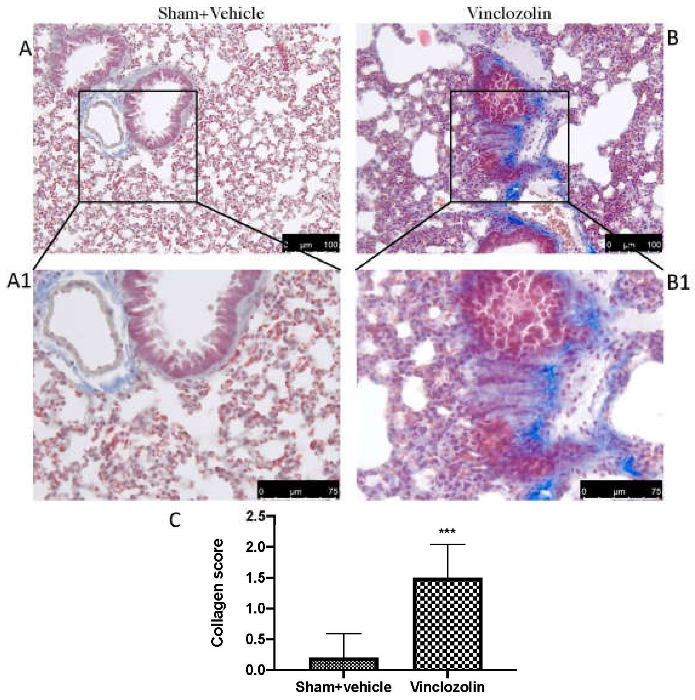
To assess pulmonary fibrosis in lung tissue, Masson’s trichrome staining was used. When compared to the sham group, vinclozolin administration caused fibrotic lesions in which the alveolar walls presented an increased amount of blue-stained fibrous connective tissue (Figure 2B, greater magnification B1). On the other hand, only a physiological deposition of collagen was seen in the sham group (Figure 2A, greater magnification A1).

2.3. Exposure to Vinclozolin Induced Inflammation and Oxidative Stress

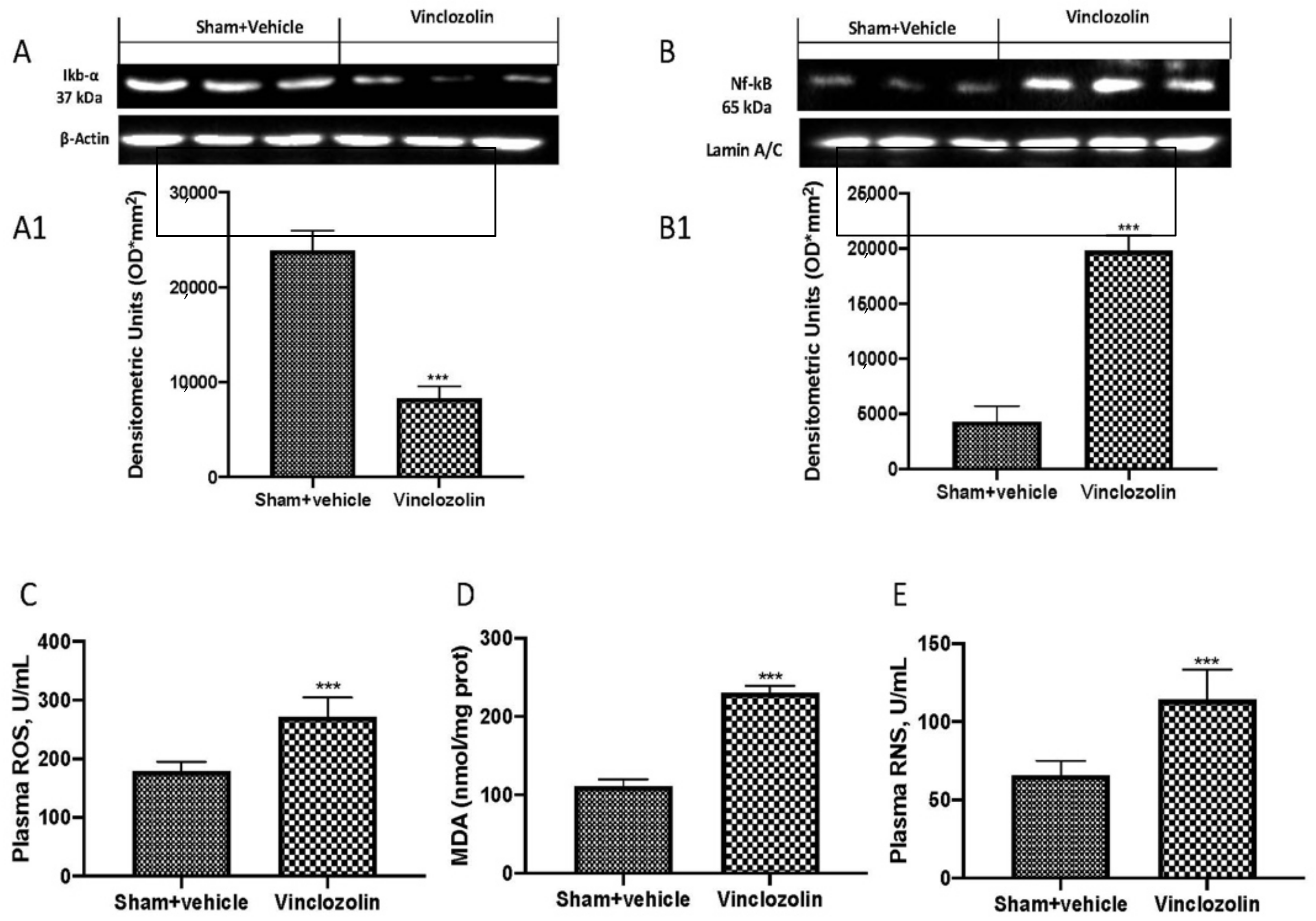
To investigate the molecular mechanism by which vinclozolin induces lung alterations, we performed Western blots of the Nf-kb pathway. We found a significant increase in Ikb-α degradation (Figure 3A and densitometric analysis A1), as well as an increase in Nf-kb nuclear translocation (Figure 3B and densitometric analysis B1) after vinclozolin exposure.
The increased generation of reactive oxygen species suggests that oxidative stress may play a role in vinclozolin-induced toxicity. For this reason, we investigated, by ELISA kits, the reactive oxygen species (ROS) production (Figure 3C), as well as H2O2 (Figure 3D) and reactive nitrogen species (RNS) (Figure 3E). We found that vinclozolin induced a significant increase in all these parameters.
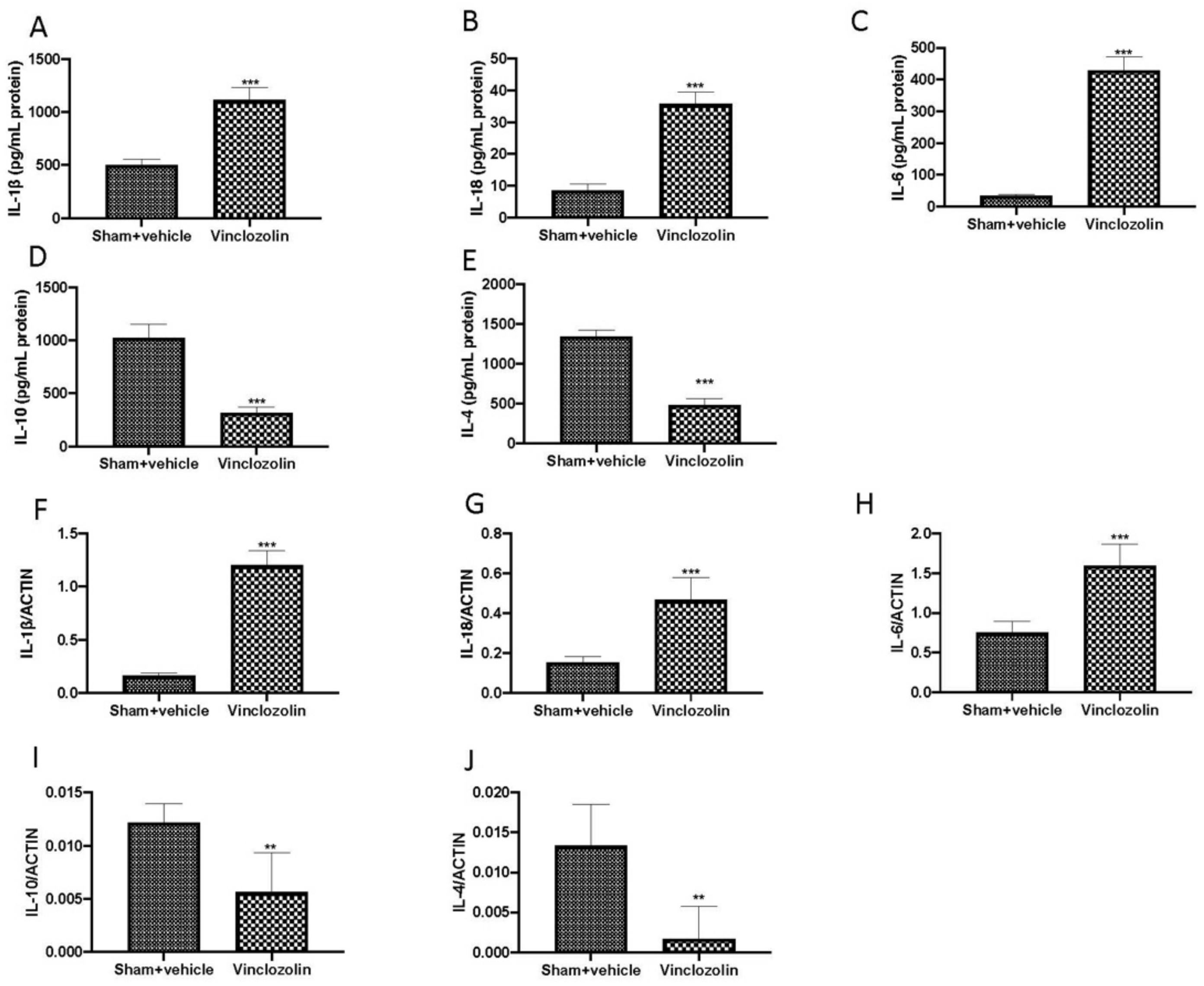
Additionally, numerous interleukins (ILs), which cause problems with cellular communication, are released when endocrine disruptors (EDs) are present, according to numerous independent investigations [13]. Using ELISA kits, we discovered that vinclozolin exposure increased the release of pro-inflammatory cytokines in BALF, specifically, IL-1β (Figure 4A), IL-18 (Figure 4B), and IL-6 (Figure 4C), whereas we discovered considerably lower levels of anti-inflammatory cytokines IL-10 (Figure 4D) and IL-4 (Figure 4E) when compared to sham groups.
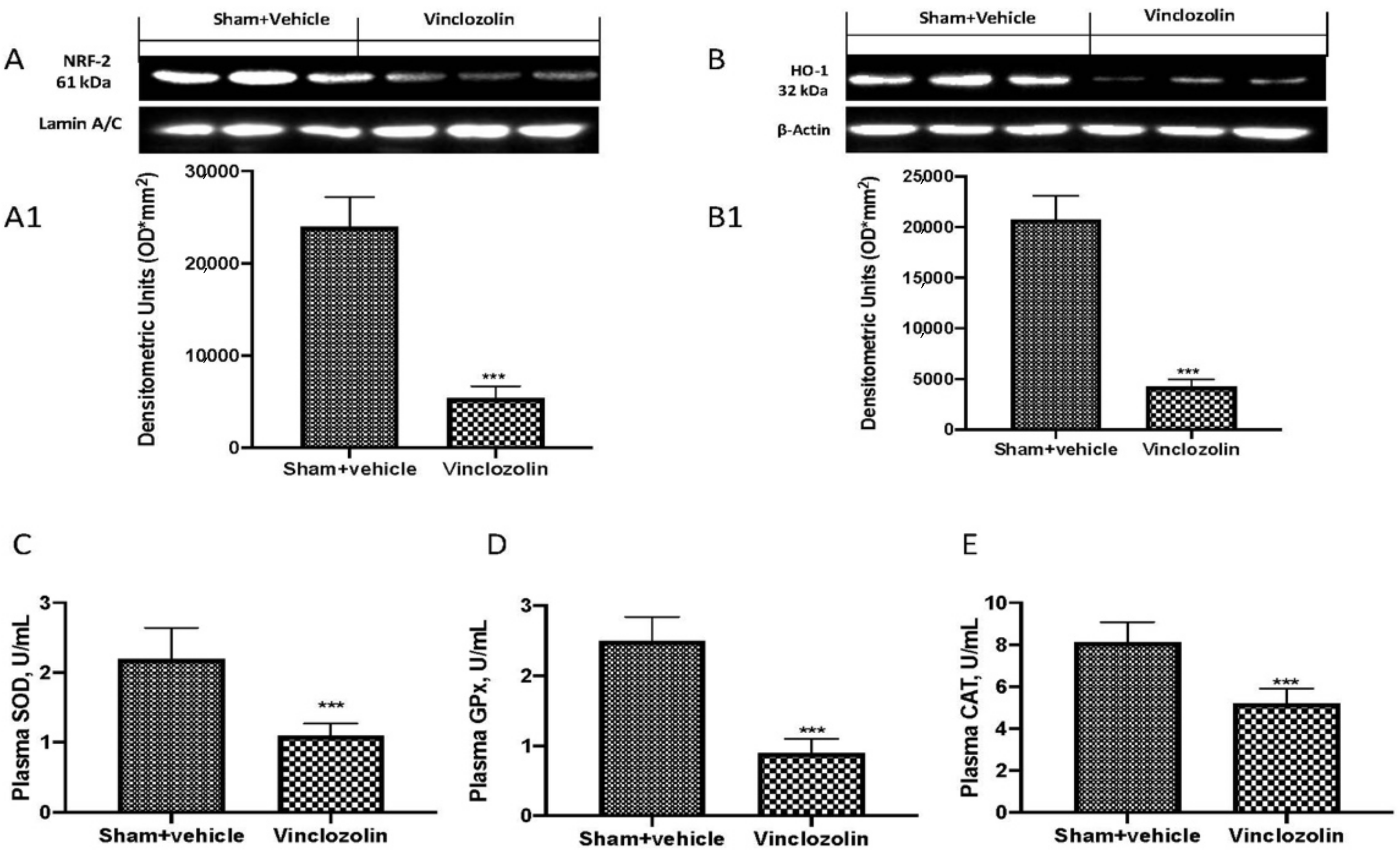
When exposed to oxidative injury, organisms have evolved defensive mechanisms that include Nrf-2-HO-1 pathway activation and antioxidant enzymes such as superoxide dismutase (SOD), glutathione peroxidase (GPx), and catalase (CAT). By Western blotting, we found a significant decrease in Nrf-2 (Figure 5A and densitometric analysis A1) and HO-1 (Figure 5B and densitometric analysis B1) and, as a consequence, a significant decrease in SOD (Figure 5C), GPx (Figure 5D), and CAT (Figure 5E).

2.4. Exposure to Vinclozolin Induced DNA Damage and Lipid Peroxidation

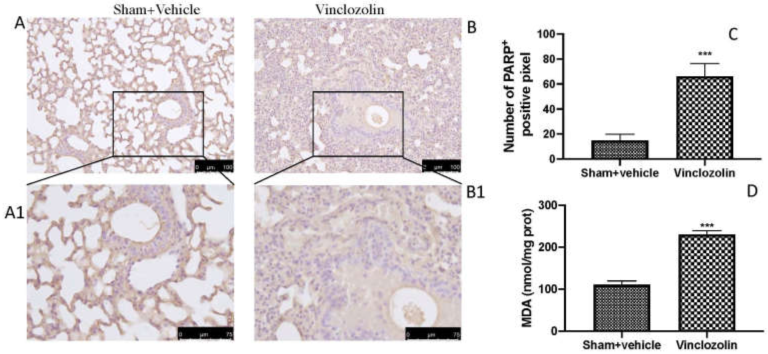
Exposure to endocrine disruptors has been linked to epigenetic alterations in a variety of tissues and organisms [14,15]. We investigated PARP by immunohistochemistry, and we found a significant increase in PARP-positive cells after vinclozolin exposure (Figure 6B with higher magnification B1 and analysis C) compared to sham (Figure 6A with higher magnification A1 and analysis C). Vinclozolin also significantly induced lipid peroxidation, as demonstrated by MDA analysis (Figure 6D).

2.5. Exposure to Vinclozolin Induced Lung Apoptosis

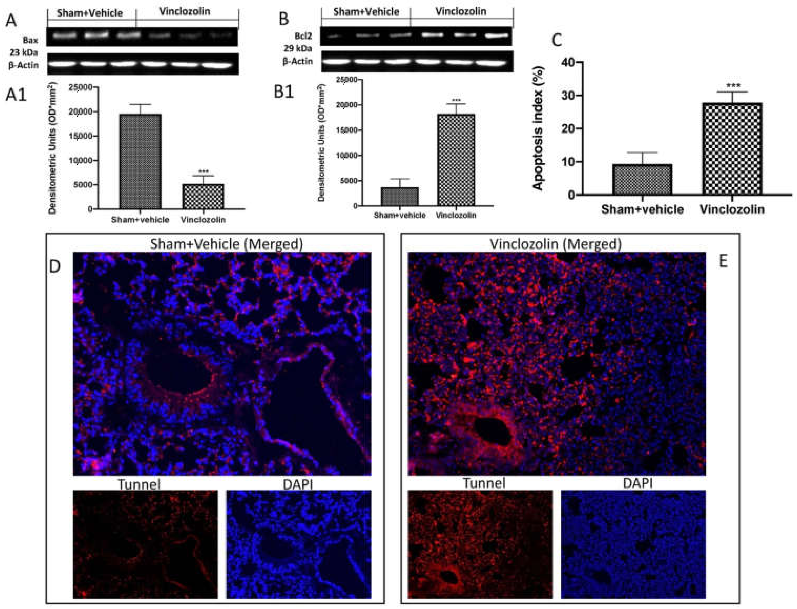
To further investigate the effects of vinclozolin exposure on the lung, we conducted a Western blot analysis of Bax and Bcl-2. We found that animals subject to vinclozolin exposure showed a significant decrease in Bax expression (Figure 7A and densitometric analysis A1) and, as a consequence, a significant increase in Bcl-2 expression (Figure 7B and densitometric analysis B1) compared to the control group. Our data were also confirmed by TUNEL analysis of lung sections. As shown in square D, sham animals showed physiological apoptosis. Vice versa, in square E, animals exposed to vinclozolin showed a significant increase in apoptotic cells (see apoptosis index C).

3. Discussion

Public interest in endocrine-disrupting chemicals’ impacts on health is growing, especially those related to long-term, low-dose exposures [16]. “Agent that interferes with the synthesis, secretion, transport, binding, or elimination of natural hormones in the body that are responsible for the maintenance of homeostasis, reproduction, development, and/or behavior” is how the U.S. Environmental Protection Agency (EPA) defines an endocrine disruptor (ED) [17]. This definition is obsolete because many animal experiments, clinical observations, and epidemiological studies have shown that endocrine disruptors can have an impact on different organs such as the reproductive system, the prostate, the breast, the liver, the thyroid, and the lungs, as well as metabolism [18]. Unfortunately, the fact that EDCs are a “shifting target” makes it difficult to comprehend how they relate to health issues. People and populations are exposed to constantly shifting patterns of chemical production and consumption. Additionally, rather than being released into the environment as separate compounds, they are frequently released as mixes. Although it is challenging to establish a direct causal link between human exposures to EDCs and disease states, findings from fundamental research and epidemiological studies demonstrate the importance of expanding screening for exposures and focusing on at-risk populations. An essential and successful strategy is disease prevention, in addition to advancing research in these fields [19]. Vinclozolin is a fungicide commonly used in fruits, vegetables, ornamental plants, and turf grasses. Vinclozolin exposure at work can happen through skin contact and dust inhalation. It may be consumed by the general population if produce they eat has been tainted with the fungicide. Vinclozolin studies have mainly focused on the reproductive system. It was demonstrated that exposure between the embryonic day and the postnatal day results in anomalies of the external genitalia in the neonatal stage, including a shortening of the anogenital distance and the retention of nipples in male pups. Due to a cleft phallus and hypospadias, male rats do not develop intromission or ejaculation during maturation. Additionally, males exhibit ectopic testicles, vaginal pouches, epididymal granulomas, accessory sex glands that are smaller or missing, and fewer cauda epididymal sperm. Additionally, vinclozolin prepubertal exposure in male rats inhibits pubertal maturation and slows the growth of the accessory sex gland and epididymis [20]. D’Amico and colleagues recently demonstrated that the effect of endocrine disruptor exposure is not limited to urogenital apparatus [9,10]. Here, we demonstrated for the first time that long-term exposure to vinclozolin induced significant damage to the respiratory system. Several cytokines, in particular, are generated by monocytes and macrophages, which attract neutrophils into lung tissues. This is important for host defense and promotes the onset of lung injury [21]. On the other hand, powerful anti-inflammatory cytokines such as IL-4 and IL-10 may be able to reduce the production of several inflammatory molecules [21]. Vinclozolin previously demonstrated the ability to induce histological alterations in the testis, prostate, ovary, kidney, and gonadal fat pad [22]. In line with this research, we found that a daily oral administration of vinclozolin significantly induced histological alterations, as well as an increase in collagen deposition that led to lung fibrosis. Unfortunately, it is well known that endocrine disruptors play a key role in inflammation and oxidative stress. In fact, they induce a significant alteration in the Nf-kb and Nrf-2 (nuclear factor erythroid 2–related factor 2) pathways, of which are master regulators of two of the most important cellular responses. During inflammation, a range of cells, including immune cells such as macrophages and lymphocytes, as well as endothelial cells and fibroblasts, produce cytokines that function as messengers between immune cells to control cell maturation, development, and responsiveness [23,24]. Additionally, in a system where oxidant species predominate, oxidative stress is defined as an imbalance between oxidant and antioxidant species [25]. As a potential modulator of ED-related outcomes, it has only recently been examined [26]. Numerous techniques have been employed to measure oxidative stress, each with its own uses and interpretations. These techniques are relatively easy to apply in both animal and human investigations because several oxidative stress assessments may be obtained by minimally invasive techniques, for instance, in blood [27]. In our study, we found a significant increase in both the Nf-kb and Nrf-2 pathways, as well as an increase in pro-inflammatory cytokines and ROS production. Radice and colleagues demonstrated that vinclozolin was able to induce lipid peroxidation in the Hep92 cell line [28]. As previously proven in other cell types, we found a significant increase in DNA damage, verified by the activation of PARP, a well-known marker of DNA damage, as well as an increase in lipid peroxidation, proven via an MDA level investigation [29,30,31,32,33,34]. Different studies demonstrated that vinclozolin administration induces granulosa cell apoptosis during follicular atresia in pigs, as well as apoptosis in testis [35,36]. Here, we demonstrated that long-term exposure to vinclozolin significantly induced lung apoptosis, as confirmed by the decrease in Bax activation and increase in Bcl-2 expression and TUNEL signal.

4. Materials and Methods

4.1. Animals

CD1 male mice (8 weeks old, 18–24 g) were acquired from Envigo (Milan, Italy) and kept in a controlled environment. All animal experiments complied with the new Italian regulations (D. Lgs 2014/26), EU regulations (EU Directive 2010/63), and ARRIVE guidelines.

4.2. Experimental Design and Groups

Mice were arbitrarily divided into three groups:
  • Sham: animals that were untreated (shown only in the histology);
  • Sham+vehicle: animals that were orally administered with corn oil;
  • Vinclozolin: animals that were orally administered 100 mg of vinclozolin daily for 28 days.
At the end of experiment, mice were sacrificed, and lung tissue and blood samples were collected for histology and biochemical analysis as previously described [37,38]. The dose, route of administration, and time of vinclozolin exposure were chosen based on another previous study and on a short exposure time at a different concentration observed in our laboratory (see Supplementary Figure S1) [9,10].

4.3. Western Blot Analysis of Cytosolic and Nuclear Extracts

Extracts from the cytosol and nucleus were prepared firstly with Buffer A containing 0.2 mM PMSF, 0·15 μM pepstatin A, 20 μM leupeptin, and 1 μM sodium orthovanadate, homogenized at the highest setting for 2 min, and centrifuged at 1000× g for 10 min at 4 °C. Following this, supernatants representing the cytosolic fraction and pellets containing enriched nuclei were re-suspended in Buffer B containing 1% Triton X-100, 150 mM NaCl, 10 mM Tris-HCl, pH 7.4, 1 mM EGTA, 1 mM EDTA, 0.2 mM PMSF, 20 μM leupeptin, and 0.2 mM sodium orthovanadate, as previously mentioned [39,40]. The following primary antibodies were used: anti-Iκbα (1:500, Santa Cruz Biotechnology (SCB), #sc-1643, Dallas, TX, USA), anti-nfκb (1:500, SCB, #sc8414), anti-NRF-2 (1-500, SCB, Heidelberg, Germany, #sc-365949), anti-heme oxygenase 1 (HO-1; 1-500, SCB, Heidelberg, Germany, #sc-136960), anti-Bax (1:500, SCB, #sc7480), and anti-Bcl-2 (1:500, SCB, #sc7382) in 1× PBS, 5% w/v non-fat dried milk, and 0.1% Tween-20 at 4 °C overnight. For the cytosolic and nuclear fractions, blots were also probed with β-actin and lamin A/C protein to ensure that they were filled with equivalent amounts of proteins (1:500; Santa Cruz Biotechnology). Signals were detected as previously described in our studies [40,41,42,43,44,45,46,47,48].

4.4. Histopathological Evaluation

Lungs were stained with hematoxylin/eosin (H/E) or Masson Trichrome and analyzed using a LeicaDM6 light microscope connected to an imaging system (LasX Navigator); they were blindly scored by two investigators without knowledge of the experimental groups. The severity of lung damage and fibrosis was assessed according to the method of Ashcroft et al. [29,49]. Briefly, histological alteration was quantified as follows: 0 for normal lung; 1 for minimal fibrous thickening of alveolar or bronchiolar walls; 3 for moderate thickening of the walls without obvious damage to lung architecture; 5 for increased fibrosis with definite damage to lung structure and the formation of fibrous bands or small fibrous masses; 7 for severe distortion of lung structure and large fibrous areas; 8 for total fibrous obliteration of fields. Collagen deposition was quantified as follows: 0 for negligible collagen deposition at the alveolar septa and thin-walled alveoli; 1 for one-layer deposition of collagen in the alveolar septa with mild fibrotic change; 2 for moderate collagen deposition and continuous fibrosis of the alveolar septa; 3 for excessive collagen deposition, thickened alveolar septa, compressed alveoli, and fibroplasia with confluent fibrotic mass.

4.5. Immunohistochemical Evaluation

Lung sections were incubated with anti-PARP antibody (1:250, Santa Cruz Biotechnology), as previously described by Cordaro et al. [50,51]. At the end of the protocol, the digital photos were analyzed by two observers blinded to the treatment, as previously performed in our laboratory [46,52,53].

4.6. Evaluation of Tissue Lipid Peroxidation

Malonaldehyde (MDA) levels were assessed, as previously described for lung tissue, at the end of the experiments. Briefly, after homogenization, MDA absorbance was measured at 650 nm using a spectrophotometer and expressed in mill-units per 100 milligram weight (mU/100 mg) of wet tissue [43,54].

4.7. Assessment of the Antioxidant System

SOD activity assay was performed following the method described by Marklund and Marklund at 420 nm [9,10,55,56]. The results are presented as SOD units/mg protein. Moreover, CAT activity was assessed following the method of Aebi in terms of mmoles of hydrogen peroxide (H2O2) consumed per min per mg of protein [57,58,59]. The results are presented as CAT units/mg protein. Furthermore, the level of the nonenzymatic cellular antioxidant glutathione was measured following the method of Moron et al. [60,61,62,63]. The results are presented as GSH units/mg protein.

4.8. Assessment of Cytokine Production

ELISA kits were used to investigate IL-1β, IL-18, IL-6, IL-10, and IL-4 in bronchoalveolar lavage fluid (BALF) at the end of experiment, as previously described [32,34,41,53,64,65,66,67]. The manufacturer’s manuals provided specific instructions for the test procedure.

4.9. Terminal Deoxynucleotidyl Nick-End Labeling (TUNEL) Assay

TUNEL staining for apoptotic cell nuclei and DAPI staining for all cell nuclei were performed in lung sections as described previously [34,55,68,69,70,71]. The index of apoptosis is expressed as the number of positively stained apoptotic cells over the total number of cells counted, multiplied by 100%.

4.10. Real-Time Quantitative Polymerase Chain Reaction Analysis (RT qPCR)

The levels of relative genes from lung tissues were measured by real-time quantitative polymerase chain reaction analysis (RT qPCR) using kits according to the manufacturer’s protocol (Aidlab Biotech, Beijing, China). The following primers were used: IL1β (F) ATGAAAGACGGCACACCCAC (R) AAGGCAGAGTCTTCGGTGAG; IL-6 (F) CAACCAAGAGGTGAGTGCTTC (R) GGTGTCCTCTTTCCCACACTG; IL-18 (F) GGAAGACCAGAGACATCCACTG (R) ACACTAGACCAAAGGGCTTG; IL-4 (F) CTGTAGGGCTTCCAAGGTGC (R) CTCTCATTGTGCCAGGTCACT; IL-10 (F) GCCAGTTAGAAAGCCACCAC (R) GGTTCAGCCTGTTTCCCAAC; ACTIN (F) GGCTGTATTCCCCTCCATCG (R) CCAGTTGGTAACAATGCCATGT. The expression levels of target mRNA were normalized to that of actin [64].

4.11. Materials

Vinclozolin (Lot #BCBZ5052) was obtained from Sigma-Aldrich INC. P.O. (St. Louis, MO, 63178, USA). Unless otherwise stated, all compounds were purchased from Sigma-Aldrich (Milan, Italy). All solutions were prepared using nonpyrogenic saline (0.9% NaCl; Baxter Healthcare Ltd., Thetford, Norfolk, UK).

4.12. Statistical Evaluation

In this study, all data are expressed as the average ± SEM and represent at least 3 experiments carried out on different days. For in vivo studies, N represents the number of animals used. The number of animals used for in vivo studies was determined using G*Power 3.1 software (Die Heinrich-Heine-Universität Düsseldorf, Düsseldorf, Germany). In all statistical studies, GraphPad Prism 8 software (La Jolla, CA, USA) was used. The results were analyzed by t-test, and a p-value less than 0.05 was considered significant.

5. Conclusions

In conclusion, in our study, we demonstrated for the first time that exposure to the fungicide vinclozolin, especially when chronic, induces serious alteration to the respiratory system, with an increase in inflammation, oxidative stress, and apoptosis. This is the first study to consider vinclozolin not only as an endocrine disruptor with androgenic activity but also as a toxin that will require a series of tests and regulations in order to evaluate its risks and hazards on the environment and animals.

Supplementary Materials

The following supporting information can be downloaded at: https://www.mdpi.com/article/10.3390/ijms231911320/s1.

Author Contributions

Conceptualization, R.S. and M.C.; Formal analysis, L.I. and D.D.P.; Funding acquisition, S.C. and R.D.P.; Methodology, R.D., D.I. and E.G.; Software, T.G. and R.F.; Supervision, R.D.P.; Validation, A.F.P. and R.C.; Writing—review and editing, R.S. and M.C. All authors have read and agreed to the published version of the manuscript.

Funding

This research was supported in part by Ministero dell’Istruzione, dell’Università e della Ricerca (MIUR), PRIN 2017 (Prot. 2017MLC3NF).

Institutional Review Board Statement

The animal study protocol was approved by the Institutional Review Board of University of Messina (protocol code 904/2021 P.R.).

Informed Consent Statement

Not applicable.

Data Availability Statement

The data used to support the findings of this study are available from the corresponding author upon request.

Conflicts of Interest

The authors declare no conflict of interest.

References

  1. Marcoccia, D.; Smeriglio, A.; Mantovani, A.; Trombetta, D.; Lorenzetti, S. Intracellular distribution of vinclozolin and its metabolites differently affects 5alpha-dihydrotestosterone (DHT)-induced PSA secretion in LNCaP cells. Reprod. Toxicol. 2022, 111, 83–91. [Google Scholar] [CrossRef]
  2. Kang, C.Q.; Meng, Q.Y.; Dang, W.; Shao, Y.J.; Lu, H.L. Effects of chronic exposure to the fungicide vinclozolin on gut microbiota community in an aquatic turtle. Ecotoxicol. Environ. Saf. 2022, 239, 113621. [Google Scholar] [CrossRef]
  3. Lu, H.L.; Kang, C.Q.; Meng, Q.Y.; Hu, J.R.; Melvin, S.D. Functional and hepatic metabolite changes in aquatic turtle hatchlings exposed to the anti-androgenic fungicide vinclozolin. Ecotoxicol. Environ. Saf. 2022, 231, 113220. [Google Scholar] [CrossRef] [PubMed]
  4. Yu, H.; Yang, J.; Zhang, Y.; Fu, H.; Yan, Z.; Zhu, Y. Vinclozolin-induced mouse penile malformation and “small testis” via miR132, miR195a together with the Hippo signaling pathway. Toxicology 2021, 460, 152842. [Google Scholar] [CrossRef]
  5. Wu, K.; Li, Y.; Pan, P.; Li, Z.; Yu, Y.; Huang, J.; Ma, F.; Tian, L.; Fang, Y.; Wang, Y.; et al. Gestational vinclozolin exposure suppresses fetal testis development in rats. Ecotoxicol. Environ. Saf. 2020, 203, 111053. [Google Scholar] [CrossRef] [PubMed]
  6. Gorczyca, G.; Wartalski, K.; Tabarowski, Z.; Duda, M. Effects of vinclozolin exposure on the expression and activity of SIRT1 and SIRT6 in the porcine ovary. J. Physiol. Pharmacol. 2019, 70, 153–165. [Google Scholar] [CrossRef]
  7. Aquilino, M.; Sanchez-Arguello, P.; Novo, M.; Martinez-Guitarte, J.L. Effects on tadpole snail gene expression after exposure to vinclozolin. Ecotoxicol. Environ. Saf. 2019, 170, 568–577. [Google Scholar] [CrossRef] [PubMed]
  8. Gazo, I.; Linhartova, P.; Shaliutina, A.; Hulak, M. Influence of environmentally relevant concentrations of vinclozolin on quality, DNA integrity, and antioxidant responses of sterlet Acipenser ruthenus spermatozoa. Chem. Biol. Interact. 2013, 203, 377–385. [Google Scholar] [CrossRef] [PubMed]
  9. D’Amico, R.; Gugliandolo, E.; Siracusa, R.; Cordaro, M.; Genovese, T.; Peritore, A.F.; Crupi, R.; Interdonato, L.; Di Paola, D.; Cuzzocrea, S.; et al. Toxic Exposure to Endocrine Disruptors Worsens Parkinson’s Disease Progression through NRF2/HO-1 Alteration. Biomedicines 2022, 10, 1073. [Google Scholar] [CrossRef] [PubMed]
  10. D’Amico, R.; Gugliandolo, E.; Cordaro, M.; Fusco, R.; Genovese, T.; Peritore, A.F.; Crupi, R.; Interdonato, L.; Di Paola, D.; Cuzzocrea, S.; et al. Toxic Effects of Endocrine Disruptor Exposure on Collagen-Induced Arthritis. Biomolecules 2022, 12, 564. [Google Scholar] [CrossRef] [PubMed]
  11. Nilsson, E.; King, S.E.; McBirney, M.; Kubsad, D.; Pappalardo, M.; Beck, D.; Sadler-Riggleman, I.; Skinner, M.K. Vinclozolin induced epigenetic transgenerational inheritance of pathologies and sperm epimutation biomarkers for specific diseases. PLoS ONE 2018, 13, e0202662. [Google Scholar] [CrossRef] [PubMed]
  12. Aquilino, M.; Sanchez-Arguello, P.; Martinez-Guitarte, J.L. Genotoxic effects of vinclozolin on the aquatic insect Chironomus riparius (Diptera, Chironomidae). Environ. Pollut. 2018, 232, 563–570. [Google Scholar] [CrossRef] [PubMed]
  13. Ochiai, S.; Roediger, B.; Abtin, A.; Shklovskaya, E.; Fazekas de St Groth, B.; Yamane, H.; Weninger, W.; Le Gros, G.; Ronchese, F. CD326(lo)CD103(lo)CD11b(lo) dermal dendritic cells are activated by thymic stromal lymphopoietin during contact sensitization in mice. J. Immunol. 2014, 193, 2504–2511. [Google Scholar] [CrossRef] [PubMed]
  14. Dolinoy, D.C.; Huang, D.; Jirtle, R.L. Maternal nutrient supplementation counteracts bisphenol A-induced DNA hypomethylation in early development. Proc. Natl. Acad. Sci. USA 2007, 104, 13056–13061. [Google Scholar] [CrossRef]
  15. Guerrero-Bosagna, C.; Settles, M.; Lucker, B.; Skinner, M.K. Epigenetic transgenerational actions of vinclozolin on promoter regions of the sperm epigenome. PLoS ONE 2010, 5, e13100. [Google Scholar] [CrossRef]
  16. Kabir, E.R.; Rahman, M.S.; Rahman, I. A review on endocrine disruptors and their possible impacts on human health. Environ. Toxicol. Pharmacol. 2015, 40, 241–258. [Google Scholar] [CrossRef]
  17. Kavlock, R.J.; Daston, G.P.; DeRosa, C.; Fenner-Crisp, P.; Gray, L.E.; Kaattari, S.; Lucier, G.; Luster, M.; Mac, M.J.; Maczka, C.; et al. Research needs for the risk assessment of health and environmental effects of endocrine disruptors: A report of the U.S. EPA-sponsored workshop. Environ. Health Perspect. 1996, 104 (Suppl. 4), 715–740. [Google Scholar] [CrossRef]
  18. Derghal, A.; Djelloul, M.; Trouslard, J.; Mounien, L. An Emerging Role of micro-RNA in the Effect of the Endocrine Disruptors. Front. Neurosci. 2016, 10, 318. [Google Scholar] [CrossRef]
  19. Diamanti-Kandarakis, E.; Bourguignon, J.P.; Giudice, L.C.; Hauser, R.; Prins, G.S.; Soto, A.M.; Zoeller, R.T.; Gore, A.C. Endocrine-disrupting chemicals: An Endocrine Society scientific statement. Endocr. Rev. 2009, 30, 293–342. [Google Scholar] [CrossRef]
  20. Wolf, C.J.; LeBlanc, G.A.; Ostby, J.S.; Gray, L.E., Jr. Characterization of the period of sensitivity of fetal male sexual development to vinclozolin. Toxicol. Sci. 2000, 55, 152–161. [Google Scholar] [CrossRef] [Green Version]
  21. Wojdasiewicz, P.; Poniatowski, L.A.; Szukiewicz, D. The role of inflammatory and anti-inflammatory cytokines in the pathogenesis of osteoarthritis. Mediat. Inflamm. 2014, 2014, 561459. [Google Scholar] [CrossRef]
  22. Nilsson, E.; Klukovich, R.; Sadler-Riggleman, I.; Beck, D.; Xie, Y.; Yan, W.; Skinner, M.K. Environmental toxicant induced epigenetic transgenerational inheritance of ovarian pathology and granulosa cell epigenome and transcriptome alterations: Ancestral origins of polycystic ovarian syndrome and primary ovarian insufiency. Epigenetics 2018, 13, 875–895. [Google Scholar] [CrossRef] [PubMed]
  23. Dinarello, C.A. Historical insights into cytokines. Eur. J. Immunol. 2007, 37 (Suppl. 1), S34–S45. [Google Scholar] [CrossRef]
  24. Akdis, M.; Aab, A.; Altunbulakli, C.; Azkur, K.; Costa, R.A.; Crameri, R.; Duan, S.; Eiwegger, T.; Eljaszewicz, A.; Ferstl, R.; et al. Interleukins (from IL-1 to IL-38), interferons, transforming growth factor beta, and TNF-alpha: Receptors, functions, and roles in diseases. J. Allergy Clin. Immunol. 2016, 138, 984–1010. [Google Scholar] [CrossRef]
  25. Lushchak, V.I. Free radicals, reactive oxygen species, oxidative stress and its classification. Chem. Biol. Interact. 2014, 224, 164–175. [Google Scholar] [CrossRef] [PubMed]
  26. Shono, T.; Taguchi, T. Short-time exposure to mono-n-butyl phthalate (MBP)-induced oxidative stress associated with DNA damage and the atrophy of the testis in pubertal rats. Environ. Sci. Pollut. Res. Int. 2014, 21, 3187–3190. [Google Scholar] [CrossRef] [PubMed]
  27. Neier, K.; Marchlewicz, E.H.; Dolinoy, D.C.; Padmanabhan, V. Assessing Human Health Risk to Endocrine Disrupting Chemicals: A Focus on Prenatal Exposures and Oxidative Stress. Endocr. Disruptors 2015, 3, e1069916. [Google Scholar] [CrossRef]
  28. Radice, S.; Marabini, L.; Gervasoni, M.; Ferraris, M.; Chiesara, E. Adaptation to oxidative stress: Effects of vinclozolin and iprodione on the HepG2 cell line. Toxicology 1998, 129, 183–191. [Google Scholar] [CrossRef]
  29. Campolo, M.; Esposito, E.; Ahmad, A.; Di Paola, R.; Paterniti, I.; Cordaro, M.; Bruschetta, G.; Wallace, J.L.; Cuzzocrea, S. Hydrogen sulfide-releasing cyclooxygenase inhibitor ATB-346 enhances motor function and reduces cortical lesion volume following traumatic brain injury in mice. J. Neuroinflamm. 2014, 11, 196. [Google Scholar] [CrossRef]
  30. Impellizzeri, D.; Talero, E.; Siracusa, R.; Alcaide, A.; Cordaro, M.; Maria Zubelia, J.; Bruschetta, G.; Crupi, R.; Esposito, E.; Cuzzocrea, S.; et al. Protective effect of polyphenols in an inflammatory process associated with experimental pulmonary fibrosis in mice. Br. J. Nutr. 2015, 114, 853–865. [Google Scholar] [CrossRef]
  31. Genovese, T.; Duranti, A.; D’Amico, R.; Fusco, R.; Impellizzeri, D.; Peritore, A.F.; Crupi, R.; Gugliandolo, E.; Cuzzocrea, S.; Di Paola, R.; et al. Fatty Acid Amide Hydrolase (FAAH) Inhibition Plays a Key Role in Counteracting Acute Lung Injury. Int. J. Mol. Sci. 2022, 23, 2781. [Google Scholar] [CrossRef] [PubMed]
  32. D’Amico, R.; Fusco, R.; Cordaro, M.; Siracusa, R.; Peritore, A.F.; Gugliandolo, E.; Crupi, R.; Scuto, M.; Cuzzocrea, S.; Di Paola, R.; et al. Modulation of NLRP3 Inflammasome through Formyl Peptide Receptor 1 (Fpr-1) Pathway as a New Therapeutic Target in Bronchiolitis Obliterans Syndrome. Int. J. Mol. Sci. 2020, 21, 2144. [Google Scholar] [CrossRef] [PubMed]
  33. Genovese, T.; D’Amico, R.; Fusco, R.; Impellizzeri, D.; Peritore, A.F.; Crupi, R.; Interdonato, L.; Gugliandolo, E.; Cuzzocrea, S.; Paola, R.D.; et al. Acai (Euterpe Oleraceae Mart.) Seeds Regulate NF-kappaB and Nrf2/ARE Pathways Protecting Lung against Acute and Chronic Inflammation. Cell. Physiol. Biochem. 2022, 56, 1–20. [Google Scholar] [CrossRef] [PubMed]
  34. Fusco, R.; Cordaro, M.; Siracusa, R.; Peritore, A.F.; Gugliandolo, E.; Genovese, T.; D’Amico, R.; Crupi, R.; Smeriglio, A.; Mandalari, G.; et al. Consumption of Anacardium occidentale L. (Cashew Nuts) Inhibits Oxidative Stress through Modulation of the Nrf2/HO-1 and NF-kB Pathways. Molecules 2020, 25, 4426. [Google Scholar] [CrossRef] [PubMed]
  35. Knet, M.; Wartalski, K.; Hoja-Lukowicz, D.; Tabarowski, Z.; Slomczynska, M.; Duda, M. Analysis of porcine granulosa cell death signaling pathways induced by vinclozolin. Theriogenology 2015, 84, 927–939. [Google Scholar] [CrossRef]
  36. Knet, M.; Tabarowski, Z.; Slomczynska, M.; Duda, M. The effects of the environmental antiandrogen vinclozolin on the induction of granulosa cell apoptosis during follicular atresia in pigs. Theriogenology 2014, 81, 1239–1247. [Google Scholar] [CrossRef]
  37. Lin, J.; Xia, J.; Zhao, H.S.; Hou, R.; Talukder, M.; Yu, L.; Guo, J.Y.; Li, J.L. Lycopene Triggers Nrf2-AMPK Cross Talk to Alleviate Atrazine-Induced Nephrotoxicity in Mice. J. Agric. Food Chem. 2018, 66, 12385–12394. [Google Scholar] [CrossRef]
  38. Li, J.; Li, X.; Bi, H.; Ma, K.; Li, B. Developmental Exposure to Atrazine Impairs Spatial Memory and Downregulates the Hippocampal D1 Dopamine Receptor and cAMP-Dependent Signaling Pathway in Rats. Int. J. Mol. Sci. 2018, 19, 2241. [Google Scholar] [CrossRef]
  39. Cordaro, M.; Paterniti, I.; Siracusa, R.; Impellizzeri, D.; Esposito, E.; Cuzzocrea, S. KU0063794, a Dual mTORC1 and mTORC2 Inhibitor, Reduces Neural Tissue Damage and Locomotor Impairment After Spinal Cord Injury in Mice. Mol. Neurobiol. 2017, 54, 2415–2427. [Google Scholar] [CrossRef]
  40. Fusco, R.; D’Amico, R.; Cordaro, M.; Gugliandolo, E.; Siracusa, R.; Peritore, A.F.; Crupi, R.; Impellizzeri, D.; Cuzzocrea, S.; Di Paola, R. Absence of formyl peptide receptor 1 causes endometriotic lesion regression in a mouse model of surgically-induced endometriosis. Oncotarget 2018, 9, 31355–31366. [Google Scholar] [CrossRef] [Green Version]
  41. Impellizzeri, D.; Siracusa, R.; Cordaro, M.; Crupi, R.; Peritore, A.F.; Gugliandolo, E.; D’Amico, R.; Petrosino, S.; Evangelista, M.; Di Paola, R.; et al. N-Palmitoylethanolamine-oxazoline (PEA-OXA): A new therapeutic strategy to reduce neuroinflammation, oxidative stress associated to vascular dementia in an experimental model of repeated bilateral common carotid arteries occlusion. Neurobiol. Dis. 2019, 125, 77–91. [Google Scholar] [CrossRef] [PubMed]
  42. Cordaro, M.; Siracusa, R.; Crupi, R.; Impellizzeri, D.; Peritore, A.F.; D’Amico, R.; Gugliandolo, E.; Di Paola, R.; Cuzzocrea, S. 2-Pentadecyl-2-Oxazoline Reduces Neuroinflammatory Environment in the MPTP Model of Parkinson Disease. Mol. Neurobiol. 2018, 55, 9251–9266. [Google Scholar] [CrossRef] [PubMed]
  43. Impellizzeri, D.; Cordaro, M.; Bruschetta, G.; Crupi, R.; Pascali, J.; Alfonsi, D.; Marcolongo, G.; Cuzzocrea, S. 2-pentadecyl-2-oxazoline: Identification in coffee, synthesis and activity in a rat model of carrageenan-induced hindpaw inflammation. Pharmacol. Res. 2016, 108, 23–30. [Google Scholar] [CrossRef] [PubMed]
  44. Di Paola, R.; Fusco, R.; Impellizzeri, D.; Cordaro, M.; Britti, D.; Morittu, V.M.; Evangelista, M.; Cuzzocrea, S. Adelmidrol, in combination with hyaluronic acid, displays increased anti-inflammatory and analgesic effects against monosodium iodoacetate-induced osteoarthritis in rats. Arthritis Res. Ther. 2016, 18, 291. [Google Scholar] [CrossRef] [PubMed]
  45. Fusco, R.; Cordaro, M.; Genovese, T.; Impellizzeri, D.; Siracusa, R.; Gugliandolo, E.; Peritore, A.F.; D’Amico, R.; Crupi, R.; Cuzzocrea, S.; et al. Adelmidrol: A New Promising Antioxidant and Anti-Inflammatory Therapeutic Tool in Pulmonary Fibrosis. Antioxidants 2020, 9, 601. [Google Scholar] [CrossRef] [PubMed]
  46. Fusco, R.; Cordaro, M.; Siracusa, R.; D’Amico, R.; Genovese, T.; Gugliandolo, E.; Peritore, A.F.; Crupi, R.; Impellizzeri, D.; Cuzzocrea, S.; et al. Biochemical Evaluation of the Antioxidant Effects of Hydroxytyrosol on Pancreatitis-Associated Gut Injury. Antioxidants 2020, 9, 781. [Google Scholar] [CrossRef] [PubMed]
  47. Cordaro, M.; Siracusa, R.; Fusco, R.; D’Amico, R.; Peritore, A.F.; Gugliandolo, E.; Genovese, T.; Scuto, M.; Crupi, R.; Mandalari, G.; et al. Cashew (Anacardium occidentale L.) Nuts Counteract Oxidative Stress and Inflammation in an Acute Experimental Model of Carrageenan-Induced Paw Edema. Antioxidants 2020, 9, 660. [Google Scholar] [CrossRef]
  48. Fusco, R.; Gugliandolo, E.; Siracusa, R.; Scuto, M.; Cordaro, M.; D’Amico, R.; Evangelista, M.; Peli, A.; Peritore, A.F.; Impellizzeri, D.; et al. Formyl Peptide Receptor 1 Signaling in Acute Inflammation and Neural Differentiation Induced by Traumatic Brain Injury. Biology 2020, 9, 238. [Google Scholar] [CrossRef] [PubMed]
  49. Wang, S.; Yang, Y.; Luo, D.; Wu, D.; Liu, H.; Li, M.; Sun, Q.; Jia, L. Lung inflammation induced by exposure to Bisphenol-A is associated with mTOR-mediated autophagy in adolescent mice. Chemosphere 2020, 248, 126035. [Google Scholar] [CrossRef]
  50. Cordaro, M.; Impellizzeri, D.; Gugliandolo, E.; Siracusa, R.; Crupi, R.; Esposito, E.; Cuzzocrea, S. Adelmidrol, a Palmitoylethanolamide Analogue, as a New Pharmacological Treatment for the Management of Inflammatory Bowel Disease. Mol. Pharmacol. 2016, 90, 549–561. [Google Scholar] [CrossRef] [Green Version]
  51. Crupi, R.; Marino, A.; Cuzzocrea, S. n-3 fatty acids: Role in neurogenesis and neuroplasticity. Curr. Med. Chem. 2013, 20, 2953–2963. [Google Scholar] [CrossRef]
  52. Fusco, R.; Siracusa, R.; Peritore, A.F.; Gugliandolo, E.; Genovese, T.; D’Amico, R.; Cordaro, M.; Crupi, R.; Mandalari, G.; Impellizzeri, D.; et al. The Role of Cashew (Anacardium occidentale L.) Nuts on an Experimental Model of Painful Degenerative Joint Disease. Antioxidants 2020, 9, 511. [Google Scholar] [CrossRef]
  53. Siracusa, R.; Paterniti, I.; Cordaro, M.; Crupi, R.; Bruschetta, G.; Campolo, M.; Cuzzocrea, S.; Esposito, E. Neuroprotective Effects of Temsirolimus in Animal Models of Parkinson’s Disease. Mol. Neurobiol. 2018, 55, 2403–2419. [Google Scholar] [CrossRef] [PubMed]
  54. Cuzzocrea, S.; Mazzon, E.; Esposito, E.; Muia, C.; Abdelrahman, M.; Di Paola, R.; Crisafulli, C.; Bramanti, P.; Thiemermann, C. Glycogen synthase kinase-3beta inhibition attenuates the development of ischaemia/reperfusion injury of the gut. Intensive Care Med. 2007, 33, 880–893. [Google Scholar] [CrossRef] [PubMed]
  55. Di Paola, D.; Natale, S.; Gugliandolo, E.; Cordaro, M.; Crupi, R.; Siracusa, R.; D’Amico, R.; Fusco, R.; Impellizzeri, D.; Cuzzocrea, S.; et al. Assessment of 2-Pentadecyl-2-oxazoline Role on Lipopolysaccharide-Induced Inflammation on Early Stage Development of Zebrafish (Danio rerio). Life 2022, 12, 128. [Google Scholar] [CrossRef] [PubMed]
  56. Cordaro, M.; Siracusa, R.; D’Amico, R.; Genovese, T.; Franco, G.; Marino, Y.; Di Paola, D.; Cuzzocrea, S.; Impellizzeri, D.; Di Paola, R.; et al. Role of Etanercept and Infliximab on Nociceptive Changes Induced by the Experimental Model of Fibromyalgia. Int. J. Mol. Sci. 2022, 23, 6139. [Google Scholar] [CrossRef] [PubMed]
  57. Aebi, H. Catalase in vitro. Methods Enzymol. 1984, 105, 121–126. [Google Scholar] [CrossRef] [PubMed]
  58. Di Paola, D.; Abbate, J.M.; Iaria, C.; Cordaro, M.; Crupi, R.; Siracusa, R.; D’Amico, R.; Fusco, R.; Impellizzeri, D.; Cuzzocrea, S.; et al. Environmental Risk Assessment of Dexamethasone Sodium Phosphate and Tocilizumab Mixture in Zebrafish Early Life Stage (Danio rerio). Toxics 2022, 10, 279. [Google Scholar] [CrossRef] [PubMed]
  59. Di Paola, D.; Capparucci, F.; Abbate, J.M.; Cordaro, M.; Crupi, R.; Siracusa, R.; D’Amico, R.; Fusco, R.; Genovese, T.; Impellizzeri, D.; et al. Environmental Risk Assessment of Oxaliplatin Exposure on Early Life Stages of Zebrafish (Danio rerio). Toxics 2022, 10, 81. [Google Scholar] [CrossRef] [PubMed]
  60. Fusco, R.; Salinaro, A.T.; Siracusa, R.; D’Amico, R.; Impellizzeri, D.; Scuto, M.; Ontario, M.L.; Crea, R.; Cordaro, M.; Cuzzocrea, S. Hidrox® Counteracts Cyclophosphamide-Induced Male Infertility through NRF2 Pathways in a Mouse Model. Antioxidants 2021, 10, 778. [Google Scholar] [CrossRef] [PubMed]
  61. Di Paola, D.; Capparucci, F.; Lanteri, G.; Crupi, R.; Marino, Y.; Franco, G.A.; Cuzzocrea, S.; Spano, N.; Gugliandolo, E.; Peritore, A.F. Environmental Toxicity Assessment of Sodium Fluoride and Platinum-Derived Drugs Co-Exposure on Aquatic Organisms. Toxics 2022, 10, 272. [Google Scholar] [CrossRef] [PubMed]
  62. Di Paola, D.; Natale, S.; Iaria, C.; Cordaro, M.; Crupi, R.; Siracusa, R.; D’Amico, R.; Fusco, R.; Impellizzeri, D.; Cuzzocrea, S.; et al. Intestinal Disorder in Zebrafish Larvae (Danio rerio): The Protective Action of N-Palmitoylethanolamide-oxazoline. Life 2022, 12, 125. [Google Scholar] [CrossRef] [PubMed]
  63. Genovese, T.; Impellizzeri, D.; D’Amico, R.; Fusco, R.; Peritore, A.F.; Di Paola, D.; Interdonato, L.; Gugliandolo, E.; Crupi, R.; Di Paola, R.; et al. Role of Bevacizumab on Vascular Endothelial Growth Factor in Apolipoprotein E Deficient Mice after Traumatic Brain Injury. Int. J. Mol. Sci. 2022, 23, 4162. [Google Scholar] [CrossRef]
  64. An, X.; Sun, X.; Hou, Y.; Yang, X.; Chen, H.; Zhang, P.; Wu, J. Protective effect of oxytocin on LPS-induced acute lung injury in mice. Sci. Rep. 2019, 9, 2836. [Google Scholar] [CrossRef] [PubMed]
  65. Esposito, E.; Impellizzeri, D.; Bruschetta, G.; Cordaro, M.; Siracusa, R.; Gugliandolo, E.; Crupi, R.; Cuzzocrea, S. A new co-micronized composite containing palmitoylethanolamide and polydatin shows superior oral efficacy compared to their association in a rat paw model of carrageenan-induced inflammation. Eur. J. Pharmacol. 2016, 782, 107–118. [Google Scholar] [CrossRef] [PubMed]
  66. Impellizzeri, D.; Siracusa, R.; Cordaro, M.; Peritore, A.F.; Gugliandolo, E.; Mancuso, G.; Midiri, A.; Di Paola, R.; Cuzzocrea, S. Therapeutic potential of dinitrobenzene sulfonic acid (DNBS)-induced colitis in mice by targeting IL-1beta and IL-18. Biochem. Pharmacol. 2018, 155, 150–161. [Google Scholar] [CrossRef] [PubMed]
  67. Cordaro, M.; Cuzzocrea, S.; Crupi, R. An Update of Palmitoylethanolamide and Luteolin Effects in Preclinical and Clinical Studies of Neuroinflammatory Events. Antioxidants 2020, 9, 216. [Google Scholar] [CrossRef]
  68. Di Paola, D.; Iaria, C.; Capparucci, F.; Cordaro, M.; Crupi, R.; Siracusa, R.; D’Amico, R.; Fusco, R.; Impellizzeri, D.; Cuzzocrea, S.; et al. Aflatoxin B1 Toxicity in Zebrafish Larva (Danio rerio): Protective Role of Hericium erinaceus. Toxins 2021, 13, 710. [Google Scholar] [CrossRef] [PubMed]
  69. Paola, D.D.; Capparucci, F.; Natale, S.; Crupi, R.; Cuzzocrea, S.; Spano, N.; Gugliandolo, E.; Peritore, A.F. Combined Effects of Potassium Perchlorate and a Neonicotinoid on Zebrafish Larvae (Danio rerio). Toxics 2022, 10, 203. [Google Scholar] [CrossRef] [PubMed]
  70. Di Paola, D.; Capparucci, F.; Lanteri, G.; Cordaro, M.; Crupi, R.; Siracusa, R.; D’Amico, R.; Fusco, R.; Impellizzeri, D.; Cuzzocrea, S.; et al. Combined Toxicity of Xenobiotics Bisphenol A and Heavy Metals on Zebrafish Embryos (Danio rerio). Toxics 2021, 9, 344. [Google Scholar] [CrossRef]
  71. Di Paola, D.; Natale, S.; Iaria, C.; Crupi, R.; Cuzzocrea, S.; Spano, N.; Gugliandolo, E.; Peritore, A.F. Environmental Co-Exposure to Potassium Perchlorate and Cd Caused Toxicity and Thyroid Endocrine Disruption in Zebrafish Embryos and Larvae (Danio rerio). Toxics 2022, 10, 198. [Google Scholar] [CrossRef] [PubMed]
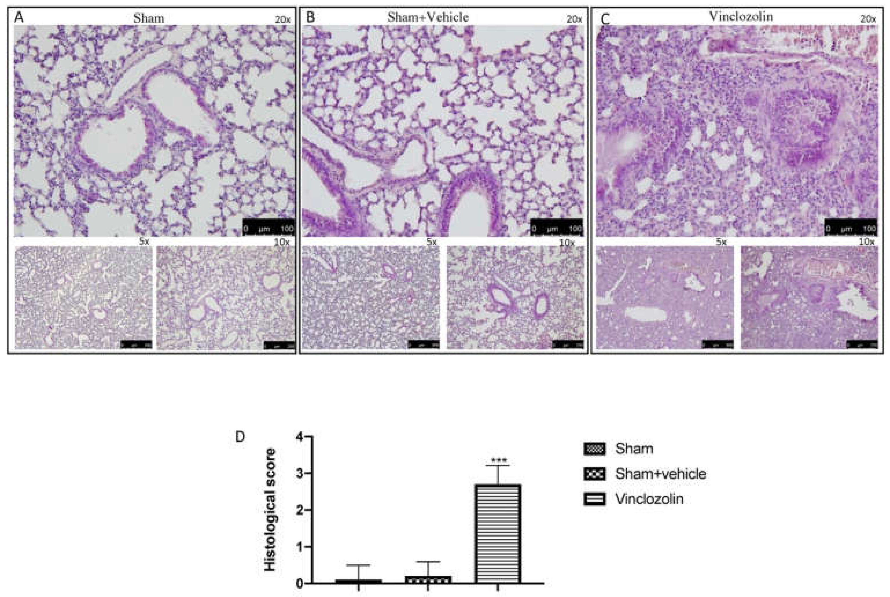
Figure 1. Vinclozolin-induced histological alterations. Square (A): images of sham animals at different magnifications (5×, 10×, and 20×); Square (B): images of sham + vehicle animals treated for 28 days with corn oil at different magnifications (5×, 10×, and 20×); Square (C): images of vinclozolin-treated animals at different magnifications (5×, 10×, and 20×); (D): Histological score. After vinclozolin administration, we observed a moderate thickening of the walls without obvious damage to the lung architecture. Values are the means ± SD of 6 mice per group. Images shown are representative of the results obtained. See manuscript for further details. *** p < 0.001 vs. sham.
Ijms 23 11320 g001
Figure 2. Collagen deposition after exposure to vinclozolin. Masson trichrome staining results for sham (A,A1) and for vinclozolin-treated (B,B1) animals; collagen score (C). Values are the means ± SD of 6 mice per group. Images shown are representative of the results obtained. After vinclozolin exposure, we found a significant increase in collagen deposition as shown by the increase in blue staining. *** p < 0.001 vs. sham.
Ijms 23 11320 g002
Figure 3. Exposure to vinclozolin induces Nf-kb activation and oxidative stress. Western blots and respective quantification of Ikb-α (A,A1) and Nf-kb pathway (B,B1); ELISA kit results in plasma for ROS (C), H2O2 (D), and reactive nitrogen species (RNS) (E). As shown, vinclozolin exposure increased inflammation and oxidative stress. Values are the means ± SD of 6 mice per group. *** p < 0.001 vs. sham.
Ijms 23 11320 g003
Figure 4. Cytokine analysis after vinclozolin exposure. ELISA kit results for IL-1β (A), IL-18 (B), IL-6 (C), IL-10 (D), and IL-4 (E). RT qPCR results for IL-1β (F), IL-18 (G), IL-6 (H), IL-10 (I), and IL-4 (J). As shown, after vinclozolin exposure, there was a significant increase in proinflammatory cytokines, as well as a significant decrease in anti-inflammatory cytokines. Values are the means ± SEM of 6 mice for all groups. ** p < 0.01 vs. sham, *** p < 0.001 vs. sham.
Ijms 23 11320 g004
Figure 5. Vinclozolin-induced Nrf-2 pathway alteration. Western blots and quantification of NRF2 (A,A1) and HO-1 (B,B1) in lung tissue. ELISA kit results for SOD (C), GPx (D), and CAT (E) levels. Vinclozolin exposure induced a significant alteration in the Nrf-2 pathway with an imbalance in antioxidant defense. Values are the means ± SD of 6 mice per group. *** p < 0.001 vs. sham.
Ijms 23 11320 g005
Figure 6. Exposure to vinclozolin induced DNA damage and lipid peroxidation. Immunohistochemistry localization of PARP for sham (A,A1) and for vinclozolin-treated (B,B1) animals; score (C); MDA levels (D). Vinclozolin induced an increase in PARP expression, as well as in lipid peroxidation. Values are the means ± SD of 6 mice per group. Images shown are representative of the results obtained. *** p < 0.001 vs. sham.
Ijms 23 11320 g006
Figure 7. Vinclozolin exposure induced lung apoptosis. Western blots and quantification of lung tissue from Bax (A,A1) and Bcl-2 (B,B1); TUNEL images from lung tissue of sham animals (D) and vinclozolin-treated animals (E); apoptotic index (C). After 28 days of vinclozolin exposure, we found a significant increase in cell death. Values are the means ± SD of 6 mice per group. Images shown are representative of the results obtained. *** p < 0.001 vs. sham.
Ijms 23 11320 g007
Publisher’s Note: MDPI stays neutral with regard to jurisdictional claims in published maps and institutional affiliations.