
Coronavirus Disease 2019-Associated Thrombotic Microangiopathy: Literature Review
Abstract
1. Introduction
Basic Concepts of Definition, Classification, Diagnosis, and Treatment of TMA
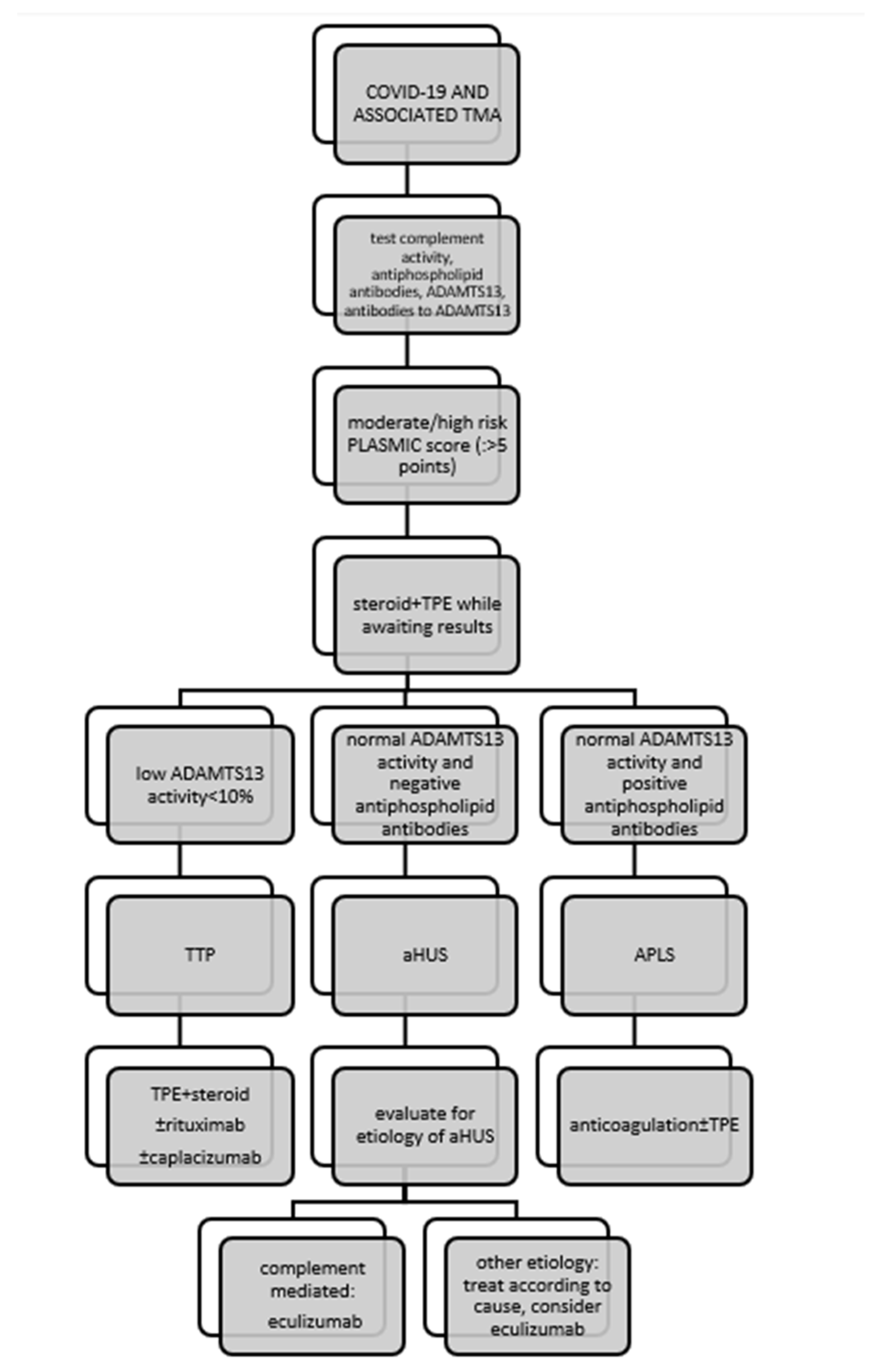
2. COVID-19-Associated TMA
2.1. Types of TMA Associated with COVID-19 and Clinical Correlates
2.1.1. Patients Presenting as TTP
2.1.2. Patients Presenting as aHUS
3. Discussion
4. Conclusions
Author Contributions
Funding
Institutional Review Board Statement
Informed Consent Statement
Data Availability Statement
Conflicts of Interest
Abbreviations
| aHUS | atypical hemolytic–uremic syndrome |
| APLS | antiphospholipid syndrome |
| AKI | acute kidney injury |
| CNI | calcineurin inhibitor |
| COVID-19 | coronavirus disease 2019 |
| ESRD | end-stage renal disease |
| FSGS | focal segmental glomerulosclerosis |
| FFP | fresh frozen plasma |
| HCQ | hydroxychloroquine |
| HCV | hepatitis C virus |
| MAHA | microangiopathic hemolytic anemia |
| PCR | polymerase chain reaction |
| SARS- CoV-2 | severe acute respiratory syndrome coronavirus 2 |
| STEC-HUS | Shiga toxin-mediated hemolytic uremic syndrome |
| TMA | thrombotic microangiopathy |
| TPE | therapeutic plasma exchange |
| TTP | thrombotic thrombocytopenic purpura |
| vWF | von Willebrand factor |
References
- Gupta, A.; Madhavan, M.V.; Sehgal, K.; Nair, N.; Mahajan, S.; Sehrawat, T.S.; Bikdeli, B.; Ahluwalia, N.; Ausiello, J.C.; Wan, E.Y.; et al. Extrapulmonary manifestations of COVID-19. Nat. Med. 2020, 26, 1017–1032. [Google Scholar] [CrossRef]
- Hirsch, J.S.; Ng, J.H.; Ross, D.W.; Sharma, P.; Shah, H.H.; Barnett, R.L.; Hazzan, A.D.; Fishbane, S.; Jhaveri, K.D.; on behalf of the Northwell COVID-19 Research Consortium; et al. Acute kidney injury in patients hospitalized with COVID-19. Kidney Int. 2020, 98, 209–218. [Google Scholar] [CrossRef]
- Sharma, P.; Uppal, N.N.; Wanchoo, R.; Shah, H.H.; Yang, Y.; Parikh, R.; Khanin, Y.; Madireddy, V.; Larsen, C.P.; Jhaveri, K.D.; et al. COVID-19–Associated Kidney Injury: A Case Series of Kidney Biopsy Findings. J. Am. Soc. Nephrol. 2020, 31, 1948–1958. [Google Scholar] [CrossRef]
- Golmai, P.; Larsen, C.P.; DeVita, M.V.; Wahl, S.J.; Weins, A.; Rennke, H.G.; Bijol, V.; Rosenstock, J.L. Histopathologic and Ultrastructural Findings in Postmortem Kidney Biopsy Material in 12 Patients with AKI and COVID-19. J. Am. Soc. Nephrol. 2020, 31, 1944–1947. [Google Scholar] [CrossRef]
- Tiwari, N.R.; Phatak, S.; Sharma, V.R.; Agarwal, S.K. COVID-19 and thrombotic microangiopathies. Thromb. Res. 2021, 202, 191–198. [Google Scholar] [CrossRef]
- Ardissino, G.; Possenti, I.; Tel, F.; Testa, S.; Paglialonga, F. Time to change the definition of hemolytic uremic syndrome. Eur. J. Intern. Med. 2014, 25, e29. [Google Scholar] [CrossRef]
- Sallée, M.; Ismail, K.; Fakhouri, F.; Vacher-Coponat, H.; Moussi-Francés, J.; Frémaux-Bacchi, V.; Burtey, S. Thrombocytopenia is not mandatory to diagnose haemolytic and uremic syndrome. BMC Nephrol. 2013, 14, 3. [Google Scholar] [CrossRef]
- George, J.N.; Nester, C.M. Syndromes of thrombotic microangiopathy. N. Engl. J. Med. 2014, 371, 654–666. [Google Scholar] [CrossRef]
- Aigner, C.; Schmidt, A.; Gaggl, M.; Sunder-Plassmann, G. An updated classification of thrombotic microangiopathies and treatment of complement gene variant-mediated thrombotic microangiopathy. Clin. Kidney J. 2019, 12, 333–337. [Google Scholar] [CrossRef]
- Brocklebank, V.; Wood, K.M.; Kavanagh, D. Thrombotic Microangiopathy and the Kidney. Clin. J. Am. Soc. Nephrol. 2018, 13, 300–317. [Google Scholar] [CrossRef]
- Goodship, T.H.; Cook, H.T.; Fakhouri, F.; Fervenza, F.C.; Frémeaux-Bacchi, V.; Kavanagh, D.; Nester, C.M.; Noris, M.; Pickering, M.C.; de Córdoba, S.R.; et al. Atypical hemolytic uremic syndrome and C3 glomerulopathy: Conclusions from a “Kidney Disease: Improving Global Outcomes” (KDIGO) Controversies Conference. Kidney Int. 2017, 91, 539–551. [Google Scholar] [CrossRef]
- Chiasakul, T.; Cuker, A. Clinical and laboratory diagnosis of TTP: An integrated approach. Hematol. Am. Soc. Hematol. Educ. Program 2018, 2018, 530–538. [Google Scholar] [CrossRef]
- Trachtman, H. HUS and TTP in Children. Pediatr. Clin. N. Am. 2013, 60, 1513–1526. [Google Scholar] [CrossRef]
- Bendapudi, P.K.; Hurwitz, S.; Fry, A.; Marques, M.B.; Waldo, S.W.; Li, A.; Sun, L.; Upadhyay, V.; Hamdan, A.; Brunner, A.M.; et al. Derivation and external validation of the PLASMIC score for rapid assessment of adults with thrombotic microangiopathies: A cohort study. Lancet Haematol. 2017, 4, e157–e164. [Google Scholar] [CrossRef]
- Palma, L.M.P.; Sridharan, M.; Sethi, S. Complement in Secondary Thrombotic Microangiopathy. Kidney Int. Rep. 2021, 6, 11–23. [Google Scholar] [CrossRef]
- Adamski, J. Thrombotic microangiopathy and indications for therapeutic plasma exchange. Hematol. Am. Soc. Hematol. Educ. Program 2014, 2014, 444–449. [Google Scholar] [CrossRef]
- Winters, J.L. Plasma exchange in thrombotic microangiopathies (TMAs) other than thrombotic thrombocytopenic purpura (TTP). Hematol. Am. Soc. Hematol. Educ. Program 2017, 2017, 632–638. [Google Scholar] [CrossRef]
- Weiler, J.M.; Packard, B.D. Methylprednisolone inhibits the alternative and amplification pathways of complement. Infect. Immun. 1982, 38, 122–126. [Google Scholar] [CrossRef]
- Dane, K.; Chaturvedi, S. Beyond plasma exchange: Novel therapies for thrombotic thrombocytopenic purpura. Hematol. Am. Soc. Hematol. Educ. Program 2018, 2018, 539–547. [Google Scholar] [CrossRef]
- Zielińska, K.A.; Van Moortel, L.; Opdenakker, G.; De Bosscher, K.; Van den Steen, P.E. Endothelial Response to Glucocorticoids in Inflammatory Diseases. Front. Immunol. 2016, 7, 592. [Google Scholar] [CrossRef]
- Jestin, M.; Benhamou, Y.; Schelpe, A.S.; Roose, E.; Provôt, F.; Galicier, L.; Hié, M.; Presne, C.; Poullin, P.; Wynckel, A.; et al. Preemptive rituximab prevents long-term relapses in immune-mediated thrombotic thrombocytopenic purpura. Blood 2018, 132, 2143–2153. [Google Scholar] [CrossRef]
- Legendre, C.M.; Licht, C.; Muus, P.; Greenbaum, L.A.; Babu, S.; Bedrosian, C.; Bingham, C.; Cohen, D.J.; Delmas, Y.; Douglas, K.; et al. Terminal complement inhibitor eculizumab in atypical hemolytic-uremic syndrome. N. Engl. J. Med. 2013, 368, 2169–2181. [Google Scholar] [CrossRef]
- Scully, M.; Cataland, S.R.; Peyvandi, F.; Coppo, P.; Knöbl, P.; Kremer Hovinga, J.A.; Metjian, A.; de la Rubia, J.; Pavenski, K.; Callewaert, F.; et al. Caplacizumab Treatment for Acquired Thrombotic Thrombocytopenic Purpura. N. Engl. J. Med. 2019, 380, 335–346. [Google Scholar] [CrossRef]
- Merrill, J.T.; Erkan, D.; Winakur, J.; James, J.A. Emerging evidence of a COVID-19 thrombotic syndrome has treatment implications. Nat. Rev. Rheumatol. 2020, 16, 581–589. [Google Scholar] [CrossRef]
- Elbadry, M.I.; Tawfeek, A.; Abdellatif, M.G.; Salama, E.H.; Abudeif, A.; Mahmoud, H.; Ezeldin, M.; Abdelkareem, R.M.; Rashad, U.M. Unusual pattern of thrombotic events in young adult non-critically ill patients with COVID-19 may result from an undiagnosed inherited and acquired form of thrombophilia. Br. J. Haematol. 2022, 196, 902–922. [Google Scholar] [CrossRef]
- Schwaegermann, M.-K.; Hobohm, L.; Rausch, J.; Reuter, M.; Griemert, T.-F.; Sivanathan, V.; Falter, T.; Sprinzl, M.F.; Lackner, K.J.; Galle, P.R.; et al. COVID-19 as a Potential Trigger for Immune Thrombotic Thrombocytopenic Purpura and Reason for an Unusual Treatment: A Case Report. Hamostaseologie 2021. [Google Scholar] [CrossRef]
- De Fabritiis, M.; Angelini, M.L.; Fabbrizio, B.; Cenacchi, G.; Americo, C.; Cristino, S.; Lifrieri, M.F.; Cappuccilli, M.; Spazzoli, A.; Zambianchi, L.; et al. Renal Thrombotic Microangiopathy in Concurrent COVID-19 Vaccination and Infection. Pathogens 2021, 10, 1045. [Google Scholar] [CrossRef]
- Al-Ansari, R.; Bakkar, M.; Abdalla, L.; Sewify, K. Critical Care COVID-19 Patient with a Picture of Thrombotic Thrombocytopenic Purpura. Eur. J. Case Rep. Intern. Med. 2020, 7, 002143. [Google Scholar] [CrossRef]
- Cohen, M.K.; Sheena, L.; Shafir, Y.; Yahalom, V.; Gafter-Gvili, A.; Spectre, G. An Early Unexpected Immune Thrombotic Thrombocytopenic Purpura Relapse Associated with SARS-CoV-2 Infection: A Case Report and Literature Review. Acta Haematol. 2021, 144, 678–682. [Google Scholar] [CrossRef]
- Tehrani, H.A.; Darnahal, M.; Vaezi, M.; Haghighi, S. COVID-19 associated thrombotic thrombocytopenic purpura (TTP); A case series and mini-review. Int. Immunopharmacol. 2021, 93, 107397. [Google Scholar] [CrossRef]
- Darnahal, M.; Tehrani, H.A.; Vaezi, M.; Haghighi, S. COVID-19 and Thrombotic Thrombocytopenic Purpura: A Case Report. Int. J. Hematol. Oncol. Stem Cell Res. 2021, 15, 72–74. [Google Scholar] [CrossRef]
- Nicolotti, D.; Bignami, E.G.; Rossi, S.; Vezzani, A. A case of thrombotic thrombocytopenic purpura associated with COVID-19. J. Thromb. Thrombolysis 2021, 52, 468–470. [Google Scholar] [CrossRef]
- Albiol, N.; Awol, R.; Martino, R. Autoimmune thrombotic thrombocytopenic purpura (TTP) associated with COVID-19. Ann. Hematol. 2020, 99, 1673–1674. [Google Scholar] [CrossRef]
- Hindilerden, F.; Yonal-Hindilerden, I.; Akar, E.; Kart-Yasar, K. COVID-19 associated autoimmune thrombotic thrombocytopenic purpura: Report of a case. Thromb. Res. 2020, 195, 136–138. [Google Scholar] [CrossRef]
- Maharaj, S.; Xue, R.; Rojan, A. Thrombotic thrombocytopenic purpura (TTP) response following COVID-19 infection: Implications for the ADAMTS-13–von Willebrand factor axis. J. Thromb. Haemost. 2021, 19, 1130–1132. [Google Scholar] [CrossRef]
- Capecchi, M.; Mocellin, C.; Abbruzzese, C.; Mancini, I.; Prati, D.; Peyvandi, F. Dramatic presentation of acquired thombotic thrombocytopenic purpura associated with COVID-19. Haematologica 2020, 105, e540. [Google Scholar] [CrossRef]
- Dhingra, G.; Maji, M.; Mandal, S.; Vaniyath, S.; Negi, G.; Nath, U.K. COVID 19 infection associated with thrombotic thrombocytopenic purpura. J. Thromb. Thrombolysis 2021, 52, 504–507. [Google Scholar] [CrossRef]
- Shankar, K.; Huffman, D.L.; Peterson, C.; Yasir, M.; Kaplan, R. A Case of COVID-19 Induced Thrombotic Thrombocytopenic Purpura. Cureus 2021, 13, e16311. [Google Scholar] [CrossRef]
- Beaulieu, M.; Mettelus, D.S.; Rioux-Massé, B.; Mahone, M. Thrombotic thrombocytopenic purpura as the initial presentation of COVID-19. J. Thromb. Haemost. 2021, 19, 1132–1134. [Google Scholar] [CrossRef]
- El-Sawalhy, E.; Shereef, H.; Manzoor, H.; Abuhmaid, F.; Patel, S. A Rare Presentation of COVID-19 Associated Thrombotic Thrombocytopenic Purpura; Therapeutic Challenges. Am. J. Med. Case Rep. 2021, 9, 147–149. [Google Scholar] [CrossRef]
- Alhomoud, M.; Alhobayb, T.; Armitage, K. COVID-19 infection triggering Thrombotic Thrombocytopenic Purpura. IDCases 2021, 26, e01256. [Google Scholar] [CrossRef]
- Law, L.; Ho, G.; Cen, D.; Stenger, J. Atypical manifestations of coronavirus disease 2019 (COVID-19)–Associated autoimmune thrombotic thrombocytopenic purpura. Clin. Case Rep. 2021, 9, 1402–1404. [Google Scholar] [CrossRef]
- Altowyan, E.; Alnujeidi, O.; Alhujilan, A.; Alkathlan, M. COVID-19 presenting as thrombotic thrombocytopenic purpura (TTP). BMJ Case Rep. 2020, 13, e238026. [Google Scholar]
- Verma, D.P.; Dandu, H.; Yadav, G.; Verma, S.P. Complicated case of COVID-19 disease with overlapping features of thrombotic thrombocytopenic purpura and haemophagocytic lymphohistiocytosis. BMJ Case Rep. 2021, 14, e242202. [Google Scholar] [CrossRef]
- Ville, S.; Le Bot, S.; Chapelet-Debout, A.; Blancho, G.; Fremeaux-Bacchi, V.; Deltombe, C.; Fakhouri, F. Atypical HUS relapse triggered by COVID-19. Kidney Int. 2021, 99, 267–268. [Google Scholar] [CrossRef]
- El Sissy, C.; Saldman, A.; Zanetta, G.; Martins, P.V.; Poulain, C.; Cauchois, R.; Kaplanski, G.; Venetz, J.-P.; Bobot, M.; Dobosziewicz, H.; et al. COVID-19 as a potential trigger of complement-mediated atypical HUS. Blood 2021, 138, 1777–1782. [Google Scholar] [CrossRef]
- Mat, O.; Ghisdal, L.; Massart, A.; Aydin, S.; Goubella, A.; Blankoff, N.; Gankam, F.; Debelle, F.; Mat, Q. Kidney Thrombotic Microangiopathy After COVID-19 Associated with C3 Gene Mutation. Kidney Int. Rep. 2021, 6, 1732–1737. [Google Scholar] [CrossRef]
- Kurian, C.J.; French, Z.; Kukulich, P.; Lankiewicz, M.; Ghimire, S.; Maarouf, O.H.; Rizk, S.; Rhoades, R. Case series: Coronavirus disease 2019 infection as a precipitant of atypical hemolytic uremic syndrome: Two case reports. J. Med. Case Rep. 2021, 15, 587. [Google Scholar] [CrossRef]
- Korotchaeva, J.; Chebotareva, N.; Andreeva, E.; Sorokin, Y.; McDonnell, V.; Stolyarevich, E.; Moiseev, S. Thrombotic Microangiopathy Triggered by COVID-19: Case Reports. Nephron 2022, 146, 197–202. [Google Scholar] [CrossRef]
- Pinte, L.; Sorohan, B.M.; Prohászka, Z.; Gherghiceanu, M.; Băicuş, C. COVID-19: A trigger for severe thrombotic microangiopathy in a patient with complement gene variant. Rom. J. Intern. Med. 2022, 60, 138–142. [Google Scholar] [CrossRef]
- Utebay, D.; Seeger, H.; Müller, A.M.S.; David, S. Complement inhibition for the treatment of COVID-19 triggered thrombotic microangiopathy with cardiac failure: A case report. Eur. Heart J. Case Rep. 2021, 5, ytab386. [Google Scholar] [CrossRef]
- Logan, D.; Jawaid, M.; Anand, U.; French, J.; Nath, V. Plasma exchange in the treatment of thrombotic microangiopathy associated with COVID-19 infection: A case report. Chest 2020, 158, A573. [Google Scholar] [CrossRef]
- Jhaveri, K.D.; Meir, L.R.; Chang, B.S.F.; Parikh, R.; Wanchoo, R.; Barilla-LaBarca, M.L.; Bijol, V.; Hajizadeh, N. Thrombotic microangiopathy in a patient with COVID-19. Kidney Int. 2020, 98, 509–512. [Google Scholar] [CrossRef]
- Bascuñana, A.; Mijaylova, A.; Vega, A.; Macías, N.; Verde, E.; Rodríguez-Ferrero, M.L.; Delgado, A.; Carbayo, J.; Goicoechea, M. Thrombotic Microangiopathy in a Kidney Transplant Patient with COVID-19. Kidney Med. 2021, 3, 124–127. [Google Scholar] [CrossRef]
- Nizamic, T.J.; Huang, Y.; Alnimri, M.; Cheng, M.; Chen, L.-X.; Jen, K.-Y. COVID-19 Manifesting as Renal Allograft Dysfunction, Acute Pancreatitis, and Thrombotic Microangiopathy: A Case Report. Transplant. Proc. 2021, 53, 1211–1214. [Google Scholar] [CrossRef]
- Sharma, S.; Pavuluri, S.; Srinivasan, K.; Ghouse, M. Thrombotic Microangiopathy in a Patient with COVID-19 Infection and Metastatic Cholangiocarcinoma. J. Hematol. 2021, 10, 83–88. [Google Scholar] [CrossRef]
- Tarasewicz, A.; Perkowska-Ptasińska, A.; Dębska-Ślizień, A. Thrombotic microangiopathy in a kidney transplant patient after COVID-19. Pol. Arch. Intern. Med. 2021, 131, 16125. [Google Scholar] [CrossRef]
- Safak, S.; Aksoy, E.; Dirim, A.B.; Demir, E.; Garayeva, N.; Oto, O.A.; Artan, A.S.; Yazici, H.; Besisik, S.; Turkmen, A. Successful treatment of a COVID-19 patient with thrombotic microangiopathy. Clin. Kidney J. 2021, 14, 1287–1288. [Google Scholar] [CrossRef]
- Gill, J.; Hebert, C.A.; Colbert, G.B. COVID-19-associated atypical hemolytic uremic syndrome and use of Eculizumab therapy. J. Nephrol. 2021, 35, 317–321. [Google Scholar] [CrossRef]
- Aminimoghaddam, S.; Afrooz, N.; Nasiri, S.; Nejad, O.M.; Mahmoudzadeh, F. A COVID-19 pregnant patient with thrombotic thrombocytopenic purpura: A case report. J. Med. Case Rep. 2021, 15, 104. [Google Scholar] [CrossRef]
- Boudhabhay, I.; Rabant, M.; Roumenina, L.T.; Coupry, L.-M.; Poillerat, V.; Marchal, A.; Frémeaux-Bacchi, V.; El Karoui, K.; Monchi, M.; Pourcine, F. Case Report: Adult Post-COVID-19 Multisystem Inflammatory Syndrome and Thrombotic Microangiopathy. Front. Immunol. 2021, 12, 680567. [Google Scholar] [CrossRef]
- Dorooshi, G.; Lalehzar, S.S.; Nasri, M.; Meamar, R. Thrombotic thrombocytopenic purpura with conjunctivitis in a patient with coronavirus disease 2019 infection. Adv. Biomed. Res. 2020, 9, 71. [Google Scholar] [CrossRef]
- Airoldi, A.; Perricone, G.; De Nicola, S.; Molisano, C.; Tarsia, P.; Belli, L. COVID-19-related thrombotic microangiopathy in a cirrhotic patient. Dig. Liver Dis. 2020, 52, 946. [Google Scholar] [CrossRef]
- Elkayam, N.; Raju, G.; Huang, Y.; Lipshitz, J.; Peeke, S.; Bluth, M.H. COVID-19-associated thrombotic angiopathy improved after plasma exchange. Clin. Case Rep. 2021, 9, e04991. [Google Scholar] [CrossRef]
- Gandhi, B.; Jebakumar, D.; Nickell, M.; Narayanan, M. Thrombotic microangiopathy with multiorgan involvement following COVID-19. Bayl. Univ. Med. Cent. Proc. 2022, 35, 204–206. [Google Scholar] [CrossRef]
- Jeyalan, V.; Storrar, J.; Wu, H.H.L.; Ponnusamy, A.; Sinha, S.; Kalra, P.A.; Chinnadurai, R. Native and transplant kidney histopathological manifestations in association with COVID-19 infection: A systematic review. World J. Transplant. 2021, 11, 480–502. [Google Scholar] [CrossRef]
- Zhang, J.; Garrett, S.; Sun, J. Gastrointestinal symptoms, pathophysiology, and treatment in COVID-19. Genes Dis. 2021, 8, 385–400. [Google Scholar] [CrossRef]
- Han, C.; Duan, C.; Zhang, S.; Spiegel, B.; Shi, H.; Wang, W.; Zhang, L.; Lin, R.; Liu, J.; Ding, Z.; et al. Digestive Symptoms in COVID-19 Patients with Mild Disease Severity: Clinical Presentation, Stool Viral RNA Testing, and Outcomes. Am. J. Gastroenterol. 2020, 115, 916–923. [Google Scholar] [CrossRef]
- Pan, L.; Mu, M.; Yang, P.; Sun, Y.; Wang, R.; Yan, J.; Li, P.; Hu, B.; Wang, J.; Hu, C.; et al. Clinical Characteristics of COVID-19 Patients with Digestive Symptoms in Hubei, China: A Descriptive, Cross-Sectional, Multicenter Study. Am. J. Gastroenterol. 2020, 115, 766–773. [Google Scholar] [CrossRef]
- Schönermarck, U.; Ries, W.; Schröppel, B.; Pape, L.; Dunaj-Kazmierowska, M.; Burst, V.; Mitzner, S.; Basara, N.; Starck, M.; Schmidbauer, D.; et al. Relative incidence of thrombotic thrombocytopenic purpura and haemolytic uraemic syndrome in clinically suspected cases of thrombotic microangiopathy. Clin. Kidney J. 2020, 13, 208–216. [Google Scholar] [CrossRef]
- Schneider, M. Thrombotic microangiopathy (TTP and HUS): Advances in differentiation and diagnosis. Am. Soc. Clin. Lab. Sci. 2007, 20, 216–220. [Google Scholar]
- Noris, M.; Galbusera, M.; Gastoldi, S.; Macor, P.; Banterla, F.; Bresin, E.; Tripodo, C.; Bettoni, S.; Donadelli, R.; Valoti, E.; et al. Dynamics of complement activation in aHUS and how to monitor eculizumab therapy. Blood 2014, 124, 1715–1726. [Google Scholar] [CrossRef]
- Levi, M.; Thachil, J.; Iba, T.; Levy, J.H. Coagulation abnormalities and thrombosis in patients with COVID-19. Lancet Haematol. 2020, 7, e438–e440. [Google Scholar] [CrossRef]
- Rahi, M.S.; Jindal, V.; Reyes, S.-P.; Gunasekaran, K.; Gupta, R.; Jaiyesimi, I. Hematologic disorders associated with COVID-19: A review. Ann. Hematol. 2021, 100, 309–320. [Google Scholar] [CrossRef]
- Grobbelaar, L.M.; Venter, C.; Vlok, M.; Ngoepe, M.; Laubscher, G.J.; Lourens, P.J.; Steenkamp, J.; Kell, D.B.; Pretorius, E. SARS-CoV-2 spike protein S1 induces fibrin(ogen) resistant to fibrinolysis: Implications for microclot formation in COVID-19. Biosci. Rep. 2021, 41, BSR20210611. [Google Scholar] [CrossRef]
- Raby, A.; Moffat, K.; Crowther, M. Anticardiolipin Antibody and Anti-beta 2 Glycoprotein I Antibody Assays. In Haemostasis; Methods in Molecular Biology Book Series; Humana: Totowa, NJ, USA, 2013; Volume 992, pp. 387–405. [Google Scholar] [CrossRef]
- Zheng, X.L.; Vesely, S.K.; Cataland, S.R.; Coppo, P.; Geldziler, B.; Iorio, A.; Matsumoto, M.; Mustafa, R.A.; Pai, M.; Rock, G.; et al. ISTH guidelines for treatment of thrombotic thrombocytopenic purpura. J. Thromb. Haemost. 2020, 18, 2496–2502. [Google Scholar] [CrossRef]
- Rock, G.A.; Shumak, K.H.; Buskard, N.A.; Blanchette, V.S.; Kelton, J.G.; Nair, R.C.; Spasoff, R.A.; Canadian Apheresis Study Group. Comparison of Plasma Exchange with Plasma Infusion in the Treatment of Thrombotic Thrombocytopenic Purpura. N. Engl. J. Med. 1991, 325, 393–397. [Google Scholar] [CrossRef]
- Terrell, D.R.; Williams, L.A.; Vesely, S.K.; Lämmle, B.; Hovinga, J.A.K.; George, J.N. The incidence of thrombotic thrombocytopenic purpura-hemolytic uremic syndrome: All patients, idiopathic patients, and patients with severe ADAMTS-13 deficiency. J. Thromb. Haemost. 2005, 3, 1432–1436. [Google Scholar] [CrossRef]
- Nester, C.M.; Barbour, T.; de Cordoba, S.R.; Dragon-Durey, M.-A.; Fremeaux-Bacchi, V.; Goodship, T.H.; Kavanagh, D.; Noris, M.; Pickering, M.; Sanchez-Corral, P.; et al. Atypical aHUS: State of the art. Mol. Immunol. 2015, 67, 31–42. [Google Scholar] [CrossRef]
- Krishnappa, V.; Gupta, M.; Elrifai, M.; Moftakhar, B.; Ensley, M.J.; Vachharajani, T.J.; Sethi, S.K.; Raina, R. Atypical Hemolytic Uremic Syndrome: A Meta-Analysis of Case Reports Confirms the Prevalence of Genetic Mutations and the Shift of Treatment Regimens. Ther. Apher. Dial. 2018, 22, 178–188. [Google Scholar] [CrossRef]
- Menne, J.; Delmas, Y.; Fakhouri, F.; Licht, C.; Lommelé, Å.; Minetti, E.E.; Provôt, F.; Rondeau, E.; Sheerin, N.S.; Wang, J.; et al. Outcomes in patients with atypical hemolytic uremic syndrome treated with eculizumab in a long-term observational study. BMC Nephrol. 2019, 20, 125. [Google Scholar] [CrossRef]
- Afshar-Kharghan, V. Atypical hemolytic uremic syndrome. Hematol. Am. Soc. Hematol. Educ. Program 2016, 2016, 217–225. [Google Scholar] [CrossRef]


| Reference | Sex | Age | Known Comorbidities | Reported Signs/Symptoms Associated with COVID-19 | Proposed Underlying Pathophysiology of TMA (Excluding COVID-19) | Biopsy-Proven TMA | PCR/Serology for SARS-CoV-2 | Kidney Function/Serum Creatinine (μmol/L) | Lowest Platelet Count (×106/L) | Accelerated Hypertension | APL Antibodies | TMA-Specific Treatment | Outcome |
|---|---|---|---|---|---|---|---|---|---|---|---|---|---|
| Kornowski Cohen [29] | F | 62 | SLE, APLS, CVA, hyperlipidemia trigeminal neuralgia, hypothyroidism | Abdominal pain, mild cough, low-grade fever | ADAMTS13 activity undetectable, borderline anti-ADAMTS13 antibodies | Not performed | + PCR, serology not done | 40 at admission, no further data | 41 | No | Not assessed (history of APLS) | TPE, steroids, caplacizumab, rituximab | Recovered, no data on renal function |
| Tehrani [30], Darnahal [31] | F | 56 | History of locally advanced breast cancer on adjuvant therapy | Fever, dyspnea | ADAMTS13 activity 0.01 IU/mL, + anti-ADAMTS13 antibodies | Not performed | + PCR, serology not done | No data | 41 | No | Not assessed | TPE, rituximab | Died—hemorrhagic CVA |
| Tehrani [30] | F | 57 | No comorbidities | Severe respiratory symptoms | ADAMTS13 activity 0,86 IU/mL, + anti-ADAMTS13 antibodies | Not performed | + PCR, serology not done | No data | 98 | No | Not assessed | Infusion of FFP (shortage of TPE setting), IVIG | Recovered, no data on renal function |
| Tehrani [30] | M | 38 | No comorbidities mentioned | No respiratory symptoms, rectorrhagia | ADAMTS13 activity 0,06 IU/mL, + anti-ADAMTS13 antibodies | Not performed | + PCR, serology not done | No data | 5 | No | Not assessed | TPE, rituximab | Recovered, no data on renal function |
| Tehrani [30] | F | 25 | Third-trimester pregnancy | Fever, dyspnea | ADAMTS13 activity 8%, + anti-ADAMTS13 antibodies | Not performed | + PCR, serology not done | No data | 10 | No | Not assessed | TPE, preterm delivery induction | Recovered, no data on renal function |
| Nicolotti [32] | F | 44 | Obesity, history of deep vein thrombosis | Weakness, dizziness, abdominal discomfort | ADAMTS13 activity <5%, + anti-ADAMTS13 antibodies | Not performed | + PCR, serology not done | 203 at admission, normal after treatment (no specific data) | 7 | No | Not assessed | TPE, steroids, rituximab, caplacizumab | Complete recovery of renal function |
| Albiol [33] | F | 57 | Arterial hypertension, breast cancer in complete remission | Fever, dry cough, anosmia, dysgeusia | ADAMTS13 activity 2%, + anti-ADAMTS13 antibodies | Not performed | − PCR, + IgG | 70 at admission, 53 after treatment | 13 | No | Not assessed | Steroids, TPE | Complete recovery of renal function |
| Hindilerden [34] | F | 74 | Arterial hypertension | Fatigue, cough | ADAMTS13 activity 0.2%, + anti-ADAMTS13 antibodies | Not performed | + PCR, serology not done | No data | 48 | No | Not assessed | TPE, steroids | Recovered, no data on renal function |
| Maharaj [35] | F | 69 | TTP with multiple relapses, status post PE and cerebral infarct | Acute hypoxic respiratory failure | ADAMTS13 activity 2.9%, + anti-ADAMTS13 antibodies | Not performed | + PCR, serology not done | No data | 100 | No | Not assessed | TPE, steroids | Died |
| Capecchi [36] | F | 55 | Secondary TTP due to bacterial pneumonia 30 years ago | Mild influenza-like symptoms 1 month prior to current admission | ADAMTS13 activity undetectable, + anti-ADAMTS13 antibodies | Not performed | − PCR, + IgG | 281 at admission, 59 at discharge | 14 | No | Not assessed | TPE, steroids, caplacizumab | Complete recovery of renal function |
| Dhingra [37] | F | 35 | No comorbidities | Loose stools 15 days prior to current admission | ADAMTS13 activity undetectable, + anti-ADAMTS13 antibodies | Not performed | + PCR, serology not done | 40–80 (66 at admission, 58 at discharge) | 20 | No | Negative | TPE, steroids, rituximab | Recovered, intact renal function from the start |
| Shankar [38] | M | 30 | No comorbidities, obesity | No respiratory symptoms, back pain, hematuria | ADAMTS13 activity 3%, + anti-ADAMTS13 antibodies | Not performed | + PCR, serology not done | 134 at admission, 68 at discharge | 4 | No | Not assessed | TPE, steroids, caplacizumab | Complete recovery of renal function |
| Beaulieu [39] | M | 70 | Peripheral artery disease, dyslipidemia | Asymptomatic | ADAMTS13 activity <10%, + anti-ADAMTS13 antibodies (low titer) | Not performed | + PCR, serology not done | 106 at admission, 67 at discharge | 18 | No | Not assessed | Steroids, TPE | Intact renal function, complete neurological recovery |
| El-Sawalhy [40] | M | 49 | Arterial hypertension, hyperlipidemia | Left-lower-quadrant abdominal tenderness | ADAMTS13 activity <5% | Not performed | + (test not specified) | No data | 12 | No | Not assessed | TPE, steroids | Recovered, no data on renal function |
| Alhomoud [41] | M | 62 | Crohn’s disease, G6PD deficiency, TTP | Shortness of breath, generalized weakness, chills | ADAMTS13 activity <5%, − anti-ADAMTS13 antibodies | Not performed | not specified | 265 at admission, normal after treatment (no specific data) | 21 | No | Not assessed | TPE, steroids, rituximab | Complete recovery of renal function |
| Law [42] | F | 47 | No comorbidities | Progressive fatigue | ADAMTS13 activity <5%, + anti-ADAMTS13 antibodies | Not performed | + PCR, serology not done | 71 at admission, 56 4 days before discharge | 6 | No | Negative | TPE, caplacizumab | Complete recovery of renal function |
| Altowyan [43] | M | 39 | Diabetes mellitus type 2 | Nausea, vomiting, no respiratory symptoms | PLASMIC score 6 (ADAMTS13 activity not assessed) | Not performed | + PCR, serology not done | 77 at admission, no further data | 6 | No | Negative | TPE, steroids, rituximab | Recovered, renal function intact |
| Verma [44] | M | 21 | No comorbidities | Fever, dyspnea, cough | PLASMIC score 7 (ADAMTS13 activity not assessed) | Not performed | + PCR, serology not done | 61 during hospitalization (not further specified) | 10 | No | Not assessed | FFP (TPE withheld—severe hypotension) | Died (sepsis, progressive TTP) |
| Reference | Sex | Age | Known Comorbidities | Reported Signs/Symptoms Associated with COVID-19 | Proposed Underlying Pathophysiology of TMA (Excluding COVID-19) | Biopsy-Proven TMA | PCR/Serology for SARS-CoV-2 | Kidney Function/Serum Creatinine (μmol/L) | Lowest Platelet Count (×106/L) | Accelerated Hypertension | APL Antibodies | TMA-Specific Treatment | Outcome |
|---|---|---|---|---|---|---|---|---|---|---|---|---|---|
| Ville [45] | F | 28 | Hereditary aHUS, CKD 3, arterial hypertension | No respiratory symptoms, fever, dysphagia, headache | Heterozygous pathogenic variant in the membrane cofactor protein-encoding gene | Not performed | + PCR, serology not performed | 230 at admission, peaked at 256, 194 at discharge, 176 on follow-up after 1 month | 106 | No | Not assessed | Eculizumab | Partial recovery of renal function |
| El Sissy [46] | M | 66 | Not mentioned | Mild respiratory symptoms | Pathogenic variant of CFH gene, reduced factor H plasma level, mildly increased sC5b-9 | Yes: glomerular + arteriolar thrombi, EC detachment | + PCR, serology not performed | 884 at admission—HD | 50 | Yes | Not assessed | None | Renal failure |
| El Sissy [46] | M | 71 | ESRD due to nephroangiosclerosis, kidney transplant recipient | Moderate respiratory symptoms (low grade oxygen therapy) | Pathogenic variant of C3 gene, very high trough levels of CNI (transplanted kidney) | Yes: performed 3 weeks after TMA resolution; GBM duplication, glomerulosclerosis | + PCR, serology not performed | 203 at admission, 150 (baseline value) after 1 month | 16 | No | Not assessed | TPE, eculizumab | Recovery of renal function to baseline |
| El Sissy [46] | M | 35 | Not mentioned | Mild respiratory symptoms | Pathogenic variant of CFI gene, reduced factor I plasma level, + factor H autoantibodies | Yes: glomerular + arteriolar thrombi | + PCR, serology not performed | 699 at admission—HD | 11 | Yes | Not assessed | TPE, eculizumab | Renal failure |
| El Sissy [46] | F | 26 | ESRD due to focal segmental glomerulosclerosis, kidney transplant recipient | Mild respiratory symptoms | Risk haplotype in the membrane-cofactor protein gene, + factor H autoantibodies, CNI (transplanted kidney) | Not performed | + PCR, serology not performed | 637 at admission, 371 after 4 months | 22 | Yes | Not assessed | TPE, eculizumab, rituximab | Partial recovery of renal function |
| Mat [47] | M | 39 | IgA nephropathy, CKD 3, hypertension, esophagitis | No respiratory symptoms, diarrhea, fever | C3 gene mutation | Yes: GBM duplication, mucoid thickening + obliteration of the lumen of a small artery | Both + | 416 at admission, became HD-dependent during hospitalization | 80 | No | Negative | TPE, steroids, eculizumab | Renal failure |
| Kurian [48] | F | 31 | No comorbidities | No respiratory symptoms, headache, nausea, diarrhea | Low C4, + factor H autoantibodies | Not performed | + PCR, serology not performed | 540 at admission, HD-dependent during hospitalization, 97 on follow-up | 25 | No | Not assessed | TPE, steroids, eculizumab | Partial recovery of renal function |
| Kurian [48] | M | 25 | Previous episode of TMA following influenza A | No respiratory symptoms, nausea, malaise, fever | Decreased C3, normal C4 | Not performed | + PCR, serology not performed | 219 at admission, peaked at 653 (no HD), 89 2 weeks after discharge | 32 | No | Not assessed | TPE, eculizumab | Complete recovery of renal function |
| Korotchaeva [49] | F | 49 | Not mentioned | No respiratory symptoms, abdominal pain, diarrhea, vomiting, fever | Decreased C3, increased C5b-9 | Yes: mucoid swelling of arterioles, glomerular capillary loops ischemia | + PCR, serology not performed | 210 at admission, peaked at 558—HD, 300 after 4 months | 157 | No | Negative | Eculizumab | Partial recovery of renal function |
| Pinte [50] | M | 23 | Arterial hypertension, previous episodes of accelerated hypertension | No respiratory symptoms, headache | Heterozygous variant with unknown significance in CFI, risk polymorphisms in CFH | Yes: glomerular capillary lumina occluded by fibrin thrombi, severely narrowed or occluded arterial lumina | + PCR, serology not performed | 424 at admission, peaked at 946—HD | 125 | Yes | + lupus anticoagulant | None (late diagnosis, lack of availability of eculizumab) | Renal failure |
| Utebay [51] | M | 76 | Atrial fibrillation | Dyspnea, cough, cardiogenic shock | Severely decreased complement levels | Not performed | + PCR, serology not performed | 88 at admission, peaked at 357—RRT, 170 at discharge | 12 | No | Negative | TPE, steroids, eculizumab | Partial recovery of renal function |
| Logan [52] | M | 40 | No comorbidities | Weakness, fever, altered mental status | Dysregulation of the alternative pathway of the complement | Not performed | + (test not specified) | 714 at admission—HD, improvement during hospitalization (not further specified) | 7 | No | Not assessed | TPE | Recovery of renal function |
| Jhaveri [53] | F | 69 | Asthma | Cough, fever, dyspnea | Low factor H, HCQ | Yes: widespread glomerular thrombi | Both + | 64 at admission, required HD later on | 14 | No | Elevated β-2-GPI IgM, other negative | Eculizumab | Died |
| El Sissy [46] | F | 38 | ESRD due to IgA nephropathy, kidney transplant recipient | Mild respiratory symptoms | CNI (transplanted kidney) | Yes: extensive EC detachment from GBM | + PCR, serology not performed | 283 at admission, 159 (baseline value) 6 months after treatment | 103 | No | Not assessed | None (decrease in tacrolimus dosage) | Recovery of renal function to baseline |
| Bascunana [54] | M | 40 | ESRD due to Liddle syndrome, kidney transplant recipient | Fever, dyspnea, diarrhea, abdominal pain | HCQ, CNI (transplanted kidney) | Not performed | + PCR, serology not performed | 326 at admission, peaked at 777—HD, 168 at discharge | 12 | No | Negative | TPE | Recovery of kidney function |
| Jespersen Nizamic [55] | F | 49 | ESRD due to FSGS, kidney transplant recipient | No respiratory symptoms, diarrhea, menorrhagia, acute pancreatitis | CNI (transplanted kidney) | Yes: significant narrowing/obliteration of the hilar arteriolar lumina, intimal edema, fragmented RBC and fibrin thrombi | + PCR, serology not performed | 357 at admission, peaked at 539, 151 10 days after discharge | 52 | No | Not assessed | Reduced dose of CNI | Complete recovery of kidney function |
| Sharma [56] | F | 63 | Metastatic cholangiocarcinoma | Dyspnea | Metastatic carcinoma, gemcitabine | Yes: Acute features of TMA–not further specified | Not specified | Approximately 100 at admission, peaked at approximately 640—HD | 53 | No | Not assessed | TPE, steroids | Palliative care, no data on kidney function |
| Tarasewicz [57] | M | 41 | ESRD due to IgA nephropathy, kidney transplant recipient | Diarrhea, hypertension; presented with pneumonia 1 month earlier | CNI (transplanted kidney) | Yes: fragmented RBCs within edematous glomerular capillary walls | + PCR 1 month prior | 325 at admission, started HD 4 months later | 115 | Yes | Not assessed | TPE | Graft failure |
| Safak [58] | M | 34 | Arterial hypertension | Blurred vision | Severe hypertension (after normalization of hypertension, TMA did not resolve) | Not performed | + PCR, serology not performed | 433 at admission, 106 at follow-up | 28 | Yes | Negative | Eculizumab | Complete recovery of renal function |
| Gill [59] | M | 32 | Childhood leukemia, orthotopic heart transplant, CKD 3 | Fever, cough, dyspnea, chest pain | Sirolimus (transplanted heart) | Yes: fibrin thrombi in arterioles without endocapillary hypercellularity or inflammation | Not specified | 685 at admission, 424 7 days after eculizumab, 202 at discharge | 43 | No | Negative | TPE, eculizumab, ravulizumab | Complete recovery of renal function |
| Aminimoghaddam [60] | F | 21 | Drug addict (amphetamine) | Fever, dry cough | Pregnancy, HCQ | Not performed | + PCR, serology not performed | 495 at admission, 80 at discharge | 20 | No | Negative | TPE | Complete recovery of renal function, stillbirth |
| Boudhabhay [61] | M | 46 | Arterial hypertension, obesity | No COVID-19 associated symptoms | Elevated C5b9, low C4, genetic testing insignificant | Yes: glomerular fibrin thrombi, myxoid intimal alterations of arterioles and arteries | − PCR, + IgG | 169 at admission, peaked at 660-RRT, 109 at discharge | 90 | Yes | Negative | Eculizumab | Complete recovery of renal function |
| Dorooshi [62] | F | 81 | Arterial hypertension | Fever, dry cough, dyspnea, anorexia, vomiting | Not known | Not performed | + PCR, serology not performed | 805 at admission | 52 | No | Not assessed | None | Died before treatment |
| Airoldi [63] | M | 56 | Liver cirrhosis (Child–Pugh B), HCV | Bilateral pneumonia | HCV, fondaparinux | Not performed | + PCR; serology not performed | No data | 36 | No | Not assessed | Steroids | Recovered, no data on kidney function |
| Korotchaeva [49] | M | 39 | Not mentioned | Fever, dry cough, malaise, dyspnea, anosmia | Not known | Not performed | + PCR, serology not performed | 1035 at admission—HD | 51 | Yes | Negative | TPE, steroids, eculizumab | Renal failure |
| Korotchaeva [49] | F | 66 | Diabetes mellitus type 2 | ARDS | Not known | Not performed | + PCR, serology not performed | 70 at admission, peaked at 193 | 6 | No | Negative | FFP transfusions, tocilizumab, eculizumab | Died |
| Elkayam [64] | F | 44 | No comorbidities | Fever, dry cough, dyspnea | Not known | Not performed | + PCR, serology not performed | 194 on day 2, worsened to HD dependency (not further specified) | 18 | No | Not assessed | TPE, FFP | Partial recovery of renal function |
| Gandhi [65] | M | 27 | Arterial hypertension, CKD | Cough, lethargy, effort intolerance, loss of vision, jejunal perforation | Not known | Yes: ischemic glomerular injury, fibrin deposition and fragmented RBCs in interlobar arterial wall | + PCR, serology not performed | 1963 at admission—HD | Lack of data | Yes | Not assessed | High-dose methylprednisolone, bevacizumab, eculizumab | Renal failure |
| Patients with TTP | FFP | TPE + Steroid | TPE ± Steroid + Caplacizumab | TPE ± Steroid + Rituximab | TPE + Steroid + Rituximab + Caplacizumab |
|---|---|---|---|---|---|
| N = 18 | 2 | 5 + 1 * | 3 | 5 | 2 |
| Recovered | 1 | 5 | 3 | 4 | 2 |
| Died | 1 | 1 | 1 | ||
| Patients with aHUS | TPE or FFP only | Steroid only | TPE + steroid only | Eculizumab or ravolizumab | No specific therapy or else |
| N= 28 | 5 | 1 | 1 | 16 * 12 also TPE ± steroid | 5 |
| Recovery of renal function | |||||
| Complete | 1 | 5 | 2 | ||
| Partial/unknown magnitude | 3 | 1 | 1 | 5 | |
| ESRD | 1 | 4 | 2 | ||
| Died | 2 | 1 | |||
Publisher’s Note: MDPI stays neutral with regard to jurisdictional claims in published maps and institutional affiliations. |
© 2022 by the authors. Licensee MDPI, Basel, Switzerland. This article is an open access article distributed under the terms and conditions of the Creative Commons Attribution (CC BY) license (https://creativecommons.org/licenses/by/4.0/).
Share and Cite
Malgaj Vrečko, M.; Aleš Rigler, A.; Večerić-Haler, Ž. Coronavirus Disease 2019-Associated Thrombotic Microangiopathy: Literature Review. Int. J. Mol. Sci. 2022, 23, 11307. https://doi.org/10.3390/ijms231911307
Malgaj Vrečko M, Aleš Rigler A, Večerić-Haler Ž. Coronavirus Disease 2019-Associated Thrombotic Microangiopathy: Literature Review. International Journal of Molecular Sciences. 2022; 23(19):11307. https://doi.org/10.3390/ijms231911307
Chicago/Turabian StyleMalgaj Vrečko, Marija, Andreja Aleš Rigler, and Željka Večerić-Haler. 2022. "Coronavirus Disease 2019-Associated Thrombotic Microangiopathy: Literature Review" International Journal of Molecular Sciences 23, no. 19: 11307. https://doi.org/10.3390/ijms231911307
APA StyleMalgaj Vrečko, M., Aleš Rigler, A., & Večerić-Haler, Ž. (2022). Coronavirus Disease 2019-Associated Thrombotic Microangiopathy: Literature Review. International Journal of Molecular Sciences, 23(19), 11307. https://doi.org/10.3390/ijms231911307