Article

Associations of miR-181a with Health-Related Quality of Life, Cognitive Functioning, and Clinical Data of Patients with Different Grade Glioma Tumors

by
Indre Valiulyte
1,*,
Aiste Pranckeviciene
2,
Adomas Bunevicius
2,
Arimantas Tamasauskas
1,
Hanna Svitina
3,
Inessa Skrypkina
3 and
Paulina Vaitkiene
4
1
Laboratory of Molecular Neurooncology, Neuroscience Institute, Lithuanian University of Health Sciences, Eiveniu Str. 4, LT-50161 Kaunas, Lithuania
2
Laboratory of Behavioral Medicine, Neuroscience Institute, Lithuanian University of Health Sciences, Eiveniu Str. 4, LT-50161 Kaunas, Lithuania
3
Laboratory of Biosynthesis of Nucleic Acids, Department of Functional Genomics, Institute of Molecular Biology and Genetics of NASU, Zabolotnogo Str. 150, 03143 Kyiv, Ukraine
4
Laboratory of Molecular Biology, Neuroscience Institute, Lithuanian University of Health Sciences, Eiveniu Str. 4, LT-50161 Kaunas, Lithuania
*
Author to whom correspondence should be addressed.
Int. J. Mol. Sci. 2022, 23(19), 11149; https://doi.org/10.3390/ijms231911149
Submission received: 29 August 2022 / Revised: 13 September 2022 / Accepted: 19 September 2022 / Published: 22 September 2022
(This article belongs to the Special Issue Role of MicroRNAs in Cancer Development and Treatment)

Abstract

:
Gliomas are central nervous system tumors with a lethal prognosis. Small micro-RNA molecules participate in various biological processes, are tissue-specific, and, therefore, could be promising targets for cancer treatment. Thus, this study aims to examine miR-181a as a potent biomarker for the diagnosis and prognosis of glioma patients and, for the first time, to find associations between the expression level of miR-181a and patient quality of life (QoL) and cognitive functioning. The expression level of miR-181a was analyzed in 78 post-operative II-IV grade gliomas by quantitative real-time polymerase chain reaction. The expression profile was compared with patient clinical data (age, survival time after the operation, tumor grade and location, mutation status of isocitrate dehydrogenase 1 (IDH1), and promoter methylation of O-6-methylguanine methyltransferase). Furthermore, the health-related QoL was assessed using the Karnofsky performance scale and the quality of life questionnaires; while cognitive assessment was assessed by the Hopkins verbal learning test-revised, trail-making test, and phonemic fluency tasks. The expression of miR-181a was significantly lower in tumors of grade III and IV and was associated with IDH1 wild-type gliomas and a worse prognosis of patient overall survival. Additionally, a positive correlation was observed between miR-181a levels and functional status and QoL of glioma patients. Therefore, miR-181a is a unique molecule that plays an important role in gliomagenesis, and is also associated with changes in patients’ quality of life.

1. Introduction

Astrocytic origin gliomas (astrocytomas) are central nervous system (CNS) tumors. According to the World Health Organization’s (WHO) 2016 classification of CNS tumors, these tumors are graded as grade II—diffuse astrocytoma, grade III—anaplastic astrocytoma, and grade IV—glioblastoma (GBM), of which GBM is the most common and malignant primary brain tumor [1]. The treatment of GBM remains a challenge. Despite available treatment options (surgical resection, adjuvant radiotherapy, and chemotherapy), the average life expectancy of patients diagnosed with GBM is only slightly above one year [2,3]. The dismal patient outcome is due to a tumor’s ability to regrow (~90%) and molecular heterogeneity [4]. Genomic and transcriptome studies revealed that histologically identical GBM forms may belong to different molecular subtypes, leading to different responses to treatment and patient life expectancy [5,6,7,8]. Therefore, more precise molecular identification of gliomas is necessary to prescribe more effective individualized therapies that would prolong patients’ survival times. This strategy is already widely used in the diagnosis of various other oncological diseases [9,10]. Nevertheless, the increased life expectancy of patients should not be the only aim of improved GBM therapies; quality of life with the oncological disease is of equal importance. Research indicates that the quality of life of GBM patients remains extremely poor [11]. Therefore, it is important to discover novel molecules that could more accurately predict the behavior of the tumor and the patient’s quality of functioning after the surgery.
In recent years, it was demonstrated that micro RNAs (miRNAs) are associated with tumor progression and drug resistance by targeting genes associated with drug resistance or by affecting genes involved in cancer cell proliferation, cell cycle, and apoptosis [12,13,14]. Mature miRNAs are short, non-coding, regulatory RNAs of 21–25 nucleotides involved in the post-transcriptional regulation of gene expression by binding to the 3′ UTR of an mRNA. According to numerous studies, miRNAs regulate about a third of human genes and are involved in many biological processes, such as nervous system regulation, angiogenesis, cell cycle control, cell differentiation, proliferation, apoptosis, and even the immune response [15,16]. Importantly, a single miRNA targets many genes, has a high specificity for tissue, and is sensitive to tumor progression. As a result, depending on the organ or tissue, miRNA molecules may act as inhibitors and/or oncogenes and could be used as a non-invasive way to diagnose and predict disease [17].
One of the most studied and promising biomarkers with predictive value for the prognosis of cancer progression is miRNA-181a, which belongs to the miR-181 family. The family of miR-181 is composed of four different mature forms, namely miR-181a, miR-181b, miR-181c, and miR-181d, localized to three separate chromosomes (1, 9, and 19) [18]. The research studies have reported the involvement of miR-181a in diverse cellular functions such as cell growth, proliferation, death, survival, and maintenance, as well as gliomagenesis [18,19,20]. Therefore, the idea of this study is to examine miR-181a as a potent biomarker for the diagnosis and prognosis of glioma patients and to find associations between the expression level of miR-181a and patients’ health-related symptoms.

2. Results

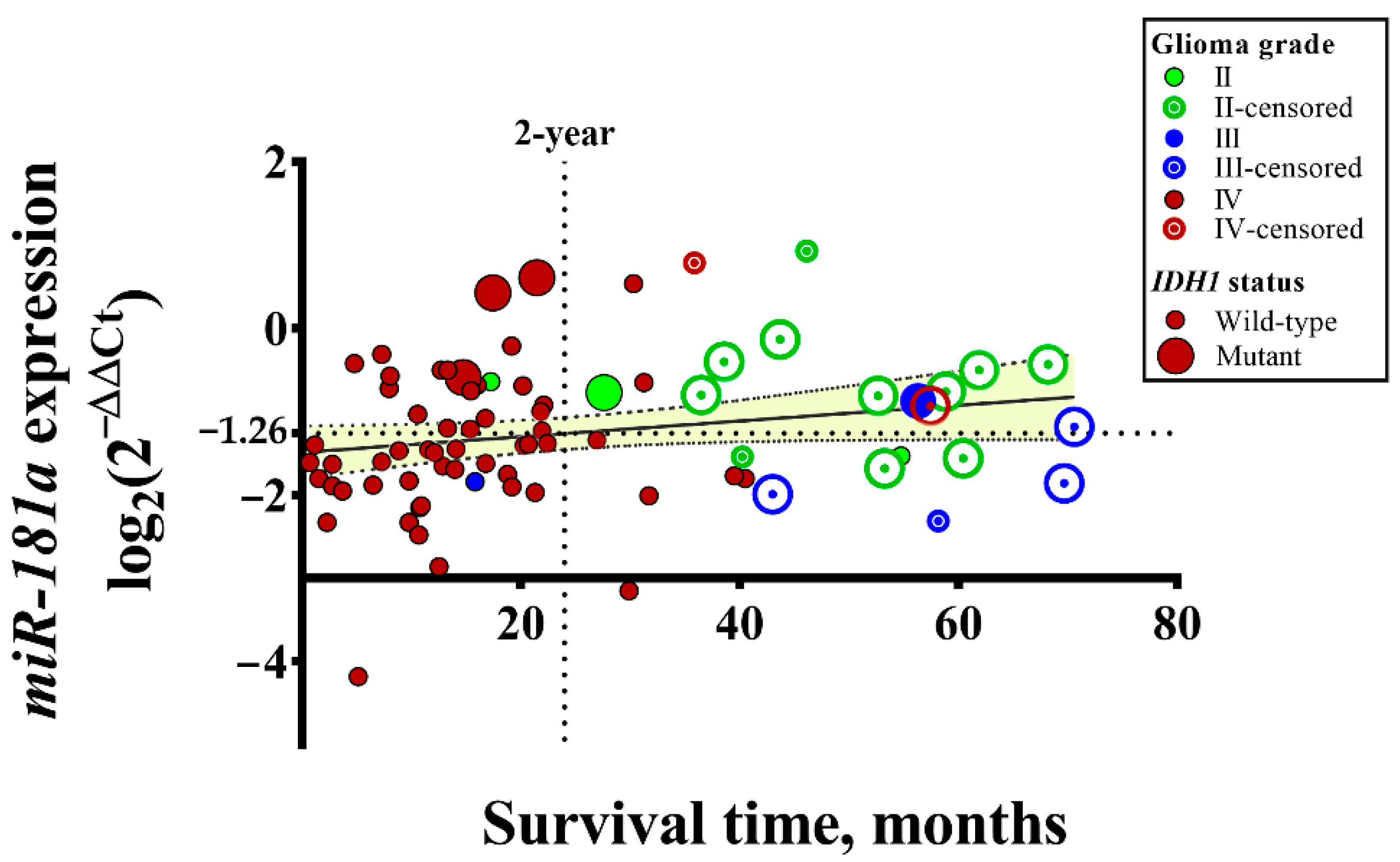
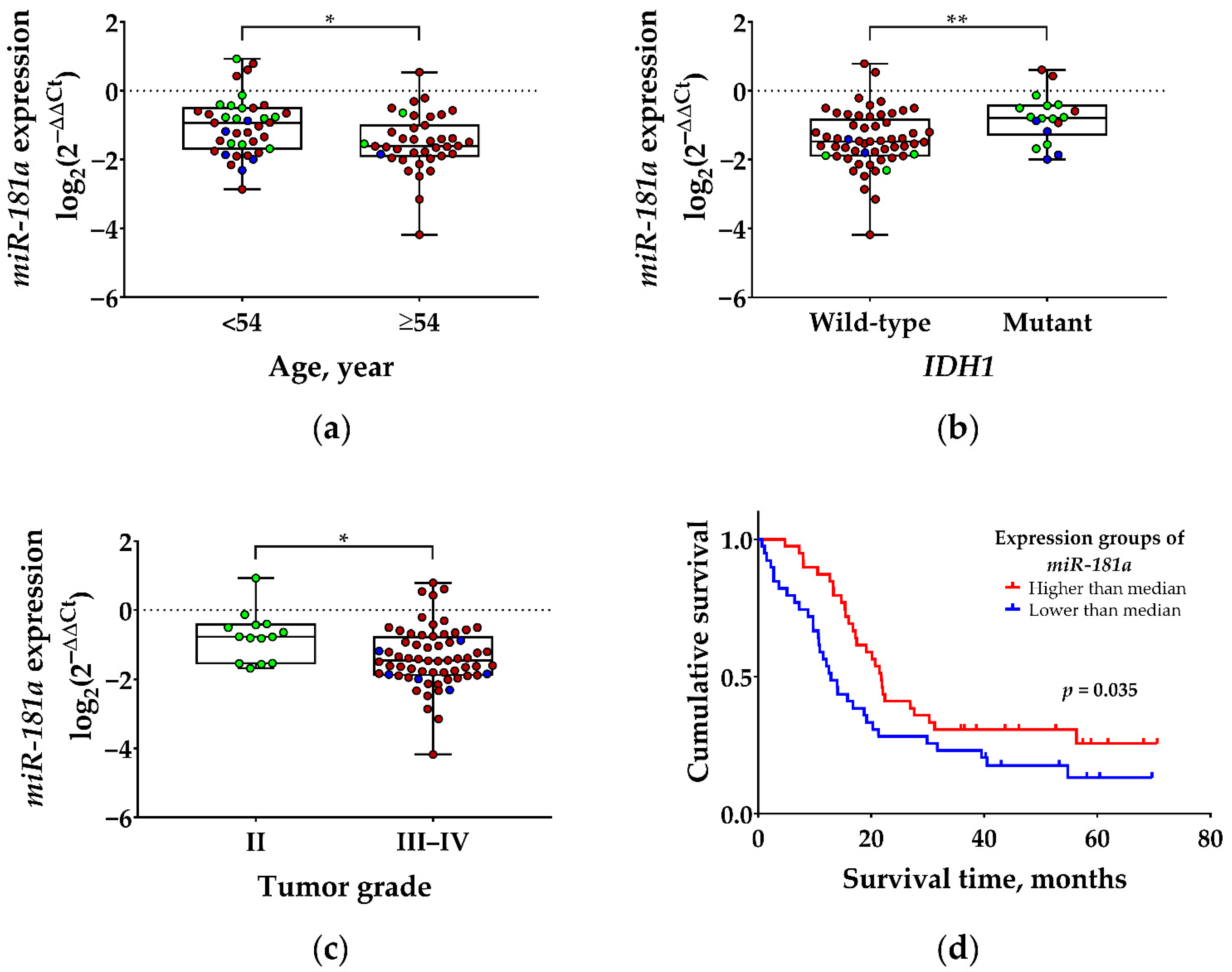
2.1. Association of miR-181a Expression with Patient Clinicopathological Data

To reveal the importance of miR-181a in the pathogenesis of astrocytomas, the mRNA expression of miR-181a was analyzed in 78 different malignancy grade tumors. In Figure 1, it was demonstrated that the gene expression was diversified. The majority of GBM patients with lower than average (<−1.26) mRNA expression died within 2 years, while patients with lower-grade tumors had a higher expression of miR-181a and survived 2 to 6 years.
As follows, to determine whether expression changes of miR-181a were significantly associated with patient clinicopathological characteristics, miR-181a expression was divided into “low” (<mean of miR-181a mRNA expression) and “high” (≥mean) gene expression groups. The analysis reveals that a higher expression of miR-181a is significantly associated with younger age of patients (<54-year, p = 0.006), lower tumor malignancy grade (p = 0.036), and gliomas with a mutant-type of IDH1 (p = 0.002) (see Table 1; Figure 2a–c). According to the Kaplan–Meier analysis, patients with a higher expression of miR-181a have a significantly higher chance of longer survival, compared with patients with low gene expression values (Log-rank test, χ2 = 4.465, df = 1, p = 0.035) (see Figure 2d). The median survival time was 8.9 months longer in the patient group with higher expression of miR-181a.
The univariate cox regression analysis reveals that patients’ clinical characteristics such as age, tumor malignancy grade, and IDH1 status, as well as the expression of miR-181a, are significantly associated with patient overall survival (OS). However, according to the multivariate cox regression analysis, the tumor stage and IDH1 status are the only covariates significantly associated with the OS of glioma patients (see Table 2).

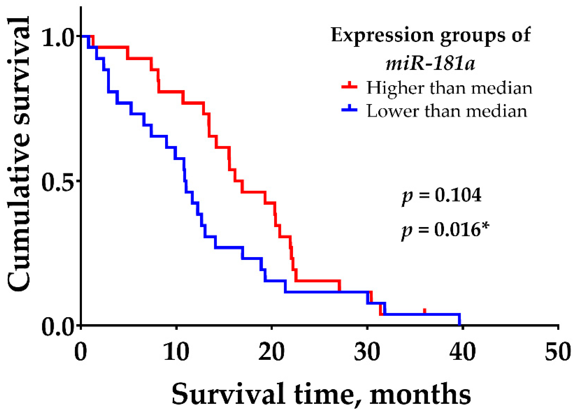
2.2. Associations of miR-181a Expression with IDH1 Status of GBM Tumors

In addition, it was noted that all IDH1 mutant GBM tumors were detected in the higher miR-181a mRNA expression group, and the majority of IDH1 wild-type GBM tumors (62%, 32/52) were detected in the lower gene expression group (see Figure 1 and Figure 2b). According to Student’s t-test, the noted difference was statistically significant (p = 0.005). To find out whether miR-181a expression was associated with patient survival, only IDH1 wild-type GBM tumors were selected and divided into two groups, according to the median of miR-181a expression. Kaplan–Meier analysis shows the tendency that patients with a higher expression of miR-181a have a significantly higher chance of longer survival, compared with patients with low gene expression values (see Figure 3). According to the log-rank test, the difference was not statistically significant (χ2 = 2.64, df = 1, p = 0.104); however, the Gehan–Breslow–Wilcoxon method, which gives more weight to deaths at early time points, shows a statistically significant difference (χ2 = 5.83, df = 1, p = 0.016).

2.3. Associations of mir181a Expression with Functional Status, QoL, and Cognitive Functioning of Patients

The correlation analysis was performed to reveal miR-181a expression associations with variables related to the quality of functioning (see Table 3). The results reveal that the expression of miR-181a positively correlates with general quality of life (EORTC QLQ-C30; r = 0.310, p = 0.010) and functional status, evaluated by a clinician (KPS; r = 0.237, p = 0.049) in all glioma patients. Interestingly, a statistically significant positive correlation between miR-181a and a better quality of life was reflected in the group of men (p = 0.009) rather than women (p = 0.469). In addition, as patients with GBM experience more severe symptoms, it was decided to analyze this subgroup separately. The expression levels of miR-181a show a significantly positive correlation with patient quality of life (EORTC QLQ-C30; r = 0.290, p = 0.041), but an inverse correlation with patient memory (HVLT-R; r = −0.291, p = 0.040). However, no statistically significant correlations of miR-181a with tumor-related symptoms (EORTC QLQ-BN20), depression (PHQ-9), cumulative learning (HVLT-R), psychomotor speed (TMT-A), executive functioning (TMT-B), and/or verbal fluency were determined.

3. Discussion

Numerous studies have shown that miRNAs, which regulate biological processes such as cell proliferation, apoptosis, metabolism, and/or differentiation, are thought to have clinical potential in cancer prognosis and treatment [21]. Among the so-far-characterized miRNAs, miR-181a is involved in several types of cancer [18]. A significant upregulation of miR-181a level has been found in breast cancer [22], ovarian cancer [23], pancreatic cancer [24], hepatocellular carcinoma [25], and oral squamous cell carcinoma [26], whereas evident downregulation of miR-181a has been detected in non-small cell lung cancer [27] and prostate cancer [28], as compared with healthy controls. Our study also reveals that the expression levels of miR-181a decrease during astrocytoma progression. Comparable results were observed by other researcher groups, which demonstrates the downregulation of miR-181a in all grade glioma tumors (WHO II–IV), GBM cell lines, and glioma stem cells (GSCs), as compared with normal brain tissue, astrocytes, and differentiated GBM cells, respectively [29,30,31,32]. Importantly, the overexpression of miR-181a inhibited proliferation, migration, invasion, epithelial-mesenchymal transition (EMT), and induced apoptosis of GBM cells [30,33,34]. The process of apoptosis was modulated by targeting the apoptosis-related genes (p53, Bax, Bcl-2, Bim, etc.) [19], while proliferation was modulated by the downregulation of the MAPK pathway [35]. In addition, the upregulation of miR-181a sensitized the GBM cells to temozolomide (TMZ) and radiation treatment [34,36] also suppressed the formation of GSCs and inhibited GBM tumorigenesis [31]. More importantly, the study by Wu et al. showed the clinical significance of circulating miR-181a in patients with glioma tumors. Before the operation, circulating miR-181a was found to be downregulated in the plasma of GBM patients as compared with lower-grade tumors [37]. After 10 days, the levels of miR-181a increased more than 10-fold. In addition, a lower expression of circulating miR-181a was significantly associated with poor OS [37], as was demonstrated in our study with astrocytoma tumors. Therefore, the aberrantly downregulated miR-181a could be a critical factor that contributes to the malignant appearance of astrocytoma.
Our study also demonstrates a significant association between the expression levels of miR-181a and patient age, as well as IDH1 status. However, according to the multivariate Cox regression analysis, only the tumor grade and IDH1 mutation were the best predictors of patient OS in our study cohort. Importantly, we noted that the mutant form of IDH1, which is the factor of a good astrocytoma patient survival prognosis, was found in GBM tumors with an increased expression of miR-181a. All these GBM patients with IDH1 mutation survived more than 14 months, and one of them survived even more than 57 months. We hypothesize that the expression of mirR-181a may influence the activity of the IDH1 protein which affects several major metabolic processes of the cells [38] and, therefore, has an impact on the OS of GBM patients. This relation between IDH1 and miR-181a in GBM patients was also observed by Sippl et al. [39]. They determined the inverse correlation between the expression of miRNA-181a2 and mRNA expression of IDH1 (p = 0.06, r = −0.55). Nevertheless, studies on a larger sample would be needed to confirm our observations.
Next, we wanted to find out whether the survival of patients with IDH1 wild-type GBM tumors depends on miR-181a expression level. Since the survival of GBM patients after surgery is generally short, we additionally used the Gehan–Breslow–Wilcoxon method, which gives more weight to deaths at early time points. The analysis demonstrates that patients with a lower expression of miR-181a have significantly worse OS as compared with those with a higher gene expression level. However, the opposite effect was noted by another research group. They demonstrated that in patients with IDH1 wild-type GBMs, low miR-181a2 expression correlated with a prolonged OS (p = 0.019), [39]. The discrepancy may have occurred because different miR-181a isoforms were used in the analyses, which might have distinct biological functions. In our study, we analyzed the miR-181a1 isoform, which is located on chromosome 1 (37.p5), while miR-181a2 is situated on chromosome 9 (37.p5) [18]. Both isoforms produce almost identical mature miR-181a, but could be regulated by distinct molecules and, therefore, be differently expressed. For example, in human blood natural-killer cells, pri-miR-181ab-2 levels were higher than pri-miR-181ab-1. The immunosuppressive cytokine, TGF-β, suppressed pri-miR-181ab-1 expression while elevating pri-miR-181ab-2 expression. On the contrary, interleukins −2, −15, and −12/−18 increased the expression of pri-miR-181ab-1, but inhibited pri-miR-181ab-2 [40].
To the best of our knowledge, this was the first study comparing expression levels of miR-181a with patient quality of life. The analysis reveals that miR-181a positively correlates with the general quality of life subjective reported by patients themselves, as well as functioning status, evaluated by the treating clinician. However, no statistically significant correlations were found between miR-181a expression and more specific tumor-related symptoms, levels of depression, or cognitive functioning. A relatively small sample size, especially of tumor grades II and III, could affect the statistical power of our analysis. Furthermore, cognitive impairment and the profile of tumor-related symptoms are highly dependent on the localization of the tumor and other clinical factors, such as edema, tumor-induced compression to nearby tissues, or the frequency of seizures. Thus, it might be that relationships were lost due to the heterogeneity of the sample regarding tumor locations and other clinical characteristics. However, previous findings by our team group showed that miR-34a or miR-181b/d expression levels were related to patients’ functioning and tumor-related symptoms [41,42]. Therefore, we believe that current findings support the idea that levels of miR-181a expression might be valuable not only in predicting longer survival but also in better general functioning. Still, further studies are needed to clarify the mixed findings in male and female subgroups.

4. Materials and Methods

4.1. Samples and Patient Clinicopathological Data

Seventy-eight samples from patients with the diagnosis of II-IV grade glioma tumors were analyzed in the study. All patients underwent neurosurgery at the Department of Neurosurgery, Hospital of Lithuanian University of Health Sciences, from 2015 to 2018. After surgical resection, tumor samples were frozen in liquid nitrogen. The diagnosis was confirmed by the pathologists. The study was approved by the Kaunas Regional Biomedical Research Ethics Committee, and written patient consent was taken from each patient before inclusion in the study.
The clinical data, such as gender, age at the time of surgery, tumor grade, isocitrate dehydrogenase 1 (IDH1) status (the R132H mutation in the IDH1 gene), and methylation of O-6-methylguanine methyltransferase (MGMT) were collected from medical records. According to the WHO classification 2016 [1], there were 14 tumor samples of grade II, 6 of grade III, and 58 of grade IV (GBM). There were 35 women and 43 men, with a mean age of 54 years (range: 24–80 years). The overall survival of the patient was calculated from the date of tumor resection to the date of patient death or database closure (5 October 2021).

4.2. Functional Status

Patient functional status was assessed by a treating neurosurgeon during the hospital stay using the Karnofsky performance scale (KPS) [43]. The KPS measures a patient’s ability to carry on his/her normal daily activities and dependence on help and nursing care using an 11-point rating scale. The total KPS score ranges from 100 (normal functioning) to 0 (death), with higher scores indicating better daily functioning and higher functional independence. Data on functional status was available for 70 (88.6%) of patients.

4.3. Quality of Life Assessment

Quality of life (QoL) assessment was performed 2–3 days before the neurosurgery. Patients were asked to fill out self-report questionnaires addressing their symptoms and quality of life. If assistance was needed due to reading, motor, or visual problems, questionnaires were filed with the help of a medical psychologist. Data on QoL was available for 68 (87.2%) of the patients.
The European Organization for Research and Treatment of Cancer quality of life questionnaires QLQ-30 [44] and QLQ-BN20 [45] for brain tumor-related symptoms were used in this study. The QLQ-C30 contains 30 items that were designed to assess global health status, subjectively reported functional status, role functioning, emotional functioning, cognitive functioning, social functioning, and various cancer-related symptoms. Raw scores were linearly transformed to 0–100 scales, with higher scores indicating better quality of life.
The QLQ-BN20 is a 20-item self-rating scale specifically developed for the assessment of health complaints in brain tumor patients. The questionnaire contains many common BT-related symptoms, including future uncertainty, visual disorder, cognitive impairment, etc. The QLQ-BN20 scores were linearly transformed to a 0–100 scale, with a higher score indicating greater BT-related symptom severity.
In addition to QoL instruments, we also included a measure of depression, as depression is significantly related to decreased QoL in many patient populations. The patient health questionnaire-9 (PHQ-9) [46] was chosen for the assessment of current depressive symptoms. The PHQ-9 is based on the diagnostic statistical manual-IV depression diagnostic criteria, and it is recognized as a valid and reliable tool for depression screening in glioma patients [47,48].

4.4. Assessment of Cognitive Functioning

A cognitive assessment was performed by a medical psychologist 2–3 days before the neurosurgery at the Department of Neurosurgery of the Hospital of LUHS. A set of neuropsychological tests, recommended for the assessment of treatment outcomes in glioma studies, was used [49].
The Hopkins verbal learning test-revised (HVLT-R) was used for verbal memory assessment [50]. The test consists of 12 words that are read aloud for three trials, each trial followed by a patient’s free recall. After an approximately 20 min. delay, during which other tests are administered, the patient is asked to recall the list of words. Two scores: cumulative learning (total number of words recalled in trials 1, 2, and 3) and delayed recall (number of words recalled after a delay) were analyzed in this study.
The Trail-making test (TMT, parts A and B) was used for the assessment of psychomotor speed and executive functioning [51]. During the task, a patient is asked to connect a sequence of 25 targets (numbers 1, 2, 3, etc. in Part A, and numbers and letters in Part B) on a sheet of paper. The time of completion (in seconds) is considered as an indicator of psychomotor speed and executive functions.
Verbal fluency was measured using phonemic fluency tasks [52]. Patients were asked to produce as many words as possible beginning with a specific letter within the one-minute interval. Three trials using the letters K, A, and S were performed. The total number of words produced during three trials is used as a verbal fluency indicator.
As cognitive performance is sensitive to aging processes, all primary scores of neuropsychological tests were transformed to age-adjusted T scores (Mean 50, SD 10) using available norms for the Lithuanian population, with higher scores indicating better cognitive function. Memory and verbal fluency data were available for 68 (87%) of the patients, and data on psychomotor speed and executive function was available for 62 (79.5%) of the patients, as some patients were not able to perform these tasks due to visual or motor impairment.

4.5. Gene Expression Analysis

Small RNAs were extracted from frozen tumor tissue using a mirVana™ miRNA Isolation Kit (Life Technologies, Carlsbad, CA, USA, cat. no. AM1560), according to the manufacturer’s instructions. The quality and concentration were determined by NanoDrop 2000 (Thermo Fisher Scientific, Wilmington, DE, USA). Following this, 10 ng of purified micro RNAs were synthesized to cDNA using the “TaqMan Advanced miRNA cDNA Synthesis Kit” (Thermo Fisher Scientific, Pleasanton, CA, USA, cat. no. A25576).
The RT-PCR with TaqMan™ Fast Advanced Master Mix (Life Technologies, Carlsbad, CA, USA, cat. no. 4444965) was performed to analyze miR-181a expression changes in II-IV grade glioma tumors and healthy human brain RNA sample “FirstChoice Human Brain Reference RNA” (RHB; Ambion, Austin, TX, USA, cat. no. AM7962). The reaction consisted of 6 µL of TaqMan® Fast Advanced Master Mix, 0.6 µL of hsa-miR-181a-3p probe (Applied Biosystems, Foster City, CA, USA, Assay ID: 479405_mir), 3 µL of cDNA sample, and nuclease-free water to a total volume of 12 µL. The reactions were performed in the RT-PCR System “Applied Biosystems 7500 Fast” (Applied Biosystems, Foster City, CA, USA) using a fast-cycling program. In addition, the housekeeping genes has-miR191-5p (Assay ID: 477952_mir), has-miR361-5p (Assay ID: 478056_mir), has-miR345-5p (Assay ID: 478366_mir), and has-miR103a-3p (Assay ID: 478253_mir) were measured to normalize the data. The average of the housekeeping genes was used for the comparative 2−∆∆Ct method, in which gene expression in tumor samples was compared to healthy brain tissue.

4.6. Statistical Analysis
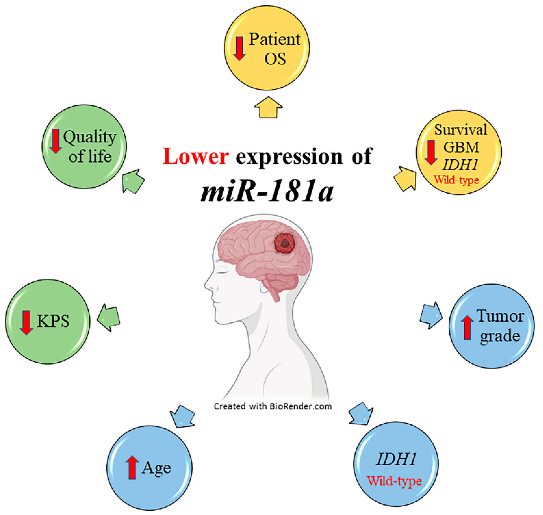
Statistical programs SPSS (version 25.0, IBM SPSS, Armonk, NY, USA) and GraphPad Prism (version 7.0, Graph-Pad Software, San Diego, CA, USA) were used for data analysis. The normality was confirmed by the Kolmogorov–Smirnov test. The Student’s independent t-test was applied to evaluate the difference in miR-181a expression between the two groups. Meanwhile, one-way ANOVA with Tukey’s correction was used for the three groups. The Chi-square test was used for the comparison of categorical variables and the Pearson correlation was used for the quantitative variables. Patient survival was evaluated according to the Kaplan–Meier curves with log-rank or Breslow tests. Univariate and multivariate (with the backward conditional method) Cox regression analyses were used to evaluate the relationships between clinical, psychological, and molecular variables and patient overall survival. The significance level was defined as p < 0.05.

5. Conclusions

The study suggests that miR-181a could be a promising biomarker for glioma patients, since the expression of miR-181a decreases during tumor progression and the downregulation of the gene is significantly associated with the worst patient survival prognosis. More importantly, lower expression levels of miR-181a are also related to wild-type IDH1, older age, as well as worse patient quality of life and functioning (see Figure 4).
However, the findings of this study should be seen in light of some limitations. One of them is a small number of tumor samples and incomplete sample information. Unfortunately, not all of the patients had information about IDH1, MGMT status, and functional data. Furthermore, patients’ genetic and medical histories were not considered during this study, which might affect the evaluation of patients’ health-related quality of life. This may have affected the reliability of the results. Therefore, follow-up studies are needed to confirm the prognostic value of miR-181a in glioma patients.

Author Contributions

Conceptualization, A.T., I.S. and P.V.; methodology A.P., A.B., I.S. and P.V.; formal analysis, H.S., I.S., I.V., A.P. and P.V.; data curation, A.P., A.B., A.T., I.S., I.V. and P.V.; writing—original draft preparation, review and editing, I.V., A.P., A.B., A.T., H.S., I.S. and P.V.; visualization, H.S. and I.V.; project administration, A.T., I.S. and P.V.; funding acquisition, P.V. All authors have read and agreed to the published version of the manuscript.

Funding

This research was funded by the Research Council of Lithuania, grant number S-LU-20-2.

Institutional Review Board Statement

The study was conducted in accordance with the Declaration of Helsinki and approved by the Ethics Committee for Biomedical Research of the Lithuanian University of Health Sciences (LUHS) (no. P2-9/2003, date: 10 December 2010).

Informed Consent Statement

Informed consent was obtained from all subjects involved in the study.

Data Availability Statement

The data is available from the corresponding author on reasonable request.

Acknowledgments

We kindly thank Jūratė Žeglienė and Raminta Bagdanavičienė for their assistance in tissue sampling and patient clinical data gathering.

Conflicts of Interest

The authors declare no conflict of interest.

References

  1. Louis, D.N.; Perry, A.; Reifenberger, G.; Von Deimling, A.; Figarella-Branger, D.; Cavenee, W.K.; Ohgaki, H.; Wiestler, O.D.; Kleihues, P.; Ellison, D.W. The 2016 World Health Organization Classification of Tumors of the Central Nervous System: A summary. Acta Neuropathol. 2016, 131, 803–820. [Google Scholar] [CrossRef] [PubMed]
  2. Weller, M.; dan der Bent, M.; Tonn, J.C.; Stupp, R.; Preusser, M.; Cohen-Jonathan-Moyal, E.; Henriksson, R.; Le Rhun, E.; Balana, C.; Chinot, O.; et al. European Association for Neuro-Oncology (EANO) guideline on the diagnosis and treatment of adult astrocytic and oligodendroglial gliomas. Lancet Oncol. 2017, 18, e315–e329. [Google Scholar] [CrossRef]
  3. Stupp, R.; Taillibert, S.; Kanner, A.A.; Read, W.; Steinberg, D.M.; Lhermitte, B.; Toms, S.; Idbaih, A.; Ahluwalia, M.S.; Fink, K.; et al. Effect of Tumor-Treating Fields Plus Maintenance Temozolomide vs Maintenance Temozolomide Alone on Survival in Patients With Glioblastoma. JAMA 2017, 318, 2306–2316. [Google Scholar] [CrossRef] [PubMed]
  4. Minniti, G.; Niyazi, M.; Alongi, F.; Navarria, P.; Belka, C. Current status and recent advances in reirradiation of glioblastoma. Radiat. Oncol. 2021, 16, 36. [Google Scholar] [CrossRef]
  5. Louis, D.N.; Perry, A.; Wesseling, P.; Brat, D.J.; Cree, I.A.; Figarella-Branger, D.; Hawkins, C.; Ng, H.K.; Pfister, S.M.; Reifenberger, G.; et al. The 2021 WHO Classification of Tumors of the Central Nervous System: A summary. Neuro-Oncology 2021, 23, 1231–1251. [Google Scholar] [CrossRef]
  6. Yang, K.; Wu, Z.; Zhang, H.; Zhang, N.; Wu, W.; Wang, Z.; Dai, Z.; Zhang, X.; Zhang, L.; Peng, Y.; et al. Glioma targeted therapy: Insight into future of molecular approaches. Mol. Cancer 2022, 21, 39. [Google Scholar] [CrossRef]
  7. Hutóczki, G.; Virga, J.; Birkó, Z.; Klekner, A. Novel Concepts of Glioblastoma Therapy Concerning Its Heterogeneity. Int. J. Mol. Sci. 2021, 22, 10005. [Google Scholar] [CrossRef]
  8. Brennan, C.W.; Verhaak, R.G.W.; McKenna, A.; Campos, B.; Noushmehr, H.; Salama, S.R.; Zheng, S.; Chakravarty, D.; Sanborn, J.Z.; Berman, S.H.; et al. The Somatic Genomic Landscape of Glioblastoma. Cell 2013, 155, 462–477. [Google Scholar] [CrossRef]
  9. Hou, J.; He, Z.; Liu, T.; Chen, D.; Wang, B.; Wen, Q.; Zheng, X. Evolution of Molecular Targeted Cancer Therapy: Mechanisms of Drug Resistance and Novel Opportunities Identified by CRISPR-Cas9 Screening. Front. Oncol. 2022, 12, 755053. [Google Scholar] [CrossRef]
  10. Malone, E.R.; Oliva, M.; Sabatini, P.J.B.; Stockley, T.; Siu, L.L. Molecular profiling for precision cancer therapies. Genome Med. 2020, 12, 8. [Google Scholar] [CrossRef] [Green Version]
  11. Korja, M.; Raj, R.; Seppä, K.; Luostarinen, T.; Malila, N.; Seppälä, M.; Mäenpää, H.; Pitkäniemi, J. Glioblastoma survival is improving despite increasing incidence rates: A nationwide study between 2000 and 2013 in Finland. Neuro-Oncology 2018, 21, 370–379. [Google Scholar] [CrossRef] [PubMed]
  12. He, B.; Zhao, Z.; Cai, Q.; Zhang, Y.; Zhang, P.; Shi, S.; Xie, H.; Peng, X.; Yin, W.; Tao, Y.; et al. miRNA-based biomarkers, therapies, and resistance in Cancer. Int. J. Biol. Sci. 2020, 16, 2628–2647. [Google Scholar] [CrossRef]
  13. Si, W.; Shen, J.; Zheng, H.; Fan, W. The role and mechanisms of action of microRNAs in cancer drug resistance. Clin. Epigenet. 2019, 11, 25. [Google Scholar] [CrossRef] [PubMed]
  14. Giovannetti, E.; Erozenci, A.; Smit, J.; Danesi, R.; Peters, G.J. Molecular mechanisms underlying the role of microRNAs (miRNAs) in anticancer drug resistance and implications for clinical practice. Crit. Rev. Oncol. Hematol. 2011, 81, 103–122. [Google Scholar] [CrossRef] [PubMed]
  15. Huang, Y.; Shen, X.J.; Zou, Q.; Wang, S.P.; Tang, S.M.; Zhang, G.Z. Biological functions of microRNAs: A review. J. Physiol. Biochem. 2010, 67, 129–139. [Google Scholar] [CrossRef] [PubMed]
  16. Acunzo, M.; Romano, G.; Wernicke, D.; Croce, C.M. MicroRNA and cancer–A brief overview. Adv. Biol. Regul. 2015, 57, 1–9. [Google Scholar] [CrossRef]
  17. Condrat, C.E.; Thompson, D.C.; Barbu, M.G.; Bugnar, O.L.; Boboc, A.; Cretoiu, D.; Suciu, N.; Cretoiu, S.M.; Voinea, S.C. miRNAs as Biomarkers in Disease: Latest Findings Regarding Their Role in Diagnosis and Prognosis. Cells 2020, 9, 276. [Google Scholar] [CrossRef] [PubMed]
  18. Indrieri, A.; Carrella, S.; Carotenuto, P.; Banfi, S.; Franco, B. The Pervasive Role of the miR-181 Family in Development, Neurodegeneration, and Cancer. Int. J. Mol. Sci. 2020, 21, 2092. [Google Scholar] [CrossRef]
  19. Feng, X.; Zhang, C.; Yang, Y.; Hou, D.; Zhu, A. Role of miR-181a in the process of apoptosis of multiple malignant tumors: A literature review. Adv. Clin. Exp. Med. 2018, 27, 263–270. [Google Scholar] [CrossRef] [PubMed]
  20. Karsy, M.; Arslan, E.; Moy, F. Current Progress on Understanding MicroRNAs in Glioblastoma Multiforme. Genes Cancer 2012, 3, 3–15. [Google Scholar] [CrossRef] [PubMed]
  21. Saliminejad, K.; Khorram Khorshid, H.R.; Soleymani Fard, S.; Ghaffari, S.H. An overview of microRNAs: Biology, functions, therapeutics, and analysis methods. J. Cell. Physiol. 2019, 234, 5451–5465. [Google Scholar] [CrossRef]
  22. Zhai, Z.; Mu, T.; Zhao, L.; Li, Y.; Zhu, D.; Pan, Y. MiR-181a-5p facilitates proliferation, invasion, and glycolysis of breast cancer through NDRG2-mediated activation of PTEN/AKT pathway. Bioengineered 2021, 13, 83–95. [Google Scholar] [CrossRef] [PubMed]
  23. Zuo, L.; Li, X.; Zhu, H.; Li, A.; Wang, Y. Expression of miR-181a in Circulating Tumor Cells of Ovarian Cancer and Its Clinical Application. ACS Omega 2021, 6, 22011–22019. [Google Scholar] [CrossRef] [PubMed]
  24. Kang, H.; Ma, D.; Zhang, J.; Zhao, J.; Yang, M. MicroRNA-18a induces epithelial-mesenchymal transition like cancer stem cell phenotype via regulating RKIP pathway in pancreatic cancer. Ann. Transl. Med. 2020, 8, 433. [Google Scholar] [CrossRef] [PubMed]
  25. Chang, S.; Chen, B.; Wang, X.; Wu, K.; Sun, Y. Long non-coding RNA XIST regulates PTEN expression by sponging miR-181a and promotes hepatocellular carcinoma progression. BMC Cancer 2017, 17, 248. [Google Scholar] [CrossRef]
  26. Yang, C.-C.; Hung, P.-S.; Wang, P.-W.; Liu, C.-J.; Chu, T.-H.; Cheng, H.-W.; Lin, S.-C. miR-181 as a putative biomarker for lymph-node metastasis of oral squamous cell carcinoma. J. Oral Pathol. Med. 2011, 40, 397–404. [Google Scholar] [CrossRef] [PubMed]
  27. Xue, W.-X.; Zhang, M.-Y.; Li, R.; Liu, X.; Yin, Y.-H.; Qu, Y.-Q. Serum miR-1228-3p and miR-181a-5p as Noninvasive Biomarkers for Non-Small Cell Lung Cancer Diagnosis and Prognosis. BioMed Res. Int. 2020, 2020, 9601876. [Google Scholar] [CrossRef]
  28. Shen, H.; Weng, X.D.; Liu, X.H.; Yang, D.; Wang, L.; Guo, J.; Wang, M.; Wang, X.; Diao, C.H. miR-181a-5p is downregulated and inhibits proliferation and the cell cycle in prostate cancer. Int. J. Clin. Exp. Pathol. 2018, 11, 3969–3976. [Google Scholar]
  29. Yang, L.; Ma, Y.; Xin, Y.; Han, R.; Li, R.; Hao, X. Role of the microRNA 181 family in glioma development. Mol. Med. Rep. 2017, 17, 322–329. [Google Scholar] [CrossRef]
  30. Shi, L.; Cheng, Z.; Zhang, J.; Li, R.; Zhao, P.; Fu, Z.; You, Y. hsa-mir-181a and hsa-mir-181b function as tumor suppressors in human glioma cells. Brain Res. 2008, 1236, 185–193. [Google Scholar] [CrossRef]
  31. Huang, S.-X.; Zhao, Z.-Y.; Weng, G.-H.; He, X.-Y.; Wu, C.-J.; Fu, C.-Y.; Sui, Z.-Y.; Ma, Y.-S.; Liu, T. Upregulation of miR-181a suppresses the formation of glioblastoma stem cells by targeting the Notch2 oncogene and correlates with good prognosis in patients with glioblastoma multiforme. Biochem. Biophys. Res. Commun. 2017, 486, 1129–1136. [Google Scholar] [CrossRef] [PubMed]
  32. Macharia, L.W.; Muriithi, W.; Heming, C.P.; Nyaga, D.K.; Aran, V.; Mureithi, M.W.; Ferrer, V.P.; Pane, A.; Filho, P.N.; Moura-Neto, V. The genotypic and phenotypic impact of hypoxia microenvironment on glioblastoma cell lines. BMC Cancer 2021, 21, 1248. [Google Scholar] [CrossRef] [PubMed]
  33. Wang, L.; Ma, J.; Wang, X.; Peng, F.; Chen, X.; Zheng, B.; Wang, C.; Dai, Z.; Ai, J.; Zhao, S. Kaiso (ZBTB33) Downregulation by miRNA-181a Inhibits Cell Proliferation, Invasion, and the Epithelial–Mesenchymal Transition in Glioma Cells. Cell. Physiol. Biochem. 2018, 48, 947–958. [Google Scholar] [CrossRef] [PubMed]
  34. Wen, X.; Li, S.; Guo, M.; Liao, H.; Chen, Y.; Kuang, X.; Liao, X.; Ma, L.; Li, Q. miR-181a-5p inhibits the proliferation and invasion of drug-resistant glioblastoma cells by targeting F-box protein 11 expression. Oncol. Lett. 2020, 20, 235. [Google Scholar] [CrossRef]
  35. Schäfer, A.; Evers, L.; Meier, L.; Schlomann, U.; Bopp, M.H.A.; Dreizner, G.-L.; Lassmann, O.; Ben Bacha, A.; Benescu, A.-C.; Pojskic, M.; et al. The Metalloprotease-Disintegrin ADAM8 Alters the Tumor Suppressor miR-181a-5p Expression Profile in Glioblastoma Thereby Contributing to Its Aggressiveness. Front. Oncol. 2022, 12, 826273. [Google Scholar] [CrossRef] [PubMed]
  36. Hu, W.; Chen, G.; Zhu, W.; Shi, D.; Lv, L.; Zhang, C.; Liu, P.; Hu, W. MicroRNA-181a sensitizes human malignant glioma U87MG cells to radiation by targeting Bcl-2. Oncol. Rep. 2010, 23, 997–1003. [Google Scholar] [CrossRef] [PubMed]
  37. Wu, J.; Al-Zahrani, A.; Beylerli, O.; Sufianov, R.; Talybov, R.; Meshcheryakova, S.; Sufianova, G.; Gareev, I.; Sufianov, A. Circulating miRNAs as Diagnostic and Prognostic Biomarkers in High-Grade Gliomas. Front. Oncol. 2022, 12, 898537. [Google Scholar] [CrossRef]
  38. Han, S.; Liu, Y.; Cai, S.J.; Qian, M.; Ding, J.; Larion, M.; Gilbert, M.R.; Yang, C. IDH mutation in glioma: Molecular mechanisms and potential therapeutic targets. Br. J. Cancer 2020, 122, 1580–1589. [Google Scholar] [CrossRef]
  39. Sippl, C.; Schoeneberger, L.; Teping, F.; Schulz-Schaeffer, W.; Urbschat, S.; Ketter, R.; Oertel, J. Impact of miRNA-181a2 on the Clinical Course of IDH1 Wild Type Glioblastoma. Processes 2021, 9, 728. [Google Scholar] [CrossRef]
  40. Presnell, S.R.; Al-Attar, A.; Cichocki, F.; Miller, J.S.; Lutz, C.T. Human natural killer cell microRNA: Differential expression of MIR181A1B1 and MIR181A2B2 genes encoding identical mature microRNAs. Genes Immun. 2015, 16, 89–98. [Google Scholar] [CrossRef]
  41. Vaitkiene, P.; Pranckeviciene, A.; Stakaitis, R.; Steponaitis, G.; Tamasauskas, A.; Bunevicius, A. Association of miR-34a Expression with Quality of Life of Glioblastoma Patients: A Prospective Study. Cancers 2019, 11, 300. [Google Scholar] [CrossRef] [PubMed] [Green Version]
  42. Stakaitis, R.; Pranckeviciene, A.; Steponaitis, G.; Tamasauskas, A.; Bunevicius, A.; Vaitkiene, P. Unique Interplay Between Molecular miR-181b/d Biomarkers and Health Related Quality of Life Score in the Predictive Glioma Models. Int. J. Mol. Sci. 2020, 21, 7450. [Google Scholar] [CrossRef] [PubMed]
  43. Karnofsky, D.A.; Abelmann, W.H.; Craver, L.F.; Burchenal, J.H. The use of the nitrogen mustards in the palliative treatment of carcinoma. With particular reference to bronchogenic carcinoma. Cancer 1948, 1, 534–656. [Google Scholar] [CrossRef]
  44. Fayers, P.; Aaronson, N.K.; Bjordal, K.; Groenvold, M.; Curran, D.; Bottomley, A. EORTC QLQ-C30 Scoring Manual, 3rd ed.; European Organisation for Research and Treatment of Cancer: Brussels, Belgium, 2001. [Google Scholar]
  45. Osoba, D.; Aaronson, N.K.; Muller, M.; Sneeuw, K.; Hsu, M.-A.; Yung, W.K.A.; Brada, M.; Newlands, E. The development and psychometric validation of a brain cancer quality-of-life questionnaire for use in combination with general cancer-specific questionnaires. Qual. Life Res. 1996, 5, 139–150. [Google Scholar] [CrossRef] [PubMed]
  46. Kroenke, K.; Spitzer, R.L.; Williams, J.B. The PHQ-9: Validity of a brief depression severity measure. J. Gen. Intern. Med. 2001, 16, 606–613. [Google Scholar] [CrossRef] [PubMed]
  47. Pranckeviciene, A.; Bunevicius, A. Depression screening in patients with brain tumors: A review. CNS Oncol. 2015, 4, 71–78. [Google Scholar] [CrossRef]
  48. Rooney, A.G.; McNamara, S.; MacKinnon, M.; Fraser, M.; Rampling, R.; Carson, A.; Grant, R. Screening for major depressive disorder in adults with cerebral glioma: An initial validation of 3 self-report instruments. Neuro-Oncology 2012, 15, 122–129. [Google Scholar] [CrossRef]
  49. Bent, M.V.D.; Wefel, J.; Schiff, D.; Taphoorn, M.; Jaeckle, K.; Junck, L.; Armstrong, T.; Choucair, A.; Waldman, A.; Gorlia, T.; et al. Response assessment in neuro-oncology (a report of the RANO group): Assessment of outcome in trials of diffuse low-grade gliomas. Lancet Oncol. 2011, 12, 583–593. [Google Scholar] [CrossRef]
  50. Benedict, R.H.; Schretlen, D.; Groninger, L.; Brandt, J. Hopkins Verbal Learning Test—Revised: Normative Data and Analysis of Inter-Form and Test-Retest Reliability. Clin. Neuropsychol. 1998, 12, 43–55. [Google Scholar] [CrossRef]
  51. Reitan, R.M. Validity of the Trail Making Test as an Indicator of Organic Brain Damage. Percept. Mot. Ski. 1958, 8, 271–276. [Google Scholar] [CrossRef]
  52. Strauss, E.; Sherman, E.M.S.; Spreen, O. A Compendium of Neuropsychological Tests: Administration, Norms, and Commentary, 3rd ed.; Oxford University Press: New York, NY, USA, 2006. [Google Scholar]
Figure 1. Distribution of patients with the diagnosis of different malignancy grade astrocytoma tumors, according to patient survival time and expression of miR-181a. The average of miR-181a mRNA expression is −1.26 (dotted line in the y-axis). Pearson correlation r = 0.206, p = 0.071, yellow area represents 95% confidence interval.
Ijms 23 11149 g001
Figure 2. Statistically significant associations of miR-181a expression with patient (a) age; (b) IDH1 status; (c) tumor grade; (d) patient survival time. The boxplots indicate mean, within 25 and 75 percentiles, min, and max values. Green dots—grade II, blue—III, and red—IV (GBM). Student’s t-test, * p < 0.05, ** p < 0.01.
Ijms 23 11149 g002
Figure 3. Survival analysis of patients with the diagnosis of IDH1 wild-type GBM in higher and lower miR-81a expression groups. p-values of log-rank test and * Gehan–Breslow–Wilcoxon test.
Ijms 23 11149 g003
Figure 4. Statistically significant associations between expression of miR-181a and patient clinical data and psychology. KPS—Karnowski performance scale. The up arrow indicates higher tumor grade and older patient age, while down arrow—shorter patient survival time, lower KPS index, and worse patient quality of life.
Ijms 23 11149 g004
Table 1. The relationships between gene expression of miR-181a and patient clinical characteristics.
VariablesTotal No.Expression of miR-181a
Low (%)High (%)p-Value
Gender
Male4320 (46.5)23 (53.5)0.15
Female3522 (62.9)13 (37.1)
Age, year
<543915 (38.5)24 (61.5)0.006
≥543927 (69.2)12 (30.8)
Grade
II144 (28.6)10 (71.4)0.036
III–IV6438 (59.4)26 (40.6)
MGMT
Unmet3419 (55.9)15 (44.1)0.978
Met3620 (55.6)16 (44.4)
IDH1
Wt5736 (63.2)21 (36.8)0.002
Mut184 (22.2)14 (77.8)
Tumor location
Right hemisphere3723 (62.2)14 (37.8)0.25
Left hemisphere3718 (48.6)19 (54.4)
Bilateral41 (25.0)3 (75.0)
Unmet—unmethylated, Met—methylated, Wt—wild-type, Mut—mutant. Significant associations p < 0.05 indicated in bold numbers.
Table 2. Univariate and multivariate Cox regression analysis of clinicopathological variables and expression of mir-181a.
VariablesUnivariateMultivariate
HR (95% CI)p-ValueHR (95% CI)p-Value
Gender0.732 (0.441–1.214)0.226N/A
Female vs. Male
Age, year3.856 (2.205–6.741)<0.0011.417 (0.778–2.581)0.254
<54 vs. ≥54
Grade9.748 (3.029–31.373)<0.0012.921 (1.459–5.846)0.002
II vs. III–IV
MGMT0.712 (0.417–1.213)0.211N/A
Unmeth vs. Meth
IDH10.105 (0.041–0.272)<0.0010.314 (0.100–0.989)0.048
Wt vs. Mut
Expression of miR-181a0.584 (0.350–0.974)0.0390.909 (0.521–1.585)0.737
Low vs. High
Unmeth—unmethylated, Meth—methylated, Wt—wild-type, Mut—mutant, N/A—not applicable, HR—hazard ratio. Significant associations p < 0.05 indicated in bold numbers.
Table 3. Relationship between health-related quality of life indicators, clinical evaluation of patient’s functioning, and expression of miR-181a in different malignancy grade glioma patients.
VariablesTotalFemaleMaleGBM
NrpNrpNrpNrp
EORTC QLQ-C30
Quality of Life
680.3100.010310.1350.469370.4230.009500.2900.041
EORTC QLQ-BN20
Tumor Related Symptoms
68−0.2090.08831−0.0050.97837−0.3110.06150−0.2230.120
PHQ-9
Depression
68−0.1280.299310.0600.75037−0.2110.21050−0.1260.385
KPS
Functional status evaluated by a clinician
700.2370.049330.0410.822370.2910.080540.2600.057
HVLT-R
Cumulative learning
68−0.1750.15532−0.1020.58036−0.1940.25650−0.2450.087
HVLT-R
Delayed recall
68−0.2260.06432−0.0940.60736−0.2820.09650−0.2910.040
TMT-A
Psychomotor speed
630.0100.94128−0.2180.265350.1120.523450.0260.866
TMT-B
Executive functions
630.0790.53828−0.2400.218350.2240.197450.0310.838
Verbal fluency68−0.1340.27632−0.2130.24136−0.0780.65250−0.1670.245
r—Pearson correlation coefficient. EORTC QLQ-30—The European Organization for Research and Treatment of Cancer quality of life questionnaire. Higher scores represent better functioning. EORTC QLQ-BN20—The European Organization for Research and Treatment of Cancer quality of life questionnaire, brain tumor module. Higher scores represent a higher symptom burden. PHQ-9—patient health questionnaire-9. Higher scores indicate higher levels of depression. KPS—Karnowski performance scale. Higher scores represent better functioning. HVLT-R—Hopkins verbal learning test-revised. Higher scores represent better functioning. TMT-A—trail-making test, part A. Higher scores represent better functioning. TMT-B—trail-making test, part B. Higher scores represent better functioning. Verbal fluency test. Higher scores represent better functioning. Significant associations p < 0.05 indicated in bold numbers.
Publisher’s Note: MDPI stays neutral with regard to jurisdictional claims in published maps and institutional affiliations.

