
Cardiovascular Disease-Associated MicroRNAs as Novel Biomarkers of First-Trimester Screening for Gestational Diabetes Mellitus in the Absence of Other Pregnancy-Related Complications


Abstract
1. Introduction
2. Results
2.1. Clinical Characteristics of GDM and Control Pregnancies
2.2. Dysregulation of Cardiovascular Disease-Associated MicroRNAs in Early Stages of Gestation in Pregnancies Destinated to Develop GDM
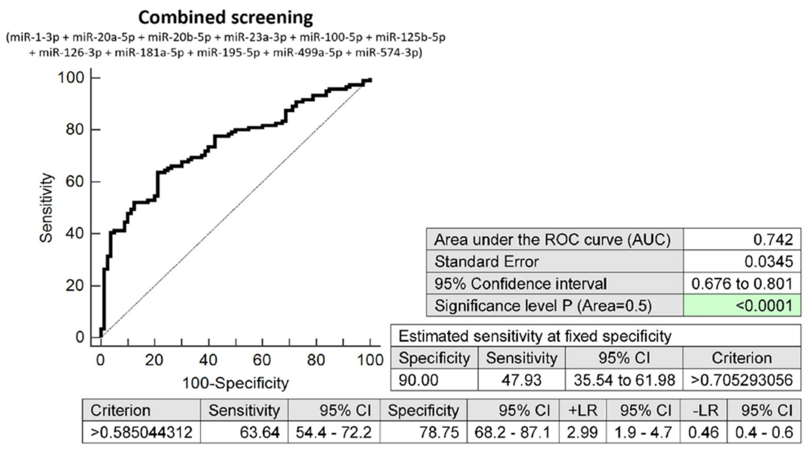
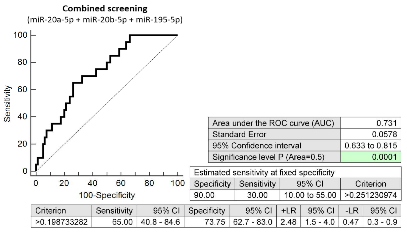
2.3. First-Trimester Combined MicroRNA Screening Is Able to Differentiate between Pregnancies Destinated to Develop GDM and Term Pregnancies with Normal Course of Gestation
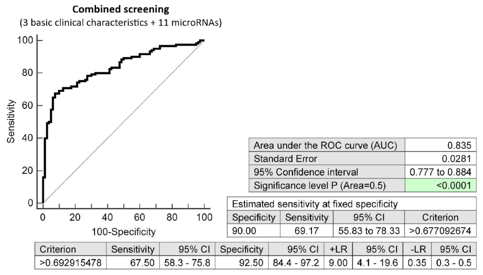
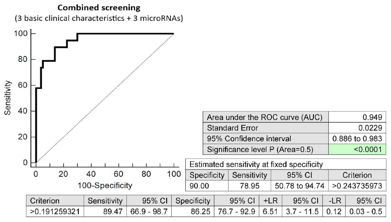
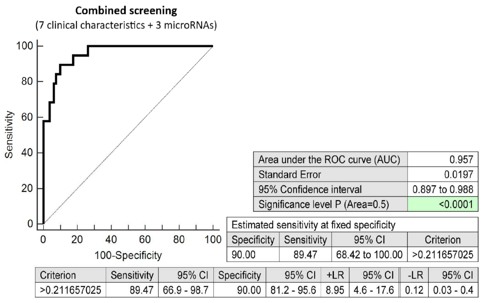
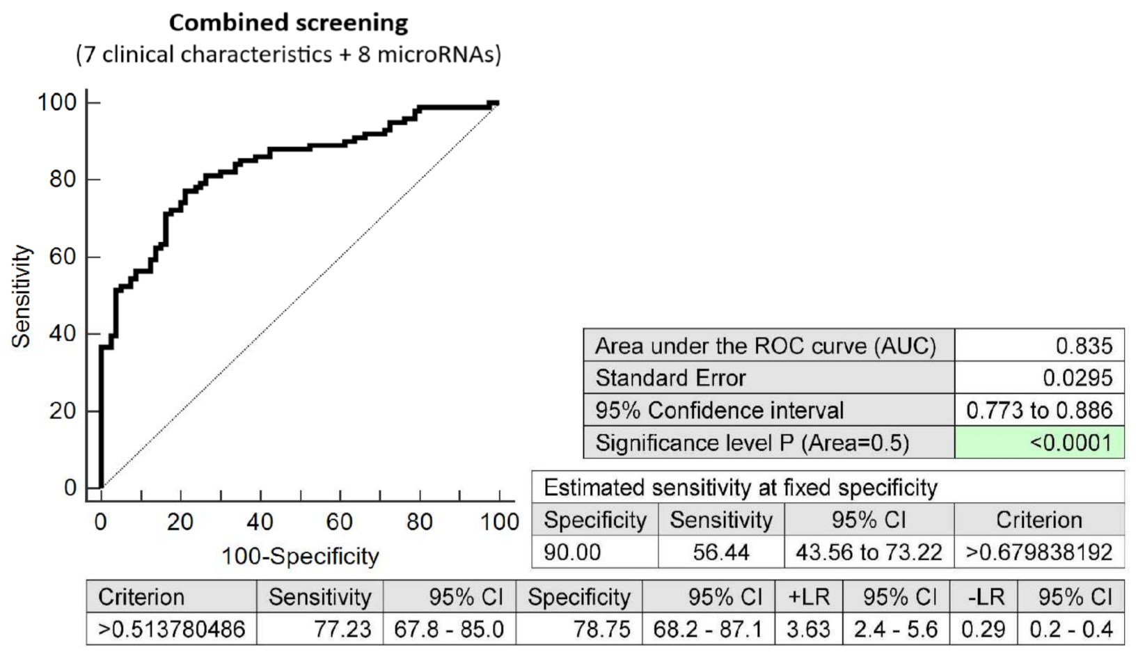
2.4. The Very Good Accuracy of First-Trimester Combined Screening (MicroRNA Biomarkers and Selected Clinical Characteristics) to Differentiate between Pregnancies Destinated to Develop GDM and Term Pregnancies with Normal Course of Gestation
2.5. Dysregulation of Cardiovascular Disease-Associated MicroRNAs in Pregnancies Destinated to Develop GDM with Respect to the Treatment Strategies (Diet Only and a Combination of Diet and Administration of Appropriate Therapy)
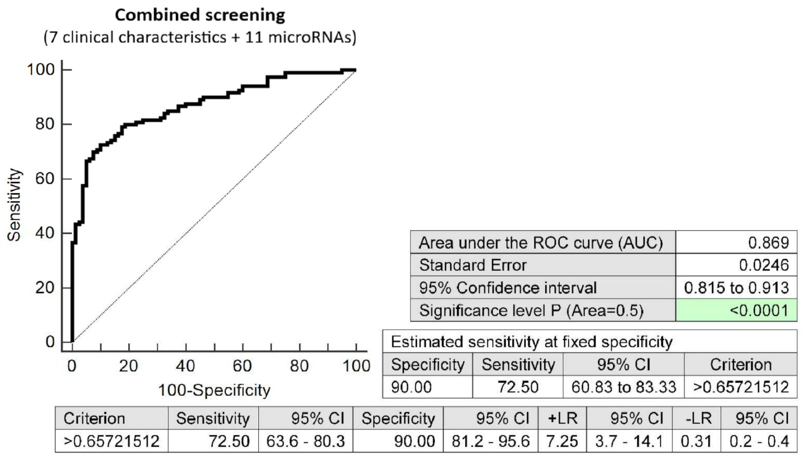
2.6. First-Trimester Combined MicroRNA Screening Is Able to Differentiate between Pregnancies Destinated to Develop GDM Requiring a Combination of Diet and Administration of Appropriate Therapy and Term Pregnancies with Normal Course of Gestation
2.7. The Very High Accuracy of First-Trimester Combined Screening (MicroRNA Biomarkers and Selected Clinical Characteristics) to Differentiate between Pregnancies Destinated to Develop GDM Requiring a Combination of Diet and Administration of Appropriate Therapy and Term Pregnancies with Normal Course of Gestation
2.8. First-Trimester Combined MicroRNA Screening Is Able to Differentiate between Pregnancies Destinated to Develop GDM Managed by Diet Only and Normal Term Pregnancies
2.9. The Very Good Accuracy of First-Trimester Combined Screening (MicroRNA Biomarkers and Selected Clinical Characteristics) to Differentiate between Pregnancies Destinated to Develop GDM Managed by Diet Only and Term Pregnancies with Normal Course of Gestation
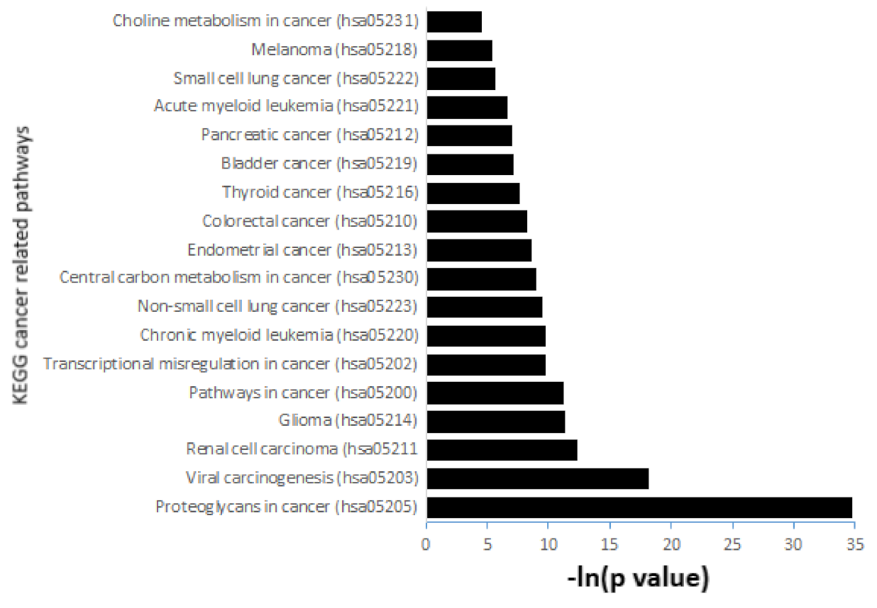
2.10. Information on MicroRNA-Gene-Biological Pathways Interactions
3. Discussion
4. Materials and Methods
4.1. Patients Cohort
4.2. Processing of Samples
4.3. Statistical Analysis
4.4. Information on MicroRNA-Gene-Biological Pathways Interactions
5. Conclusions
6. Patents
Supplementary Materials
Author Contributions
Funding
Institutional Review Board Statement
Informed Consent Statement
Data Availability Statement
Acknowledgments
Conflicts of Interest
References
- Committee on Practice Bulletins—Obstetrics. ACOG Practice Bulletin No. 190: Gestational Diabetes Mellitus. Obstet. Gynecol. 2018, 131, e49–e64. [Google Scholar] [CrossRef]
- International Association of Diabetes and Pregnancy Study Groups Consensus Panel; Metzger, B.E.; Gabbe, S.G.; Persson, B.; Buchanan, T.A.; Catalano, P.A.; Damm, P.; Dyer, A.R.; Leiva, A.D.; Hod, M.; et al. International association of diabetes and pregnancy study groups recommendations on the diagnosis and classification of hyperglycemia in pregnancy. Diabetes Care 2010, 33, 676–682. [Google Scholar] [CrossRef]
- American Diabetes Association. Diagnosis and classification of diabetes mellitus (Position Statement). Diabetes Care 2009, 32, S62–S67. [Google Scholar] [CrossRef]
- England, L.J.; Dietz, P.M.; Njoroge, T.; Callaghan, W.M.; Bruce, C.; Buus, R.M.; Williamson, D.F. Preventing type 2 diabetes: Public health implications for women with a history of gestational diabetes mellitus. Am. J. Obstet. Gynecol. 2009, 200, 365.e1–365.e8. [Google Scholar] [CrossRef]
- O’Sullivan, J.B. Body weight and subsequent diabetes mellitus. JAMA 1982, 248, 949–952. [Google Scholar] [CrossRef]
- Kim, C.; Newton, K.M.; Knopp, R.H. Gestational diabetes and the incidence of type 2 diabetes: A systematic review. Diabetes Care 2002, 25, 1862–1868. [Google Scholar] [CrossRef]
- International Diabetes Federation. IDF GDM Model of Care. Available online: https://www.idf.org/e-library/guidelines/77-idf-gdm-model-of-care-implementation-protocol-guidelines-for-healthcare-professionals.html (accessed on 16 May 2022).
- National Institute for Health and Care Excellence (NICE). Diabetes in Pregnancy: Management from Preconception to the Postnatal Period. Available online: https://www.guidelines.co.uk/diabetes/nice-diabetes-in-pregnancy-guideline/252595.article (accessed on 16 May 2022).
- Syngelaki, A.; Pastides, A.; Kotecha, R.; Wright, A.; Akolekar, R.; Nicolaides, K.H. First-Trimester Screening for Gestational Diabetes Mellitus Based on Maternal Characteristics and History. Fetal Diagn. Ther. 2015, 38, 14–21. [Google Scholar] [CrossRef]
- Nanda, S.; Savvidou, M.; Syngelaki, A.; Akolekar, R.; Nicolaides, K.H. Prediction of gestational diabetes mellitus by maternal factors and biomarkers at 11 to 13 weeks. Prenat. Diagn. 2011, 31, 135–141. [Google Scholar] [CrossRef] [PubMed]
- Sweeting, A.N.; Appelblom, H.; Ross, G.P.; Wong, J.; Kouru, H.; Williams, P.F.; Sairanen, M.; Hyett, J.A. First trimester prediction of gestational diabetes mellitus: A clinical model based on maternal demographic parameters. Diabetes Res. Clin. Pract. 2017, 127, 44–50. [Google Scholar] [CrossRef]
- Sweeting, A.N.; Wong, J.; Appelblom, H.; Ross, G.P.; Kouru, H.; Williams, P.F.; Sairanen, M.; Hyett, J.A. A Novel Early Pregnancy Risk Prediction Model for Gestational Diabetes Mellitus. Fetal Diagn. Ther. 2019, 45, 76–84. [Google Scholar] [CrossRef]
- Wang, P.; Ma, H.H.; Hou, X.Z.; Song, L.L.; Song, X.L.; Zhang, J.F. Reduced plasma level of irisin in first trimester as a risk factor for the development of gestational diabetes mellitus. Diabetes Res. Clin. Pract. 2018, 142, 130–138. [Google Scholar] [CrossRef]
- Wu, Y.T.; Zhang, C.J.; Mol, B.W.; Kawai, A.; Li, C.; Chen, L.; Wang, Y.; Sheng, J.Z.; Fan, J.X.; Shi, Y.; et al. Early Prediction of Gestational Diabetes Mellitus in the Chinese Population via Advanced Machine Learning. J. Clin. Endocrinol. Metab. 2021, 106, e1191–e1205. [Google Scholar] [CrossRef]
- Kumar, M.; Chen, L.; Tan, K.; Ang, L.T.; Ho, C.; Wong, G.; Soh, S.E.; Tan, K.H.; Chan, J.K.Y.; Godfrey, K.M.; et al. Population-centric risk prediction modeling for gestational diabetes mellitus: A machine learning approach. Diabetes Res. Clin. Pract. 2022, 185, 109237. [Google Scholar] [CrossRef]
- Sakurai, K.; Eguchi, A.; Watanabe, M.; Yamamoto, M.; Ishikawa, K.; Mori, C. Exploration of predictive metabolic factors for gestational diabetes mellitus in Japanese women using metabolomic analysis. J. Diabetes Investig. 2019, 10, 513–520. [Google Scholar] [CrossRef]
- Tenenbaum-Gavish, K.; Sharabi-Nov, A.; Binyamin, D.; Møller, H.J.; Danon, D.; Rothman, L.; Hadar, E.; Idelson, A.; Vogel, I.; Koren, O.; et al. First trimester biomarkers for prediction of gestational diabetes mellitus. Placenta 2020, 101, 80–89. [Google Scholar] [CrossRef]
- Zheng, Y.; Hou, W.; Xiao, J.; Huang, H.; Quan, W.; Chen, Y. Application Value of Predictive Model Based on Maternal Coagulation Function and Glycolipid Metabolism Indicators in Early Diagnosis of Gestational Diabetes Mellitus. Front. Public Health 2022, 10, 850191. [Google Scholar] [CrossRef]
- Xiong, Y.; Lin, L.; Chen, Y.; Salerno, S.; Li, Y.; Zeng, X.; Li, H. Prediction of gestational diabetes mellitus in the first 19 weeks of pregnancy using machine learning techniques. J. Matern. Fetal Neonatal Med. 2022, 35, 2457–2463. [Google Scholar] [CrossRef]
- Zhang, Y.Z.; Zhou, L.; Tian, L.; Li, X.; Zhang, G.; Qin, J.Y.; Zhang, D.D.; Fang, H. A mid-pregnancy risk prediction model for gestational diabetes mellitus based on the maternal status in combination with ultrasound and serological findings. Exp. Ther. Med. 2020, 20, 293–300. [Google Scholar] [CrossRef]
- Cremona, A.; O’Gorman, C.S.; Ismail, K.I.; Hayes, K.; Donnelly, A.E.; Hamilton, J.; Cotter, A. A risk-prediction model using parameters of maternal body composition to identify gestational diabetes mellitus in early pregnancy. Clin. Nutr. ESPEN 2021, 45, 312–321. [Google Scholar] [CrossRef]
- Shaarbaf Eidgahi, E.; Nasiri, M.; Kariman, N.; Safavi Ardebili, N.; Salehi, M.; Kazemi, M.; Zayeri, F. Diagnostic accuracy of first and early second trimester multiple biomarkers for prediction of gestational diabetes mellitus: A multivariate longitudinal approach. BMC Pregnancy Childbirth 2022, 22, 13. [Google Scholar] [CrossRef]
- Lappharat, S.; Rothmanee, P.; Jandee, K.; Suksai, M.; Liabsuetrakul, T. A model for predicting gestational diabetes mellitus in early pregnancy: A prospective study in Thailand. Obstet. Gynecol. Sci. 2022, 65, 156–165. [Google Scholar] [CrossRef]
- Wang, X.; Zheng, X.; Yan, J.; Xu, R.; Xu, M.; Zheng, L.; Xu, L.; Lin, Z. The Clinical Values of Afamin, Triglyceride and PLR in Predicting Risk of Gestational Diabetes During Early Pregnancy. Front. Endocrinol. 2021, 12, 723650. [Google Scholar] [CrossRef]
- Wang, J.; Lv, B.; Chen, X.; Pan, Y.; Chen, K.; Zhang, Y.; Li, Q.; Wei, L.; Liu, Y. An early model to predict the risk of gestational diabetes mellitus in the absence of blood examination indexes: Application in primary health care centres. BMC Pregnancy Childbirth 2021, 21, 814. [Google Scholar] [CrossRef]
- Kang, M.; Zhang, H.; Zhang, J.; Huang, K.; Zhao, J.; Hu, J.; Lu, C.; Shao, J.; Weng, J.; Yang, Y.; et al. A Novel Nomogram for Predicting Gestational Diabetes Mellitus During Early Pregnancy. Front. Endocrinol. 2021, 12, 779210. [Google Scholar] [CrossRef]
- Buerger, O.; Elger, T.; Varthaliti, A.; Syngelaki, A.; Wright, A.; Nicolaides, K.H. First-Trimester Screening for Gestational Diabetes Mellitus in Twin Pregnancies. J. Clin. Med. 2021, 10, 3814. [Google Scholar] [CrossRef]
- Snyder, B.M.; Baer, R.J.; Oltman, S.P.; Robinson, J.G.; Breheny, P.J.; Saftlas, A.F.; Bao, W.; Greiner, A.L.; Carter, K.D.; Rand, L.; et al. Early pregnancy prediction of gestational diabetes mellitus risk using prenatal screening biomarkers in nulliparous women. Diabetes Res. Clin. Pract. 2020, 163, 108139. [Google Scholar] [CrossRef]
- Zhang, X.; Zhao, X.; Huo, L.; Yuan, N.; Sun, J.; Du, J.; Nan, M.; Ji, L. Risk prediction model of gestational diabetes mellitus based on nomogram in a Chinese population cohort study. Sci. Rep. 2020, 10, 21223. [Google Scholar] [CrossRef]
- van Hoorn, F.; Koster, M.; Naaktgeboren, C.A.; Groenendaal, F.; Kwee, A.; Lamain-de Ruiter, M.; Franx, A.; Bekker, M.N. Prognostic models versus single risk factor approach in first-trimester selective screening for gestational diabetes mellitus: A prospective population-based multicentre cohort study. BJOG 2021, 128, 645–654. [Google Scholar] [CrossRef]
- Benhalima, K.; Van Crombrugge, P.; Moyson, C.; Verhaeghe, J.; Vandeginste, S.; Verlaenen, H.; Vercammen, C.; Maes, T.; Dufraimont, E.; De Block, C.; et al. Estimating the risk of gestational diabetes mellitus based on the 2013 WHO criteria: A prediction model based on clinical and biochemical variables in early pregnancy. Acta Diabetol. 2020, 57, 661–671. [Google Scholar] [CrossRef]
- Meertens, L.; Smits, L.; van Kuijk, S.; Aardenburg, R.; van Dooren, I.; Langenveld, J.; Zwaan, I.M.; Spaanderman, M.; Scheepers, H. External validation and clinical usefulness of first-trimester prediction models for small- and large-for-gestational-age infants: A prospective cohort study. BJOG 2019, 126, 472–484. [Google Scholar] [CrossRef]
- Pan, Y.; Hu, J.; Zhong, S. The joint prediction model of pBMI and eFBG in predicting gestational diabetes mellitus. J. Int. Med. Res. 2020, 48, 300060519889199. [Google Scholar] [CrossRef] [PubMed]
- Zheng, T.; Ye, W.; Wang, X.; Li, X.; Zhang, J.; Little, J.; Zhou, L.; Zhang, L. A simple model to predict risk of gestational diabetes mellitus from 8 to 20 weeks of gestation in Chinese women. BMC Pregnancy Childbirth 2019, 19, 252. [Google Scholar] [CrossRef] [PubMed]
- Lamain-de Ruiter, M.; Kwee, A.; Naaktgeboren, C.A.; de Groot, I.; Evers, I.M.; Groenendaal, F.; Hering, Y.R.; Huisjes, A.J.; Kirpestein, C.; Monincx, W.M.; et al. External validation of prognostic models to predict risk of gestational diabetes mellitus in one Dutch cohort: Prospective multicentre cohort study. BMJ 2016, 354, i4338. [Google Scholar] [CrossRef] [PubMed]
- Ravnsborg, T.; Andersen, L.L.; Trabjerg, N.D.; Rasmussen, L.M.; Jensen, D.M.; Overgaard, M. First-trimester multimarker prediction of gestational diabetes mellitus using targeted mass spectrometry. Diabetologia 2016, 59, 970–979. [Google Scholar] [CrossRef]
- Hassiakos, D.; Eleftheriades, M.; Papastefanou, I.; Lambrinoudaki, I.; Kappou, D.; Lavranos, D.; Akalestos, A.; Aravantinos, L.; Pervanidou, P.; Chrousos, G. Increased Maternal Serum Interleukin-6 Concentrations at 11 to 14 Weeks of Gestation in Low Risk Pregnancies Complicated with Gestational Diabetes Mellitus: Development of a Prediction Model. Horm. Metab. Res. 2016, 48, 35–41. [Google Scholar] [CrossRef]
- Alptekin, H.; Çizmecioğlu, A.; Işık, H.; Cengiz, T.; Yildiz, M.; Iyisoy, M.S. Predicting gestational diabetes mellitus during the first trimester using anthropometric measurements and HOMA-IR. J. Endocrinol. Investig. 2016, 39, 577–583. [Google Scholar] [CrossRef]
- Papastefanou, I.; Eleftheriades, M.; Kappou, D.; Lambrinoudaki, I.; Lavranos, D.; Pervanidou, P.; Sotiriadis, A.; Hassiakos, D.; Chrousos, G.P. Maternal serum osteocalcin at 11–14 weeks of gestation in gestational diabetes mellitus. Eur. J. Clin. Investig. 2015, 45, 1025–1031. [Google Scholar] [CrossRef]
- Eleftheriades, M.; Papastefanou, I.; Lambrinoudaki, I.; Kappou, D.; Lavranos, D.; Akalestos, A.; Souka, A.P.; Pervanidou, P.; Hassiakos, D.; Chrousos, G.P. Elevated placental growth factor concentrations at 11–14 weeks of gestation to predict gestational diabetes mellitus. Metabolism 2014, 63, 1419–1425. [Google Scholar] [CrossRef]
- Li, J.; Dong, X.; Wang, Z.; Wu, J. MicroRNA-1 in Cardiac Diseases and Cancers. Korean J. Physiol. Pharmacol. 2014, 18, 359–363. [Google Scholar] [CrossRef]
- Li, Y.Q.; Zhang, M.F.; Wen, H.Y.; Hu, C.L.; Liu, R.; Wei, H.Y.; Ai, C.M.; Wang, G.; Liao, X.X.; Li, X. Comparing the diagnostic values of circulating microRNAs and cardiac troponin T in patients with acute myocardial infarction. Clinics 2013, 68, 75–80. [Google Scholar] [CrossRef]
- Gasiulė, S.; Stankevičius, V.; Patamsytė, V.; Ražanskas, R.; Zukovas, G.; Kapustina, Z.; Zaliaduonytė, D.; Benetis, R.; Lesauskaitė, V.; Vilkaitis, G. Tissue-Specific miRNAs Regulate the Development of Thoracic Aortic Aneurysm: The Emerging Role of KLF4 Network. J. Clin. Med. 2019, 8, 1609. [Google Scholar] [CrossRef] [PubMed]
- Gerlinger-Romero, F.; Yonamine, C.Y.; Junior, D.C.; Esteves, J.V.; Machado, U.F. Dysregulation between TRIM63/FBXO32 expression and soleus muscle wasting in diabetic rats: Potential role of miR-1-3p, -29a/b-3p, and -133a/b-3p. Mol. Cell. Biochem. 2017, 427, 187–199. [Google Scholar] [CrossRef] [PubMed]
- Kokkinopoulou, I.; Maratou, E.; Mitrou, P.; Boutati, E.; Sideris, D.C.; Fragoulis, E.G.; Christodoulou, M.I. Decreased expression of microRNAs targeting type-2 diabetes susceptibility genes in peripheral blood of patients and predisposed individuals. Endocrine 2019, 66, 226–239. [Google Scholar] [CrossRef]
- Hromadnikova, I.; Kotlabova, K.; Dvorakova, L.; Krofta, L. Evaluation of Vascular Endothelial Function in Young and Middle-Aged Women with Respect to a History of Pregnancy, Pregnancy-Related Complications, Classical Cardiovascular Risk Factors, and Epigenetics. Int. J. Mol. Sci. 2020, 21, 430. [Google Scholar] [CrossRef] [PubMed]
- Wang, X.; Shang, Y.; Dai, S.; Wu, W.; Yi, F.; Cheng, L. MicroRNA-16-5p aggravates myocardial infarction injury by targeting expression of insulin receptor substrates 1 and mediating myocardial apoptosis and angiogenesis. Curr. Neurovasc. Res. 2019. Epub ahead of print. [Google Scholar] [CrossRef]
- O’Sullivan, J.F.; Neylon, A.; McGorrian, C.; Blake, G.J. miRNA-93-5p and other miRNAs as predictors of coronary artery disease and STEMI. Int. J. Cardiol. 2016, 224, 310–316. [Google Scholar] [CrossRef]
- Vegter, E.L.; Schmitter, D.; Hagemeijer, Y.; Ovchinnikova, E.S.; van der Harst, P.; Teerlink, J.R.; O’Connor, C.M.; Metra, M.; Davison, B.A.; Bloomfield, D.; et al. Use of biomarkers to establish potential role and function of circulating microRNAs in acute heart failure. Int. J. Cardiol. 2016, 224, 231–239. [Google Scholar] [CrossRef]
- Gacoń, J.; Badacz, R.; Stępień, E.; Karch, I.; Enguita, F.J.; Żmudka, K.; Przewłocki, T.; Kabłak-Ziembicka, A. Diagnostic and prognostic micro-RNAs in ischaemic stroke due to carotid artery stenosis and in acute coronary syndrome: A four-year prospective study. Kardiol. Pol. 2018, 76, 362–369. [Google Scholar] [CrossRef]
- Zhu, Y.; Tian, F.; Li, H.; Zhou, Y.; Lu, J.; Ge, Q. Profiling maternal plasma microRNA expression in early pregnancy to predict gestational diabetes mellitus. Int. J. Gynaecol. Obstet. 2015, 130, 49–53. [Google Scholar] [CrossRef]
- Cao, Y.L.; Jia, Y.J.; Xing, B.H.; Shi, D.D.; Dong, X.J. Plasma microRNA-16-5p, -17-5p and -20a-5p: Novel diagnostic biomarkers for gestational diabetes mellitus. J. Obstet. Gynaecol. Res. 2017, 43, 974–981. [Google Scholar] [CrossRef]
- Hocaoglu, M.; Demirer, S.; Senturk, H.; Turgut, A.; Komurcu-Bayrak, E. Differential expression of candidate circulating microRNAs in maternal blood leukocytes of the patients with preeclampsia and gestational diabetes mellitus. Pregnancy Hypertens. 2019, 17, 5–11. [Google Scholar] [CrossRef] [PubMed]
- Duan, Y.R.; Chen, B.P.; Chen, F.; Yang, S.X.; Zhu, C.Y.; Ma, Y.L.; Li, Y.; Shi, J. Exosomal microRNA-16-5p from human urine-derived stem cells ameliorates diabetic nephropathy through protection of podocyte. J. Cell Mol. Med. 2019; Epub ahead of print. [Google Scholar] [CrossRef] [PubMed]
- Assmann, T.S.; Recamonde-Mendoza, M.; Costa, A.R.; Puñales, M.; Tschiedel, B.; Canani, L.H.; Bauer, A.C.; Crispim, D. Circulating miRNAs in diabetic kidney disease: Case-control study and in silico analyses. Acta Diabetol. 2019, 56, 55–65. [Google Scholar] [CrossRef]
- Alicka, M.; Major, P.; Wysocki, M.; Marycz, K. Adipose-Derived Mesenchymal Stem Cells Isolated from Patients with Type 2 Diabetes Show Reduced “Stemness” through an Altered Secretome Profile, Impaired Anti-Oxidative Protection, and Mitochondrial Dynamics Deterioration. J. Clin. Med. 2019, 8, 765. [Google Scholar] [CrossRef] [PubMed]
- Mogilyansky, E.; Rigoutsos, I. The miR-17/92 cluster: A comprehensive update on its genomics, genetics, functions and increasingly important and numerous roles in health and disease. Cell Death Differ. 2013, 20, 1603–1614. [Google Scholar] [CrossRef] [PubMed]
- Zhou, L.; Qi, R.Q.; Liu, M.; Xu, Y.P.; Li, G.; Weiland, M.; Kaplan, D.H.; Mi, Q.S. microRNA miR-17-92 cluster is highly expressed in epidermal Langerhans cells but not required for its development. Genes Immun. 2014, 15, 57–61. [Google Scholar] [CrossRef]
- Danielson, L.S.; Park, D.S.; Rotllan, N.; Chamorro-Jorganes, A.; Guijarro, M.V.; Fernandez-Hernando, C.; Fishman, G.I.; Phoon, C.K.; Hernando, E. Cardiovascular dysregulation of miR-17-92 causes a lethal hypertrophic cardiomyopathy and arrhythmogenesis. FASEB J. 2013, 27, 1460–1467. [Google Scholar] [CrossRef]
- Du, W.; Pan, Z.; Chen, X.; Wang, L.; Zhang, Y.; Li, S.; Liang, H.; Xu, C.; Zhang, Y.; Wu, Y.; et al. By targeting Stat3 microRNA-17-5p promotes cardiomyocyte apoptosis in response to ischemia followed by reperfusion. Cell. Physiol. Biochem. 2014, 34, 955–965. [Google Scholar] [CrossRef]
- Kaucsár, T.; Révész, C.; Godó, M.; Krenács, T.; Albert, M.; Szalay, C.I.; Rosivall, L.; Benyó, Z.; Bátkai, S.; Thum, T.; et al. Activation of the miR-17 family and miR-21 during murine kidney ischemia-reperfusion injury. Nucleic Acid Ther. 2013, 23, 344–354. [Google Scholar] [CrossRef]
- Fang, L.; Ellims, A.H.; Moore, X.L.; White, D.A.; Taylor, A.J.; Chin-Dusting, J.; Dart, A.M. Circulating microRNAs as biomarkers for diffuse myocardial fibrosis in patients with hypertrophic cardiomyopathy. J. Transl. Med. 2015, 13, 314. [Google Scholar] [CrossRef]
- Wu, J.; Du, K.; Lu, X. Elevated expressions of serum miR-15a, miR-16, and miR-17-5p are associated with acute ischemic stroke. Int. J. Clin. Exp. Med. 2015, 8, 21071–21079. [Google Scholar] [PubMed]
- Chen, J.; Xu, L.; Hu, Q.; Yang, S.; Zhang, B.; Jiang, H. MiR-17-5p as circulating biomarkers for the severity of coronary atherosclerosis in coronary artery disease. Int. J. Cardiol. 2015, 197, 123–124. [Google Scholar] [CrossRef] [PubMed]
- Tian, L.; Song, Z.; Shao, W.; Du, W.W.; Zhao, L.R.; Zeng, K.; Yang, B.B.; Jin, T. Curcumin represses mouse 3T3-L1 cell adipogenic differentiation via inhibiting miR-17-5p and stimulating the Wnt signalling pathway effector Tcf7l. Cell Death Dis. 2017, 8, e2559. [Google Scholar] [CrossRef] [PubMed]
- Chen, T.C.; Sung, M.L.; Kuo, H.C.; Chien, S.J.; Yen, C.K.; Chen, C.N. Differential regulation of human aortic smooth muscle cell proliferation by monocyte-derived macrophages from diabetic patients. PLoS ONE 2014, 9, e113752. [Google Scholar]
- Mendell, J.T. miRiad roles for the miR-17-92 cluster in development and disease. Cell 2008, 133, 217–222. [Google Scholar] [CrossRef]
- Brock, M.; Samillan, V.J.; Trenkmann, M.; Schwarzwald, C.; Ulrich, S.; Gay, R.E.; Gassmann, M.; Ostergaard, L.; Gay, S.; Speich, R.; et al. AntagomiR directed against miR-20a restores functional BMPR2 signalling and prevents vascular remodelling in hypoxia-induced pulmonary hypertension. Eur. Heart J. 2014, 35, 3203–3211. [Google Scholar] [CrossRef]
- Pheiffer, C.; Dias, S.; Rheeder, P.; Adam, S. Decreased Expression of Circulating miR-20a-5p in South African Women with Gestational Diabetes Mellitus. Mol. Diagn. Ther. 2018, 22, 345–352. [Google Scholar] [CrossRef]
- Platania, C.B.M.; Maisto, R.; Trotta, M.C.; D’Amico, M.; Rossi, S.; Gesualdo, C.; D’Amico, G.; Balta, C.; Herman, H.; Hermenean, A.; et al. Retinal and circulating miRNA expression patterns in diabetic retinopathy: An in silico and in vivo approach. Br. J. Pharmacol. 2019, 176, 2179–2194. [Google Scholar]
- Lareyre, F.; Clément, M.; Moratal, C.; Loyer, X.; Jean-Baptiste, E.; Hassen-Khodja, R.; Chinetti, G.; Mallat, Z.; Raffort, J. Differential micro-RNA expression in diabetic patients with abdominal aortic aneurysm. Biochimie 2019, 162, 1–7. [Google Scholar] [CrossRef]
- Dickinson, B.A.; Semus, H.M.; Montgomery, R.L.; Stack, C.; Latimer, P.A.; Lewton, S.M.; Lynch, J.M.; Hullinger, T.G.; Seto, A.G.; van Rooij, E. Plasma microRNAs serve as biomarkers of therapeutic efficacy and disease progression in hypertension-induced heart failure. Eur. J. Heart Fail. 2013, 15, 650–659. [Google Scholar] [CrossRef]
- Flowers, E.; Aouizerat, B.E.; Abbasi, F.; Lamendola, C.; Grove, K.M.; Fukuoka, Y.; Reaven, G.M. Circulating microRNA-320a and microRNA-486 predict thiazolidinedione response: Moving towards precision health for diabetes prevention. Metabolism 2015, 64, 1051–1059. [Google Scholar] [CrossRef] [PubMed]
- Katayama, M.; Wiklander, O.P.B.; Fritz, T.; Caidahl, K.; El-Andaloussi, S.; Zierath, J.R.; Krook, A. Circulating Exosomal miR-20b-5p Is Elevated in Type 2 Diabetes and Could Impair Insulin Action in Human Skeletal Muscle. Diabetes 2019, 68, 515–526. [Google Scholar] [CrossRef] [PubMed]
- Xiong, Y.; Chen, L.; Yan, C.; Zhou, W.; Endo, Y.; Liu, J.; Hu, L.; Hu, Y.; Mi, B.; Liu, G. Circulating Exosomal miR-20b-5p Inhibition Restores Wnt9b Signaling and Reverses Diabetes-Associated Impaired Wound Healing. Small 2020, 16, e1904044. [Google Scholar] [CrossRef] [PubMed]
- Zhu, K.; Hu, X.; Chen, H.; Li, F.; Yin, N.; Liu, A.L.; Shan, K.; Qin, Y.W.; Huang, X.; Chang, Q.; et al. Downregulation of circRNA DMNT3B contributes to diabetic retinal vascular dysfunction through targeting miR-20b-5p and BAMBI. EBioMedicine 2019, 49, 341–353. [Google Scholar] [CrossRef]
- Sekar, D.; Venugopal, B.; Sekar, P.; Ramalingam, K. Role of microRNA 21 in diabetes and associated/related diseases. Gene 2016, 582, 14–18. [Google Scholar] [CrossRef] [PubMed]
- Suárez, Y.; Fernández-Hernando, C.; Pober, J.S.; Sessa, W.C. Dicer dependent microRNAs regulate gene expression and functions in human endothelial cells. Circ. Res. 2007, 100, 1164–1173. [Google Scholar] [CrossRef]
- Dong, S.; Ma, W.; Hao, B.; Hu, F.; Yan, L.; Yan, X.; Wang, Y.; Chen, Z.; Wang, Z. microRNA-21 promotes cardiac fibrosis and development of heart failure with preserved left ventricular ejection fraction by up-regulating Bcl-2. Int. J. Clin. Exp. Pathol. 2014, 7, 565–574. [Google Scholar]
- Zhang, J.; Xing, Q.; Zhou, X.; Li, J.; Li, Y.; Zhang, L.; Zhou, Q.; Tang, B. Circulating miRNA 21 is a promising biomarker for heart failure. Mol. Med. Rep. 2017, 16, 7766–7774. [Google Scholar] [CrossRef]
- Licholai, S.; Blaż, M.; Kapelak, B.; Sanak, M. Unbiased Profile of MicroRNA Expression in Ascending Aortic Aneurysm Tissue Appoints Molecular Pathways Contributing to the Pathology. Ann. Thorac. Surg 2016, 102, 1245–1252. [Google Scholar] [CrossRef]
- Kriegel, A.J.; Baker, M.A.; Liu, Y.; Liu, P.; Cowley, A.W., Jr.; Liang, M. Endogenous microRNAs in human microvascular endothelial cells regulate mRNAs encoded by hypertension-related genes. Hypertension 2015, 66, 793–799. [Google Scholar] [CrossRef]
- Velle-Forbord, T.; Eidlaug, M.; Debik, J.; Sæther, J.C.; Follestad, T.; Nauman, J.; Gigante, B.; Røsjø, H.; Omland, T.; Langaas, M.; et al. Circulating microRNAs as predictive biomarkers of myocardial infarction: Evidence from the HUNT study. Atherosclerosis 2019, 289, 1–7. [Google Scholar] [CrossRef] [PubMed]
- Demirsoy, İ.H.; Ertural, D.Y.; Balci, Ş.; Çınkır, Ü.; Sezer, K.; Tamer, L.; Aras, N. Profiles of Circulating MiRNAs Following Metformin Treatment in Patients with Type 2 Diabetes. J. Med. Biochem. 2018, 37, 499–506. [Google Scholar] [CrossRef] [PubMed]
- Olivieri, F.; Spazzafumo, L.; Bonafè, M.; Recchioni, R.; Prattichizzo, F.; Marcheselli, F.; Micolucci, L.; Mensà, E.; Giuliani, A.; Santini, G.; et al. MiR-21-5p and miR-126a-3p levels in plasma and circulating angiogenic cells: Relationship with type 2 diabetes complications. Oncotarget 2015, 6, 35372–35382. [Google Scholar] [CrossRef] [PubMed]
- Assmann, T.S.; Recamonde-Mendoza, M.; De Souza, B.M.; Crispim, D. MicroRNA expression profiles and type 1 diabetes mellitus: Systematic review and bioinformatic analysis. Endocr. Connect. 2017, 6, 773–790. [Google Scholar] [CrossRef] [PubMed]
- Lakhter, A.J.; Pratt, R.E.; Moore, R.E.; Doucette, K.K.; Maier, B.F.; DiMeglio, L.A.; Sims, E.K. Beta cell extracellular vesicle miR-21-5p cargo is increased in response to inflammatory cytokines and serves as a biomarker of type 1 diabetes. Diabetologia 2018, 61, 1124–1134. [Google Scholar] [CrossRef]
- Grieco, G.E.; Cataldo, D.; Ceccarelli, E.; Nigi, L.; Catalano, G.; Brusco, N.; Mancarella, F.; Ventriglia, G.; Fondelli, C.; Guarino, E.; et al. Serum Levels of miR-148a and miR-21-5p Are Increased in Type 1 Diabetic Patients and Correlated with Markers of Bone Strength and Metabolism. Noncoding RNA 2018, 4, 37. [Google Scholar] [CrossRef]
- Gholaminejad, A.; Abdul Tehrani, H.; Gholami Fesharaki, M. Identification of candidate microRNA biomarkers in diabetic nephropathy: A meta-analysis of profiling studies. J. Nephrol. 2018, 31, 813–831. [Google Scholar] [CrossRef]
- Long, B.; Gan, T.Y.; Zhang, R.C.; Zhang, Y.H. miR-23a Regulates Cardiomyocyte Apoptosis by Targeting Manganese Superoxide Dismutase. Mol. Cells 2017, 40, 542–549. [Google Scholar] [CrossRef]
- Wang, S.; He, W.; Wang, C. MiR-23a Regulates the Vasculogenesis of Coronary Artery Disease by Targeting Epidermal Growth Factor Receptor. Cardiovasc. Ther. 2016, 34, 199–208. [Google Scholar] [CrossRef]
- Cong, X.; Li, Y.; Lu, N.; Dai, Y.; Zhang, H.; Zhao, X.; Liu, Y. Resveratrol attenuates the inflammatory reaction induced by ischemia/reperfusion in the rat heart. Mol. Med. Rep. 2014, 9, 2528–2532. [Google Scholar] [CrossRef]
- Černá, V.; Ostašov, P.; Pitule, P.; Moláček, J.; Třeška, V.; Pešta, M. The Expression Profile of MicroRNAs in Small and Large Abdominal Aortic Aneurysms. Cardiol. Res. Pract. 2019, 2019, 8645840. [Google Scholar] [CrossRef] [PubMed]
- Lozano-Bartolomé, J.; Llauradó, G.; Portero-Otin, M.; Altuna-Coy, A.; Rojo-Martínez, G.; Vendrell, J.; Jorba, R.; Rodríguez-Gallego, E.; Chacón, M.R. Altered Expression of miR-181a-5p and miR-23a-3p Is Associated With Obesity and TNFα-Induced Insulin Resistance. J. Clin. Endocrinol. Metab. 2018, 103, 1447–1458. [Google Scholar] [CrossRef] [PubMed]
- Dolz, S.; Górriz, D.; Tembl, J.I.; Sánchez, D.; Fortea, G.; Parkhutik, V.; Lago, A. Circulating MicroRNAs as Novel Biomarkers of Stenosis Progression in Asymptomatic Carotid Stenosis. Stroke 2017, 48, 10–16. [Google Scholar] [CrossRef] [PubMed]
- De Gonzalo-Calvo, D.; Cenarro, A.; Garlaschelli, K.; Pellegatta, F.; Vilades, D.; Nasarre, L.; Camino-Lopez, S.; Crespo, J.; Carreras, F.; Leta, R.; et al. Translating the microRNA signature of microvesicles derived from human coronary artery smooth muscle cells in patients with familial hypercholesterolemia and coronary artery disease. J. Mol. Cell. Cardiol. 2017, 106, 55–67. [Google Scholar] [CrossRef]
- Gecys, D.; Tatarunas, V.; Veikutiene, A.; Lesauskaite, V. New potential modulators of CYP4F2 enzyme activity in angina pectoris: Hsa-miR-24-3p and hsa-miR-34a-5p. Biomarkers 2020, 25, 40–47. [Google Scholar] [CrossRef] [PubMed]
- Onrat, S.T.; Onrat, E.; Ercan Onay, E.; Yalım, Z.; Avşar, A. The Genetic Determination of the Differentiation between Ischemic Dilated Cardiomyopathy and Idiopathic Dilated Cardiomyopathy. Genet. Test. Mol. Biomark. 2018, 22, 644–651. [Google Scholar] [CrossRef] [PubMed]
- Tan, H.; Qi, J.; Fan, B.Y.; Zhang, J.; Su, F.F.; Wang, H.T. MicroRNA-24-3p Attenuates Myocardial Ischemia/Reperfusion Injury by Suppressing RIPK1 Expression in Mice. Cell. Physiol. Biochem. 2018, 51, 46–62. [Google Scholar] [CrossRef]
- Xiao, X.; Lu, Z.; Lin, V.; May, A.; Shaw, D.H.; Wang, Z.; Che, B.; Tran, K.; Du, H.; Shaw, P.X. MicroRNA miR-24-3p Reduces Apoptosis and Regulates Keap1-Nrf2 Pathway in Mouse Cardiomyocytes Responding to Ischemia/Reperfusion Injury. Oxidative Med. Cell. Longev. 2018, 2018, 7042105. [Google Scholar] [CrossRef]
- Gao, J.; Liu, Q.G. The role of miR-26 in tumors and normal tissues. Oncol. Lett. 2011, 2, 1019–1023. [Google Scholar] [CrossRef]
- Zheng, L.; Lin, S.; Lv, C. MiR-26a-5p regulates cardiac fibroblasts collagen expression by targeting ULK1. Sci Rep. 2018, 8, 2104. [Google Scholar] [CrossRef]
- Bye, A.; Røsjø, H.; Nauman, J.; Silva, G.J.; Follestad, T.; Omland, T.; Wisløff, U. Circulating microRNAs predict future fatal myocardial infarction in healthy individuals—The HUNT study. J. Mol. Cell. Cardiol. 2016, 97, 162–168. [Google Scholar] [CrossRef]
- Hsu, A.; Chen, S.J.; Chang, Y.S.; Chen, H.C.; Chu, P.H. Systemic approach to identify serum microRNAs as potential biomarkers for acute myocardial infarction. BioMed Res. Int. 2014, 2014, 418628. [Google Scholar] [CrossRef] [PubMed]
- Xing, X.; Guo, S.; Zhang, G.; Liu, Y.; Bi, S.; Wang, X.; Lu, Q. miR-26a-5p protects against myocardial ischemia/reperfusion injury by regulating the PTEN/PI3K/AKT signaling pathway. Braz. J. Med. Biol. Res. 2020, 53, e9106. [Google Scholar] [CrossRef]
- Chouvarine, P.; Geldner, J.; Giagnorio, R.; Legchenko, E.; Bertram, H.; Hansmann, G. Trans-Right-Ventricle and Transpulmonary MicroRNA Gradients in Human Pulmonary Arterial Hypertension. Pediatr. Crit. Care Med. 2019. Epub ahead of print. [Google Scholar] [CrossRef]
- Garavelli, S.; Bruzzaniti, S.; Tagliabue, E.; Prattichizzo, F.; Di Silvestre, D.; Perna, F.; La Sala, L.; Ceriello, A.; Mozzillo, E.; Fattorusso, V.; et al. Blood Co-Circulating Extracellular microRNAs and Immune Cell Subsets Associate with Type 1 Diabetes Severity. Int. J. Mol. Sci. 2020, 21, 477. [Google Scholar] [CrossRef]
- Ye, Y.; Hu, Z.; Lin, Y.; Zhang, C.; Perez-Polo, J.R. Downregulation of microRNA-29 by antisense inhibitors and a PPAR-gamma agonist protects against myocardial ischaemia-reperfusion injury. Cardiovasc. Res. 2010, 87, 535–544. [Google Scholar] [CrossRef]
- Moraes, L.N.; Fernandez, G.J.; Vechetti-Júnior, I.J.; Freire, P.P.; Souza, R.W.A.; Villacis, R.A.R.; Rogatto, S.R.; Reis, P.P.; Dal-Pai-Silva, M.; Carvalho, R.F. Integration of miRNA and mRNA expression profiles reveals microRNA-regulated networks during muscle wasting in cardiac cachexia. Sci. Rep. 2017, 7, 6998. [Google Scholar] [CrossRef]
- Zhao, Y.; Yuan, Y.; Qiu, C. Underexpression of CACNA1C Caused by Overexpression of microRNA-29a Underlies the Pathogenesis of Atrial Fibrillation. Med. Sci. Monit. 2016, 22, 2175–2181. [Google Scholar] [CrossRef]
- Zhang, L.; Zhang, Y.; Xue, S.; Ding, H.; Wang, Y.; Qi, H.; Wang, Y.; Zhu, W.; Li, P. Clinical significance of circulating microRNAs as diagnostic biomarkers for coronary artery disease. J. Cell. Mol. Med. 2020, 24, 1146–1150. [Google Scholar] [CrossRef] [PubMed]
- Wander, P.L.; Boyko, E.J.; Hevner, K.; Parikh, V.J.; Tadesse, M.G.; Sorensen, T.K.; Williams, M.A.; Enquobahrie, D.A. Circulating early- and mid-pregnancy microRNAs and risk of gestational diabetes. Diabetes Res. Clin. Pract. 2017, 132, 1–9. [Google Scholar] [CrossRef]
- Kong, L.; Zhu, J.; Han, W.; Jiang, X.; Xu, M.; Zhao, Y.; Dong, Q.; Pang, Z.; Guan, Q.; Gao, L.; et al. Significance of serum microRNAs in pre-diabetes and newly diagnosed type 2 diabetes: A clinical study. Acta Diabetol. 2011, 48, 61–69. [Google Scholar] [CrossRef] [PubMed]
- Widlansky, M.E.; Jensen, D.M.; Wang, J.; Liu, Y.; Geurts, A.M.; Kriegel, A.J.; Liu, P.; Ying, R.; Zhang, G.; Casati, M.; et al. miR-29 contributes to normal endothelial function and can restore it in cardiometabolic disorders. EMBO Mol. Med. 2018, 10, e8046. [Google Scholar] [CrossRef]
- Bulent Vatan, M.; Kalaycı Yigin, A.; Akdemir, R.; Tarik Agac, M.; Akif Cakar, M.; Aksoy, M.; Tatli, E.; Kilic, H.; Gunduz, H.; Guzel, D.; et al. Altered Plasma MicroRNA Expression in Patients with Mitral Chordae Tendineae Rupture. J. Heart Valve Dis. 2016, 25, 580–588. [Google Scholar] [PubMed]
- Gumus, G.; Giray, D.; Bobusoglu, O.; Tamer, L.; Karpuz, D.; Hallioglu, O. MicroRNA values in children with rheumatic carditis: A preliminary study. Rheumatol. Int. 2018, 38, 1199–1205. [Google Scholar] [CrossRef] [PubMed]
- Rogg, E.M.; Abplanalp, W.T.; Bischof, C.; John, D.; Schulz, M.H.; Krishnan, J.; Fischer, A.; Poluzzi, C.; Schaefer, L.; Bonauer, A.; et al. Analysis of Cell Type-Specific Effects of MicroRNA-92a Provides Novel Insights Into Target Regulation and Mechanism of Action. Circulation 2018, 138, 2545–2558. [Google Scholar] [CrossRef]
- Marques, F.Z.; Vizi, D.; Khammy, O.; Mariani, J.A.; Kaye, D.M. The transcardiac gradient of cardio-microRNAs in the failing heart. Eur. J. Heart Fail. 2016, 18, 1000–1008. [Google Scholar] [CrossRef]
- Liu, Y.; Li, Q.; Hosen, M.R.; Zietzer, A.; Flender, A.; Levermann, P.; Schmitz, T.; Frühwald, D.; Goody, P.; Nickenig, G.; et al. Atherosclerotic Conditions Promote the Packaging of Functional MicroRNA-92a-3p Into Endothelial Microvesicles. Circ. Res. 2019, 124, 575–587. [Google Scholar] [CrossRef]
- Wiese, C.B.; Zhong, J.; Xu, Z.Q.; Zhang, Y.; Ramirez Solano, M.A.; Zhu, W.; Linton, M.F.; Sheng, Q.; Kon, V.; Vickers, K.C. Dual inhibition of endothelial miR-92a-3p and miR-489-3p reduces renal injury-associated atherosclerosis. Atherosclerosis 2019, 282, 121–131. [Google Scholar] [CrossRef]
- Moncini, S.; Salvi, A.; Zuccotti, P.; Viero, G.; Quattrone, A.; Barlati, S.; De Petro, G.; Venturin, M.; Riva, P. The role of miR-103 and miR-107 in regulation of CDK5R1 expression and in cellular migration. PLoS ONE 2011, 6, e20038. [Google Scholar] [CrossRef]
- Huang, L.; Li, L.; Chen, X.; Zhang, H.; Shi, Z. MiR-103a targeting Piezo1 is involved in acute myocardial infarction through regulating endothelium function. Cardiol. J. 2016, 23, 556–562. [Google Scholar] [CrossRef]
- Deng, B.; Du, J.; Hu, R.; Wang, A.P.; Wu, W.H.; Hu, C.P.; Li, Y.J.; Li, X.H. MicroRNA-103/107 is involved in hypoxia-induced proliferation of pulmonary arterial smooth muscle cells by targeting HIF-1β. Life Sci. 2016, 147, 117–124. [Google Scholar] [CrossRef] [PubMed]
- Trajkovski, M.; Hausser, J.; Soutschek, J.; Bhat, B.; Akin, A.; Zavolan, M.; Heim, M.H.; Stoffel, M. MicroRNAs 103 and 107 regulate insulin sensitivity. Nature 2011, 474, 649–653. [Google Scholar] [CrossRef]
- Assmann, T.S.; Recamonde-Mendoza, M.; Puñales, M.; Tschiedel, B.; Canani, L.H.; Crispim, D. MicroRNA expression profile in plasma from type 1 diabetic patients: Case-control study and bioinformatic analysis. Diabetes Res. Clin. Pract. 2018, 141, 35–46. [Google Scholar] [CrossRef] [PubMed]
- Shaham, L.; Binder, V.; Gefen, N.; Borkhardt, A.; Izraeli, S. MiR-125 in normal and malignant hematopoiesis. Leukemia 2012, 26, 2011–2018. [Google Scholar] [CrossRef]
- Tiedt, S.; Prestel, M.; Malik, R.; Schieferdecker, N.; Duering, M.; Kautzky, V.; Stoycheva, I.; Böck, J.; Northoff, B.H.; Klein, M.; et al. RNA-Seq Identifies Circulating miR-125a-5p, miR-125b-5p, and miR-143-3p as Potential Biomarkers for Acute Ischemic Stroke. Circ. Res. 2017, 121, 970–980. [Google Scholar] [CrossRef]
- Jia, K.; Shi, P.; Han, X.; Chen, T.; Tang, H.; Wang, J. Diagnostic value of miR-30d-5p and miR-125b-5p in acute myocardial infarction. Mol. Med. Rep. 2016, 14, 184–194. [Google Scholar] [CrossRef]
- Bayoumi, A.S.; Park, K.M.; Wang, Y.; Teoh, J.P.; Aonuma, T.; Tang, Y.; Su, H.; Weintraub, N.L.; Kim, I.M. A carvedilol-responsive microRNA, miR-125b-5p protects the heart from acute myocardial infarction by repressing pro-apoptotic bak1 and klf13 in cardiomyocytes. J. Mol. Cell. Cardiol 2018, 114, 72–82. [Google Scholar] [CrossRef]
- Lamadrid-Romero, M.; Solís, K.H.; Cruz-Reséndiz, M.S.; Pérez, J.E.; Díaz, N.F.; Flores-Herrera, H.; García-López, G.; Perichart, O.; Reyes-Muñoz, E.; Arenas-Huertero, F.; et al. Central nervous system development-related microRNAs levels increase in the serum of gestational diabetic women during the first trimester of pregnancy. Neurosci. Res. 2018, 130, 8–22. [Google Scholar] [CrossRef]
- Satake, E.; Pezzolesi, M.G.; Md Dom, Z.I.; Smiles, A.M.; Niewczas, M.A.; Krolewski, A.S. Circulating miRNA Profiles Associated With Hyperglycemia in Patients With Type 1 Diabetes. Diabetes 2018, 67, 1013–1023. [Google Scholar] [CrossRef]
- Samandari, N.; Mirza, A.H.; Kaur, S.; Hougaard, P.; Nielsen, L.B.; Fredheim, S.; Mortensen, H.B.; Pociot, F. Influence of Disease Duration on Circulating Levels of miRNAs in Children and Adolescents with New Onset Type 1 Diabetes. Noncoding RNA 2018, 4, 35. [Google Scholar] [CrossRef]
- Yu, C.Y.; Yang, C.Y.; Rui, Z.L. MicroRNA-125b-5p improves pancreatic β-cell function through inhibiting JNK signaling pathway by targeting DACT1 in mice with type 2 diabetes mellitus. Life Sci. 2019, 224, 67–75. [Google Scholar] [CrossRef] [PubMed]
- Wu, X.J.; Zhao, Z.F.; Kang, X.J.; Wang, H.J.; Zhao, J.; Pu, X.M. MicroRNA-126-3p suppresses cell proliferation by targeting PIK3R2 in Kaposi’s sarcoma cells. Oncotarget 2016, 7, 36614–36621. [Google Scholar] [CrossRef] [PubMed]
- Matsha, T.E.; Kengne, A.P.; Hector, S.; Mbu, D.L.; Yako, Y.Y.; Erasmus, R.T. MicroRNA profiling and their pathways in South African individuals with prediabetes and newly diagnosed type 2 diabetes mellitus. Oncotarget 2018, 9, 30485–30498. [Google Scholar] [CrossRef] [PubMed][Green Version]
- Lan, X.; Wu, L.; Wu, N.; Chen, Q.; Li, Y.; Du, X.; Wei, C.; Feng, L.; Li, Y.; Osoro, E.K.; et al. Long Noncoding RNA lnc-HC Regulates PPARγ-Mediated Hepatic Lipid Metabolism through miR-130b-3p. Mol. Ther. Nucleic Acids 2019, 18, 954–965. [Google Scholar] [CrossRef]
- Tryggestad, J.B.; Vishwanath, A.; Jiang, S.; Mallappa, A.; Teague, A.M.; Takahashi, Y.; Thompson, D.M.; Chernausek, S.D. Influence of gestational diabetes mellitus on human umbilical vein endothelial cell miRNA. Clin. Sci. 2016, 130, 1955–1967. [Google Scholar] [CrossRef] [PubMed]
- Zhang, J.; Jazii, F.R.; Haghighi, M.M.; Alvares, D.; Liu, L.; Khosraviani, N.; Adeli, K. miR-130b is a potent stimulator of hepatic very-low-density lipoprotein assembly and secretion via marked induction of microsomal triglyceride transfer protein. Am. J. Physiol. Endocrinol. Metab. 2020, 318, E262–E275. [Google Scholar] [CrossRef]
- Li, P.; Zhang, Q.; Wu, X.; Yang, X.; Zhang, Y.; Li, Y.; Jiang, F. Circulating microRNAs serve as novel biological markers for intracranial aneurysms. J. Am. Heart Assoc. 2014, 3, e000972. [Google Scholar] [CrossRef]
- Tian, C.; Li, Z.; Yang, Z.; Huang, Q.; Liu, J.; Hong, B. Plasma MicroRNA-16 Is a Biomarker for Diagnosis, Stratification, and Prognosis of Hyperacute Cerebral Infarction. PLoS ONE 2016, 11, e0166688. [Google Scholar] [CrossRef]
- Prabu, P.; Rome, S.; Sathishkumar, C.; Aravind, S.; Mahalingam, B.; Shanthirani, C.S.; Gastebois, C.; Villard, A.; Mohan, V.; Balasubramanyam, M. Circulating MiRNAs of ‘Asian Indian Phenotype’ Identified in Subjects with Impaired Glucose Tolerance and Patients with Type 2 Diabetes. PLoS ONE 2015, 10, e0128372. [Google Scholar] [CrossRef]
- Feng, T.; Li, K.; Zheng, P.; Wang, Y.; Lv, Y.; Shen, L.; Chen, Y.; Xue, Z.; Li, B.; Jin, L.; et al. Weighted Gene Coexpression Network Analysis Identified MicroRNA Coexpression Modules and Related Pathways in Type 2 Diabetes Mellitus. Oxidative Med. Cell. Longev. 2019, 2019, 9567641. [Google Scholar] [CrossRef]
- Liang, H.W.; Yang, X.; Wen, D.Y.; Gao, L.; Zhang, X.Y.; Ye, Z.H.; Luo, J.; Li, Z.Y.; He, Y.; Pang, Y.Y.; et al. Utility of miR 133a 3p as a diagnostic indicator for hepatocellular carcinoma: An investigation combined with GEO, TCGA, meta analysis and bioinformatics. Mol. Med. Rep. 2018, 17, 1469–1484. [Google Scholar] [CrossRef] [PubMed]
- van Rooij, E.; Olson, E.N. MicroRNAs: Powerful new regulators of heart disease and provocative therapeutic targets. J. Clin. Investig. 2007, 117, 2369–2376. [Google Scholar] [CrossRef] [PubMed]
- Wang, J.; Xu, R.; Lin, F.; Zhang, S.; Zhang, G.; Hu, S.; Zheng, Z. MicroRNA: Novel regulators involved in the remodeling and reverse remodeling of the heart. Cardiology 2009, 113, 81–88. [Google Scholar] [CrossRef]
- Kukreja, R.C.; Yin, C.; Salloum, F.N. MicroRNAs: New players in cardiac injury and protection. Mol. Pharmacol. 2011, 80, 558–564. [Google Scholar] [CrossRef]
- Duisters, R.F.; Tijsen, A.J.; Schroen, B.; Leenders, J.J.; Lentink, V.; van der Made, I.; Herias, V.; van Leeuwen, R.E.; Schellings, M.W.; Barenbrug, P.; et al. miR-133 and miR-30 regulate connective tissue growth factor: Implications for a role of microRNAs in myocardial matrix remodeling. Circ. Res. 2009, 104, 170–178. [Google Scholar] [CrossRef]
- Liu, W.; Ling, S.; Sun, W.; Liu, T.; Li, Y.; Zhong, G.; Zhao, D.; Zhang, P.; Song, J.; Jin, X.; et al. Circulating microRNAs correlated with the level of coronary artery calcification in symptomatic patients. Sci. Rep. 2015, 5, 16099. [Google Scholar] [CrossRef]
- Jiang, Y.; Zhang, M.; He, H.; Chen, J.; Zeng, H.; Li, J.; Duan, R. MicroRNA/mRNA profiling and regulatory network of intracranial aneurysm. BMC Med. Genom. 2013, 6, 36. [Google Scholar] [CrossRef]
- Liu, H.; Xiong, W.; Liu, F.; Lin, F.; He, J.; Liu, C.; Lin, Y.; Dong, S. Significant role and mechanism of microRNA-143-3p/KLLN axis in the development of coronary heart disease. Am. J. Transl. Res. 2019, 11, 3610–3619. [Google Scholar]
- Li, C.; Li, J.; Xue, K.; Zhang, J.; Wang, C.; Zhang, Q.; Chen, X.; Gao, C.; Yu, X.; Sun, L. MicroRNA-143-3p promotes human cardiac fibrosis via targeting sprouty3 after myocardial infarction. J. Mol. Cell. Cardiol. 2019, 129, 281–292. [Google Scholar] [CrossRef]
- Yu, B.; Zhao, Y.; Zhang, H.; Xie, D.; Nie, W.; Shi, K. Inhibition of microRNA-143-3p attenuates myocardial hypertrophy by inhibiting inflammatory response. Cell Biol. Int. 2018, 42, 1584–1593. [Google Scholar] [CrossRef]
- Jiao, M.; You, H.Z.; Yang, X.Y.; Yuan, H.; Li, Y.L.; Liu, W.X.; Jin, M.; Du, J. Circulating microRNA signature for the diagnosis of childhood dilated cardiomyopathy. Sci. Rep. 2018, 8, 724. [Google Scholar] [CrossRef] [PubMed]
- Deng, L.; Blanco, F.J.; Stevens, H.; Lu, R.; Caudrillier, A.; McBride, M.; McClure, J.D.; Grant, J.; Thomas, M.; Frid, M.; et al. MicroRNA-143 Activation Regulates Smooth Muscle and Endothelial Cell Crosstalk in Pulmonary Arterial Hypertension. Circ. Res. 2015, 117, 870–883. [Google Scholar] [CrossRef]
- Shi, L.; Tian, C.; Sun, L.; Cao, F.; Meng, Z. The lncRNA TUG1/miR-145-5p/FGF10 regulates proliferation and migration in VSMCs of hypertension. Biochem. Biophys. Res. Commun. 2018, 501, 688–695. [Google Scholar] [CrossRef] [PubMed]
- Yang, X.; Niu, X.; Xiao, Y.; Lin, K.; Chen, X. MiRNA expression profiles in healthy OSAHS and OSAHS with arterial hypertension: Potential diagnostic and early warning markers. Respir. Res. 2018, 19, 194. [Google Scholar] [CrossRef] [PubMed]
- Toro, R.; Blasco-Turrión, S.; Morales-Ponce, F.J.; Gonzalez, P.; Martínez-Camblor, P.; López-Granados, A.; Brugada, R.; Campuzano, O.; Pérez-Serra, A.; Rosa Longobardo, F.; et al. Plasma microRNAs as biomarkers for Lamin A/C-related dilated cardiomyopathy. J. Mol. Med. 2018, 96, 845–856. [Google Scholar] [CrossRef]
- Yuan, M.; Zhang, L.; You, F.; Zhou, J.; Ma, Y.; Yang, F.; Tao, L. MiR-145-5p regulates hypoxia-induced inflammatory response and apoptosis in cardiomyocytes by targeting CD40. Mol. Cell. Biochem. 2017, 431, 123–131. [Google Scholar] [CrossRef]
- Wu, G.; Tan, J.; Li, J.; Sun, X.; Du, L.; Tao, S. miRNA-145-5p induces apoptosis after ischemia-reperfusion by targeting dual specificity phosphatase J. Cell. Physiol. 2019, 234, 16281–16289. [Google Scholar] [CrossRef]
- Xie, X.; Peng, L.; Zhu, J.; Zhou, Y.; Li, L.; Chen, Y.; Yu, S.; Zhao, Y. miR-145-5p/Nurr1/TNF-α Signaling-Induced Microglia Activation Regulates Neuron Injury of Acute Cerebral Ischemic/Reperfusion in Rats. Front. Mol. Neurosci. 2017, 10, 383. [Google Scholar] [CrossRef]
- Nunez Lopez, Y.O.; Retnakaran, R.; Zinman, B.; Pratley, R.E.; Seyhan, A.A. Predicting and understanding the response to short-term intensive insulin therapy in people with early type 2 diabetes. Mol. Metab. 2019, 20, 63–78. [Google Scholar] [CrossRef]
- Zhang, J.; Cui, C.; Xu, H. Downregulation of miR-145-5p elevates retinal ganglion cell survival to delay diabetic retinopathy progress by targeting FGF5. Biosci. Biotechnol. Biochem. 2019, 83, 1655–1662. [Google Scholar] [CrossRef]
- Zamanian Azodi, M.; Rezaei-Tavirani, M.; Rezaei-Tavirani, M.; Robati, R.M. Gestational Diabetes Mellitus Regulatory Network Identifies hsa-miR-145-5p and hsa-miR-875-5p as Potential Biomarkers. Int. J. Endocrinol. Metab. 2019, 17, e86640. [Google Scholar] [CrossRef] [PubMed]
- Taganov, K.D.; Boldin, M.P.; Chang, K.J.; Baltimore, D. NF-kappaB-dependent induction of microRNA miR-146, an inhibitor targeted to signaling proteins of innate immune responses. Proc. Natl. Acad. Sci. USA 2006, 103, 12481–12486. [Google Scholar] [CrossRef] [PubMed]
- Paterson, M.R.; Kriegel, A.J. MiR-146a/b: A family with shared seeds and different roots. Physiol. Genom. 2017, 49, 243–252. [Google Scholar] [CrossRef] [PubMed]
- Zhang, X.; Ye, Z.H.; Liang, H.W.; Ren, F.H.; Li, P.; Dang, Y.W.; Chen, G. Down-regulation of miR-146a-5p and its potential targets in hepatocellular carcinoma validated by a TCGA- and GEO-based study. FEBS Open Bio 2017, 7, 504–521. [Google Scholar] [CrossRef]
- Wang, X.; Ha, T.; Liu, L.; Zou, J.; Zhang, X.; Kalbfleisch, J.; Gao, X.; Williams, D.; Li, C. Increased expression of microRNA-146a decreases myocardial ischaemia/reperfusion injury. Cardiovasc. Res. 2013, 97, 432–442. [Google Scholar] [CrossRef]
- Quan, X.; Ji, Y.; Zhang, C.; Guo, X.; Zhang, Y.; Jia, S.; Ma, W.; Fan, Y.; Wang, C. Circulating MiR-146a May be a Potential Biomarker of Coronary Heart Disease in Patients with Subclinical Hypothyroidism. Cell. Physiol. Biochem. 2018, 45, 226–236. [Google Scholar] [CrossRef]
- Li, S.H.; Chen, L.; Pang, X.M.; Su, S.Y.; Zhou, X.; Chen, C.Y.; Huang, L.G.; Li, J.P.; Liu, J.L. Decreased miR-146a expression in acute ischemic stroke directly targets the Fbxl10 mRNA and is involved in modulating apoptosis. Neurochem. Int. 2017, 107, 156–167. [Google Scholar] [CrossRef]
- Barberio, M.D.; Kasselman, L.J.; Playford, M.P.; Epstein, S.B.; Renna, H.A.; Goldberg, M.; DeLeon, J.; Voloshyna, I.; Barlev, A.; Salama, M.; et al. Cholesterol efflux alterations in adolescent obesity: Role of adipose-derived extracellular vesical microRNAs. J. Transl. Med. 2019, 17, 232. [Google Scholar] [CrossRef]
- Gaudet, A.D.; Fonken, L.K.; Gushchina, L.V.; Aubrecht, T.G.; Maurya, S.K.; Periasamy, M.; Nelson, R.J.; Popovich, P.G. miR-155 Deletion in Female Mice Prevents Diet-Induced Obesity. Sci. Rep. 2016, 6, 22862. [Google Scholar] [CrossRef]
- Chen, L.; Zheng, S.Y.; Yang, C.Q.; Ma, B.M.; Jiang, D. MiR-155-5p inhibits the proliferation and migration of VSMCs and HUVECs in atherosclerosis by targeting AKT1. Eur. Rev. Med. Pharmacol. Sci 2019, 23, 2223–2233. [Google Scholar]
- Zhu, M.; Wei, Y.; Geißler, C.; Abschlag, K.; Corbalán Campos, J.; Hristov, M.; Möllmann, J.; Lehrke, M.; Karshovska, E.; Schober, A. Hyperlipidemia-Induced MicroRNA-155-5p Improves β-Cell Function by Targeting Mafb. Diabetes 2017, 66, 3072–3084. [Google Scholar] [CrossRef] [PubMed]
- Li, S.; Lee, C.; Song, J.; Lu, C.; Liu, J.; Cui, Y.; Liang, H.; Cao, C.; Zhang, F.; Chen, H. Circulating microRNAs as potential biomarkers for coronary plaque rupture. Oncotarget 2017, 8, 48145–48156. [Google Scholar] [CrossRef]
- Mukai, N.; Nakayama, Y.; Murakami, S.; Tanahashi, T.; Sessler, D.I.; Ishii, S.; Ogawa, S.; Tokuhira, N.; Mizobe, T.; Sawa, T.; et al. Potential contribution of erythrocyte microRNA to secondary erythrocytosis and thrombocytopenia in congenital heart disease. Pediatr. Res. 2018, 83, 866–873. [Google Scholar] [CrossRef]
- Klimczak, D.; Kuch, M.; Pilecki, T.; Żochowska, D.; Wirkowska, A.; Pączek, L. Plasma microRNA-155-5p is increased among patients with chronic kidney disease and nocturnal hypertension. J. Am. Soc. Hypertens. 2017, 11, 831–841.e4. [Google Scholar] [CrossRef] [PubMed]
- Wang, M.; Sun, L.; Ding, W.; Cai, S.; Zhao, Q. Ablation alleviates atrial fibrillation by regulating the signaling pathways of endothelial nitric oxide synthase/nitric oxide via miR-155-5p and miR-24-3p. J. Cell. Biochem. 2019, 120, 4451–4462. [Google Scholar] [CrossRef]
- Sun, X.; Sit, A.; Feinberg, M.W. Role of miR-181 family in regulating vascular inflammation and immunity. Trends Cardiovasc. Med. 2014, 24, 105–112. [Google Scholar] [CrossRef] [PubMed]
- Hulsmans, M.; Sinnaeve, P.; Van der Schueren, B.; Mathieu, C.; Janssens, S.; Holvoet, P. Decreased miR-181a expression in monocytes of obese patients is associated with the occurrence of metabolic syndrome and coronary artery disease. J. Clin. Endocrinol. Metab. 2012, 97, E1213–E1218. [Google Scholar] [CrossRef]
- Du, X.; Yang, Y.; Xu, C.; Peng, Z.; Zhang, M.; Lei, L.; Gao, W.; Dong, Y.; Shi, Z.; Sun, X.; et al. Upregulation of miR-181a impairs hepatic glucose and lipid homeostasis. Oncotarget 2017, 8, 91362–91378. [Google Scholar] [CrossRef]
- Wu, J.; Fan, C.L.; Ma, L.J.; Liu, T.; Wang, C.; Song, J.X.; Lv, Q.S.; Pan, H.; Zhang, C.N.; Wang, J.J. Distinctive expression signatures of serum microRNAs in ischaemic stroke and transient ischaemic attack patients. Thromb. Haemost. 2017, 117, 992–1001. [Google Scholar]
- Zhu, J.; Yao, K.; Wang, Q.; Guo, J.; Shi, H.; Ma, L.; Liu, H.; Gao, W.; Zou, Y.; Ge, J. Circulating miR-181a as a Potential Novel Biomarker for Diagnosis of Acute Myocardial Infarction. Cell. Physiol. Biochem. 2016, 40, 1591–1602. [Google Scholar] [CrossRef]
- Nabih, E.S.; Andrawes, N.G. The Association Between Circulating Levels of miRNA-181a and Pancreatic Beta Cells Dysfunction via SMAD7 in Type 1 Diabetic Children and Adolescents. J. Clin. Lab. Anal. 2016, 30, 727–731. [Google Scholar] [CrossRef] [PubMed]
- He, J.F.; Luo, Y.M.; Wan, X.H.; Jiang, D. Biogenesis of MiRNA-195 and its role in biogenesis, the cell cycle, and apoptosis. J. Biochem. Mol. Toxicol. 2011, 25, 404–408. [Google Scholar] [CrossRef] [PubMed]
- van Rooij, E.; Sutherland, L.B.; Liu, N.; Williams, A.H.; McAnally, J.; Gerard, R.D.; Richardson, J.A.; Olson, E.N. A signature pattern of stress-responsive microRNAs that can evoke cardiac hypertrophy and heart failure. Proc. Natl. Acad. Sci. USA 2006, 103, 18255–18260. [Google Scholar] [CrossRef] [PubMed]
- You, X.Y.; Huang, J.H.; Liu, B.; Liu, S.J.; Zhong, Y.; Liu, S.M. HMGA1 is a new target of miR-195 involving isoprenaline-induced cardiomyocyte hypertrophy. Biochemistry 2014, 79, 538–544. [Google Scholar] [CrossRef]
- Zampetaki, A.; Attia, R.; Mayr, U.; Gomes, R.S.; Phinikaridou, A.; Yin, X.; Langley, S.R.; Willeit, P.; Lu, R.; Fanshawe, B.; et al. Role of miR-195 in aortic aneurysmal disease. Circ. Res. 2014, 115, 857–866. [Google Scholar] [CrossRef] [PubMed]
- Du, J.; Zheng, R.; Xiao, F.; Zhang, S.; He, K.; Zhang, J.; Shao, Y. Downregulated MicroRNA-195 in the Bicuspid Aortic Valve Promotes Calcification of Valve Interstitial Cells via Targeting SMAD7. Cell. Physiol. Biochem. 2017, 44, 884–896. [Google Scholar] [CrossRef] [PubMed]
- Collares, C.V.; Evangelista, A.F.; Xavier, D.J.; Rassi, D.M.; Arns, T.; Foss-Freitas, M.C.; Foss, M.C.; Puthier, D.; Sakamoto-Hojo, E.T.; Passos, G.A.; et al. Identifying common and specific microRNAs expressed in peripheral blood mononuclear cell of type 1, type 2, and gestational diabetes mellitus patients. BMC Res. Notes 2013, 6, 491. [Google Scholar] [CrossRef]
- Tagoma, A.; Alnek, K.; Kirss, A.; Uibo, R.; Haller-Kikkatalo, K. MicroRNA profiling of second trimester maternal plasma shows upregulation of miR-195-5p in patients with gestational diabetes. Gene 2018, 672, 137–142. [Google Scholar] [CrossRef]
- Massaro, J.D.; Polli, C.D.; Costa e Silva, M.; Alves, C.C.; Passos, G.A.; Sakamoto-Hojo, E.T.; de Holanda Miranda, W.R.; Bispo Cezar, N.J.; Rassi, D.M.; Crispim, F.; et al. Post-transcriptional markers associated with clinical complications in Type 1 and Type 2 diabetes mellitus. Mol. Cell. Endocrinol. 2019, 490, 1–14. [Google Scholar] [CrossRef]
- Li, M.; Luan, L.; Liu, Q.; Liu, Y.; Lan, X.; Li, Z.; Liu, W. MiRNA-199a-5p Protects Against Cerebral Ischemic Injury by Down-Regulating DDR1 in Rats. World Neurosurg. 2019, 131, e486–e494. [Google Scholar] [CrossRef]
- Yan, M.; Yang, S.; Meng, F.; Zhao, Z.; Tian, Z.; Yang, P. MicroRNA 199a-5p induces apoptosis by targeting JunB. Sci. Rep. 2018, 8, 6699. [Google Scholar] [CrossRef] [PubMed]
- Lynch, S.M.; Ward, M.; McNulty, H.; Angel, C.Z.; Horigan, G.; Strain, J.J.; Purvis, J.; Tackett, M.; McKenna, D.J. Serum levels of miR-199a-5p correlates with blood pressure in premature cardiovascular disease patients homozygous for the MTHFR 677C > T polymorphism. Genomics 2020, 112, 669–676. [Google Scholar] [CrossRef] [PubMed]
- Tian, X.; Yu, C.; Shi, L.; Li, D.; Chen, X.; Xia, D.; Zhou, J.; Xu, W.; Ma, C.; Gu, L.; et al. MicroRNA-199a-5p aggravates primary hypertension by damaging vascular endothelial cells through inhibition of autophagy and promotion of apoptosis. Exp. Ther. Med. 2018, 16, 595–602. [Google Scholar] [CrossRef]
- Zhou, Y.; Pang, B.; Xiao, Y.; Zhou, S.; He, B.; Zhang, F.; Liu, W.; Peng, H.; Li, P. The protective microRNA-199a-5p-mediated unfolded protein response in hypoxic cardiomyocytes is regulated by STAT3 pathway. J. Physiol. Biochem. 2019, 75, 73–81. [Google Scholar] [CrossRef] [PubMed]
- Liu, Y.; Liu, G.; Zhang, H.; Wang, J. MiRNA-199a-5p influences pulmonary artery hypertension via downregulating Smad3. Biochem. Biophys. Res. Commun. 2016, 473, 859–866. [Google Scholar] [CrossRef] [PubMed]
- Wang, J.; Yu, G. A Systems Biology Approach to Characterize Biomarkers for Blood Stasis Syndrome of Unstable Angina Patients by Integrating MicroRNA and Messenger RNA Expression Profiling. Evid.-Based Complement. Altern. Med. 2013, 2013, 510208. [Google Scholar] [CrossRef]
- Yu, L.; Gu, T.; Shi, E.; Wang, Y.; Fang, Q.; Wang, C. Dysregulation of renal microRNA expression after deep hypothermic circulatory arrest in rats. Eur. J. Cardiothorac Surg 2016, 49, 1725–1731. [Google Scholar] [CrossRef]
- Hirt, M.N.; Werner, T.; Indenbirken, D.; Alawi, M.; Demin, P.; Kunze, A.C.; Stenzig, J.; Starbatty, J.; Hansen, A.; Fiedler, J.; et al. Deciphering the microRNA signature of pathological cardiac hypertrophy by engineered heart tissue- and sequencing-technology. J. Mol. Cell. Cardiol. 2015, 81, 1–9. [Google Scholar] [CrossRef]
- Aguado-Fraile, E.; Ramos, E.; Conde, E.; Rodríguez, M.; Martín-Gómez, L.; Lietor, A.; Candela, Á.; Ponte, B.; Liaño, F.; García-Bermejo, M.L. A Pilot Study Identifying a Set of microRNAs As Precise Diagnostic Biomarkers of Acute Kidney Injury. PLoS ONE 2015, 10, e0127175. [Google Scholar] [CrossRef]
- Ma, H.; Chen, P.; Sang, C.; Huang, D.; Geng, Q.; Wang, L. Modulation of apoptosis-related microRNAs following myocardial infarction in fat-1 transgenic mice vs wild-type mice. J. Cell. Mol. Med. 2018, 22, 5698–5707. [Google Scholar] [CrossRef]
- Qiao, X.R.; Wang, L.; Liu, M.; Tian, Y.; Chen, T. MiR-210-3p attenuates lipid accumulation and inflammation in atherosclerosis by repressing IGF2. Biosci. Biotechnol. Biochem. 2020, 84, 321–329. [Google Scholar] [CrossRef] [PubMed]
- Derda, A.A.; Pfanne, A.; Bwangär, C.; Schimmel, K.; Kennel, P.J.; Xiao, K.; Schulze, P.C.; Bauersachs, J.; Thum, T. Blood-based microRNA profiling in patients with cardiac amyloidosis. PLoS ONE 2018, 13, e0204235. [Google Scholar]
- Verjans, R.; Peters, T.; Beaumont, F.J.; van Leeuwen, R.; van Herwaarden, T.; Verhesen, W.; Munts, C.; Bijnen, M.; Henkens, M.; Diez, J.; et al. MicroRNA-221/222 Family Counteracts Myocardial Fibrosis in Pressure Overload-Induced Heart Failure. Hypertension 2018, 71, 280–288. [Google Scholar] [CrossRef] [PubMed]
- Zhuang, X.; Li, R.; Maimaitijiang, A.; Liu, R.; Yan, F.; Hu, H.; Gao, X.; Shi, H. miR-221-3p inhibits oxidized low-density lipoprotein induced oxidative stress and apoptosis via targeting a disintegrin and metalloprotease-22. J. Cell. Biochem. 2019, 120, 6304–6314. [Google Scholar] [CrossRef] [PubMed]
- Pereira-da-Silva, T.; Coutinho Cruz, M.; Carrusca, C.; Cruz Ferreira, R.; Napoleão, P.; Mota Carmo, M. Circulating microRNA profiles in different arterial territories of stable atherosclerotic disease: A systematic review. Am. J. Cardiovasc. Dis. 2018, 8, 1–13. [Google Scholar]
- Coffey, S.; Williams, M.J.; Phillips, L.V.; Galvin, I.F.; Bunton, R.W.; Jones, G.T. Integrated microRNA and messenger RNA analysis in aortic stenosis. Sci. Rep. 2016, 6, 36904. [Google Scholar] [CrossRef]
- Coskunpinar, E.; Cakmak, H.A.; Kalkan, A.K.; Tiryakioglu, N.O.; Erturk, M.; Ongen, Z. Circulating miR-221-3p as a novel marker for early prediction of acute myocardial infarction. Gene 2016, 591, 90–96. [Google Scholar] [CrossRef]
- Sørensen, S.S.; Nygaard, A.B.; Nielsen, M.Y.; Jensen, K.; Christensen, T. miRNA expression profiles in cerebrospinal fluid and blood of patients with acute ischemic stroke. Transl. Stroke Res. 2014, 5, 711–718. [Google Scholar] [CrossRef]
- Gusar, V.A.; Timofeeva, A.V.; Zhanin, I.S.; Shram, S.I.; Pinelis, V.G. Estimation of Time-Dependent microRNA Expression Patterns in Brain Tissue, Leukocytes, and Blood Plasma of Rats under Photochemically Induced Focal Cerebral Ischemia. Mol. Biol. 2017, 51, 683–695. [Google Scholar] [CrossRef]
- Nie, X.; Chen, Y.; Tan, J.; Dai, Y.; Mao, W.; Qin, G.; Ye, S.; Sun, J.; Yang, Z.; Chen, J. MicroRNA-221-3p promotes pulmonary artery smooth muscle cells proliferation by targeting AXIN2 during pulmonary arterial hypertension. Vascul. Pharmacol. 2019, 116, 24–35. [Google Scholar] [CrossRef]
- Villard, A.; Marchand, L.; Thivolet, C.; Rome, S. Diagnostic Value of Cell-free Circulating MicroRNAs for Obesity and Type 2 Diabetes: A Meta-analysis. J. Mol. Biomark. Diagn. 2015, 6, 251. [Google Scholar] [CrossRef] [PubMed]
- Wang, L.; Xu, L.; Xu, M.; Liu, G.; Xing, J.; Sun, C.; Ding, H. Obesity-Associated MiR-342-3p Promotes Adipogenesis of Mesenchymal Stem Cells by Suppressing CtBP2 and Releasing C/EBPα from CtBP2 Binding. Cell. Physiol. Biochem. 2015, 35, 2285–2298. [Google Scholar] [CrossRef] [PubMed]
- Hezova, R.; Slaby, O.; Faltejskova, P.; Mikulkova, Z.; Buresova, I.; Raja, K.R.; Hodek, J.; Ovesna, J.; Michalek, J. microRNA-342, microRNA-191 and microRNA-510 are differentially expressed in T regulatory cells of type 1 diabetic patients. Cell. Immunol. 2010, 260, 70–74. [Google Scholar] [CrossRef]
- Eissa, S.; Matboli, M.; Bekhet, M.M. Clinical verification of a novel urinary microRNA panal: 133b, -342 and -30 as biomarkers for diabetic nephropathy identified by bioinformatics analysis. Biomed. Pharmacother. 2016, 83, 92–99. [Google Scholar] [CrossRef] [PubMed]
- Cheng, S.; Cui, Y.; Fan, L.; Mu, X.; Hua, Y. T2DM inhibition of endothelial miR-342-3p facilitates angiogenic dysfunction via repression of FGF11 signaling. Biochem. Biophys. Res. Commun. 2018, 503, 71–78. [Google Scholar] [CrossRef]
- Khalyfa, A.; Kheirandish-Gozal, L.; Bhattacharjee, R.; Khalyfa, A.A.; Gozal, D. Circulating microRNAs as Potential Biomarkers of Endothelial Dysfunction in Obese Children. Chest 2016, 149, 786–800. [Google Scholar] [CrossRef]
- Hoekstra, M. MicroRNA-499-5p: A therapeutic target in the context of cardiovascular disease. Ann. Transl. Med. 2016, 4, 539. [Google Scholar] [CrossRef]
- Zhao, L.; Wang, B.; Zhang, W.; Sun, L. Effect of miR-499a-5p on damage of cardiomyocyte induced by hypoxia-reoxygenation via downregulating CD38 protein. J. Cell. Biochem. 2020, 121, 996–1004. [Google Scholar] [CrossRef]
- Neshati, V.; Mollazadeh, S.; Fazly Bazzaz, B.S.; de Vries, A.A.F.; Mojarrad, M.; Naderi-Meshkin, H.; Neshati, Z.; Mirahmadi, M.; Kerachian, M.A. MicroRNA-499a-5p Promotes Differentiation of Human Bone Marrow-Derived Mesenchymal Stem Cells to Cardiomyocytes. Appl. Biochem. Biotechnol. 2018, 186, 245–255. [Google Scholar] [CrossRef]
- Boštjančič, E.; Zidar, N.; Glavač, D. MicroRNAs and cardiac sarcoplasmic reticulum calcium ATPase-2 in human myocardial infarction: Expression and bioinformatic analysis. BMC Genom. 2012, 13, 552. [Google Scholar] [CrossRef]
- Salinas, J.; Lin, H.; Aparico, H.J.; Huan, T.; Liu, C.; Rong, J.; Beiser, A.; Himali, J.J.; Freedman, J.E.; Larson, M.G.; et al. Whole blood microRNA expression associated with stroke: Results from the Framingham Heart Study. PLoS ONE 2019, 14, e0219261. [Google Scholar] [CrossRef] [PubMed]
- Baldeón Rojas, L.; Weigelt, K.; de Wit, H.; Ozcan, B.; van Oudenaren, A.; Sempértegui, F.; Sijbrands, E.; Grosse, L.; van Zonneveld, A.J.; Drexhage, H.A.; et al. Study on inflammation-related genes and microRNAs, with special emphasis on the vascular repair factor HGF and miR-574-3p, in monocytes and serum of patients with T2D. Diabetol. Metab. Syndr. 2016, 8, 6. [Google Scholar] [CrossRef]
- Hromadnikova, I.; Kotlabova, K.; Dvorakova, L.; Krofta, L.; Sirc, J. Substantially Altered Expression Profile of Diabetes/Cardiovascular/Cerebrovascular Disease Associated microRNAs in Children Descending from Pregnancy Complicated by Gestational Diabetes Mellitus-One of Several Possible Reasons for an Increased Cardiovascular Risk. Cells 2020, 9, 1557. [Google Scholar]
- Yoffe, L.; Polsky, A.; Gilam, A.; Raff, C.; Mecacci, F.; Ognibene, A.; Crispi, F.; Gratacós, E.; Kanety, H.; Mazaki-Tovi, S.; et al. Early diagnosis of gestational diabetes mellitus using circulating microRNAs. Eur. J. Endocrinol. 2019, 181, 565–577. [Google Scholar] [CrossRef] [PubMed]
- Légaré, C.; Desgagné, V.; Poirier, C.; Thibeault, K.; White, F.; Clément, A.A.; Scott, M.S.; Jacques, P.É.; Perron, P.; Guérin, R.; et al. First trimester plasma microRNAs levels predict Matsuda Index-estimated insulin sensitivity between 24th and 29th week of pregnancy. BMJ Open Diabetes Res. Care 2022, 10, e002703. [Google Scholar] [CrossRef]
- Juchnicka, I.; Kuźmicki, M.; Niemira, M.; Bielska, A.; Sidorkiewicz, I.; Zbucka-Krętowska, M.; Krętowski, A.J.; Szamatowicz, J. miRNAs as Predictive Factors in Early Diagnosis of Gestational Diabetes Mellitus. Front. Endocrinol. 2022, 13, 839344. [Google Scholar] [CrossRef]
- Zhang, L.; Zhang, T.; Sun, D.; Cheng, G.; Ren, H.; Hong, H.; Chen, L.; Jiao, X.; Du, Y.; Zou, Y.; et al. Diagnostic value of dysregulated microribonucleic acids in the placenta and circulating exosomes in gestational diabetes mellitus. J. Diabetes Investig. 2021, 12, 1490–1500. [Google Scholar] [CrossRef]
- Wang, J.; Pan, Y.; Dai, F.; Wang, F.; Qiu, H.; Huang, X. Serum miR-195-5p is upregulated in gestational diabetes mellitus. J. Clin. Lab. Anal. 2020, 34, e23325. [Google Scholar] [CrossRef]
- Sørensen, A.E.; van Poppel, M.N.M.; Desoye, G.; Damm, P.; Simmons, D.; Jensen, D.M.; Dalgaard, L.T.; The DALI Core Investigator Group. The Predictive Value of miR-16, -29a and -134 for Early Identification of Gestational Diabetes: A Nested Analysis of the DALI Cohort. Cells 2021, 10, 170. [Google Scholar] [CrossRef]
- Zhao, C.; Dong, J.; Jiang, T.; Shi, Z.; Yu, B.; Zhu, Y.; Chen, D.; Xu, J.; Huo, R.; Dai, J.; et al. Early second-trimester serum miRNA profiling predicts gestational diabetes mellitus. PLoS ONE 2011, 6, e23925. [Google Scholar] [CrossRef]
- Kennedy, M.; Cartland, S.; Saravanan, P.; Simpson, N.; Scott, E.; Forbes, K. miR-1-3p and miR-133-3p are altered in maternal serum EVs and placenta in pregnancies complicated by gestational diabetes with large-for-gestational age babies. Endocr. Abstr. 2019, 65, 349. [Google Scholar] [CrossRef]
- Wang, F.; Zhang, X.; Zhou, H. Role of cell free microRNA-19a and microRNA-19b in gestational diabetes mellitus patients. 3 Biotech. 2019, 9, 406. [Google Scholar] [CrossRef]
- Feng, Y.; Qu, X.; Chen, Y.; Feng, Q.; Zhang, Y.; Hu, J.; Li, X. MicroRNA-33a-5p sponges to inhibit pancreatic β-cell function in gestational diabetes mellitus LncRNA DANCR. Reprod. Biol. Endocrinol. 2020, 18, 61. [Google Scholar] [CrossRef] [PubMed]
- Sebastiani, G.; Guarino, E.; Grieco, G.E.; Formichi, C.; Delli Poggi, C.; Ceccarelli, E.; Dotta, F. Circulating microRNA (miRNA) Expression Profiling in Plasma of Patients with Gestational Diabetes Mellitus Reveals Upregulation of miRNA miR-330-3p. Front. Endocrinol. 2017, 8, 345. [Google Scholar] [CrossRef] [PubMed]
- Stirm, L.; Huypens, P.; Sass, S.; Batra, R.; Fritsche, L.; Brucker, S.; Abele, H.; Hennige, A.M.; Theis, F.; Beckers, J.; et al. Maternal whole blood cell miRNA-340 is elevated in gestational diabetes and inversely regulated by glucose and insulin. Sci. Rep. 2018, 8, 1366. [Google Scholar] [CrossRef] [PubMed]
- Xu, K.; Bian, D.; Hao, L.; Huang, F.; Xu, M.; Qin, J.; Liu, Y. microRNA-503 contribute to pancreatic beta cell dysfunction by targeting the mTOR pathway in gestational diabetes mellitus. EXCLI J. 2017, 16, 1177–1187. [Google Scholar]
- Liu, L.; Zhang, J.; Liu, Y. MicroRNA-1323 serves as a biomarker in gestational diabetes mellitus and aggravates high glucose-induced inhibition of trophoblast cell viability by suppressing TP53INP1. Exp. Ther. Med. 2021, 21, 230. [Google Scholar]
- Hromadnikova, I.; Kotlabova, K.; Krofta, L. Cardiovascular Disease-Associated MicroRNA Dysregulation during the First Trimester of Gestation in Women with Chronic Hypertension and Normotensive Women Subsequently Developing Gestational Hypertension or Preeclampsia with or without Fetal Growth Restriction. Biomedicines 2022, 10, 256. [Google Scholar]
- Hromadnikova, I.; Kotlabova, K.; Krofta, L. First-Trimester Screening for Fetal Growth Restriction and Small-for-Gestational-Age Pregnancies without Preeclampsia Using Cardiovascular Disease-Associated MicroRNA Biomarkers. Biomedicines 2022, 10, 718. [Google Scholar] [CrossRef]
- Hromadnikova, I.; Kotlabova, K.; Krofta, L. First Trimester Prediction of Preterm Delivery in the Absence of Other Pregnancy-Related Complications Using Cardiovascular-Disease Associated MicroRNA Biomarkers. Int. J. Mol. Sci. 2022, 23, 3951. [Google Scholar] [CrossRef]
- Hromadnikova, I.; Kotlabova, K.; Dvorakova, L.; Krofta, L. Diabetes Mellitus and Cardiovascular Risk Assessment in Mothers with a History of Gestational Diabetes Mellitus Based on Postpartal Expression Profile of MicroRNAs Associated with Diabetes Mellitus and Cardiovascular and Cerebrovascular Diseases. Int. J. Mol. Sci. 2020, 21, 2437. [Google Scholar] [CrossRef] [PubMed]
- Shah, B.R.; Retnakaran, R.; Booth, G.L. Increased risk of cardiovascular disease in young women following gestational diabetes mellitus. Diabetes Care 2008, 31, 1668–1669. [Google Scholar] [CrossRef] [PubMed]
- Kramer, C.K.; Campbell, S.; Retnakaran, R. Gestational diabetes and the risk of cardiovascular disease in women: A systematic review and meta-analysis. Diabetologia 2019, 62, 905–914. [Google Scholar] [CrossRef]
- Yu, Y.; Soohoo, M.; Sørensen, H.T.; Li, J.; Arah, O.A. Gestational Diabetes Mellitus and the Risks of Overall and Type-Specific Cardiovascular Diseases: A Population- and Sibling-Matched Cohort Study. Diabetes Care 2022, 45, 151–159. [Google Scholar] [CrossRef] [PubMed]
- Bentley-Lewis, R. Late cardiovascular consequences of gestational diabetes mellitus. Semin. Reprod. Med. 2009, 27, 322–329. [Google Scholar] [CrossRef]
- Shostrom, D.C.V.; Sun, Y.; Oleson, J.J.; Snetselaar, L.G.; Bao, W. History of Gestational Diabetes Mellitus in Relation to Cardiovascular Disease and Cardiovascular Risk Factors in US Women. Front. Endocrinol. 2017, 8, 144. [Google Scholar] [CrossRef]
- Peng, Y.S.; Lin, J.R.; Cheng, B.H.; Ho, C.; Lin, Y.H.; Shen, C.H.; Tsai, M.H. Incidence and relative risk for developing cancers in women with gestational diabetes mellitus: A nationwide cohort study in Taiwan. BMJ Open 2019, 9, e024583. [Google Scholar] [CrossRef]
- Han, K.T.; Cho, G.J.; Kim, E.H. Evaluation of the Association between Gestational Diabetes Mellitus at First Pregnancy and Cancer within 10 Years Postpartum Using National Health Insurance Data in South Korea. Int. J. Environ. Res. Public Health 2018, 15, 2646. [Google Scholar] [CrossRef]
- Liu, Y.; Chen, X.; Sheng, J.; Sun, X.; Chen, G.Q.; Zhao, M.; Chen, Q. Complications of Pregnancy and the Risk of Developing Endometrial or Ovarian Cancer: A Case-Control Study. Front. Endocrinol. 2021, 12, 642928. [Google Scholar] [CrossRef]
- Simon, J.; Goueslard, K.; Arveux, P.; Bechraoui-Quantin, S.; Petit, J.M.; Quantin, C. Increased Risk of Hospitalization for Pancreatic Cancer in the First 8 Years after a Gestational Diabetes Mellitus regardless of Subsequent Type 2 Diabetes: A Nationwide Population-Based Study. Cancers 2021, 13, 308. [Google Scholar] [CrossRef]
- Fuchs, O.; Sheiner, E.; Meirovitz, M.; Davidson, E.; Sergienko, R.; Kessous, R. The association between a history of gestational diabetes mellitus and future risk for female malignancies. Arch. Gynecol. Obstet. 2017, 295, 731–736. [Google Scholar] [PubMed]
- Dawson, S.I. Long-term risk of malignant neoplasm associated with gestational glucose intolerance. Cancer 2004, 100, 149–155. [Google Scholar] [CrossRef] [PubMed]
- Perrin, M.C.; Terry, M.B.; Kleinhaus, K.; Deutsch, L.; Yanetz, R.; Tiram, E.; Calderon-Margalit, R.; Friedlander, Y.; Paltiel, O.; Harlap, S. Gestational diabetes and the risk of breast cancer among women in the Jerusalem Perinatal Study. Breast Cancer Res. Treat. 2008, 108, 129–135. [Google Scholar] [PubMed]
- Perrin, M.C.; Terry, M.B.; Kleinhaus, K.; Deutsch, L.; Yanetz, R.; Tiram, E.; Calderon, R.; Friedlander, Y.; Paltiel, O.; Harlap, S. Gestational diabetes as a risk factor for pancreatic cancer: A prospective cohort study. BMC Med. 2007, 5, 25. [Google Scholar] [CrossRef]
- Metzger, B.E.; Coustan, D.R. Summary and recommendations of the Fourth International Workshop-Conference on Gestational Diabetes Mellitus. The Organizing Committee. Diabetes Care 1998, 21, B161–B167. [Google Scholar]
- Livak, K.J.; Schmittgen, T.D. Analysis of relative gene expression data using real-time quantitative PCR and the 2(-Delta Delta C(T)) Method. Methods 2001, 25, 402–408. [Google Scholar]
- Vandesompele, J.; De Preter, K.; Pattyn, F.; Poppe, B.; Van Roy, N.; De Paepe, A.; Speleman, F. Accurate normalization of re-al-time quantitative RT-PCR data by geometric averaging of multiple internal control genes. Genome Biol. 2002, 3, research0034.1. [Google Scholar]
- Hromadnikova, I.; Kotlabova, K.; Hympanova, L.; Krofta, L. Gestational hypertension, preeclampsia and intrauterine growth restriction induce dysregulation of cardiovascular and cerebrovascular disease associated microRNAs in maternal whole peripheral blood. Thromb. Res. 2016, 137, 126–140. [Google Scholar]
- Andersen, C.L.; Jensen, J.L.; Ørntoft, T.F. Normalization of real-time quantitative reverse transcription-PCR data: A model-based variance estimation approach to identify genes suited for normalization, applied to bladder and colon can-cer data sets. Cancer Res. 2004, 64, 5245–5250. [Google Scholar]
- Benjamini, Y.; Hochberg, Y. Controlling the false discovery rate: A practical and powerful approach to multiple testing. J. R. Stat. Soc. Ser. B 1995, 57, 289–300. [Google Scholar]


| miRBase ID | Gene Location on Chromosome | Role in the Pathogenesis of Diabetes Mellitus and Cardiovascular/Cerebrovascular Diseases |
|---|---|---|
| hsa-miR-1-3p | 20q13.3 [41] 18q11.2 | Acute myocardial infarction, heart ischemia, post-myocardial infarction complications, thoracic aortic aneurysm [43], diabetes mellitus [44,45], and vascular endothelial dysfunction [46] |
| hsa-miR-16-5p | 13q14.2 | Myocardial infarction [47,48], heart failure [49], acute coronary syndrome, cerebral ischaemic events [50], gestational diabetes mellitus [51,52,53], and diabetes mellitus [54,55,56] |
| hsa-miR-17-5p | 13q31.3 [57,58] | Cardiac development [59], ischemia/reperfusion-induced cardiac injury [60], kidney ischemia-reperfusion injury [61], diffuse myocardial fibrosis in hypertrophic cardiomyopathy [62], acute ischemic stroke [63], coronary artery disease [64], adipogenic differentiation [65], gestational diabetes mellitus [51,52], and diabetes mellitus [56,66] |
| hsa-miR-20a-5p | 13q31.3 [67] | Pulmonary hypertension [68], gestational diabetes mellitus [51,52,69], diabetic retinopathy [70], and diabetes with abdominal aortic aneurysm [71] |
| hsa-miR-20b-5p | Xq26.2 [67] | Hypertension-induced heart failure [72], insulin resistance [73], T2DM [74,75], and diabetic retinopathy [76] |
| hsa-miR-21-5p | 17q23.2 [77] | Homeostasis of the cardiovascular system [78], cardiac fibrosis and heart failure [79,80], thoracic aortic aneurysm [43], ascending aortic aneurysm [81], regulation of hypertension-related genes [82], myocardial infarction [83], insulin resistance [73], T2DM [84], T2DM with major cardiovascular events [85], T1DM [86,87,88], and diabetic nephropathy [89] |
| hsa-miR-23a-3p | 19p13.12 | Heart failure [90], coronary artery disease [91], cerebral ischemia-reperfusion [92], vascular endothelial dysfunction [46], small and large abdominal aortic aneurysm [93], obesity and insulin resistance [94] |
| hsa-miR-24-3p | 19p13.12 | Asymptomatic carotid stenosis [95], familial hypercholesterolemia and coronary artery disease [96], angina pectoris [97], ischemic dilated cardiomyopathy [98], small and large abdominal aortic aneurysm [93], myocardial ischemia/reperfusion [99,100], and diabetes mellitus [45,56,60,62] |
| hsa-miR-26a-5p | 3p22.2 [101] 12q14.1 | Heart failure, cardiac hypertrophy, myocardial infarction [83,103,104], ischemia/reperfusion injury [105], pulmonary arterial hypertension [106], T1DM [107], and diabetic nephropathy [89] |
| hsa-miR-29a-3p | 7q32.3 | Ischemia/reperfusion-induced cardiac injury [108], cardiac cachexia, heart failure [109], atrial fibrillation [110], diffuse myocardial fibrosis in hypertrophic cardiomyopathy [62], coronary artery disease [111], pulmonary arterial hypertension [106], gestational diabetes mellitus [112], and diabetes mellitus [44,55,113,114] |
| hsa-miR-92a-3p | 13q31.3 Xq26.2 | Mitral chordae tendineae rupture [115], children with rheumatic carditis [116], myocardial infarction [117], heart failure [118], coronary artery disease [119], and renal injury-associated atherosclerosis [120] |
| hsa-miR-100-5p | 11q24.1 | Failing human heart, idiopathic dilated cardiomyopathy, ischemic cardiomyopathy [98], regulation of hypertension-related genes [82], and T1DM [86] |
| hsa-miR-103a-3p | 5q34 [121] 20p13 | Hypertension, hypoxia-induced pulmonary hypertension [123], myocardial ischemia/reperfusion injury, acute myocardial infarction [124], ischemic dilated cardiomyopathy [99], obesity, and regulation of insulin sensitivity [125], T1DM [126] |
| hsa-miR-125b-5p | 11q24.1 [126] 21q21.1 | Acute ischemic stroke, acute myocardial infarction [128,129], ischemic dilated cardiomyopathy [98], ascending aortic aneurysm [81], gestational diabetes mellitus [130], T1DM [131,132], and T2DM [133] |
| hsa-miR-126-3p | 9q34.3 [134] | Acute myocardial infarction [104], thoracic aortic aneurysm [43], T2DM [85,135], T2DM with major cardiovascular events [85], and gestational diabetes mellitus [136] |
| hsa-miR-130b-3p | 22q11.21 | Hypertriglyceridemia [137,138], intracranial aneurysms [139], hyperacute cerebral infarction [140], T2DM [84,141,142], and gestational diabetes mellitus [136] |
| hsa-miR-133a-3p | 18q11.2 [143] 20q13.33 | Heart failure, myocardial fibrosis in hypertrophic cardiomyopathy [62,145], arrhythmogenesis in the hypertrophic and failing hearts [146,147], coronary artery calcification [148], thoracic aortic aneurysm [43], ascending aortic aneurysm [81], and diabetes mellitus [41,45] |
| hsa-miR-143-3p | 5q33 | Intracranial aneurysms [149], coronary heart disease [150], myocardial infarction [151], myocardial hypertrophy [152], dilated cardiomyopathy [153], pulmonary arterial hypertension [154], acute ischemic stroke [127], and ascending aortic aneurysm [81], |
| hsa-miR-145-5p | 5q33 | Hypertension [155,156], dilated cardiomyopathy [157], myocardial infarction [158], stroke [159], acute cerebral ischemic/reperfusion [160], T2DM [56,161], T1DM [84], diabetic retinopathy [162], and gestational diabetes mellitus [163] |
| hsa-miR-146a-5p | 5q33.3 [164,165] | Angiogenesis [166], hypoxia, ischemia/reperfusion-induced cardiac injury [167], myocardial infarction [48], coronary atherosclerosis, coronary heart disease in patients with subclinical hypothyroidism [168], thoracic aortic aneurysm [43], acute ischemic stroke, acute cerebral ischemia [169], T2DM [56,84], T1DM [107], and diabetic nephropathy [89] |
| hsa-miR-155-5p | 21q21.3 | Thoracic aortic aneurysm [43], type 1 diabetes [125], gestational diabetes mellitus [53], adolescent obesity [170], diet-induced obesity and obesity resistance [171], atherosclerosis [172], hyperlipidemia-associated endotoxemia [173], coronary plaque rupture [174], children with cyanotic heart disease [175], chronic kidney disease and nocturnal hypertension [176], and atrial fibrillation [177] |
| hsa-miR-181a-5p | 1q32.1 [178] 9q33.3 | Regulation of hypertension-related genes, atherosclerosis [178], metabolic syndrome, coronary artery disease [179], non-alcoholic fatty liver disease [180], ischaemic stroke, transient ischaemic attack, acute myocardial infarction [181,182], obesity and insulin resistance [94,178,179], T1DM [84,183], and T2DM [178,182] |
| hsa-miR-195-5p | 17p13.1 [184] | Cardiac hypertrophy, heart failure [185,186], abdominal aortic aneurysms [187], aortic stenosis [188], T2DM [161], and gestational diabetes mellitus [189] |
| hsa-miR-199a-5p | 1q24.3 19p13.2 | T1DM, T2DM, gestational diabetes mellitus [190], diabetic retinopathy [191], cerebral ischemic injury [192], heart failure [193], hypertension [194,195], congenital heart disease [196], pulmonary artery hypertension [197], unstable angina [198], hypoxia in myocardium [196], and acute kidney injury [199] |
| hsa-miR-210-3p | 11p15.5 | Cardiac hypertrophy [200], acute kidney injury [201], myocardial infarction [202], and atherosclerosis [203] |
| hsa-miR-221-3p | Xp11.3 | Asymptomatic carotid stenosis [95], cardiac amyloidosis [204], heart failure [205], atherosclerosis [206,207], aortic stenosis [208], acute myocardial infarction [209], acute ischemic stroke [210], focal cerebral ischemia [211], pulmonary artery hypertension [212], and obesity [213] |
| hsa-miR-342-3p | 14q32.2 | Cardiac amyloidosis [204], obesity [214], T1DM [84,190,215], T2DM [190,216,217, gestational diabetes mellitus [190] and endothelial dysfunction [218] |
| hsa-miR-499a-5p | 20q11.22 | Myocardial infarction [48,219], hypoxia [220], cardiac regeneration [221], and vascular endothelial dysfunction [46] |
| hsa-miR-574-3p | 4p14 | Myocardial infarction [222], coronary artery disease [138], cardiac amyloidosis [204], stroke [223], and T2DM [142,224] |
| Normal Term Pregnancies (n = 80) | GDM Overall (n = 121) | GDM Managed by Diet Only (n = 101) | GDM Managed by Diet and Therapy (n = 20) | p-Value 1 | p-Value 2 | p-Value 3 | |
|---|---|---|---|---|---|---|---|
| Maternal characteristics | |||||||
| Autoimmune diseases (SLE/APS/RA) | 0 (0%) | 1 (0.83%) | 1 (RA, 1.0%) | 0 (0%) | 0.672 OR: 2.004 95% CI: 0.081–49.814 | 0.593 OR: 2.403 95% CI: 0.096–59.786 | 0.497 OR: 3.927 95% CI: 0.076–203.916 |
| Other autoimmune diseases | 0 (0%) | 1 (0.83%) | 1 (vasculitis; 1.0%) | 0 (0%) | 0.672 OR: 2.004 95% CI: 0.081–49.814 | 0.593 OR: 2.403 95% CI: 0.096–59.786 | 0.497 OR: 3.927 95% CI: 0.076–203.916 |
| Any kind of autoimmune disease (SLE/APS/RA/other) | 0 (0%) | 2 (1.65%) | 2 (1.98%) | 0 (0%) | 0.435 OR: 3.368 95% CI: 0.160–71.088 | 0.369 OR: 4.045 95% CI: 0.191–85.468 | 0.497 OR: 3.927 95% CI: 0.076–203.916 |
| Trombophilic gene mutations | 0 (0%) | 11 (9.09%) | 9 (8.91%) | 2 (10.0%) | 0.052 OR: 16.756 95% CI: 0.973–288.513 | 0.055 OR: 16.535 95% CI: 0.947–288.589 | 0.050 OR: 21.757 95% CI: 1.002–472.533 |
| Family history of diabetes | |||||||
| First-degree relative with DM | 10 (12.50%) | 30 (24.79%) | 26 (25.74%) | 4 (20.0%) | 0.036 OR: 2.308 95% CI: 1.057–5.037 | 0.030 OR: 2.427 95% CI: 1.092–5.394 | 0.392 OR: 1.750 95% CI: 0.486–6.297 |
| Second-degree relative with DM | 21 (26.25%) | 44 (36.36%) | 36 (35.64%) | 8 (40.0%) | 0.135 OR: 1.605 95% CI: 0.863–2.986 | 0.178 OR: 1.556 95% CI: 0.818–2.961 | 0.230 OR: 1.873 95% CI: 0.673–5.215 |
| Parity | |||||||
| Nulliparous—no previous pregnancy | 40 (50.0%) | 54 (44.63%) | 46 (45.54%) | 8 (40.0%) | 0.455 OR: 0.806 95% CI: 0.458–1.419 | 0.551 OR: 0.836 95% CI: 0.465–1.505 | 0.425 OR: 0.667 95% CI: 0.246–1.805 |
| Parous—no prior GDM | 39 (48.75%) | 61 (50.41%) | 50 (49.50%) | 11 (55.0%) | 0.817 OR: 1.069 95% CI: 0.608–1.880 | 0.919 OR: 1.031 95% CI: 0.573–1.853 | 0.618 OR: 1.285 95% CI: 0.480–3.437 |
| Parous—prior GDM | 1 (1.25%) | 6 (4.96%) | 5 (4.95%) | 1 (5.0%) | |||
| History of macrosomia (FBW > 4000 g) | 4 (5.0%) | 2 (1.65%) | 1 (0.99%) | 1 (5.0%) | 0.194 OR: 0.319 95% CI: 0.057–1.786 | 0.141 OR: 0.190 95% CI: 0.021–1.735 | 1.0 OR: 1.000 95% CI: 0.106–9.471 |
| History of miscarriage spontaneous loss of a pregnancy before 22 weeks of gestation | 16 (20.0%) | 42 (34.71%) | 36 (35.64%) | 6 (30.0%) | 0.026 OR: 2.127 95% CI: 1.095–4.129 | 0.022 OR: 2.215 95% CI: 1.119–4.384 | 0.338 OR: 1.714 95% CI: 0.569–5.161 |
| History of perinatal death the death of a baby between 22 weeks of gestation (or weighing 500 g) and 7 days after birth | 0 (0%) | 4 (3.31%) | 3 (2.97%) | 1 (5.0%) | 0.224 OR: 6.166 95% CI: 0.327–116.113 | 0.251 OR: 5.721 95% CI: 0.291–112.387 | 0.128 OR: 12.385 95% CI: 0.486–315.805 |
| ART (IVF/ICSI/other) | 2 (2.5%) | 20 (16.53%) | 15 (14.85%) | 5 (25.0%) | 0.007 OR: 7.723 95% CI: 1.752–34.038 | 0.013 OR: 6.802 95% CI: 1.507–30.698 | 0.004 OR: 13.000 95% CI: 2.304–73.362 |
| Smoking during pregnancy | 2 (2.5%) | 6 (4.96%) | 4 (3.96%) | 2 (10.0%) | 0.392 OR: 2.035 95% CI: 0.108–10.343 | 0.589 OR: 1.608 95% CI: 0.287–9.012 | 0.156 OR: 4.333 95% CI: 0.572–32.859 |
| Pregnancy details (First trimester of gestation) | |||||||
| Maternal age (years) | 32 (25–42) | 33 (21–42) | 33 (21–42) | 32 (25–42) | 0.635 | 0.572 | 0.950 |
| Advanced maternal age (≥35 years old at early stages of gestation) | 18 (22.50%) | 49 (40.49%) | 42 (41.58%) | 7 (35.0%) | 0.009 OR: 2.618 95% CI: 1.238–4.437 | 0.007 OR: 2.675 95% CI: 1.271–4.731 | 0.252 OR: 1.144 95% CI: 0.644–5.343 |
| BMI (kg/m2) | 21.28 (17.16–29.76) | 24.24 (17.37–40.76) | 23.89 (17.37–40.76) | 26.55 (19.33–39.79) | <0.001 | <0.001 | <0.001 |
| BMI ≥ 30 kg/m2 | 0 (0%) | 25 (20.66%) | 17 (16.83%) | 8 (40%) | 0.009 OR: 42.544 95% CI: 2.550–709.837 | 0.015 OR: 33.343 95% CI: 1.972–563.719 | 0.002 OR: 109.480 95% CI: 5.941–2017.344 |
| Gestational age at sampling (weeks) | 10.29 (9.57–13.71) | 10.29 (9.43–13.57) | 10.29 (9.43–13.57) | 10.21 (9.43–12.71) | 0.737 | 0.548 | 0.521 |
| MAP (mmHg) | 88.75 (67.67–103.83) | 92.0 (72.83–127.58) | 91.96 (72.83–127.58) | 92.58 (82.85–101.92) | 0.051 | 0.083 | 0.022 |
| MAP (MoM) | 1.05 (0.84–1.25) | 1.05 (0.90–1.44) | 1.05 (0.90–1.44) | 1.07 (0.97–1.13) | 0.656 | 0.574 | 0.361 |
| Mean UtA-PI | 1.39 (0.56–2.43) | 1.35 (0.42–2.30) | 1.35 (0.42–2.30) | 1.25 (0.74–1.84) | 0.591 | 0.831 | 0.495 |
| Mean UtA-PI (MoM) | 0.90 (0.37–1.55) | 0.88 (0.26–1.48) | 0.89 (0.26–1.48) | 0.85 (0.52–1.26) | 0.539 | 0.710 | 0.402 |
| PIGF serum levels (pg/mL) | 27.1 (8.1–137.0) | 26.7 (9.2–71.0) | 26.8 (9.2–71.0) | 25.5 (14.5–46.0) | 0.420 | 0.377 | 0.375 |
| PIGF serum levels (MoM) | 1.04 (0.38–2.61) | 1.09 (0.44–2.0) | 1.06 (0.44–2.0) | 1.15 (0.62–1.59) | 0.934 | 0.690 | 0.065 |
| PAPP-A serum levels (IU/L) | 1.49 (0.48–15.69) | 1.28 (0.22–11.45) | 1.35 (0.22–11.45) | 1.0 (0.26–6.83) | 0.063 | 0.123 | 0.158 |
| PAPP-A serum levels (MoM) | 1.17 (0.37–3.18) | 1.05 (1.19–3.67) | 1.04 (0.28–3.02) | 1.43 (0.19–3.67) | 0.606 | 0.434 | 0.362 |
| Free b-hCG serum levels (μg/L) | 60.21 (9.9–200.6) | 50.25 (9.31–211.3) | 53.82 (9.31–211.3) | 32.62 (16.55–153.2) | 0.043 | 0.123 | 0.037 |
| Free b-hCG serum levels (MoM) | 1.02 (0.31–3.57) | 0.98 (0.18–4.54) | 1.0 (0.18–4.54) | 0.97 (0.33–2.74) | 0.317 | 0.437 | 0.446 |
| Screen positive for PE and/or FGR by FMF algorithm | 0 (0%) | 11 (9.09%) | 10 (9.90%) | 1 (5.0%) | 0.052 OR: 16.756 95% CI: 0.973–288.513 | 0.045 OR: 18.475 95% CI: 1.066–320.312 | 0.128 OR: 12.385 95% CI: 0.486–315.805 |
| Aspirin intake during pregnancy | 0 (0%) | 8 (6.61%) | 7 (6.93%) | 1 (5.0%) | 0.089 OR: 12.057 95% CI: 0.686–211.908 | 0.083 OR: 12.778 95% CI: 0.717–227.208 | 0.128 OR: 12.385 95% CI: 0.486–315.806 |
| Pregnancy details (At delivery) | |||||||
| BMI (kg/m2) | 26.66 (21.71–34.82) | 28.41 (20.11–49.31) | 28.24 (20.11–49.31) | 32.11 (23.23–44.98) | 0.004 | 0.042 | <0.001 |
| SBP (mmHg) | 122 (100–155) | 120 (90–160) | 121 (90–160) | 120 (100–140) | 0.823 | 0.950 | 0.330 |
| DBP (mmHg) | 76 (60–90) | 79 (57–109) | 79 (57–109) | 79 (60–89) | 0.898 | 0.945 | 0.816 |
| Gestational age at delivery (weeks) | 40.07 (37.57–42.0) | 39.14 (36.14–41.29) | 39.14 (36.14–41.29) | 38.93 (36.57–41.0) | <0.001 | <0.001 | 0.009 |
| Delivery at gestational age < 37 weeks | 0 (0%) | 6 (4.96%) | 4 (3.96%) 1 CS for vasculitis-associated adverse obstetric history 3 CS for abnormal CTG | 2 (10.0%) 1 CS for vasculitis-associated adverse obstetric history 1 CS for abnormal CTG | 0.135 OR: 9.061 95% CI: 0.503–163.118 | 0.181 OR: 7.431 95% CI: 0.394–140.092 | 0.050 OR: 21.757 95% CI: 1.002–472.533 |
| Polyhydramnios | 1 (1.25%) | 28 (23.14%) | 21 (20.79%) | 7 (35.0%) | 0.002 OR: 23.785 95% CI: 3.164–178.781 | 0.003 OR: 20.738 95% CI: 2.723–157.908 | <0.001 OR: 42.538 95% CI: 4.828–374.768 |
| Fetal birth weight (grams) | 3470 (2920–4240) | 3370 (2430–4340) | 3310 (2430–4340) | 3625 (2950–4220) | 0.043 | 0.003 | 0.046 |
| LGA (FBW > 90th percentile) | 2 (2.5%) | 11 (9.09%) | 7 (6.93%) | 4 (20.0%) | 0.082 OR: 3.900 95% CI: 0.841–18.089 | 0.192 OR: 2.904 95% CI: 0.586–14.384 | 0.012 OR: 9.750 95% CI: 1.643–57.851 |
| Macrosomia (FBW > 4000g) | 5 (6.25%) | 10 (8.26%) | 8 (7.92%) | 2 (10.0%) | 0.596 OR: 1.351 95% CI: 0.444–4.112 | 0.666 OR: 1.290 95% CI: 0.405–4.108 | 0.560 OR: 1.667 95% CI: 0.299–9.295 |
| Fetal sex | |||||||
| Boy | 40 (50.0%) | 60 (49.59%) | 49 (48.51%) | 11 (55.0%) | 0.954 OR: 0.984 95% CI: 0.559–1.730 | 0.843 OR: 0.942 95% CI: 0.524–1.695 | 0.689 OR: 1.222 95% CI: 0.457–3.269 |
| Girl | 40 (50.0%) | 61 (50.41%) | 52 (51.49%) | 9 (45.0%) | |||
| Induced delivery | 8 (10.0%) 4 postterm pregnancy 1 polyhydramnios 1 suspicious CTG 2 programmed labour | 39 (32.23%) | 32 (31.68%) 29 term or postterm GDM pregnancy 2 suspicious CTG 1 hepatopathy | 7 (35.0%) 7 term or postterm GDM pregnancy | <0.001 OR: 4.281 95% CI: 1.878–9.757 | <0.001 OR: 4.174 95% CI: 1.798–9.689 | 0.008 OR: 4.846 95% CI: 1.498–15.674 |
| Mode of delivery | |||||||
| Vaginal | 69 (86.25%) | 66 (54.55%) | 58 (57.43%) | 8 (40.0%) | <0.001 OR: 5.227 95% CI: 2.519–10.848 | <0.001 OR: 4.651 95% CI: 2.199–9.832 | <0.001 OR: 9.409 95% CI: 3.139–28.205 |
| CS | 11 (13.75%) | 55 (45.45%) | 43 (42.57%) | 12 (60.0%) | |||
| Apgar score < 7, 5 min | 0 (0%) | 0 (0%) | 0 (0%) | 0 (0%) | 0.837 OR: 0.663 95% CI: 0.013–33.732 | 0.908 OR: 0.793 95% CI: 0.015–40.411 | 0.497 OR: 3.927 95% CI: 0.076–203.916 |
| Apgar score < 7, 10 min | 0 (0%) | 0 (0%) | 0 (0%) | 0 (0%) | 0.837 OR: 0.663 95% CI: 0.013–33.732 | 0.908 OR: 0.793 95% CI: 0.015–40.411 | 0.497 OR: 3.927 95% CI: 0.076–203.916 |
| Umbilical blood pH | 7.3 (7.29–7.38) | 7.3 (7.12–7.39) | 7.3 (7.29–7.30) | 0.981 | 0.796 |
| Mann-Whitney Test Results GDM Overall (n = 121) vs. Normal Term Pregnancies (n = 80) | |||
|---|---|---|---|
| Median (IQR) | Mean (SD) | p-Value | |
| miR-1-3p | 0.135 (0.071–0.254) vs. 0.075 (0.033–0.198) | 0.259 (0.525) vs. 0.176 (0.303) | p= 0.0028 ** |
| miR-16-5p | 1.216 (0.968–1.725) vs. 1.411 (0.890–1.980) | 1.495 (0.981) vs. 1.646 (1.129) | p = 0.5781 |
| miR-17-5p | 1.527 (1.181–2.311) vs. 1.384 (0.971–1.923) | 1.973 (1.473) vs. 1.748 (1.312) | p = 0.0538 |
| miR-20a-5p | 2.215 (1.493–3.398) vs. 1.576 (0.991–2.413) | 3.037 (3.068) vs. 1.909 (1.370) | p < 0.001 *** |
| miR-20b-5p | 2.662 (1.812–3.959) vs. 1.976 (1.111–2.675) | 3.706 (3.878) vs. 2.377 (2.291) | p < 0.001 *** |
| miR-21-5p | 0.344 (0.231–0.460) vs. 0.320 (0.167–0.538) | 0.433 (0.420) vs. 0.394 (0.219) | p = 0.2418 |
| miR-23a-3p | 0.239 (0.168–0.436) vs. 0.185 (0.103–0.376) | 0.367 (0.337) vs. 0.296 (0.329) | p = 0.0065 * |
| miR-24-3p | 0.292 (0.228–0.372) vs. 0.326 (0.196–0.468) | 0.331 (0.197) vs. 0.384 (0.284) | p = 0.5730 |
| miR-26a-5p | 0.699 (0.500–0.926) vs. 0.633 (0.410–1.066) | 0.837 (0.670) vs. 0.776 (0.521) | p = 0.3022 |
| miR-29a-3p | 0.405 (0.282–0.575) vs. 0.372 (0.221–0.545) | 0.510 (0.396) vs. 0.407 (0.245) | p = 0.0840 |
| miR-92a-3p | 2.179 (1.604–3.084) vs. 2.327 (1.188–3.743) | 2.702 (2.226) vs. 2.807 (2.132) | p = 0.9812 |
| miR-100-5p | 0.0023 (0.0013–0.0036) vs. 0.0013 (0.0006–0.0027) | 0.0030 (0.0039) vs. 0.0018 (0.0016) | p < 0.001 *** |
| miR-103a-3p | 1.565 (0.963–2.541) vs. 1.203 (0.815–2.425) | 2.121 (2.252) vs. 1.770 (1.466) | p = 0.1547 |
| miR-125b-5p | 0.0041 (0.0025–0.0057) vs. 0.0030 (0.0016–0.0054) | 0.0049 (0.0046) vs. 0.0036 (0.0027) | p = 0.0034 ** |
| miR-126-3p | 0.328 (0.231–0.509) vs. 0.272 (0.140–0.432) | 0.462 (0.551) vs. 0.336 (0.270) | p = 0.0137 * |
| miR-130b-3p | 0.745 (0.476–1.409) vs. 0.702 (0.407–1.157) | 1.075 (0.960) vs. 1.163 (2.425) | p = 0.2105 |
| miR-133a-3p | 0.109 (0.061–0.220) vs. 0.110 (0.550–0.233) | 0.193 (0.265) vs. 0.232 (0.483) | p = 0.8750 |
| miR-143-3p | 0.048 (0.030–0.880) vs. 0.038 (0.016–0.089) | 0.073 (0.086) vs. 0.058 (0.057) | p = 0.0260 |
| miR-145-5p | 0.176 (0.125–0.236) vs. 0.161 (0.980–0.243) | 0.209 (0.153) vs. 0.195 (0.143) | p = 0.2025 |
| miR-146a-5p | 1.224 (0.821–1.843) vs. 1.225 (0.578–1.765) | 1.658 (1.541) vs. 1.388 (1.096) | p = 0.1415 |
| miR-155-5p | 0.619 (0.434–0.778) vs. 0.607 (0.361–1.614) | 0.703 (0.523) vs 1.247 (1.439) | p = 0.2987 |
| miR-181a-5p | 0.250 (0.175–0.379) vs 0.181 (0.141–0.330) | 0.330 (0.318) vs 0.246 (0.184) | p = 0.0065 * |
| miR-195-5p | 0.267 (0.168–0.487) vs 0.106 (0.048–0.271) | 0.470 (0.690) vs 0.227 (0.364) | p < 0.001 *** |
| miR-199a-5p | 0.080 (0.037–0.159) vs 0.058 (0.023–0.111) | 0.136 (0.223) vs 0.096 (0.131) | p = 0.0288 |
| miR-210-3p | 0.102 (0.074–0.154) vs 0.138 (0.075–0.224) | 0.134 (0.105) vs 0.186 (0.180) | p = 0.0952 |
| miR-221-3p | 0.644 (0.448–0.969) vs 0.548 (0.293–0.906) | 0.815 (0.736) vs 0.693 (0.561) | p = 0.0947 |
| miR-342-3p | 3.069 (2.122–4.110) vs 2.542 (1.551–4.206) | 3.605 (2.724) vs 3.307 (2.383) | p = 0.1947 |
| miR-499a-5p | 0.460 (0.231–0.780) vs 0.269 (0.089–0.587) | 0.758 (1.070) vs 0.477 (0.566) | p < 0.001 *** |
| miR-574-3p | 0.275 (0.180–0.395) vs 0.181 (0.117–0.292) | 0.354 (0.332) vs 0.222 (0.156) | p < 0.001 *** |
| Kruskal–Wallis Test Results GDM Managed by Diet Only (n = 101) vs. Normal Term Pregnancies (n = 80) GDM Managed by Diet and Therapy (n = 20) vs. Normal Term Pregnancies (n = 80) | |||
|---|---|---|---|
| Median (IQR) | Mean (SD) | p-Value | |
| miR-1-3p | 0.141 (0.075–0.274) vs. 0.075 (0.033–0.198) 0.099 (0.071–0.175) vs. 0.075 (0.033–0.198) | 0.278 (0.568) vs. 0.176 (0.303) 0.162 (0.190) vs. 0.176 (0.303) | p = 0.0045 * p = 1.000 |
| miR-16-5p | 1.216 (0.981–1. 785) vs. 1.411 (0.890–1.980) 1.268 (0.923–2.007) vs. 1.411 (0.890–1.980) | 1.469 (0.976) vs. 1.646 (1.129) 1.626 (1.019) vs. 1.646 (1.129) | p = 1.000 p = 1.000 |
| miR-17-5p | 1.480 (1.166–2.267) vs. 1.384 (0.971–1.923) 1.893 (1.346–2.362) vs. 1.384 (0.971–1.923) | 1.950 (1.553) vs. 1.748 (1.312) 2.085 (0.996) vs. 1.748 (1.312) | p = 0.3822 p = 0.1019 |
| miR-20a-5p | 2.144 (1.486–3.398) vs. 1.576 (0.991–2.413) 2.598 (1.787–3.384) vs. 1.576 (0.991–2.413) | 3.019 (3.220) vs. 1.909 (1.370) 3.130 (2.204) vs. 1.909 (1.370) | p = 0.0015 ** p = 0.0098 * |
| miR-20b-5p | 2.577 (1.784–3.719) vs. 1.976 (1.111–2.675) 3.072 (2.085–5.484) vs. 1.976 (1.111–2.675) | 3.678 (4.112) vs. 2.377 (2.291) 3.850 (2.439) vs. 2.377 (2.291) | p < 0.001 *** p = 0.0054 ** |
| miR-21-5p | 0.339 (0.222–0.460) vs. 0.320 (0.167–0.538) 0.352 (0.260–0.464) vs. 0.320 (0.167–0.538) | 0.426 (0.436) vs. 0.394 (0.219) 0.472 (0.332) vs. 0.394 (0.219) | p = 1.000 p = 0.4483 |
| miR-23a-3p | 0.229 (0.160–0.444) vs. 0.185 (0.103–0.376) 0.299 (0.219–0.344) vs. 0.185 (0.103–0.376) | 0.364 (0.346) vs. 0.296 (0.329) 0.383 (0.293) vs. 0.296 (0.329) | p = 0.0627 p = 0.0371 |
| miR-24-3p | 0.292 (0.222–0.370) vs. 0.326 (0.196–0.468) 0.301 (0.241–0.377) vs. 0.326 (0.196–0.468) | 0.330 (0.206) vs. 0.384 (0.284) 0.339 (0.147) vs. 0.384 (0.284) | p = 1.000 p = 1.000 |
| miR-26a-5p | 0.729 (0.497–0.938) vs. 0.633 (0.410–1.066) 0.658 (0.560–0.917) vs. 0.633 (0.410–1.066) | 0.841 (0.705) vs. 0.776 (0.521) 0.815 (0.462) vs. 0.776 (0.521) | p = 0.9599 p = 1.000 |
| miR-29a-3p | 0.404 (0.276–0.571) vs. 0.372 (0.221–0.545) 0.435 (0.358–0.666) vs. 0.372 (0.221–0.545) | 0.486 (0.377) vs. 0.407 (0.245) 0.630 (0.471) vs. 0.407 (0.245) | p = 0.5656 p = 0.1198 |
| miR-92a-3p | 2.171 (1.604–3.036) vs. 2.327 (1.188–3.743) 2.258 (1.603–3.681) vs. 2.327 (1.188–3.743) | 2.647 (2.217) vs. 2.807 (2.132) 2.979 (2.3086) vs. 2.807 (2.132) | p = 1.000 p = 1.000 |
| miR-100-5p | 0.0024 (0.0013–0.0036) vs. 0.0013 (0.0006–0.0027) 0.0014 (0.0012–0.0037) vs. 0.0013 (0.0006–0.0027) | 0.0031 (0.0041) vs. 0.0018 (0.0016) 0.0028 (0.0025) vs. 0.0018 (0.0016) | p = 0.0010 ** p = 0.2898 |
| miR-103a-3p | 1.531 (0.949–2.533) vs. 1.203 (0.815–2.425) 1.618 (1.234–2.554) vs. 1.203 (0.815–2.425) | 2.085 (2.294) vs. 1.770 (1.466) 2.304 (2.075) vs. 1.770 (1.466) | p = 0.7368 p = 0.4354 |
| miR-125b-5p | 0.0041 (0.0026–0.0057) vs. 0.0030 (0.0016–0.0054) 0.0038 (0.0021–0.0055) vs. 0.0030 (0.0016–0.0054) | 0.0050 (0.0048) vs. 0.0036 (0.0027) 0.0045 (0.0029) vs. 0.0036 (0.0027) | p = 0.0109 * p = 0.4855 |
| miR-126-3p | 0.332 (0.219–0.500) vs. 0.272 (0.140–0.432) 0.324 (0.280–0.546) vs. 0.272 (0.140–0.432) | 0.470 (0.595) vs. 0.336 (0.270) 0.418 (0.228) vs. 0.336 (0.270) | p = 0.0842 p = 0.1516 |
| miR-130b-3p | 0.707 (0.453–1.315) vs. 0.702 (0.407–1.157) 1.087 (0.577–1.481) vs. 0.702 (0.407–1.157) | 1.051 (0.995) vs. 1.163 (2.425) 1.194 (0.769) vs. 1.163 (2.425) | p = 1.000 p = 0.1983 |
| miR-133a-3p | 0.118 (0.066–0.228) vs. 0.110 (0.550–0.233) 0.071 (0.055–0.105) vs. 0.110 (0.550–0.233) | 0.209 (0.283) vs. 0.232 (0.483) 0.113 (0.109) vs. 0.232 (0.483) | p = 1.000 p = 0.4015 |
| miR-143-3p | 0.048 (0.029–0.087) vs. 0.038 (0.016–0.089) 0.049 (0.033–0.090) vs. 0.038 (0.016–0.089) | 0.072 (0.088) vs. 0.058 (0.057) 0.078 (0.077) vs. 0.058 (0.057) | p = 0.1327 p = 0.2766 |
| miR-145-5p | 0.176 (0.122–0.235) vs. 0.161 (0.980–0.243) 0.171 (0.131–0.242) vs. 0.161 (0.980–0.243) | 0.210 (0.162) vs. 0.195 (0.143) 0.200 (0.100) vs. 0.195 (0.143) | p = 0.6997 p = 1.000 |
| miR-146a-5p | 1.116 (0.800–1.798) vs. 1.225 (0.578–1.765) 1.451 (1.167–2.129) vs. 1.225 (0.578–1.765) | 1.634 (1.621) vs. 1.388 (1.096) 1.780 (1.068) vs. 1.388 (1.096) | p = 0.8676 p = 0.1619 |
| miR-155-5p | 0.624 (0.432–0.820) vs. 0.607 (0.361–1.614) 0.566 (0.448–0.695) vs. 0.607 (0.361–1.614) | 0.701 (0.516) vs. 1.247 (1.439) 0.710 (0.573) vs. 1.247 (1.439) | p = 1.000 p = 1.000 |
| miR-181a-5p | 0.246 (0.175–0.375) vs. 0.181 (0.141–0.330) 0.260 (0.190–0.393) vs. 0.181 (0.141–0.330) | 0.331 (0.336) vs. 0.246 (0.184) 0.326 (0.208) vs. 0.246 (0.184) | p = 0.0399 p = 0.1367 |
| miR-195-5p | 0.269 (0.154–0.487) vs. 0.106 (0.048–0.271) 0.246 (0.210–0.522) vs. 0.106 (0.048–0.271) | 0.460 (0.707) vs. 0.227 (0.364) 0.520 (0.609) vs. 0.227 (0.364) | p < 0.001 *** p < 0.001 *** |
| miR-199a-5p | 0.073 (0.033–0.139) vs. 0.058 (0.023–0.111) 0.088 (0.052–0.163) vs. 0.058 (0.023–0.111) | 0.134 (0.233) vs. 0.096 (0.131) 0.148 (0.165) vs. 0.096 (0.131) | p = 0.1575 p = 0.1701 |
| miR-210-3p | 0.102 (0.074–0.154) vs. 0.138 (0.075–0.224) 0.099 (0.075–0.155) vs. 0.138 (0.075–0.224) | 0.134 (0.109) vs. 0.186 (0.180) 0.131 (0.080) vs. 0.186 (0.180) | p = 0.2982 p = 1.000 |
| miR-221-3p | 0.644 (0.448–0.948) vs. 0.548 (0.293–0.906) 0.616 (0.459–1.032) vs. 0.548 (0.293–0.906) | 0.819 (0.776) vs. 0.693 (0.561) 0.796 (0.503) vs. 0.693 (0.561) | p = 0.3698 p = 0.7241 |
| miR-342-3p | 3.093 (2.070–3.955) vs. 2.542 (1.551–4.206) 2.884 (2.159–4.844) vs. 2.542 (1.551–4.206) | 3.555 (2.756) vs. 3.307 (2.383) 3.858 (2.610) vs. 3.307 (2.383) | p = 0.6912 p = 1.000 |
| miR-499a-5p | 0.459 (0.218–0.881) vs. 0.269 (0.089–0.587) 0.472 (0.285–0.611) vs. 0.269 (0.089–0.587) | 0.771 (1.104) vs. 0.477 (0.566) 0.692 (0.902) vs. 0.477 (0.566) | p= 0.0043 * p = 0.1765 |
| miR-574-3p | 0.275 (0.182–0.392) vs. 0.181 (0.117–0.292) 0.279 (0.178–0.485) vs. 0.181 (0.117–0.292) | 0.350 (0.339) vs. 0.222 (0.156) 0.375 (0.301) vs. 0.222 (0.156) | p < 0.001 *** p = 0.0356 |
| Assay Name | ID | NCBI Location Chromosome | Sequence |
|---|---|---|---|
| hsa-miR-1 | hsa-miR-1-3p | Chr.20: 62554306–62554376 [+] | 5′-UGGAAUGUAAAGAAGUAUGUAU-3′ |
| hsa-miR-16 | hsa-miR-16-5p | Chr.13: 50048973–50049061 [−] | 5′-UAGCAGCACGUAAAUAUUGGCG- 3′ |
| hsa-miR-17 | hsa-miR-17-5p | Chr.13: 91350605–91350688 [+] | 5′-CAAAGUGCUUACAGUGCAGGUAG-3′ |
| hsa-miR-20a | hsa-miR-20a-5p | Chr.13: 91351065–91351135 [+] | 5′-UAAAGUGCUUAUAGUGCAGGUAG-3′ |
| hsa-miR-20b | hsa-miR-20b-5p | Chr.X: 134169809–134169877 [−] | 5′-CAAAGUGCUCAUAGUGCAGGUAG-3′ |
| hsa-miR-21 | hsa-miR-21-5p | Chr.17: 59841266–59841337 [+] | 5′-UAGCUUAUCAGACUGAUGUUGA-3′ |
| hsa-miR-23a | hsa-miR-23a-3p | Chr.19: 13836587–13836659 [−] | 5′-AUCACAUUGCCAGGGAUUUCC-3′ |
| hsa-miR-24 | hsa-miR-24-3p | Chr.9: 95086021–95086088 [+] | 5′-UGGCUCAGUUCAGCAGGAACAG-3′ |
| hsa-miR-26a | hsa-miR-26a-5p | Chr.3: 37969404–37969480 [+] | 5′-UUCAAGUAAUCCAGGAUAGGCU-3′ |
| hsa-miR-29a | hsa-miR-29a-3p | Chr.7: 130876747–130876810 [−] | 5′-UAGCACCAUCUGAAAUCGGUUA-3′ |
| hsa-miR-92a | hsa-miR-92a-3p | Chr.13: 91351314–91351391 [+] | 5′-UAUUGCACUUGUCCCGGCCUGU-3′ |
| hsa-miR-100 | hsa-miR-100-5p | Chr.11: 122152229–122152308 [−] | 5′-AACCCGUAGAUCCGAACUUGUG-3′ |
| hsa-miR-103 | hsa-miR-103a-3p | Chr.5: 168560896–168560973 [−] | 5′-AGCAGCAUUGUACAGGGCUAUGA-3′ |
| hsa-miR-125b | hsa-miR-125b-5p | Chr.11: 122099757–122099844 [−] | 5′-UCCCUGAGACCCUAACUUGUGA-3′ |
| hsa-miR-126 | hsa-miR-126-3p | Chr.9: 136670602–136670686 [+] | 5′-UCGUACCGUGAGUAAUAAUGCG-3′ |
| hsa-miR-130b | hsa-miR-130b-3p | Chr.22: 21653304–21653385 [+] | 5′-CAGUGCAAUGAUGAAAGGGCAU-3′ |
| hsa-miR-133a | hsa-miR-133a-3p | Chr.18: 21825698–21825785 [−] | 5′-UUUGGUCCCCUUCAACCAGCUG-3′ |
| hsa-miR-143 | hsa-miR-143-3p | Chr.5: 149428918–149429023 [+] | 5′-UGAGAUGAAGCACUGUAGCUC-3′ |
| hsa-miR-145 | hsa-miR-145-5p | Chr.5: 149430646–149430733 [+] | 5′-GUCCAGUUUUCCCAGGAAUCCCU-3′ |
| hsa-miR-146a | hsa-miR-146a-5p | Chr.5: 160485352–160485450 [+] | 5′-UGAGAACUGAAUUCCAUGGGUU-3′ |
| hsa-miR-155 | hsa-miR-155-5p | Chr.21: 25573980–25574044 [+] | 5′-UUAAUGCUAAUCGUGAUAGGGGU-3′ |
| hsa-miR-181a | hsa-miR-181a-5p | Chr.1: 198859044–198859153 [−] | 5′-AACAUUCAACGCUGUCGGUGAGU-3′ |
| hsa-miR-195 | hsa-miR-195-5p | Chr.17: 7017615–7017701 [−] | 5′-UAGCAGCACAGAAAUAUUGGC-3′ |
| hsa-miR-199a | hsa-miR-199a-5p | Chr.19: 10817426–10817496 [−] | 5′-CCCAGUGUUCAGACUACCUGUUC-3′ |
| hsa-miR-210 | hsa-miR-210-3p | Chr.11: 568089–568198 [−] | 5′-CUGUGCGUGUGACAGCGGCUGA-3′ |
| hsa-miR-221 | hsa-miR-221-3p | Chr.X: 45746157–45746266 [−] | 5′-AGCUACAUUGUCUGCUGGGUUUC-3′ |
| hsa-miR-342-3p | hsa-miR-342-3p | Chr.14: 100109655–100109753 [+] | 5′-UCUCACACAGAAAUCGCACCCGU-3′ |
| mmu-miR-499 | hsa-miR-499a-5p | Chr.20: 34990376–34990497 [+] | 5′-UUAAGACUUGCAGUGAUGUUU-3′ |
| hsa-miR-574-3p | hsa-miR-574-3p | Chr.4: 38868032–38868127 [+] | 5′-CACGCUCAUGCACACACCCACA-3′ |
| RNU58A | 664243 | Chr.18: 49491283–49491347 [−] | 5′-CTGCAGTGATGACTTTCTTGGGACACCTTTGGA TTTACCGTGAAAATTAATAAATTCTGAGCAGC-3′ |
| RNU38B | 568914 | Chr.1: 44778390–44778458 [+] | 5′-CCAGTTCTGCTACTGACAGTAAGTGAAGATAA AGTGTGTCTGAGGAGA-3′ |
| K | i | Alpha = 0.05 | Alpha = 0.01 | Alpha = 0.001 |
|---|---|---|---|---|
| 2 | 0.05 | 0.01 | 0.001 | |
| 1 | 0.025 | 0.005 | 0.001 |
| K | i | Alpha = 0.05 | Alpha = 0.01 | Alpha = 0.001 |
|---|---|---|---|---|
| 3 | 0.05 | 0.01 | 0.001 | |
| 1 | 0.017 | 0.003 | 0.000 | |
| 2 | 0.033 | 0.007 | 0.001 | |
| 3 | 0.050 | 0.010 | 0.001 |
Publisher’s Note: MDPI stays neutral with regard to jurisdictional claims in published maps and institutional affiliations. |
© 2022 by the authors. Licensee MDPI, Basel, Switzerland. This article is an open access article distributed under the terms and conditions of the Creative Commons Attribution (CC BY) license (https://creativecommons.org/licenses/by/4.0/).
Share and Cite
Hromadnikova, I.; Kotlabova, K.; Krofta, L. Cardiovascular Disease-Associated MicroRNAs as Novel Biomarkers of First-Trimester Screening for Gestational Diabetes Mellitus in the Absence of Other Pregnancy-Related Complications. Int. J. Mol. Sci. 2022, 23, 10635. https://doi.org/10.3390/ijms231810635
Hromadnikova I, Kotlabova K, Krofta L. Cardiovascular Disease-Associated MicroRNAs as Novel Biomarkers of First-Trimester Screening for Gestational Diabetes Mellitus in the Absence of Other Pregnancy-Related Complications. International Journal of Molecular Sciences. 2022; 23(18):10635. https://doi.org/10.3390/ijms231810635
Chicago/Turabian StyleHromadnikova, Ilona, Katerina Kotlabova, and Ladislav Krofta. 2022. "Cardiovascular Disease-Associated MicroRNAs as Novel Biomarkers of First-Trimester Screening for Gestational Diabetes Mellitus in the Absence of Other Pregnancy-Related Complications" International Journal of Molecular Sciences 23, no. 18: 10635. https://doi.org/10.3390/ijms231810635
APA StyleHromadnikova, I., Kotlabova, K., & Krofta, L. (2022). Cardiovascular Disease-Associated MicroRNAs as Novel Biomarkers of First-Trimester Screening for Gestational Diabetes Mellitus in the Absence of Other Pregnancy-Related Complications. International Journal of Molecular Sciences, 23(18), 10635. https://doi.org/10.3390/ijms231810635