Article

SGLT2 Inhibitor Empagliflozin Modulates Ion Channels in Adult Zebrafish Heart

by
Alexey V. Karpushev
1,2,*,
Valeria B. Mikhailova
2,
Ekaterina S. Klimenko
2,
Alexander N. Kulikov
3,
Dmitry Yu. Ivkin
4,
Elena Kaschina
5,6 and
Sergey V. Okovityi
4
1
Sechenov Institute of Evolutionary Physiology and Biochemistry RAS, 44 Thorez Ave., 194223 Saint Petersburg, Russia
2
Almazov National Medical Research Centre, 2 Akkuratova St., 197341 Saint Petersburg, Russia
3
Pavlov First State Medical University of St. Petersburg, 6-8 Ulitsa L’va Tolstovo, 197022 Saint Petersburg, Russia
4
Saint Petersburg State Chemical Pharmaceutical University, 14, Prof. Popov Str., 197376 Saint Petersburg, Russia
5
Charité-Universitätsmedizin Berlin, Corporate Member of Freie Universität Berlin and Humboldt-Universität zu Berlin, Institute of Pharmacology, Cardiovascular–Metabolic–Renal (CMR)-Research Center, 10115 Berlin, Germany
6
DZHK (German Centre for Cardiovascular Research), Partner Site Berlin, 10115 Berlin, Germany
*
Author to whom correspondence should be addressed.
Int. J. Mol. Sci. 2022, 23(17), 9559; https://doi.org/10.3390/ijms23179559
Submission received: 28 June 2022 / Revised: 14 August 2022 / Accepted: 20 August 2022 / Published: 23 August 2022
(This article belongs to the Special Issue Zebrafish Models in Toxicology and Disease Studies)

Abstract

:
Empagliflozin, an inhibitor of sodium-glucose co-transporter 2 (iSGLT2), improves cardiovascular outcomes in patients with and without diabetes and possesses an antiarrhythmic activity. However, the mechanisms of these protective effects have not been fully elucidated. This study aimed to explore the impact of empagliflozin on ion channel activity and electrophysiological characteristics in the ventricular myocardium. The main cardiac ionic currents (INa, ICaL, ICaT, IKr, IKs) and action potentials (APs) were studied in zebrafish. Whole-cell currents were measured using the patch clamp method in the isolated ventricular cardiomyocytes. The conventional sharp glass microelectrode technique was applied for the recording of APs from the ventricular myocardium of the excised heart. Empagliflozin pretreatment compared to the control group enhanced potassium IKr step current density in the range of testing potentials from 0 to +30 mV, IKr tail current density in the range of testing potentials from +10 to +70 mV, and IKs current density in the range of testing potentials from −10 to +20 mV. Moreover, in the ventricular myocardium, empagliflozin pretreatment shortened AP duration APD as shown by reduced APD50 and APD90. Empagliflozin had no influence on sodium (INa) and L- and T-type calcium currents (ICaL and ICaT) in zebrafish ventricular cardiomyocytes. Thus, we conclude that empagliflozin increases the rapid and slow components of delayed rectifier K+ current (IKr and IKs). This mechanism could be favorable for cardiac protection.

1. Introduction

Despite the great advances in medicine, sudden cardiac death remains a leading cause of mortality and is responsible for more than 60% of all deaths from cardiovascular diseases [1]. Sodium-glucose cotransporter 2 inhibitors (isSGLT2) reduce hospitalizations and death from heart failure (HF) [2,3,4]. The underlying mechanisms of their beneficial effects are being intensively studied.
Recent experimental data provided evidence that isSGLT2 empagliflozin, canagliflozin, and dapagliflozin slowed the progression of heart failure in normoglycemic animals [5,6,7,8], and their effectiveness was comparable with ACE inhibitors [9]. The results of DAPA HF- and EMPEROR-reduced randomized clinical trials demonstrated the beneficial effects of isSGLT2 in non-diabetic patients with heart failure [10,11]. Moreover, isSGLT2 exerts cardiorenal protection that co-insides with antiarrhythmic effects [12,13,14]. For instance, dapagliflozin decreased the incidence of reported episodes of atrial fibrillation and atrial flutter adverse events in high-risk patients with type 2 diabetes mellitus [12]. Moreover, a recent meta-analysis of 34 randomized trials with 63,166 patients demonstrated that isSGLT2 is are associated with significantly reduced risks of incident atrial arrhythmias and sudden cardiac death in patients with T2DM [15].
Cardioprotective effects of isSGLT2 in the context of anti-arrhythmias may be attributed to reduced fibrosis and decreased left ventricular hypertrophy [16] as well as to reduced sympathetic activity [17,18]. Recently, several important cellular mechanisms of action of isSGLT2 have been identified, e.g., anti-oxidative and improvement of the cardiac metabolome [19], increased energy production from glucose, ketone bodies, and fatty acid oxidation [20,21], enhanced mitochondrial respiratory capacity [22], anti-inflammation [23], and anti-proteolysis [24]. These mechanisms may contribute to fibroblast activation-related electrical remodeling or the function of different cardiac ion channels.
Importantly, the effects of empagliflozin on the late Na+ current [25,26], L-type Ca2+ and Na+/Ca2+ exchanger (NCX) currents [27] have been recently shown. Moreover, empagliflozin reduced calcium-calmodulin kinase II (CaMKII) activity and CaMKII-dependent SR Ca2+ leak [28]. Several recent studies have suggested that iSGLT2 also inhibits cardiac Na+/H+ exchanger (NHE1) activity and expression. Nevertheless, these data are controversial [29,30,31] and the exact anti-arrhythmic mechanism of action of iSGLT2 remains unclear.
Therefore, this study aimed to investigate the influence of empagliflozin on the main ionic currents in the cardiomyocytes and the action potential (AP) profile in the ventricular myocardium of an isolated heart.

2. Results

In our study, we used the zebrafish (Danio rerio), a tropical freshwater teleost. Isolated zebrafish ventricular cardiomyocytes appear rod-shaped and quite narrow compared to those of mammals (Figure 1A), as shown previously [32,33]. Phalloidin conjugated with Alexa Fluor 488 was used to visualize the sarcomeric organization of actin, which is clearly seen in the cross-striations (Figure 1B).
To elucidate whether empagliflozin has effects on the cardiac electrical activity we performed experiments to register main ionic currents in freshly isolated cardiomyocytes from zebrafish. The concentration range of empagliflozin was used based on the plasma levels observed clinically [34]. Cardiomyocyte viability was assessed using the MTT assay, the results of which are presented in Figure 1C. No significant differences in cell viability were determined between the control and empagliflozin-treated groups.
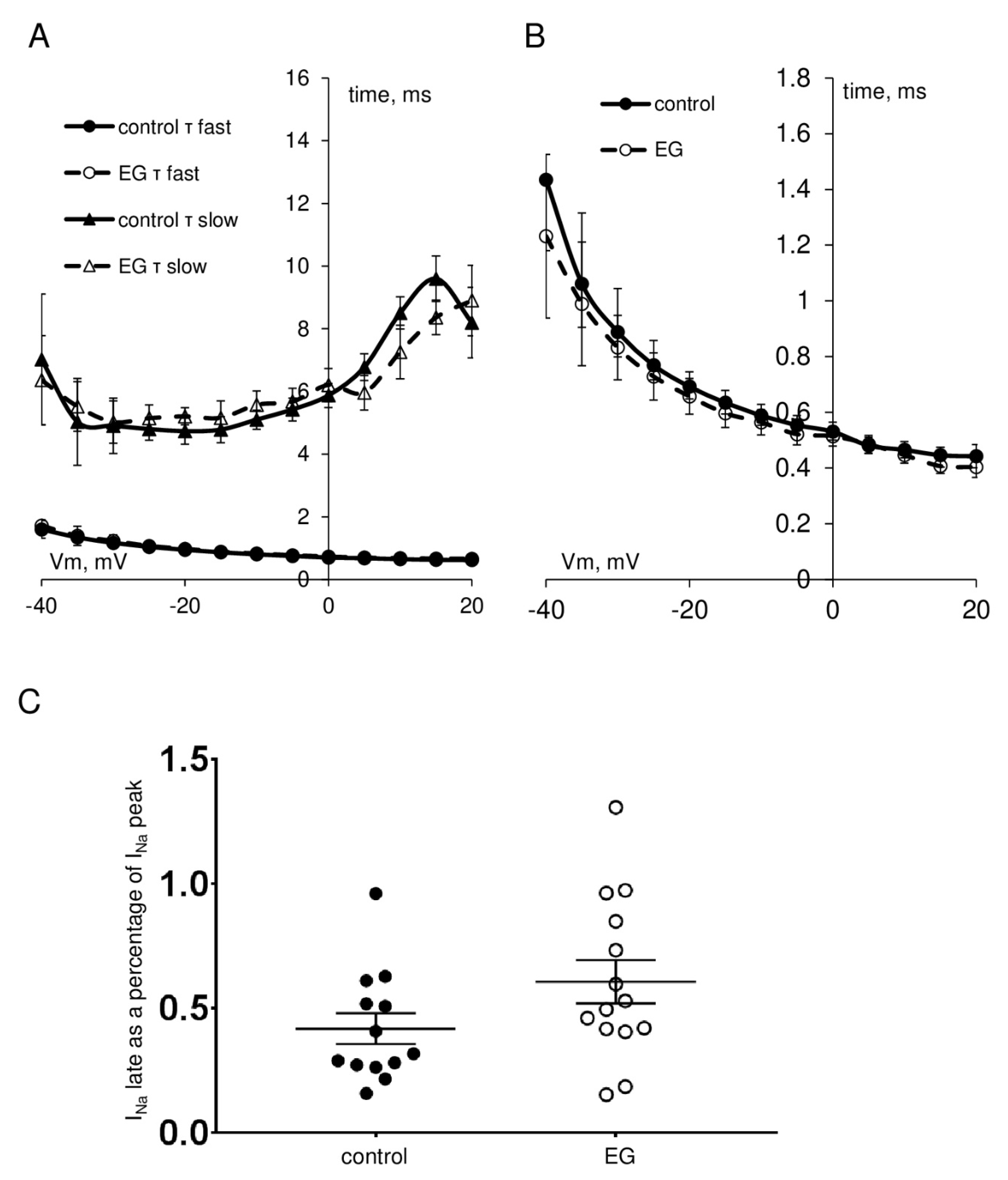
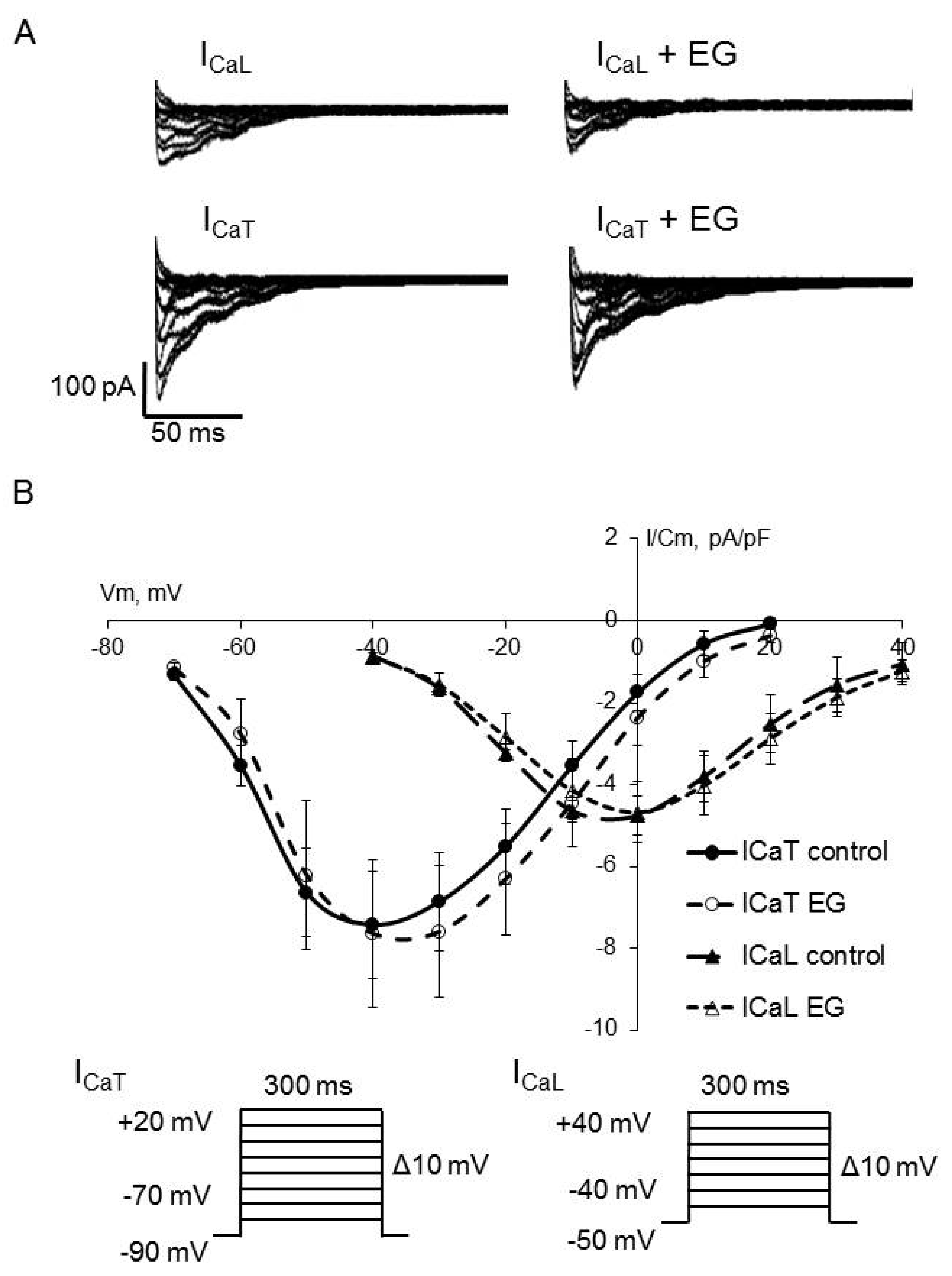
As shown in Figure 2, Figure 3 and Figure 4, the extended incubation of ventricular cardiomyocytes for 2 h in the presence of empagliflozin at the concentration of 5 μM had no effect on INa, ICaL, and ICaT. Analysis of the current-voltage characteristics of INa, ICaL, and ICaT and the parameters of the voltage-dependence of activation and inactivation revealed no significant differences between the control and empagliflozin-treated groups. Table 1 summarizes the biophysical characteristics of examined ionic currents.
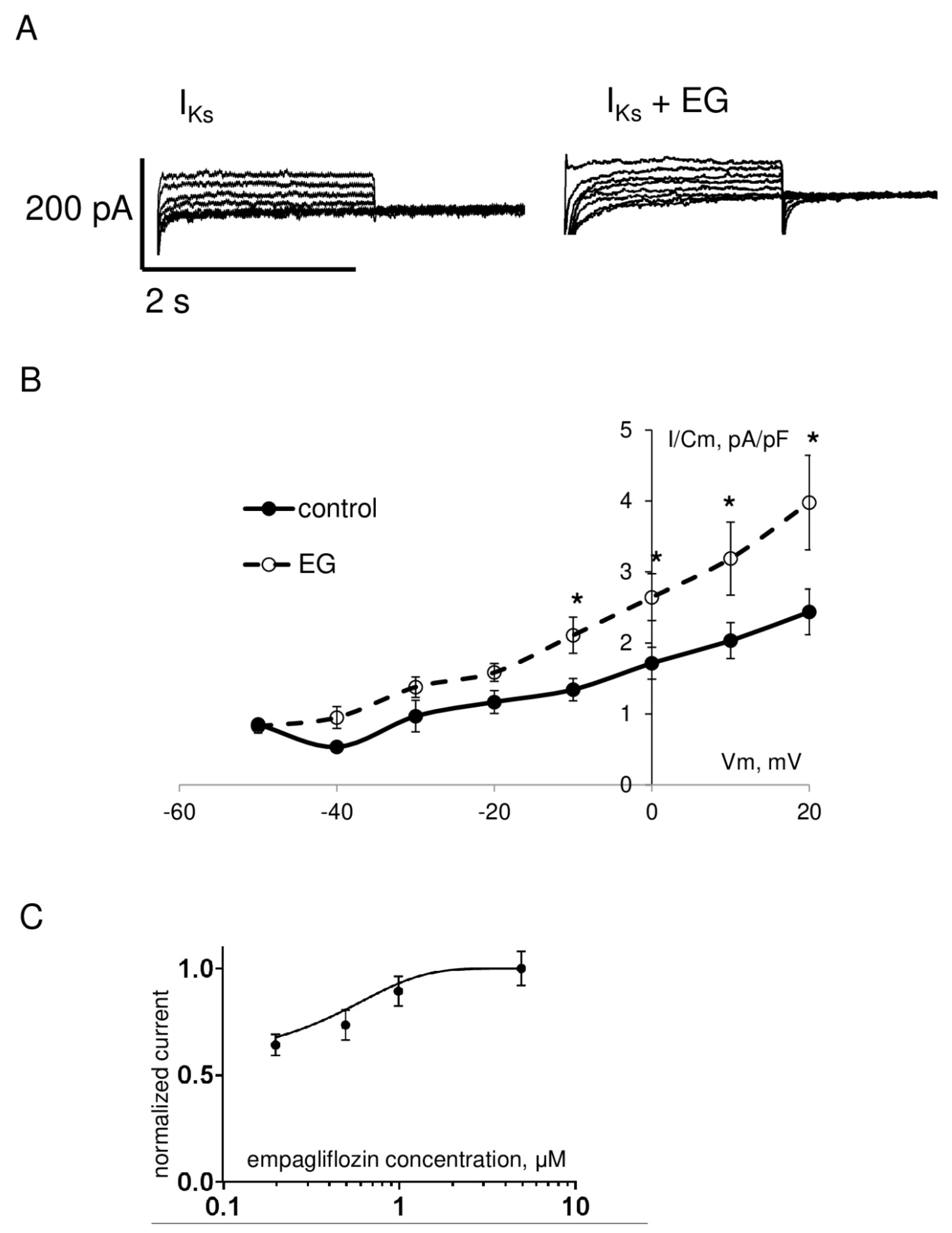
However, in contrast to the aforementioned currents, functional analysis of the rapid component of delayed rectifier potassium current IKr exhibited a significant increase in the amplitude of the step and tail currents. As shown in Figure 5 and Table 1, IKr step current density in the range of testing potentials from 0 to +30 mV and IKr tail current density in the range of testing potentials from +10 to +70 mV in cardiomyocytes after empagliflozin pretreatment was significantly enhanced compared to the control group. Analysis of the slow component of delayed rectifier potassium current IKs revealed a significant increase in the amplitude in the range of testing potentials from −10 to +20 mV in the empagliflozin-treated group (Figure 6). The effect of empagliflozin on the IKr tail and IKs current density had a concentration-dependent manner in the range from 0.2 to 5 μM with a half maximal effective concentration (EC50) of 0.56 μM and 0.76 μM, respectively.
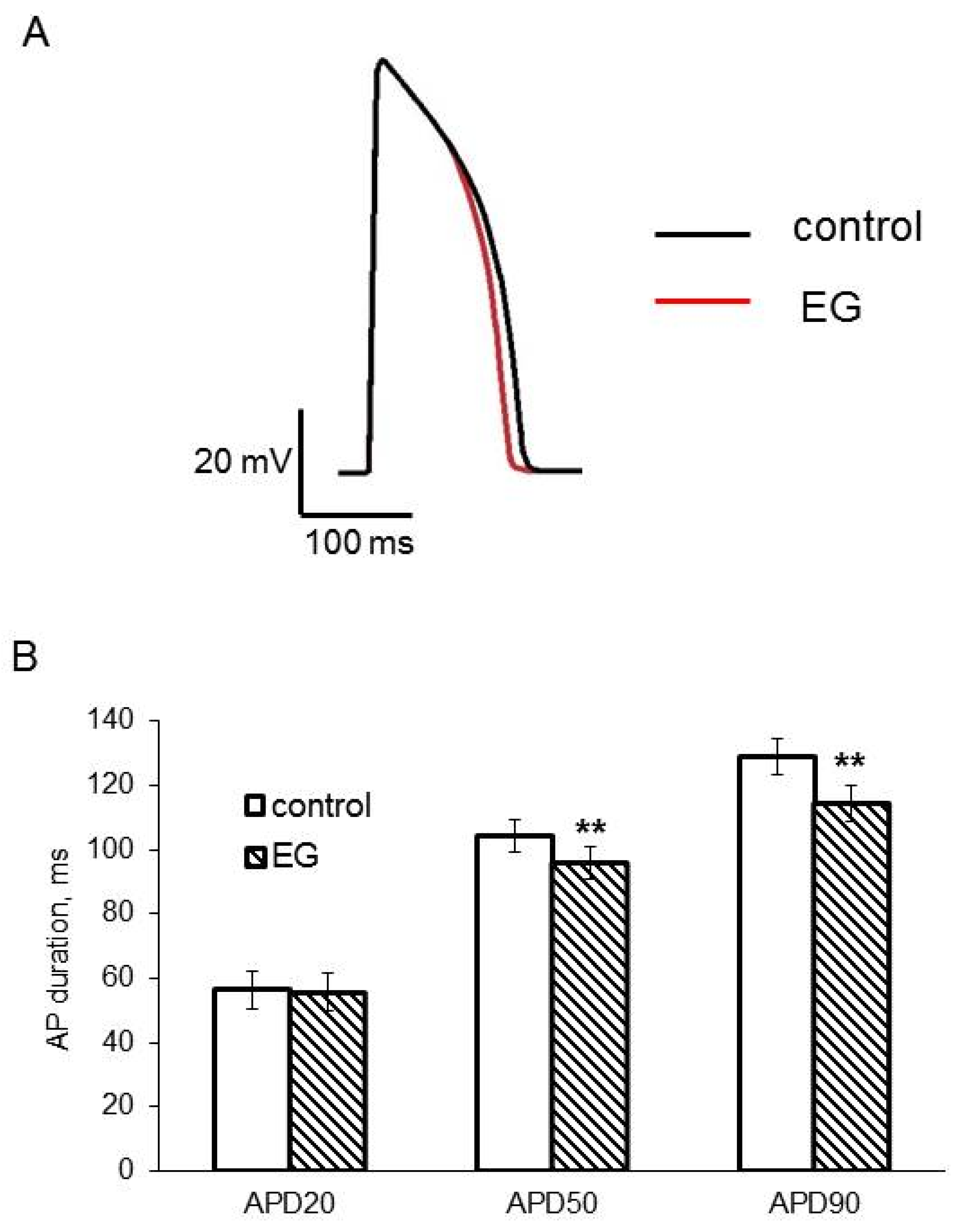
Since IKr is one of the major repolarizing currents in zebrafish hearts and an increase in the IKr current density can result in a change in AP duration, our next step was to explore the AP profile in the ventricular myocardium of an isolated heart. We found, as expected, that the perfusion of the excised heart for 2 h with 5 μM empagliflozin-containing buffer solution significantly reduced APD50 and APD90, but did not affect other AP parameters (Figure 7 and Table 2).

3. Discussion

Recently, SGLT2 inhibitors have come into the focus of research due to the cardioprotective effects demonstrated by the EMPA-REG OUTCOME [2]. Dramatic cardiac benefits include the reduced rate of death from cardiovascular causes and the reduction in heart failure hospitalization in people with and without diabetes [10,35]. Despite a growing number of studies investigating the cardiovascular effects of empagliflozin, the underlying mechanisms are still not fully understood and research results are sometimes contradictory [29,36].
The present study was designed to determine the potential effects on the main ionic currents responsible for AP generation in the model object, zebrafish. Zebrafish are widely used as an adequate vertebrate model of human cardiac function due to the presence of a similar set of ionic currents responsible for the AP in cardiomyocytes. For example, while the use of small mammals such as mice and rats can be limited due to the little expression of delayed rectifier potassium current [37] the advantage of using zebrafish is the presence of both rapid and slow components of this current [38]. Zebrafish have similarities with humans in resting membrane potential, AP amplitude, and shape, in particular in the presence of a clear plateau phase [39]. The similarity with mammals is the involvement of INa in the AP upstroke and ICaL in the plateau phase. Although a fast phase-1 repolarization is not present in zebrafish AP by reason of the absence of the transient outward current ITo.
A number of studies have already shown the restorative effects of empagliflozin on the increased by some impacts late Na+ current [25,26], on the disturbed L-type Ca2+ and Na+/Ca2+ exchanger (NCX) currents in the ventricular myocytes of diabetes mellitus rats [27]. There are controversial data regarding the inhibition of Na+/H+ exchanger activity by empagliflozin [29,30,31]. In this study, we demonstrate for the first time that empagliflozin affects the rapid and slow components of delayed rectifier potassium current IKr and IKs. It was revealed significant increase in the amplitude of the IKr step and tail and IKs currents in empagliflozin-treated cardiomyocytes. However, we failed to detect any effects on INa, ICaL, and ICaT.
In the human heart, the IKr current is conducted by the hERG channel, also known as KV11.1 or KCNH2 [40]. IKr is essential for proper electrical activity in the heart. IKr is crucially important for determining the cardiac AP duration due to effective control of repolarization [41]. Reduction in IKr produced by either direct channel block or inhibition of trafficking results in prolonged AP duration that is linked to an increased risk for Torsade de Pointes and, as a consequence, sudden cardiac death [42]. The pore-forming subunit of the IKs channel, known as KvLQT1 or Kv7.1, is encoded by the KCNQ1 gene [40]. Reduction in current densities due to loss-of-function KCNQ1 mutations or a reduction in repolarization reserve during β-adrenergic stimulation is thought to underlie long QT syndrome phenotypes with increasing susceptibility to arrhythmia [43]. Thus, drugs that increase hERG or KvLQT1 activity might have potential antiarrhythmic effects.
Our experiments in the recording of APs from ventricular myocardium have shown a significant reduction in APD50 and APD90 in empagliflozin-treated zebrafish hearts. These data, as expected, show good agreement with those of IK outward current enhancement.
It has been reported that the expression of the hERG channel is significantly downregulated in diabetic hearts due to high-glucose-induced inhibition of channel trafficking, and this downregulation is a critical contributor to the slowing of repolarization [44]. Studies performed using various animal models have reported a decrease in IKr and IKs current along with a prolonged QT interval in diabetic dog and rabbit hearts [45]. It is also well established that a reduction in the expression of KV channels in hypertrophied and failing myocardium can result in AP prolongation, which is known to be a pro-arrhythmogenic substrate [46]. Thus, the obtained results allow speculation that the gain of function effect of empagliflozin on IK outward current might be considered as a mechanism of cardioprotective action of the drug. However, when trying to extrapolate the results of our research to humans, the temperature sensitivity of delayed rectifier potassium current should be taken into account [47,48]. The recordings of ionic currents were performed at +28 °C, within the range of physiological temperatures for zebrafish. Moreover, it is noteworthy that IKr is produced predominantly by a channel encoded by the zebrafish ortholog to the mammalian KCNH6 gene [49]. These facts should determine objectives of the further study on the effects of empagliflozin.
It should be noted that in some previous studies SGLT2 has not been detected in cardiomyocytes and the heart [50,51,52]. On the other hand, Kwong-Man Ng and coworkers reported SGLT2 expression in hiPSC-derived cardiomyocytes and human heart tissue and they showed that high glucose culture significantly increased SGLT1 and SGLT2 expression in cardiomyocytes [53]. Therefore, what is the pathway of the empagliflozin effect remains a question to be solved. Molecular modeling of empagliflozin docking has shown that the drug has binding affinities to a region in NaV1.5 that is a binding site for known sodium channel inhibitors [25]. However, it seems more likely indirect effect, through the activation of signaling cascades. Empagliflozin has been shown to induce vasodilation in the rabbit aorta by activating protein kinase G PKG and KV channels [54]. Empagliflozin reduces the activity of Ca2+/calmodulin-dependent kinase II CaMKII in mouse and human failing ventricular myocytes [28]. This makes it possible to assume the presence of signaling pathways mediating empagliflozin effects on IK.
In conclusion, results from this study revealed the following key observations: (1) empagliflozin increases IKr and IKs currents and has no effects on INa, ICaL, and ICaT in zebrafish ventricular cardiomyocytes, (2) empagliflozin shortens AP duration in ventricular myocardium. Summing up the aforementioned, suppose that the cardioprotective effect of the SGLT2 inhibitor may be attributed to the upregulation effect on IK outward current.

4. Materials and Methods

4.1. Isolation of Ventricular Cardiomyocytes

All animal handling was performed in accordance with the Helsinki convention. One-year-old wild-type zebrafish were used in the experiments.
Ventricular cardiomyocytes were obtained from the heart by enzymatic dissociation. The fish were killed by decapitation. The heart was rapidly excised. A cannula, blunted syringe needle 32 gauge, was introduced through the aortic bulb of the isolated heart for retrograde perfusion for 10–15 min with a Ca2+-free solution of the following composition (in mM): 100 NaCl, 10 KCl, 1.2 KH2PO4, 4 MgSO4, 10 HEPES, 50 taurine, 20 glucose, pH 6.9 (adjusted with KOH at room temperature). Then the heart was perfused for 25–30 min with the same solution containing proteolytic enzymes: 0.7 mg/mL collagenase type IA (Sigma-Aldrich, St. Louis, MO, USA); 0.6 mg/mL trypsin, type IX (Sigma-Aldrich, St. Louis, MO, USA), and 1 mg/mL bovine serum albumin (DIA-M, Moscow, Russia). All perfusion was carried out at room temperature, trypsin was used only to obtain cells for registration of Na+ current. After the end of perfusion, the atrium was removed and ventricular myocardium was destroyed mechanically (by cutting with surgical scissors and pipetting) to isolate individual cells. Cardiomyocytes were stored in the Ca2+-free solution at +4 °C for no more than 8 h.

4.2. Cell Viability Assay

Cell viability was determined using 3-(4,5-dimethylthiaz-ol-2-yl)-2,5-diphenyltetrazolium bromide MTT (Sigma-Aldrich, St. Louis, MO, USA). Cardiomyocytes were plated in 96-well plates at 1000 cells per well. Cells were exposed to empagliflozin in the concentration range from 0.2 to 5 μM of empagliflozin for 2 h at +28 °C. All groups were repeated in triplicate and were repeated in three independent experiments. After the treatment, MTT solution was added to the final concentration of 0.5 mg/mL for incubation at +28 °C for 4 h. Then DMSO was added to dissolve formazan crystals. Finally, the absorbance was measured by microplate reader CLARIOstar® Plus (BMG LABTECH, Ortenberg, Germany) at 570 nm. The absorbance reading at 630 nm was used as a reference and was subtracted from the 570-nm absorbance reading. The percentage of living cells was calculated using the following formula:
% of living cells = 100% × [(sample absorbance − blank absorbance)/(control absorbance − blank absorbance)],
where blank absorbance is the absorbance in wells with the buffer solution without cells and control absorbance is the absorbance in wells with untreated cells.

4.3. Actin Fluorescent Staining

Cardiomyocytes plated on gelatin-coated coverslips were fixed with 4% paraformaldehyde in PBS for 15 min. After the fixation, cells were soaked with PBS three times for 5 min and then permeabilized with 0.05% Triton X-100 in PBS for 5 min at room temperature. Subsequently, the cells were again washed with PBS twice for 5 min. To visualize actin filaments, the cells were incubated with phalloidin conjugated with Alexa Fluor 488 (Thermo Fisher Scientific, Waltham, MA, USA) for 40 min at room temperature and analyzed under a fluorescence microscope Axio Observer Z1 (Carl Zeiss, Oberkochen, Germany) after counterstaining of nuclei with 4′,6-diamidino-2-phenylindole (DAPI). Images were obtained at a magnification of ×63.

4.4. Recording of Ionic Currents

The whole-cell voltage clamp recordings of ionic currents were performed in the freshly isolated ventricular myocytes at +28 °C, which is the standard temperature for zebrafish maintenance in the lab [55] and within the range of physiological temperatures for zebrafish populations reported in the wild [56]. Each control or empagliflozin-treated group consisted of 13–19 cardiomyocytes, 3–5 cells per fish. Data acquisition was performed with amplifier Axopatch 200B and Clampfit software, version 10.3 (Molecular Devices, San Jose, CA, USA). The ionic currents were acquired at 20–50 kHz and low-pass filtered at 5 kHz using the analog-to-digital interface Digidata 1440A acquisition system (Molecular Devices, San Jose, CA, USA). All pulse protocols were applied more than 5 min after membrane rupture. Patch pipettes of 2.5–3.5 MΩ resistance were pulled from the borosilicate glass B150-110-10 (Sutter Instrument, Novato, CA, USA) with a puller P-1000 (Sutter Instrument, Novato, CA, USA). The pipette and cell capacities and access resistance were completely compensated. The series resistance was compensated by 85–90%.
Na+ current INa was recorded in the bath solution contained in mM: 150 NaCl, 3 KCl, 1.8 CaCl2, 1.2 MgCl2, 10 HEPES, 10 glucose, pH 7.6 (adjusted with NaOH at room temperature). The pipette solution contained in mM: 5 NaCl, 130 CsCl, 1 MgCl2, 5 EGTA, 5 HEPES, 5 MgATP, pH 7.2 (adjusted with CsOH). Ca2+ and K+ currents, ICa and IKr, were blocked with 10μM nifedipine and 2 μM E-4031 (Tocris, Bristol, UK) added to the external solution.
INa was elicited from the holding potential of −120 mV with 40 ms depolarizing voltage steps from −80 to +60 mV in 5 mV increment at the frequency of 1 Hz. The current density, INa normalized to the cell membrane capacitance, was plotted against the voltage steps. Sodium conductance GNa was determined using the equation
GNa = INa/(V − Vrev),
where V is the voltage step, and Vrev is the reversal potential of INa calculated by a linear extrapolation of peak INa in the range of depolarization potentials from +10 to +40 mV. The voltage dependence of steady-state activation of INa was estimated by normalized GNa/GNa max plotted against voltage steps and fitted by the Boltzmann equation
GNa/GNa max = 1 − 1/(1 + exp((V − V1/2)/k)),
where GNa max is the maximal sodium conductance, V1/2 is the potential of half-maximal activation of INa, and k is the curve slope factor.
The voltage dependence of steady-state inactivation of INa was estimated using a double-step protocol with 40 ms testing steps to −20 mV following a conditioning 500 ms prepulse ranging from −120 mV to 0 mV in 5 mV step increment. Normalized INa/INa max elicited by the testing steps was plotted against the voltage of conditioning prepulse and fitted by the Boltzmann equation.
The time to peak was analyzed as a measure of activation kinetics. Time constants of inactivation were obtained by fitting the decaying phase of current trace with a biexponential equation:
It/Imax = Afast × (1 + exp(−t/τ fast)) + Aslow × (1 + exp(−t/τ slow)),
where Afast and Aslow are the fractions of fast and slow inactivating components, respectively, and τ fast and τ slow are their time constants. INa late current was measured at 100 ms after INa peak at −35 mV and the data are presented as a percentage of INa peak current.
ICa was recorded in the bath solution contained in mM: 130 NaCl, 5 CsCl, 2 CaCl2, 1 MgCl2, 5 Na-pyruvate, 10 HEPES, 10 glucose, pH 7.4 (adjusted with NaOH at room temperature). The pipette solution contained in mM: 130 CsCl, 1 MgCl2, 0.345 CaCl2, 5 EGTA, 10 HEPES, 5 MgATP, 15 TEA-Cl, pH 7.2 (adjusted with CsOH). INa and IKr were blocked with 2 μM tetrodotoxin TTX and 2 μM E-4031 (Tocris, Bristol, UK) added to the external solution.
The total ICa, including ICaT and ICaL, was elicited from the holding potential of −90 mV with 300 ms depolarizing voltage steps from −70 to +20 mV in 10 mV increment. ICaL was recorded at depolarization in the range from −40 to +40 mV following the 300 ms step of depolarization up to −50 mV. ICaT was obtained as the difference current between these two protocols.
Delayed rectifier potassium current IK was recorded in the bath solution contained in mM: 150 NaCl, 5.4 KCl, 1.8 CaCl2, 1.2 MgCl2, 10 HEPES, 10 glucose, pH 7.6 (adjusted with NaOH at room temperature). The pipette solution contained in mM: 140 KCl, 1 MgCl2, 5 EGTA, 10 HEPES, 4 MgATP, 0.03 Na2GTP, pH 7.2 (adjusted with KOH). INa, ICa, and IK1 were blocked with 2 μM TTX, 10 μM nifedipine, (Tocris, Bristol, UK), and 2 mM BaCl2 added to the external solution.
IK was elicited by a double-pulse protocol from the holding potential of −80 mV. Initial 2 s depolarization from −50 to +70 mV in 10 mV step increment was followed by 2 s repolarization to −40 mV. Rapid component of delayed rectifier potassium current IKr was measured as an E-4031-sensitive current. The step and tail current peak amplitude after subtraction of the E-4031-sensitive current was used to assess IKr. Slow component of delayed rectifier potassium current IKs was obtained as outward step current in the presence of E-4031.

4.5. Recording of Action Potentials

APs were recorded from ex vivo heart after cutting off the pacemaker area of the heart (sinoatrial junction). The excised heart preparation consisting of the ventricle and a part of the atrium was pinned on the bottom of the Sylgard-coated chamber and continuously perfused at +28 °C with oxygenated solution contained in mM: 150 NaCl, 5.4 KCl, 1.8 CaCl2, 1.2 MgCl2, 10 HEPES, 10 glucose, pH 7.4 (adjusted with NaOH). The continuous pacing at 2 Hz frequency was performed. After an hour of equilibration in experimental conditions, the recording of electrical activity was started. The conventional sharp glass microelectrodes technique was used for intracellular recording of APs in ventricular myocardium. The microelectrodes of 20–40 MΩ resistance were filled with 3 M KCl and connected to a high input impedance amplifier model 1600 (A-M Systems, Sequim, WA, USA). The signal was digitized and recorded using PowerGraph 3.3 (DI-Soft, Moscow, Russia) and analyzed using Mini Analysis 3.0 software (Synaptosoft, Fort Lee, NJ, USA). AP duration at 20%, 50%, and 90% of repolarization (APD20, APD50, and APD90, respectively), AP amplitude, and AP upstroke velocity (dV/dt) were determined during offline analysis. Drug was added to the perfusion solution from concentrated stock solutions to yield the final concentration.

4.6. Empagliflozin Treatment

To elucidate empagliflozin effects on the main ionic currents in freshly isolated zebrafish cardiomyocytes, cells were incubated for 2 h in the presence of various concentrations of empagliflozin. To determine empagliflozin effects on the AP parameters, the isolated heart was perfused for 2 h with empagliflozin-containing oxygenated buffer solution. Incubation and perfusion were carried out at +28 °C. Further, the recordings of ionic currents or AP were performed in the presence of empagliflozin in the buffer solutions.

4.7. Statistical Analysis

Data are presented as mean values ± standard errors (SEM). After checking the normality of the distribution of data obtained by ionic currents recording, statistical comparisons were made using Student’s t-test. AP parameters measured in zebrafish heart before and after application of 5 μM empagliflozin were compared using paired Student’s t-test. Results with p < 0.05 were considered to be statistically significant.

Author Contributions

A.V.K.: conception and design, data analysis and interpretation, manuscript writing; V.B.M. and E.S.K.: performing animal experiments, collection, and assembly of data; A.N.K., D.Y.I., E.K. and S.V.O.: conception and design, administrative support, manuscript writing. All authors have read and agreed to the published version of the manuscript.

Funding

This work was supported by the grant from the Russian Science Foundation 22-15-00186.

Institutional Review Board Statement

The study was conducted according to the guidelines of the Declaration of Helsinki, and approval was obtained from Almazov National Medical Research Centre Ethical Committee (reference number: 0913-22-02B).

Informed Consent Statement

Not applicable.

Data Availability Statement

The data presented in this study are available on request from the corresponding author.

Conflicts of Interest

The authors declare no conflict of interest.

Abbreviations

APAction potential
APD20, APD50, and APD90Action potential duration at 20%, 50%, and 90% of repolarization, respectively
hERGHuman ether-a-go-go related gene
hiPSCHuman induced pluripotent stem cell
ICaLL-type calcium current
ICaTT-type calcium current
INaSodium current
IKrRapid component of delayed rectifier potassium current
IKsSlow component of delayed rectifier potassium current
KVVoltage-gated potassium channel
SGLT2Sodium-glucose co-transporter 2
i(s)SGLT2Sodium-glucose cotransporter type 2 inhibitor(s)

References

  1. Adabag, A.S.; Luepker, R.V.; Roger, V.L.; Gersh, B.J. Sudden cardiac death: Epidemiology and risk factors. Nat. Rev. Cardiol. 2010, 7, 216–225. [Google Scholar] [CrossRef] [PubMed] [Green Version]
  2. Zinman, B.; Wanner, C.; Lachin, J.M.; Fitchett, D.; Bluhmki, E.; Hantel, S.; Mattheus, M.; Devins, T.; Johansen, O.E.; Woerle, H.J.; et al. Empagliflozin, Cardiovascular Outcomes, and Mortality in Type 2 Diabetes. N. Engl. J. Med. 2015, 373, 2117–2128. [Google Scholar] [CrossRef] [Green Version]
  3. Neal, B.; Perkovic, V.; Mahaffey, K.W.; de Zeeuw, D.; Fulcher, G.; Erondu, N.; Shaw, W.; Law, G.; Desai, M.; Matthews, D.R.; et al. Canagliflozin and Cardiovascular and Renal Events in Type 2 Diabetes. N. Engl. J. Med. 2017, 377, 644–657. [Google Scholar] [CrossRef]
  4. Wiviott, S.D.; Raz, I.; Bonaca, M.P.; Mosenzon, O.; Kato, E.T.; Cahn, A.; Silverman, M.G.; Zelniker, T.A.; Kuder, J.F.; Murphy, S.A.; et al. Dapagliflozin and Cardiovascular Outcomes in Type 2 Diabetes. N. Engl. J. Med. 2019, 380, 347–357. [Google Scholar] [CrossRef] [PubMed]
  5. Byrne, N.J.; Parajuli, N.; Levasseur, J.L.; Boisvenue, J.; Beker, D.L.; Masson, G.; Fedak, P.W.; Verma, S.; Dyck, J.R. Empagliflozin Prevents Worsening of Cardiac Function in an Experimental Model of Pressure Overload-Induced Heart Failure. JACC Basic Transl. Sci. 2017, 2, 347–354. [Google Scholar] [CrossRef] [PubMed]
  6. Yurista, S.; Silljé, H.H.; Oberdorf Maass, S.U.; Schouten, E.; Giani, M.G.P.; Hillebrands, J.; Van Goor, H.; Van Veldhuisen, D.J.; De Boer, R.A.; Westenbrink, B.D. Sodium-glucose co-transporter 2 inhibition with empagliflozin improves cardiac function in non-diabetic rats with left ventricular dysfunction after myocardial infarction. Eur. J. Heart Fail. 2019, 21, 862–873. [Google Scholar] [CrossRef]
  7. Lim, V.G.; Bell, R.M.; Arjun, S.; Kolatsi-Joannou, M.; Long, D.A.; Yellon, D.M. SGLT2 Inhibitor, Canagliflozin, Attenuates Myocardial Infarction in the Diabetic and Nondiabetic Heart. JACC Basic Transl. Sci. 2019, 4, 15–26. [Google Scholar] [CrossRef]
  8. Cappetta, D.; De Angelis, A.; Ciuffreda, L.P.; Coppini, R.; Cozzolino, A.; Miccichè, A.; Dell'Aversana, C.; D’Amario, D.; Cianflone, E.; Scavone, C.; et al. Amelioration of diastolic dysfunction by dapagliflozin in a non-diabetic model involves coronary endothelium. Pharmacol. Res. 2020, 157, 104781. [Google Scholar] [CrossRef]
  9. Krasnova, M.; Kulikov, A.; Okovityi, S.; Ivkin, D.; Karpov, A.; Kaschina, E.; Smirnov, A. Comparative efficacy of empagliflozin and drugs of baseline therapy in post-infarct heart failure in normoglycemic rats. Naunyn Schmiedebergs Arch. Exp. Pathol. Pharmakol. 2020, 393, 1649–1658. [Google Scholar] [CrossRef]
  10. McMurray, J.J.V.; Solomon, S.D.; Inzucchi, S.E.; Køber, L.; Kosiborod, M.N.; Martinez, F.A.; Ponikowski, P.; Sabatine, M.S.; Anand, I.S.; Bělohlávek, J.; et al. Dapagliflozin in Patients with Heart Failure and Reduced Ejection Fraction. N. Engl. J. Med. 2019, 381, 1995–2008. [Google Scholar] [CrossRef] [Green Version]
  11. Packer, M.; Anker, S.D.; Butler, J.; Filippatos, G.; Pocock, S.J.; Carson, P.; Januzzi, J.; Verma, S.; Tsutsui, H.; Brueckmann, M.; et al. Cardiovascular and Renal Outcomes with Empagliflozin in Heart Failure. N. Engl. J. Med. 2020, 383, 1413–1424. [Google Scholar] [CrossRef] [PubMed]
  12. Zelniker, T.A.; Braunwald, E. Mechanisms of Cardiorenal Effects of Sodium-Glucose Cotransporter 2 Inhibitors. J. Am. Coll. Cardiol. 2020, 75, 422–434. [Google Scholar] [CrossRef] [PubMed]
  13. Okunrintemi, V.; Mishriky, B.M.; Powell, J.R.; Cummings, D.M. Sodium glucose co transporter 2 inhibitors and atrial fibrillation in the cardiovascular and renal outcome trials. Diabetes Obes. Metab. 2020, 23, 276–280. [Google Scholar] [CrossRef]
  14. Li, H.-L.; Lip, G.Y.H.; Feng, Q.; Fei, Y.; Tse, Y.-K.; Wu, M.-Z.; Ren, Q.-W.; Tse, H.-F.; Cheung, B.-M.Y.; Yiu, K.-H. Sodium-glucose cotransporter 2 inhibitors (SGLT2i) and cardiac arrhythmias: A systematic review and meta-analysis. Cardiovasc. Diabetol. 2021, 20, 1–13. [Google Scholar] [CrossRef]
  15. Fernandes, G.C.; Fernandes, A.; Cardoso, R.; Penalver, J.; Knijnik, L.; Mitrani, R.D.; Myerburg, R.J.; Goldberger, J.J. Association of SGLT2 inhibitors with arrhythmias and sudden cardiac death in patients with type 2 diabetes or heart failure: A meta-analysis of 34 randomized controlled trials. Hearth Rhythm 2021, 18, 1098–1105. [Google Scholar] [CrossRef] [PubMed]
  16. Santos-Gallego, C.G.; Requena-Ibanez, J.A.; Antonio, R.S.; Garcia-Ropero, A.; Ishikawa, K.; Watanabe, S.; Picatoste, B.; Vargas-Delgado, A.P.; Flores-Umanzor, E.J.; Sanz, J.; et al. Empagliflozin Ameliorates Diastolic Dysfunction and Left Ventricular Fibrosis/Stiffness in Nondiabetic Heart Failure. JACC Cardiovasc. Imaging 2020, 14, 393–407. [Google Scholar] [CrossRef]
  17. Matthews, V.B.; Elliot, R.H.; Rudnicka, C.; Hricova, J.; Herat, L.; Schlaich, M. Role of the sympathetic nervous system in regulation of the sodium glucose cotransporter 2. J. Hypertens. 2017, 35, 2059–2068. [Google Scholar] [CrossRef]
  18. Wan, N.; Rahman, A.; Hitomi, H.; Nishiyama, A. The Effects of Sodium-Glucose Cotransporter 2 Inhibitors on Sympathetic Nervous Activity. Front. Endocrinol. 2018, 9, 421. [Google Scholar] [CrossRef] [Green Version]
  19. Oshima, H.; Miki, T.; Kuno, A.; Mizuno, M.; Sato, T.; Tanno, M.; Yano, T.; Nakata, K.; Kimura, Y.; Abe, K.; et al. Empagliflozin, an SGLT2 Inhibitor, Reduced the Mortality Rate after Acute Myocardial Infarction with Modification of Cardiac Metabolomes and Antioxidants in Diabetic Rats. J. Pharmacol. Exp. Ther. 2018, 368, 524–534. [Google Scholar] [CrossRef] [Green Version]
  20. Santos-Gallego, C.G.; Requena-Ibanez, J.A.; Antonio, R.S.; Ishikawa, K.; Watanabe, S.; Picatoste, B.; Flores, E.; Garcia-Ropero, A.; Sanz, J.; Hajjar, R.J.; et al. Empagliflozin Ameliorates Adverse Left Ventricular Remodeling in Nondiabetic Heart Failure by Enhancing Myocardial Energetics. J. Am. Coll. Cardiol. 2019, 73, 1931–1944. [Google Scholar] [CrossRef]
  21. Verma, S.; McMurray, J.J.V. SGLT2 inhibitors and mechanisms of cardiovascular benefit: A state-of-the-art review. Diabetologia 2018, 61, 2108–2117. [Google Scholar] [CrossRef] [PubMed] [Green Version]
  22. Seefeldt, J.M.; Lassen, T.R.; Hjortbak, M.V.; Jespersen, N.R.; Kvist, F.; Hansen, J.; Bøtker, H.E. Cardioprotective effects of empagliflozin after ischemia and reperfusion in rats. Sci. Rep. 2021, 11, 1–13. [Google Scholar] [CrossRef]
  23. Byrne, N.J.; Matsumura, N.; Maayah, Z.H.; Ferdaoussi, M.; Takahara, S.; Darwesh, A.M.; Levasseur, J.L.; Jahng, J.W.S.; Vos, D.; Parajuli, N.; et al. Empagliflozin Blunts Worsening Cardiac Dysfunction Associated With Reduced NLRP3 (Nucleotide-Binding Domain-Like Receptor Protein 3) Inflammasome Activation in Heart Failure. Circ. Heart Fail. 2020, 13, e006277. [Google Scholar] [CrossRef] [PubMed]
  24. Hirata, Y.; Nomura, K.; Senga, Y.; Okada, Y.; Kobayashi, K.; Okamoto, S.; Minokoshi, Y.; Imamura, M.; Takeda, S.; Hosooka, T.; et al. Hyperglycemia induces skeletal muscle atrophy via a WWP1/KLF15 axis. JCI Insight 2019, 4, e124952. [Google Scholar] [CrossRef] [PubMed] [Green Version]
  25. Philippaert, K.; Kalyaanamoorthy, S.; Fatehi, M.; Long, W.; Soni, S.; Byrne, N.J.; Barr, A.; Singh, J.; Wong, J.; Palechuk, T.; et al. Cardiac Late Sodium Channel Current Is a Molecular Target for the Sodium/Glucose Cotransporter 2 Inhibitor Empagliflozin. Circulation 2021, 143, 2188–2204. [Google Scholar] [CrossRef] [PubMed]
  26. Hegyi, B.; Hernandez, J.M.; Shen, E.Y.; Habibi, N.R.; Bossuyt, J.; Bers, D.M. Empagliflozin Reverses Late Na + Current Enhancement and Cardiomyocyte Proarrhythmia in a Translational Murine Model of Heart Failure with Preserved Ejection Fraction. Circulation 2022, 145, 1029–1031. [Google Scholar] [CrossRef]
  27. Lee, T.-I.; Chen, Y.-C.; Lin, Y.-K.; Chung, C.-C.; Lu, Y.-Y.; Kao, Y.-H.; Chen, Y.-J. Empagliflozin Attenuates Myocardial Sodium and Calcium Dysregulation and Reverses Cardiac Remodeling in Streptozotocin-Induced Diabetic Rats. Int. J. Mol. Sci. 2019, 20, 1680. [Google Scholar] [CrossRef] [Green Version]
  28. Mustroph, J.; Wagemann, O.; Lücht, C.M.; Trum, M.; Hammer, K.; Sag, C.M.; Lebek, S.; Tarnowski, D.; Reinders, J.; Perbellini, F.; et al. Empagliflozin reduces Ca/calmodulin-dependent kinase II activity in isolated ventricular cardiomyocytes. ESC Heart Fail. 2018, 5, 642–648. [Google Scholar] [CrossRef] [Green Version]
  29. Chung, Y.J.; Park, K.C.; Tokar, S.; Eykyn, T.R.; Fuller, W.; Pavlovic, D.; Swietach, P.; Shattock, M.J. Off-target effects of sodium-glucose co-transporter 2 blockers: Empagliflozin does not inhibit Na+/H+ exchanger-1 or lower [Na+]i in the heart. Cardiovasc. Res. 2020, 117, 2794–2806. [Google Scholar] [CrossRef]
  30. Baartscheer, A.; Schumacher, C.A.; Wust, R.C.; Fiolet, J.W.T.; Stienen, G.; Coronel, R.; Zuurbier, C.J. Empagliflozin decreases myocardial cytoplasmic Na+ through inhibition of the cardiac Na+/H+ exchanger in rats and rabbits. Diabetologia 2016, 60, 568–573. [Google Scholar] [CrossRef] [Green Version]
  31. Uthman, L.; Baartscheer, A.; Bleijlevens, B.; Schumacher, C.A.; Fiolet, J.W.T.; Koeman, A.; Jancev, M.; Hollmann, M.W.; Weber, N.C.; Coronel, R.; et al. Class effects of SGLT2 inhibitors in mouse cardiomyocytes and hearts: Inhibition of Na+/H+ exchanger, lowering of cytosolic Na+ and vasodilation. Diabetologia 2017, 61, 722–726. [Google Scholar] [CrossRef] [PubMed] [Green Version]
  32. Brette, F.; Luxan, G.; Cros, C.; Dixey, H.; Wilson, C.; Shiels, H.A. Characterization of isolated ventricular myocytes from adult zebrafish (Danio rerio). Biochem. Biophys. Res. Commun. 2008, 374, 143–146. [Google Scholar] [CrossRef]
  33. Verkerk, A.O.; Remme, C.A. Zebrafish: A novel research tool for cardiac (patho)electrophysiology and ion channel disorders. Front. Physiol. 2012, 3, 255. [Google Scholar] [CrossRef] [PubMed] [Green Version]
  34. Scheen, A.J. Pharmacokinetic and Pharmacodynamic Profile of Empagliflozin, a Sodium Glucose Co-Transporter 2 Inhibitor. Clin. Pharmacokinet. 2014, 53, 213–225. [Google Scholar] [CrossRef] [PubMed] [Green Version]
  35. Zannad, F.; Ferreira, J.P.; Pocock, S.J.; Anker, S.D.; Butler, J.; Filippatos, G.; Brueckmann, M.; Ofstad, A.P.; Pfarr, E.; Jamal, W.; et al. SGLT2 inhibitors in patients with heart failure with reduced ejection fraction: A meta-analysis of the EMPEROR-Reduced and DAPA-HF trials. Lancet 2020, 396, 819–829. [Google Scholar] [CrossRef]
  36. Uthman, L.; Baartscheer, A.; Schumacher, C.A.; Fiolet, J.W.T.; Kuschma, M.C.; Hollmann, M.W.; Coronel, R.; Weber, N.C.; Zuurbier, C.J. Direct Cardiac Actions of Sodium Glucose Cotransporter 2 Inhibitors Target Pathogenic Mechanisms Underlying Heart Failure in Diabetic Patients. Front. Physiol. 2018, 9, 1575. [Google Scholar] [CrossRef]
  37. Sutanto, H.; Heijman, J. Integrative Computational Modeling of Cardiomyocyte Calcium Handling and Cardiac Arrhythmias: Current Status and Future Challenges. Cells 2022, 11, 1090. [Google Scholar] [CrossRef]
  38. Abramochkin, D.V.; Hassinen, M.; Vornanen, M. Transcripts of Kv7.1 and MinK channels and slow delayed rectifier K+ current (IKs) are expressed in zebrafish (Danio rerio) heart. Pflügers Arch. Eur. J. Physiol. 2018, 470, 1753–1764. [Google Scholar] [CrossRef]
  39. Gauvrit, S.; Bossaer, J.; Lee, J.; Collins, M.M. Modeling Human Cardiac Arrhythmias: Insights from Zebrafish. J. Cardiovasc. Dev. Dis. 2022, 9, 13. [Google Scholar] [CrossRef]
  40. Chen, L.; Sampson, K.J.; Kass, R.S. Cardiac Delayed Rectifier Potassium Channels in Health and Disease. Card. Electrophysiol. Clin. 2016, 8, 307–322. [Google Scholar] [CrossRef] [Green Version]
  41. Sanguinetti, M.C.; Tristani-Firouzi, M. hERG potassium channels and cardiac arrhythmia. Nature 2006, 440, 463–469. [Google Scholar] [CrossRef] [PubMed]
  42. Brown, A.M. hERG Assay, QT Liability, and Sudden Cardiac Death. In Cardiac Safety of Noncardiac Drugs; Springer: Berlin/Heidelberg, Germany, 2007; pp. 67–81. [Google Scholar] [CrossRef]
  43. Bohnen, M.S.; Peng, G.; Robey, S.H.; Terrenoire, C.; Iyer, V.; Sampson, K.J.; Kass, R.S. Molecular Pathophysiology of Congenital Long QT Syndrome. Physiol. Rev. 2017, 97, 89–134. [Google Scholar] [CrossRef] [PubMed] [Green Version]
  44. Shi, Y.-Q.; Yan, M.; Liu, L.-R.; Zhang, X.; Wang, X.; Geng, H.-Z.; Zhao, X.; Li, B.-X. High Glucose Represses hERG K+ Channel Expression through Trafficking Inhibition. Cell. Physiol. Biochem. 2015, 37, 284–296. [Google Scholar] [CrossRef] [Green Version]
  45. Ozturk, N.; Uslu, S.; Ozdemir, S. Diabetes-induced changes in cardiac voltage-gated ion channels. World J. Diabetes 2021, 12, 1–18. [Google Scholar] [CrossRef] [PubMed]
  46. Mustroph, J.; Maier, L.S.; Wagner, S. CaMKII regulation of cardiac K channels. Front. Pharmacol. 2014, 5, 20. [Google Scholar] [CrossRef] [Green Version]
  47. Vandenberg, J.I.; Varghese, A.; Lu, Y.; Bursill, J.A.; Mahaut-Smith, M.P.; Huang, C.L.-H. Temperature dependence of human ether-à-go-go-related gene K+ currents. Am. J. Physiol. Physiol. 2006, 291, C165–C175. [Google Scholar] [CrossRef]
  48. Walsh, K.B.; Begenisich, T.B.; Kass, R.S. Beta-adrenergic modulation of cardiac ion channels. Differential temperature sensitivity of potassium and calcium currents. J. Gen. Physiol. 1989, 93, 841–854. [Google Scholar] [CrossRef] [Green Version]
  49. Vornanen, M.; Hassinen, M. Zebrafish heart as a model for human cardiac electrophysiology. Channels 2016, 10, 101–110. [Google Scholar] [CrossRef] [Green Version]
  50. Vrhovac, I.; Eror, D.B.; Klessen, D.; Burger, C.; Breljak, D.; Kraus, O.; Radović, N.; Jadrijević, S.; Aleksic, I.; Walles, T.; et al. Localizations of Na+-d-glucose cotransporters SGLT1 and SGLT2 in human kidney and of SGLT1 in human small intestine, liver, lung, and heart. Pflügers Arch. Eur. J. Physiol. 2014, 467, 1881–1898. [Google Scholar] [CrossRef]
  51. Chen, J.; Williams, S.; Ho, S.; Loraine, H.; Hagan, D.; Whaley, J.M.; Feder, J.N. Quantitative PCR tissue expression profiling of the human SGLT2 gene and related family members. Diabetes Ther. 2010, 1, 57–92. [Google Scholar] [CrossRef] [Green Version]
  52. Zhou, L.; Cryan, E.V.; D'Andrea, M.R.; Belkowski, S.; Conway, B.R.; Demarest, K.T. Human cardiomyocytes express high level of Na+/glucose cotransporter 1 (SGLT1). J. Cell. Biochem. 2003, 90, 339–346. [Google Scholar] [CrossRef]
  53. Ng, K.-M.; Lau, Y.-M.; Dhandhania, V.; Cai, Z.-J.; Lee, Y.-K.; Lai, W.-H.; Tse, H.-F.; Siu, C.-W. Empagliflozin Ammeliorates High Glucose Induced-Cardiac Dysfuntion in Human iPSC-Derived Cardiomyocytes. Sci. Rep. 2018, 8, 14872. [Google Scholar] [CrossRef] [PubMed]
  54. Seo, M.S.; Jung, H.S.; An, J.R.; Kang, M.; Heo, R.; Li, H.; Han, E.-T.; Yang, S.-R.; Cho, E.-H.; Bae, Y.M.; et al. Empagliflozin dilates the rabbit aorta by activating PKG and voltage-dependent K+ channels. Toxicol. Appl. Pharmacol. 2020, 403, 115153. [Google Scholar] [CrossRef] [PubMed]
  55. Westerfield, M. The Zebrafish Book. A Guide for the Laboratory Use of Zebrafish (Danio rerio), 4th ed.; University of Oregon Press: Eugene, OR, USA, 2000. [Google Scholar]
  56. Engeszer, R.E.; Patterson, L.B.; Rao, A.A.; Parichy, D.M. Zebrafish in The Wild: A Review of Natural History And New Notes from The Field. Zebrafish 2007, 4, 21–40. [Google Scholar] [CrossRef] [PubMed]
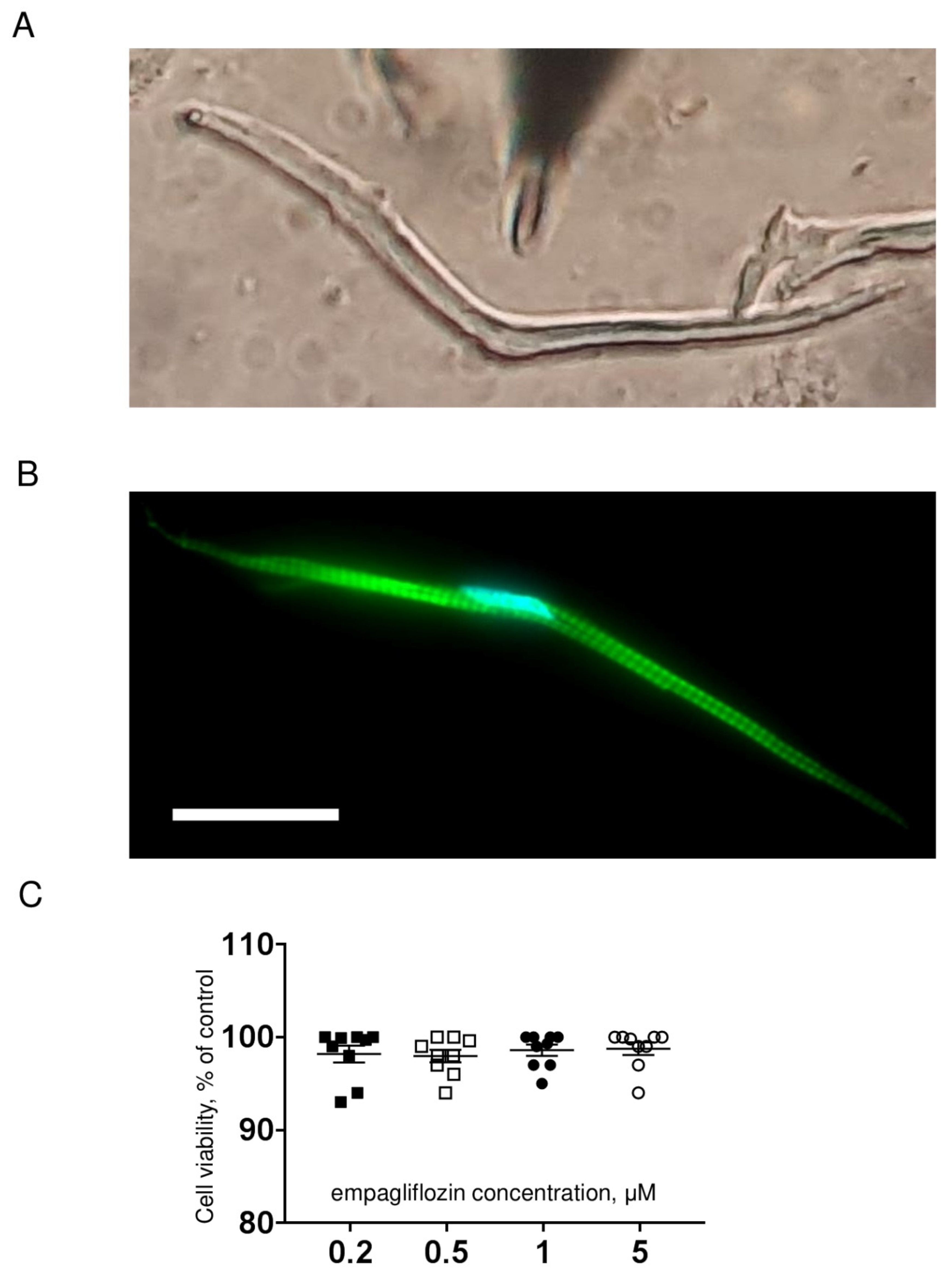
Figure 1. (A) Representative image of an isolated zebrafish ventricular cardiomyocyte used for patch clamp experiments. Magnification: 40×. (B) Representative image of actin filaments (green) staining using phalloidin in zebrafish cardiomyocytes. Nucleus (blue) was visualized with DAPI. Scale bar is 20 μm. (C) Grouped data on MTT test for cardiomyocytes treated with empagliflozin. Data are expressed as % of value obtained for untreated cells.
Ijms 23 09559 g001
Figure 2. (A) Representative whole–cell current traces of INa in freshly isolated ventricular cardiomyocytes from zebrafish in control (left) and after 2 h incubation with 5 μM empagliflozin (right). (B) The current density–voltage relationship of the INa in control (n = 13) and in empagliflozin–treated (n = 14) cardiomyocytes. (C) The voltage dependence of steady–state activation and inactivation. The solid and dash lines show least–squares fits to the Boltzmann function. Voltage clamp protocols used to determine activation and inactivation characteristics are shown at the bottom.
Ijms 23 09559 g002
Figure 3. (A) The voltage dependence of the inactivation fast (triangles) and slow (circles) time constants τfast, τslow of INa in control (filled) and after 2 h incubation with 5 μM empagliflozin (open) cardiomyocytes. (B) The voltage dependence of the time to peak of INa. (C) Grouped data on INa late current as a percentage of INa peak current in control and after 2 h incubation with 5 μM empagliflozin.
Ijms 23 09559 g003
Figure 4. (A) Representative whole–cell current traces of ICa in freshly isolated ventricular cardiomyocytes from zebrafish in control (left) and after 2 h incubation with 5 μM empagliflozin (right). (B) The current density–voltage relationship of the ICa in control (n = 14) and in empagliflozin–treated (n = 14) cardiomyocytes. Voltage clamp protocols used to estimate ICaT and ICaL are shown at the bottom.
Ijms 23 09559 g004
Figure 5. (A) Representative whole-cell current traces of IKr in freshly isolated ventricular cardiomyocytes from zebrafish in control (left) and after 2 h incubation with 5 μM empagliflozin (right). (B) The current density–voltage relationship of the IKr step (left) and tail (right) current in control (n = 18) and in empagliflozin–treated (n = 19) cardiomyocytes. * p < 0.05 obtained by Student’s t-test. Voltage clamp protocols used to estimate IKr are shown at the bottom. (C) Concentration–response curve for empagliflozin effect on the IKr tail current density at +10 mV. The solid line shows least-squares fit to the Hill function.
Ijms 23 09559 g005
Figure 6. (A) Representative whole–cell current traces of IKs in freshly isolated ventricular cardiomyocytes from zebrafish in control (left) and after 2 h incubation with 5 μM empagliflozin (right). (B) The current density–voltage relationship of the IKs in control (n = 18) and in empagliflozin–treated (n = 19) cardiomyocytes. * p < 0.05 obtained by Student’s t-test. (C) Concentration–response curve for empagliflozin effect on the IKs current density at +20 mV. The solid line shows least–squares fit to the Hill function.
Ijms 23 09559 g006
Figure 7. (A) Representative recording of ventricular AP from ex vivo zebrafish heart. Black line shows AP in control and red in 5 μM empagliflozin–treated heart. (B) Bar graphs of mean results for APD20, APD50, and APD90 in control (n = 5) and empagliflozin–treated (n = 5) heart. ** p < 0.01 obtained by paired Student’s t-test.
Ijms 23 09559 g007
Table 1. Biophysical characteristics of INa, ICaL, ICaT, and IKr in ventricular cardiomyocytes from zebrafish. Values are represented as mean ± SEM. * p < 0.05 obtained by Student’s t-test.
ControlnEmpagliflozinn
INa peak current density
at −35 mV, pA/pF
−317.9 ± 34.613−334.4 ± 37.414
INa steady-state activation,
V1/2, mV
k, mV/e-fold

−46.5 ± 1.1
3.7 ± 0.2
13
−48.3 ± 0.9
3.9 ± 0.3
14
INa steady-state inactivation,
V1/2, mV
k, mV/e-fold

−77.3 ± 2.0
5.1 ± 0.2
13
−76.1 ± 0.9
5.0 ± 0.1
14
INa late current,
% of INa peak current
0.42 ± 0.09130.61 ± 0.1014
ICaL peak current density
at 0 mV, pA/pF
−4.8 ± 0.714−4.7 ± 0.714
ICaT peak current density
at −40 mV, pA/pF, ms
−7.4 ± 1.314−7.6 ± 1.814
ICaL steady-state activation,
V1/2, mV
k, mV/e-fold

−19.3 ± 1.6
7.7 ± 0.3
14
−16.0 ± 1.7
7.9 ± 0.5
14
ICaT steady-state activation,
V1/2, mV
k, mV/e-fold

−50.2 ± 2.2
9.5 ± 1.3
14
−46.6 ± 2.8
9.1 ± 1.2
14
IKr peak step current density
at 0 mV, pA/pF
2.8 ± 0.318* 4.2 ± 0.719
IKr peak tail current density
at +10 mV, pA/pF
5.1 ± 0.418* 7.6 ± 1.019
IKs peak current density
at +20 mV, pA/pF
2.4 ± 0.318* 4.0 ± 0.719
Table 2. Amplitude and time parameters of the AP in ventricular myocardium of isolated zebrafish heart. Values are represented as mean ± SEM. ** p < 0.01 obtained by paired Student’s t-test.
Control
n = 5
Empagliflozin
n = 5
AP amplitude, mV82.8 ± 3.081.0 ± 2.7
AP upstroke velocity, mV/ms11.2 ± 0.913.8 ± 2.0
APD20, ms56.5 ± 5.955.8 ± 5.9
APD50, ms104.3 ± 5.0** 95.7 ± 5.0
APD90, ms128.8 ± 5.5** 114.5 ± 5.8
Publisher’s Note: MDPI stays neutral with regard to jurisdictional claims in published maps and institutional affiliations.