Article

Cloning of the Maternal Effector Gene org and Its Regulation by lncRNA ORG-AS in Chinese Tongue Sole (Cynoglossus semilaevis)

1
School of Marine Sciences, Ningbo University, Ningbo 315211, China
2
Key Lab of Sustainable Development of Marine Fisheries, Ministry of Agriculture and Rural Affairs, Yellow Sea Fisheries Research Institute, Chinese Academy of Fishery Sciences, Qingdao 266071, China
3
Laboratory for Marine Fisheries Science and Food Production Processes, Pilot National Laboratory for Marine Science and Technology (Qingdao), Qingdao 266237, China
*
Author to whom correspondence should be addressed.
Int. J. Mol. Sci. 2022, 23(15), 8605; https://doi.org/10.3390/ijms23158605
Submission received: 21 June 2022 / Revised: 24 July 2022 / Accepted: 27 July 2022 / Published: 3 August 2022
(This article belongs to the Special Issue Molecular Sexual Determination and Differentiation in Fishes)

Abstract

:
Maternal effector genes (MEGs) encode maternal RNA and protein, accumulating in the cytoplasm of oocytes. During oocyte development, MEGs participate in oocyte meiosis and promote oocyte development. And MEGs can also regulate maternal transcriptome stability and promote maternal–zygotic transition (MTZ) in early embryonic development. Long noncoding RNAs (lncRNAs), as new epigenetic regulators, can regulate gene expression at both the transcriptional and post-transcriptional levels through cis- or trans-regulation. The oogenesis-related gene org is a germ-cell-specific gene in fish, but the role of org in embryonic development and oogenesis has rarely been studied, and the knowledge of the lncRNA-mediated regulation of org is limited. In this study, we cloned and identified the org gene of Chinese tongue sole (Cynoglossus semilaevis), and we identified a lncRNA named lncRNA ORG-anti-sequence (ORG-AS), located at the reverse overlapping region of org. The results of qRT-PCR and FISH demonstrated that org was highly expressed during the early stages of embryonic development and oogenesis and was located in the cytoplasm of oocytes. ORG-AS was expressed at low levels in the ovary and colocalized with org in the cytoplasm of oocytes. In vitro experiments showed that overexpression of ORG-AS inhibited org expression. These results suggest that org, as a MEG in C. semilaevis, participates in the MTZ and the oogenesis. The lncRNA ORG-AS negatively regulates the gene expression of org through trans-regulation. These new findings broaden the function of MEGs in embryonic development and the oogenesis of bony fish and prove that lncRNAs are important molecular factors regulating org.

1. Introduction

In mammals, the development of early embryos depends on maternal complexes, including subcellular organelles and biomacromolecules stored in the oocyte cytoplasm, encoded by maternal effector genes (MEGs) [1,2]. These maternal complexes participate in oocyte genome transcriptional activation, meiosis, and fertilization, and maintain oocyte growth and development [3]. MEGs were first found in Drosophila [4] and then in Nematode [5] and Xenopus [6]. For example, Zar1 is specifically expressed in oocytes, and it has been found that the targeted knockout of Zar1/2 blocks the process of maternal–zygotic transition (MTZ) in humans and mice [7,8]. Researchers also found some germ-cell-specific MEGs in bony fish, such as vasa, nanos1, and ziwi in zebrafish, which can maintain early embryonic development and oogenesis [9,10,11].
Oogenesis is a long process and is the basis of embryonic development, which starts from the differentiation of primary oocytes after mitosis and the proliferation of oocytes [12]. Oogenesis is regulated by a large number of factors in the ovary, including histone modification, nonhistone acetylation, alternative splicing, and other epigenetic modifications, as well as communication between oocytes and their surrounding granulosa cells [13,14]. Ma et al. found that protein deacetylases HDAC1 and HDAC2 in mice can regulate transcription and apoptosis in the development of mice oocytes [15]. In addition, Lu et al. found a large number of differentially expressed genes and differentially alternative splicing genes through a comparative transcriptome analysis of the gonads of Chinese tongue sole (Cynoglossus semilaevis), and they proved that alternative splicing, as an important mechanism of epigenetic modification, can regulate reproductive cell development [16,17]. Consequently, epigenetic regulation plays an important role in the development of oocytes.
Noncoding RNAs with a length of more than 200 nucleotides are considered long noncoding RNAs (lncRNAs), which are widely transcribed in eukaryotic genomes with the same 5′ cap structure and 3′ polyA tail as mRNA [18]. According to the position relationship between lncRNAs and adjacent mRNAs in the genome, lncRNAs can be divided into seven categories, namely, sense, antisense, bidirectional, intron, intergenic, promoter, and enhancer lncRNAs [19]. As a new epigenetic regulator, lncRNAs can regulate gene expression by regulating the modification of histones and DNA [20]. In addition, the cis- and trans-regulation of lncRNA is also important for the regulation of local gene expression [21]. Feng et al. found that lncRNA DMART2-AS is an important regulatory factor of dmrt2 in C. semilaevis, reversing complementation with exon 4 of dmrt2 and inducing the transcription of dmrt2 [22]. In the crustacean Daphnia magna, lncRNA DAPALR overlaps with dsx1 5′UTR and can activate and maintain the expression of dsx1 [23]. In conclusion, all these findings show the important role of lncRNAs in regulating gene expression.
As an economically important fish in China, the female C. semilaevis grows 2–4 times faster than males, and females have a greater economic value [24]. MEGs are crucial for embryonic development and oogenesis in female fish. However, there is no study on the function and regulation of MEGs in C. semilaevis at present. Recently, Li et al. found a new gene in zebrafish, zorg, a germ-cell-specific gene that is abundant in the oocyte cortex of zebrafish oocytes. zorg gene products may play an important role in zebrafish oogenesis and early embryonic development [25]. org, as a germ-cell-specific gene only found in fish, has important research value in embryonic development and oogenesis. In this study, we cloned and identified the first MEG org in C. semilaevis, and we identified the lncRNA, ORG-anti sequence (ORG-AS), that regulates org in C. semilaevis.

2. Results

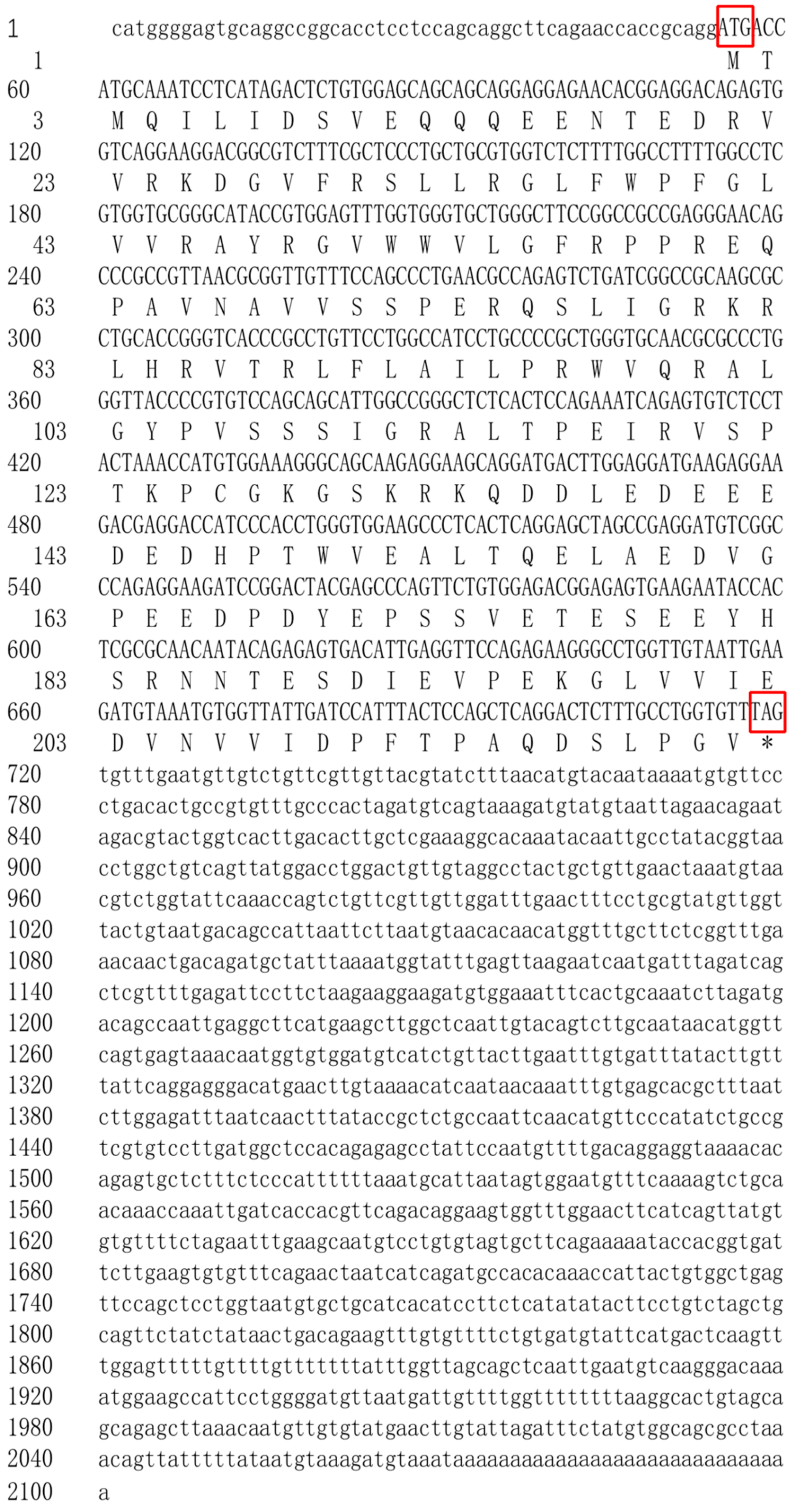
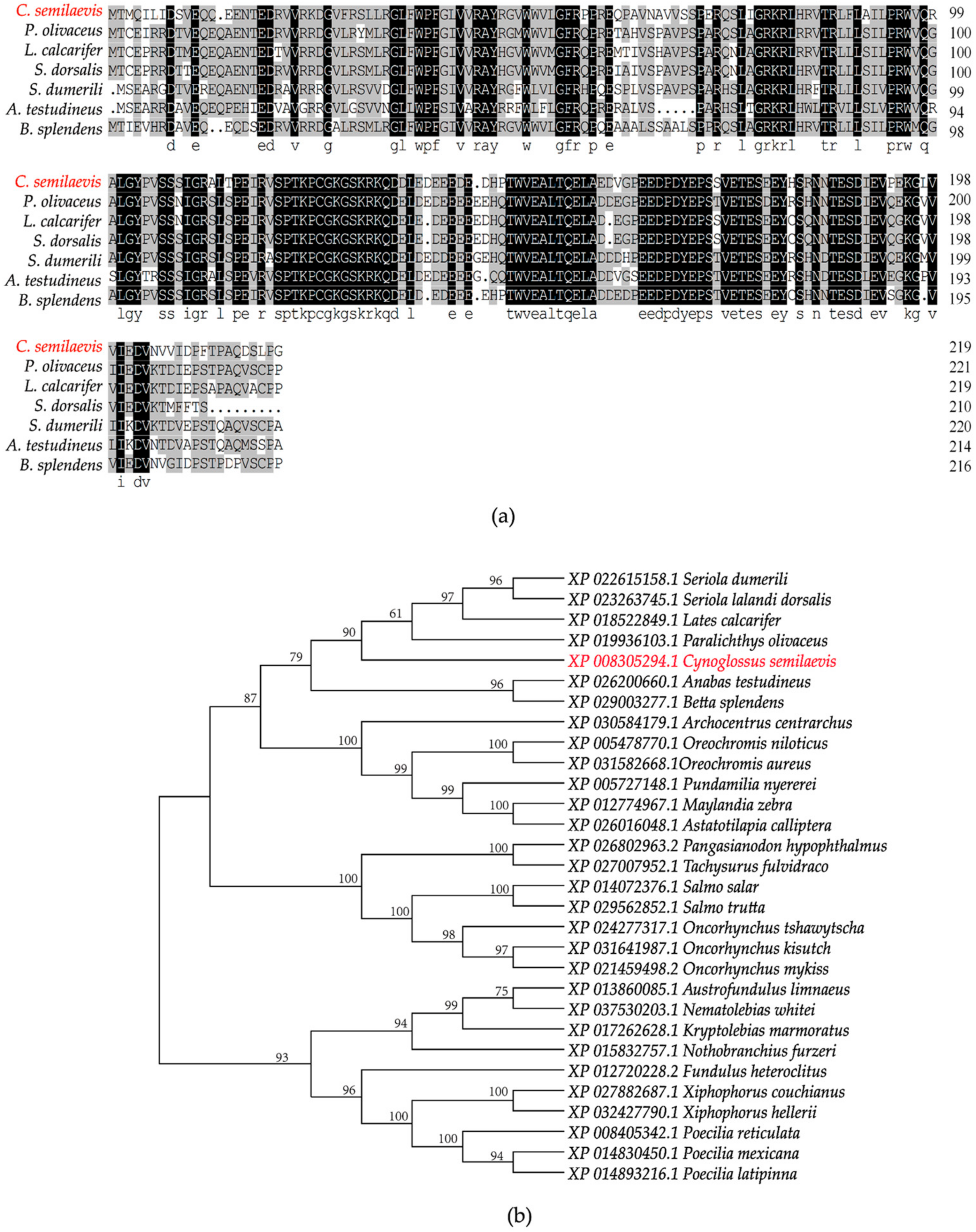
2.1. Cloning and Sequence Analysis of org

We obtained the full-length sequence of the org gene using 5′ and 3′ RACE amplification. A sequence analysis revealed that the total org cDNA length was 2101 bp, comprising six exons and five introns, and had a poly (A) tail at the 3′ end. The org cDNA had a 666 bp open reading frame and encoded a 221 amino acid protein (Figure 1). To further understand the homology and evolution of org, we compared the amino acid sequences of org with those of other species. We found that org only exists in fish and that the amino acid sequence of org shares 84.88% homology with other teleost amino acid sequences (Figure 2a). The results of the phylogenetic tree show that C. semilaevis formed a clade with Paralichthys olivaceus, Lates calcarifer, Seriola lalandi dorsalis, and Seriola dumerili, indicating a relatively close evolutionary relationship (Figure 2b).

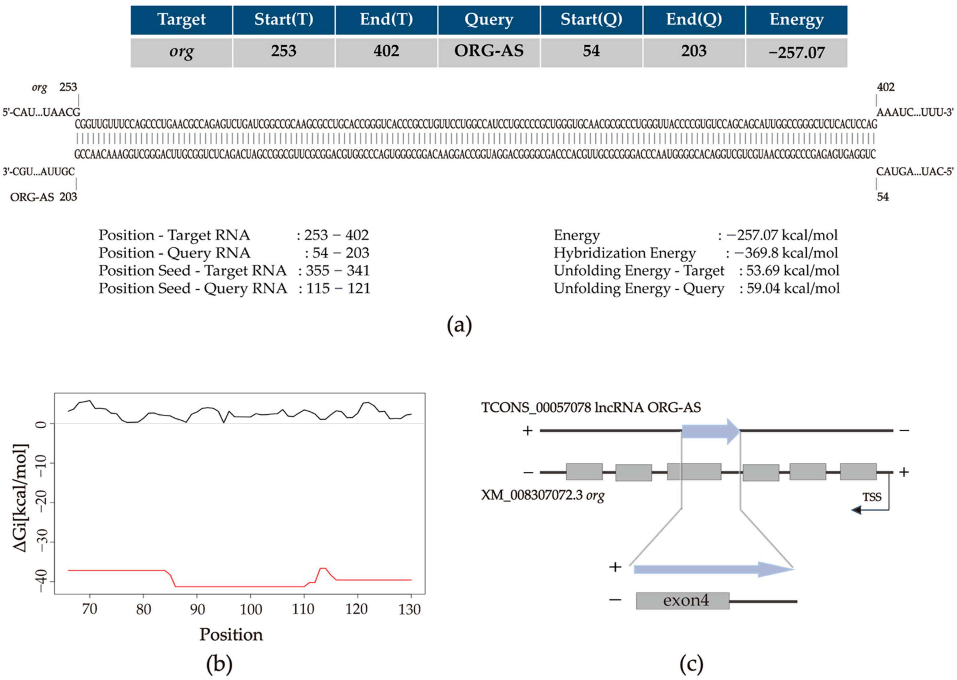
2.2. ORG-AS Is Predicted to Bind to org

We found that there is a reverse complementary sequence between org upstream and a lncRNA ORG-AS in the C. semilaevis genome. We hypothesized that ORG-AS might regulate org, and we predicted the interaction sites between ORG-AS and org. The IntaRNA results showed that 54–203 nucleotides of ORG-AS were inversely complementary to nucleotides 253–402 of org (Figure 3a). ORG-AS (nucleotides 86–110) and org (nucleotides 348–372) might interact according to the RNAup prediction (Figure 3b). We observed that ORG-AS was inversely complementary to exon 4 of org by building the model presented in Figure 3c.

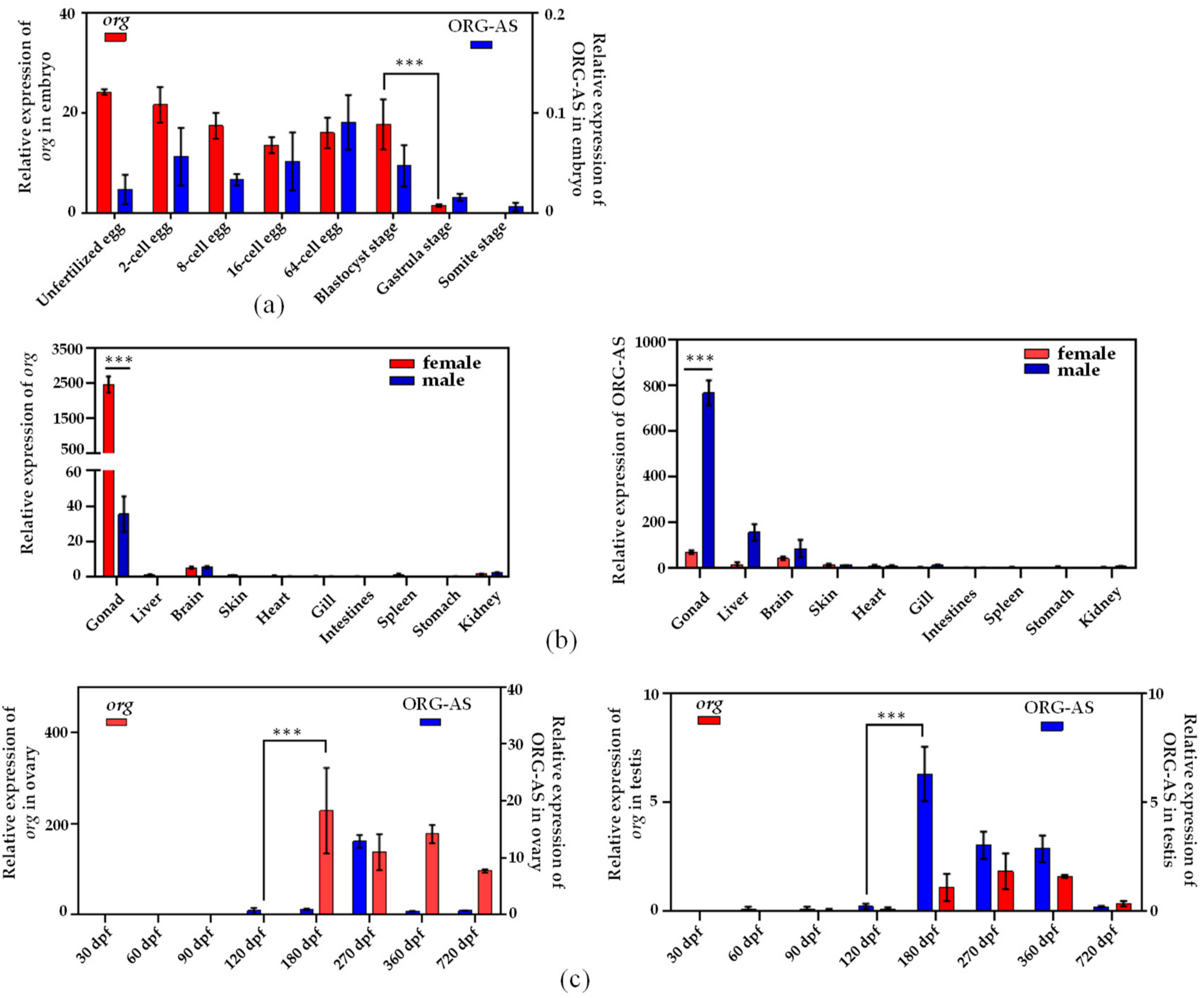
2.3. Expression Patterns of org and ORG-AS

We measured org and ORG-AS expression levels using qRT-PCR on different tissues in adult C. semilaevis, as well as in the early embryos of C. semilaevis. The org and ORG-AS expression patterns are shown in Figure 4. The results showed that there were high org mRNA levels in early embryos and unfertilized eggs. The high expression levels were sustained from unfertilized eggs to the blastocyst stage, and they decreased rapidly in the gastrulation stage. However, the expression of ORG-AS in unfertilized eggs and early embryos was much lower than that of org, and the expression of ORG-AS showed a rapid decrease in the gastrulation stage (Figure 4a). In sexually mature C. semilaevis, org was mainly expressed in the gonads, especially in the ovary, and it was rarely expressed in other tissues. ORG-AS was also mainly expressed in the gonads, but in contrast to org, it was highly expressed in testis (Figure 4b).
We further assessed org and ORG-AS expression patterns during gonadal development (Figure 4c). The expression of org in the ovary increased sharply at 180 dpf, and then it remained stable. Moreover, the expression of org was higher than that of ORG-AS in each developmental stage of the ovary. In the testis, the expression of ORG-AS increased rapidly at 180 dpf and was higher than that of org, and then it gradually decreased with org. Furthermore, in the testis and ovary, ORG-AS was always expressed earlier than org.

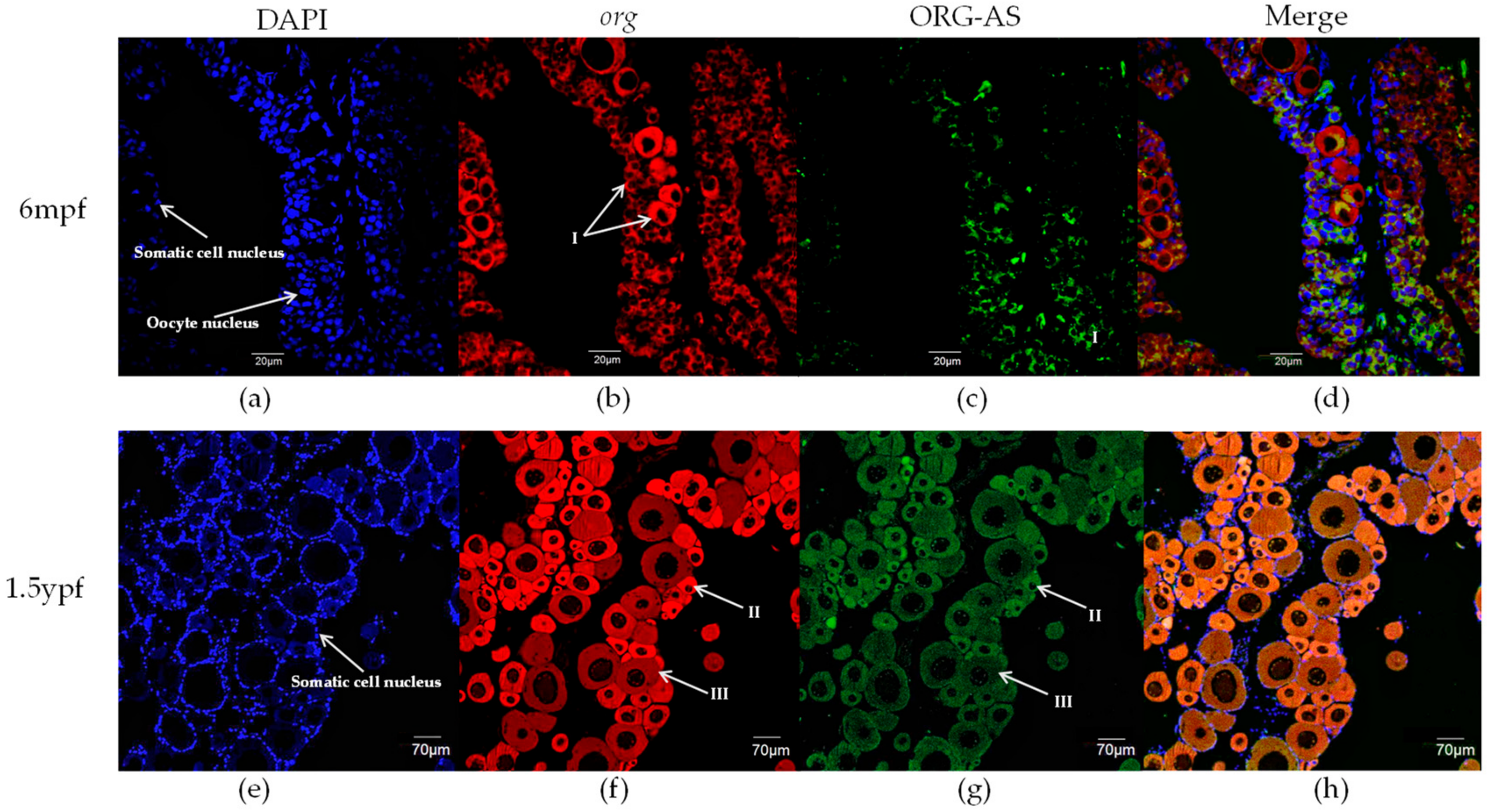
2.4. FISH of org and ORG-AS in Gonads

We investigated the localization of org and ORG-AS in oocytes using fluorescence in situ hybridization. In 6 mpf and 1.5 ypf ovaries, there were many somatic cells around the oocytes (Figure 5a,e). The oocytes in the 6 mpf ovaries were mainly phase I oogonia with large nucleoli in the center of the nucleus (Figure 5b). The oocytes in the 1.5 ypf ovaries were mainly phase II and early phase III oocytes, which contain multiple small nucleoli, distributed around the nucleolar membrane (Figure 5f). In 6 mpf and 1.5 ypf ovaries, org was mainly localized in the oocyte cytoplasm (Figure 5b,f). In addition, similar to the org expression pattern, ORG-AS was mainly localized in both 6 mpf and 1.5 ypf ovaries (Figure 5c,g). Intriguingly, the merged image showed that ORG-AS and org were co-located in the cytoplasm of oocyte (Figure 5d,h).

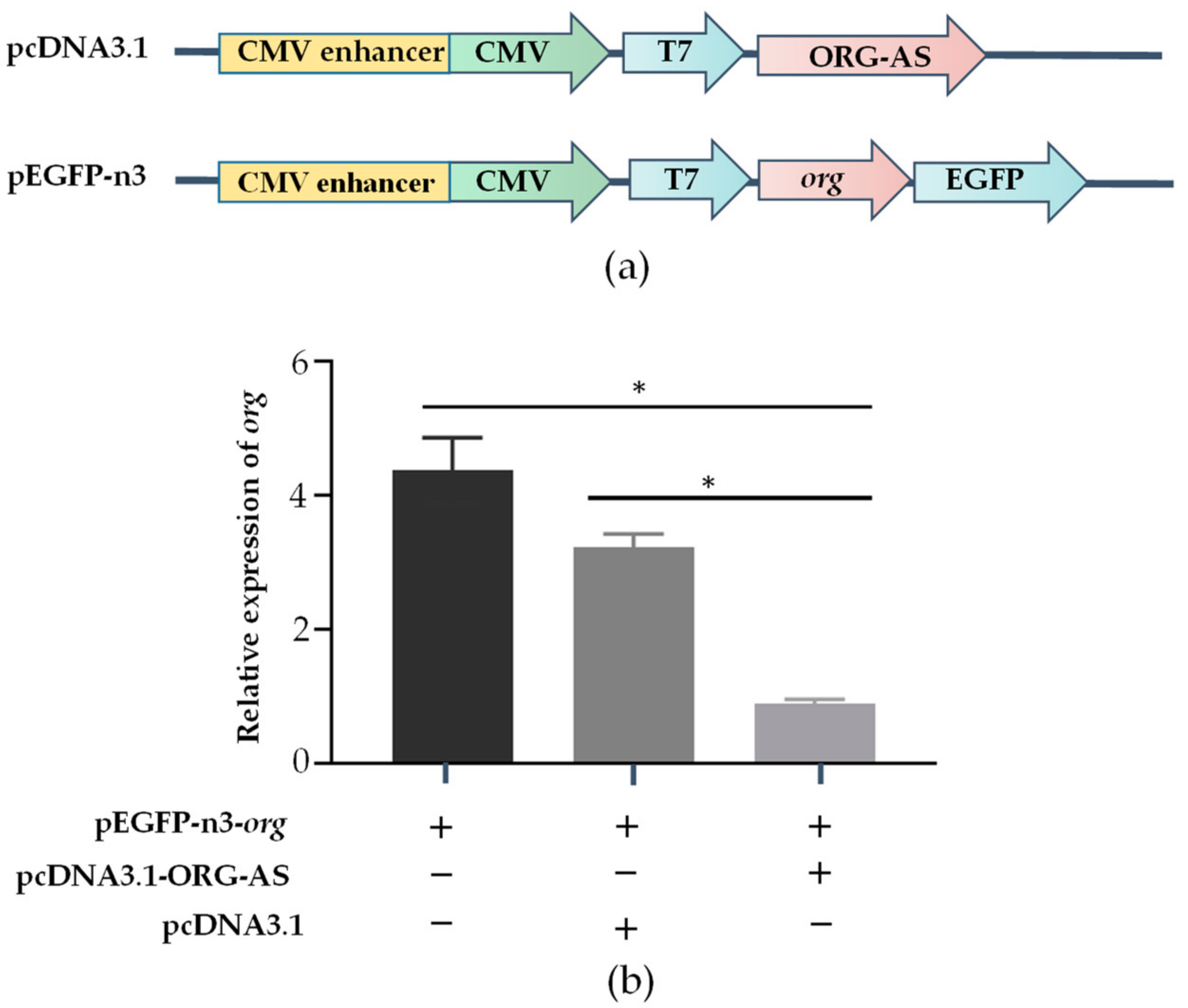
2.5. ORG-AS Negatively Regulates the Expression of org

To determine whether ORG-AS negatively regulates org, we constructed overexpression vectors for org and ORG-AS and then transfected 293T cells (Figure 6a). The qRT-PCR results showed that compared with the org transfection groups, org expression decreased significantly in the ORG-AS and org co-transfection groups. While the expression of org in the ORG-AS and pcDNA3.1 co-transfection groups did not significantly change (Figure 6b). The results indicate that ORG-AS can inhibit the transcription of org.

3. Discussion

We used RACE to clone the cDNA of the org gene from the ovary of C. semilaevis. org contains a 5′-cap and 3′poly-A tail, encoding 221 amino acids, including six exons and five introns. Homology and evolutionary analysis showed that org was only found in fish and was highly conserved. We presumed that org is only a MEG in fish. MEGs activate the embryonic genome by encoding maternal effector complexes, and they degrade rapidly after completing their duties, which promotes the transformation of embryos from a maternal regulatory mechanism to a zygotic regulatory mechanism [26]. In addition, maternal complexes encoded by MEGs are involved in oocyte meiosis and genomic transcriptome activation, and they promote oocyte development [3]. Then, we analyzed the expression pattern and cellular localization of org. The qRT-PCR results showed that org was highly expressed in unfertilized eggs, and after fertilization, the expression abundance decreased significantly after the blastocyst stage. Previous studies have shown that the activation of zygote genes usually occurs around the blastocyst stage in bony fish [27]. These findings are consistent with the expression pattern of org in early embryos. The results indicate that org may play a key role in the development of early embryos and MTZ.
In sexually mature C. semilaevis, the qRT-PCR results showed that org was relatively abundant in the gonads, especially in the ovary. The significant difference in the expression of org between the ovary and testis indicates that org may play an important role in the ovary. In addition, the qRT-PCR results showed that org was barely expressed in the early stage of ovarian development. Then, org was highly expressed at the beginning of reproductive cell maturation into the division period. According to the FISH results, org was mainly localized in the cytoplasm of oocytes. This finding is consistent with the expression patterns of the MEGs vasa, zorg, npm2, and foxr1 in zebrafish [9,25,28,29]. According to the development of oocytes in the ovary, fish ovaries can be divided into three categories: the complete synchronous spawning type, the batch synchronous spawning type, and the asynchronous spawning type. The ovary of C. semilaevis belongs to the asynchronous spawning type; the ovary contains oocytes of different developmental stages [30]. This indicates the continuity of oogenesis in C. semilaevis. Liu et al. also revealed that the oogenesis of C. semilaevis is continuous by coordinating different granulosa cells through single-cell sequencing [31]. Previous studies have shown that the oogenesis of bony fish begins with the differentiation and proliferation of oocytes. Oocytes undergo meiosis and differentiation to form primary oocytes and secondary oocytes, which gather in the ovarian matrix, manage the stage of primary oocytes and secondary oocytes, and finally form eggs [32]. Ma et al. showed that the differentiation of C. semilaevis oocytes begins at 150 dpf. At this time, the primary oocytes begin to disperse in the reproductive epithelium, indicating that some reproductive cells in the ovary begin to enter mature division, marking the beginning of oogenesis [30]. These findings are consistent with the expression pattern of org during ovarian development. The above results suggests that org might function as a MEG and maintain the continuity of oogenesis in the ovary.
In addition, we found that there was a 150 bp reverse complementary sequence between lncRNA ORG-AS and org exon 4, and we predicted the interaction between them by IntaRNA and RNAup. To study the regulatory effect of ORG-AS on org, we analyzed the expression patterns and cellular localization of org and ORG-AS. During the whole embryonic development period, a high level of org was accompanied by a low expression of ORG-AS. Intriguingly, we observed the decrease of org and ORG-AS in the gastrula stage. Both org and ORG-AS were hardly detectable during the early stage of gonad development. The expressions of org and ORG-AS increased rapidly after the onset of oogenesis. More importantly, ORG-AS was expressed earlier than org in the testis and ovary. These results indicate that the expression of ORG-AS might play an important role in regulating the precise level of org. FISH showed that ORG-AS colocalized with org in oocytes. Furthermore, the overexpression of ORG-AS suppressed the expression level of org. LncRNA, as an important epigenetic regulator, plays an important role in gene transcription and post-transcriptional regulation [33]. Previous studies have shown that lncRNAs can not only regulate gene expression through cis-regulation but can also function in trans-regulation by interacting with proteins or RNA molecules [34]. This indicates that the org gene expression was regulated by the trans-regulation function of lncRNA ORG-AS. Our results suggest that high levels of ORG-AS maintain low expression of org and promote the testis development. In the ovary, ORG-AS might bind to the mRNA of org and regulate oogenesis.
In summary, we first identified the MEG org in C. semilaevis and cloned the full-length cDNA of org. The evolutionary analysis showed that org may be a specific MEG in fish. In the early embryonic development of C. semilaevis, org was involved in MTZ as a MEG to ensure the stability of MTZ. During oogenesis, org was highly expressed and mainly localized in the oocyte cytoplasm. According to the localization of org in oocytes, it can be used as a reproductive development marker gene in the oocytes of C. semilaevis. Second, we identified the lncRNA ORG-AS, determined its expression pattern and location, and found that it inhibited org expression through trans-regulation. However, the mechanisms of org in MTZ and oogenesis are still unclear and need further study.

4. Materials and Methods

4.1. Experimental Fish and Tissue Sampling

The Chinese tongue sole animals used in this experiment were all from Huanghai Aquiculture, Ltd. (Yantai, China). The “Guidelines for Experimental Animals” of the Yellow Sea Fisheries Research Institute (YSFRI) (Qingdao, China) were used to collect and process animals. The anatomical collection of the gonads, kidney, intestine, liver, spleen, heart, brain, skin, gill, and stomach of adult (360 days post-fertilization) C. semilaevis was performed. An anatomical analysis of the gonads of C. semilaevis at 180–720 dpf and an anatomical analysis of body parts that contained gonads at 30, 60, 90, and 120 dpf were performed. The early embryos of C. semilaevis were collected at the unfertilized stage, cell differentiation stage, blastocyst stage, proembryo stage, and somite stage. Each anatomical tissue was frozen in liquid nitrogen and stored at −80 °C. A caudal fin sample was collected from each fish; stored in ethanol for the purposes of obtaining genomic DNA and identifying their sex; and then classified according to stage, sex, and tissue.

4.2. Cloning of org

TRIzol was used to extract the RNA from each tissue of adult C. semilaevis and early embryos and dissolve RNA in DEPC water. RNA purification and concentration were determined using a NanoDrop2000 spectrophotometer (Thermo, Waltham, MA, USA). The integrity of the product was detected by 1% agarose gel electrophoresis, and qualified RNA was stored at −80 °C. Then, a Prime Script™ II 1st Strand cDNA Synthesis Kit (TaKaRa, Shiga, Japan) was used for cDNA transcription and synthesis. A lysis method was used to extract genomic DNA from the caudal fin and to identify fish sex using methods and primers recorded in the literature (sex-F and sex-R, Table 1) [35].
With the NCBI database, primers (org-F, org-R; Table 1) were designed according to predicted sequences (Accession No. XM_008307072.3), and PCR was performed using the gonad cDNA as the template for the amplification of org with Repaid DNA polymerase (TaKaRa, Shiga, Japan). In PCR, the following conditions were used: 94 °C for 5 min, followed by 35 cycles of 94 °C for 15 s, 55 °C for 15 s, and 72 °C for 5 s, and then a 72 °C extension for 5 min. A FastPure® Gel DNA Extraction Mini Kit was used to purify the PCR product (Vazyme, Nanjing, China), and the target fragment was ligated into the pEASY-T1 cloning vector (TransGene, Beijing, China). Positive clones were screened and sequenced by Ruibo, Ltd., (Beijing, China) after being transformed into DH5α competent cells (TaKaRa, Shiga, Japan).
After confirming the sequence, we used a SMARTer RACE 5′/3′ Kit to amplify the full length of org (Clontech, Mountain View, CA, USA). Long-end primers in the kit and gene-specific primers (5′GSP, 3′GSP; Table 1) were used for the first round of PCR amplification. The products of the second round of amplification were amplified using the short-end primers in the kit and gene-specific primers (5′NGSP, 3′NGSP; Table 1). PCR products were transformed, linked, and sequenced as described above.

4.3. Real-Time Quantitative PCR

Primers were designed according to the org and ORG-AS sequences, and Cs-18S-F, Cs-18S-R, β-actin-Q-F, and β-actin-Q-R were used as internal reference genes (org-Q-F, org-Q-R, ORG-AS-Q-F, ORG-AS-Q-R, Cs-18S-F, Cs-18S-R, β-actin-Q-F, β-actin-Q-R; Table 1). The cDNA of each tissue was used as a template for qRT-PCR amplification to determine the expression of org and ORG-AS in the tissue. With a reaction volume of 20 μL, qRT-PCR was performed on a rapid real-time PCR system, the ABI 7500, real-time fluorescent quantitative PCR, Lightcycler 480II (Basel, Switzerland), using the QuantiNova™ SYBR Green PCR Kit (Qiagen, Hilden, Germany). For qRT-PCR, the following conditions were used: 95 °C for 2 min, 40 cycles of 95 °C for 5 s, and 60 °C for 10 s. The reaction conditions for the melting curve were 95 °C for 5 s, 60 °C for 1 min, +1 °C/min, and 95 °C for 15 s. Three biological and three technical repeats were required for the qRT-PCR of each tissue. Relative quantitative analysis using the 2−ΔΔCt method and the normalization of org and ORG-AS expression using β-actin and Cs-18S rRNA were performed. For an analysis of the data, GraphPad Prism 6 was used, along with a t test to assess statistical significance (GraphPad Software, La Jolla, CA, USA).

4.4. Fluorescence In Situ Hybridization of org and ORG-AS

The ovaries at 6 mpf and 1.5 ypf were stored in 4% PFA, fixed in paraffin wax, and sliced into 3 μm sections. org and ORG-AS-specific fluorescent in situ hybridization probes were designed; org was assessed using FISH with a 5′Cy3-labeled fluorescent probe, and ORG-AS was assessed using FISH with a 5′6-FAM-labeled fluorescent probe (Table 1). Fluorescence in situ hybridization was performed with an RNA FISH kit (Gene Pharma, Shanghai, China) according to the manufacturer’s requirements. The images were acquired with a laser confocal microscope (Olympus, Tokyo, Japan) and analyzed.

4.5. Cell Culture, Vectors, and Transfection

ORG-AS and org overexpression vectors were established to confirm the role of ORG-AS. The primers shown in Table 1 were used for PCR amplification of the ORF CDS region and the binding site of ORG-AS. The ORG-AS and pcDNA3.1+ vectors (Invitrogen, Carlsbad, CA, USA) were cut by EcoRI and XbaI, and the org and pEGFP-n3 vectors were cut by EcoRI and KpnI. pcDNA3.1-ORG-AS and pEGFP-n3-org were ligated with T4 DNA ligase (NEB, Inc., Ipswich, MA, USA). The connected plasmids were transformed into Escherichia coli, pcDNA3.1-ORG-AS was coated on the plate containing ampicillin, and pEGFP-n3-org was coated on the plate containing Kana. After 12 h, a positive clone was selected for colony PCR, and the positive clones were sent for sequencing to verify whether the recombinant plasmids were connected correctly. The recombinant plasmid was extracted using an EndoFree Mini Plasmid Kit II (Tiangen Biotech, Beijing, China) for subsequent transfection experiments.
The 293T cell line was purchased from the Shanghai Institute of Cell Biology. L-15 Medium Leibovitz (Thermo Fisher, Carlsbad, CA, USA) was used to culture the cells at 37 °C with 5% CO2. Cells logarithmically grew to 80–90% density in medium, were digested with 0.25% trypsin, and were transferred to 24-well cell culture plates. Then, pEGFP-n3-org and pcDNA3.1-ORG-AS vectors were cotransfected into 293T cells using Lipofectamine™ 3000 (Thermo Fisher, Invitrogen, Carlsbad, CA, USA). The negative control group comprised pEGFP-n3-org and pcDNA3.1 vector-cotransfected 293T cells, and the control group comprised pEGFP-n3-org vector-transfected 293T cells. Cells were collected 48 h after transfection, and RNA was extracted as mentioned above. The expression of org was detected using qRT-PCR.

4.6. Sequence Analysis

RNA/RNA binding sites were predicted using IntaRNA (accessed on 13 March 2022, http://rna.informatik.uni-freiburg.de/IntaRNA/Input.jsp) and RNAup (accessed on 13 March 2022, http://rna.tbi.Univie.ac.at//cgi-bin/RNAWebSuite/RNAup.cgi). Gene homology searches were performed using the Blast tool in NCBI (accessed on 7 April 2022, http://www.ncbi.nlm.nih.gov/BLAST/). Multiple sequence alignment was performed using DNAMAN (accessed on 4 June 2022, http://www.clustal.org/omega/). A NJ phylogenetic tree was constructed using the MEGA maximum likelihood method.

4.7. Statistical Analysis

Experimental data are shown as the mean ± standard error and were obtained in triplicate with three independent experiments. The data were analyzed using t tests and plotted with the GraphPad Prism 6 software package (GraphPad Software, San Diego, CA, USA). p values of ≤0.05 were considered statistically significant (* p < 0.05, ** p < 0.01, *** p < 0.001).

5. Conclusions

Our study showed that org, as a MEG only present in fish, plays an important role in the early embryonic development and oogenesis of C. semilaevis. The lncRNA ORG-AS negatively regulates org expression through trans-regulation. These studies broaden our knowledge of the function of MEGs in embryonic development and oogenesis and further enrich the knowledge of lncRNAs in regulating genes.

Author Contributions

Conceptualization, C.S., C.L. and X.Z.; methodology, X.Z., L.T., Q.W. and B.F.; validation, X.Z., Q.L. and W.M.; investigation, X.Z.; validation, X.Z.; writing—original draft preparation, X.Z.; writing—review and editing, X.Z., C.S. and Q.W.; supervision, C.S. All authors have read and agreed to the published version of the manuscript.

Funding

This study was supported the National Nature Science Foundation of China (grant numbers 31722058 and 31472269); the AoShan Talents Cultivation Program Supported by Qingdao National Laboratory for Marine Science and Technology (grant number 2017ASTCP-ES06); the Taishan Scholar Project Fund of Shandong of China to C.S.; the National Ten-Thousands Talents Special Support Program to C.S.; the Central Public-interest Scientific Institution Basal Research Fund, CAFS (grant number No.2020TD19); and the China Agriculture Research System (CARS-47-G03); the project by National Marine Genetic Resource Center.

Institutional Review Board Statement

The animal study protocol was approved by the Institutional Animal Care and Use Committee (IACUC) of the Yellow Sea Fisheries Research Institute (CAFS) (Qingdao, China) (Approval No.: YSFRI-2022022).

Informed Consent Statement

Not applicable.

Conflicts of Interest

The authors declare no conflict of interest.

References

  1. Gouw, J.W.; Pinkse, M.W.; Vos, H.R.; Moshkin, Y.; Verrijzer, C.P.; Heck, A.J.; Krijgsveld, J. In vivo stable isotope labeling of fruit flies reveals post-transcriptional regulation in the maternal-to-zygotic transition. Mol. Cell Proteom. 2009, 8, 1566–1578. [Google Scholar] [CrossRef] [PubMed] [Green Version]
  2. Tadros, W.; Lipshitz, H.D. The maternal-to-zygotic transition: A play in two acts. Development 2009, 136, 3033–3042. [Google Scholar] [CrossRef] [PubMed] [Green Version]
  3. Yu, C.; Ji, S.-Y.; Sha, Q.-Q.; Dang, Y.; Zhou, J.-J.; Zhang, Y.-L.; Liu, Y.; Wang, Z.-W.; Hu, B.; Sun, Q.-Y. BTG4 is a meiotic cell cycle–coupled maternal-zygotic-transition licensing factor in oocytes. Nat. Struct. Mol. Biol. 2016, 23, 387–394. [Google Scholar] [CrossRef] [PubMed]
  4. Nüsslein-Volhard, C.; Lohs-Schardin, M.; Sander, K.; Cremer, C. A dorso-ventral shift of embryonic primordia in a new maternal-effect mutant of Drosophila. Nature 1980, 283, 474–476. [Google Scholar] [CrossRef] [PubMed]
  5. Bowerman, B. 3 Maternal Control of Pattern Formation in Early Caenorhabditis elegans Embryos. Curr. Top. Dev. Biol. 1998, 39, 73–117. [Google Scholar] [PubMed]
  6. Heasman, J. Maternal determinants of embryonic cell fate. Semin. Cell Dev. Biol. 2006, 17, 93–98. [Google Scholar] [CrossRef] [PubMed]
  7. Wu, X.; Viveiros, M.M.; Eppig, J.J.; Bai, Y.; Fitzpatrick, S.L.; Matzuk, M.M. Zygote arrest 1 (Zar1) is a novel maternal-effect gene critical for the oocyte-to-embryo transition. Nat. Genet. 2003, 33, 187–191. [Google Scholar] [CrossRef] [PubMed]
  8. Rong, Y.; Ji, S.-Y.; Zhu, Y.-Z.; Wu, Y.-W.; Shen, L.; Fan, H.-Y. ZAR1 and ZAR2 are required for oocyte meiotic maturation by regulating the maternal transcriptome and mRNA translational activation. Nucleic Acids Res. 2019, 47, 11387–11402. [Google Scholar] [CrossRef] [PubMed]
  9. Yoon, C.; Kawakami, K.; Hopkins, N. Zebrafish vasa homologue RNA is localized to the cleavage planes of 2-and 4-cell-stage embryos and is expressed in the primordial germ cells. Development 1997, 124, 3157–3165. [Google Scholar] [CrossRef] [PubMed]
  10. Köprunner, M.; Thisse, C.; Thisse, B.; Raz, E. A zebrafish nanos-related gene is essential for the development of primordial germ cells. Genes Dev. 2001, 15, 2877–2885. [Google Scholar] [CrossRef]
  11. Tan, C.-H.; Lee, T.-C.; Weeraratne, S.D.; Korzh, V.; Lim, T.-M.; Gong, Z. RETRACTED: Ziwi, the zebrafish homologue of the Drosophila piwi: Co-localization with vasa at the embryonic genital ridge and gonad-specific expression in the adults. Gene Expr. Patterns 2002, 2, 257–260. [Google Scholar] [CrossRef]
  12. Gosden, R. Oogenesis as a foundation for embryogenesis. Mol. Cell Endocrinol. 2002, 186, 149–153. [Google Scholar] [CrossRef]
  13. Rodrigues, P.; Limback, D.; McGinnis, L.K.; Plancha, C.E.; Albertini, D.F. Oogenesis: Prospects and challenges for the future. J. Cell Physiol. 2008, 216, 355–365. [Google Scholar] [CrossRef] [PubMed]
  14. Sánchez, F.; Smitz, J. Molecular control of oogenesis. Biochim. Biophys. Acta (BBA)-Mol. Basis Dis. 2012, 1822, 1896–1912. [Google Scholar] [CrossRef] [PubMed] [Green Version]
  15. Ma, P.; Pan, H.; Montgomery, R.L.; Olson, E.N.; Schultz, R.M. Compensatory functions of histone deacetylase 1 (HDAC1) and HDAC2 regulate transcription and apoptosis during mouse oocyte development. Proc. Natl. Acad. Sci. USA 2012, 109, E481–E489. [Google Scholar] [CrossRef] [PubMed] [Green Version]
  16. Lu, Y.-F.; Liu, Q.; Liu, K.-Q.; Wang, H.-Y.; Li, C.-H.; Wang, Q.; Shao, C.-W. Identification of global alternative splicing and sex-specific splicing via comparative transcriptome analysis of gonads of Chinese tongue sole (Cynoglossus semilaevis). Zool. Res. 2022, 43, 319. [Google Scholar] [CrossRef]
  17. Zhu, L.-Y.; Zhu, Y.-R.; Dai, D.-J.; Wang, X.; Jin, H.-C. Epigenetic regulation of alternative splicing. Am. J. Cancer Res. 2018, 8, 2346. [Google Scholar] [PubMed]
  18. Ulitsky, I.; Bartel, D.P. lincRNAs: Genomics, evolution, and mechanisms. Cell 2013, 154, 26–46. [Google Scholar] [CrossRef] [Green Version]
  19. Dahariya, S.; Paddibhatla, I.; Kumar, S.; Raghuwanshi, S.; Pallepati, A.; Gutti, R.K. Long non-coding RNA: Classification, biogenesis and functions in blood cells. Mol. Immunol. 2019, 112, 82–92. [Google Scholar] [CrossRef] [PubMed]
  20. Bridges, M.C.; Daulagala, A.C.; Kourtidis, A. LNCcation: lncRNA localization and function. J. Cell Biol. 2021, 220, e202009045. [Google Scholar] [CrossRef] [PubMed]
  21. Gomez, J.A.; Wapinski, O.L.; Yang, Y.W.; Bureau, J.F.; Gopinath, S.; Monack, D.M.; Chang, H.Y.; Brahic, M.; Kirkegaard, K. The NeST long ncRNA controls microbial susceptibility and epigenetic activation of the interferon-gamma locus. Cell 2013, 152, 743–754. [Google Scholar] [CrossRef] [PubMed] [Green Version]
  22. Feng, B.; Li, S.; Wang, Q.; Tang, L.; Huang, F.; Zhang, Z.; Mahboobe, S.; Shao, C. lncRNA DMRT2-AS acts as a transcriptional regulator of dmrt2 involving in sex differentiation in the Chinese tongue sole (Cynoglossus semilaevis). Comp. Biochem. Physiol. B Biochem. Mol. Biol. 2021, 253, 110542. [Google Scholar] [CrossRef]
  23. Kato, Y.; Perez, C.A.G.; Ishak, N.S.M.; Nong, Q.D.; Sudo, Y.; Matsuura, T.; Wada, T.; Watanabe, H. A 5′ UTR-Overlapping LncRNA Activates the Male-Determining Gene doublesex1 in the Crustacean Daphnia magna. Curr. Biol. 2018, 28, 1811–1817.e1814. [Google Scholar] [CrossRef] [PubMed] [Green Version]
  24. Liao, X.; Ma, H.Y.; Xu, G.B.; Shao, C.W.; Tian, Y.S.; Ji, X.S.; Yang, J.F.; Chen, S.L. Construction of a genetic linkage map and mapping of a female-specific DNA marker in half-smooth tongue sole (Cynoglossus semilaevis). Mar. Biotechnol. 2009, 11, 699–709. [Google Scholar] [CrossRef] [PubMed]
  25. Dai, L.; Ma, W.; Li, J.; Xu, Y.; Li, W.; Zhao, Y.; Deng, F. Cloning and characterization of a novel oocyte-specific gene zorg in zebrafish. Theriogenology 2009, 71, 441–449. [Google Scholar] [CrossRef]
  26. Li, L.; Zheng, P.; Dean, J. Maternal control of early mouse development. Development 2010, 137, 859–870. [Google Scholar] [CrossRef] [PubMed] [Green Version]
  27. Lee, M.T.; Bonneau, A.R.; Takacs, C.M.; Bazzini, A.A.; DiVito, K.R.; Fleming, E.S.; Giraldez, A.J. Nanog, Pou5f1 and SoxB1 activate zygotic gene expression during the maternal-to-zygotic transition. Nature 2013, 503, 360–364. [Google Scholar] [CrossRef] [PubMed] [Green Version]
  28. Bouleau, A.; Desvignes, T.; Traverso, J.M.; Nguyen, T.; Chesnel, F.; Fauvel, C.; Bobe, J. Maternally inherited npm2 mRNA is crucial for egg developmental competence in zebrafish. Biol. Reprod. 2014, 91, 43. [Google Scholar] [CrossRef] [PubMed]
  29. Cheung, C.T.; Patinote, A.; Guiguen, Y.; Bobe, J. foxr1 is a novel maternal-effect gene in fish that is required for early embryonic success. PeerJ 2018, 6, e5534. [Google Scholar] [CrossRef]
  30. Ma, X.; Liu, X.; Wen, H.; Xu, Y.; Zhang, L. Histological observation on gonadal sex differentiation in Cynoglossus semilaevis Günther. Mar. Fish Res. 2006, 27, 55–61. [Google Scholar]
  31. Liu, X.; Huang, Y.; Tan, F.; Wang, H.-Y.; Chen, J.-Y.; Zhang, X.; Zhao, X.; Liu, K.; Wang, Q.; Liu, S. Single-Cell Atlas of the Chinese Tongue Sole (Cynoglossus semilaevis) Ovary Reveals Transcriptional Programs of Oogenesis in Fish. Front. Cell Dev. Biol. 2022, 10, 828124. [Google Scholar] [CrossRef]
  32. Andrade, R.; Bazzoli, N.; Rizzo, E.; Sato, Y. Continuous gametogenesis in the neotropical freshwater teleost, Bryconops affinis (Pisces: Characidae). Tissue Cell 2001, 33, 524–532. [Google Scholar] [CrossRef] [PubMed] [Green Version]
  33. Zhang, B.; Arun, G.; Mao, Y.S.; Lazar, Z.; Hung, G.; Bhattacharjee, G.; Xiao, X.; Booth, C.J.; Wu, J.; Zhang, C.; et al. The lncRNA Malat1 is dispensable for mouse development but its transcription plays a cis-regulatory role in the adult. Cell Rep. 2012, 2, 111–123. [Google Scholar] [CrossRef] [PubMed] [Green Version]
  34. Kopp, F.; Mendell, J.T. Functional classification and experimental dissection of long noncoding RNAs. Cell 2018, 172, 393–407. [Google Scholar] [CrossRef] [PubMed] [Green Version]
  35. Liu, Y.; Chen, S.; Gao, F.; Meng, L.; Hu, Q.; Song, W.; Shao, C.; Lv, W. SCAR-transformation of sex-specific SSR marker and its application in half-smooth tongue sole (Cynoglossus semiliaevis). J. Agric. Biotechnol. 2014, 22, 787–792. [Google Scholar]
Figure 1. Full-length cDNA sequence and amino acid sequence of org gene. The ORF of org cDNA encoded 221 amino acids. The amino acid sequence is in uppercase letters. The red box represents the start codon and stop codon of org, and the stop codon is indicated by an asterisk (*).
Ijms 23 08605 g001
Figure 2. Homology analysis and phylogenetic tree of ORG proteins in bony fish. (a) DNAMAN was used to perform multisequence alignment of org amino acid sequences. Black boxes indicate sequence identity between amino acid sequences, while gray boxes indicate more than 75% similarity between amino acid sequences. Note: S. dumerili (Sequence ID: XP_022615158.1), L. calcarifer (Sequence ID: XP_018522849.1), S. dorsalis (Sequence ID: XP_023263745.1), P. olivaceus (Sequence ID: XP_019936103.1), A. testudineus (Sequence ID: XP_026200660.1), and B. splendens (Sequence ID: XP_029003277.1); (b) Phylogenetic tree based on the org amino acid sequence, using the neighbor-joining method. The number displayed on the branch node represents the guiding value (%). The C. semilaevis org is marked in red color.
Ijms 23 08605 g002
Figure 3. org and ORG-AS interaction site diagram. (a) IntaRNA results showed ORG-AS binding sites to org; (b) RNAup showed the interaction between nucleotides 86–110 of ORG-AS and nucleotides 348–372 of org. ORG-AS is represented by a black line; org is represented by a red line; (c) Diagram of ORG-AS and org interactions.
Ijms 23 08605 g003
Figure 4. Expression of org and ORG-AS in C. semilaevis. (a) org and ORG-AS expression patterns in early embryos, and org and ORG-AS expression levels normalized to 18S rRNA expression; (b) Expression of org and ORG-AS in sexually mature C. semilaevis, and org and ORG-AS expression levels normalized to β-actin expression; (c) Expression patterns of org and ORG-AS during gonad development in C. semilaevis, and org and ORG-AS expression levels normalized to β-actin expression. The mean ± SEM from three independent individuals (n = 3) is shown. (*** p < 0.001).
Ijms 23 08605 g004
Figure 5. org and ORG-AS localization in the ovary of C. semilaevis at 6 mpf and 1.5 ypf. (a) Oocyte nuclei and somatic nuclei were stained with DAPI in 6 mpf ovaries; (b) Localization of org detected using 5′Cy3-labeled fluorescent probe in 6 mpf ovaries; (c) Localization of ORG-AS detected using 5′6-FAM-labeled fluorescent probe in 6 mpf ovaries; (e) Somatic nuclei were stained with DAPI in 1.5 ypf ovaries; (f) Localization of org detected using 5′Cy3-labeled fluorescent probe in 1.5 ypf ovaries; (g) Localization of ORG-AS detected using 5′6-FAM-labeled fluorescent probe in 1.5 ypf ovaries; (d,h) Combination of org, ORG-AS, and nucleus localization. I: phase I oogonia; II: phase II oocyte; III: early phase III oocyte.
Ijms 23 08605 g005
Figure 6. The effect of ORG-AS overexpression on org expression levels. (a) Construction of pcDNA3.1-ORG-AS and pEGFP-n3-org overexpression vectors; (b) After transfection of 293T cells, the expression level of org was detected and normalized to ACTINB expression, and the mean ± SEM from three independent individuals (n = 3) is shown. * p < 0.05.
Ijms 23 08605 g006
Table 1. Primers used for cloning and gene expression analysis.
Primer NameSequence (5′-3′)
org-FCCAGCAGGCTTCAGAACCACCG
org-RCAGTAACCAACATACGCAGGAA
org-3-GSPCCCTGACACTGCCGTGTTTGCCCACTAGA
org-3-NGSPACTGGTCACTTGACACTTGCTCG
org-5-GSPAGCCCAGCACCCACCAAACTCCACGGT
org-5-NGSPCCACTCTGTCCTCCGTGTTCTC
org-Q-FCAGCAGGAGGAGAACACGGA
org-Q-RACCAAACTCCACGGTATGCC
ORG-AS-Q-FCACTTCGGAGAACACTGGCG
ORG-AS-Q-RTGGGGCTCTTCTATTGGTCG
β-actin-Q-FGCTGTGCTGTCCCTGTA
β-actin-Q-RGAGTAGCCACGCTCTGTC
Cs-18S rRNA-FGGTCTGTGATGCCCTTAGATGTC
Cs-18S rRNA-RAGTGGGGTTCAGCGGGGTTAC
hACTINB-Q-FGATGATATCGCCGCGCTCGT
hACTINB-Q-RGTAGATGGGCACAGTGTGGGTG
org-EcoRI-FCGGAATTCATGACCATGCAAATCCTC
org-KpnI-RCGGGTACCAACACCAGGCAAAGAGT
ORG-AS-KpnI-FCGGAATTCCTGGAGTGAGAGCCCGG
ORG-AS-EcoRI-RGCTCTAGAACGCTTGGTTTGAACTTGATG
org-5′Cy3TGGGCTCGTAGTCCGGATCTTCCTCTGGGCC
ORG-AS-5′6-FAMCCTCATAGACTCTTCAGGACAGAGTGGTCAGGAAGG
sex-FCCTAAATGATGGATGTAGATTCTGTC
sex-RGATCCAGAGAAAATAAACCCAGG
Publisher’s Note: MDPI stays neutral with regard to jurisdictional claims in published maps and institutional affiliations.

