Article

Synovial Fluid Regulates the Gene Expression of a Pattern of microRNA via the NF-κB Pathway: An In Vitro Study on Human Osteoarthritic Chondrocytes

Rheumatology Unit, Department of Medicine, Surgery and Neuroscience, Azienda Ospedaliera Universitaria Senese, Policlinico Le Scotte, 53100 Siena, Italy
*
Author to whom correspondence should be addressed.
Current address: Rheumatology Unit, Department of Medicine, Surgery and Neuroscience, University of Siena, 53100 Siena, Italy.
Int. J. Mol. Sci. 2022, 23(15), 8334; https://doi.org/10.3390/ijms23158334
Submission received: 2 July 2022 / Revised: 22 July 2022 / Accepted: 26 July 2022 / Published: 28 July 2022
(This article belongs to the Special Issue Osteoarthritis 2.0: From Molecular Pathways to Therapeutic Advances)

Abstract

:
Synovial fluid (SF) represents the primary source of nutrients of articular cartilage and is implicated in maintaining cartilage metabolism. We investigated the effects of SF, from patients with osteoarthritis (OA), rheumatoid arthritis (RA), and controls, on a pattern of microRNA (miRNA) in human OA chondrocytes. Cells were stimulated with 50% or 100% SF for 24 h and 48 h. Apoptosis and superoxide anion production were detected by cytometry; miRNA (34a, 146a, 155, 181a), cytokines, metalloproteinases (MMPs), type II collagen (Col2a1), antioxidant enzymes, B-cell lymphoma (BCL)2, and nuclear factor (NF)-κB by real-time PCR. The implication of the NF-κB pathway was assessed by the use of NF-κB inhibitor (BAY-11-7082). RA and OA SF up-regulated miR-34a, -146a, -155, -181a, interleukin (IL)-1β, IL-6, tumor necrosis factor (TNF)-α, MMP-1, MMP-13, and ADAMTs-5 gene expression, while it down-regulated Col2a1. Pathological SF also induced apoptosis, reduced viability, and decreased BCL2 mRNA, whereas it increased superoxide anions, the expression of antioxidant enzymes, p65 and p50 NF-κB. Opposite and positive results were obtained with 100% control SF. Pre-incubation with BAY-11-7082 counteracted SF effects on miRNA. We highlight the role of the SF microenvironment in regulating some miRNA involved in inflammation and cartilage degradation during OA and RA, via the NF-κB pathway.

1. Introduction

Osteoarthritis (OA) is the most common chronic and degenerative musculoskeletal disorder and the leading cause of disability and impairment in middle-aged and older individuals [1,2]. The disease involves the entire joint and is characterized by progressive cartilage degradation, osteophytes formation, and synovial inflammation [3,4]. Rheumatoid arthritis (RA) is considered a chronic systemic, inflammatory, and progressive disease; it affects 1% of individuals aged 20–40 years worldwide, causing lifelong disability [5,6,7]. The etiology of RA is multifactorial and remains largely unknown; the disorder primarily involves the joints, leading to marked synovial inflammation, cartilage destruction, and bone erosion [8,9].
Progressive cartilage degradation and loss of extracellular matrix (ECM) elements, together with different grades of inflammation, are important hallmarks of both OA and RA diseases.
Synovial fluid (SF) represents the primary source of joint lubricants and nutrients of articular cartilage, and it is mainly implicated in the maintenance of cartilage metabolism and homeostasis. Inflammatory and degenerative processes occurring in OA and RA induced modification in the molecular and cellular composition of SF. Indeed, in these conditions, it becomes less viscous and loses its unique viscoelastic properties due to a reduction in the molecular weight and concentration of hyaluronic acid (HA), as well as due to changes in cytokines, enzymes profiles, and inflammatory white blood cells (WBCs) concentrations [10,11,12,13,14]. In particular, in RA and OA, the SF is characterized by an increased content of inflammatory mediators (prostaglandin E2, leukotriene B4), cytokines (interleukin (IL)-1β, IL-6, and tumor necrosis factor (TNF)-α), nitric oxide (NO), and reactive oxygen species (ROS), released from chondrocytes, synoviocytes, and WBCs, which contributes to articular cartilage degradation [3,5,8,15,16].
At present, only a few studies have investigated the biological effects of SF on cartilage metabolism, showing that SF from RA and OA patients stimulated pro-inflammatory and catabolic processes in human primary chondrocytes [17,18,19,20,21], while SF taken from human healthy joints seemed to have a chondroprotective role [22,23,24]. However, the results remain still limited and need to be further elucidated.
To the best of our knowledge, there is no evidence reporting the biological activities of SF on the microRNA (miRNA) expression profile. MiRNA are a class of small non-coding RNAs molecules, around 22 nucleotides long, which regulate gene expression by binding specific sequences within target messenger RNA (mRNA). A large number of evidence demonstrated that some miRNA are differentially expressed in OA cartilage samples and synovial tissue of OA and RA patients, confirming their role in the development and progression of these pathological conditions [25,26,27]. Some of the dysregulated miRNA target genes encoding for proteins implicated in extracellular matrix remodeling, pro-inflammatory activities, and redox balance in OA chondrocytes and RA fibroblast-like synoviocytes [27,28,29].
In the present study, we investigated the biological effects of SF taken from patients with OA or RA compared with control SF, in the regulation of some miRNA, cartilage metabolism, and inflammation in human primary OA chondrocytes. In this regard, we analyzed cell viability, the ratio of apoptosis, and the gene expression of miR-34a, miR-146a, miR-155, and miR-181a, the main matrix-degrading enzymes and type II collagen alpha 1 chain (Col2a1), and a pattern of cytokines. In addition, the production of mitochondrial superoxide anion and the mRNA levels of antioxidant enzymes were also evaluated. Finally, the possible mechanism underlying SF effects was assessed by analysis of the nuclear factor (NF)-κB pathway.

2. Results

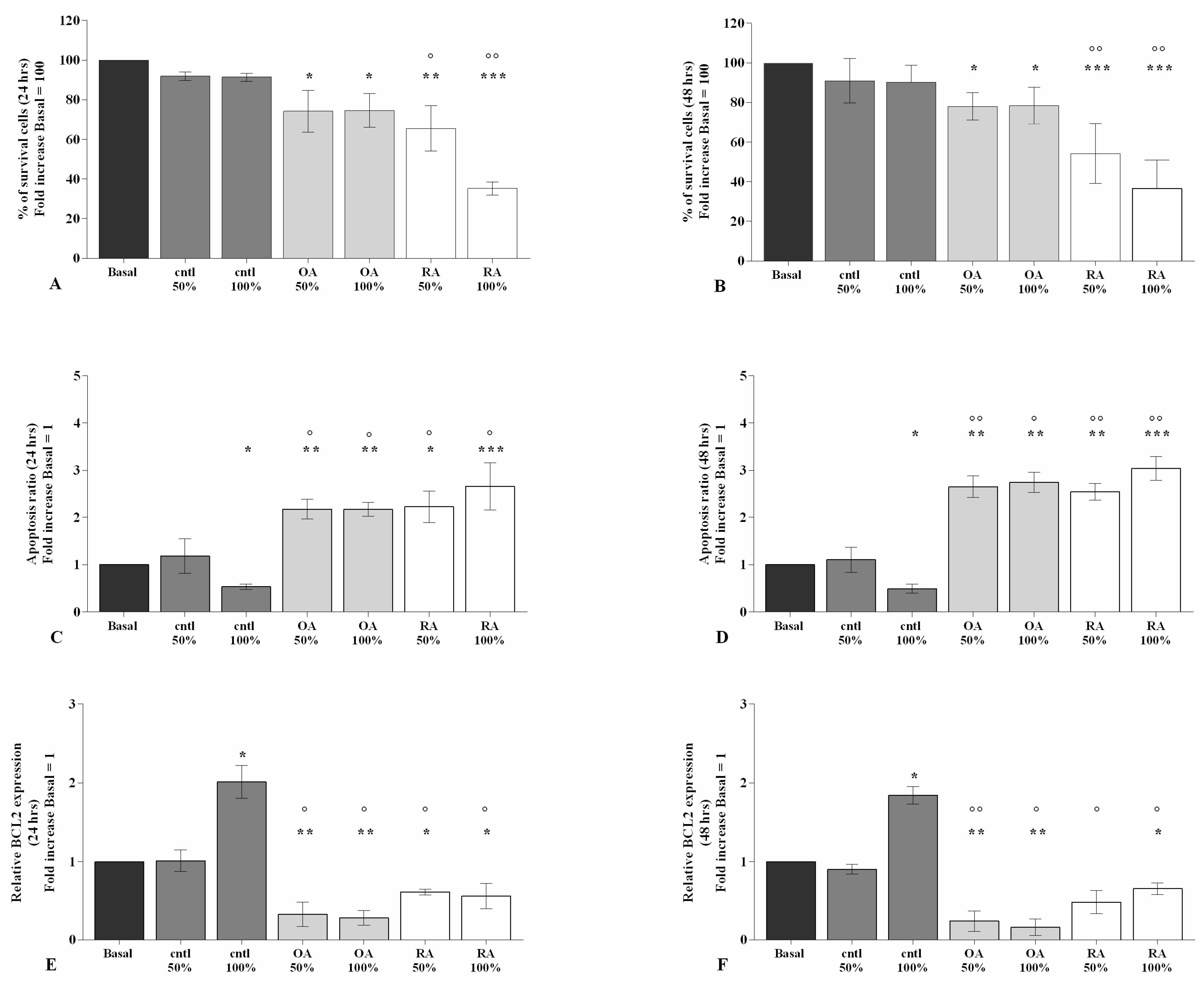
2.1. SF Modulates Chondrocytes Viability and Apoptosis

Figure 1 and Figure S1 show the effects of the stimulus of 50% or 100% SF taken from patients with OA, RA, and controls, for 24 h and 48 h, on cell viability and apoptosis ratio evaluated by MTT assay and flow cytometry.
After 24 h and 48 h of stimulus of the cells with 50% or 100% OA SF, the percentage of survival (p < 0.05) and the gene levels of BCL2 (p < 0.01) were significantly reduced, whereas an increase in apoptosis ratio (p < 0.01) was observed. Similar results were found when our cultures were stimulated with 50% or 100% RA SF (p < 0.05, p < 0.01, p < 0.001) (Figure 1A–F). Furthermore, viability and apoptosis were significantly affected by the stimulus with 50% or 100% OA and RA SF with respect to control SF (p < 0.05, p < 0.01); no changes were found when OA SF was compared with RA SF (Figure 1A–F).
Surprisingly, the incubation of the cells with control SF showed a significant up-regulation of the gene expression of the anti-apoptotic marker BCL2 (p < 0.05), along with a decrease in apoptosis (p < 0.05), when tested at 100% concentration, in comparison to the basal condition (Figure 1A–F).

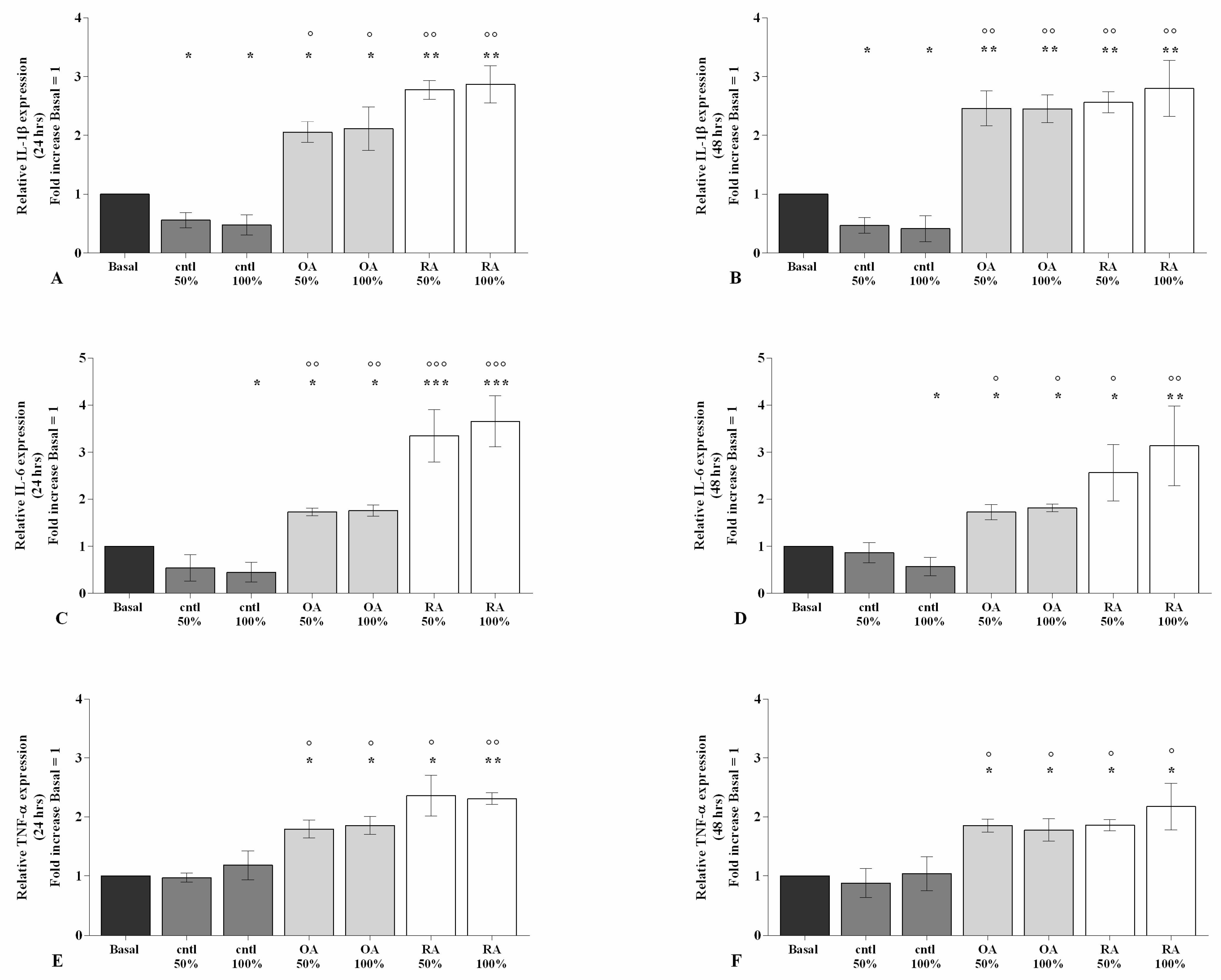
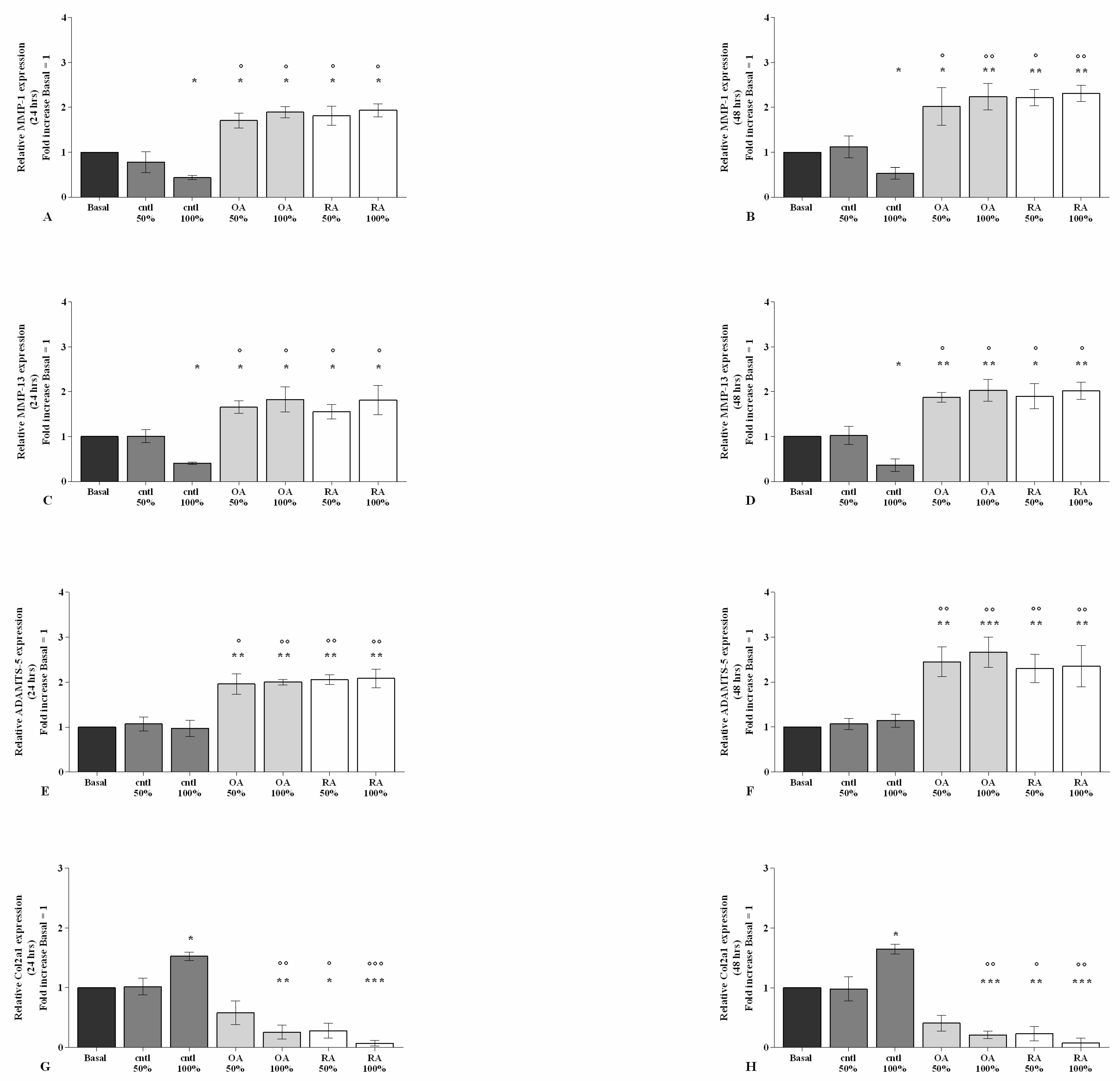
2.2. SF Regulates Inflammation and Chondrocyte Metabolism

The stimulus of chondrocytes with 50% or 100% OA and RASF significantly increased the gene expression of IL-1β, IL-6, and TNF-α (p < 0.05, p < 0.01, p < 0.001), at both time-points, in comparison to the basal condition (Figure 2A–F). This effect was also reported when OA and RA SF were compared with control SF (p < 0.05, p < 0.01) (Figure 2A–F), while no modifications of the pathological SF on cytokine regulation were observed.
On the contrary, when the cells were incubated with control SF, a significant down-regulation of the transcriptional levels of IL-1β at both 50% and 100% concentrations (p < 0.05), as well as that of IL-6 when tested at 100% concentration (p < 0.05), were found in comparison to the baseline (Figure 2A–F).
To examine the maintenance of cartilage integrity and structure under SF condition, the main matrix degrading enzymes and matrix components were tested using real-time PCR (Figure 3).
The incubation of chondrocytes with 100% control SF demonstrated a significant reduction in MMP-1 and MMP-13 (p < 0.05) and an increase in Col2a1 (p < 0.05) gene expression compared to basal condition; no detectable changes were observed for ADAMTS-5 (Figure 3A–H). On the other hand, 24 h and 48 h of stimulus with 50% or 100% OA SF significantly increased the gene levels of MMP-1, MMP-13, and ADAMTS-5 (p < 0.05, p < 0.01), whereas it decreased the levels of Col2a1 (p < 0.01, p < 0.001) in comparison to the baseline; 50% or 100% RA SF induced similar effects on the expression of matrix degrading enzymes and Col2a1 (p < 0.05, p < 0.01, p < 0.001) (Figure 3A–H). In addition, OA and RA SF significantly affected the regulation of cartilage turnover when compared to control SF (p < 0.05, p < 0.01). Conversely, no differences were found when compared to each other (Figure 3A–F).

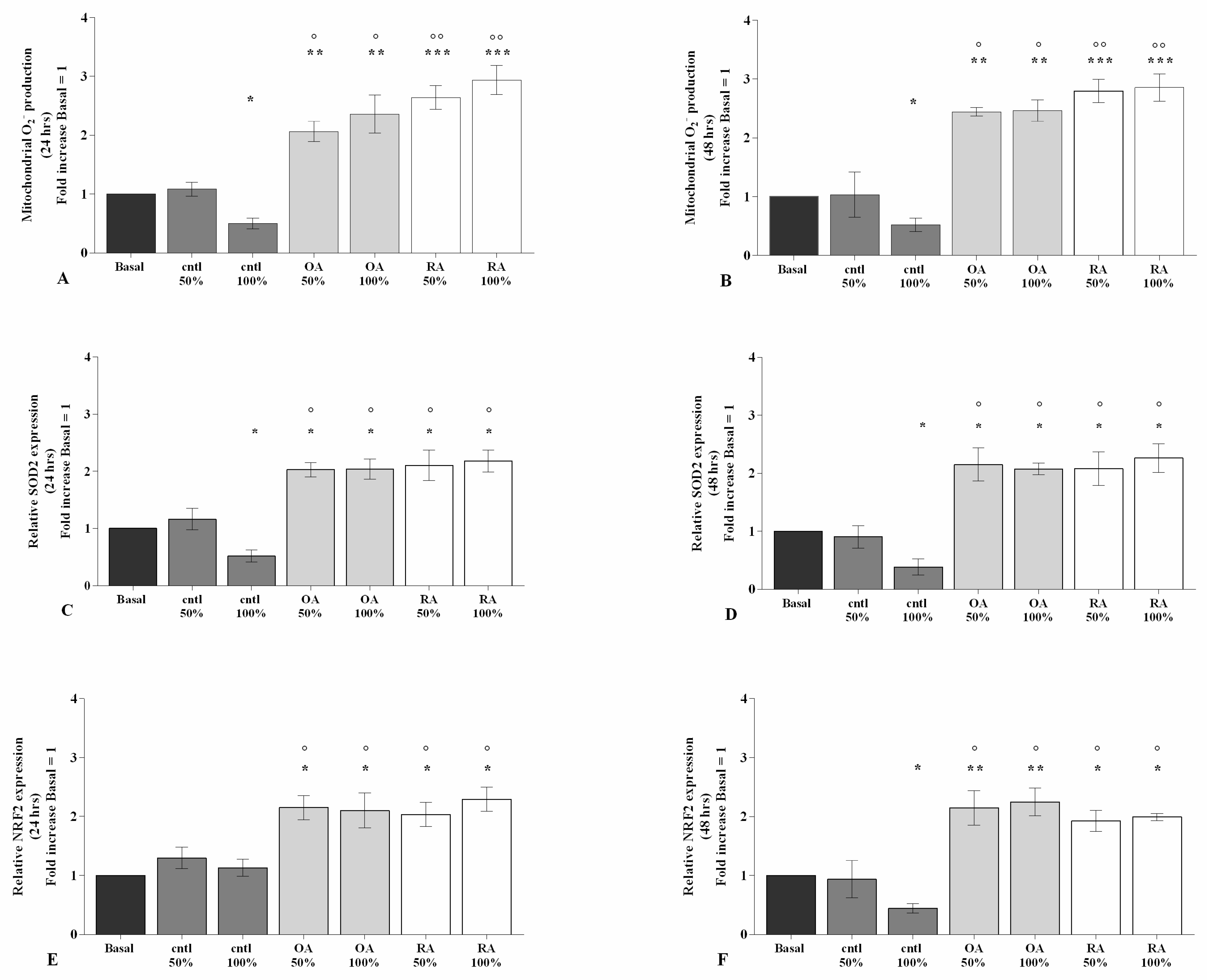
2.3. SF Affects Oxidative Stress Balance

The potential effect of SF on the regulation of oxidant/antioxidant balance is reported in Figure 4 and Figure S2.
Flow cytometry and PCR analyses demonstrated that 100% control SF significantly limited the production of mitochondrial superoxide anion (p < 0.05) and the gene expression of the antioxidant enzyme SOD-2 (p < 0.05), at both 24 h and 48 h of stimulus, and of transcriptional factor NRF2 (p < 0.05) at 48 h, with respect to basal condition (Figure 4A–F). On the contrary, 50% or 100% OA and RA SF induced a significant increase in superoxide anion production (p < 0.01, p < 0.001), as well as in the gene expression of SOD-2 (p < 0.05, p < 0.01) and NRF2 (p < 0.05, p < 0.01) gene expression (Figure 4A–F). OA and RA SF also promoted the oxidative stress status when compared to control SF, at both analyzed time points (p < 0.05, p < 0.01), with no differences between them (Figure 4A–F).

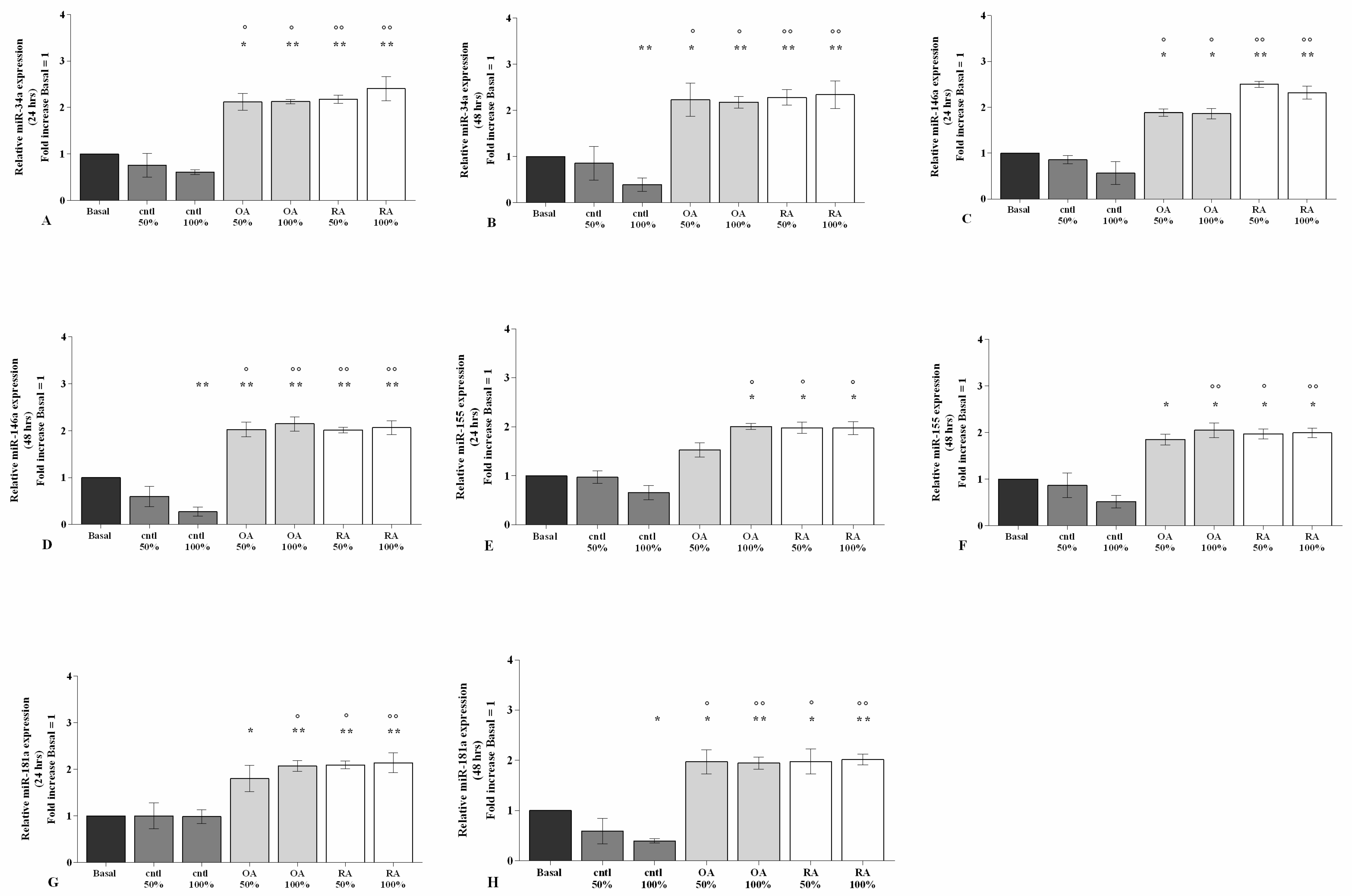
2.4. SF Regulates the Gene Expression of a Pattern of miRNA

The gene expressions of miR-34a, miR-146a, miR-155, and miR-181a, evaluated by real-time PCR, were significantly increased (p < 0.05, p < 0.01), at both 24 h and 48 h, in our cell cultures stimulated with 50% or 100% of OA or RA SF when compared to the baseline (p < 0.05, p < 0.01) (Figure 5A–H). A similar trend was also maintained when OA and RA SF were compared with control SF (p < 0.05, p < 0.01) (Figure 5A–H). No differences between OA and RA SF on miRNA regulation were observed.
Surprisingly, 48 h of treatment with control SF significantly reduced the levels of miR-34a, miR-146a, and miR-181a when tested at 100% concentration (p < 0.05) with respect to the baseline (Figure 5A–H).

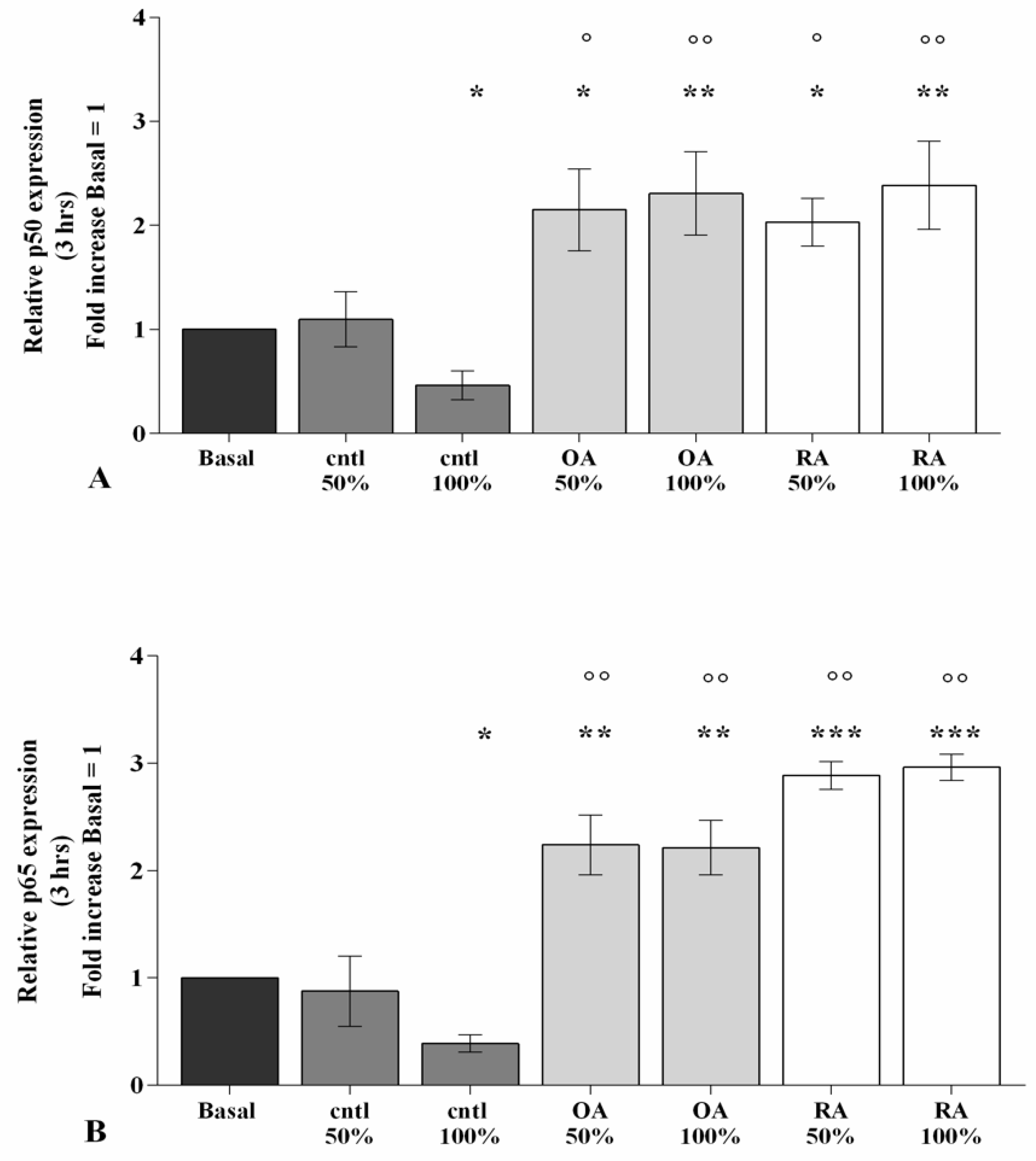
2.5. SF Induces NF-κB Pathway Activation

Figure 6 reports the effect of SF on the regulation of the NF-κB signaling pathway after 3 h of stimulating OA chondrocytes with 50% or 100% SF from OA, RA, and controls. The real-time PCR analysis showed a significant reduction in p50 and p65 subunits gene expression after incubation with 100% control SF in comparison to the basal condition (Figure 6A,B). Conversely, 50% or 100% OA and RA SF significantly up-regulated the transcriptional levels of both subunits (p < 0.05, p < 0.01, p < 0.001), with respect to the baseline, revealing a particular exacerbation of p65. OA and RA SF stimulus was also effective when compared to control SF (p < 0.05, p < 0.01), while no modifications were observed among the pathological SF samples (Figure 6A,B).

2.6. SF Regulates miRNA Expression via the NF-κB Pathway

The involvement of the NF-κB pathway in mediating SF-induced effects on miRNA expression has been assessed using a specific NF-κB inhibitor (IKKα/β, BAY 11-7082) (Figure 7).
Real-time PCR analysis revealed that the gene expression of the studied miRNA was significantly decreased (p < 0.05, p < 0.01) in OA chondrocytes incubated with BAY 11-7082 in comparison to the basal condition (Figure 7A–D). The co-treatment of the cells with BAY 11-7082 and 100% OA and RA SF did not show any change in miRNA expression with respect to chondrocytes incubated with BAY 11-7082 alone (Figure 7A–D).
Furthermore, the presence of the NF-κB inhibitor significantly limited the effect of OA and RA SF on the expression levels of the analyzed miRNA (p < 0.05, p < 0.01). No modifications with 100% control SF were observed (Figure 7A–D).

3. Discussion

MiRNA play an important role in the pathogenesis of different musculoskeletal diseases, including OA and RA [30]. Data suggest that miRNA have both beneficial and detrimental effects on the joints. In fact, a lot of studies proved their activity in cartilage metabolism, as well as in pathological processes including inflammation, apoptosis, and oxidative stress [25,30].
SF is mainly involved in the maintenance and health of the articular cartilage. It has been observed that chondrocyte metabolism is deeply influenced by the specific composition of SF [12,14]. This evidence stimulated an increasing interest in comprehending the exact effect of the SF microenvironment in cartilage homeostasis.
In this study, we examined the effects of OA and RA SF, tested at 50% and 100%, in a pattern of miRNA associated to the pathogenesis of OA and RA. The possible implication of the NF-κB pathway was also assessed. Furthermore, we evaluated the effects of the OA and RA SF on inflammation, apoptosis, and oxidative stress. SF from patients with knee injuries who underwent anterior cruciate ligament surgery was used as a control.
First of all, our data confirmed a significant increase in the gene expression of the main pro-inflammatory cytokines IL-1β, IL-6, and TNF-α in OA chondrocytes incubated with RA SF for 24 h and 48 h. Similarly, SF from patients with OA showed the same regulation of the studied cytokines. These results are in agreement with previous studies. In particular, some authors demonstrated a relevant increase in IL-1β, IL-6, IL-8, and TNF-α expression in human primary chondrocytes and mesenchymal stem cells treated with OA or RA SF, for 24 h and 48 h [28,29,31]. Furthermore, the stimulus of our cells derived from OA and RA SF determined a marked increase in the gene expression of the main matrix-degrading enzymes MMP-1, MMP-13, and ADAMTS-5, while a down-regulation of Col2a1 was observed. In a similar manner, Carballo et al. [21] found raised gene levels of MMP-3, MMP-13, ADAMTS-5, and IL-18 in human primary chondrocytes stimulated with OA SF for 24 h and 72 h. These findings suggest the contribution of SF to the ECM-degrading processes occurring over the course of this disease.
The elevated levels of IL-1β, IL-6, and TNF-α, as well as of MMPs, found in the SF of both OA and RA patients confirmed their important role in the pathogenesis of these joint diseases [12,32]. Therefore, an excess of these pro-inflammatory mediators and enzymes in SF can affect chondrocyte homeostasis and induce the production of other cytokines and matrix-degrading enzymes; in addition, they also inhibit the synthesis of proteoglycans and type II collagen, thereby causing cartilage degradation in OA and RA [8,32].
Surprisingly, when our chondrocytes were incubated with 50% or 100% control SF, at both analyzed time points, a significant reduction in the gene expression of IL-1β and IL-6 was observed, as well as of MMP-1 and MMP-13, while an over-expression of Col2a1 was found. These results are partially consistent with a study performed on bovine cartilage explant cultures incubated with different SF percentages isolated from healthy bovine joints, for 12 days, demonstrating the anabolic and anti-catabolic activities of the SF used [33].
The regulation of chondrocyte survival is important for the maintenance of proper cartilage structure and function. Apoptosis is an active, physiological process of cell death, playing a critical role in retaining the homeostasis of tissues and cells [34]; it is regulated by various genes, such as those of the BCL-2 family, identified as controllers and inductors of this mechanism [35]. Dysregulation of apoptosis is, thus, associated with a variety of diseases including inflammatory and degenerative disorders such as OA and RA [35,36].
Cell viability and apoptosis analyses performed in the present study showed a reduction in survival and an increase in apoptotic ratio, with a concomitant down-regulation of the anti-apoptotic marker BCL2, in OA chondrocytes stimulated with 50% and 100% RA or OA SF. These data confirm what was found in previous studies. In 2007, Schuerwegh et al. [18] observed a significant decrease in cell viability and cell proliferation, along with an increase in apoptosis, in bovine articular chondrocytes incubated for 48 h with SF derived from patients with RA. Later, a group of investigators reported that the number of vital cells appeared clearly reduced after treatment of human OA chondrocytes with RA or OA SF, from 24 h to 72 h; conversely a strong induction of early apoptosis was observed in a time-dependent manner [19,20]. These authors pointed out that the highest effect of pathological SF on cell survival was achieved after 24 h of treatment. During further incubation (until 72 h), the number of vital cells remained stable, suggesting a sort of adaptation of chondrocytes to the surrounding microenvironment after initial damage. This hypothesis could be in line with our results considering that we observed SF effects at 24 h of stimulus, and a similar trend, without substantial differences, was maintained at 48 h.
Interestingly, to the best of our knowledge, we demonstrated for the first time the anti-apoptotic activity of control SF, at 100% concentrations at both tested time points.
In recent years, the integrity of the mitochondrial structure has been considered a prerequisite for normal chondrocyte survival. Degenerative factors and inflammatory mediators, such as cytokines, induce defects in mitochondrial function and lead to excessive ROS generation; this condition contributes to cartilage degradation and synovial inflammation [3,37]. Accumulation of ROS in the joint, with a concomitant failure of the antioxidant scavenging system, progressively inhibits the synthesis of glycosaminoglycans and type II collagen fibers, induces apoptosis, and activates the production of matrix-degrading enzymes [3,37]. In addition, ROS overproduction exacerbates synovitis and leads to the release of pro-inflammatory cytokines, in turn, stimulating the synthesis of new ROS [37,38].
In this study, the analysis of oxidative stress showed an overproduction of mitochondrial superoxide anions in OA chondrocytes treated with 50% or 100% RA or OA SF. Pathological SF also increased the gene expression of antioxidant enzyme, SOD2, and the transcriptional factor NRF2. This is the first experience revealing the involvement of SF components in the regulation of redox balance. However, a study by Schuerwegh and collaborators [18] seems to partially sustain our results, albeit not comparable; the authors reported a significant increase in NO production after treatment of bovine articular chondrocytes with RA SF, for a period of 48 h.
Furthermore, Sun at el. [33] demonstrated the ability of 1% normal SF to reduce the production of NO, induced by IL-1β, in bovine articular cartilage explants; this activity was effective in a time-dependent manner, until 12 days of incubation. These data appear in contrast with our findings since we did not observe any substantial effect of control SF in regulating oxidative stress. We hypothesize that this could be ascribable to the different experimental procedures employed, such as the samples used, the parameters analyzed, and the treatment conditions applied.
The increase in antioxidant agents observed after stimulus of chondrocytes with pathological SF, concomitant with excessive ROS production, could be explained as an acute adaptive mechanism of the cell against the overproduction of free radicals. We speculate that, in response to continuous and cumulative oxidative stress status, a protective increase in ROS scavenging activity can be induced through the activation of transcription factor NRF2 [28,38,39,40], as confirmed by various authors [41,42,43].
In the present study, for the first time, we investigated the potential effect of SF in the regulation of some miRNA associated to the joint damage occurring in OA and RA.
In particular, miR-34a has been known to be implicated in activating apoptosis signaling, limiting cell proliferation, and modulating redox balance in human OA chondrocytes and synovial fibroblasts [28,29,43,44,45]. In addition, miR-146a, traditionally identified as a regulator of inflammation, has been recently recognized as an activator of apoptosis and oxidative stress by its direct effect on SMAD4 and NRF2 transcriptional factors, in OA cells [28,29,46]. Similarly, miR-181a activates apoptosis and oxidative stress by targeting the anti-apoptotic marker BCL2 and modulating the oxidant/antioxidant system in different cell types [28,47,48]. Furthermore, it is a regulator of B and T cell development and stimulates the production of pro-inflammatory cytokines in RA synovial fibroblasts [49]. MiR-155 has been primarily associated to the maturation and activation of innate and adaptive immune system cells, and also found highly expressed in synovial fluid of RA patients [50]. The described properties of these miRNA make them very suitable candidates for regulating inflammatory and degenerative articular processes [25,30]. Thus, we explored the possible influence of pathological SF on their expression profile in chondrocyte cultures to provide new information on the complex mechanisms underlying OA and RA.
Interestingly, for the first one, we demonstrated the up-regulation of miR-34a, miR-146a, miR-155 and miR-181a gene expression induced by OA and RA SF, while opposite effects were observed when the cells were incubated with 100% control SF. These data partially sustained the results on miR-155 obtained by Li et al. in PBMC-derivative macrophages incubated with OA SF for 24 h and 48 h [51]. To the best of our knowledge, no data from the literature are available about the regulation of SF on the other studied miRNA.
Lastly, we supposed that the observed effects induced by SF can be associated to the regulation of the NF-κB signaling pathway.
It is well known that NF-κB signaling represents one of the most prominent mechanisms activated in the pathogenesis of inflammation-related diseases, such as OA and RA [52,53]. Indeed, in these pathological conditions, NF-κB is highly expressed in articular cartilage and synovial tissue [52,53,54]. It is triggered by pro-inflammatory cytokines, such as IL-1β, and ECM degradation products, and the activated NF-κB modulates the expression of several cytokines, chemokines, and matrix-degrading enzymes; all these features accelerate catabolic events responsible for promoting cartilage degradation and inflammation [55,56]. Moreover, the direct involvement of the NF-κB pathway on miRNA-related post-transcriptional regulation of inflammation, apoptosis, and oxidative stress in OA chondrocytes and synovial fibroblasts has been reported [28,29,57,58,59].
In this experience, we found an increase in the gene expression of the main NF-κB subunits, p50 and p65, after stimulus of OA chondrocytes, for 3 h, with 50% or 100% RA and OA SF, while their reduction in the presence of 100% control SF was observed. The results obtained by Sayegh et al. [31] are in line with our outcomes. In particular, they revealed that pro-inflammatory RA SF, added to the culture medium of mesenchymal stem cells, was able to significantly inhibit the protein expression of NF-κB inhibitor, IκB, resulting in a consequent activation of the pathway; the authors assumed that the presence of different cytokines in SF, including TNF-α, was essential to regulate the activation of the signaling pathway, through the TNF/NF-κB axis.
Our results also confirmed the regulation of miRNA by NF-κB, as shown by the use of a specific NF-κB inhibitor, BAY11-7082, according to previous observations [28,29,57,58,59]. Furthermore, we proved, for the first time, that the presence of BAY11-7082 in our cultures, down-regulating NF-κB, reduced the effect of SF on miR-34a, miR-146a, miR-155, and miR-181a gene expression.
Taken together, our results suggest that a pathological SF, abundant in pro-inflammatory mediators and degrading factors, can induce the expression of some miRNA; miRNAs, in turn, have an active role on inflammation, cartilage turnover, apoptosis and oxidative stress, through the activation of the NF-κB pathway, triggering a vicious circle (Figure S3).
Otherwise, the observed protective effects of control SF can be attributed to low content of cytokines and other inflammatory mediators, and to a high amount of high-molecular-weight HA [60]. High-molecular-weight HA has demonstrated to have potent anti-inflammatory effects in synovial fibroblasts, reducing NF-κB activation and IL-1β, IL-6, and TNF-α production [61,62,63].

4. Materials and Methods

4.1. Synovial Fluid Collection

SF was obtained from the knees of four patients with OA (two females and two males, with age ranging from 62 to 70 years) and four patients suffering from RA (three females and one male, with age ranging from 55 to 59 years). Diagnoses of knee OA and RA were ensured by ACR/EULAR classification criteria [64,65] (Table S1). All patients were followed in our Rheumatology Unit (Azienda Ospedaliera Universitaria Senese, Siena, Italy) and underwent knee aspirations for diagnostic evaluation or prior to an intra-articular glucocorticoid or hyaluronic acid injection. Four healthy subjects (one female and three males, with age ranging from 30 to 35 years) with knee injuries who underwent anterior cruciate ligament reconstruction under spinal anesthesia were used as controls. They have no signs or symptoms related to OA, RA, or other inflammatory or degenerative joint diseases. During surgery, SF was aseptically aspirated with an 18-gauge needle. There was no evidence of contamination of the SF samples at the time of aspiration. The samples from healthy subjects were provided by the Orthopedic Surgery Unit (University of Siena, Italy).
The participants presented no clinical or laboratory signs of infection and gave their informed consent prior to SF collection.
SF was put in a sterile container and stored in a refrigerator at 4 °C until it was examined within 24 h of joint aspiration. SF samples were analyzed by an expert examiner on site, blinded to the clinical findings of the subjects. After the first macroscopic analysis, SF was microscopically examined under optical light microscopy for total and differential WBC count using a Bürker counting chamber and pre-stained slides for cell morphology [66,67]. A crystal search was performed using polarized compensated light microscopy, and a negative result was necessary before using SF for the study [66,67].
Following the analysis, SF samples were diluted to 20% in DMEM with 10% FBS, due to their high viscosity, and then filtered through a 70-μm nylon mesh to remove debris and prevent possible contamination of the cultures. Samples were transferred to microcentrifuge tubes, and centrifuged at 2000 rpm for 10 min before storing the supernatant at −80 °C until use.

4.2. Cell Cultures

Human OA articular cartilage was obtained from the femoral heads of five non-obese (BMI ranging from 20 to 23 kg/m2) and non-diabetic patients (two men and three women, age ranging from 65 to 75) with hip OA according to ACR criteria [64], subjected to total hip replacement surgery. OA chondrocytes were derived from the area adjacent to the OA lesion [68]. Articular cartilage was supplied by the Orthopedic Surgery Unit, University of Siena, Italy. The use of human articular specimens was allowed by the authorization of the Ethic Committee of Azienda Ospedaliera Universitaria Senese/Siena University Hospital (decision no. 13931/18), and informed consent was provided by each donor.
Chondrocytes isolation was provided immediately after surgery. In particular, cartilage fragments were aseptically dissected from each donor and processed by an enzymatic digestion, as previously described [48]. For growth and expansion, cells were cultured in Dulbecco’s modified Eagle’s medium (DMEM) (Euroclone, Milan, Italy) with phenol red and L-glutamine, supplemented with 10% fetal bovine serum (FBS) (Euroclone, Milan, Italy), 200 U/mL penicillin, and 200 µg/mL streptomycin (P/S) (Sigma-Aldrich, Milan, Italy). The medium was changed every 2–3 days, and the cell morphology was examined daily with an inverted microscope (Olympus IMT-2, Tokyo, Japan). OA primary chondrocytes at the first passage were employed for the experiments.

4.3. Treatment of Cell Cultures

Human OA chondrocytes were plated in six-well dishes at a starting density of 1 × 10⁵ cells/well until 85% confluence. SF samples (4 OA, 4 RA, and 4 healthy controls) were diluted to 20% in DMEM with 10% FBS, and then used for the treatment. Medium was removed from the cultures and replaced with 50% (50% SF at 20% in DMEM 10% FBS + 50% DMEM 10% FBS) or 100% (100% SF at 20% in DMEM 10% FBS) SF from patients with knee OA, RA, and controls, for a period of 24 h and 48 h.
After the treatment, the cells were recovered and immediately processed to carry out the MTT assay, flow cytometry analysis, and quantitative real-time PCR.
Some cultures were pre-incubated for 2 h with BAY 11-7082 1 μM (NF-κB inhibitor, IKKα/β, Sigma–Aldrich, Milan, Italy) and then treated with SF. Afterwards, the gene expression of the studied miRNA (miR-34a, miR-146a, miR-155, and miR-181a) was evaluated.

4.4. MTT Assay

After treatment, the viability of the cells was evaluated by a 3-(4,4-dimethylthiazol-2-yl)-2,5-diphenyl-tetrazoliumbromide (MTT) (Sigma-Aldrich, Milan, Italy) test to assess the percentage of survival cells.
Chondrocytes were grown in 12-well dishes at a starting density of 5 × 10⁴ and, after treatment, were incubated for 3 h at 37 °C in a culture medium containing 10% of 5 mg/mL of MTT (Sigma–Aldrich, Milan, Italy). Then, the medium was removed and 0.2 mL of dimethyl sulfoxide (DMSO) (Rottapharm Biotech, Monza, Italy) were added to the wells to solubilize the formazan crystals. The absorbance was measured at 570 nm in a microplate reader (BioTek Instruments, Inc., Winooski, VT, USA).
The percentage of survival cells was evaluated as follows: (absorbance of considered sample)/(absorbance of control) × 100. The obtained data were reported as optical density units per 104 adherent cells.

4.5. Apoptosis Detection

Apoptotic cells were evaluated using an annexin V-FITC and propidium iodide (PI) (ThermoFisher Scientific, Milan, Italy) kit. Chondrocytes were seeded in 12-well plates (8 × 10⁴ cells/well) for 24 h in DMEM with 10% FBS, before the treatment procedure. Then, the cells were washed and harvested using trypsin, collected into cytometry tubes, and centrifuged at 1500 rpm for 5 min. The supernatant was replaced, and the pellet was re-suspended in 100 μL of 1 × annexin-binding buffer, 5 μL of Alexa Fluor 488 annexin-V conjugated to fluorescein (FITC, green fluorescence), and 1 μL of 100 μg/mL PI (red fluorescence) working solution, incubated at room temperature for 15 min in the dark. Afterward, 600 μL of 1 × annexin-binding buffer was added to the tubes before analysis using a flow cytometer.
A total of 10,000 events (1 × 10⁴ cells per assay) were measured by the instrument, and the results were analyzed with Cell Quest software (Version 4.0, Becton Dickinson, San Jose, CA, USA). Apoptosis analysis was carried out considering the simultaneous staining of cells with Alexa Fluor 488 annexin-V and PI. The results were expressed as the percentage of cells positively stained by each dye (total apoptosis) [69].

4.6. Mitochondrial Superoxide Anion (•O2−) Production

OA chondrocytes were seeded into 12-well plates (8 × 10⁴ cells/well) for 24 h in DMEM with 10% FBS, before the treatment procedure. Then, the cells were incubated in PBS (Euroclone, Milan, Italy) and MitoSOX Red for 15 min at 37 °C in the dark, to evaluate mitochondrial superoxide anion (•O2−) production. Cells were then harvested by trypsin, collected into cytometry tubes, and centrifuged at 1500 rpm for 10 min. Cells were then dissolved in saline solution before flow cytometry analysis. A density of 1 × 10⁴ cells per assay (a total of 10,000 events) was measured by the instrument and data were analyzed with CellQuest software (Version 4.0, Becton Dickinson, San Jose, CA, USA). Results were collected as the median fluorescence (AU), represented by the mean of three independent experiments [29].

4.7. RNA Isolation and Quantitative Real-Time PCR

Cells were grown and maintained in six-well dishes at a starting density of 1 × 10⁵ cells/well until they became 85% confluent in DMEM supplemented with 10% FBS, before the treatment procedure. After treatment, cells were collected, and total RNA was extracted using TriPure Isolation Reagent (Euroclone, Milan, Italy), according to the manufacturer’s instructions, before storing at −80 °C. The concentration, purity, and integrity of RNA were evaluated by measuring the OD at 260 nm and the 260/280 and 260/230 ratios using a Nanodrop-1000 (Celbio, Milan, Italy).
Reverse transcription for target genes was carried out by the QuantiTect Reverse Transcription Kit (Qiagen, Germany), while for miRNA it was carried out by the cDNA miScript PCR Reverse Transcription Kit (Qiagen, Hilden, Germany), according to the manufacturer’s instructions.
Target genes and miRNA were examined by real-time PCR using QuantiFast SYBR Green PCR (Qiagen, Hilden, Germany) and miScript SYBR Green (Qiagen, Hilden, Germany) kits, respectively. A list of primers used for PCR reactions is reported in Table S2.
All qPCR reactions were achieved in glass capillaries by a LightCycler 1.0 (Roche Molecular Biochemicals, Mannheim, Germany) with LightCycler Software Version 3.5. The reaction procedure was described in detail in our previous studies [57,69].
For the data analysis, the Ct values of each sample and the efficiency of the primer set were calculated and converted into relative quantities [70,71]. The normalization was performed considering actin beta (ACTB) for target genes and Small Nucleolar RNA, C/D Box 25 (SNORD-25) for miRNA as the housekeeping genes [72].

4.8. Statistical Analysis

Three experiments were performed for each SF (4 OA, 4 RA, and 4 healthy controls), and the results were expressed as the mean ± standard deviation (SD) of triplicate values for each experiment. The normal distribution of data was evaluated by Shapiro–Wilk, D’Agostino and Pearson, and Kolmogorov–Smirnov tests. MTT, flow cytometry results, and quantitative real-time PCR, were each analyzed via a mixed model ANOVA with Bonferroni post hoc test. All analyses were carried out using the SAS System (SAS Institute Inc., Cary, NC, USA) and GraphPad Prism 6.1. A p-value of < 0.05 was defined as statistically significant.

5. Conclusions

The results of the present study highlight the potential role of SF in the regulation of a pattern of miRNA in human OA chondrocytes, via the NF-κB signaling pathway.
First of all, we observed that SF, taken from patients with RA or OA, has inflammatory and degrading activities on cartilage, the expression of cytokines, and matrix degrading enzymes, activating apoptosis and oxidative stress, as well as the NF-κB pathway, in human OA chondrocytes. Conversely, healthy SF showed opposite and positive effects.
Our data demonstrated, for the first time, the role of SF in modulating the expression levels of miR-34a, miR-146a, miR-155, and miR-181a, mainly involved in inflammation and cartilage metabolism occurring in OA and RA; moreover, the observed transcriptional modifications of miRNA seem to be due to the modulation of the NF-κB signaling pathway.
Thus, we underline that a specific composition of SF microenvironment may differently influence inflammation and chondrocyte integrity and metabolism. We suppose that this aspect is in part related to (1) the presence of pro-inflammatory cytokines and matrix degrading factors inside the fluid, (2) the number of WBCs, attesting the presence of an inflammatory state, and (3) the content of high-molecular-weight HA, lubricin, and other bioactive molecules.
Taken together, our data provide preliminary fundamentals to improve the knowledge about the relevance of SF, as well as its composition, in the regulation of cartilage homeostasis, even through miRNA regulation. Our observations can help to improve the understanding of the complex mechanisms underlying the pathogenesis of OA and RA.
However, further experiments are required to support our hypothesis, considering the difficulty of translating the results obtained from in vitro studies into clinical practice.
In addition, our study presents some limitations that have to be noticed. The main limitation is related to the discrepancy in age between the groups of patients and controls. This is due to the difficulty to find normal controls, without any sign of rheumatologic disease, in older subjects. Second, the analysis of the rheological properties of the studied SF, as well as of their specific content in pro-inflammatory mediators and HA, should provide further information useful to investigate the relationship more deeply between the different components of SF and their effects in inflammatory and degrading processes and in miRNA expression profiles. Furthermore, a multivariate analysis investigating the possible association among the different studied mediators could be useful to improve the knowledge about the pathogenetic mechanisms underlying inflammatory and/or degenerative diseases. Another important limitation is the lack of the analysis of protein expression of the evaluated target genes, to confirm whether the changes occurring at the gene expression level are also reflected in protein regulation. In addition, transfection experiments using specific miRNA inhibitors may point out the implication of these molecules in mediating SF effects.

Supplementary Materials

The following supporting information can be downloaded at: https://www.mdpi.com/article/10.3390/ijms23158334/s1.

Author Contributions

Conceptualization, S.C., S.T. and A.F.; methodology, S.C., S.L. and I.S.; validation, S.L.; investigation, S.C. and S.L.; resources, S.B., S.T. and A.F.; data curation, S.C.; writing—original draft, S.C., S.L. and A.F.; writing—review and editing, S.C., S.T., S.L., I.S., S.B., E.F. and A.F.; funding acquisition, E.F. All authors have read and agreed to the published version of the manuscript.

Funding

This research was partially supported by a grant (funds for the research) of the University of Siena, PSR-2022.

Institutional Review Board Statement

All methods performed in this study abide by the Declaration of Helsinki and the study protocols used in this research were approved by the Ethic Committee of Azienda Ospedaliera Universitaria Senese/Siena University Hospital (decision no. 13931/18).

Informed Consent Statement

All patients signed an informed written consent form to participate in the study.

Data Availability Statement

The data presented in this study are available on request from the corresponding author.

Conflicts of Interest

The authors declare no conflict of interest.

References

  1. Hunter, D.J.; Bierma-Zeinstra, S. Osteoarthritis. Lancet 2019, 393, 1745–1759. [Google Scholar] [CrossRef]
  2. Ratneswaran, A.; Kapoor, M. Osteoarthritis year in review: Genetics, genomics, epigenetics. Osteoarthr. Cartil. 2021, 29, 151–160. [Google Scholar] [CrossRef] [PubMed]
  3. Zheng, L.; Zhang, Z.; Sheng, P.; Mobasheri, A. The role of metabolism in chondrocyte dysfunction and the progression of osteoarthritis. Ageing Res. Rev. 2021, 66, 101249. [Google Scholar] [CrossRef]
  4. Sanchez-Lopez, E.; Coras, R.; Torres, A.; Lane, N.E.; Guma, M. Synovial inflammation in osteoarthritis progression. Nat. Rev. Rheumatol. 2022, 18, 258–275. [Google Scholar] [CrossRef] [PubMed]
  5. Smolen, J.S.; Aletaha, D.; McInnes, I.B. Rheumatoid arthritis. Lancet 2016, 388, 2023–2038, Erratum in Lancet 2016, 388, 1984. [Google Scholar] [CrossRef]
  6. Chauhan, K.; Jandu, J.S.; Goyal, A.; Bansal, P.; Al-Dhahir, M.A. Rheumatoid Arthritis; StatPearls Publishing: Treasure Island, FL, USA, 2021. [Google Scholar]
  7. Mueller, A.L.; Payandeh, Z.; Mohammadkhani, N.; Mubarak, S.M.H.; Zakeri, A.; Alagheband Bahrami, A.; Brockmueller, A.; Shakibaei, M. Recent Advances in Understanding the Pathogenesis of Rheumatoid Arthritis: New Treatment Strategies. Cells 2021, 10, 3017. [Google Scholar] [CrossRef]
  8. Kondo, N.; Kuroda, T.; Kobayashi, D. Cytokine Networks in the Pathogenesis of Rheumatoid Arthritis. Int. J. Mol. Sci. 2021, 22, 10922. [Google Scholar] [CrossRef]
  9. Romão, V.C.; Fonseca, J.E. Etiology and Risk Factors for Rheumatoid Arthritis: A State-of-the-Art Review. Front. Med. 2021, 8, 689698. [Google Scholar] [CrossRef]
  10. Altobelli, E.; Angeletti, P.M.; Piccolo, D.; De Angelis, R. Synovial Fluid and Serum Concentrations of Inflammatory Markers in Rheumatoid Arthritis, Psoriatic Arthritis and Osteoarthitis: A Systematic Review. Curr. Rheumatol. Rev. 2017, 13, 170–179. [Google Scholar] [CrossRef]
  11. Birkelund, S.; Bennike, T.B.; Kastaniegaard, K.; Lausen, M.; Poulsen, T.B.G.; Kragstrup, T.W.; Deleuran, B.W.; Christiansen, G.; Stensballe, A. Proteomic analysis of synovial fluid from rheumatic arthritis and spondyloarthritis patients. Clin. Proteom. 2020, 17, 29. [Google Scholar] [CrossRef]
  12. Meehan, R.T.; Regan, E.A.; Hoffman, E.D.; Wolf, M.L.; Gill, M.T.; Crooks, J.L.; Parmar, P.J.; Scheuring, R.A.; Hill, J.C.; Pacheco, K.A.; et al. Synovial Fluid Cytokines, Chemokines and MMP Levels in Osteoarthritis Patients with Knee Pain Display a Profile Similar to Many Rheumatoid Arthritis Patients. J. Clin. Med. 2021, 10, 5027. [Google Scholar] [CrossRef]
  13. Qin, L.; Wang, H.; Zhao, C.; Chen, C.; Chen, H.; Li, X.; Wang, J.; Hu, N.; Huang, W. Serum and Synovial Biomarkers for Distinguishing Between Chronic Periprosthetic Joint Infections and Rheumatoid Arthritis: A Prospective Cohort Study. J. Arthroplast. 2021, 37, 342–346. [Google Scholar] [CrossRef]
  14. Nieminen, P.; Hämäläinen, W.; Savinainen, J.; Lehtonen, M.; Lehtiniemi, S.; Rinta-Paavola, J.; Lehenkari, P.; Kääriäinen, T.; Joukainen, A.; Kröger, H.; et al. Metabolomics of Synovial Fluid and Infrapatellar Fat Pad in Patients with Osteoarthritis or Rheumatoid Arthritis. Inflammation 2022, 45, 1101–1117. [Google Scholar] [CrossRef]
  15. Ostrowska, M.; Maśliński, W.; Prochorec-Sobieszek, M.; Nieciecki, M.; Sudoł-Szopińska, I. Cartilage and bone damage in rheumatoid arthritis. Reumatologia 2018, 56, 111–120. [Google Scholar] [CrossRef] [Green Version]
  16. Hu, Q.; Ecker, M. Overview of MMP-13 as a Promising Target for the Treatment of Osteoarthritis. Int. J. Mol. Sci. 2021, 22, 1742. [Google Scholar] [CrossRef]
  17. Verbruggen, A.; De Clerck, L.S.; Bridts, C.H.; Breedveld, F.C.; Stevens, W.J. Influence of blood and synovial fluid immune complexes of patients with rheumatoid arthritis on production of nitric oxide and growth and viability of chondrocytes. J. Rheumatol. 2000, 27, 35–40. [Google Scholar] [PubMed]
  18. Schuerwegh, A.J.; Dombrecht, E.J.; Stevens, W.J.; Van Offel, J.F.; Kockx, M.M.; Bridts, C.H.; De Clerck, L.S. Synovial fluid and peripheral blood immune complexes of patients with rheumatoid arthritis induce apoptosis in cytokine-activated chondrocytes. Rheumatol. Int. 2007, 27, 901–909. [Google Scholar] [CrossRef] [PubMed]
  19. Röhner, E.; Matziolis, G.; Perka, C.; Füchtmeier, B.; Gaber, T.; Burmester, G.R.; Buttgereit, F.; Hoff, P. Inflammatory synovial fluid microenvironment drives primary human chondrocytes to actively take part in inflammatory joint diseases. Immunol. Res. 2012, 52, 169–175. [Google Scholar] [CrossRef] [PubMed]
  20. Hoff, P.; Buttgereit, F.; Burmester, G.R.; Jakstadt, M.; Gaber, T.; Andreas, K.; Matziolis, G.; Perka, C.; Röhner, E. Osteoarthritis synovial fluid activates pro-inflammatory cytokines in primary human chondrocytes. Int. Orthop. 2013, 37, 145–151. [Google Scholar] [CrossRef] [Green Version]
  21. Carballo, C.B.; Coelho, T.R.P.; de Holanda Afonso, R.C.; Faria, J.C.O.; Alves, T.; Monte, S.M.; Ventura Matioszek, G.M.; Moura-Neto, V.; Brito, J.M. Osteoarthritic Synovial Fluid and TGF-β1 Induce Interleukin-18 in Articular Chondrocytes. Cartilage 2020, 11, 385–394. [Google Scholar] [CrossRef]
  22. Lee, D.A.; Salih, V.; Stockton, E.F.; Stanton, J.S.; Bentley, G. Effect of normal synovial fluid on the metabolism of articular chondrocytes in vitro. Clin. Orthop. Relat. Res. 1997, 342, 228–238. [Google Scholar] [CrossRef]
  23. Brand, J.A.; McAlindon, T.E.; Zeng, L. A 3D system for culturing human articular chondrocytes in synovial fluid. J. Vis. Exp. 2012, 59, e3587. [Google Scholar] [CrossRef] [PubMed] [Green Version]
  24. Hegewald, A.A.; Ringe, J.; Bartel, J.; Krüger, I.; Notter, M.; Barnewitz, D.; Kaps, C.; Sittinger, M. Hyaluronic acid and autologous synovial fluid induce chondrogenic differentiation of equine mesenchymal stem cells: A preliminary study. Tissue Cell 2004, 36, 431–438. [Google Scholar] [CrossRef] [PubMed]
  25. De Palma, A.; Cheleschi, S.; Pascarelli, N.A.; Tenti, S.; Galeazzi, M.; Fioravanti, A. Do MicroRNAs have a key epigenetic role in osteoarthritis and in mechanotransduction? Clin. Exp. Rheumatol. 2017, 35, 518–526. [Google Scholar] [PubMed]
  26. Chang, C.; Xu, L.; Zhang, R.; Jin, Y.; Jiang, P.; Wei, K.; Xu, L.; Shi, Y.; Zhao, J.; Xiong, M.; et al. MicroRNA-Mediated Epigenetic Regulation of Rheumatoid Arthritis Susceptibility and Pathogenesis. Front. Immunol. 2022, 13, 838884. [Google Scholar] [CrossRef]
  27. Ghafouri-Fard, S.; Poulet, C.; Malaise, M.; Abak, A.; Mahmud Hussen, B.; Taheriazam, A.; Taheri, M.; Hallajnejad, M. The Emerging Role of Non-Coding RNAs in Osteoarthritis. Front. Immunol. 2021, 12, 773171. [Google Scholar] [CrossRef]
  28. Cheleschi, S.; Gallo, I.; Barbarino, M.; Giannotti, S.; Mondanelli, N.; Giordano, A.; Tenti, S.; Fioravanti, A. MicroRNA Mediate Visfatin and Resistin Induction of Oxidative Stress in Human Osteoarthritic Synovial Fibroblasts Via NF-κB Pathway. Int. J. Mol. Sci. 2019, 20, 5200. [Google Scholar] [CrossRef] [Green Version]
  29. Cheleschi, S.; Barbarino, M.; Gallo, I.; Tenti, S.; Bottaro, M.; Frati, E.; Giannotti, S.; Fioravanti, A. Hydrostatic Pressure Regulates Oxidative Stress through microRNA in Human Osteoarthritic Chondrocytes. Int. J. Mol. Sci. 2020, 21, 3653. [Google Scholar] [CrossRef]
  30. Ali, S.A.; Peffers, M.J.; Ormseth, M.J.; Jurisica, I.; Kapoor, M. The non-coding RNA interactome in joint health and disease. Nat. Rev. Rheumatol. 2021, 17, 692–705. [Google Scholar] [CrossRef]
  31. Sayegh, S.; El Atat, O.; Diallo, K.; Rauwel, B.; Degboé, Y.; Cavaignac, E.; Constantin, A.; Cantagrel, A.; Trak-Smayra, V.; Alaaeddine, N.; et al. Rheumatoid Synovial Fluids Regulate the Immunomodulatory Potential of Adipose-Derived Mesenchymal Stem Cells Through a TNF/NF-κB-Dependent Mechanism. Front. Immunol. 2019, 10, 1482, Erratum in Front Immunol. 2019, 10, 1961. [Google Scholar] [CrossRef]
  32. Wang, T.; He, C. Pro-inflammatory cytokines: The link between obesity and osteoarthritis. Cytokine Growth Factor Rev. 2018, 44, 38. [Google Scholar] [CrossRef] [PubMed]
  33. Sun, E.Y.; Fleck, A.K.M.; Abu-Hakmeh, A.E.; Kotsakis, A.; Leonard, G.R.; Wan, L.Q. Cartilage Metabolism is Modulated by Synovial Fluid Through Metalloproteinase Activity. Ann. Biomed. Eng. 2018, 46, 810–818. [Google Scholar] [CrossRef] [PubMed]
  34. Yang, J.; Hu, S.; Bian, Y.; Yao, J.; Wang, D.; Liu, X.; Guo, Z.; Zhang, S.; Peng, L. Targeting Cell Death: Pyroptosis, Ferroptosis, Apoptosis and Necroptosis in Osteoarthritis. Front. Cell Dev. Biol. 2022, 9, 789948. [Google Scholar] [CrossRef]
  35. Hwang, H.S.; Kim, H.A. Chondrocyte Apoptosis in the Pathogenesis of Osteoarthritis. Int. J. Mol. Sci. 2015, 16, 26035–26054. [Google Scholar] [CrossRef]
  36. Zhou, L.; Li, L.; Wang, Y.; Gao, Q.; Geng, Y.Q. Effects of RANKL on the proliferation and apoptosis of fibroblast-like synoviocytes in rheumatoid arthritis through regulating the NF-κB signaling pathway. Eur. Rev. Med. Pharmacol. Sci. 2019, 23, 9215–9221. [Google Scholar] [CrossRef]
  37. Kan, S.; Duan, M.; Liu, Y.; Wang, C.; Xie, J. Role of Mitochondria in Physiology of Chondrocytes and Diseases of Osteoarthritis and Rheumatoid Arthritis. Cartilage 2021, 13, 1102S–1121S. [Google Scholar] [CrossRef] [PubMed]
  38. Huang, M.L.; Chiang, S.; Kalinowski, D.S.; Bae, D.H.; Sahni, S.; Richardson, D.R. The Role of the Antioxidant Response in Mitochondrial Dysfunction in Degenerative Diseases: Cross-Talk between Antioxidant Defense, Autophagy, and Apoptosis. Oxidative Med. Cell. Longev. 2019, 2019, 6392763. [Google Scholar] [CrossRef] [Green Version]
  39. Marchev, A.S.; Dimitrova, P.A.; Burns, A.J.; Kostov, R.V.; Dinkova-Kostova, A.T.; Georgiev, M.I. Oxidative stress and chronic inflammation in osteoarthritis: Can NRF2 counteract these partners in crime? Ann. N. Y. Acad. Sci. 2017, 1401, 114–135. [Google Scholar] [CrossRef] [Green Version]
  40. Cheleschi, S.; Tenti, S.; Giannotti, S.; Veronese, N.; Reginster, J.Y.; Fioravanti, A. A Combination of Celecoxib and Glucosamine Sulfate Has Anti-Inflammatory and Chondroprotective Effects: Results from an In Vitro Study on Human Osteoarthritic Chondrocytes. Int. J. Mol. Sci. 2021, 22, 8980. [Google Scholar] [CrossRef] [PubMed]
  41. Rabinowitch, H.D.; Clare, D.A.; Crapo, J.D.; Fridovich, I. Positive correlation between superoxide dismutase and resistance to paraquat toxicity in the green alga Chlorella sorokiniana. Arch. Biochem. Biophys. 1983, 225, 640–648. [Google Scholar] [CrossRef]
  42. Ding, X.; Wang, D.; Li, L.; Ma, H. Dehydroepiandrosterone ameliorates H2O2-induced Leydig cells oxidation damage and apoptosis through inhibition of ROS production and activation of PI3K/Akt pathways. Int. J. Biochem. Cell Biol. 2016, 70, 126–139. [Google Scholar] [CrossRef] [PubMed]
  43. Cheleschi, S.; De Palma, A.; Pascarelli, N.A.; Giordano, N.; Galeazzi, M.; Tenti, S.; Fioravanti, A. Could Oxidative Stress Regulate the Expression of MicroRNA-146a and MicroRNA-34a in Human Osteoarthritic Chondrocyte Cultures? Int. J. Mol. Sci. 2017, 18, 2660. [Google Scholar] [CrossRef] [Green Version]
  44. Zhong, X.; Li, P.; Li, J.; He, R.; Cheng, G.; Li, Y. Downregulation of microRNA-34a inhibits oxidized low-density lipoprotein-induced apoptosis and oxidative stress in human umbilical vein endothelial cells. Int. J. Mol. Med. 2018, 42, 1134–1144. [Google Scholar] [CrossRef] [Green Version]
  45. Endisha, H.; Datta, P.; Sharma, A.; Nakamura, S.; Rossomacha, E.; Younan, C. MicroRNA-34a-5p Promotes Joint Destruction During Osteoarthritis. Arthritis Rheumatol. 2021, 73, 426–439. [Google Scholar] [CrossRef] [PubMed]
  46. Liu, J.N.; Lu, S.; Fu, C.M. MiR-146a expression profiles in osteoarthritis in different tissue sources: A meta-analysis of observational studies. J. Orthop. Surg. Res. 2022, 17, 148. [Google Scholar] [CrossRef] [PubMed]
  47. Feng, X.; Zhang, C.; Yang, Y.; Hou, D.; Zhu, A. Role of miR-181a in the process of apoptosis of multiple malignant tumors: A literature review. Adv. Clin. Exp. Med. 2018, 27, 263–270. [Google Scholar] [CrossRef] [Green Version]
  48. Cheleschi, S.; Tenti, S.; Mondanelli, N.; Corallo, C.; Barbarino, M.; Giannotti, S.; Gallo, I.; Giordano, A.; Fioravanti, A. MicroRNA-34a and MicroRNA-181a Mediate Visfatin-Induced Apoptosis and Oxidative Stress via NF-κB Pathway in Human Osteoarthritic Chondrocytes. Cells 2019, 8, 874. [Google Scholar] [CrossRef] [Green Version]
  49. Chen, C.Z.; Li, L.; Lodish, H.F.; Bartel, D.P. MicroRNAs modulate hematopoietic lineage differentiation. Science 2004, 303, 83–86. [Google Scholar] [CrossRef] [Green Version]
  50. Stanczyk, J.; Pedrioli, D.M.; Brentano, F.; Sanchez-Pernaute, O.; Kolling, C.; Gay, R.E.; Detmar, M.; Gay, S.; Kyburz, D. Altered expression of MicroRNA in synovial fibroblasts and synovial tissue in rheumatoid arthritis. Arthritis Rheum. 2008, 58, 1001–1009. [Google Scholar] [CrossRef]
  51. Li, G.S.; Cui, L.; Wang, G.D. miR-155-5p regulates macrophage M1 polarization and apoptosis in the synovial fluid of patients with knee osteoarthritis. Exp. Ther. Med. 2021, 21, 68. [Google Scholar] [CrossRef]
  52. Rigoglou, S.; Papavassiliou, A.G. The NF-κB signalling pathway in osteoarthritis. Int. J. Biochem. Cell Biol. 2013, 45, 2580–2584. [Google Scholar] [CrossRef] [PubMed]
  53. Sun, S.C. The non-canonical NF-κB pathway in immunity and inflammation. Nat. Rev. Immunol. 2017, 17, 545–558. [Google Scholar] [CrossRef] [PubMed]
  54. Noort, A.R.; Tak, P.P.; Tas, S.W. Non-canonical NF-κB signaling in rheumatoid arthritis: Dr Jekyll and Mr Hyde? Arthritis Res. Ther. 2015, 17, 15. [Google Scholar] [CrossRef] [PubMed] [Green Version]
  55. Chow, Y.Y.; Chin, K.Y. The Role of Inflammation in the Pathogenesis of Osteoarthritis. Mediat. Inflamm. 2020, 2020, 8293921. [Google Scholar] [CrossRef] [PubMed] [Green Version]
  56. Liu, S.; Ma, H.; Zhang, H.; Deng, C.; Xin, P. Recent advances on signaling pathways and their inhibitors in rheumatoid arthritis. Clin. Immunol. 2021, 230, 108793. [Google Scholar] [CrossRef] [PubMed]
  57. Cheleschi, S.; Giordano, N.; Volpi, N.; Tenti, S.; Gallo, I.; Di Meglio, M.; Giannotti, S.; Fioravanti, A. A Complex Relationship between Visfatin and Resistin and microRNA: An In Vitro Study on Human Chondrocyte Cultures. Int. J. Mol. Sci. 2018, 19, 3909. [Google Scholar] [CrossRef] [Green Version]
  58. Wu, M.H.; Tsai, C.H.; Huang, Y.L.; Fong, Y.C.; Tang, C.H. Visfatin Promotes IL-6 and TNF-α Production in Human Synovial Fibroblasts by Repressing miR-199a-5p through ERK, p38 and JNK Signaling Pathways. Int. J. Mol. Sci. 2018, 19, 190. [Google Scholar] [CrossRef] [Green Version]
  59. Shao, J.; Ding, Z.; Peng, J.; Zhou, R.; Li, L.; Qian, Q.; Chen, Y. MiR-146a-5p promotes IL-1β-induced chondrocyte apoptosis through the TRAF6-mediated NF-kB pathway. Inflamm. Res. 2020, 69, 619–630. [Google Scholar] [CrossRef]
  60. More, S.; Kotiya, A.; Kotia, A.; Ghosh, S.K.; Spyrou, L.A.; Sarris, I.E. Rheological properties of synovial fluid due to viscosupplements: A review for osteoarthritis remedy. Comput. Methods Prog. Biomed. 2020, 196, 105644. [Google Scholar] [CrossRef]
  61. Campo, G.M.; Avenoso, A.; D’Ascola, A.; Prestipino, V.; Scuruchi, M.; Nastasi, G.; Calatroni, A.; Campo, S. Inhibition of hyaluronan synthesis reduced inflammatory response in mouse synovial fibroblasts subjected to collagen-induced arthritis. Arch. Biochem. Biophys. 2012, 518, 42–52. [Google Scholar] [CrossRef]
  62. Rodriguez-García, S.C.; Castellanos-Moreira, R.; Uson, J.; Naredo, E.; O’Neill, T.W.; Doherty, M.; Boesen, M.; Pandit, H.; Möller Parera, I.; Vardanyan, V.; et al. Efficacy and safety of intra-articular therapies in rheumatic and musculoskeletal diseases: An overview of systematic reviews. RMD Open 2021, 7, e001658. [Google Scholar] [CrossRef] [PubMed]
  63. Altman, R.D.; Manjoo, A.; Fierlinger, A.; Niazi, F.; Nicholls, M. The mechanism of action for hyaluronic acid treatment in the osteoarthritic knee: A systematic review. BMC Musculoskelet. Disord. 2015, 16, 321. [Google Scholar] [CrossRef] [PubMed] [Green Version]
  64. Altman, R.; Asch, E.; Bloch, D.; Bole, G.; Borenstein, D.; Brandt, K.; Christy, W.; Cooke, T.D.; Greenwald, R.; Hochberg, M. Development of criteria for the classification and reporting of osteoarthritis. Classification of osteoarthritis of the knee. Diagnostic and Therapeutic Criteria Committee of the American Rheumatism Association. Arthritis Rheum. 1986, 29, 1039–1049. [Google Scholar] [CrossRef]
  65. Aletaha, D.; Neogi, T.; Silman, A.J.; Funovits, J.; Felson, D.T.; Bingham, C.O. Rheumatoid arthritis classification criteria: An American College of Rheumatology/European League Against Rheumatism collaborative initiative. Arthritis Rheum. 2010, 62, 2569–2581. [Google Scholar] [CrossRef] [PubMed]
  66. Shmerling, R.H. Synovial fluid analysis. A critical reappraisal. Rheum. Dis. Clin. N. Am. 1994, 20, 503–512. [Google Scholar] [CrossRef]
  67. Swan, A.; Amer, H.; Dieppe, P. The value of synovial fluid assays in the diagnosis of joint disease: A literature survey. Ann. Rheum. Dis. 2002, 61, 493–498. [Google Scholar] [CrossRef]
  68. Mankin, H.J.; Dorfman, H.; Lippiello, L.; Zarins, A. Biochemical and metabolic abnormalities in articular cartilage from osteo-arthritic human hips. II. Correlation of morphology with biochemical and metabolic data. J. Bone Joint. Surg. Am. 1971, 53, 523–537. [Google Scholar] [CrossRef] [PubMed]
  69. Cheleschi, S.; Tenti, S.; Barbarino, M.; Giannotti, S.; Bellisai, F.; Frati, E.; Fioravanti, A. Exploring the Crosstalk between Hydrostatic Pressure and Adipokines: An In Vitro Study on Human Osteoarthritic Chondrocytes. Int. J. Mol. Sci. 2021, 22, 2745. [Google Scholar] [CrossRef]
  70. Pfaffl, M.W. A new mathematical model for relative quantification in real-time RT-PCR. Nucleic Acids Res. 2001, 29, e45. [Google Scholar] [CrossRef]
  71. Ramakers, C.; Ruijter, J.M.; Deprez, R.H.; Moorman, A.F. Assumption-free analysis of quantitative real-time polymerase chain reaction (PCR) data. Neurosci. Lett. 2003, 339, 62–66. [Google Scholar] [CrossRef]
  72. Vandesompele, J.; De Preter, K.; Pattyn, F.; Poppe, B.; Van Roy, N.; De Paepe, A.; Speleman, F. Accurate normalization of real-time quantitative RT-PCR data by geometric averaging of multiple internal control genes. Genome Biol. 2002, 3, 34. [Google Scholar] [CrossRef] [PubMed] [Green Version]
Figure 1. SF regulates viability and apoptosis. Human osteoarthritic (OA) chondrocytes were incubated for 24 h and 48 h with 50% and 100% SF derived from patients with OA, rheumatoid arthritis (RA), or controls. (A,B) Evaluation of cell viability by MTT assay. (C,D) Apoptosis detection performed by flow cytometry analysis and measured with annexin Alexa fluor 488 assay. Data were expressed as the percentage of positive cells for annexin-V and propidium iodide (PI) staining. (E,F) Expression levels of B-cell lymphoma (BCL2) analyzed by quantitative real-time PCR. The percentage of survival cells, the ratio of apoptosis, and the gene expression were referenced to the ratio of the value of interest and the value of basal condition, reported equal to 100 or 1. Data were represented as mean ± SD of triplicate values. * p < 0.05, ** p < 0.01, *** p < 0.001 versus basal condition. ° p < 0.05, °° p < 0.01 versus control SF.
Ijms 23 08334 g001
Figure 2. SF regulation on inflammation. Human osteoarthritic (OA) chondrocytes were incubated for 24 h and 48 h with 50% and 100% SF derived from patients with OA, rheumatoid arthritis (RA), or controls. (AF) Expression levels of interleukin (IL)-1β, IL-6, and tumor necrosis factor (TNF) analyzed by quantitative real-time PCR. The gene expression was referenced to the ratio of the value of interest and the value of basal condition, reported equal to 1. Data were represented as mean ± SD of triplicate values. * p < 0.05, ** p < 0.01, *** p < 0.001 versus basal condition. ° p < 0.05, °° p < 0.01, °°° p < 0.001 versus control SF.
Ijms 23 08334 g002
Figure 3. SF regulates chondrocyte metabolism. Human osteoarthritic (OA) chondrocytes were incubated for 24 h and 48 h with 50% and 100% SF derived from patients with OA, rheumatoid arthritis (RA), or controls. (AH) Expression levels of metalloproteinase (MMP)-1, -13, metalloproteinase with thrombospondin motif (ADAMTS-5), and type II collagen (Col2a1) analyzed by quantitative real-time PCR. The gene expression was referenced to the ratio of the value of interest and the value of basal condition, reported equal to 1. Data were represented as mean ± SD of triplicate values. * p < 0.05, ** p < 0.01, *** p < 0.001 versus basal condition. ° p < 0.05, °° p < 0.01, °°° p < 0.001 versus control SF.
Ijms 23 08334 g003
Figure 4. SF modulates oxidative stress balance. Human osteoarthritic (OA) chondrocytes were incubated for 24 h and 48 h with 50% and 100% SF derived from patients with OA, rheumatoid arthritis (RA), or controls. (A,B) Mitochondrial superoxide anion production evaluated by MitoSox Red staining at flow cytometry. (CF) Expression levels of superoxide dismutase (SOD)-2 and nuclear factor erythroid-derived 2-like 2 (NRF2) analyzed by quantitative real-time PCR. The production of superoxide anion and the gene expression were referenced to the ratio of the value of interest and the value of basal condition, reported equal to 1. Data were represented as mean ± SD of triplicate values. * p < 0.05, ** p < 0.01, *** p < 0.001 versus basal condition. ° p < 0.05, °° p < 0.01 versus control SF.
Ijms 23 08334 g004
Figure 5. SF regulation on miRNA. Human osteoarthritic (OA) chondrocytes were incubated for 24 h and 48 h with 50% and 100% SF derived from patients with OA, rheumatoid arthritis (RA), or controls. (AH) Expression levels of microRNA (miR)-34a, miR-146a, miR-155, and miR-181a analyzed by quantitative real-time PCR. The gene expression was referenced to the ratio of the value of interest and the value of basal condition, reported equal to 1. Data were represented as mean ± SD of triplicate values. * p < 0.05, ** p < 0.01 versus basal condition. ° p < 0.05, °° p < 0.01 versus control SF.
Ijms 23 08334 g005
Figure 6. SF modulates the NF-κB signaling pathway. Human osteoarthritic (OA) chondrocytes were incubated for 3 h with 50% and 100% SF derived from patients with OA, rheumatoid arthritis (RA), or controls. (A,B) Expression levels of p50 and p65 subunits analyzed by quantitative real-time PCR. The gene expression was referenced to the ratio of the value of interest and the value of the basal condition, reported equal to 1. Data were represented as mean ± SD of triplicate values. * p < 0.05, ** p < 0.01, *** p < 0.001 versus basal condition. ° p < 0.05, °° p < 0.01 versus control SF.
Ijms 23 08334 g006
Figure 7. SF modulates the NF-κB pathway. Human osteoarthritic (OA) chondrocytes were pre-incubated for 3 h with a specific nuclear factor (NF)-κB inhibitor (BAY 11-7082, IKKα/β, 1 μM), and then for 48 h with 100% SF derived from patients with OA, rheumatoid arthritis (RA), or controls. (AD) Expression levels of microRNA (miR)-34a, miR-146a, miR-155, and miR-181a analyzed by quantitative real-time PCR. The gene expression was referenced to the ratio of the value of interest and the value of basal condition, reported equal to 1. Data were represented as mean ± SD of triplicate values. * p < 0.05, ** p < 0.01, versus basal condition. °° p < 0.01, °°° p < 0.001 versus control SF.
Ijms 23 08334 g007
Publisher’s Note: MDPI stays neutral with regard to jurisdictional claims in published maps and institutional affiliations.

