Article

Uptake of Phosphate, Calcium, and Vitamin D by the Pregnant Uterus of Sheep in Late Gestation: Regulation by Chorionic Somatomammotropin Hormone

1
Department of Animal Science, Texas A&M University, College Station, TX 77843, USA
2
College of Veterinary Medicine, Colorado State University, Fort Collins, CO 80523, USA
3
Department of Veterinary Physiology and Pharmacology, Texas A&M University, College Station, TX 77843, USA
4
Department of Pediatrics, University of Colorado Anschutz Medical Campus, Aurora, CO 80045, USA
*
Author to whom correspondence should be addressed.
Int. J. Mol. Sci. 2022, 23(14), 7795; https://doi.org/10.3390/ijms23147795
Submission received: 24 May 2022 / Revised: 28 June 2022 / Accepted: 12 July 2022 / Published: 14 July 2022
(This article belongs to the Special Issue The Role of Vitamin D in Human Health and Diseases 2.0)

Abstract

:
Minerals are required for the establishment and maintenance of pregnancy and regulation of fetal growth in mammals. Lentiviral-mediated RNA interference (RNAi) of chorionic somatomammotropin hormone (CSH) results in both an intrauterine growth restriction (IUGR) and a non-IUGR phenotype in sheep. This study determined the effects of CSH RNAi on the concentration and uptake of calcium, phosphate, and vitamin D, and the expression of candidate mRNAs known to mediate mineral signaling in caruncles (maternal component of placentome) and cotyledons (fetal component of placentome) on gestational day 132. CSH RNAi Non-IUGR pregnancies had a lower umbilical vein–umbilical artery calcium gradient (p < 0.05) and less cotyledonary calcium (p < 0.05) and phosphate (p < 0.05) compared to Control RNAi pregnancies. CSH RNAi IUGR pregnancies had less umbilical calcium uptake (p < 0.05), lower uterine arterial and venous concentrations of 25(OH)D (p < 0.05), and trends for lower umbilical 25(OH)D uptake (p = 0.059) compared to Control RNAi pregnancies. Furthermore, CSH RNAi IUGR pregnancies had decreased umbilical uptake of calcium (p < 0.05), less uterine venous 25(OH)D (vitamin D metabolite; p = 0.055), lower caruncular expression of SLC20A2 (sodium-dependent phosphate transporter; p < 0.05) mRNA, and lower cotyledonary expression of KL (klotho; p < 0.01), FGFR1 (fibroblast growth factor receptor 1; p < 0.05), FGFR2 (p < 0.05), and TRPV6 (transient receptor potential vanilloid member 6; p < 0.05) mRNAs compared to CSH RNAi Non-IUGR pregnancies. This study has provided novel insights into the regulatory role of CSH for calcium, phosphate, and vitamin D utilization in late gestation.

1. Introduction

Across vertebrate species, a constant and extensive supply of minerals is required to ensure the formation and maintenance of a strong, durable, dynamic, and renewable skeleton [1]. Phosphorous and calcium, two of the most abundant minerals in the body, have critical roles in the development and post-natal function of both the kidney and skeleton. Additionally, these mineral ions have important roles in the regulation of many physiological processes including cellular metabolism, proliferation, and protein synthesis [1,2,3,4,5,6,7].
Postnatally, mineral and bone homeostasis are tightly regulated by interactions of parathyroid hormone (PTH), PTH related protein (PTHrP), 1,25-dihydroxyvitamin D3 (1,25[OH]2D3), fibroblast growth factor 23 (FGF23)-klotho (KL) signaling with FGF receptors, and sex steroids (estradiol, progesterone, and testosterone) [1,8,9]. These hormones regulate mineral metabolism via the expression of regulatory molecules such as calcium-transporting ATPases, calcium-binding proteins, sodium dependent phosphate transporters, and the transient receptor potential vanilloid (TRPV) family members [6,10,11,12,13,14,15]. Emerging evidence suggests that many of these molecules with important roles in the regulation of mineral transport postnatally are expressed at the ovine maternal-conceptus interface throughout gestation [16,17]. As such, these molecules regulate the active transport of minerals from the maternal circulation to the fetal-placental circulation where they are essential for the appropriate regulation of fetal development [1,18].
It has been suggested that interferon tau (IFNT), the maternal signal for pregnancy recognition in ruminants, and progesterone (P4) act both independently and synergistically to alter endometrial expression of mRNAs and proteins important for calcium, phosphate, and vitamin D signaling, transport, and utilization during the peri-implantation period of pregnancy [19]. Furthermore, it was recently reported that exogenous P4 supplementation for the first eight days of pregnancy altered expression of mRNAs and proteins of regulators of calcium, phosphate, and vitamin D signaling, transport, and utilization in both the endometrium and placentomes on Day 125 of pregnancy [20]. While these studies have provided compelling evidence for endocrine (P4) and paracrine (IFNT) regulation of mineral transport in the pregnant ewe, other conceptus (embryo/fetus and associated extra-embryonic membranes) derived signals may be important regulators of phosphate, calcium, and vitamin D signaling, transport, and utilization at the maternal-conceptus interface.
The binucleate cells of the ovine conceptus begin secreting chorionic somatomammotropin hormone (CSH; also known as placental lactogen) into the uterine lumen from approximately Day 16 of gestation [21]. CSH continues to be expressed by the binucleate cells and the syncytium of the ovine placentomes throughout gestation [22] and is detectable in the maternal uterine vein from approximately Day 45 of gestation and in peripheral serum from approximately Day 48 of gestation [23]. Peak concentrations of CSH in peripheral serum are between Days 120 to 140 of gestation, before rapidly declining approximately five days prior to parturition [23].
Recent studies utilizing lentiviral-mediated RNA interference (RNAi) in pregnant sheep have demonstrated an important role of CSH in the regulation of fetal growth. The knockdown of CSH protein led to the formation of two fetal phenotypes: non-intrauterine growth restricted and intrauterine growth restricted (IUGR) [24,25]. In non-IUGR pregnancies, CSH RNAi reduced uterine blood flow and the expression of endothelial nitric oxide synthase (NOS3) protein in caruncles, while increasing utero-placental glucose utilization [25]. In contrast, in CSH RNAi IUGR pregnancies, reduced uterine and umbilical blood flow, utero-placental glucose utilization, and uterine uptake of amino acids was accompanied by lighter uterine and fetal weights [24]. These findings highlight the potential role of CSH in the regulation of nutrient transport in the pregnant sheep.
The relative abundance of phosphate and calcium in ovine fetal fluids (allantoic and amniotic fluids) in late gestation have been reported [16,17]. However, the concentration of phosphate, calcium, and vitamin D in uterine and umbilical vessels and how these nutrients are utilized by the fetus, placental membranes, and uterus are not well documented. Given the previously described effects of CSH RNAi on the regulation of fetal growth, uterine and umbilical blood flow, and nutrient transport, it was hypothesized that CSH RNAi would decrease the utero-placental utilization of phosphate, calcium, and vitamin D, with a more severe phenotype observed in IUGR pregnancies. This study aimed to determine concentrations of phosphate, calcium, and the vitamin D metabolite 25(OH)D in uterine and umbilical blood vessels. Additionally, the study was designed to determine the impact of CSH RNAi on concentrations of phosphate, calcium, and 25(OH)D in uterine vessels, caruncles, and cotyledons, as well as the expression of candidate mRNAs with known roles in the regulation of phosphate, calcium, and vitamin D in caruncles and cotyledons in both Non-IUGR and IUGR pregnancies.

2. Results

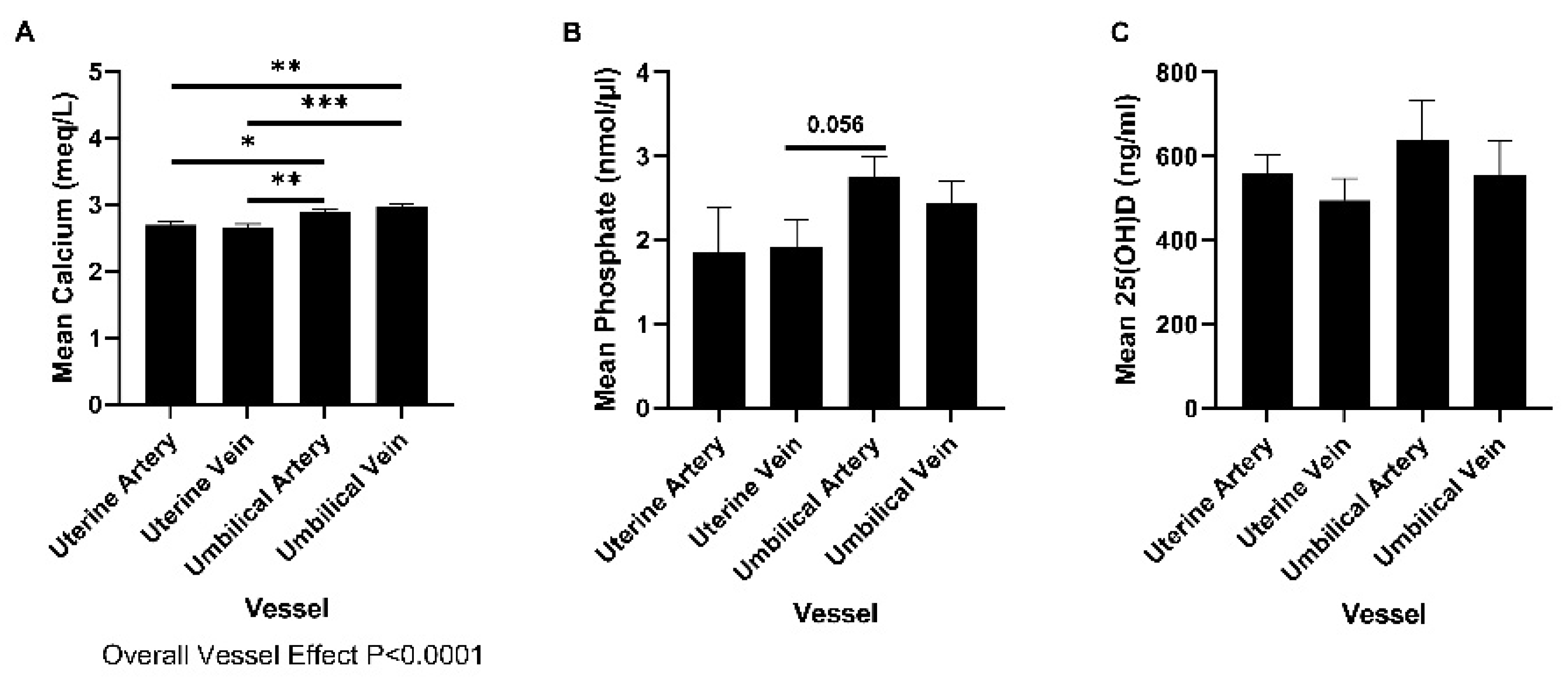
2.1. Concentrations of Phosphate, Calcium, and 25(OH)D in Uterine and Umbilical Blood

Plasma samples from Control RNAi pregnancies on Day 132 of gestation were used to determine the concentrations of phosphate, calcium, and 25(OH)D in uterine and umbilical vessels in late gestation. Calcium was more abundant in umbilical blood vessels than uterine blood vessels (Figure 1A; Overall vessel effect p < 0.0001). Phosphate tended to be more abundant in umbilical arteries than uterine veins on Day 132 of gestation (post-hoc comparison p = 0.056); Figure 1B). The concentrations of 25(OH)D were not different in plasma from each of the vessel types investigated (Figure 1C).

2.2. The Effect of CSH RNAi on Calcium, Phosphate, and Vitamin D in Plasma

2.2.1. Calcium

The uterine uptake of calcium was less in CSH RNAi IUGR pregnancies compared to CSH RNAi Non-IUGR pregnancies (p < 0.05; Table 1). Similarly, the umbilical uptake of calcium was less in CSH RNAi IUGR pregnancies compared to Control RNAi pregnancies (p < 0.05; Table 1. Overall treatment effect p = 0.089). Furthermore, CSH RNAi Non-IUGR pregnancies had a lower umbilical vein–umbilical artery calcium gradient than Control RNAi pregnancies (p < 0.05; Table 1). CSH RNAi did not affect concentrations of calcium in plasma from uterine arteries or umbilical vessels, the uterine artery–vein calcium gradient, the uteroplacental utilization of calcium, or the uterine and umbilical uptakes of calcium relative to uterine, fetal and placental weights (Table 1).

2.2.2. Phosphate

It is important to note that plasma from uterine arterial samples from non-IUGR pregnancies was unavailable for phosphate analyses. There were no significant differences in concentrations of phosphate in uterine or umbilical vessels, uterine artery-vein or umbilical vein-artery gradients, uterine or umbilical uptakes, or uteroplacental utilization of phosphate between Control RNAi and CSH RNAi IUGR pregnancies (Supplementary Table S1).

2.2.3. 25(OH)D

It is important to note that plasma from uterine arterial samples from non-IUGR pregnancies was unavailable for 25(OH)D analyses. 25(OH)D was less abundant in plasma from uterine arteries (p < 0.05; Table 2) and veins (p < 0.05; Table 2) from CSH RNAi IUGR pregnancies compared to Control RNAi pregnancies. Similarly, 25(OH)D tended to be less abundant in plasma from uterine veins from CSH RNAi IUGR pregnancies compared to CSH RNAi Non-IUGR pregnancies (p = 0.055; Table 2). Furthermore, there tended to be an effect of CSH RNAi on the umbilical uptake of 25(OH)D per kg of fetus (Overall Treatment Effect p = 0.09), with less 25(OH)D uptake in the CSH RNAi IUGR pregnancies compared to Control RNAi pregnancies (posthoc comparison p = 0.059; Table 2). There were no significant differences in concentrations of 25(OH)D in umbilical vessels, uterine artery-vein or umbilical vein-artery gradients, uterine or umbilical uptakes, or uteroplacental utilization of 25(OH)D between Control RNAi and CSH RNAi IUGR pregnancies (Table 2).

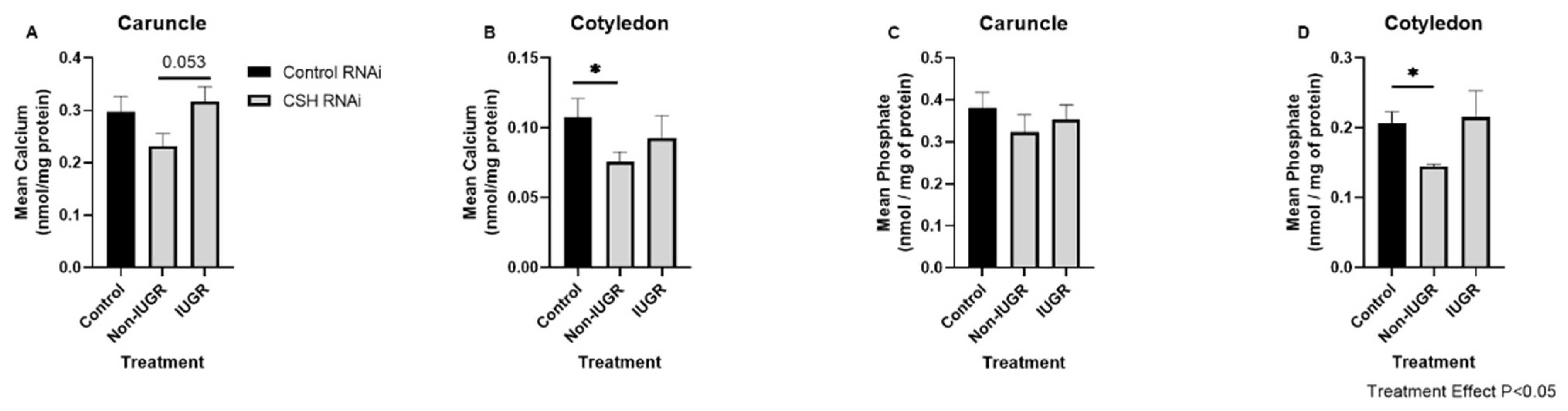
2.3. Effect of CSH RNAi on Abundances of Calcium and Phosphate in Cotyledons and Caruncles

Caruncles from CSH RNAi Non-IUGR pregnancies tended to have less calcium than CSH RNAi IUGR pregnancies (p = 0.053; Figure 2A). Similarly, calcium (p < 0.05; Figure 2B) and phosphate (p < 0.05; Figure 2D) were less abundant in cotyledons from CSH RNAi Non-IUGR pregnancies compared to Control RNAi pregnancies. CSH RNAi did not alter abundance of phosphate in caruncles (Figure 2C) on Day 132 of gestation.

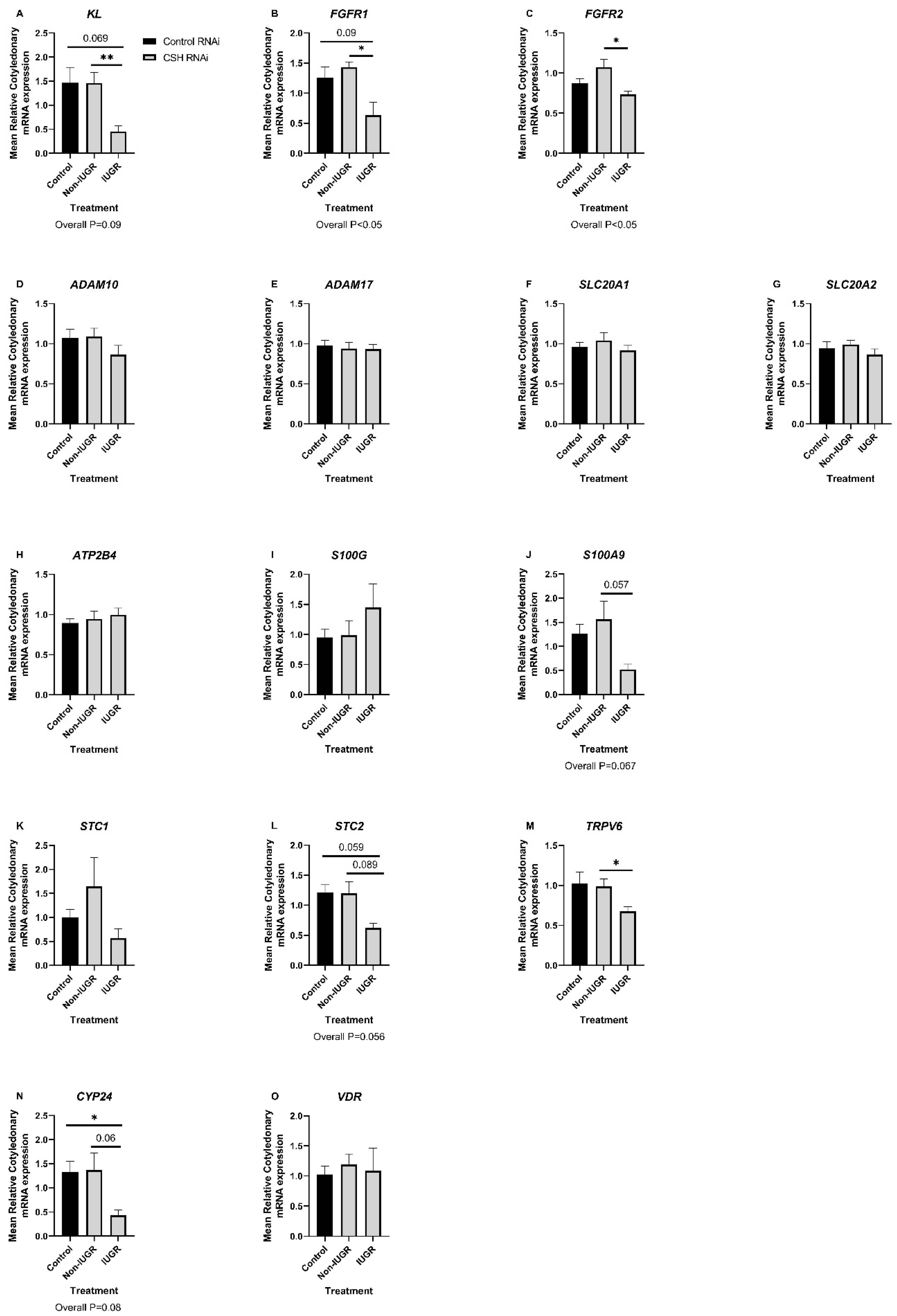
2.4. Effect of CSH RNAi on the Expression of Candidate mRNAs with Roles in the Regulation of Calcium, Phosphate, and Vitamin D Signaling in Caruncles and Cotyledons

The expression of the sodium dependent phosphate transporter (solute carrier family 20 member 2) SLC20A2 mRNA was lower in caruncles from CSH RNAi IUGR pregnancies compared to CSH RNAi Non-IUGR pregnancies (posthoc comparison p < 0.05; overall treatment effect p = 0.056. Supplementary Figure S1G). CSH RNAi did not affect the expression of KL, FGFR1, FGFR2, ADAM10, ADAM17, PTHRP, SLC20A1, STC1, STC2, ATP2B4, PTHRP, S100G, TRPV6, CYP24, or VDR mRNAs in caruncles (p > 0.10; Supplementary Figure S1). There were treatment effects of CSH RNAi on the expression of KL (p = 0.098; Figure 3A), FGFR1 (p < 0.05; Figure 3B), FGFR2 (p < 0.05; Figure 3C), S100A9 (p = 0.067; Figure 3J), STC2 (p = 0.067; Figure 3L) and CYP24 (p = 0.096; Figure 3N) mRNAs in cotyledons. Cotyledons from CSH RNAi IUGR pregnancies had lower expression of CYP24 (p < 0.05; Figure 3N) mRNA compared to Control RNAi pregnancies. Similarly, the expression of KL (p = 0.069; Figure 3A), FGFR1 (p = 0.09; Figure 3B), and STC2 (p = 0.059; Figure 3L) mRNAs tended to be lower in cotyledons from CSH RNAi IUGR pregnancies compared to Control RNAi pregnancies. Furthermore, the expression of KL (p < 0.01; Figure 3A), FGFR1 (p < 0.05; Figure 3B), FGFR2 (p < 0.05; Figure 3C), S100A9 (p = 0.057; Figure 3J), STC2 (p = 0.089; Figure 3L), TRPV6 (p < 0.05; Figure 3M), and CYP24 (p = 0.06; Figure 3N) mRNAs was lower in cotyledons from CSH RNAi IUGR pregnancies compared to CSH RNAi Non-IUGR pregnancies. CSH RNAi did not affect the expression of ADAM10, ADAM17, SLC20A1, SLC20A2, STC1, ATP2B4, S100G, TRPV6, or VDR mRNAs in cotyledons (p > 0.10; Figure 3).

2.5. Correlations between Biometric Data and Caruncular and Cotyledonary Phosphate and Calcium Abundance

To further assess the relationship between the abundance of calcium and phosphate and biometric data from this study, maternal, uterine, fetal, and placental weights were correlated with the abundance of phosphate and calcium in caruncular and cotyledonary homogenates (Supplementary Table S2). The abundance of calcium in caruncles tended to be negatively associated with fetal weight (r = 0.181; p = 0.06). In contrast, the abundance of both calcium (r = 0.163; p = 0.09) and phosphate (r = 0.365; p < 0.01) were positively correlated to placental weight.

2.6. Correlations between Biometric Data and Calcium, Phosphate, and Vitamin D Utilization

Maternal and uterine (Supplementary Table S3), and fetal and placental (Supplementary Table S4) weights were correlated against the abundance, uterine uptake, umbilical uptake, uterine artery-umbilical artery gradient, uterine artery-uterine vein gradient, and uteroplacental utilization of phosphate, calcium, and 25(OH)D. The uteroplacental utilization of phosphate tended to be positively correlated with fetal weight (r = 0.483; p = 0.056). The abundance of calcium in umbilical arterial (r = 0.161; p = 0.08) and venous (r = 0.181; p = 0.06) plasma tended to be positively correlated with placental weight. Furthermore, umbilical calcium uptake was positively correlated with uterine weight (r = 0.174; p = 0.067), fetal weight (r = 0.499; p < 0.001), and placental weight (r = 0.319; p < 0.01). The abundance of 25(OH)D in uterine venous plasma was positively correlated with fetal weight (r = 0.294; p < 0.05). In contrast, the uterine artery-uterine vein calcium gradient was negatively correlated with uterine weight (r = 0.276; p < 0.05), fetal weight (r = 0.334; p < 0.01), and placental weight (r = 0.210; p < 0.05).

2.7. Correlations between Biometric Data and the Caruncular and Cotyledonary mRNA Expression of Calcium, Phosphate, and Vitamin D Regulatory Molecules

Maternal, uterine, fetal, and placental weights were correlated against the caruncular and cotyledonary expression of calcium, phosphate, and vitamin D regulatory mRNAs (Table 3). Maternal weight was negatively correlated with the caruncular expression of PTHrP (r = 0.262; p < 0.05) and STC1 (r = 0.233; p < 0.05) mRNAs. Uterine weight was negatively correlated with the caruncular expression of ADAM10 (r = 0.226; p < 0.05). Likewise, ATP2B4 (r = 0.185; p = 0.058), PTHrP (r = 0.177; p = 0.072), and STC1 (r = 0.199; p = 0.06) mRNAs tended to be negatively correlated with uterine weight. Furthermore, fetal weight was negatively correlated with the caruncular expression of ADAM10 (r = 0.161; p = 0.079), SLC20A1 (r = 0.241; p < 0.05), and PTHrP (r = 0.281; p < 0.05) mRNAs. Similarly, the caruncular expression of ADAM10 (r = 0.159; p = 0.08) and SLC20A1 (r = 0.288; p < 0.05) mRNAs was negatively correlated with placental weight. In contrast, the expression of SLC20A2 mRNA by caruncles tended to be positively correlated with fetal weight (r = 0.167; p = 0.074). The cotyledonary expression of VDR mRNA was negatively correlated with fetal (r = 0.224; p < 0.05) and placental (r = 0.208; p < 0.05) weight. Maternal weight was positively correlated with the cotyledonary expression of FGFR2 mRNA (r = 0.389; p < 0.01) and tended to be positively correlated with ADAM10 (r = 0.158; p = 0.083), and SLC20A1 (r = 0.195; p = 0.059) mRNAs. Similarly, cotyledonary KL mRNA expression tended to be correlated with uterine weight (r = 0.184; p = 0.067). Furthermore, fetal weight was positively correlated with the cotyledonary expression of KL (r = 0.222; p < 0.05), FGFR1 (r = 0.194; p = 0.05), and S100A9 (r = 0.225; p < 0.05) mRNAs.

3. Discussion

Findings from studies across eutherian mammals suggest a critical role for the utero-placental transport of minerals for the establishment and maintenance of pregnancy and for the regulation of fetal-placental growth and development [1,18]. Despite the wide acceptance that phosphate, calcium, and vitamin D are essential during pregnancy, few details exist regarding the hormonal regulation of mechanisms required for transport and metabolism of these fundamental nutrients.
While several techniques exist to study placental mineral transport in pregnant ewes [1], the most widely accepted is in situ placental perfusion [26,27]. However, although this technique has provided significant enhancements in the understanding of mineral transport in the pregnant sheep, it requires removal of the fetus that leads to a rapid decline in placental function. Thus, caution should be taken when extrapolating the significance of these findings. In contrast, the present study utilized catheters which did not disrupt placental or fetal functions and, therefore, provides important new advances regarding the understanding of mineral transport, uptake, and utilization by the uterus and conceptus in late gestation. The utilization of real-time, steady-state quantification of the abundance of calcium in relation to uterine and umbilical blood flow provides a reliable assessment of calcium flow through the feto-placental unit. Phosphate, calcium, and vitamin D signaling pathways are intimately interrelated [18] and, therefore, placental transport and utilization of these nutrients must not be considered in isolation. Our real-time calcium measurements, in combination with the quantification of phosphate and 25(OH)D in uterine and umbilical samples by colorimetric methods and ELISA, respectively, provide important insights into mineral transport during late gestation in pregnant ewes.
It is widely appreciated that mammalian fetuses are hypercalcemic compared to maternal calcium levels in late gestation, a process ensuring appropriate skeletal mineralization [1]. Considering this, it was unsurprising to find a greater abundance of calcium in umbilical vessels (both artery and vein) compared to uterine vessels (both artery and vein) in control animals in the present study. This is consistent with extensive transport of calcium to the fetus, as demonstrated in Table 1, which was expected as substantial amounts of calcium accumulate in both amniotic and allantoic fluid for utilization by the fetus in late gestation [17]. Further, placental transport of minerals is primarily uni-directional, ensuring that adequate mineral supplies are available for the fetus. Thus, the back-flux of calcium from conceptus to mother is estimated to be as little as 0.4% in sheep in late gestation [28].
Prolactin and CSH have been suggested to regulate intestinal calcium absorption, reduce urinary calcium excretion [29], and stimulate synthesis of PTHrP [30]. Regulation of intestinal mineral absorption is a critical adaptation to increase the availability of minerals from the maternal circulation [18], thereby preventing hypocalcemia during periods of high demands of the fetus for minerals, particularly during late gestation. Intrauterine infusions of ovine CSH increase the abundance of STC1 in progestinized ewes [31], demonstrating important roles for P4 and CSH in the regulation of calcium transport by the ovine uterus. However, to date, there have been limited investigations into the role of CSH in the regulation of calcium homeostasis in late gestation, when there is abundant expression of CSH by binucleate cells and the syncytium of ovine placentomes and its release into the maternal peripheral circulation [22,23,32]. The present study provides novel evidence for a role of CSH in the regulation of calcium homeostasis during late gestation in sheep. CSH RNAi IUGR pregnancies had less uterine calcium uptake than Control RNAi pregnancies. Similarly, CSH RNAi IUGR pregnancies had less umbilical calcium uptakes and lower cotyledonary expression of TRPV6 mRNA than CSH RNAi Non-IUGR pregnancies. Interestingly, calcium homeostasis also appeared to be altered in the CSH RNAi Non-IUGR pregnancies. The umbilical vein-umbilical artery calcium gradient was lower in CSH RNAi Non-IUGR pregnancies, suggesting either lower utilization or greater availability of calcium for CSH RNAi Non-IUGR fetuses compared to both the Control RNAi and CSH RNAi Non-IUGR fetuses. Additionally, results of this study revealed a lower abundance of calcium in the cotyledons of CSH RNAi Non-IUGR pregnancies when compared to Control RNAi pregnancies. Although these fetuses were not considered to be intrauterine growth restricted, CSH RNAi does reduce uterine blood flow, decreases the abundance of NOS3 protein, and increases the utero-placental utilization of glucose compared to Control RNAi pregnancies [25]. Clearly, despite no gross differences in fetal weight, there are nutritional and potentially metabolic perturbations in the CSH RNAi Non-IUGR pregnancies which warrant further investigation.
Interestingly, the culture of human term trophoblast cells with the calcium ionophore A23187 inhibited the secretion of CSH [33], suggesting that calcium exerts a negative effect on the release of CSH from term human trophoblast cells. A relationship between calcium and CSH release in vitro has also been suggested in the sheep [34]. Ovine cotyledonary cells release greater amounts of CSH in response to culture with (1) calcium-depleted medium, (2) the calcium antagonist MgCl, and (3) the calcium channel blocking agents verapamil and nefidipine, suggesting an inverse relationship between extracellular concentrations of calcium and CSH secretion.
This study, to our knowledge, is the first to report a relationship between CSH and phosphate signaling and transport at the ovine maternal-conceptus interface. In the present study, there was less phosphate in cotyledons of CSH RNAi Non-IUGR pregnancies compared to Control RNAi pregnancies. Furthermore, the expression of sodium dependent phosphate transporter SLC20A2 mRNA was lower in caruncles from CSH RNAi IUGR compared to CSH RNAi Non-IUGR pregnancies, and this was accompanied by the lower expression of mRNAs for components of the KL-FGF signaling cascade (KL, FGFR1, and FGFR2) in cotyledons from CSH RNAi IUGR pregnancies compared to Control RNAi and/or CSH RNAi Non-IUGR pregnancies. Given the intimate relationship between phosphate and calcium homeostasis [35], and the essential role of CSH in the regulation of placental nutrient transport, fetal growth, and development [36], it is perhaps unsurprising that alterations in phosphate signaling were observed in this study. As there were no effects of CSH RNAi treatment on abundance in uterine and umbilical vessels, uterine or umbilical uptake or utero-placental utilization of phosphate, local alterations in phosphate signaling may occur within placentomes to counteract the effects of CSH deficiency on calcium and vitamin D abundance. Further studies are required to fully understand the relationship between phosphate, calcium, vitamin D, and CSH at the maternal conceptus in sheep throughout gestation.
In sheep it is known that radiolabeled 1,25-dihydroxycholecalciferol can be transported across the placenta in both directions [37], and that ovine fetuses have greater metabolism of vitamin D compared to maternal vitamin D metabolism [38]. It has been suggested that, in pregnant ewes, the placenta is responsible for the generation of approximately 40% of the 1,25-dihydroxycholecalciferol in fetal blood and that local metabolism of vitamin D in the pregnant uterus is critical for the regulation of fetal growth. In fact, the cytochrome P450 (CYP) enzymes critical for the metabolism of vitamin D, and the vitamin D receptor, are expressed at the maternal-conceptus interface in late gestation (Day 125) in sheep [17,20]. This finding is not unique to sheep as the expression of vitamin D regulatory molecules in the female reproductive tract in pigs, humans, and rodents has been reported [39,40,41,42], suggesting important roles of vitamin D in the establishment and maintenance of pregnancy, and in the regulation of conceptus growth and development.
In the present study, there were lower concentrations of 25(OH)D in uterine arterial and venous blood from CSH RNAi IUGR pregnancies, suggesting a systemic effect of CSH RNAi on maternal vitamin D status in pregnancies with IUGR fetuses. Human CSH has been reported to stimulate the activity of 1α-hydroxylase (CYP27B1) [43], an important enzyme for the generation of ‘active’ vitamin D in vivo. Furthermore, CSH can increase plasma 1,25-dihydroxyvitamin D in hypophysectomized, non-pregnant rats [44]. Collectively, these findings suggest that CSH may act upon the liver and kidneys to alter the metabolism of vitamin D. Thus, it could be speculated that CSH RNAi that resulted in IUGR pregnancies may have systemic effects on maternal vitamin D status that must be elucidated in future studies. The umbilical uptake of 25(OH)D was negative across treatment groups, indicating production of 25(OH)D by the fetus. Furthermore, the decreased expression of CYP24 mRNA was observed in the cotyledons of CSH RNAi IUGR pregnancies compared to Control RNAi pregnancies. The activity of vitamin D is regulated by the catabolic activity of CYP24A1 (24-hydroxylase) which inactivates 1,25[OH]2D3 via conversion to 1,24,25[OH]3D3 [45]. Collectively, these findings suggest (1) alterations in maternal vitamin D status in response to CSH RNAi in ewes which have IUGR fetuses; and (2) that conceptuses (embryo/fetus and associated extra-embryonic membranes) from CSH RNAi IUGR pregnancies may increase the metabolism of vitamin D to compensate for the decrease in available maternal vitamin D. As these differences were unique to pregnancies with IUGR fetuses, they provide additional evidence for an important role of vitamin D in the regulation of fetal growth and suggest that CSH influences vitamin D homeostasis in the pregnant ewe.
It is important to note that the majority of differences in the tissues in response to CSH RNAi were observed in the cotyledon and not the caruncle. This is perhaps unsurprising given that the CSH is expressed by the binucleate cells and the syncytium of the ovine placentomes [21,22]. Thus, the findings presented in this study support the concept that CSH regulates mineral transport in the fetal portion of the placentome.

4. Materials and Methods

4.1. Experimental Animals and Sample Collection

The experimental design and sample collection methods have been published with regard to the impact of chorionic somatomammotropin RNA interference (CSH RNAi) on uterine and umbilical blood flows, and nutrient utilization for lentiviral scrambled control RNAi (control, n = 10), normal-weight (non-IUGR, n = 6) and growth restricted sheep fetuses (IUGR, n = 4) [24,25]. In brief, all ewes (Dorper breed composition) were group housed in pens at the Colorado State University Animal Reproduction and Biotechnology Laboratory, and were provided access to hay, trace minerals, and water in order to meet or slightly exceed their National Research Council requirements. Following synchronization and subsequent breeding, fully expanded and hatched blastocysts were collected by flushing the uteri on Day nine of pregnancy. Each blastocyst was infected with 100,000 transducing units of either control RNAi or CSH RNAi virus. After infection for approximately 5 h, each blastocyst was washed, and a single blastocyst was transferred surgically into a synchronized recipient ewe. Each recipient ewe was monitored daily for return to standing estrus and confirmed pregnant on Day 50 of gestation by ultrasound (Mindray Medical Equipment, Mahwah, NJ, USA).

4.2. Quantification of Calcium, Phosphate, and 25(OH)D in Uterine Flushings, Plasma, and Endometrial Homogenates

As described previously, on Day 126 of gestation, ewes underwent surgical placement of fetal and maternal catheters in order to determine blood flow and nutrient flux. Briefly, the following catheters were placed: fetal descending aorta (representing umbilical artery blood), fetal femoral vein and umbilical vein, maternal femoral artery (representing uterine artery blood), maternal femoral vein, and uterine vein [25]. On Day 132 of gestation, uterine and umbilical blood flows were determined using the steady state 3H2O transplacental diffusion technique. Using these catheters, four samples of blood were obtained on Day 132 at 20-min intervals for the real time measurement of calcium (Ca2+) using a blood gas analyzer (ABL800 Flex, Radiometer, Brea, CA, USA). Day 132 was selected as peak concentrations of CSH in peripheral serum are found between Days 120 to 140 of gestation [23].
Snap-frozen samples from caruncles (maternal component of placentome) and cotyledons (fetal component of placentomes) (300–500 mg of tissue) were homogenized in 1 mL of lysis buffer (60 mM Tris-HCl (Sigma Aldrich, St. Louis, MO, USA), 1 mM Na3VO4 (Fisher Scientific, Waltham, MA, USA), 10% glycerol (Fisher Scientific), and 1% sodium dodecyl sulfate (BioRad, Hercules, CA, USA), containing an EDTA-free protease inhibitor (Roche, Indianapolis, IN, USA)). Homogenates were centrifuged at 14,000× g for 15 min at 4 °C, and supernatants were stored at −80 °C until assayed. The concentration of total proteins in the tissue homogenates was quantified spectrophotometrically (SynergyH1, BioTek, Winooski, VT, USA. Absorbance 562 nm) using a protein assay dye reagent (BioRad; 500-0006) according to the manufacturer’s instructions.
The concentrations of calcium in caruncular and cotyledonary homogenates were quantified using a colorimetric assay (Sigma Aldrich; MAK022), as described previously [17]. The concentrations of phosphate in uterine and umbilical plasma samples, and caruncular and cotyledonary homogenates were quantified using a colorimetric assay (Abcam, Cambridge, MA, USA; ab65622), as described previously [16]. Additionally, the concentrations of the vitamin D metabolite 25(OH)D in uterine and umbilical plasma samples were quantified by ELISA (Eagle Biosciences, Amherst, NH, USA; VID91-K01), as described previously [17].
Uterine and umbilical gradients, and uptakes of calcium, phosphate, and 25(OH)D relative to uterine, fetal, and placental weights were calculated using mathematical formulas published previously [25] (see Table 4). It is important to note that plasma from uterine arterial samples from non-IUGR pregnancies was unavailable for phosphate and 25(OH)D analyses. The abundance of calcium and phosphate in caruncular and cotyledonary homogenates are expressed relative to concentration of protein in each sample.

4.3. Analysis of Candidate Gene Expression by qPCR

Total RNA was extracted from ovine caruncles and cotyledons using Trizol (15596018 Invitrogen, Carlsbad, CA, USA) and further purified using the RNeasy Mini Kit (74104 Qiagen, Hilden, Germany) as described previously [17]. Concentrations of RNA were quantified using a Nanodrop (ND-1000 Spectrophotometer), and all samples had a 260/280 value >2. Complementary DNA (cDNA) was synthesized from 1 µg of RNA with SuperScript II reverse transcriptase and oligo (deoxythymidine) primers (Invitrogen, Carlsbad, CA, USA), as per the manufacturer’s instructions. Negative controls without reverse transcriptase were included to test for genomic contamination and all cDNA was stored at −20 °C until required.
The relative expression of candidate genes in cotyledonary and caruncular samples was quantified by quantitative polymerase chain reaction (qPCR) as described previously [16,17]. To investigate the effects of CSH on phosphate signaling in the sheep placentome, the mRNA expression of the sodium dependent phosphate transporters solute carrier family 20 member 1 (SLC20A1) and SLC20A2, and components of the fibroblast growth factor-klotho signalling cascade (fibroblast growth factor receptor 1 [FGFR1], FGFR2, klotho [KL], a disintegrin and metalloproteinase domain-containing protein 10 [ADAM10], and ADAM17) was quantified by qPCR. To investigate the effects of CSH on calcium signalling in sheep placentomes, the mRNA expression of the calcium binding proteins S100 calcium-binding protein G (S100G) and S100A9, plasma membrane calcium-transporting ATPase 4 (ATP2B4), PTH related protein (PTHrP), stanniocalcin 1 (STC1), STC2, and transient receptor potential cation channel subfamily v member 6 (TRPV6) was quantified by qPCR. Additionally, the expression of the 25-hydroxyvitamin D3-24-hydroxylase (CYP24), which inactivates 1,25[OH]2D3 via conversion to 1,24,25[OH]3D3, and vitamin D receptor (VDR) was quantified by qPCR. qPCR was performed using the ABI prism 7900HT system (Applied Biosystems, Foster City, CA, USA) with Power SYBR Green PCR Master Mix (Applied Biosystems), as per the manufacturer’s instructions to determine expression of mRNAs encoding for genes of interest. Primer sequences are provided in Supplementary Table S5. Primer efficiency and specificity were evaluated by generating a standard curve from pooled cDNA and by the inclusion of a dissociation curve to the RT reaction, respectively. Serial dilutions of pooled cDNA in nuclease-free water ranging from 1:2 to 1:256 were used as standards. All primer sets used amplified a single product and had an efficiency of between 95% and 105%. Each well contained 10% cDNA, 30% nuclease-free water, 10% primer, and 50% SYBR Green reaction mix in a 10 µL reaction volume. All reactions were performed at an annealing temperature of 60 °C. The stability of reference genes was assessed by geNORM V3.5 (Ghent University Hospital, Centre for Medical Genetics, Ghent, Belgium) in each tissue, with an M value of <1.5, and the reference genes used for normalization had stable expression. The reference genes GAPDH and TUB, and ACTB and SDHA were determined to have stable expression in caruncles and cotyledons, respectively, by geNORM V3.5 (Ghent University Hospital, Centre for Medical Genetics). Additionally, the effects of CSH RNAi were determined using GenStat (Version 13.1; VSN International Ltd., Indore, India) to ensure that there was no effect of CSH knockdown on reference gene expression. The abundances of the candidate mRNAs in the samples were quantified using the ΔΔCq method.

4.4. Statistical Analysis

All statistical analyses were performed using Minitab (Minitab, LLC, Chicago, IL, USA) or Graphpad Prism 9 Software (Graphstats Technologies Private Limited, Karnataka, India). Mean values were calculated for each individual sample for each parameter investigated and the normality of the distribution of the data was assessed using the Anderson-Darling test. If p < 0.05, the data were not considered to have a normal distribution. Transformations were carried out if necessary to achieve a Gaussian distribution. Outliers identified by a ROUT outlier test were excluded. ANOVA with a Tukey post-hoc analysis or t-tests were performed where appropriate to assess the effects of CSH RNAi on the parameters investigated. The effect of CSH RNAi on all parameters investigated was compared between (1) control RNAi, (2) CSH RNAi Non-IUGR, and (3) CSH RNAi IUGR groups. The results were considered significant at p < 0.05, trending towards significant at p > 0.05 < 0.1 and not significant at p > 0.1. Correlations were performed between maternal, uterine, fetal, and placental weights and all parameters investigated in this study.

5. Conclusions

This study has provided novel insights into the abundance and relative utilization of calcium, phosphate, and vitamin D by ovine uteri and conceptuses in late gestation. The results provide evidence supporting a role for CSH in the regulation of mineral transport and utilization at the ovine maternal-conceptus interface, thus providing a platform upon which further mechanistic understanding of the hormonal regulation of mineral transport during pregnancy in mammalian species can be achieved.

Supplementary Materials

The following supporting information can be downloaded at: https://www.mdpi.com/article/10.3390/ijms23147795/s1.

Author Contributions

The animal experimentation was planned and executed by R.V.A., A.R.T. and P.J.R. The samples for analyses were generously provided by A.R.T. and R.V.A., and analyses were performed by C.S. and K.M.H. The first draft of the manuscript was written by C.S. and F.W.B., and edited by K.M.H., A.R.T., L.J.S., R.V.A. and P.J.R. The final version of the manuscript was prepared by C.S. and F.W.B. and approved by K.M.H., A.R.T., P.J.R. and R.V.A. All authors have read and agreed to the published version of the manuscript.

Funding

This work was supported by National Institutes of Health Grants HD102909, HD093701, HD094952, and DK08813, and Agriculture and Food Research Initiative Grants 2016–38420-25289 and 2019–67011-29614 from the United State Department of Agriculture.

Institutional Review Board Statement

All animal procedures were approved by the Colorado State University Institutional Animal Care and Use Committee (Protocol Number 18-7866A), the Institutional Biosafety Committee (18-029B), and the University of Colorado Anschutz Medial Campus Institutional Animal Care and Use Committee (Protocol Number 00714) [24,25].

Informed Consent Statement

Not applicable.

Data Availability Statement

All data generated or analyzed during this study are included in this published article and its supplementary files.

Acknowledgments

The authors sincerely thank those involved in animal care and technical support for the generation and collection of these samples.

Conflicts of Interest

The authors declare that they have no conflict of interest.

References

  1. Kovacs, C.S. Bone Development and Mineral Homeostasis in the Fetus and Neonate: Roles of the Calciotropic and Phosphotropic Hormones. Physiol. Rev. 2014, 94, 1143–1218. [Google Scholar] [CrossRef] [PubMed]
  2. Santella, L. The Role of Calcium in the Cell Cycle: Facts and Hypotheses. Biochem. Biophys. Res. Commun. 1998, 244, 317–324. [Google Scholar] [CrossRef] [PubMed]
  3. Brostrom, M.A.; Brostrom, C.O. Calcium Dynamics and Endoplasmic Reticular Function in the Regulation of Protein Synthesis: Implications for Cell Growth and Adaptability. Cell Calcium 2003, 34, 345–363. [Google Scholar] [CrossRef]
  4. Chin, K.V.; Cade, C.; Brostrom, C.O.; Galuska, E.M.; Brostrom, M.A. Calcium-Dependent Regulation of Protein Synthesis at Translational Initiation in Eukaryotic Cells. J. Biol. Chem. 1987, 262, 16509–16514. [Google Scholar] [CrossRef]
  5. Glancy, B.; Balaban, R.S. Role of Mitochondrial Ca2+ in the Regulation of Cellular Energetics. Biochemistry 2012, 51, 2959–2973. [Google Scholar] [CrossRef] [Green Version]
  6. Jeon, U.S. Kidney and Calcium Homeostasis. Electrolyte Blood Press. 2008, 6, 68–76. [Google Scholar] [CrossRef] [Green Version]
  7. Penido, M.G.M.G.; Alon, U.S. Phosphate Homeostasis and Its Role in Bone Health. Pediatr. Nephrol. 2012, 27, 2039–2048. [Google Scholar] [CrossRef] [Green Version]
  8. Nabeshima, Y.I.; Imura, H. A-Klotho: A Regulator That Integrates Calcium Homeostasis. Am. J. Nephrol. 2008, 28, 455–464. [Google Scholar] [CrossRef]
  9. Khalil, R.; Ri, N.; Jardi, F.; Vanderschueren, D.; Claessens, F.; Decallonne, B. Sex Steroids and the Kidney: Role in Renal Calcium and Phosphate Handling. Mol. Cell. Endocrinol. 2018, 465, 61–72. [Google Scholar] [CrossRef]
  10. Bagur, R.; Hajnoczky, G. Intracellular Ca2+ Sensing: Role in Calcium Homeostasis and Signaling. Mol. Cell 2017, 66, 780–788. [Google Scholar] [CrossRef] [Green Version]
  11. Stafford, N.; Wilson, C.; Oceandy, D.; Neyses, L.; Cartwright, E.J. The Plasma Membrane Calcium ATPases and Their Role as Major New Players in Human Disease. Physiol. Rev. 2017, 97, 1089–1125. [Google Scholar] [CrossRef] [PubMed] [Green Version]
  12. Hermann, A.; Donato, R.; Weiger, T.M.; Chazin, W.J. S100 Calcium Binding Proteins and Ion Channels. Front. Pharmacol. 2012, 3, 67. [Google Scholar] [CrossRef] [PubMed] [Green Version]
  13. Islam, M.S. Transient Receptor Potential Channels. Adv. Exp. Med. Biol. 2011, 704, 218. [Google Scholar] [CrossRef]
  14. Segawa, H.; Shiozaki, Y.; Kaneko, I.; Miyamoto, K.I. The Role of Sodium-Dependent Phosphate Transporter in Phosphate Homeostasis. J. Nutr. Sci. Vitaminol. 2015, 61, S119–S121. [Google Scholar] [CrossRef] [PubMed] [Green Version]
  15. Lederer, E. Renal Phosphate Transporters. Curr. Opin. Nephrol. Hypertens. 2014, 23, 502–506. [Google Scholar] [CrossRef]
  16. Stenhouse, C.; Halloran, K.M.; Newton, M.G.; Gaddy, D.; Suva, L.J.; Bazer, F.W. Novel Mineral Regulatory Pathways in Ovine Pregnancy: 1. Phosphate, Klotho Signaling, and Sodium Dependent Phosphate Transporters. Biol. Reprod. 2021, 104, 1084–1096. [Google Scholar] [CrossRef]
  17. Stenhouse, C.; Halloran, K.M.; Newton, M.G.; Gaddy, D.; Suva, L.J.; Bazer, F.W. Novel Mineral Regulatory Pathways in Ovine Pregnancy: II. Calcium Binding Proteins, Calcium Transporters, and Vitamin D Signaling. Biol. Reprod. 2021, 105, 232–243. [Google Scholar] [CrossRef]
  18. Stenhouse, C.; Suva, L.J.; Gaddy, D.; Wu, G.; Bazer, F.W. Phosphate, Calcium, and Vitamin D: Key Regulators of Fetal and Placental Development in Mammals. Adv. Exp. Med. Biol. 2022, 1354, 77–107. [Google Scholar] [CrossRef]
  19. Stenhouse, C.; Halloran, K.; Moses, R.; Seo, H.; Gaddy, D.; Johnson, G.; Wu, G.; Suva, L.; Bazer, F. Effects of Progesterone and Interferon Tau on Ovine Endometrial Phosphate, Calcium, and Vitamin D Signaling. Biol. Reprod. 2022, 106, 888–899. [Google Scholar] [CrossRef]
  20. Stenhouse, C.; Halloran, K.; Hoskins, E.; Newton, M.; Moses, R.; Seo, H.; Dunlap, K.; Satterfield, M.; Gaddy, D.; Johnson, G.; et al. Effects of Exogenous Progesterone on Expression of Mineral Regulatory Molecules by Ovine Endometrium and Placentomes. Biol. Reprod. 2022, 106, 1126–1142. [Google Scholar] [CrossRef]
  21. Martal, J.; Djiane, J. The Production of Chorionic Somatomammotrophin in Sheep. J. Reprod. Fertil. 1977, 49, 285–289. [Google Scholar] [CrossRef] [PubMed] [Green Version]
  22. Wooding, F.B.P.; Morgan, G.; Forsyth, I.A.; Butcher, G.; Hutchings, A.; Billingsley, S.A.; Gluckman, P.D. Light and Electron Microscopic Studies of Cellular Localization of OPL with Monoclonal and Polyclonal Antibodies. J. Histochem. Cytochem. 1992, 40, 1001–1009. [Google Scholar] [CrossRef] [PubMed] [Green Version]
  23. Chan, J.; Robertson, H.; Friesen, H. Maternal and Fetal Concentrations of Ovine Placental Lactogen Measured by Radioimmunoassay. Endocrinology 1978, 102, 1606–1613. [Google Scholar] [CrossRef] [PubMed]
  24. Tanner, A.R.; Lynch, C.S.; Kennedy, V.C.; Ali, A.; Winger, Q.A.; Rozance, P.J.; Anthony, R.V. CSH RNA Interference Reduces Global Nutrient Uptake and Umbilical Blood Flow Resulting in Intrauterine Growth Restriction. Int. J. Mol. Sci. 2021, 22, 8150. [Google Scholar] [CrossRef]
  25. Tanner, A.R.; Lynch, C.S.; Ali, A.; Winger, Q.A.; Rozance, P.J.; Anthony, R.V. Impact of Chorionic Somatomammotropin RNA Interference on Uterine Blood Flow and Placental Glucose Uptake in the Absence of Intrauterine Growth Restriction. Am. J. Physiol.-Regul. Integr. Comp. Physiol. 2021, 320, 138–148. [Google Scholar] [CrossRef]
  26. Weatherley, A.J.; Ross, R.; Pickard, D.W.; Care, A.D. The Transfer of Calcium during Perfusion of the Placenta in Intact and Thyroparathyroidectomized Sheep. Placenta 1983, 4, 271–278. [Google Scholar] [CrossRef]
  27. Care, A.; Caple, I.; Abbas, S.; Pickard, D. The Effect of Fetal Thyroparathyroidectomy on the Transport of Calcium across the Ovine Placenta to the Fetus. Placenta 1986, 7, 417–424. [Google Scholar] [CrossRef]
  28. Symonds, H.; Sansom, B.; Twardock, A. The Measurement of the Transfer of Calcium and Phosphorus from Foetus to Dam in the Sheep Using a Whole Body Counter. Res. Vet. Sci. 1972, 13, 272–275. [Google Scholar] [CrossRef]
  29. Burstyn, P.; Lloyd, I.; McKillop, W. The Effect of Human Placental Lactogen on the Renal Excretion of Calcium in the Rabbit. IRCS J. Med. Sci. 1975, 3, 30. [Google Scholar]
  30. Dvir, R.; Golander, A.; Jaccard, N.; Yedwab, G.; Otremski, I.; Spirer, Z.; Weisman, Y. Amniotic Fluid and Plasma Levels of Parathyrod Hormone-Related Protein and Hormonal Modulation of Its Secretion by Amniotic Fluid Cells. Eur. J. Endocrinol. 1995, 133, 277–282. [Google Scholar] [CrossRef] [Green Version]
  31. Song, G.; Bazer, F.W.; Wagner, G.F.; Spencer, T.E. Stanniocalcin (STC) in the Endometrial Glands of the Ovine Uterus: Regulation by Progesterone and Placental Hormones. Biol. Reprod. 2006, 74, 913–922. [Google Scholar] [CrossRef] [PubMed] [Green Version]
  32. Lea, R.G.; Wooding, P.; Stewart, I.; Hannah, L.T.; Morton, S.; Wallace, K.; Aitken, R.P.; Milne, J.S.; Regnault, T.R.; Anthony, R.V.; et al. The Expression of Ovine Placental Lactogen, StAR and Progesterone-Associated Steroidogenic Enzymes in Placentae of Overnourished Growing Adolescent Ewes. Reproduction 2007, 133, 785–796. [Google Scholar] [CrossRef] [PubMed] [Green Version]
  33. Hochberg, Z.; Bick, T.; Perlman, R.; Lahav, M.; Barzilai, D. The Modulation of Placental Lactogen Secretion by Calcium: Studies with Cultured Human Term Trophoblast. Mol. Cell. Biol. 1984, 37, 359–362. [Google Scholar] [CrossRef]
  34. Burke, J.; Rice, G.; Ralph, M.; Thorburn, G. Effects of Calcium Availability on the Release of Ovine Choriomammoropin from Cotyledonary Cells Incubated in Vitro. Comp. Biochem. Physiol. 1989, 93A, 489–492. [Google Scholar] [CrossRef]
  35. Renkema, K.Y.; Alexander, R.T.; Bindels, R.J.; Hoenderop, J.G. Calcium and Phosphate Homeostasis: Concerted Interplay of New Regulators. Ann. Med. 2008, 40, 82–91. [Google Scholar] [CrossRef]
  36. Anthony, R.V.; Limesand, S.W.; Fanning, M.D.; Liang, R. Placental Lactogen and Growth Hormone. Regulation and Action. In Endocrinology of Pregnancy; Bazer, F., Ed.; Humana Press: Totowa, NJ, USA, 1998; pp. 461–490. [Google Scholar]
  37. Devaskar, U.P.; Ho, M.; Devaskar, S.U.; Tsang, R.C. 25-Hydroxy- and 1 Alpha,25-Dihydroxyvitamin D. Maternal-Fetal Relationship and the Transfer of 1,25-Dihydroxyvitamin D3 across the Placenta in an Ovine Model. Dev. Pharmacol. Ther. 1984, 7, 213–220. [Google Scholar] [CrossRef]
  38. Ross, R.; Halbert, K.; Tsang, R.C. Determination of the Production and Metabolic Clearance Rates of 1,25-Dihydroxyvitamin D3 in the Pregnant Sheep and Its Chronically Catheterized Fetus by Primed Infusion Technique. Pediatr. Res. 1989, 26, 633–634. [Google Scholar] [CrossRef] [Green Version]
  39. Jang, H.; Choi, Y.; Yoo, I.; Han, J.; Hong, J.S.; Kim, Y.Y.; Ka, H. Vitamin D-Metabolic Enzymes and Related Molecules: Expression at the Maternal-Conceptus Interface and the Role of Vitamin D in Endometrial Gene Expression in Pigs. PLoS ONE 2017, 12, e0187221. [Google Scholar] [CrossRef] [Green Version]
  40. Shahbazi, M.; Jeddi-tehrani, M.; Zareie, M.; Salek-moghaddam, A.; Akhondi, M.M.; Bahmanpoor, M.; Sadeghi, M.R.; Zarnani, A.H. Expression Profiling of Vitamin D Receptor in Placenta, Decidua and Ovary of Pregnant Mice. Placenta 2011, 32, 657–664. [Google Scholar] [CrossRef]
  41. O’Brien, K.O.; Li, S.; Cao, C.; Kent, T.; Young, B.V.; Queenan, R.A.; Pressman, E.K.; Cooper, E.M. Placental CYP27B1 and CYP24A1 Expression in Human Placental Tissue and Their Association With Maternal and Neonatal Calcitropic Hormones. Endocr. Res. 2014, 99, 1348–1356. [Google Scholar] [CrossRef] [Green Version]
  42. Bergada, L.; Pallares, J.; Arcidiacono, M.; Cardus, A.; Santacan, M.; Valls, J.; Cao, G.; Fernandez, E.; Dolcet, X.; Dusso, A.; et al. Role of Local Bioactivation of Vitamin D by CYP27A1 and CYP2R1 in the Control of Cell Growth in Normal Endometrium and Endometrial Carcinoma. Lab. Investig. 2014, 94, 608–622. [Google Scholar] [CrossRef] [PubMed] [Green Version]
  43. Spanos, E.; Brown, D.; Stevenson, J.; Macintyre, I. Stimulation of 1,25-Dihydroxycholecalciferol Production by Prolactin and Related Peptides in Intact Renal Cell Preparations in Vitro. Biochim. Biophys. Acta 1981, 672, 7–15. [Google Scholar] [CrossRef]
  44. Takeuchi, K.; Morikawa, H.; Ueda, Y.; Mochizuki, M. Studies on the Effects of Placental Lactogen on Calcium Metabolism during Pregnancy. Nihon Naibunpi Gakkai Zasshi 1988, 64, 1175–1186. [Google Scholar] [CrossRef] [PubMed] [Green Version]
  45. Bikle, D. Vitamin D Metabolism, Mechanism of Action, and Clinical Applications. Chem. Biol. 2014, 21, 319–329. [Google Scholar] [CrossRef] [Green Version]
Figure 1. Concentration of calcium (A), phosphate (B), and 25(OH)D (C) in plasma from control RNAi uterine and umbilical blood collected on Day 132 of gestation. Data are presented as means ± SEM. Control RNAi, n = 10. * p < 0.05, ** p < 0.01, *** p < 0.001.
Ijms 23 07795 g001
Figure 2. Abundances of calcium (A,B) and phosphate (C,D) in caruncles (A,C) and cotyledons (B,D) from Control RNAi, CSH RNAi Non-IUGR, and CSH RNAi IUGR pregnancies. Data are presented as means ± SEM. Control RNAi, n = 10; CSH RNAi Non-IUGR, n = 6; CSH RNAi IUGR, n = 4. CSH, chorionic somatomammotropin; IUGR, intrauterine growth restriction; RNAi, RNA interference. * p < 0.05.
Ijms 23 07795 g002
Figure 3. Relative expression of candidate mRNAs with roles in calcium, phosphate, and vitamin D signaling, transport, and metabolism in cotyledons from Control RNAi, CSH RNAi Non-IUGR, and CSH RNAi IUGR pregnancies. Data are presented as means ± SEM. Control RNAi, n = 10; CSH RNAi Non-IUGR, n = 6; CSH RNAi IUGR, n = 4. CSH, chorionic somatomammotropin; IUGR, intrauterine growth restriction; RNAi, RNA interference. * p < 0.05, ** p < 0.01.
Ijms 23 07795 g003
Table 1. Effect of CSH RNAi on serum calcium abundance. Data are presented as means ± SEM. Different superscripts indicate statistical significance. (Control RNAi n = 10; CSH RNAi Non-IUGR n = 6; CSH RNAi IUGR n = 4). CSH, chorionic somatomammotropin; IUGR, intrauterine growth restriction; N/A, samples/results not available; RNAi, RNA interference.
CSH RNAiOverall
p Value
Control RNAiNon-IUGRIUGR
Uterine
Uterine Arterial Calcium (meq/L)2.694 ± 0.0542.594 ± 0.0262.776 ± 0.029>0.10
Uterine Venous Calcium (meq/L)2.654 ± 0.0522.538 ± 0.0262.718 ± 0.0160.09
Uterine Artery-Vein Calcium Gradient (meq/L)0.040 ± 0.0070.056 ± 0.0020.058 ± 0.016>0.10
Uterine Calcium Uptake (meq/min)0.071 ± 0.012 ab0.080 ± 0.004 a0.057 ± 0.010 b>0.10
Uterine Calcium Uptake per kg of Uterus (meq/min)0.109 ± 0.0230.133 ± 0.0040.112 ± 0.014>0.10
Uterine Calcium Uptake per kg Fetus (meq/min)0.019 ± 0.0030.021 ± 0.0010.021 ± 0.003>0.10
Uterine Calcium Uptake per kg Placenta (meq/min)0.161 ± 0.0250.209 ± 0.0090.172 ± 0.032>0.10
Umbilical
Umbilical Arterial Calcium (meq/L)2.896 ± 0.0422.878 ± 0.0642.899 ± 0.104>0.10
Umbilical Venous Calcium (meq/L)2.977 ± 0.0422.937 ± 0.0642.984 ± 0.107>0.10
Umbilical Vein-Artery Gradient (meq/L)0.081 ± 0.005 a0.060 ± 0.009 b0.086 ± 0.004 ab<0.05
Umbilical Calcium Uptake (meq/min)0.063 ± 0.006 a0.048 ± 0.008 ab0.038 ± 0.008 b0.089
Umbilical Calcium Uptake per kg of Uterus (meq/min)0.089 ± 0.0110.078 ± 0.0130.075 ± 0.014>0.10
Umbilical Calcium Uptake per kg Fetus (meq/min)3.759 ± 0.71712.325 ± 0.43012.078 ± 0.695>0.10
Umbilical Calcium Uptake per kg Placenta (meq/min)0.145 ± 0.0160.123 ± 0.0190.105 ± 0.007>0.10
Uteroplacental
Uteroplacental Calcium Utilization (meq/L)0.008 ± 0.0160.032 ± 0.0060.019 ± 0.012>0.10
Table 2. Effect of CSH RNAi on serum 25(OH)D abundance. Data are presented as means ± SEM. Different superscripts indicate statistical significance. (Control RNAi n = 4–10; CSH RNAi Non-IUGR n = 6; CSH RNAi IUGR n = 4). CSH, chorionic somatomammotropin; IUGR, intrauterine growth restriction; N/A, samples/results not available; RNAi, RNA interference.
CSH RNAiOverall p Value
Control RNAiNon-IUGRIUGR
Uterine
Uterine Arterial 25(OH)D (ng/mL)560.5 ± 43.74 aN/A322.9 ± 68.68 b<0.05
Uterine Venous 25(OH)D (ng/mL)495.0 ± 51.87 a485.3 ± 52.62 ab267.2 ± 28.82 b<0.05
Uterine Artery-Vein 25(OH)D Gradient (ng/mL)61.63 ± 26.96N/A59.07 ± 28.82>0.10
Uterine 25(OH)D Uptake (µg/min)38.77 ± 12.81N/A84.47 ± 33.26>0.10
Uterine 25(OH)D Uptake per kg of Uterus (µg/min)85.59 ± 36.63N/A157.7 ± 58.69>0.10
Uterine 25(OH)D Uptake per kg Fetus (µg/min)21.74 ± 12.31N/A25.71 ± 9.403>0.10
Uterine 25(OH)D Uptake per kg Placenta (µg/min)192.0 ± 113.6N/A204.2 ± 87.33>0.10
Umbilical
Umbilical Arterial 25(OH)D (ng/mL)639.7 ± 93.39683.3 ± 127.6830.3 ± 230.2>0.10
Umbilical Venous 25(OH)D (ng/mL)555.4 ± 80.09573.0 ± 74.25586.9 ± 170.4>0.10
Umbilical Vein-Artery 25(OH)D Gradient (ng/mL)−84.26 ± 71.34−110.3 ± 132.6−243.4 ± 104.3>0.10
Umbilical 25(OH)D Uptake (µg/min)−63.13 ± 51.85−63.28 ± 90.15−227.4 ± 105.2>0.10
Umbilical 25(OH)D Uptake per kg of Uterus (µg/min)−99.51 ± 62.51−118.7 ± 150.5−468.5 ± 226.8>0.10
Umbilical 25(OH)D Uptake per kg Fetus (µg/min)−16.92 ± 13.12−14.89 ± 22.05−98.13 ± 55.180.09
Umbilical 25(OH)D Uptake per kg Placenta (µg/min)−162.9 ± 122.0−165.6 ± 234.2−723.6 ± 434.9>0.10
Uteroplacental
Uteroplacental 25(OH)D Utilization (µg/min)155.3 ± 84.27N/A252.9 ± 139.7>0.10
Table 3. Correlations between biometric data and the caruncular and cotyledonary mRNA expression of calcium, phosphate, and vitamin D regulatory molecules.
Biometric ParametermRNACaruncleCotyledon
rp-Valuerp-Value
Maternal WeightKL0.0980.2050.0830.232
Maternal WeightFGFR10.0010.8960.1160.142
Maternal WeightFGFR20.0700.2610.3890.006
Maternal WeightADAM100.0030.8150.1580.083
Maternal WeightADAM170.0210.5380.0050.783
Maternal WeightSLC20A10.0600.2990.1950.059
Maternal WeightSLC20A20.0720.2510.0190.586
Maternal WeightATP2B40.0070.7310.0390.433
Maternal WeightPTHrP0.2620.025N/AN/A
Maternal WeightS100G0.0490.3460.0240.522
Maternal WeightS100A9N/AN/A0.0860.223
Maternal WeightSTC10.2330.0420.1050.205
Maternal WeightSTC20.0590.3140.0930.203
Maternal WeightTRPV60.0220.5350.0910.210
Maternal WeightCYP240.0500.3410.1150.168
Maternal WeightVDR0.0140.6150.0120.645
Uterine WeightKL0.0580.3380.1840.067
Uterine WeightFGFR10.0250.5190.0090.699
Uterine WeightFGFR20.0020.8510.0040.803
Uterine WeightADAM100.2260.0340.0080.714
Uterine WeightADAM170.1400.1050.0010.880
Uterine WeightSLC20A10.0850.2140.0010.879
Uterine WeightSLC20A20.0080.7100.1270.147
Uterine WeightATP2B40.1850.0580.0450.398
Uterine WeightPTHrP0.1770.072N/AN/A
Uterine WeightS100G0.00030.9527.535 × 10−50.972
Uterine WeightS100A9N/AN/A0.0600.312
Uterine WeightSTC10.1990.060.0580.351
Uterine WeightSTC20.00030.9420.0180.584
Uterine WeightTRPV60.0230.5240.0940.201
Uterine WeightCYP240.0240.5110.0390.432
Uterine WeightVDR0.0800.2260.0910.197
Fetal WeightKL0.060.3260.2220.041
Fetal WeightFGFR10.0550.3330.1940.05
Fetal WeightFGFR20.0410.3940.1200.160
Fetal WeightADAM100.1610.0790.0760.238
Fetal WeightADAM170.0050.7670.0020.854
Fetal WeightSLC20A10.2410.0280.0040.806
Fetal WeightSLC20A20.1670.0740.0900.227
Fetal WeightATP2B40.090.200.1150.170
Fetal WeightPTHrP0.2810.02N/AN/A
Fetal WeightS100G0.0070.7250.0020.854
Fetal WeightS100A9N/AN/A0.2250.04
Fetal WeightSTC10.1300.1420.2090.065
Fetal WeightSTC20.0020.8660.2020.053
Fetal WeightTRPV64.328 × 10−50.9780.0020.863
Fetal WeightCYP240.1390.1050.1050.190
Fetal WeightVDR5.289 × 10−60.9920.2240.035
Placental WeightKL0.0490.3760.0450.383
Placental WeightFGFR10.0150.6150.0450.369
Placental WeightFGFR28.461 × 10−60.9900.0310.486
Placental WeightADAM100.1590.083.854 × 10−50.979
Placental WeightADAM170.0540.3250.0020.848
Placental WeightSLC20A10.2880.0150.0340.451
Placental WeightSLC20A20.0040.7890.0020.878
Placental WeightATP2B40.0840.2150.0680.296
Placental WeightPTHrP0.0990.190N/AN/A
Placental WeightS100G0.0320.4510.0140.625
Placental WeightS100A9N/AN/A0.0220.548
Placental WeightSTC10.0320.4780.0810.270
Placental WeightSTC20.0280.4920.0770.249
Placental WeightTRPV60.1280.1220.0180.585
Placental WeightCYP240.010.6780.0070.736
Placental WeightVDR0.00060.9180.2080.043
Table 4. Calculations for Nutrient Uptake and Utilization. Abbreviations: UTA = uterine artery, UTV = uterine vein, UMA = umbilical artery, UMV = umbilical vein, [ ] = concentration.
ParameterFormula
Uterine uptakeuterine blood flow (mL/min) × ([UTA] − [UTV])
Umbilical uptakeumbilical blood flow (mL/min) × ([UMV] − [UMA])
Uteroplacental utilizationuterine uptake–umbilical uptake
Uterine uptake relative to uterine, fetal, or placental weightuterine uptake/uterine, fetal, or placental weight
Umbilical uptake relative to uterine, fetal, or placental weightumbilical uptake/uterine, fetal, or placental weight
Uterine artery–uterine vein gradient[UTA] − [UTV]
Umbilical vein–umbilical artery gradient[UMV] − [UMA]
Uterine artery-umbilical artery gradient[UTA] − [UMA]
Publisher’s Note: MDPI stays neutral with regard to jurisdictional claims in published maps and institutional affiliations.

