Article

Mitochondrial rRNA Methylation by Mettl15 Contributes to the Exercise and Learning Capability in Mice

by
Olga A. Averina
1,2,†,
Ivan G. Laptev
2,†,
Mariia A. Emelianova
3,
Oleg A. Permyakov
1,
Sofia S. Mariasina
1,
Alyona I. Nikiforova
4,
Vasily N. Manskikh
2,
Olga O. Grigorieva
1,
Anastasia K. Bolikhova
5,
Gennady A. Kalabin
6,
Olga A. Dontsova
2,3,7,8 and
Petr V. Sergiev
1,2,3,7,*
1
Institute of Functional Genomics, Lomonosov Moscow State University, 119991 Moscow, Russia
2
Belozersky Institute of Physico-Chemical Biology, Lomonosov Mosco16w State University, 119991 Moscow, Russia
3
Center of Life Sciences, Skolkovo Institute of Science and Technology, 121205 Moscow, Russia
4
Institute of Mitoengineering, Lomonosov Moscow State University, 119234 Moscow, Russia
5
Faculty of Bioengineering and Bioinformatics, Lomonosov Moscow State University, 119234 Moscow, Russia
6
Pharmacy Resource Center, Peoples’ Friendship University of Russia (RUDN), 117198 Moscow, Russia
7
Department of Chemistry, Lomonosov Moscow State University, 119991 Moscow, Russia
8
Shemyakin-Ovchinnikov Institute of Bioorganic Chemistry, 117997 Moscow, Russia
*
Author to whom correspondence should be addressed.
These authors contributed equally to this work.
Int. J. Mol. Sci. 2022, 23(11), 6056; https://doi.org/10.3390/ijms23116056
Submission received: 3 April 2022 / Revised: 22 May 2022 / Accepted: 26 May 2022 / Published: 27 May 2022

Abstract

:
Mitochondrial translation is a unique relic of the symbiotic origin of the organelle. Alterations of its components cause a number of severe human diseases. Hereby we report a study of mice devoid of Mettl15 mitochondrial 12S rRNA methyltransferase, responsible for the formation of m4C839 residue (human numbering). Homozygous Mettl15−/− mice appeared to be viable in contrast to other mitochondrial rRNA methyltransferase knockouts reported earlier. The phenotype of Mettl15−/− mice is much milder than that of other mutants of mitochondrial translation apparatus. In agreement with the results obtained earlier for cell cultures with an inactivated Mettl15 gene, we observed accumulation of the RbfA factor, normally associated with the precursor of the 28S subunit, in the 55S mitochondrial ribosome fraction of knockout mice. A lack of Mettl15 leads to a lower blood glucose level after physical exercise relative to that of the wild-type mice. Mettl15−/− mice demonstrated suboptimal muscle performance and lower levels of Cox3 protein synthesized by mitoribosomes in the oxidative soleus muscles. Additionally, we detected decreased learning capabilities in the Mettl15−/− knockout mice in the tests with both positive and negative reinforcement. Such properties make Mettl15−/− knockout mice a suitable model for mild mitochondriopathies.

1. Introduction

Mitochondria, the powerhouses of eukaryotic cells, possess their own genome as a remnant of its endosymbiotic origin [1]. To synthesize 13 protein components of the oxidative phosphorylation (OXPHOS) complexes encoded in the mitochondrial genome, the organelle makes use of the specialized translation apparatus [2,3,4,5] inherited from the endosymbiotic proteobacteria. Although ribosomal and transfer RNAs necessary for the mitochondrial protein biosynthesis are encoded within the organelle, ribosomal proteins, translation, and biogenesis factors [6] are encoded by the nuclear DNA, synthesized by the cytosolic ribosomes, and imported into the mitochondria.
Mutations in genes’ coding for the components of mitochondrial protein biosynthesis apparatus cause a number of severe human diseases, such as mitochondrial encephalopathy, lactic acidosis, and stroke-like episodes (MELAS), caused by a mutation in the mitochondrial tRNALeu(UUR) gene [7] and myoclonic epilepsy with ragged red fibers (MERRF) caused by a mutation in the mitochondrial tRNALys gene [8]. Later, a number of other mutations within the mitochondrial tRNA genes were found to be associated with similar pathologies [9]. Mutation A1555G in the mitochondrial 12S rRNA gene was found to cause sensorineural hearing loss exacerbated by aminoglycoside antibiotics [10]. A number of pathologies were found to be caused by mutations in nuclear genes’ coding for mitochondrial aminoacyl-tRNA synthetases, mitochondrial ribosomal proteins, as well as translation elongation and termination factors [11,12,13,14,15]. Mutations in genes’ coding for the proteins involved in mitochondrial ribosome biogenesis, such as ERAL1 [16], FASTKD2 [17], C1QBP/p32 [18], DHX30 [19], and GTPBP5 [20] are suggested as causative for human genetic disorders [15].
Mammalian mitochondrial rRNAs are modified at ten nucleotide residues [21,22,23], which are clustered in the functional centers of the small and large ribosomal subunits [24]. Mutations of the mitochondrial rRNA methyltransferase genes, such as TFB1M [25,26,27] and MRM2 [28], are also implicated in human diseases, such as maternally inherited deafness, diabetes, and MELAS-like syndrome. Although the list of mitochondrial rRNA modification enzymes was recently completed with the identification of the TRMT2B protein [29,30] as a 12S rRNA methyltransferase, the physiological impact of mitochondrial rRNA modifications at the level of an organism has been addressed for a limited number of related enzymes, such as TFB1M [31] and NSUN4 [32].
METTL15 is a mitochondrial rRNA methyltransferase, responsible for the 12S rRNA m4C839 modification [33,34,35]. Inactivation of the METTL15 gene in various human [33,34] and murine [35] cell lines was accompanied by a variable degree of mitochondrial translation impairment ranging from almost negligible to severe, which could be attributed to the difference in cell lines used. To address the influence of the mitochondrial 12S rRNA modification at m4C839 residue (human numbering will be used throughout the manuscript for consistency with other publications) at the level of an organism, herein we describe the creation and study of a knockout mice line with homozygous inactivation of the Mettl15 gene.

2. Results

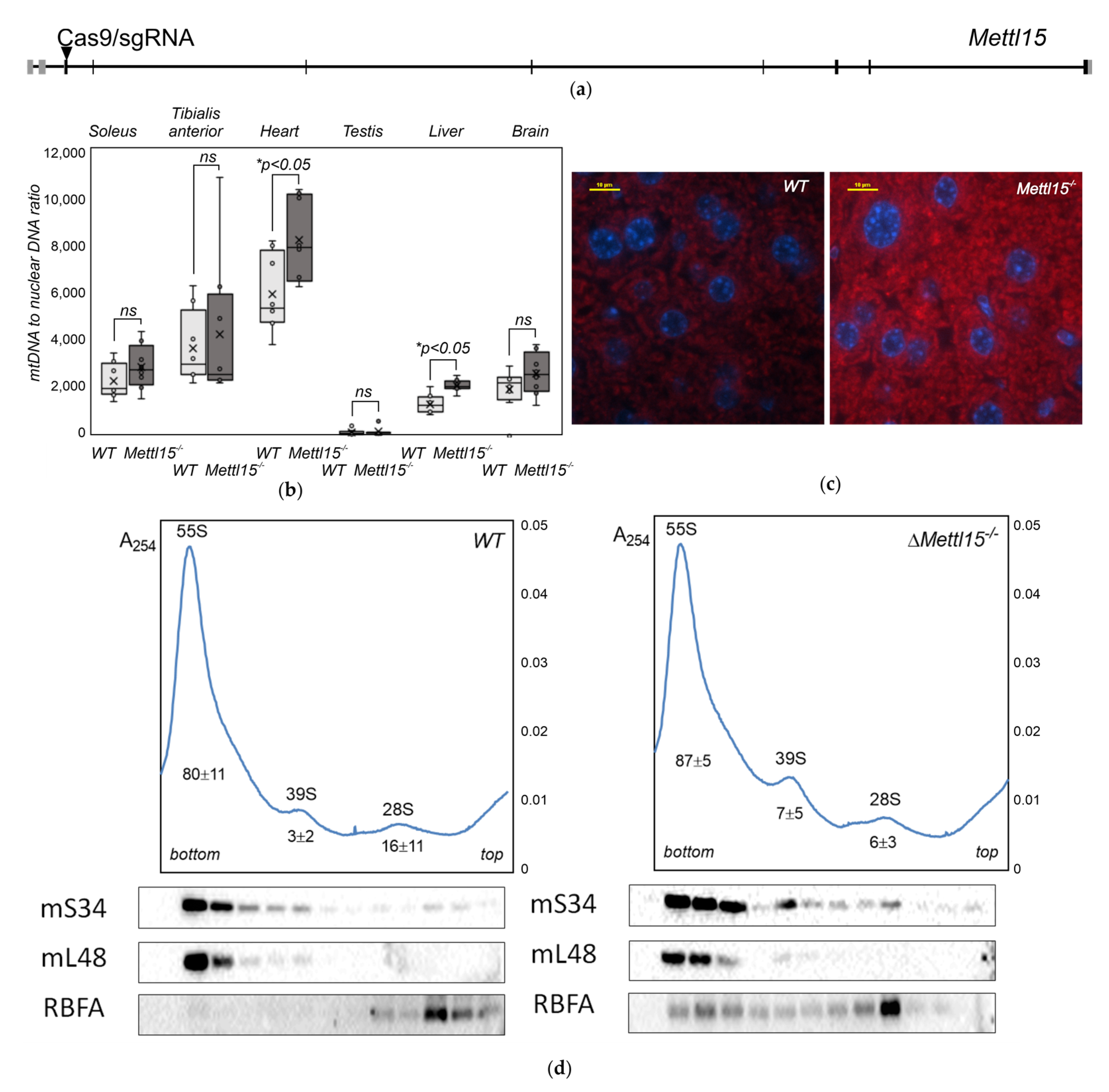
2.1. Generation of Mettl15 Knockout Mice

To inactivate the Mettl15 gene in mice we applied a CRISPR/Cas9 system [36] to cleave the third (first coding) exon of the Mettl15 gene (Figure 1a). To this end, a solution of the corresponding sgRNA obtained by in vitro transcription was mixed with S. pyogenes Cas9 coding mRNA and microinjected into zygotes of C57BL/6xCBA F1 mice. Following microinjection, the surviving zygotes were introduced to the oviducts of pseudopregnant mice. An inactivating two non-adjacent nucleotide deletion frameshift allele variant, hereafter referenced to as Mettl15 (Supplementary Materials Figure S1a,b), was found in the genotype of the progeny, which was used for further mating. Obtained heterozygous Mettl15+/− mice were backcrossed to a C57BL/6 background followed by mating to produce homozygous Mettl15−/− mice. Unlike knockouts for other mitochondrial rRNA methyltransferase genes [31,32], the homozygous Mettl15−/− mice were viable, borne at expected Mendelian ratios, and appeared normal.
The status of the 12S rRNA nucleotide C839 modification in the homozygous knockout mice was verified for several mice organs. To this end, the total RNA from the hearts, livers, and kidneys of four wild-type and four Mettl15−/− knockout mice were hybridized with the biotinylated oligonucleotide, complementary to the modification site. An excess of unhybridized RNA was digested by RNase T1, whereas the RNA fragment of interest was purified via immobilized streptavidin resin and subjected to RNase T1 hydrolysis followed by MALDI MS analysis (Supplementary Materials Figure S1c). As expected, we observed a complete lack of the 12S rRNA nucleotide C839 modification in all analyzed organs of the Mettl15−/− knockout mice, in contrast to a nearly complete modification in the corresponding organs of the wild-type mice. A complete lack of the nucleotide C839 modification in the 12S rRNA of Mettl15−/− knockout mice is accompanied by a substoichiometric modification of the neighboring nucleotide C837, in agreement with the observations obtained earlier for the Mettl15 knockout cell lines [33,35].

2.2. Abundance of Mitochondria in Mettl15−/− Knockout Mice

A malfunction of the mitochondrial rRNA modification machinery might lead to compensatory activation of the mitochondrial biogenesis and a consequent increase in the mtDNA copy number and the content of mitochondria in the cell [31,32]. To check whether Mettl15 gene inactivation would result in an excessive mitochondria accumulation in mice tissues, we determined the mtDNA to nuclear DNA ratio in the cardiac and skeletal muscles, testis, liver, and brain via the qPCR method (Figure 1b). Although a tendency for an increase in the mtDNA copy number was observed in all analyzed tissues of the Mettl15−/− knockout mice except the glycolytic tibialis anterior muscles, it reached statistical significance in the liver and cardiac muscle.
Mitochondria were stained using the Altmann’s method [37] and with anti-VDAC1 antibodies in the samples of the liver and kidneys (Figure 1c, Supplementary Materials Figure S2). Although staining with the Altmann’s method revealed the tendency for increased mitochondrial content (Supplementary Materials Figure S2a,b,e) it does not reach statistical significance. Staining with anti-VDAC1 antibodies supported the same tendency (Supplementary Materials Figure S2c,f) for the kidney samples and demonstrated a significant (p < 0.05) excess of mitochondria in the liver of Mettl15−/− knockout mice (Figure 1c, Supplementary Materials Figure S2d,f).
To assess the possible pathological consequences of Mettl15 gene inactivation we performed an assessment of the average body mass (Supplementary Materials Figure S3a) and temperature (Supplementary Materials Figure S3b) as well as a complete histopathology analysis of Mettl15−/− knockout mice and their wild-type littermates (Supplementary Materials Figure S3c).
No apparent differences that could be explained by Mettl15 gene inactivation have been observed in the mass, thermogenesis, and histopathology analysis. The accelerated aging phenotype was described for mice carrying mutations in the components of the mitochondrial ribosome [38]. To assess the accumulation of senescent cells in the tissues of Mettl15−/− knockout mice we applied senescence-associated b-galactosidase activity tests. Sparse cells positive for b-galactosidase activity were observed in the kidneys of both the wild-type and Mettl15−/− knockout mice at 4 months of age (Supplementary Materials Figure S4a). The senescent cell density was almost equal for the four-month-old wild-type and Mettl15−/− mice (Supplementary Materials Figure S4d). Other observed tissue samples, such as from the liver (Supplementary Materials Figure S4b), spleen, heart, brain, skeletal muscles, skin, and intestine (data not shown), were negative for b-galactosidase activity. However, an examination of a limited number of one-year-old wild-type (n = 2) and Mettl15−/− knockout mice (n = 2) allowed us to observe a nearly 2-fold difference in the number of senescent cells (Supplementary Materials Figure S4c) in the kidney slices. This remarkable tendency, albeit not conclusive due to the limited number of aged Mettl15−/− mice, could be the subject of a separate study. To search for signs of mitochondrial myopathy, we looked for the red-stained pathological mitochondria using Gomori trichrome staining of frozen unfixed muscle sections (Supplementary Materials Figure S5). However, clear signs of pathological mitochondria were not observed in either of the samples.

2.3. Alteration in the Composition of the Mitochondrial Ribosomes in Mettl15−/− Knockout Mice

Previously, we assessed an influence of the 12S rRNA modification at the nucleotide m4C839 on the proportion of mitochondrial 55S ribosomes and free 28S and 39S ribosomal subunits on the model of the murine NS0 cell line [35]. Although we found a very minute change in the ratio of the subunits and the ribosome caused by Mettl15 inactivation, the composition of mitochondrial ribosomal fractions was found to depend on the Mettl15 activity. Remarkably, RbfA, a mammalian mitoribosome assembly factor [39] homologous to the corresponding bacterial protein [40], was found to be associated with the 55S mitochondrial ribosome upon Mettl15 inactivation. Other groups observed variable degrees of mitoribosome assembly defects using human cell lines with an inactivated Mettl15 gene [33,34].
In order to check whether Mettl15 inactivation in mice would have an influence on mitoribosome association from the subunits and composition of mitochondrial ribosomes similar to those observed in cell lines, we prepared mitochondrial fractions from the liver samples of the Mettl15−/− knockout mice and their wild-type littermates. Mitochondrial lysates were subjected to ultracentrifugation through the sucrose-density gradients (Figure 1d, Supplementary Materials Figure S6a–h, upper panels). An influence of Mettl15 knockout on the ratio of the ribosomal subunits and ribosomes in mice liver mitochondria was found to be minimal (Supplementary Materials Figure S6i), reminiscent of a similar observation made for the knockout murine cell line [35]. At the same time, immunoblotting of the sucrose-density gradient fractions (Figure 1d, Supplementary Materials Figure S6a–h, lower panels) confirmed an increased association of the RbfA assembly factor with the 55S ribosomes observed previously on a cell line model.

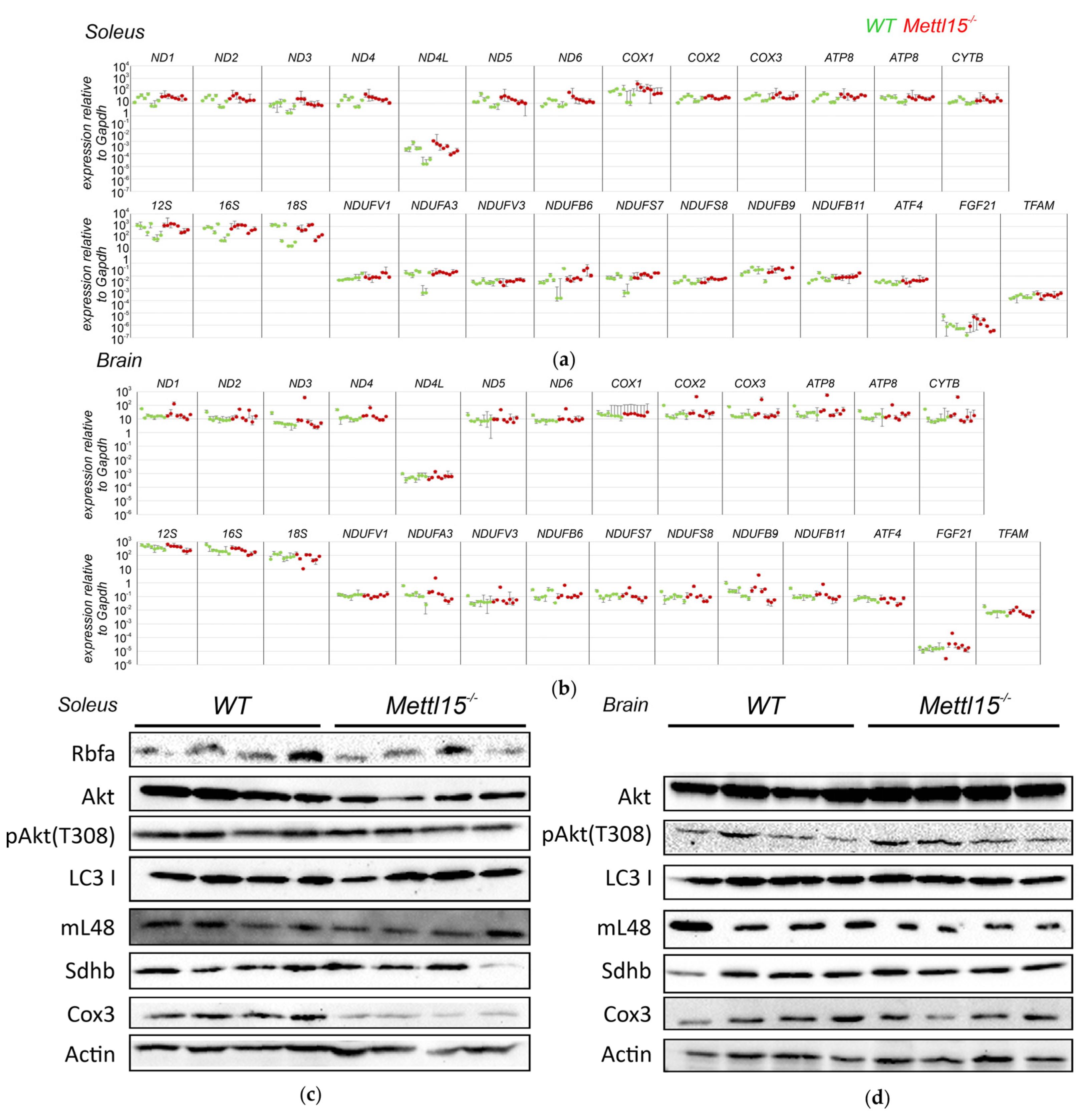
2.4. An Influence of Mettl15 Knockout on Gene Expression in Mice

In a number of studies, various mutations in the components of the mitochondrial translation apparatus were found to initiate a compensatory response manifested in increased transcription of the mitochondrial genome, an increase in the biogenesis of nuclear-encoded mitochondrial components, and boosting of the cytosolic translation efficiency via activation of mTOR signaling [41,42,43]. To check whether this is the case for the inactivation of Mettl15 in mice, we analyzed the expression levels of the 16S and 12S mitochondrial rRNAs, all protein-coding genes of mitochondria, a number of nuclear genes of the mitochondrial proteins, and genes whose expression level was reported to be increased upon impairment of mitochondrial translation [42] in the heart, oxidative (soleus), and glycolytic (tibialis anterior) skeletal muscles, liver, testis, and brain. Cytosolic 18S rRNA and Gapdh mRNA were used as controls (Figure 2a,b, Supplementary Materials Figure S7). No significant alteration in gene expression was found to be associated with Mettl15 gene knockout.
Additionally, quantities of the mitochondrial ribosomal protein Mrpl48, assembly factor RbfA, nuclear-encoded Sdhb, and mitochondrially encoded Cox3 protein components of the respiratory chain, as well as protein kinase Akt involved in mTOR signaling, its phosphorylated form, and autophagy factor LC3 have been assessed by immunoblotting of oxidative muscle (soleus) and brain lysates (Figure 2c,d, Supplementary Materials Figure S8). In line with the results of the RT qPCR analysis of the gene expression, we have detected neither a difference in the amounts of nuclear-encoded proteins nor the activities of mTOR signaling or autophagy between the wild-type and Mettl15−/− knockout mice. However, we observed a reduction in the amount of mitochondrially encoded Cox3 protein in the lysates of the oxidative soleus muscle. Since the Cox3 mRNA level was not affected by Mettl15 gene inactivation (Figure 2a), it is likely that Cox3 protein level reduction is a result of a decrease in the mitochondrial translation efficiency caused by Mettl15 inactivation. Such a difference, however, was not observed for the brain extracts.

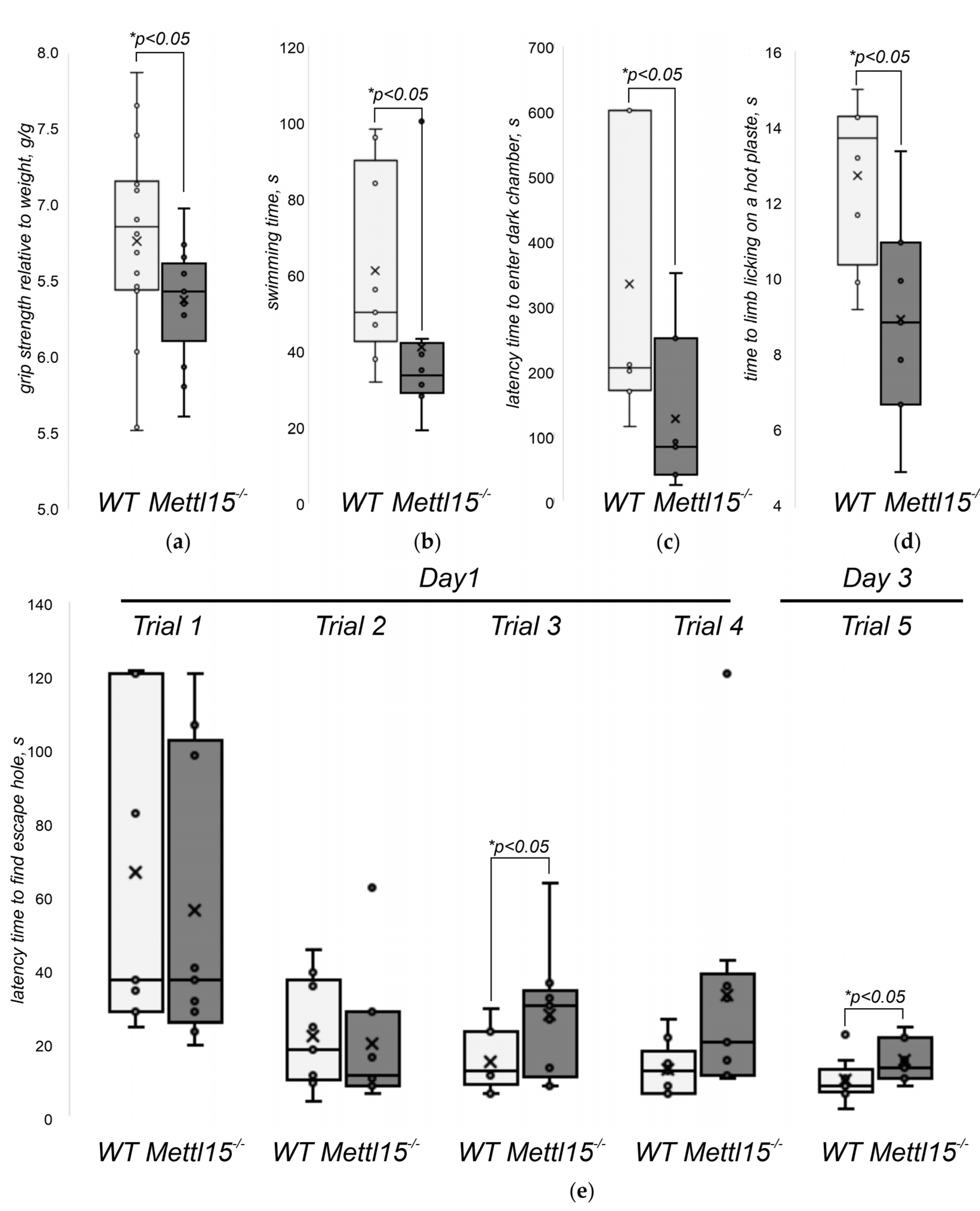
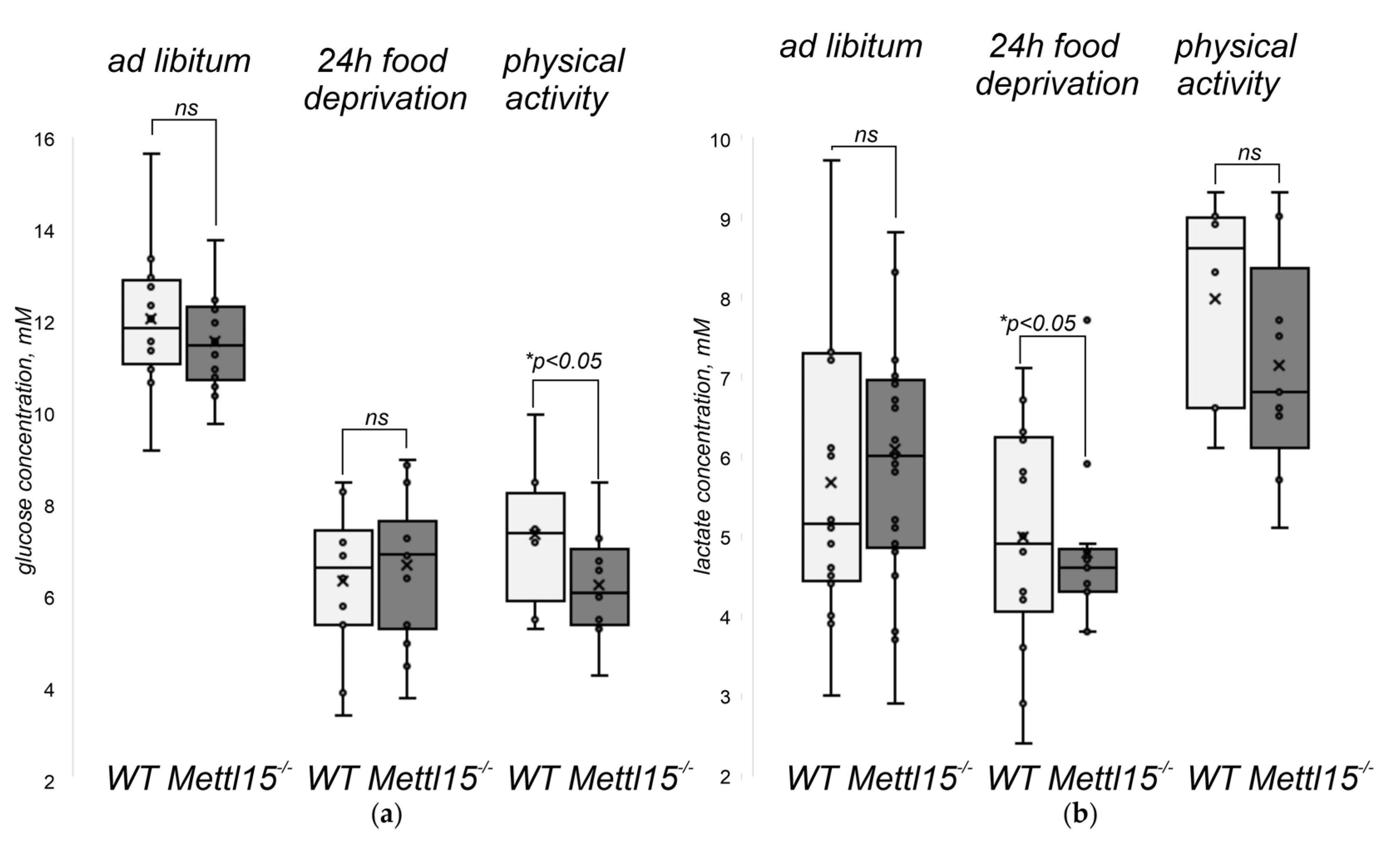
2.5. An Influence of Mettl15 Knockout on the Physiology of Mice

The suboptimal function of mitochondria in general and mitochondrial protein biosynthesis in particular frequently leads to a decrease in the performance of systems that rely heavily on the oxidative phosphorylation, such as skeletal muscles, and a disturbance of the biochemical composition of body fluids is indicative of the increased usage of anaerobic glycolysis as an alternative to OXPHOS in energy production. To test whether this is the case for Mettl15−/− knockout mice we determined forelimb grip strength (Figure 3a) and forced floating time (Figure 3b) after 24 h of food deprivation. The performance of knockout mice in both tests was found to be significantly lower than that of the wild-type controls. Neurological manifestations were frequently found in mitochondrial diseases [44] and several mouse models with impaired mitochondrial functions demonstrated this [45,46,47]. We subjected the Mettl15−/− knockout mice to the open field test for general exploratory activity (Supplementary Materials Figure S9a) and preference for exploration of a new object (Supplementary Materials Figure S9b), and the light–dark box test to assess anxiety (Supplementary Materials Figure S9c) and learning (Figure 3c). Although the majority of tests revealed no difference between the wild-type and Mettl15 knockout mice, we detected a significant difference between the groups in a learning test in a light–dark box with electric foot shock. In this test, mice were placed in a compartment with two boxes, a brightly illuminated one and a dark one, and were allowed to explore it freely. Mice tend to avoid illuminated boxes but were faced with a mild electric foot shock in the dark box. After placement in the home cage for 24 h, the mice were subjected to the same light–dark box to monitor their memories, which should suggest they avoid the dark box. To control for the possibility that Mettl15−/− knockout mice are more tolerant to the mild pain stimuli rather than their learning ability be compromised, we tested their response time latency in a hot plate test (Figure 3d). Apparently, mice with a Mettl15 deficiency are even more sensitive to pain than their wild-type littermates. Thus, we concluded that Mettl15 and hence mitoribosome modification contributes to the efficiency of learning or memory.
To explore further the learning deficit of the Mettl15−/− knockout mice, we used a memory test with positive rather than negative reinforcement. In the box maze test [48], mice were placed into the box with several dead-end appendices and an escape hole leading to a refuge. After initial free exploration of the maze, mice were subjected to four trials to find refuge. After 48 h, mice were subjected to a single trial to find refuge. The latency time before entering the escape hole is a readout (Figure 3e). As a result, we found that Mettl15−/− knockout mice performed worse in the box maze test spending significantly (p < 0.05) more time to recall the way to a known escape hole 2 days after training (Figure 3e, rightmost bars). This tendency was notable even during the training trials but reached statistical significance only for the third attempt (Figure 3e, third group of bars).
Patients suffering from a number of mitochondrial diseases frequently have lactic acidosis and a number of other differences in the plasma metabolites concentration [49]. The inactivation of genes related to the functioning of mitochondria in mice likewise results in plasma metabolite profile alterations [50]. To this end, we performed NMR-based metabolite profiling in the serum of the Mettl15−/− knockout mice and the corresponding control group fed ad libitum (Supplementary Materials Figure S10). The inactivation of the Mettl15 gene appeared not to cause any significant difference in the metabolite profiles of mice under normal conditions.
In addition, we monitored the serum concentration of glucose (Figure 4a) and lactate (Figure 4b) of mice fed ad libitum (Figure 4a,b, leftmost bars), the same mice cohort after a 24 h food deprivation (Figure 4a,b, central bars), and the same mice after 24 h of food deprivation and forced swimming (Figure 4a,b, rightmost bars). As a result, we found that the fed Mettl15−/− mice demonstrate levels of plasma glucose and lactate concentrations nearly similar to the wild-type. After food deprivation, the plasma glucose concentrations dropped in both mice cohorts, whereas the lactate concentration remained nearly the same for the wild-type mice but decreased for the Mettl15−/− knockout mice to a level significantly lower than that of the wild-type. The intense physical exercise led to the elevation of the plasma lactate concentration for both the wild-type and knockout mice. Although the lactate concentration remained somewhat lower in the plasma of Mettl15−/− knockout mice, this difference was not significant. During this physical exercise, the glucose level of the wild-type mice increased, whereas that of the knockout mice decreased. As a result, the plasma glucose concentration following exercise was significantly lower for Mettl15−/− knockout mice.
Translation, as well as ribosome biogenesis, are energy-consuming processes, hence, even a moderate decrease in translation efficiency might lead to increased energy expenditure in mitochondria, which could be manifested in a lower reserve of energy left after a starvation period. Although the wild-type animals are likely to retain reserves of glycogen sufficient to supply glucose to the bloodstream in a period of high physical activity, Mettl15−/− knockout mice might be lacking it. To check for this possibility, we stained the samples of liver and skeletal muscle (gastrocnemius, the mixed-type muscle relying on both OXPHOS and glycolysis) for glycogen in the wild-type and Mettl15−/− knockout mice (Supplementary Materials Figure S11). In contrast to the expectation, no significant difference in the amount of glycogen was found between the wild-type and knockout mice. Thus, a somewhat lower concentration of glucose in the bloodstream of Mettl15−/− knockout mice following physical exercise is not a consequence of a systemic shortage of glycogen.

3. Discussion

Mitochondrial ribosomes are related to the ribosomes of their proteobacterial predecessors. Several modified nucleotides present in the mitochondrial rRNA are conserved between bacterial and mitochondrial ribosomes or universally conserved [24]. Three mitochondrial rRNA methyltransferases whose physiological significance has thus far been assessed on a knockout mice model belong to this category. Mitochondrial TFB1M methyltransferase [51] responsible for the formation of 12S rRNA dimethyladenosine residues m62A936/7 is a homologue of bacterial KsgA protein [52] and Dim1/Dim1L protein [53] involved in the same modification of cytosolic ribosomes. Although an inactivation of KsgA in bacteria is not lethal, it leads to a ribosome biogenesis defect [54,55]; an inactivation of the Dim1 gene responsible for the modification of the cytosolic ribosomes is lethal for yeast [53]; and an inactivation of the mitochondrial rRNA methyltransferase gene Tfb1m is lethal for mice [31]. Lethality is likewise associated with the inactivation of mice mitochondrial rRNA methyltransferase gene Nsun4, responsible for the formation of m5C841 in the 12S rRNA [32]. Deletion of its bacterial homologue rsmF [56], is not lethal but rather has a mild phenotype [55]. The essentiality of NSUN4 in mammals could be associated with an additional function of its complex with MTERF4 in the assembly of the large 39S mitochondrial ribosome subunit [32]. An inactivation of the Mettl15 gene in mice reported here is not lethal and in contrast to other published knockouts of mitochondrial rRNA methyltransferase genes has a very mild phenotype. This is reminiscent of the mild phenotype of its bacterial ortholog rsmH knockout manifested only in a decrease in translation initiation fidelity [57] and the ability of the translation apparatus to cope with the burden of an exogenous gene expression [55].
We have observed neither an accumulation of ribosome assembly intermediates, which could be detected by a sucrose-gradient centrifugation, nor any defects in the formation of the mitochondrial 55S ribosomes in accordance with our earlier data obtained with the cell line with inactivated Mettl15 [35]. However, we recapitulated the presence of the RbfA assembly factor [35] in the 55S ribosome fraction in Mettl15−/− knockout mice. It is likely that suboptimality of mitochondrial ribosome biogenesis caused by Mettl15 inactivation is moderate and could have a physiological consequence only for tissues highly dependent on OXPHOS and under challenging conditions as will be discussed below. This statement is supported by the observation of the reduced level of Cox3 protein, synthesized by the mitoribosome, in the oxidative soleus muscle. Interestingly, the Cox3 protein amount in the brain is refractory to the Mettl15 inactivation perhaps due to slower mitochondrial biogenesis and turnover.
Apart from the reported mice knockouts of Tfb1m [31] and Nsun4 [32], the significance of mitochondrial protein biosynthesis components for mice physiology has been assessed for the mutations in genes’ coding for mitochondrial ribosomal proteins Mrps5 [58], Mrps34 [59], assembly factor Ptcd1 [42], and mt-mRNA binding proteins Slirp [60] and Taco1 [61], as well as translation factors Mtif3 [62] and Mtef4/Guf1 [41]. In many cases mutations in the assembly factors or ribosomal proteins lead to decreased stability of either 12S rRNA [31,59] or 16S rRNA [42] accompanied by an increase in transcription of rRNA whose stability was unaffected [31,32,42,60]; mitochondrially encoded mRNAs [31,32,42]; mRNAs coding for mitochondrial ribosomal proteins and transcription factors such as TFAM [32,59]; and a number of other nuclear genes such as ATF4 and FGF21, whose expression is indicative of mitochondrial dysfunction [59]. At the posttranslational level, the excessive activation of mTOR signaling was likewise associated with suboptimal mitochondrial function [42,59]. Although we thoroughly assessed the expression of all protein-coding genes in the mitochondrial genome, 12S and 16S rRNA, and the indicative nuclear genes related to the mitochondria, we found that an inactivation of the Mettl15 gene has a minor influence on gene expression in a representative range of tissues. Likewise, we could not detect a difference in the amounts of proteins related to mitochondrial function, mTOR signaling, and autophagy between the wild-type and Mettl15 knockout mice. However, we observed the tendency for an increase in mtDNA content in the Mettl15−/− knockout mice tissues that was significant for the liver and heart. Increased mitochondrial content was corroborated by immunohistochemical staining of the liver mitochondria. These results are in line with the much milder effect of Mettl15 gene knockout on the mitochondrial ribosome assembly and protein synthesis we observed in this study and the previous study on the knockout cell line [35].
Severe defects of the mitochondrial translation were reported to affect a number of tissues, such as the heart and skeletal muscles, where necrotic foci and centralized nuclei were observed [42,59,63], the liver where excessive lipid accumulation and steatosis were found [59], and the testis, where size decrease and spermatogenesis defects were noted [41]. As Mettl15 knockout has a much milder influence on mitochondrial ribosome assembly, it was unsurprising to observe no significant differences in the histopathology of the knockout mice tissues from that of their wild-type littermates. More specific changes, such as an accumulation of senescent cells and damaged mitochondria, were not observed at a significant level in the young Mettl15−/− knockout mice. However, we observed an interesting tendency for excessive senescent cell accumulation in one-year-old mice kidneys, which could be an interesting subject for further study.
Mitochondrial dysfunction is idiosyncratically associated with lactic acidosis [12,13] as well as elevated alanine, creatine, and lactate/pyruvate ratio [49]. In accordance with the mild phenotype of Mettl15 knockout at the molecular level, we observed no difference in the metabolite concentrations at the steady-state condition. However, challenging mice's physiological state by food deprivation and physical exercise allowed us to reveal a number of differences from the wild-type. Although the wild-type mice were found capable to elevate serum glucose levels upon forced swimming after 24 h of starvation, Mettl15−/− knockout mice demonstrated a small but significant decrease in the concentration of glucose under the same conditions. The lower glucose concentration could not be explained by lower glycogen storage, but perhaps by higher glucose expenditure due to lower efficiency of mitochondrial function. Starvation also led to a decrease in serum lactate concentration for Mettl15−/− knockout mice, and even after forced swimming the lactate concentration increase was less than that for the wild-type mice, although this difference was below statistical confidence (p > 0.05). A decrease in the glucose concentration associated with mitochondrial dysfunction provoked by starvation has been documented in a number of patients [64] including those who have alterations in the components of mitochondrial ribosomes [65]; in these cases, the molecular mechanisms leading to hypoglycemia have likewise remained enigmatic. Lower lactate levels in the serum of starved Mettl15−/− knockout mice could be a consequence of gluconeogenesis converting this metabolite into glucose to cope with glucose limitation.
Unsurprisingly, manifestations of the mitochondrial pathologies are associated with organs with high-energy demands, such as skeletal muscles and the brain [12]. Mettl15−/− knockout mice demonstrated a statistically significant force and performance reduction in the grip strength and forced swimming tests after a challenge of 24 h starvation. This effect might be explained by a lower energy production or higher expenditure; the latter being supported by a decreased plasma glucose concentration. A number of studies revealed a learning deficit caused by mutations in the components of the mitochondrial translation apparatus, such as Mrps34, Taco1, and Mrps5 [58,59,61]. Likewise, Mettl15−/− knockout mice demonstrated a significantly decreased learning capability. Moreover, we observed a learning deficit in the tests with either positive or negative reinforcement. The learning deficit in the Mrps5 mutant mice is attributed to the decrease in translation fidelity of the mitochondrial ribosomes [58]. A decrease in the fidelity of translation initiation was observed for a knockout of the bacterial ortholog of Mettl15, rRNA methyltransferase RsmH [57]. Whether phenotype manifestations of the Mettl15 knockout are associated with mitochondrial mistranslation is a subject for further studies.
Mice lines with mutations in the genes coding for the components of mitochondrial translation apparatus are precious models for human mitochondrial diseases. Inactivation of other tested mitochondrial rRNA methyltransferase genes [31,32] were reported to be lethal, which speaks in favor of their importance but precludes their use as models of mitochondrial diseases. Obviously, inactivation of the Mettl15 gene in mice has a way milder, yet significant muscular and neurological phenotype. This peculiarity paves the way for the application of Mettl15−/− knockout mice as a model for mild mitochondrial dysfunction.

4. Materials and Methods

4.1. Mettl15 Gene Inactivation and RNA Modification Analysis

For inactivation of the M. musculus Mettl15 gene, we injected mouse zygotes with a mixture of 12 ng/uL sgRNA (target sequence GCATACTGAATCTAAAGCTG) mixed with 25 ng/uL mRNA coding for S. pyogenesis Cas9 (Thermo scientific). The genome-edited mice were generated as previously described [66]. All manipulations were conducted in compliance with protocol №182 approved by the Local Bioethics Commission of the Research Center “Institute of Mitoengineering of Moscow State University” LLC, (Moscow, Russia).
For the monitoring of the modification status of the 12S rRNA nucleotide C839, total RNA was isolated from the hearts, livers, and kidneys of four Mettl15−/− mice and four mice of the control group by Trizol extraction. A 12S rRNA fragment was isolated by a previously published procedure [35] using 5′-biotinylated oligodeoxyribonucleotide (5′-[biotin]- TGTTAAGTTTAATTTAATTTGAGGAGGGTGACGGGCGGTG) complementary to the region of interest of the 12S rRNA and digested by RNase T1. Presence of the modification was analyzed by Ultraflex III BRUKER mass spectrometer equipped with a UV laser (Nd, 335 nm).

4.2. Tests with Live Mice

The experiments with live animals were performed on 8 wild-type animals and 8 Mettl15−/− knockout mice. Statistical data analysis was performed using the nonparametric Mann–Whitney U test. The chosen significance level was p < 0.05.
The grip force test was done according to previously published procedure [67]. Each animal procedure was repeated 10 times with 15–30 s between attempts; five maximum values are averaged.
Forced swimming test [68,69,70,71,72,73] recording the animal's maximum swimming time represent physical performance capability. A mouse with 13% of body weight load fixed on the middle part of the tail was placed in water. The time until an animal’s refusal to swim is recorded. Mice were rescued alive from the water after the endpoint.
Novel object recognition test is designed to evaluate general exploratory activity and object recognition memory in rodents [74,75]. This methodology assesses the natural preference of rodents to explore novel objects [76]. Mice were allowed to explore two objects of the same shape and color for 5 min. The test was repeated the next day, one of the objects being replaced with a new one, different in color and shape from the initial one. The number of approximations, sniffing, and direct contacts of mice with the objects were recorded.
Passive avoidance test is fear-motivated test designed to evaluate memory in rodents. The test unit consists of a dark chamber with an electrode floor and a light chamber, separated by a wall with a sliding door. The mouse is placed in a brightly lit compartment, after which the door is opened and the latency period of passage to the compartment with the electrode floor is recorded. One day later, the animal is again placed in the illuminated compartment and the latency to cross through the opened gate between the compartments is assessed [77,78,79]. Memory performance is positively correlated with the latency of passage to the dark compartment on the second experimental day; the better the memory, the greater the latency.
Hot plate test is designed to assess nociception. The mouse is placed on a hot plate at a controlled temperature (52–55 °C) and the time until the first hind paw licking is recorded [80].
The light/dark transition test (LDT) is one of the most widely used tests to measure anxiety-like behavior in mice [81]. The LDT test is based on the innate aversion of rodents to brightly illuminated areas and on the spontaneous exploratory behavior of rodents in response to mild stressors, that is, novel environment and light [82]. The test unit consists of dark and light chambers. The latency of passage from the bright to the dark chamber was the studied research parameter.
Similar to the LDT test, the box-maze test [48] is based on rodents’ avoidance of brightly illuminated areas. The box-maze apparatus contains an illuminated central box with five holes leading to immediate dead ends and a single escape hole that is connected to the standard housing cage. Mice were placed into the illuminated box maze and allowed to explore it and behave freely for up to 2 min. The latency time to fully enter the escape hole is recorded. Mice are subjected to a total of four such trials, all on the same day. Over the course of the four trials, mice learn the location of the escape hole and exit the maze through it with progressively shorter escape latency. After 48 h, the procedure is repeated and mice are subjected to only one trial.

4.3. Histopathological Examination

The brain, heart, spleen, kidneys, liver, testis, lungs, tibialis anterior, and soleus muscle specimens were fixed with formalin, dehydrated and paraffin-embedded. Microtome sections with thickness of 3 µm were stained with hematoxylin and eosin. Images were observed and recorded using an Axiosope A1 microscope with a CCD-camera AxioCam MRc.5 (Carl Zeiss, Oberkochen, Germany).
To stain senescence-associated b-galactosidase positive senescent cells, we used the protocol for paraffin sections: organ samples were fixed with Baker’s formol-calcium solution for 24 h at +4 °C, washed in distilled water for 24 h, dehydrated in acetone, and embedded in paraffin [83,84]. Microtome sections with thickness of 5 µm were stained for 3 h with X-gal solution at suboptimal pH 6.0 and 37 °C as recommended [85] and counterstained with nuclear red. Number of senescent cells in full organ section was counted manually and averaged to 1 mm2 of section area with ImageJ software. To stain the mitochondria, the kidney and liver samples were fixed with Regaud fixative and stained according to classical Altmann’s method [37]. Area of mitochondria was estimated with ImageJ software on 3 photomicrographs (100×) of sections of renal tubules and liver. For study of myopathy lesions, unfixed cryotome sections with thickness of 5 µm were stained with Gomori trichrome [86].
Immunohistochemical analysis of mitochondrial content in the wild-type and Mettl15−/− knockout mice was done with formaldehyde-fixed paraffin-embedded slices of liver and kidneys. Staining was done with rabbit anti-VDAC1 antibodies (Abcam ab15895) and Cy5 conjugated goat anti-rabbit antibodies (Invitrogen, A10523). Microphotographs were taken by Nikon C2 microscope and analyzed by Nikon NIS-Elements software.

4.4. Sucrose-Gradient Profiling of Mitochondrial Ribosomes

Mitochondria and mitochondrial ribosomes were isolated by combining the methods described [87,88] with some changes. Mice were sacrificed by cervical dislocation and liver was homogenized in MIBSM buffer (50 mM HEPES-KOH, pH 7.5, 70 mM sucrose, 210 mM mannitol, 10 mM KCl, 1.5 mM MgCl2, 2 mM EDTA, 1 mM DTT, protease inhibitors). Lysate was clarified and mitochondria were pelleted by 7000× g 20 min centrifugation at +4 °C, resuspended and washed using the same procedure, and finally resuspended in 1 mL of SEM (20 mM HEPES-KOH, 250 mM sucrose, pH 7.5, 1 mM EDTA). Mitochondrial suspension was loaded on top of the step sucrose gradient (2 mL 60%, 4 mL 32%, 2 mL 23% and 2 mL 15% sucrose in 20 mM HEPES-KOH, pH 7.5, 1 mM EDTA) and centrifuged in SW41Ti rotor at 27,000 rpm for 1 h at +4 °C. Brown migrating band of mitochondria between 60% and 32% sucrose was collected and pelleted at 8000× g 10 min at +4 °C.
Mitoribosome profiles were obtained as described [35], fractionated into 0.5 mL fractions using ACTA purifier (GE Healthcare, Chicago, IL, USA) while monitoring absorbance at 254 nm. Proteins from gradient fractions were isolated by TCA/Deoxycholate precipitation as described [89], dissolved in 1× Laemmli buffer, and separated by SDS PAGE.

4.5. Western Blot Analysis

For Western blot analysis, mice organs were lysed in RIPA buffer with Halt Protease and Phosphatase Inhibitor Cocktail (ThermoFisher Scientific, Waltham, MA, USA) using Precellys Evolution homogenizer (Bertin Technologies, Montigny-le-Bretonneux, France). Protein concentration in cleared lysates was measured by Bradford assay and 30 µg of each lysate was used for Western blotting.
For detection of the proteins, the following antibodies were used: RBFA antibody (PA5-59587, Invitrogen, Carlsbad, CA, USA), MRPS34 antibody (PA5-59872, Invitrogen, Carlsbad, CA, USA), MRPL48 antibody (ab194826, Abcam, Waltham, MA, USA), AKT (9272, Cell Signaling Technology, Danvers, MA, USA), pAKT (Thr308) (4056, Cell Signaling Technology, Danvers, MA, USA), LC3A/B (PA1-16931, ThermoFisher Scientific, Waltham, MA, USA), SDHB (PA5-29843, ThermoFisher Scientific, Waltham, MA, USA), Cox3 (55082-1-AP, Proteintech, Rosemont, IL, USA), and Actin (ab8229, Abcam, Waltham, MA, USA).

4.6. Quantitative RT–PCR

Total RNA was extracted from mice tissue homogenates using QIAzol Lysis Reagent (Qiagen, Hilden, Germany). cDNA was synthesized from total RNA using Maxima First Strand cDNA Synthesis Kit for RT-qPCR (ThermoFisher Scientific, Waltham, MA, USA). The RT-PCR was performed on individual cDNAs by using SYBR® Green PCR master mix in the CFX384 Touch Real-Time PCR System. The primer sequences are available in the Supplementary Materials (Supplementary Table S1). The mRNA expression was calculated by the 2−ΔΔCT method and normalized to the expression of Gapdh.
Copy number of mtDNA was estimated by quantitative PCR as previously described [35].

4.7. Metabolome Analysis

For the analysis of lactate and glucose concentrations, blood samples were taken from the facial vein [90] and analyzed by a portable biochemical analyzer Accutrend Plus (Roche Diagnostics, Basel, Switzerland). For complete NMR analysis of metabolome, blood was collected by cardiac puncture [91] from animals anesthetized by isoflurane inhalation. Plasma was separated by centrifugation and used immediately for metabolite extraction by methanol and chloroform 1:1 mixture. Water layer was removed and dried. Before NMR measurements, the solid metabolites were dissolved in 50 mM sodium phosphate at pH 7.0, 0.107 mM d6-DSS, 0.13 mg/mL NaN3 in D2O. NMR spectra were acquired at 35 °C on a Bruker Avance 700 MHz NMR spectrometer equipped with a Z-axis-gradient 5 mm HCN Prodigy cryoprobe. One-dimensional 1H NMR spectra were acquired from each sample using the standard 1D NOESY-presat experiment. A total of 400 scans were accumulated for plasma samples. 1H,13C-HSQC spectra were measured for one sample from each group to confirm the metabolites assignments. All steps of 1D spectra processing and metabolite identification were performed using Chenomx NMR Suite program. 1H,13C-HSQC spectra were analysed in TopSpin program.

Supplementary Materials

The following supporting information can be downloaded at: https://www.mdpi.com/article/10.3390/ijms23116056/s1.

Author Contributions

Conceptualization, O.A.D. and P.V.S.; methodology, O.A.A., S.S.M., I.G.L. and V.N.M.; formal analysis, O.A.A., S.S.M., I.G.L., V.N.M., A.K.B. and O.O.G.; investigation, O.A.A., I.G.L., M.A.E., O.A.P., S.S.M., A.I.N., V.N.M., A.K.B., O.O.G. and G.A.K.; writing—original draft preparation, P.V.S.; writing—review and editing, P.V.S.; visualization, O.A.A., V.N.M., S.S.M. and P.V.S.; supervision, P.V.S.; funding acquisition, O.A.D. and P.V.S. All authors have read and agreed to the published version of the manuscript.

Funding

This research was funded by the Russian Science Foundation, grant number 19-14-00043, in part related to the genome editing and analysis of mice physiology, grant number 21-64-00006, in part related to the experiments with mitochondria isolation and ribosome analysis, grant number 19-14-00115, in part related to the NMR metabolomics; the Russian Foundation for Basic Research, grant number 20-04-00736 in part related to methylation analysis. All authors from the Lomonosov Moscow University were supported by the leading scientific school in molecular technology of biological systems and synthetic biology.

Institutional Review Board Statement

The study was conducted in accordance with the Declaration of Helsinki, and approved by the Ethics Committee of the Research Center “Institute of Mitoengineering of Moscow State University” LLC, (Moscow, Russia) (protocol code 182, accessed on 12 May 2021).

Informed Consent Statement

Not applicable.

Data Availability Statement

Not applicable.

Acknowledgments

Authors are indebted to Mikhail Vysokikh for inspiration in the field of mitochondrial biochemistry and generous sharing of antibodies and Alexey Nesterenko for expert assistance in the light–dark box test with electric foot shock. We are thankful to Ivan Chicherin for the generous sharing of antibodies.

Conflicts of Interest

The authors declare no conflict of interest.

References

  1. Sagan, L. On the Origin of Mitosing Cells. 1967. J. NIH Res. 1993, 5, 65–72. [Google Scholar]
  2. Christian, B.E.; Spremulli, L.L. Mechanism of Protein Biosynthesis in Mammalian Mitochondria. Biochim. Biophys. Acta (BBA) Gene Regul. Mech. 2012, 1819, 1035–1054. [Google Scholar] [CrossRef] [Green Version]
  3. Ott, M.; Amunts, A.; Brown, A. Organization and Regulation of Mitochondrial Protein Synthesis. Annu. Rev. Biochem. 2016, 85, 77–101. [Google Scholar] [CrossRef]
  4. Mai, N.; Chrzanowska-Lightowlers, Z.M.A.; Lightowlers, R.N. The Process of Mammalian Mitochondrial Protein Synthesis. Cell Tissue Res. 2017, 367, 5–20. [Google Scholar] [CrossRef] [Green Version]
  5. Ayyub, S.A.; Varshney, U. Translation Initiation in Mammalian Mitochondria- a Prokaryotic Perspective. RNA Biol. 2020, 17, 165–175. [Google Scholar] [CrossRef]
  6. Lopez Sanchez, M.I.G.; Krüger, A.; Shiriaev, D.I.; Liu, Y.; Rorbach, J. Human Mitoribosome Biogenesis and Its Emerging Links to Disease. Int. J. Mol. Sci. 2021, 22, 3827. [Google Scholar] [CrossRef]
  7. Goto, Y.; Nonaka, I.; Horai, S. A Mutation in the TRNA(Leu)(UUR) Gene Associated with the MELAS Subgroup of Mitochondrial Encephalomyopathies. Nature 1990, 348, 651–653. [Google Scholar] [CrossRef]
  8. Shoffner, J.M.; Lott, M.T.; Lezza, A.M.; Seibel, P.; Ballinger, S.W.; Wallace, D.C. Myoclonic Epilepsy and Ragged-Red Fiber Disease (MERRF) Is Associated with a Mitochondrial DNA TRNA(Lys) Mutation. Cell 1990, 61, 931–937. [Google Scholar] [CrossRef]
  9. Ruiz-Pesini, E.; Lott, M.T.; Procaccio, V.; Poole, J.C.; Brandon, M.C.; Mishmar, D.; Yi, C.; Kreuziger, J.; Baldi, P.; Wallace, D.C. An Enhanced MITOMAP with a Global MtDNA Mutational Phylogeny. Nucleic Acids Res. 2007, 35, D823–D828. [Google Scholar] [CrossRef] [Green Version]
  10. Prezant, T.R.; Agapian, J.V.; Bohlman, M.C.; Bu, X.; Oztas, S.; Qiu, W.Q.; Arnos, K.S.; Cortopassi, G.A.; Jaber, L.; Rotter, J.I. Mitochondrial Ribosomal RNA Mutation Associated with Both Antibiotic-Induced and Non-Syndromic Deafness. Nat. Genet. 1993, 4, 289–294. [Google Scholar] [CrossRef]
  11. Rötig, A. Human Diseases with Impaired Mitochondrial Protein Synthesis. Biochim. Biophys. Acta (BBA) Bioenerget. 2011, 1807, 1198–1205. [Google Scholar] [CrossRef] [Green Version]
  12. Vafai, S.B.; Mootha, V.K. Mitochondrial Disorders as Windows into an Ancient Organelle. Nature 2012, 491, 374–383. [Google Scholar] [CrossRef]
  13. Boczonadi, V.; Horvath, R. Mitochondria: Impaired Mitochondrial Translation in Human Disease. Int. J. Biochem. Cell Biol. 2014, 48, 77–84. [Google Scholar] [CrossRef] [Green Version]
  14. Rudler, D.L.; Hughes, L.A.; Viola, H.M.; Hool, L.C.; Rackham, O.; Filipovska, A. Fidelity and Coordination of Mitochondrial Protein Synthesis in Health and Disease. J. Physiol. 2020, 599, 3449–3462. [Google Scholar] [CrossRef]
  15. Ferrari, A.; Del’Olio, S.; Barrientos, A. The Diseased Mitoribosome. FEBS Lett. 2021, 595, 1025–1061. [Google Scholar] [CrossRef]
  16. Chatzispyrou, I.A.; Alders, M.; Guerrero-Castillo, S.; Zapata Perez, R.; Haagmans, M.A.; Mouchiroud, L.; Koster, J.; Ofman, R.; Baas, F.; Waterham, H.R.; et al. A Homozygous Missense Mutation in ERAL1, Encoding a Mitochondrial RRNA Chaperone, Causes Perrault Syndrome. Hum. Mol. Genet. 2017, 26, 2541–2550. [Google Scholar] [CrossRef] [Green Version]
  17. Wei, X.; Du, M.; Li, D.; Wen, S.; Xie, J.; Li, Y.; Chen, A.; Zhang, K.; Xu, P.; Jia, M.; et al. Mutations in FASTKD2 Are Associated with Mitochondrial Disease with Multi-OXPHOS Deficiency. Hum. Mutat. 2020, 41, 961–972. [Google Scholar] [CrossRef]
  18. Feichtinger, R.G.; Oláhová, M.; Kishita, Y.; Garone, C.; Kremer, L.S.; Yagi, M.; Uchiumi, T.; Jourdain, A.A.; Thompson, K.; D’Souza, A.R.; et al. Biallelic C1QBP Mutations Cause Severe Neonatal-, Childhood-, or Later-Onset Cardiomyopathy Associated with Combined Respiratory-Chain Deficiencies. Am. J. Hum. Genet. 2017, 101, 525–538. [Google Scholar] [CrossRef]
  19. Lessel, D.; Schob, C.; Küry, S.; Reijnders, M.R.F.; Harel, T.; Eldomery, M.K.; Coban-Akdemir, Z.; Denecke, J.; Edvardson, S.; Colin, E.; et al. De Novo Missense Mutations in DHX30 Impair Global Translation and Cause a Neurodevelopmental Disorder. Am. J. Hum. Genet. 2017, 101, 716–724. [Google Scholar] [CrossRef] [Green Version]
  20. Solomon, B.D.; Pineda-Alvarez, D.E.; Hadley, D.W.; Keaton, A.A.; Agochukwu, N.B.; Raam, M.S.; Carlson-Donohoe, H.E.; Kamat, A.; Chandrasekharappa, S.C. De Novo Deletion of Chromosome 20q13.33 in a Patient with Tracheo-Esophageal Fistula, Cardiac Defects and Genitourinary Anomalies Implicates GTPBP5 as a Candidate Gene. Birth Defects Res. A Clin. Mol. Teratol. 2011, 91, 862–865. [Google Scholar] [CrossRef] [Green Version]
  21. Bohnsack, M.T.; Sloan, K.E. The Mitochondrial Epitranscriptome: The Roles of RNA Modifications in Mitochondrial Translation and Human Disease. Cell. Mol. Life Sci. 2018, 75, 241–260. [Google Scholar] [CrossRef]
  22. Laptev, I.; Dontsova, O.; Sergiev, P. Epitranscriptomics of Mammalian Mitochondrial Ribosomal RNA. Cells 2020, 9, 2181. [Google Scholar] [CrossRef]
  23. Lopez Sanchez, M.I.G.; Cipullo, M.; Gopalakrishna, S.; Khawaja, A.; Rorbach, J. Methylation of Ribosomal RNA: A Mitochondrial Perspective. Front. Genet. 2020, 11, 761. [Google Scholar] [CrossRef]
  24. Sergiev, P.V.; Aleksashin, N.A.; Chugunova, A.A.; Polikanov, Y.S.; Dontsova, O.A. Structural and Evolutionary Insights into Ribosomal RNA Methylation. Nat. Chem. Biol. 2018, 14, 226–235. [Google Scholar] [CrossRef]
  25. Bykhovskaya, Y.; Mengesha, E.; Wang, D.; Yang, H.; Estivill, X.; Shohat, M.; Fischel-Ghodsian, N. Human Mitochondrial Transcription Factor B1 as a Modifier Gene for Hearing Loss Associated with the Mitochondrial A1555G Mutation. Mol. Genet. Metab. 2004, 82, 27–32. [Google Scholar] [CrossRef]
  26. Cotney, J.; McKay, S.E.; Shadel, G.S. Elucidation of Separate, but Collaborative Functions of the RRNA Methyltransferase-Related Human Mitochondrial Transcription Factors B1 and B2 in Mitochondrial Biogenesis Reveals New Insight into Maternally Inherited Deafness. Hum. Mol. Genet. 2009, 18, 2670–2682. [Google Scholar] [CrossRef]
  27. Koeck, T.; Olsson, A.H.; Nitert, M.D.; Sharoyko, V.V.; Ladenvall, C.; Kotova, O.; Reiling, E.; Rönn, T.; Parikh, H.; Taneera, J.; et al. A Common Variant in TFB1M Is Associated with Reduced Insulin Secretion and Increased Future Risk of Type 2 Diabetes. Cell Metab. 2011, 13, 80–91. [Google Scholar] [CrossRef] [Green Version]
  28. Garone, C.; D’Souza, A.R.; Dallabona, C.; Lodi, T.; Rebelo-Guiomar, P.; Rorbach, J.; Donati, M.A.; Procopio, E.; Montomoli, M.; Guerrini, R.; et al. Defective Mitochondrial RRNA Methyltransferase MRM2 Causes MELAS-like Clinical Syndrome. Hum. Mol. Genet. 2017, 26, 4257–4266. [Google Scholar] [CrossRef]
  29. Laptev, I.; Shvetsova, E.; Levitskii, S.; Serebryakova, M.; Rubtsova, M.; Bogdanov, A.; Kamenski, P.; Sergiev, P.; Dontsova, O. Mouse Trmt2B Protein Is a Dual Specific Mitochondrial Metyltransferase Responsible for M5U Formation in Both TRNA and RRNA. RNA Biol. 2020, 17, 441–450. [Google Scholar] [CrossRef]
  30. Powell, C.A.; Minczuk, M. TRMT2B Is Responsible for Both TRNA and RRNA M5U-Methylation in Human Mitochondria. RNA Biol. 2020, 17, 451–462. [Google Scholar] [CrossRef] [Green Version]
  31. Metodiev, M.D.; Lesko, N.; Park, C.B.; Cámara, Y.; Shi, Y.; Wibom, R.; Hultenby, K.; Gustafsson, C.M.; Larsson, N.-G. Methylation of 12S RRNA Is Necessary for in Vivo Stability of the Small Subunit of the Mammalian Mitochondrial Ribosome. Cell Metab. 2009, 9, 386–397. [Google Scholar] [CrossRef] [PubMed] [Green Version]
  32. Metodiev, M.D.; Spåhr, H.; Loguercio Polosa, P.; Meharg, C.; Becker, C.; Altmueller, J.; Habermann, B.; Larsson, N.-G.; Ruzzenente, B. NSUN4 Is a Dual Function Mitochondrial Protein Required for Both Methylation of 12S RRNA and Coordination of Mitoribosomal Assembly. PLoS Genet. 2014, 10, e1004110. [Google Scholar] [CrossRef] [PubMed] [Green Version]
  33. Haute, L.V.; Hendrick, A.G.; D’Souza, A.R.; Powell, C.A.; Rebelo-Guiomar, P.; Harbour, M.E.; Ding, S.; Fearnley, I.M.; Andrews, B.; Minczuk, M. METTL15 Introduces N4-Methylcytidine into Human Mitochondrial 12S RRNA and Is Required for Mitoribosome Biogenesis. Nucleic Acids Res. 2019, 47, 10267–10281. [Google Scholar] [CrossRef] [PubMed] [Green Version]
  34. Chen, H.; Shi, Z.; Guo, J.; Chang, K.; Chen, Q.; Yao, C.-H.; Haigis, M.C.; Shi, Y. The Human Mitochondrial 12S RRNA m 4C Methyltransferase METTL15 Is Required for Mitochondrial Function. J. Biol. Chem. 2020, 295, 8505–8513. [Google Scholar] [CrossRef]
  35. Laptev, I.; Shvetsova, E.; Levitskii, S.; Serebryakova, M.; Rubtsova, M.; Zgoda, V.; Bogdanov, A.; Kamenski, P.; Sergiev, P.; Dontsova, O. METTL15 Interacts with the Assembly Intermediate of Murine Mitochondrial Small Ribosomal Subunit to Form M4C840 12S RRNA Residue. Nucleic Acids Res. 2020, 48, 8022–8034. [Google Scholar] [CrossRef]
  36. Ran, F.A.; Hsu, P.D.; Wright, J.; Agarwala, V.; Scott, D.A.; Zhang, F. Genome Engineering Using the CRISPR-Cas9 System. Nat. Protoc. 2013, 8, 2281–2308. [Google Scholar] [CrossRef] [Green Version]
  37. Romeis, B. Mikroskopische Technik; R. Oldenbourg: München, Germany, 1948. [Google Scholar]
  38. Scherbakov, D.; Duscha, S.; Juskeviciene, R.; Restelli, L.; Frank, S.; Laczko, E.; Boettger, E.C. Mitochondrial Misreading in Skeletal Muscle Accelerates Metabolic Aging and Confers Lipid Accumulation and Increased Inflammation. RNA 2020, 27, 265–272. [Google Scholar] [CrossRef]
  39. Rozanska, A.; Richter-Dennerlein, R.; Rorbach, J.; Gao, F.; Lewis, R.J.; Chrzanowska-Lightowlers, Z.M.; Lightowlers, R.N. The Human RNA-Binding Protein RBFA Promotes the Maturation of the Mitochondrial Ribosome. Biochem. J. 2017, 474, 2145–2158. [Google Scholar] [CrossRef] [Green Version]
  40. Xia, B.; Ke, H.; Shinde, U.; Inouye, M. The Role of RbfA in 16S RRNA Processing and Cell Growth at Low Temperature in Escherichia Coli. J. Mol. Biol. 2003, 332, 575–584. [Google Scholar] [CrossRef]
  41. Gao, Y.; Bai, X.; Zhang, D.; Han, C.; Yuan, J.; Liu, W.; Cao, X.; Chen, Z.; Shangguan, F.; Zhu, Z.; et al. Mammalian Elongation Factor 4 Regulates Mitochondrial Translation Essential for Spermatogenesis. Nat. Struct. Mol. Biol. 2016, 23, 441–449. [Google Scholar] [CrossRef]
  42. Perks, K.L.; Rossetti, G.; Kuznetsova, I.; Hughes, L.A.; Ermer, J.A.; Ferreira, N.; Busch, J.D.; Rudler, D.L.; Spahr, H.; Schöndorf, T.; et al. PTCD1 Is Required for 16S RRNA Maturation Complex Stability and Mitochondrial Ribosome Assembly. Cell Rep. 2018, 23, 127–142. [Google Scholar] [CrossRef] [PubMed] [Green Version]
  43. Ferreira, N.; Perks, K.L.; Rossetti, G.; Rudler, D.L.; Hughes, L.A.; Ermer, J.A.; Scott, L.H.; Kuznetsova, I.; Richman, T.R.; Narayana, V.K.; et al. Stress Signaling and Cellular Proliferation Reverse the Effects of Mitochondrial Mistranslation. EMBO J. 2019, 38, e102155. [Google Scholar] [CrossRef] [PubMed]
  44. McFarland, R.; Taylor, R.W.; Turnbull, D.M. A Neurological Perspective on Mitochondrial Disease. Lancet Neurol. 2010, 9, 829–840. [Google Scholar] [CrossRef]
  45. Sörensen, L.; Ekstrand, M.; Silva, José, P.; Lindqvist, E.; Xu, B.; Rustin, P.; Olson, L.; Larsson, N.-G. Late-Onset Corticohippocampal Neurodepletion Attributable to Catastrophic Failure of Oxidative Phosphorylation in MILON Mice. J. Neurosci. 2001, 21, 8082–8090. [Google Scholar] [CrossRef] [PubMed]
  46. Weeber, E.J.; Levy, M.; Sampson, M.J.; Anflous, K.; Armstrong, D.L.; Brown, S.E.; Sweatt, J.D.; Craigen, W.J. The Role of Mitochondrial Porins and the Permeability Transition Pore in Learning and Synaptic Plasticity. J. Biol. Chem. 2002, 277, 18891–18897. [Google Scholar] [CrossRef] [Green Version]
  47. Aloni, E.; Ruggiero, A.; Gross, A.; Segal, M. Learning Deficits in Adult Mitochondria Carrier Homolog 2 Forebrain Knockout Mouse. Neuroscience 2018, 394, 156–163. [Google Scholar] [CrossRef]
  48. Darvas, M.; Mukherjee, K.; Lee, A.; Ladiges, W. A Novel One-Day Learning Procedure for Mice. Curr. Protoc. Mouse Biol. 2020, 10, e68. [Google Scholar] [CrossRef]
  49. Shaham, O.; Slate, N.G.; Goldberger, O.; Xu, Q.; Ramanathan, A.; Souza, A.L.; Clish, C.B.; Sims, K.B.; Mootha, V.K. A Plasma Signature of Human Mitochondrial Disease Revealed through Metabolic Profiling of Spent Media from Cultured Muscle Cells. Proc. Natl. Acad. Sci. USA 2010, 107, 1571–1575. [Google Scholar] [CrossRef] [Green Version]
  50. Graham, B.H.; Waymire, K.G.; Cottrell, B.; Trounce, I.A.; MacGregor, G.R.; Wallace, D.C. A Mouse Model for Mitochondrial Myopathy and Cardiomyopathy Resulting from a Deficiency in the Heart/Muscle Isoform of the Adenine Nucleotide Translocator. Nat. Genet. 1997, 16, 226–234. [Google Scholar] [CrossRef]
  51. Seidel-Rogol, B.L.; McCulloch, V.; Shadel, G.S. Human Mitochondrial Transcription Factor B1 Methylates Ribosomal RNA at a Conserved Stem-Loop. Nat. Genet. 2003, 33, 23–24. [Google Scholar] [CrossRef]
  52. Poldermans, B.; Roza, L.; Van Knippenberg, P.H. Studies on the Function of Two Adjacent N6,N6-Dimethyladenosines near the 3’ End of 16 S Ribosomal RNA of Escherichia Coli. III. Purification and Properties of the Methylating Enzyme and Methylase-30 S Interactions. J. Biol. Chem. 1979, 254, 9094–9100. [Google Scholar] [CrossRef]
  53. Lafontaine, D.; Vandenhaute, J.; Tollervey, D. The 18S RRNA Dimethylase Dim1p Is Required for Pre-Ribosomal RNA Processing in Yeast. Genes Dev. 1995, 9, 2470–2481. [Google Scholar] [CrossRef] [PubMed] [Green Version]
  54. Connolly, K.; Rife, J.P.; Culver, G. Mechanistic Insight into the Ribosome Biogenesis Functions of the Ancient Protein KsgA. Mol. Microbiol. 2008, 70, 1062–1075. [Google Scholar] [CrossRef] [PubMed] [Green Version]
  55. Pletnev, P.; Guseva, E.; Zanina, A.; Evfratov, S.; Dzama, M.; Treshin, V.; Pogorel’skaya, A.; Osterman, I.; Golovina, A.; Rubtsova, M.; et al. Comprehensive Functional Analysis of Escherichia Coli Ribosomal RNA Methyltransferases. Front. Genet. 2020, 11, 97. [Google Scholar] [CrossRef] [PubMed] [Green Version]
  56. Andersen, N.M.; Douthwaite, S. YebU Is a M5C Methyltransferase Specific for 16 S RRNA Nucleotide 1407. J. Mol. Biol. 2006, 359, 777–786. [Google Scholar] [CrossRef] [PubMed]
  57. Kimura, S.; Suzuki, T. Fine-Tuning of the Ribosomal Decoding Center by Conserved Methyl-Modifications in the Escherichia Coli 16S RRNA. Nucleic Acids Res. 2010, 38, 1341–1352. [Google Scholar] [CrossRef] [PubMed] [Green Version]
  58. Akbergenov, R.; Duscha, S.; Fritz, A.-K.; Juskeviciene, R.; Oishi, N.; Schmitt, K.; Shcherbakov, D.; Teo, Y.; Boukari, H.; Freihofer, P.; et al. Mutant MRPS5 Affects Mitoribosomal Accuracy and Confers Stress-Related Behavioral Alterations. EMBO Rep. 2018, 19. [Google Scholar] [CrossRef]
  59. Richman, T.R.; Ermer, J.A.; Davies, S.M.K.; Perks, K.L.; Viola, H.M.; Shearwood, A.-M.J.; Hool, L.C.; Rackham, O.; Filipovska, A. Mutation in MRPS34 Compromises Protein Synthesis and Causes Mitochondrial Dysfunction. PLoS Genet. 2015, 11, e1005089. [Google Scholar] [CrossRef]
  60. Lagouge, M.; Mourier, A.; Lee, H.J.; Spåhr, H.; Wai, T.; Kukat, C.; Silva Ramos, E.; Motori, E.; Busch, J.D.; Siira, S.; et al. SLIRP Regulates the Rate of Mitochondrial Protein Synthesis and Protects LRPPRC from Degradation. PLoS Genet. 2015, 11, e1005423. [Google Scholar] [CrossRef]
  61. Richman, T.R.; Spåhr, H.; Ermer, J.A.; Davies, S.M.K.; Viola, H.M.; Bates, K.A.; Papadimitriou, J.; Hool, L.C.; Rodger, J.; Larsson, N.-G.; et al. Loss of the RNA-Binding Protein TACO1 Causes Late-Onset Mitochondrial Dysfunction in Mice. Nat. Commun. 2016, 7, 11884. [Google Scholar] [CrossRef] [Green Version]
  62. Rudler, D.L.; Hughes, L.A.; Perks, K.L.; Richman, T.R.; Kuznetsova, I.; Ermer, J.A.; Abudulai, L.N.; Shearwood, A.-M.J.; Viola, H.M.; Hool, L.C.; et al. Fidelity of Translation Initiation Is Required for Coordinated Respiratory Complex Assembly. Sci. Adv. 2019, 5, eaay2118. [Google Scholar] [CrossRef] [PubMed] [Green Version]
  63. Rackham, O.; Busch, J.D.; Matic, S.; Siira, S.J.; Kuznetsova, I.; Atanassov, I.; Ermer, J.A.; Shearwood, A.-M.J.; Richman, T.R.; Stewart, J.B.; et al. Hierarchical RNA Processing Is Required for Mitochondrial Ribosome Assembly. Cell Rep. 2016, 16, 1874–1890. [Google Scholar] [CrossRef] [PubMed] [Green Version]
  64. Mochel, F.; Slama, A.; Touati, G.; Desguerre, I.; Giurgea, I.; Rabier, D.; Brivet, M.; Rustin, P.; Saudubray, J.-M.; DeLonlay, P. Respiratory Chain Defects May Present Only with Hypoglycemia. J. Clin. Endocrinol. Metab. 2005, 90, 3780–3785. [Google Scholar] [CrossRef] [PubMed] [Green Version]
  65. Gardeitchik, T.; Mohamed, M.; Ruzzenente, B.; Karall, D.; Guerrero-Castillo, S.; Dalloyaux, D.; van den Brand, M.; van Kraaij, S.; van Asbeck, E.; Assouline, Z.; et al. Bi-Allelic Mutations in the Mitochondrial Ribosomal Protein MRPS2 Cause Sensorineural Hearing Loss, Hypoglycemia, and Multiple OXPHOS Complex Deficiencies. Am. J. Hum. Genet. 2018, 102, 685–695. [Google Scholar] [CrossRef] [PubMed] [Green Version]
  66. Averina, O.A.; Vysokikh, M.Y.; Permyakov, O.A.; Sergiev, P.V. Simple Recommendations for Improving Efficiency in Generating Genome-Edited Mice. Acta Nat. 2020, 12, 42–50. [Google Scholar] [CrossRef] [PubMed]
  67. Rogers, D.C.; Peters, J.; Martin, J.E.; Ball, S.; Nicholson, S.J.; Witherden, A.S.; Hafezparast, M.; Latcham, J.; Robinson, T.L.; Quilter, C.A.; et al. SHIRPA, a Protocol for Behavioral Assessment: Validation for Longitudinal Study of Neurological Dysfunction in Mice. Neurosci. Lett. 2001, 306, 89–92. [Google Scholar] [CrossRef]
  68. Huang, C.-C.; Hsu, M.-C.; Huang, W.-C.; Yang, H.-R.; Hou, C.-C. Triterpenoid-Rich Extract from Antrodia Camphorata Improves Physical Fatigue and Exercise Performance in Mice. Evid.-Based Complement. Altern. Med. 2012, 2012, 1–8. [Google Scholar] [CrossRef] [Green Version]
  69. Wang, S.-Y.; Huang, W.-C.; Liu, C.-C.; Wang, M.-F.; Ho, C.-S.; Huang, W.-P.; Hou, C.-C.; Chuang, H.-L.; Huang, C.-C. Pumpkin (Cucurbita Moschata) Fruit Extract Improves Physical Fatigue and Exercise Performance in Mice. Molecules 2012, 17, 11864–11876. [Google Scholar] [CrossRef]
  70. Chen, K.; Li, N.; Fan, F.; Geng, Z.; Zhao, K.; Wang, J.; Zhang, Y.; Tang, C.; Wang, X.; Meng, X. Tibetan Medicine Duoxuekang Capsule Ameliorates High-Altitude Polycythemia Accompanied by Brain Injury. Front. Pharmacol. 2021, 12, 680636. [Google Scholar] [CrossRef]
  71. Li, Q.; Wang, Y.; Cai, G.; Kong, F.; Wang, X.; Liu, Y.; Yang, C.; Wang, D.; Teng, L. Antifatigue Activity of Liquid Cultured Tricholoma Matsutake Mycelium Partially via Regulation of Antioxidant Pathway in Mouse. BioMed Res. Int. 2015, 2015, 1–10. [Google Scholar] [CrossRef] [Green Version]
  72. Kaur, H.; Kaur, R.; Jaggi, A.S.; Bali, A. Beneficial Role of Central Anticholinergic Agent in Preventing the Development of Symptoms in Mouse Model of Post-Traumatic Stress Disorder. J. Basic Clin. Physiol. Pharmacol. 2020, 31, 20190196. [Google Scholar] [CrossRef] [PubMed]
  73. Hult, E.M.; Bingaman, M.J.; Swoap, S.J. A Robust Diving Response in the Laboratory Mouse. J. Comp. Physiol. B 2019, 189, 685–692. [Google Scholar] [CrossRef] [PubMed]
  74. Reger, M.L.; Hovda, D.A.; Giza, C.C. Ontogeny of Rat Recognition Memory Measured by the Novel Object Recognition Task. Dev. Psychobiol. 2009, 51, 672–678. [Google Scholar] [CrossRef] [PubMed] [Green Version]
  75. Gaskin, S.; Tardif, M.; Cole, E.; Piterkin, P.; Kayello, L.; Mumby, D.G. Object Familiarization and Novel-Object Preference in Rats. Behav. Process. 2010, 83, 61–71. [Google Scholar] [CrossRef]
  76. Antunes, M.; Biala, G. The Novel Object Recognition Memory: Neurobiology, Test Procedure, and Its Modifications. Cogn. Process 2012, 13, 93–110. [Google Scholar] [CrossRef] [Green Version]
  77. Eagle, A.; Wang, H.; Robison, A. Sensitive Assessment of Hippocampal Learning Using Temporally Dissociated Passive Avoidance Task. BIO-PROTOCOL 2016, 6, e1821. [Google Scholar] [CrossRef] [Green Version]
  78. Xiang, C.; Li, Z.-N.; Huang, T.-Z.; Li, J.-H.; Yang, L.; Wei, J.-K. Threshold for Maximal Electroshock Seizures (MEST) at Three Developmental Stages in Young Mice. Zool Res. 2019, 40, 231–235. [Google Scholar] [CrossRef]
  79. Ågamo, A.; Ögren, S.O.; Abizaid, A.; Aboitiz, F.; Absalom, A.; Adell, A.; Adelt, I.; Alici, Y.; Allgulander, C.; Almeida, O.; et al. Encyclopedia of Psychopharmacology: A Springer Live Reference; Springer: Berlin/Heidelberg, Germany, 2011; ISBN 978-3-642-27772-6. [Google Scholar]
  80. Graham, B. Noninvasive, in Vivo Approaches to Evaluating Behavior and Exercise Physiology in Mouse Models of Mitochondrial Disease. Methods 2002, 26, 364–370. [Google Scholar] [CrossRef]
  81. Serchov, T.; van Calker, D.; Biber, K. Light/Dark Transition Test to Assess Anxiety-like Behavior in Mice. BIO-PROTOCOL 2016, 6, e1957. [Google Scholar] [CrossRef]
  82. Bourin, M.; Hascoët, M. The Mouse Light/Dark Box Test. Eur. J. Pharmacol. 2003, 463, 55–65. [Google Scholar] [CrossRef]
  83. Lojda, Z.; Gossrau, R.; Schiebler, T.H. Enzyme Histochemistry: A Laboratory Manual; Springer: Berlin/Heidelberg, Germany, 1979; ISBN 978-3-540-09269-8. [Google Scholar]
  84. Luna, L. Histopathologic Methods and Color Atlas of Special Stains and Tissue Artifacts; American Histolabs: Gaithersburg, MD, USA, 1993. [Google Scholar]
  85. Debacq-Chainiaux, F.; Erusalimsky, J.D.; Campisi, J.; Toussaint, O. Protocols to Detect Senescence-Associated Beta-Galactosidase (SA-Βgal) Activity, a Biomarker of Senescent Cells in Culture and in Vivo. Nat. Protoc. 2009, 4, 1798–1806. [Google Scholar] [CrossRef] [PubMed]
  86. Suvarna, S.K.; Layton, C.; Bancroft, J.D. (Eds.) Bancroft’s Theory and Practice of Histological Techniques, 8th ed.; Elsevier: Amsterdam, The Netherlands, 2019; ISBN 978-0-7020-6864-5. [Google Scholar]
  87. Aibara, S.; Andréll, J.; Singh, V.; Amunts, A. Rapid Isolation of the Mitoribosome from HEK Cells. J. Vis. Exp. 2018, 140, 57877. [Google Scholar] [CrossRef] [PubMed] [Green Version]
  88. Dayal, A.A.; Medvedeva, N.V.; Nekrasova, T.M.; Duhalin, S.D.; Surin, A.K.; Minin, A.A. Desmin Interacts Directly with Mitochondria. Int. J. Mol. Sci. 2020, 21, 8122. [Google Scholar] [CrossRef] [PubMed]
  89. Reschke, M.; Clohessy, J.G.; Seitzer, N.; Goldstein, D.P.; Breitkopf, S.B.; Schmolze, D.B.; Ala, U.; Asara, J.M.; Beck, A.H.; Pandolfi, P.P. Characterization and Analysis of the Composition and Dynamics of the Mammalian Riboproteome. Cell Rep. 2013, 4, 1276–1287. [Google Scholar] [CrossRef] [Green Version]
  90. Golde, W.T.; Gollobin, P.; Rodriguez, L.L. A Rapid, Simple, and Humane Method for Submandibular Bleeding of Mice Using a Lancet. Lab. Anim. 2005, 34, 39–43. [Google Scholar] [CrossRef]
  91. Diehl, K.H.; Hull, R.; Morton, D.; Pfister, R.; Rabemampianina, Y.; Smith, D.; Vidal, J.M.; van de Vorstenbosch, C. European Federation of Pharmaceutical Industries Association and European Centre for the Validation of Alternative Methods A Good Practice Guide to the Administration of Substances and Removal of Blood, Including Routes and Volumes. J. Appl. Toxicol. 2001, 21, 15–23. [Google Scholar] [CrossRef]
Figure 1. Influence of Mettl15 gene inactivation on mitochondrial ribosomes in mice. (a) Scheme of M. musculus Mettl15 gene structure with the site of CRISPR/Cas9 cleavage used to generate knockout. (b) Quantity of mtDNA relative to nuclear DNA assessed by qPCR in the wild-type (left box plot, light grey, n = 8) and Mettl15−/− knockout (right box plot, dark grey, n = 8) mice. Interquartile ranges are shown as solid bars and the entire data range by thin lines. Horizontal line corresponds to median while crossing to the average. Significance level calculated accordingly to the nonparametric Mann–Whitney U test is shown at p < 0.05. (c) Staining of liver mitochondria (red) by anti-VDAC1 antibodies for the wild type (left) and Mettl15−/− knockout (right) mice. Additional experiments and quantitation are shown in the Supplementary Materials Figure S2. (d) Sucrose-density gradient profiles of the liver mitochondrial extracts for the wild-type (left panel) and Mettl15−/− knockout (right panel) mice. A254 is plotted on the upper panels and immunoblotting of the fractions on the lower panels. Antibodies used are indicated next to the panels. Peak quantitation averages on the basis of sucrose-density centrifugations of liver mitochondrial lysates of n = 8 mice on the basis of areas under curves are shown next to corresponding peaks. Additional experiments are shown in the Supplementary Materials Figure S6.
Ijms 23 06056 g001
Figure 2. Influence of Mettl15 gene inactivation on gene expression in mice. (a) RT qPCR quantification of the cytosolic and mitochondrial ribosomal RNA, mitochondrial mRNAs, and mRNAs of nuclear-encoded mitochondrial proteins as well as mRNAs reported to be overexpressed upon other mitochondrial deficiencies (designations shown above the lanes). Shown are values normalized to the level of Gapdh mRNA. Within the lane, each point corresponds to a wild-type (green dots, n = 8) or Mettl15−/− knockout (red dots, n = 8) animal (i.e., biological replicates). Error bars are used to illustrate the difference between technical replicates. RNA was extracted from oxidative muscle (soleus). Data for other tissues are shown in the Supplementary Materials Figure S7. (b) RT qPCR quantification of representative RNA amounts in the brains of the wild-type (green dots, n = 8) or Mettl15−/− knockout (red dots, n = 8) animals. Designations are similar to that of the panel (a). (c) Immunoblotting of the wild-type (left lanes, see additional experiments shown in the Supplementary Materials Figure S8, total n = 8) and Mettl15−/− knockout (right three lanes, see additional experiments shown in the Supplementary Materials Figure S8, total n = 8) extracts from oxidative skeletal muscle (soleus). Antibodies used are indicated next to the panels. (d) Immunoblotting of the wild and Mettl15−/− knockout extracts from the brain. Designations were similar to that of the panel (b).
Ijms 23 06056 g002
Figure 3. Influence of Mettl15 gene inactivation on mice behavior. (a) Grip strength of the wild-type (left box plot, light grey, n = 16) and Mettl15−/− knockout (right box plot, dark grey, n = 13) normalized to the body weight. Interquartile ranges are shown as solid bars and the entire data range by thin lines. Horizontal lines correspond to median while crossing to the average. Significance level calculated accordingly to the nonparametric Mann–Whitney U test is shown at p < 0.05. (b) Forced swimming time in seconds with the weight load of 13% body weight of the wild-type (left box plot, light grey, n = 8) and Mettl15−/− knockout (right box plot, dark grey, n = 8). Designations are similar to the panel (a). (c) Learning efficiency with negative reinforcement. Shown is the latency time in seconds spent in the illuminated compartment prior to entering the dark compartment following the preceding training with electric foot shock in the dark compartment. Designations and number of animals similar to the panel (b). (d) Pain tolerance. Shown is the time spent on a heated plate (52–55 °C) before hind licking. Designations and number of animals similar to the panel (b). (e) Learning efficiency with positive reinforcement. Shown is the latency time in seconds spent in the illuminated box prior to entering the escape hole. The training trials 1–4 performed on the first day correspond to the bar groups 1–4. The test trial performed 48 h after learning corresponds to the rightmost group of bars. Designations and number of animals are similar to the panel (b).
Ijms 23 06056 g003
Figure 4. Influence of Mettl15 gene inactivation on plasma glucose and lactate concentration. (a) Glucose concentration in the blood plasma of the wild-type (WT, left box plot, light grey) and Mettl15−/− knockout (Mettl15−/−, right box plot, dark grey) mice. Leftmost bars correspond to mice fed ad libitum (WT, n = 12; Mettl15−/−, n = 14). Central bars correspond to mice after 24 h food deprivation (WT, n = 14; Mettl15−/−, n = 14) and the rightmost bars to the mice after food deprivation and forced swimming (WT, n = 8; Mettl15−/−, n = 8). Interquartile ranges are shown as solid bars and the entire data range by thin lines. Horizontal lines correspond to median while crossing to the average. Significance level calculated accordingly to the nonparametric Mann–Whitney U test is shown at p < 0.05. (b) Lactate concentration in the blood plasma of the wild-type and Mettl15−/− knockout mice. Same conditions were compared: ad libitum nutrition (WT, n = 16; Mettl15−/−, n = 21), 24 h food deprivation (WT, n = 14; Mettl15−/−, n = 14) and following forced swimming (WT, n = 8; Mettl15−/−, n = 8). Designations similar to the panel (a).
Ijms 23 06056 g004
Publisher’s Note: MDPI stays neutral with regard to jurisdictional claims in published maps and institutional affiliations.

