Article

HDACs Gene Family Analysis of Eight Rosaceae Genomes Reveals the Genomic Marker of Cold Stress in Prunus mume

Beijing Key Laboratory of Ornamental Plants Germplasm Innovation and Molecular Breeding, National Engineering Research Center for Floriculture, Beijing Laboratory of Urban and Rural Ecological Environment, School of Landscape Architecture, Beijing Forestry University, Beijing 100083, China
*
Author to whom correspondence should be addressed.
Int. J. Mol. Sci. 2022, 23(11), 5957; https://doi.org/10.3390/ijms23115957
Submission received: 27 April 2022 / Revised: 19 May 2022 / Accepted: 20 May 2022 / Published: 25 May 2022
(This article belongs to the Special Issue Comparative Genomics and Functional Genomics Analysis in Plants)

Abstract

:
Histone deacetylases (HDACs) play important roles in plant growth, development, and stress response. However, the pattern of how they are expressed in response to cold stress in the ornamental woody plant Prunus mume is poorly understood. Here, we identify 121 RoHDACs from eight Rosaceae plants of which 13 PmHDACs genes are from P. mume. A phylogenetic analysis suggests that the RoHDACs family is classified into three subfamilies, HDA1/RPD3, HD2, and SIR2. We identify 11 segmental duplication gene pairs of RoHDACs and find, via a sequence alignment, that the HDACs gene family, especially the plant-specific HD2 family, has experienced gene expansion and contraction at a recent genome evolution history. Each of the three HDACs subfamilies has its own conserved domains. The expression of PmHDACs in mei is found to be tissue-specific or tissue-wide. RNA-seq data and qRT-PCR experiments in cold treatments suggest that almost all PmHDACs genes—especially PmHDA1/6/14, PmHDT1, and PmSRT1/2—significantly respond to cold stress. Our analysis provides a fundamental insight into the phylogenetic relationship of the HDACs family in Rosaceae plants. Expression profiles of PmHDACs in response to cold stress could provide an important clue to improve the cold hardiness of mei.

1. Introduction

Epigenetics occurs at the stage of transcriptional regulation, and it affects the pattern of gene expression [1]. Histone acetylation and deacetylation are a dynamic and reversible epigenetics process to regulate the transcription of genes [2]. Histone acetyltransferases (HATs) bind the acetyl of acetyl coenzyme A (COA) to the N-terminal residues of specific amino acids in histones, facilitating the binding of transcription factors to DNA and activating the transcription of genes, whereas histone deacetylases (HDACs or HDAs) remove the acetyl- from the N-terminal residue of specific lysines to repress gene expression [3,4,5,6,7].
Since the HDACs gene (ZmRPD3) was isolated in maize (Zea mays) [8,9], it has been successively discovered in plants, which is classified into three distinct families: reduced potassium dependence3 (RPD3/HDA1), nicotinamide adenine dinucleotide (NAD)-dependent enzyme silent information regulator 2 (SIR2), and the plant-specific Histone Deacetylase 2 (HD2) family [10,11]. The HDA1 type deacetylases are further divided into three groups (class I, II, and IV) based on conserved domains and a homology of sequences [12]. In Arabidopsis thaliana, 18 HDACs were identified, i.e., 12 in the HDA1/RPD3 family, 4 in the HD2 family, and 2 in the SIR family [13]. The HDACs family has also been detected in rice (Oryza sativa japonica) [14], poplar (Populus trichocarpa) [15], and other plants [16,17,18,19,20,21,22,23]. However, it is unclear how frequently they occur in ornamental plants.
Previse studies suggested that the HDACs family plays an important role in plant growth and development [24,25,26,27]. In a cold vernalization pathway, HDA1/9 interact with VP1/ABI3-LIKE 1 (VAL1/2) and CURLY LEAF (CLF)-polycomb group repressive complex2 (PRC2) to decrease the expression of Flowering Locus C (FLC) [28,29]. Plant response to stress includes multiple processes, involving translational regulation, post-translational regulation, and epigenetic modifications [30,31]. Epigenetic regulation via HDACs plays an integral role in plant responses to high or low temperature, salinity, and drought abiotic stresses [32]. In Arabidopsis, AtHDAs, AtSin3, AtERF4, and AtERF7 protein complexes inhibit ABA and abiotic stress response through deacetylation modification [33]. HDA9 interacts with WRKY to participate in abiotic stress regulation [34]. In cold stress, HDA6 plays a key role in cold tolerance by regulating the expression of cold stress responsive genes [35,36,37]. The cold-treated axe1-5 mutant was highly sensitive to freezing temperature (−18 °C) compared to the wild type in Arabidopsis [36]. AtHDT1/3/4, the plant-specific HD2 proteins, can interact with RPD3/HDA1 histone deacetylases such as HDA6, indicating that HD2s functionally associate with RPD3-type HDACs in the same multiprotein complex to regulate stress response genes in plants [38,39,40]. Overexpressed AtHDT4 showed a higher tolerance to low temperature and drought abiotic stresses [41].
Mei (Prunus mume) is an ornamental woody plant, naturally distributed in southern China. In recent years, some cold resistant cultivars, such as apricot mei ‘Songchun’ and plum mei ‘Meirenmei’ have been cultivated by interspecies hybridization between mei, apricot, and plum. However, the molecular mechanisms underlying the responses of mei to cold stress remain unclear. Besides, the functions of HDACs in ornamental plants growth, development and stress, especially in woody plants, have been less studied. In this study, we first identify HDACs family members from 8 Rosaceae plants and further study their gene sequences, phylogenetic tree, expression profiles, and putative function. Itreveal that PmHDACs in mei could play an important role in mediating this species’ response to cold stress. Our research provides a comprehensive insight into the phylogenetic relationship of HDACs family in Rosaceae plants and expression profiles of PmHDACs response to cold stress in mei.

2. Results

2.1. Identification of Rosaceae HDACs Gene Families

To identify HDACs genes, hidden Markov models (HMMs) and BLASTP methods were used. We initially queried a total of 125 members in Rosaceae (Tables S1 and S2), from which 4 were deleted based on sequence alignment and domain confirmation. Finally, a total of 121 RoHDACs genes were identified in 8 species including mei, apricot (Prunus armeniaca), and Chinese plum (Prunus salicina) in the Rosaceae family (Table S3). It suggests that HDACs gene numbers in Prunus species are relatively similar, most of which contain 13 members, such as mei, Chinese plum, peach (Prunus persica), and Somei-Yoshino (Prunus yedoensis). There are 12 and 16 members in apricot and sweet cherry (Prunus avium), respectively (Table 1). Compared to Prunus species, rose (Rosa chinensis) and apple (Malus domenstica) have more than 20 HDAC members, 20 for rose and 21 for apple (Table 1). Previous studies showed that there are 16 members in P. trichocarpa [15], all of which, except for PtHDA912, are identified in this study. For all 13 mei HDACs, 7 PmHDACs proteins (PmHDA1/6/8-1/8-2/15, PmHDT1/3) are located into nucleus, of which PmHDT1 and PmHDT3 have typical nuclear localization sites (NLS) belonging to HD2 family(-KKAK-) (Tables S3 and S4). Protein molecular weights of PmHDACs range from 26,188.27 to 188,010.2 kDa (Table S4). The characteristic of all putative HDACs sequences, predicted conserved domains and other structures of the proteins were listed in Table S3.

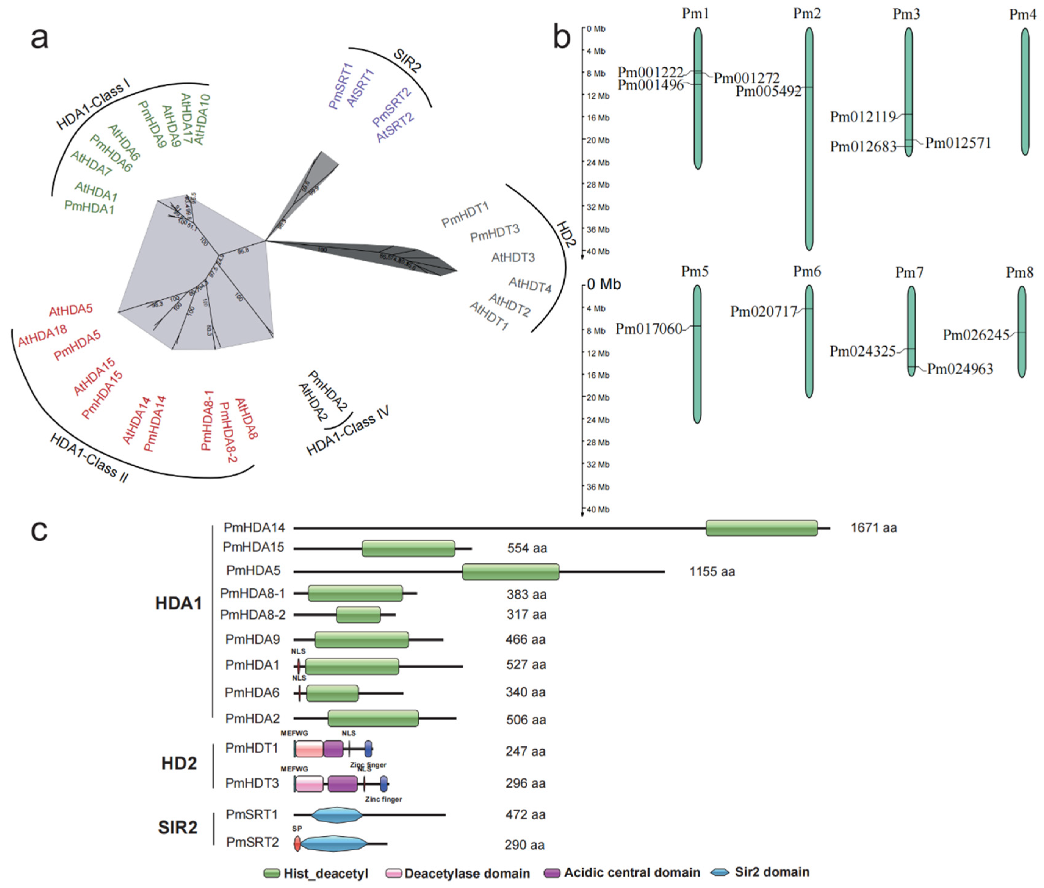
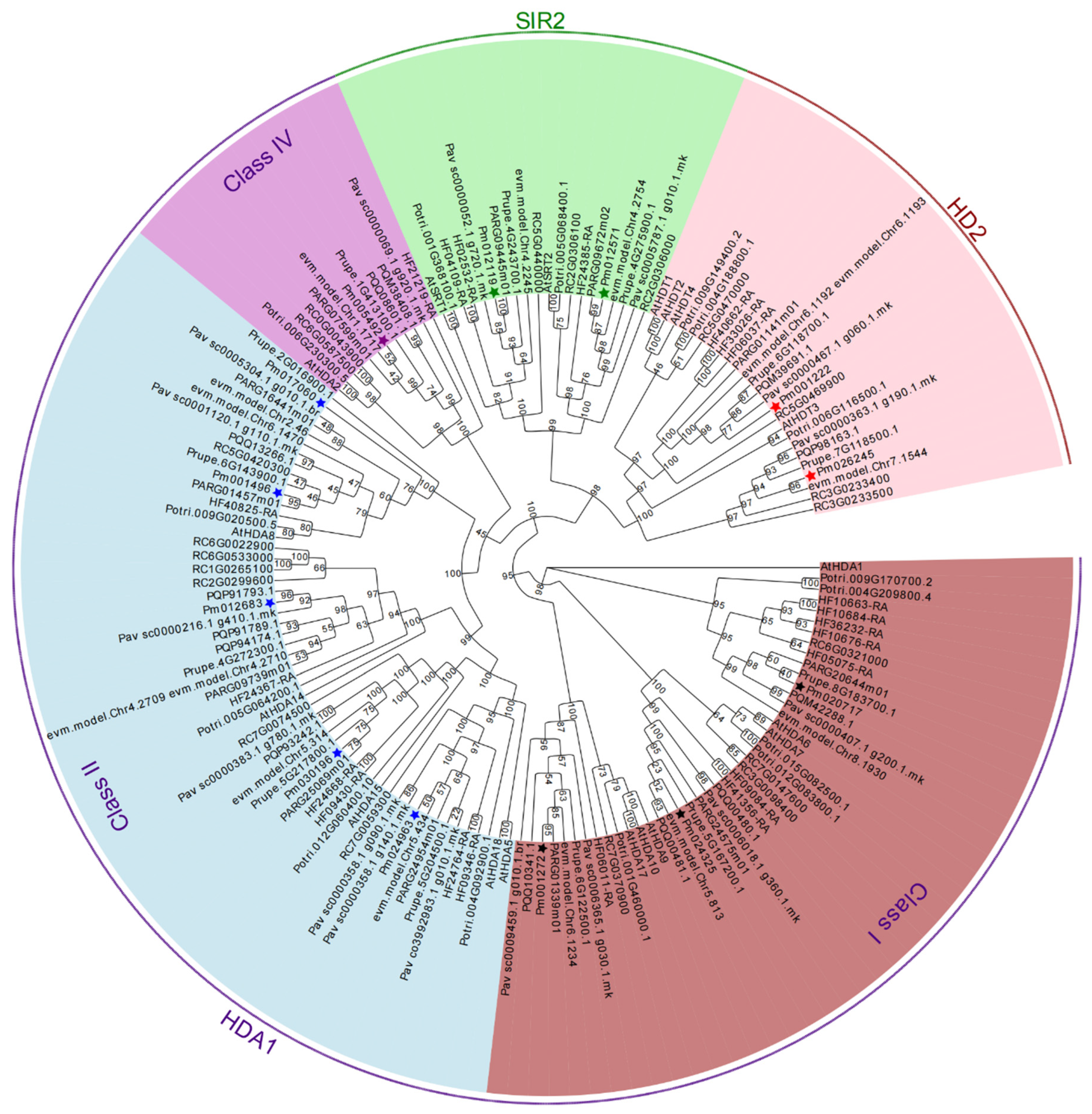
2.2. Multiple Sequences Alignment, Phylogenetic and Classification Analysis of RoHDACs

To evaluate the phylogenetic relationship of all putative 121 RoHDACs, a maximum likelihood phylogenetic tree of 10 plants was constructed and shown in Figure 1. It indicates that all RoHDACs can be classified into three families: RPD3/HDA1 superfamily, HD2 family, and SIR2 family, among which HDA1 family can further divided into Class I, Class II, and Class IV groups characterized by conserved domain and sequence similarity. We name these 121 RoHDACs genes according to the homology with Arabidopsis HDACs genes (Table S3). As depicted in Table 1 and Figure 1. The Class I group was composed of three clades based on the core Arabidopsis members AtHDA1/6/9. There are three Arabidopsis orthologues genes (HDA1/6/9) in mei, apricot, plum, and peach; four genes in sweet cherry, Somei-Yoshino and rose; and eight in apple. Among them, PavHDA9 contained two homologous genes, namely PavHDA9-1/9-2, PyHDA7 was found to be orthologues of Arabidopsis AtHDA7, and RcHDA6 has two homologous genes, namely RcHDA6-1/6-2.There are at most eight HDA1 genes in apple: three (MdHDA1-1 to 1-3) identified to be homologous of MdHDA1 and two of MdHDA6. For the Class II group, five members were identified in five Prunus plants including mei, seven members in sweet cherry and rosa, and six members in apple. We found that HDA8 in most plants consisted of two homologous genes, i.e., namely HDA8-1/8-2 (Figure 2a), with HDA8-2 genes having a shorter sequence and sharing a high sequence similarity (Table S3, Figure S1). Most class IV species contain a single HDA2 gene, and two homologous genes that only appear in Somei-Yoshino and rose. Several HDACs with high-similarity short sequences probably display expansion and contraction during the gene family evolution throng sequence alignment analyzed. For example, there are no HDA15 and SIR2 homologous genes, but there are three HDA14 homologies (PyHDA14-1/14-2/14-3) in Somei-Yoshino (Table S3, Figure S2). Besides, three high homologous genes (MdHDA1-1/1-2/1-3) are confirmed in apple and four homologous genes (RcHDA14-1/4-2/4-3/4-4) in rose (Table S3, Figures S3 and S4).
Unlike HDA1 superfamily, SIR2 family consists of only two members in most Rosaceae species, namely SIR1 and SIR2. Three members were identified in apple and rose but no SIR2 genes in Somei-Yoshino (Table 1). For plants specific HD2 family, only rose has four members corresponding to Arabidopsis, other Rosaceae plants have one, two, or three members. The phylogenetic tree of 13 PmHDACs and 18 AtHDACs proteins infers that all the 31 prospective proteins have a great bootstrap value showing the evolutionary relationships between mei and Arabidopsis HDACs proteins (Figure 2a). In our results, the phylogenetic trees of HDACs (Figure 1 and Figure 3) can also highlight the evolutionary relationship of Rosaceae plants, indicating that PmHDACs are more closely related to Prunus species like apricot and peach, which corresponds to the previous study [42,43,44].

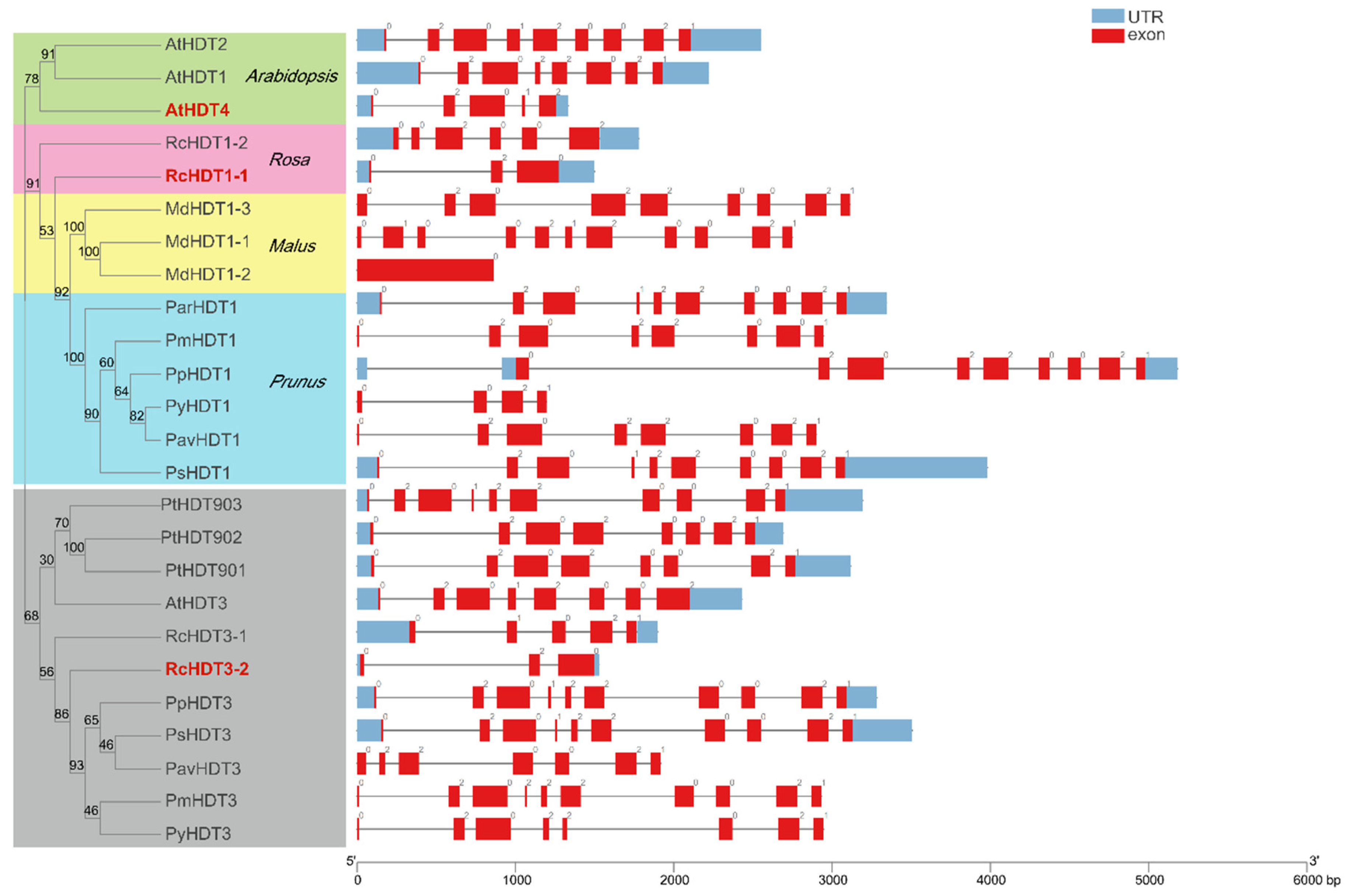
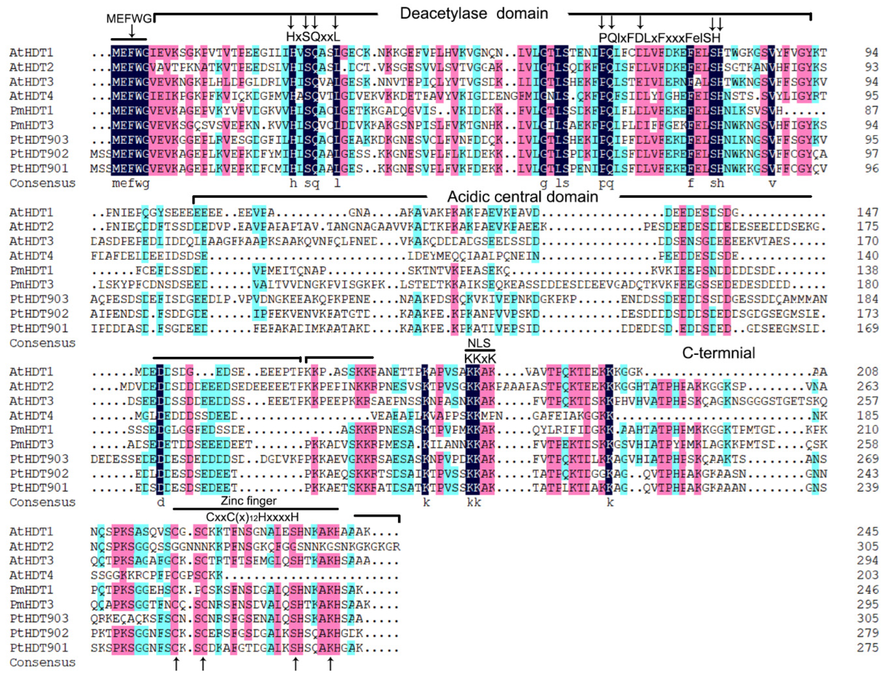
2.3. Gene Structure and Conversed Domains Analysis

To conform all the putative RoHDACs gene structures and conversed domains of proteins, we acquire gene exon numbers and positions in the genome as well as motifs and conserved domains of proteins (Figure S5, Table S3). The number of exons in three families exhibit distinct differences, which were ranged from 1 to 24. In the HDA1 superfamily, the orthologous genes of different species seem to contain a similar exon number. For instance, almost all RoHDA1 genes have 7 exons and most RoHDA9 genes have 14 exons. RoHDA15 and RoHDA5 genes, containing the average numbers of 16.89 and 14.33 exons, have significantly more exons than RoHDA8 and RoHDA14 (4.0 and 7.31) in the Class II group. In the plant-specific HD2 family, most members contain 8 or 10 exons, with an average of 7.52. We identified 8 and 10 exons in PmHDT1 and PmHDT3, respectively (Figure 3). It indicates that the distribution of exons in most HD2 genes seems to reveal a relatively stable evolutionary trend except some short paralogous genes. Besides, Gr2 group members (AtHDT4, RcHDT1-1, RcHDT3-2) contain less exons with 3-5. We also found that exon numbers among the paralogous genes, such as PmHDA8s, RcHDA6s, and RcHDA14s are dramatically different in terms of sequence size (Figure S5, Table S3).
Conserved motifs and domains are important components shared by members of gene family as well as the main regions playing functions. In this study, we identified 20 motifs, ranging from 21–80 aa, in all 154 HDACs proteins of 10 plants. It indicates that both the type and the number of motifs in these three types of HDACs are obviously diverse (Figure S5, Table S3). Almost all Class I type proteins have 9 kinds of motifs (motif7,14,10,4,9,5,1,2, and 11). On the contrary, in the class II, the distribution of motif types in HDA5, HDA15, HDA8, and HDA14 clades are slightly diverse. Class IV contains 4 motifs and the SIR2 family contains 3, where motif 16 and 17 are shared both in SRT1 and SRT2, but motif 18 is specific in the SRT2 proteins. We identified only one motif in the HD2 family. Furthermore, we predict the number and the distribution of completely conserved domains in each PmHDACs protein (Figure 2c). Over 8 HDA1 type proteins have a highly characteristic histone deacetylase domain that ranges from 140 to 348 aa, and 2 SIR2-type HDACs have a well conserved Sir2 domain. Moreover, sequence alignment indicates that two HD2 proteins (PmHDT1 and PmHDT3) contain 5 conserved domains: an N-terminal MEFWG- motif, followed by an around 86aa deacetylase catalytic domain, a long highly-variable acidic central domain, a 4 amino acid -KKAK- NLS, and a 22aa C2H2 zinc finger domain in the C-terminal end (Figure 2c and Figure 4). Previse study indicates that the HD2 family are classified into two groups: Gr1 and Gr2 [45]. We propose to define PmHDT1 and PmHDT3 as the Gr1 group based on the zinc finger they have. These domains play the important roles in the function and the classification of HD2 proteins [45].

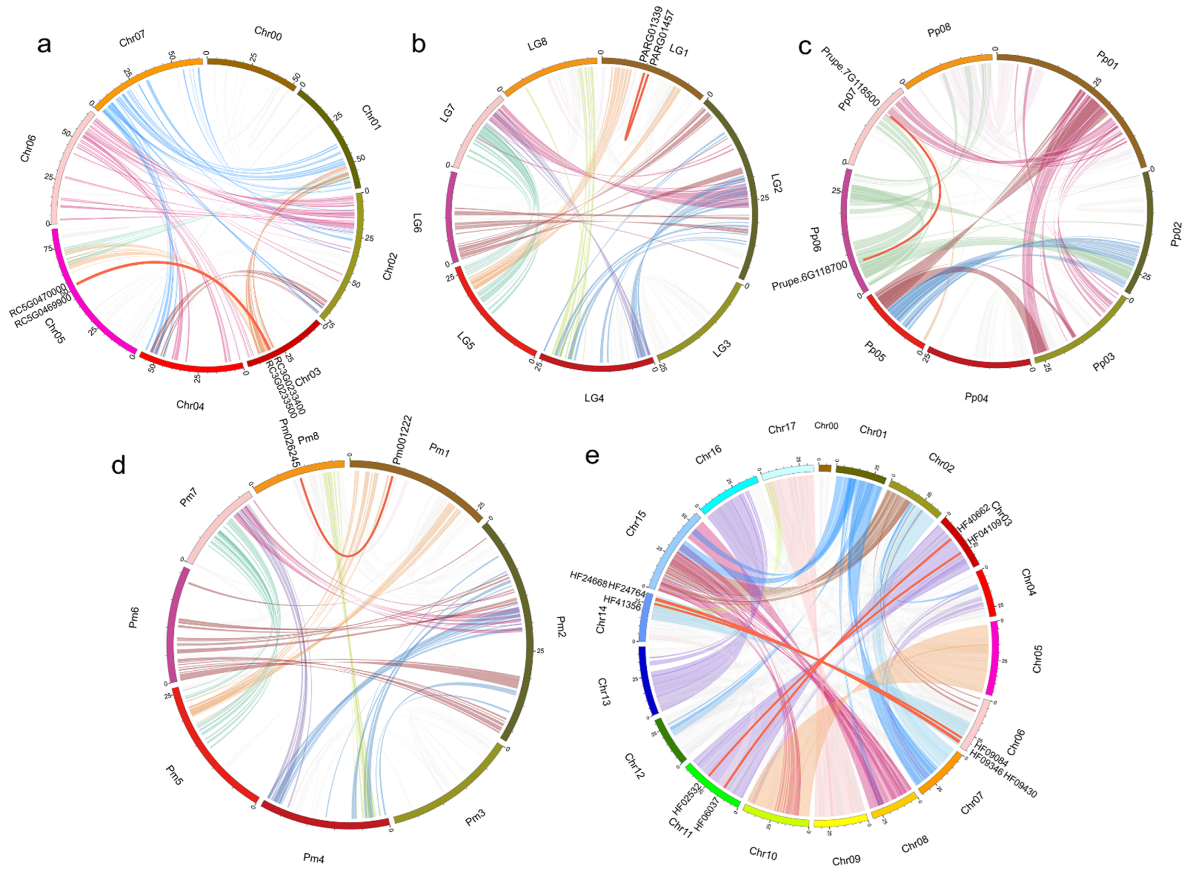
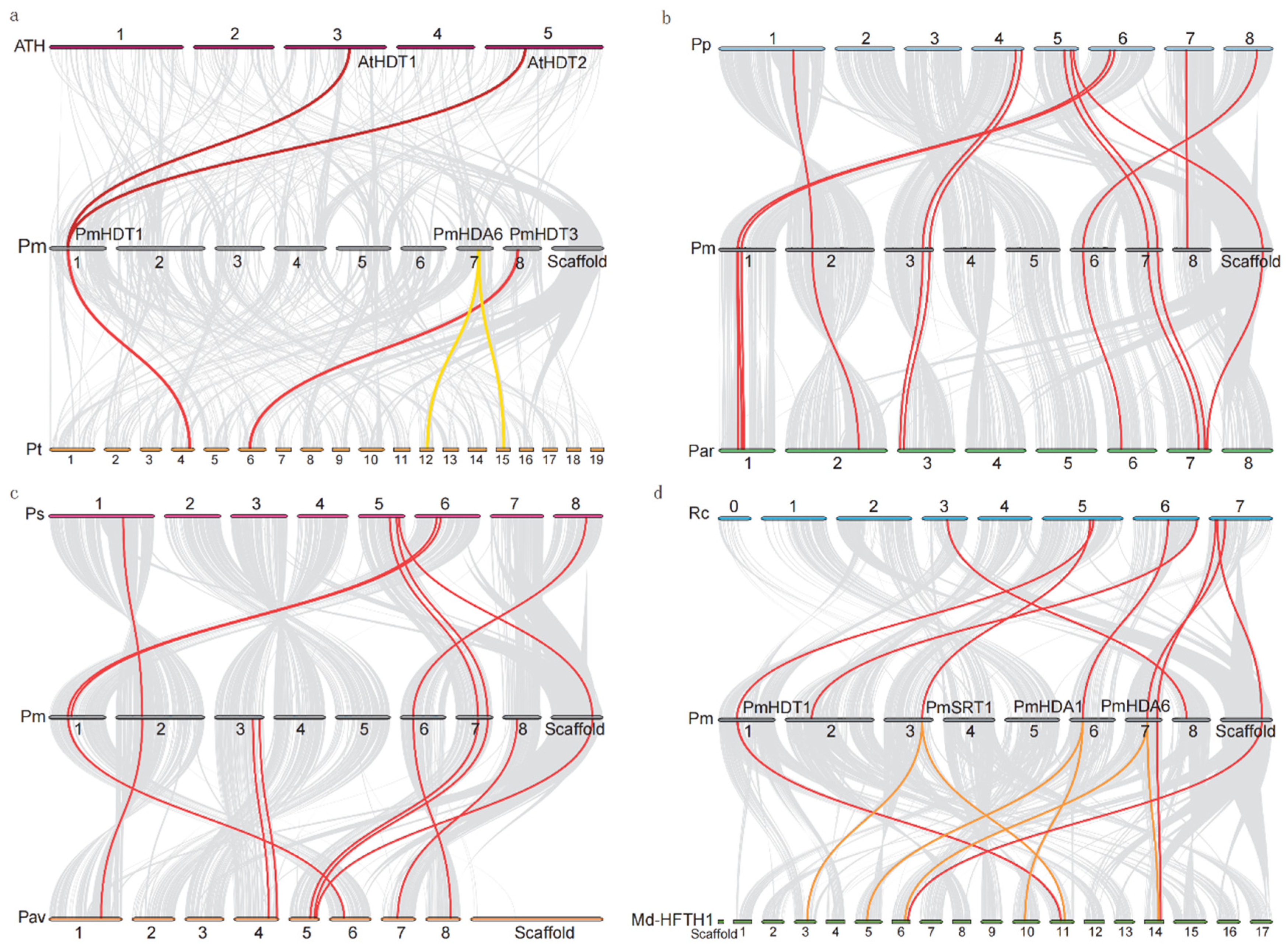
2.4. RoHDACs Gene Locations on Chromosome, Segmental Duplications and Synteny Analysis

The positions of overall 108 RoHDAC genes on the chromosome are determined by their genomic distributions, except for 13 PyHDAC genes that fail to be assembled into the chromosomal level. The results show that 12 of 13 PmHDAC genes are unevenly distributed on 8 chromosomes (Figure 2b). These are the largest three members on chromosomes one and three, respectively, and two genes on chromosome seven. Only one gene is found on chromosomes five and six, and no PmHDAC genes are found on the chromosome four. Besides, the location of other RoHDACs genes on the chromosome can be found in the Figures S6 and S7.
For TD and SD events, we firstly conducted a genomic comparison among Rosaceae species to identify the syntenic blocks and tandem duplications. Orthologous gene pairs of HDACs indicate that there are no TD events but SD events in RoHDACs genes. We investigated a total of 11 SD gene pairs in 6 HDACs families, among which 5 gene pairs belonged to MdHDACs, 2 belonged to RcHDACs, and 1 belonged to PmHDACs, ParHDACs, PavHDACs, and PpHDACs (Figure 5, Table S5). It is worth noting that over a half of the SD gene pairs occur in HD2 families involved in five species, corresponding to the previous study that the HD2 family expands via several rounds of successive duplication [45]. In mei, only the Pm001222 and the Pm026245, located on chromosomes one and eight, respectively, have a collinearity relationship. However, there are mostly five SD gene pairs in apple HDACs, distributed in all three families (Table S4). Previous study showed that a recent whole-genome duplication (WGD) shaped the genome of the domesticated apple, and we compared these five SD gene pairs that belong to duplication regions of the chromosome [42,46], which indicates that the WGD event may mainly contribute to the expansion of the MdHDACs family, resulting in the expansion of paralogous genes.
In addition, to further confirm the evolution of Rosacea plants, we conducted intergenomic comparisons and synteny analysis against Arabidopsis, P. trichocarpa, mei and other Rosacea plants. Only two and four HDACs orthologous gene pairs are identified between Arabidopsis and mei, as well as P. trichocarpa and mei, respectively (Figure 6a). Among them, PmHDT1 (Pm001222), located on chromosome one, has two collinearities (AtHDT1/2) in Arabidopsis, whereas only one homology gene in other Rosaceas, suggesting that HD2 family genes contracted in some Rosacea genomes. However, we found at least eight orthologous gene pairs among mei and other Rosaceae plants (Figure 6b,c), suggesting that HDACs among Rosaceae plants have significantly richer homologous relationships than those in model plants. In addition, there are more chromosomal synchronization collinearity genes between mei and apricot, indicating that they have a very closely relationship. Synteny between mei and apple shows that some PmHDAC genes (PmHDA1/6, PmSRT1) have two homologies in apple, suggesting the HDACs gene family expanded in the apple genome. In summary, it is obvious that the members of the HDACs gene family expanded or contracted significantly in various species during the evolution of Rosaceae, which may be related to the genome duplication event [42].

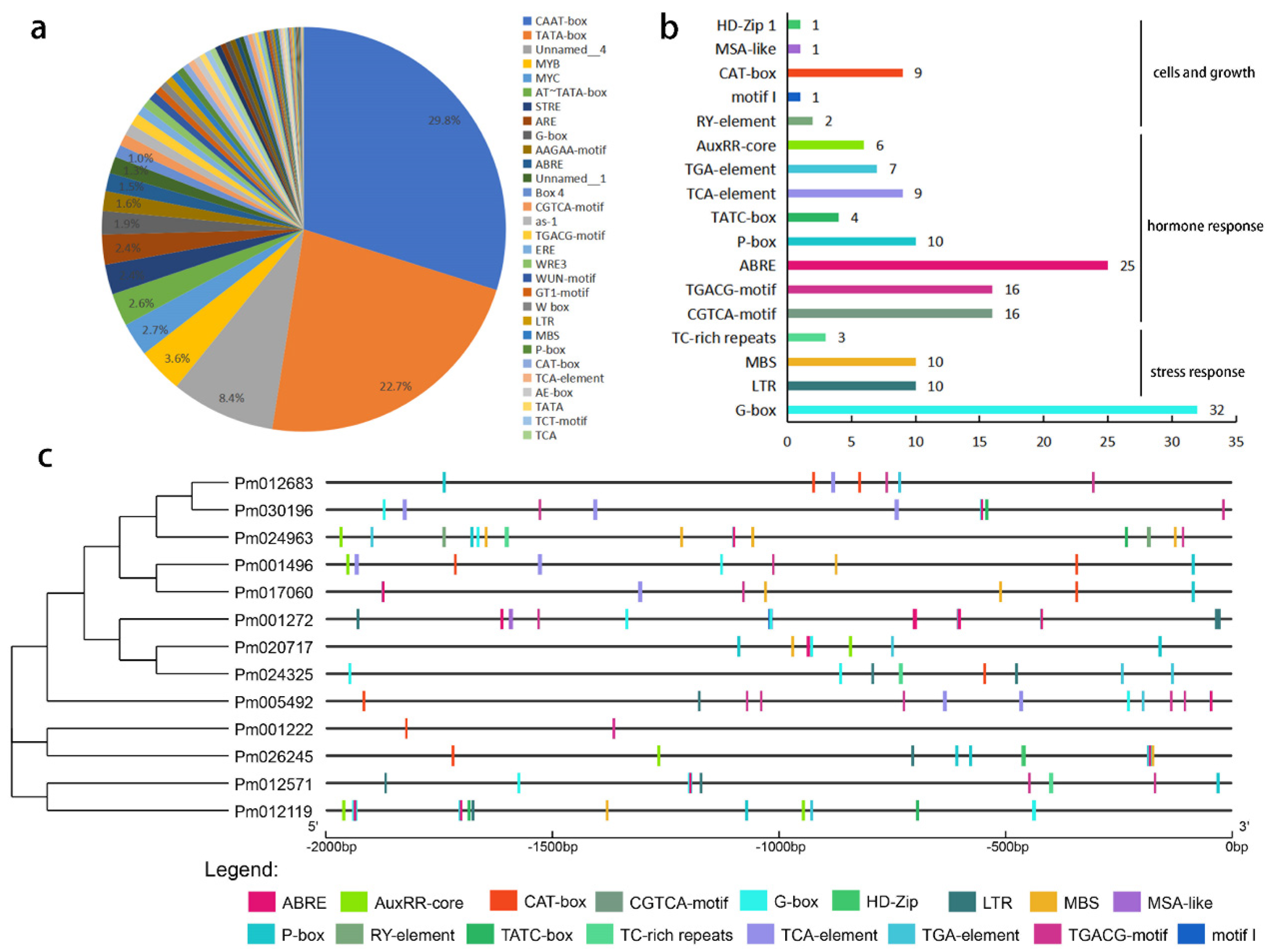
2.5. Cis-Element of PmHDACs

To confirm the potential regulatory mechanisms of PmHDACs in the plant development process, we analyzed the cis-element located on the 2.0 kb of all PmHDAC genes upstream using the PlantCARE website. Bioinformatics analysis showed that the TATA box and the CAAT-box, defined as common cis-acting elements in promoter and enhancer regions, are still the widely distrusted elements (52.5%) (Figure 7a). Then, we selected 17 cis-elements related to light, plant hormones, stress response, and the meristem development processes (Table S6). G-box elements, involved in plant light responsiveness, are widely distributed in PmHDACs promoters (Figure 7b,c). Notably, some cis-elements that have been confirmed as involved in biotic and abiotic stress, such as LTR, MBS, and TC-rich repeats, were comprised in PmHDACs (Table S5, Figure 7b). We found that 6 of 13 PmHDAC genes have LTR elements: PmHDA9 contains 3 LTRs, PmHDA6 and PmSRT2 have 2 LTRs, and PmHDA2, PmHDT3, and PmSRT1 have only one LTR (Figure 7c). These cis-elements identified related to hormones and plant stress processes may play an important role in plant resistance to environmental changes. Besides, we found a large number of hormone-related elements such as ABRE, CGTCA-motif, TCACG-motif, P-box, TCA-element, TGA-element, AuxRR-core, and TATC-box that participate in hormonal responses (i.e., gibberellin, abscisic acid, MeJA, auxin, and salicylic acid).

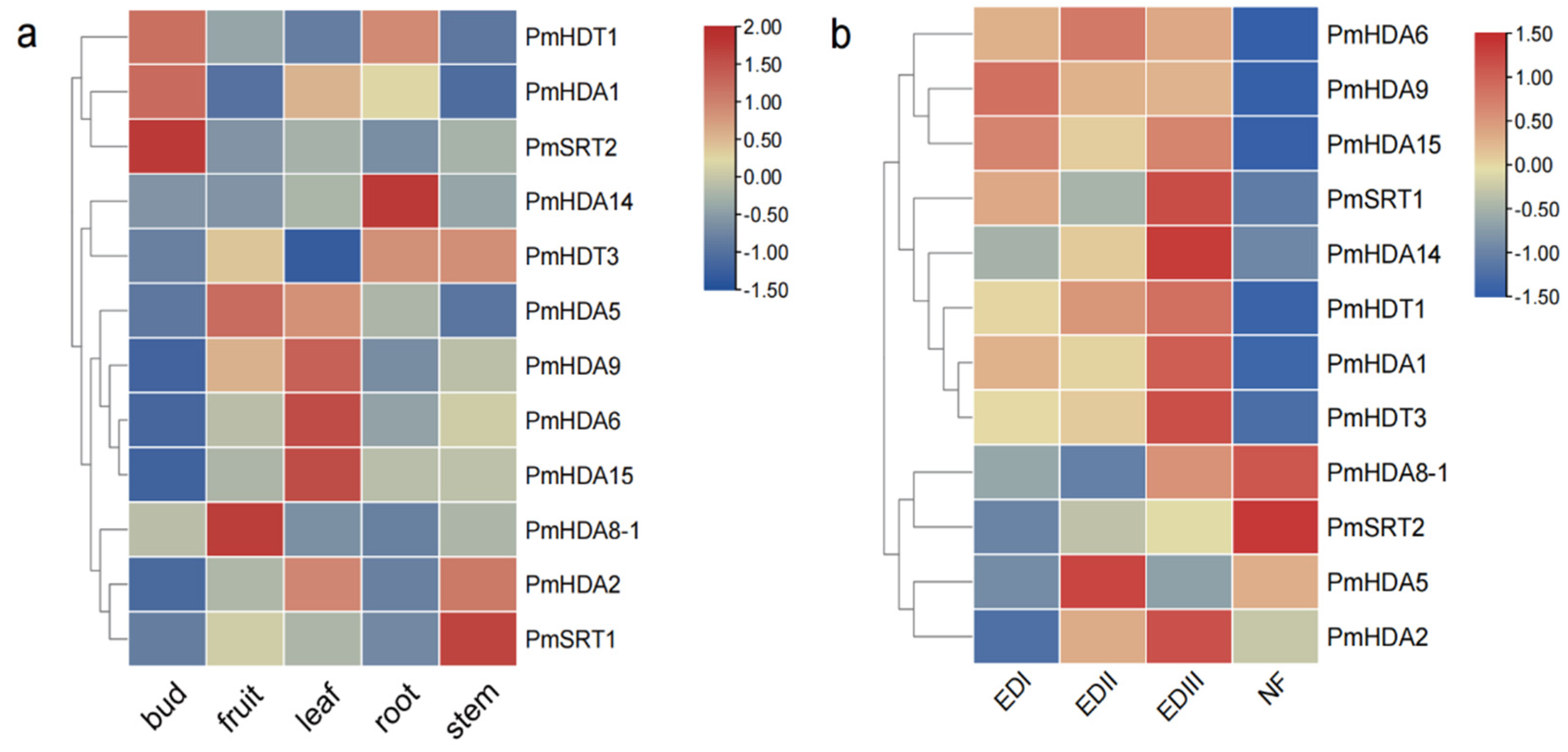
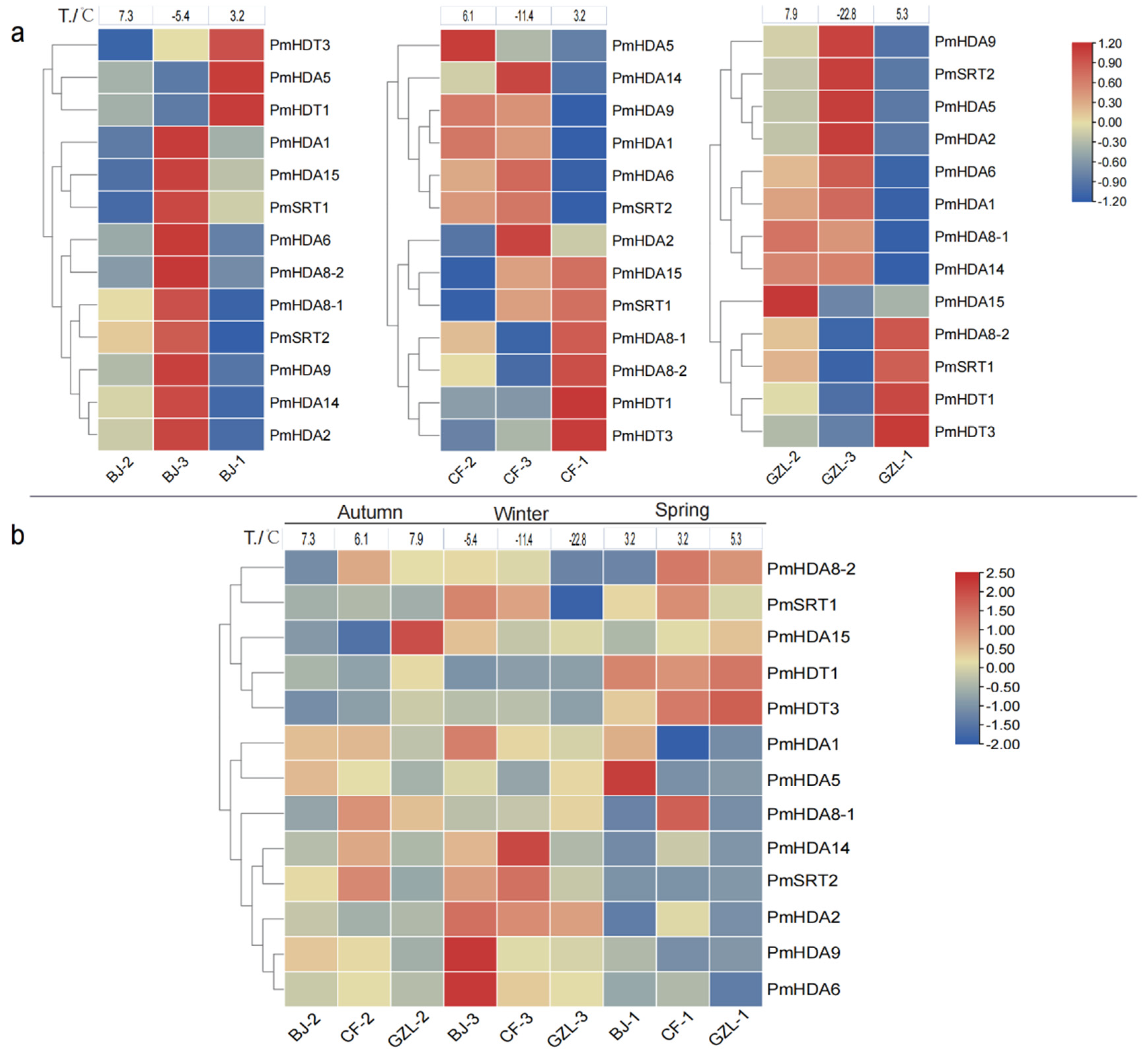
2.6. PmHDACs Expression Profiles in Tissues and Treatments

PmHDACs genes are differentially expressed in flower buds, fruits, leaves, roots, and stems, especially genes PmSRT2, PmHDA14, PmHDA6, PmHDA15, PmHDA8-1, PmSRT1 are tissue-specific (Figure 8a). For example, PmSRT2 is expressed in flowering buds, but it has a relatively low expression in other tissues. PmHDA14 and PmHDA8-1 are specifically expressed in roots and fruits, respectively. All PmHDACs genes show two expression trends during the flowering bud dormancy process (Figure 8b). Four genes (PmHDA14, PmHDT1, PmHDT3, and PmHDA2) are gradually up-regulated from EDI to EDIII and up to the peak in the EDIII stage, then abruptly decrease in the fourth NF stage, particularly PmHDA14 and PmHDT3. The expression of PmHDA6/9/15 genes is stably high in three endodormancy stages and also decreases in the NF stage. PmSRT1 gene is highly expressed in EDII. On the contrary, the expression of PmHDA8-1 and PmSRT2 gradually accumulates and reaches a peak in the NF stage when the bud dormancy is released. These expression patterns suggest that PmHDACs may be involved in the bud dormancy release process by deacetylating the target genes of different signaling pathways at different development stages.
We also detect the expression profiles in stems of cold-insensitive cultivar ‘Songchun’ under three low temperatures at three test sites: Beijing (BJ), Chifeng (CF), and Gongzhuling (GZL) (Figure 9). Some of PmHDACs genes show similar expression patterns during the change of temperature in different phenological periods at the same latitude test site, respectively (Figure 9a). For example, at the Beijing site, a total of 10 genes (PmHDA1/6/9/15/2/14/8-1/8-2, PmSRT1/2) show up-regulation in autumn and in winter (2nd to 3rd stages) but then decrease in the early-spring (1st stage). However, the expression levels of PmHDA5 and PmHDT1 genes are down-regulated from the 2nd to the 3rd stages and then increase in the first stage. We find the gene expression trend of Gongzhuling and Chifeng sites are relatively consistent with that in Beijing. In summary, we find PmHDA1/6/9 and PmSRT2 genes are all up-regulated at the cold acclimation period (2 to 3), then down-regulated at the cold acclimation lost period (3 to 1) in three sites, while PmHDT1/3 has the opposite expression profiles, indicating that these genes are positively or negatively correlated with low temperature response. However, the expressions of most PmHDACs in the same season have slight differences at the different test sites, among which some genes like PmHDA15 are highly expressed at the GZL site in autumn, PmHDA6/9 are highly expressed at the BJ site, while PmHDA14 shows high expressions at the GZL site in winter. More than half of genes have a relatively low expression in spring, except PmHDT1/3 (Figure 9b). These results indicate that the expression diversities of PmHDACs in different sites may be caused by geographical environmental factors or cold-resistant differences of plant individuals.

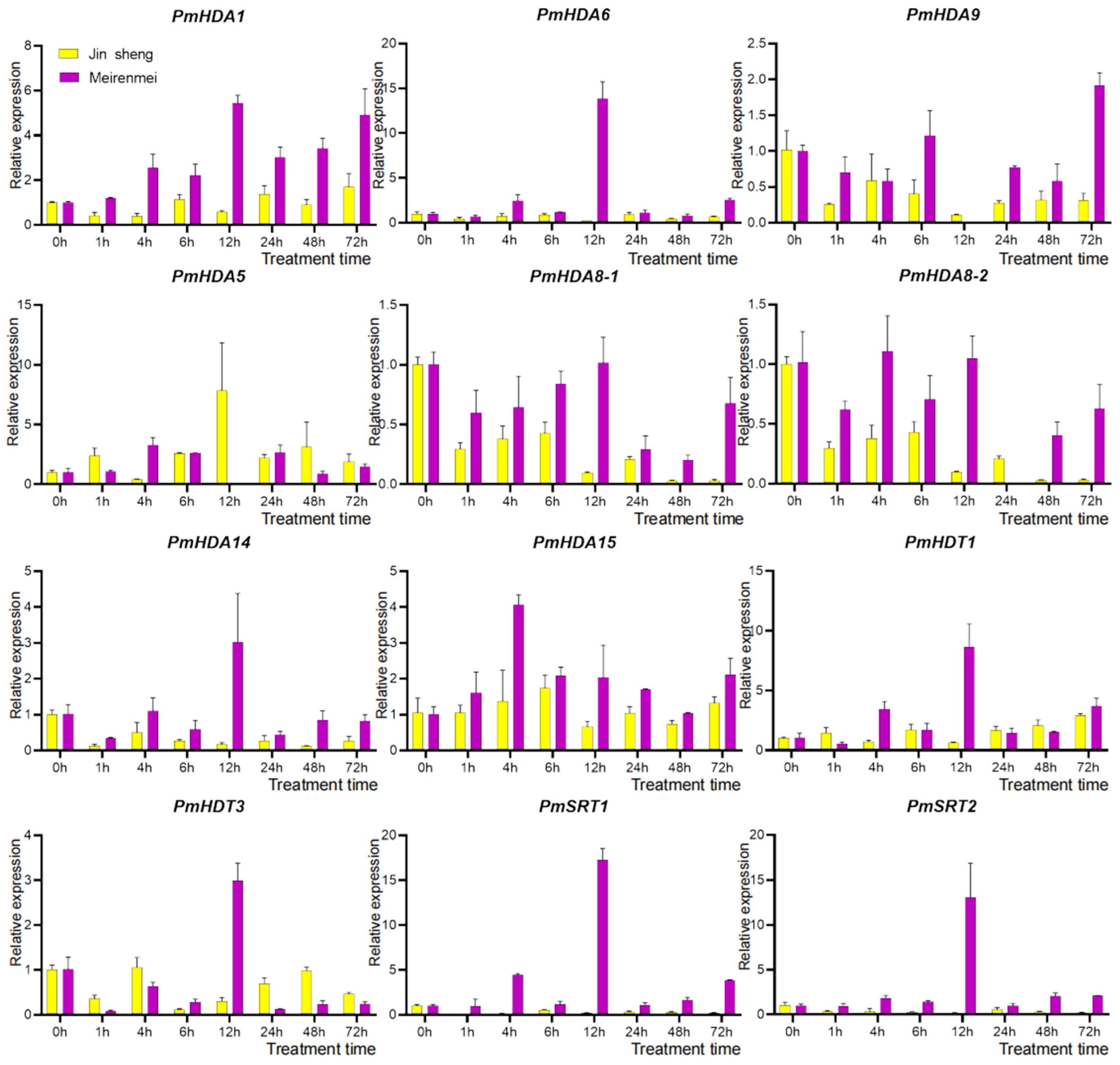
2.7. qRT-PCR of PmHDACs under Cold Stress and Gene Functional Annotation

To further confirm the putative roles of PmHDACs genes in response to cold stress in mei, the expression levels of PmHDACs under 4 °C low temperature treatment are determined by qRT-PCR in cold-sensitive cultivar ‘Jinsheng’ and cold-insensitive cultivar ‘Meirenmei’ (Figure 10). All PmHDACs members show significant expression profiles under cold treatments in the two cultivars. Obviously, most of the genes are induced with higher expression levels in ‘Meirenmei’, by contrast, repressed in the cold sensitive cultivar ‘Jinsheng’. For instance, PmHDA6, PmHDA14, PmHDT1, PmHDT3, PmSRT1, and PmSRT2 genes are significantly induced and highly expressed at 12 h under cold, indicating that they might be involved in the cold stress response process of ‘Meirenmei’ at this stage. In addition, the expression of the PmHDA1 gene is markedly up-regulated at 1 h to 72 h under cold treatment compared without cold (0 h), also peaking at 12 h. These above genes may have a close positive correlation with cold tolerance of ‘Meirenmei’, all of which are induced by low-temperature, and are likely to be involved in the process of cold stress regulation by modifying low temperature sensitive key genes through deacetylation. Importantly, we find that genes PmHDA6, PmHDA9, PmHDA8-1/8-2, PmHDA14, PmSRT1, and PmSRT2 are strongly down-regulated in ‘Jinsheng’: their expressions are barely detectable in the later cold treatment stages (12–72 h), which indicates that these genes are also likely to be negatively involved in the cold intolerance of ‘Jinsheng’. The PmHDA5 gene is the only member highly expressed in the ‘Jinsheng’, and the expression peak also occurs at 12 h after cold treatment, indicating that it may be positively involved in the regulation of the cold intolerance in ‘Jinsheng’. Thus, these expression results demonstrate that HDACs genes show the functional diversity of a cold stress response and the similarity in functional mechanisms. All of them mainly enrich in protein deacetylation, chromosome organization, and other epigenetic processes, and they may target activated or inhibitive genes by deacetylation modification to participate in gene silencing regulation, thereby positively or negatively regulating mei cold sensory processes (Figure S8).

3. Discussion

The essential histone deacetylation genes HDACs can interact with other transcription factors to silence gene expression through deacetylation modification [47]. They are widely involved in plant vegetative and reproductive growth processes, seed maturation and stress responses [15,48]. In this study, we have identified a total of 121 RoHDACs genes in 8 Rosaceae genomes, which are classified into 3 families (RPD3/HDA1, HD2 and SIR2). For the HDA1 superfamily, all 12 AtHDA1-type genes are initially divided into classes I, II, and III [13] of which, AtHDA8, AtHDA14, AtHDA10, and AtHDA17 are still unclassified into any class genes [13,48]. Later research indicates that AtHDA8 and AtHDA14 are classified into Class II and three clusters (Class I, II, and IV) confirmed [12]. However, the rice HDA1 proteins are divided into four classes [14]. Our study suggests that the orthologues genes of HDA8 and HDA14 are clustered as class II proteins with a high bootstrap value (Figure 1) as well as shared at least five motifs (Figure S5), which corresponds to the later classification of Arabidopsis HDA1 [12]. This study also indicates that each of the three HDACs subfamilies has its own conserved domains. It is worth noting that the plant-specific HD2 family contains five conserved domains, of which the N-terminal motif MEFWG-, important for the gene regulation activity of HDs [49], is also highly conserved among most of the Rosaceae HDs identified (Figure 2 and Figure S9), which is consistent with the previous research [45]. Besides, previous study shows that the length of almost all HD2 protein differences is strongly correlated with the length of the highly-variable acidic central domain [45]. Sequence alignment shows that PmHDT1/3 acidic central domains are relatively less conserved, which is probably the main reason for the differences in gene functions.
Gene duplication events are considered to be the major forms of gene family expansion in the history of gene evolution [50]. In our study, a total of 11 SD gene pairs were identified in 8 Rosacea HDACs. Previous phylogenetic research confirms that gene duplication events or sequence rearrangements occur successively during the evolution of the Arabidopsis HDA1 superfamily, resulting in an increase in gene members (AtHDA10/17/18) [13,51]. Notably, similar homologous gene duplication and rearrangement events also occur in eight Rosaceae HDA1 superfamily, leading to an uneven distribution of HDA1 genes in different species. Having the most numbers of HDACs, a total of 21 genes are identified in the apple genome. So why are there significantly more HDACs in apples than in the Prunus species? Firstly, some paralogous genes are searched in MdHDACs, where MdHDA1 has three paralogous genes (Figure S3) and MdHDA5/6/15 have two. These homologous genes pairs are found at the same time as the SD gene pairs (consistent with WGD blocks [42]) in apple genome (Table S4). For the collinearity results in apple, not only HDA1 family but also the SIR2 and the HD2 genes have obvious SD events, which further indicates that the recent WGD event and the increase in chromosomes as an ancient tetraploid plant lead to the significant expansion of the apple HDACs family [42]. Besides, we found that most HDA8 genes of the HDA1 family in Rosaceae plants contain two paralogous genes (Table S3). For instant, PmHDA8-1 protein is highly similar with the PmHDA8-2 (Figure S1). Similar gene expansions are observed in other RoHDACs like PyHDA14 and RcHDAC14 genes (Figures S2 and S4). In our research, the SD gene pairs of RoHDACs exist in most HD2 family members, such as PmHDT1 and PmHDAT3 (Table S4), demonstrating that the HD2 family may have expanded via several rounds of successive duplication [45]. Obviously, we find that some HD2 proteins lost almost all of the C-terminal region, including the C2H2 zinc finger domain, such as RcHDT1-1 and RcHDT3-2 that can be defined as the Gr2 group (Figure S9). However, the sequences of these two Gr2 HD2s are conserved with those of Gr1 members, and it is reasonable to postulate that Gr2 evolves from Gr1 gene [45].
Epigenetic modification control plant cold responses [52]. In Arabidopsis, HDA6 is one of the HDAC genes analyzed that plays a key role in cold tolerance. The mutant axe1-5 shows reduced freezing tolerance compared with the wild-type plants under cold treatment, indicating that HDA6 plays a critical role in regulating the cold acclimation process that confers freezing resistance in Arabidopsis [36]. In our research, three cold stress treatments were analyzed in different mei cultivars and tissues. Based on the above results, we also obtained some genes closely related to cold stress. First, the important gene PmHDA6 responded to cold stress in both flower buds and stems at different sites (Figure 8 and Figure 9). Furthermore, qRT-PCR results indicate that the expression levels of PmHDA6 are strongly up-regulated at 12 h under 4 °C cold treatments, then abruptly reduce later in cold tolerance cultivar ‘Meirenmei’. Besides, the expression levels of PmHDA1 gene in ‘Meirenmei’ are strongly increased during the cold stress treatment, which is quite similar to the expression of AtHDA6 [36]. These results suggest that PmHDA1 and PmHDA6, as hub genes, are probably involved in the cold response to promote the freezing resistance of ‘Meirenmei’ through regulation of cold-related genes. Other PmHDA1 genes PmHDA9, PmHDA8-1/8-2, PmHDA14, PmHDA5, and PmHDA2 are also significantly negatively or positively responsive to cold stress in two cultivars. In banana, MaMYB4 recruits MaHDA2 to modulate mechanisms of fatty acid biosynthesis during a cold stress response in fruits [53]. Except for the cold stress, HDA1 genes play important roles in other abiotic stress through forming protein complex with TFs, such as WRKY and hormone signals [32,33,34,54,55]. The Arabidopsis HOS15, a WD40-repeat protein, interacts with HDACs to regulate the plant development process [56,57], particularly, functions as a repressor to control cold stress-regulated gene expression through chromatin modification [58,59]. HD2 proteins also play important roles in abiotic stress responses, including cold [41,59,60,61]. AtHD2C (AtHDT3) interacts with HOS15, associated with cold signaling gene COR (COLD RESPONSIVE) and CBF (C-REPEAT (CRT) BINDING FACTOR) in response to cold stress [58]. Recent study indicates that HD2 proteins can interact with HDA1-type histone deacetylases, such as HDA6 and HDA9 [38,40]. In mei, the expression accumulation of PmHD2 genes is detected in stems and flower buds under cold treatment. Among them, PmHDT1 is highly expressed in ‘Meirenmei’ under cold treatment, which is consistent with PmHDA6 (Figure 10). Collectively, we infer that PmHDACs may function as the similar protein complex to be involved in cold stress in mei plant.
Additionally, it is worthy to note that mei SIR2 genes are strongly up-regulated in ‘Meirenmei’ cultivar while down-regulated in ‘Jinsheng’, which shows the significant cultivar association. We find that the motif 18 that is highly conserved only consist in the SRT2 proteins but not in the SRT1 (Figure S10). Furthermore, the sequence conservation of SRT1 proteins or SRT2 proteins are significantly higher than that between the two proteins (Figure S10). It suggests that PmSRT1 and PmSRT2 may be involved in response to cold stress with different regulatory mechanisms in different cultivars, although there is little function data available about plant SIR2-type HDACs [48]. Combined with cis-elements identified in PmHDACs (Figure 7), we find that almost a half of genes contain the low temperature response element LTR, which have been detected to bind to transcript factors like bHLH to regulate plant development [62,63,64]. In summary, our results confirm that histone deacetylases PmHDACs play important roles in the regulation of the abiotic stress response regarding the cold tolerance of mei in north China. However, how their interactions form the protein complex that regulates those genes related to cold stress and affect the cold resistance of mei requires further research in the future.

4. Materials and Methods

4.1. Plants Genome Resources

The high-quality genome assembly and annotation files of A. thaliana (TAIR10.41), P. trichocarpa (v4.0), P. mume (v1.0), P. salicina ‘Sanyueli’ (v2.0), P. armeniaca (v1.0), P. persica ‘Lovell’ (v2.0), P. avium (v1.0.a1), P. yedoensis var. nudiflora (v1.0), M. domenstica ‘HFTH1′ (v1.0), and R. chinensis ‘Old Blush’ (v1.0) were download from the Genome Database for Rosaceae (GDR, https://www.rosaceae.org, accessed on 5 July 2021) [65] and from Phytozome v12.1 (https://phytozome.jgi.doe.gov/pz/portal.html, accessed on 5 July 2021) [66].

4.2. Identification of HDACs Gene Family

The HMM search 2.0 was used to identified the HDA1 and the SIR2 families with a histone deacetylase domain and a SIR2 domain (PF00850, PF02146) from the Pfam database v32.0 [67] (http://pfam.xfam.org, accessed on 7 July 2021). HD2 genes were identified by BLASTP according to the Arabidopsis HD2 proteins. After proofreading using a phylogenetic tree, sequence size, and conserved domains, we deleted the wrong gene members. Ultimately, the HDACs members of 8 Rosaceae plants were acquired.
According to gene numbers and conserved domains, transmembrane domains and signal peptides were acquired on the SMART v9.0 [68] (http://smart.embl-heidelberg.de, accessed on 23 July 2021), TMHMM-2.0 [69] (https://services.healthtech.dtu.dk/service.php?TMHMM-2.0, accessed on 6 August 2021), and SignalP-5.0 [70] (https://services.healthtech.dtu.dk/service.php?SignalP-5.0, accessed on 6 August 2021) websites. The NLS and the physical and chemical parameters of proteins were predicted on the WoLF PSORT (https://wolfpsort.hgc.jp/, accessed on 6 August 2021) and ProParam (https://web.expasy.org/protparam/, accessed on 7 August 2021).

4.3. Phylogenetic Analysis and Classification of HDACs Genes

With identified HDACs, we first conducted a codon align of mei and Arabidopsis protein sequences. Using a Muscle method, neighbor-joining trees were constructed by MEGA 7.0 [71]. To obtain the maximum-likelihood tree, we generated a set of protein multiple sequence alignments of HDACs proteins with MAFFT v7 [72] (https://mafft.cbrc.jp/alignment/server/, accessed on 10 August 2021), and we constructed the phylogenetic trees using IQtree 2.1.3 [73] (http://iqtree.cibiv.univie.ac.at/, accessed on 10 August 2021). For the putative name of Rosaceae HDACs genes, we divided all the Rosaceae HDACs into subgroups and named the homologous gene based on Arabidopsis HDACs genes.

4.4. Gene Structure and Protein Conserved Motif Analysis

We obtained the exons, introns, and UTR (untranslated Region) location information of the genes from the genome annotation file. The RoHDACs proteins were submitted to MEME-v4.12.0 [74] with settings: -maxsize 6,000,000 -mod anr -nmotifs 20 -minw 6 -maxw 100 to search for conversed motifs. Finally, TBtools [75] was used to conduct the tree-structure-motif map.

4.5. Duplications, Synteny and Genes Chromosome Location Analysis

Protein sequences of each Rosaceae specie were used to makeblastb and blastp all-vs-all with Blast 2.6 software, respectively. We analyzed eight Rosaceae WGD events by McscanX [76] with default settings. The segmental duplication (SD) gene pairs and tandem duplication (TD) events of RoHDACs were acquired from synteny blocks and the tandem gene pairs. Synteny circle-map were plotted by CIRCOS [77]. The collinearity of HDACs in different genomes was analyzed by McscanX as above. Chromosomal location of RoHDACs and total length of chromosomes were extracted from a genome annotation gff file and a genome sequence, and they were marked by MG2C_v2.0 (http://mg2c.iask.in/mg2c_v2.0/, accessed on 13 August 2021) [78].

4.6. PmHDACs Cis-Acting Element Analysis

We obtained the 2000 bp promoter sequences upstream of the PmHDACs gene members, and we submitted it on the website plantCARE [79] to analyze cis-acting elements they contained; then, we constructed the element distribution with GSDS3.0 (http://gsds.gao-lab.org, accessed on 17 August 2021).

4.7. Expression Profiles of PmHDACs

To investigate the potential functions of PmHDACs in different tissues and cold stress response, RNA-seq data of five different tissues of mei (flower buds, fruits, leaves, roots, and stems) [80] and flower buds of mei ‘Lve’ cultivar exposed in three dormancy status EDI (Endodormancy I, 0% flush rate), EDII (Endodormancy II, 45% flush rate), EDIII (Endodormancy III, 100% flush rate), and NF stage (Natural Flush) [81] were obtained. The expression profiles were also analyzed in RNA-seq data of ‘Songchun’ cultivar stems at three different geographical sites: Beijing (BJ, 54′39° N, 28′116° E), Chifeng (CF, 17′42° N, 58′118° E), and Gongzhuling (GZL, 42′43° N, 47′124° E) in three phenological stages (autumn deciduous initiation, winter dormancy, and spring germination) under natural cold.
The heatmap of gene expression was mapped by FPKM values using TBtools [75] with a normalized row scale and row cluster method.

4.8. Plant Materials, Cold Stress Treatments and qRT-PCR

Branches of cold-sensitive cultivar ‘Jinsheng’ and cold-tolerated cultivar ‘Meirenmei’ were collected from Jiufeng International Plum Blossom Garden, Beijing, China (07′40° N, 11′116° E). Plants were maintained in water overnight at 22 °C and treated under 4 °C with 1, 3, 6 12, 24, 36, 48, and 72 h under long-day conditions (16-h light/8-h dark). Then we obtained annul branches to extract total RNA.
Total RNA was isolated by an RNAprep Pure Plant Plus Kit (Qiagen, Beijing, China) and then synthesized with cDNA using the ReverTra Ace®qPCR RT Master Mix with gDNA Remover (Toyobo, Osaka, Japan). A quantitative RT-PCR was performed on a qTOWER2.2 System (analytikjena, Jena, German), using a SYBR®Green Premix Pro Taq HS qPCR Kit (AccurateBiology, Hunan, China). The reaction system was a total of 20 μL with a 10 μL SYBR®Green Premix Pro Taq HS qPCR Kit, 8 μL 10× forward and reverse primers mix, and 2 μL 10× cDNA samples. The reaction program was set as follows: an initial denaturation step (30 s at 95 °C), followed by 40 cycles of 5 s at 95 °C, 30 s at 50–60 °C, and 30 s 72 °C. The relative expression levels of the genes were calculated using the ∆∆Ct method and normalized using the PmActin reference gene. The primers used in this study were listed in Table S7.

4.9. GO Annotation and Enrichment Analysis

GO annotation data of PmHDACs genes were extracted from mei genome GO annotation files (background) and then submitted on the website omicshare (https://www.omicshare.com/, accessed on 13 September 2021) to perform further GO enrichment analysis.

5. Conclusions

In this study, a total of 121 HDACs are identified in 8 Rosaceae plants, among which 13 genes are from woody plant mei. The detailed genome-wide characterization analyses of HDACs are first confirmed in Rosaceae plants, including phylogenetic evolution, subfamily classification, gene structure, conserved domain, gene locations, and SD events. Besides, we focus on the expression profiles of HDACs in mei. The mRNA accumulation levels of PmHDACs in mei various tissues reveal relatively tissue-specific or wide expressions profiles. RNA-seq data and qRT-PCR experiment in cold treatment suggest that PmHDAC genes significantly respond to cold stress with up or down regulated expression pattens. Our research provides an insight into the phylogenetic relationship of the HDACs family in Rosaceaes, and it functions as an investigation of PmHDACs response to cold stress in mei.

Supplementary Materials

The following supporting information can be downloaded: at https://www.mdpi.com/article/10.3390/ijms23115957/s1.

Author Contributions

Conceptualization, J.M. and L.S.; methodology, J.M. and Z.W.; software, J.M., M.L. and Z.W.; validation, J.M.; formal analysis, J.M.; investigation, J.M.; resources, Q.Z., T.C. and L.S.; data curation, J.M.; writing—original draft preparation, J.M.; writing—review and editing, L.S.; visualization, L.S.; supervision, L.S. All authors have read and agreed to the published version of the manuscript.

Funding

This research was funded by Forestry and Grassland Science and Technology Innovation Youth Top Talent Project of China (No. 2020132608), the National Key Research and Development Program of China (2018YFD1000401), and the National Natural Science Foundation of China (No. 31870689).

Data Availability Statement

The data are included into supplementary materials.

Conflicts of Interest

The authors declare no conflict of interest.

References

  1. Campbell, R.R.; Wood, M.A. How the epigenome integrates information and reshapes the synapse. Nat. Rev. Neurosci. 2019, 20, 133–147. [Google Scholar] [CrossRef] [PubMed]
  2. Loidl, P. A plant dialect of the histone language. Trends Plant Sci. 2004, 9, 84–90. [Google Scholar] [CrossRef] [PubMed]
  3. Pazin, M.J.; Kadonaga, J.T. What’s Up and Down with Histone Deacetylation and Transcription. Cell 1997, 89, 325–328. [Google Scholar] [CrossRef] [Green Version]
  4. Kiermaier, A.; Eilers, M. Transcriptional control: Calling in histone deacetylase. Curr. Biol. 1997, 7, R505–R507. [Google Scholar] [CrossRef] [Green Version]
  5. Kuo, M.H.; Allis, C.D. Roles of histone acetyltransferases and deacetylases in gene regulation. BioEssays News Rev. Mol. Cell. Dev. Biol. 1998, 20, 615–626. [Google Scholar] [CrossRef]
  6. Shen, Y.; Wei, W.; Zhou, D. Histone Acetylation Enzymes Coordinate Metabolism and Gene Expression. Trends Plant Sci. 2015, 20, 614–621. [Google Scholar] [CrossRef]
  7. Sharon, Y.; Roth, J.M.D.A. Histone acetyltransferases. Annu. Rev. Biochem. 2001, 70, 81–120. [Google Scholar] [CrossRef]
  8. Gerald Brosch, M.G.P.L. Purification of histone deacetylase HD 1-A of germinating maize embryos. FEBS Lett. 1996, 393, 287–291. [Google Scholar] [CrossRef] [Green Version]
  9. Rossi, V.; Hartings, H.; Motto, M. Identification and characterisation of an RPD3 homologue from maize (Zea mays L.) that is able to complement an rpd3 null mutant of Saccharomyces cerevisiae. Mol. Gen. Genet. MGG 1998, 258, 288. [Google Scholar] [CrossRef]
  10. Lusser, A.; Brosch, G.; Loidl, A.; Haas, H.; Loidl, P. Identification of maize histone deacetylase HD2 as an acidic nucleolar phosphoprotein. Science 1997, 277, 88–91. [Google Scholar] [CrossRef]
  11. Wu, K.; Tian, L.; Malik, K.; Brown, D.; Miki, B. Functional analysis of HD2 histone deacetylase homologues in Arabidopsis thaliana. Plant J. 2000, 22, 19–27. [Google Scholar] [CrossRef] [PubMed]
  12. Alinsug, M.V.; Yu, C.; Wu, K. Phylogenetic analysis, subcellular localization, and expression patterns of RPD3/HDA1 family histone deacetylases in plants. BMC Plant Biol. 2009, 9, 37. [Google Scholar] [CrossRef] [PubMed] [Green Version]
  13. Pandey Ritu, E.A. Analysis of histone acetyltransferase and histone deacetylase families of Arabidopsis thaliana suggests functional diversification of chromatin modification among multicellular eukaryotes. Nucleic Acids Res. 2002, 30, 5033–5036. [Google Scholar] [CrossRef] [Green Version]
  14. Fu, W.; Wu, K.; Duan, J. Sequence and expression analysis of histone deacetylases in rice. Biochem. Biophys. Res. Commun. 2007, 356, 843–850. [Google Scholar] [CrossRef] [PubMed]
  15. Ma, X.; Lv, S.; Zhang, C.; Yang, C. Histone deacetylases and their functions in plants. Plant Cell Rep. 2013, 32, 465–478. [Google Scholar] [CrossRef]
  16. Aquea, F.; Timmermann, T.; Arce-Johnson, P. Analysis of histone acetyltransferase and deacetylase families of Vitis vinifera. Plant Physiol. Biochem. 2010, 48, 194–199. [Google Scholar] [CrossRef]
  17. Hou, J.; Ren, R.; Xiao, H.; Chen, Z.; Yu, J.; Zhang, H.; Shi, Q.; Hou, H.; He, S.; Li, L. Characteristic and evolution of HAT and HDAC genes in Gramineae genomes and their expression analysis under diverse stress in Oryza sativa. Planta 2021, 253, 72. [Google Scholar] [CrossRef]
  18. Yang, C.; Shen, W.; Chen, H.; Chu, L.; Xu, Y.; Zhou, X.; Liu, C.; Chen, C.; Zeng, J.; Liu, J.; et al. Characterization and subcellular localization of histone deacetylases and their roles in response to abiotic stresses in soybean. BMC Plant Biol. 2018, 18, 226. [Google Scholar] [CrossRef]
  19. Eom, S.H.; Hyun, T.K. Comprehensive Analysis of the Histone Deacetylase Gene Family in Chinese Cabbage (Brassica rapa): From Evolution and Expression Pattern to Functional Analysis of BraHDA3. Agriculture 2021, 11, 244. [Google Scholar] [CrossRef]
  20. Jin, P.; Gao, S.; He, L.; Xu, M.; Zhang, T.; Zhang, F.; Jiang, Y.; Liu, T.; Yang, J.; Yang, J.; et al. Genome-Wide Identification and Expression Analysis of the Histone Deacetylase Gene Family in Wheat (Triticum aestivum L.). Plants 2021, 10, 19. [Google Scholar] [CrossRef]
  21. Yuan, L.; Dai, H.; Zheng, S.; Huang, R.; Tong, H. Genome-wide identification of the HDAC family proteins and functional characterization of CsHD2C, a HD2-type histone deacetylase gene in tea plant (Camellia sinensis L. O. Kuntze). Plant Physiol. Bioch. 2020, 155, 898–913. [Google Scholar] [CrossRef] [PubMed]
  22. Imran, M.; Shafiq, S.; Naeem, M.K.; Widemann, E.; Munir, M.Z.; Jensen, K.B.; Wang, R.R.C. Histone Deacetylase (HDAC) Gene Family in Allotetraploid Cotton and Its Diploid Progenitors: In Silico Identification, Molecular Characterization, and Gene Expression Analysis under Multiple Abiotic Stresses, DNA Damage and Phytohormone Treatments. Int. J. Mol. Sci. 2020, 21, 321. [Google Scholar] [CrossRef] [PubMed] [Green Version]
  23. Zhao, L.; Lu, J.; Zhang, J.; Wu, P.; Yang, S.; Wu, K. Identification and characterization of histone deacetylases in tomato (Solanum lycopersicum). Front. Plant Sci. 2015, 5, 760. [Google Scholar] [CrossRef] [PubMed] [Green Version]
  24. Krogan, N.T.; Hogan, K.; Long, J.A. APETALA2 negatively regulates multiple floral organ identity genes in Arabidopsis by recruiting the co-repressor TOPLESS and the histone deacetylase HDA19. Development 2012, 139, 4180–4190. [Google Scholar] [CrossRef] [Green Version]
  25. Yu, C.; Chang, K.; Wu, K. Genome-Wide Analysis of Gene Regulatory Networks of the FVE-HDA6-FLD Complex in Arabidopsis. Front. Plant Sci. 2016, 7, 555. [Google Scholar] [CrossRef] [Green Version]
  26. Gonzalez, D.; Bowen, A.J.; Carroll, T.S.; Conlan, R.S. The Transcription Corepressor LEUNIG Interacts with the Histone Deacetylase HDA19 and Mediator Components MED14 (SWP) and CDK8 (HEN3) To Repress Transcription. Mol. Cell. Biol. 2007, 27, 5306–5315. [Google Scholar] [CrossRef] [Green Version]
  27. Kim, W.; Latrasse, D.; Servet, C.; Zhou, D. Arabidopsis histone deacetylase HDA9 regulates flowering time through repression of AGL19. Biochem. Bioph. Res. Commun. 2013, 432, 394–398. [Google Scholar] [CrossRef]
  28. de Rooij, P.G.H.; Perrella, G.; Kaiserli, E.; van Zanten, M. The diverse and unanticipated roles of histone deacetylase 9 in coordinating plant development and environmental acclimation. J. Exp. Bot. 2020, 71, 6211–6225. [Google Scholar] [CrossRef]
  29. Jiang, D.; Wang, Y.; Wang, Y.; He, Y. Repression of FLOWERING LOCUS C and FLOWERING LOCUS T by the Arabidopsis Polycomb repressive complex 2 components. PLoS ONE 2008, 3, e3404. [Google Scholar] [CrossRef] [Green Version]
  30. Zhang, H.; Zhu, J.; Gong, Z.; Zhu, J. Abiotic stress responses in plants. Nat. Reviews. Genet. 2021, 23, 104–119. [Google Scholar] [CrossRef]
  31. Zhu, J. Abiotic Stress Signaling and Responses in Plants. Cell 2016, 167, 313–324. [Google Scholar] [CrossRef] [Green Version]
  32. Chen, L.; Luo, M.; Wang, Y.; Wu, K. Involvement of Arabidopsis histone deacetylase HDA6 in ABA and salt stress response. J. Exp. Bot. 2010, 61, 3345–3353. [Google Scholar] [CrossRef] [Green Version]
  33. Chen, L.; Wu, K. Role of histone deacetylases HDA6 and HDA19 in ABA and abiotic stress response. Plant Signal. Behav. 2010, 5, 1318–1320. [Google Scholar] [CrossRef] [PubMed] [Green Version]
  34. Zheng, Y.; Ge, J.; Bao, C.; Chang, W.; Liu, J.; Shao, J.; Liu, X.; Su, L.; Pan, L.; Zhou, D.X. Histone deacetylase HDA9 and transcription factor WRKY53 are mutual antagonists in regulation of plant stress response. Mol. Plant 2019, 13, 598–611. [Google Scholar] [CrossRef]
  35. Jung, J.H.; Park, J.H.; Lee, S.; To, T.K.; Kim, J.M.; Seki, M.; Park, C.M. The cold signaling attenuator HIGH EXPRESSION OF OSMOTICALLY RESPONSIVE GENE1 activates FLOWERING LOCUS C transcription via chromatin remodeling under short-term cold stress in Arabidopsis. Plant Cell 2013, 25, 4378–4390. [Google Scholar] [CrossRef] [PubMed] [Green Version]
  36. To, T.K.; Nakaminami, K.; Kim, J.; Morosawa, T.; Ishida, J.; Tanaka, M.; Yokoyama, S.; Shinozaki, K.; Seki, M. Arabidopsis HDA6 is required for freezing tolerance. Biochem. Bioph. Res. Commun. 2011, 406, 414–419. [Google Scholar] [CrossRef] [PubMed]
  37. Kim, J.M.; To, T.K.; Seki, M. An epigenetic integrator: New insights into genome regulation, environmental stress responses and developmental controls by histone deacetylase 6. Plant Cell Physiol. 2012, 53, 794–800. [Google Scholar] [CrossRef] [Green Version]
  38. Luo, M.; Wang, Y.; Liu, X.; Yang, S.; Lu, Q.; Cui, Y.; Wu, K. HD2C interacts with HDA6 and is involved in ABA and salt stress response in Arabidopsis. J. Exp. Bot. 2012, 63, 3297–3306. [Google Scholar] [CrossRef] [PubMed]
  39. Luo, M.; Cheng, K.; Xu, Y.; Yang, S.; Wu, K. Plant Responses to Abiotic Stress Regulated by Histone Deacetylases. Front. Plant Sci. 2017, 8, 2147. [Google Scholar] [CrossRef] [Green Version]
  40. Luo, M.; Wang, Y.Y.; Liu, X.; Yang, S.; Wu, K. HD2 proteins interact with RPD3-type histone deacetylases. Plant Signal. Behav. 2012, 7, 608–610. [Google Scholar] [CrossRef] [Green Version]
  41. Han, Z.; Yu, H.; Zhao, Z.; Hunter, D.; Luo, X.; Duan, J.; Tian, L. AtHD2D Gene Plays a Role in Plant Growth, Development, and Response to Abiotic Stresses in Arabidopsis thaliana. Front. Plant Sci. 2016, 7, 310. [Google Scholar] [CrossRef] [PubMed] [Green Version]
  42. Zhang, L.; Hu, J.; Han, X.; Li, J.; Gao, Y.; Richards, C.M.; Zhang, C.; Tian, Y.; Liu, G.; Gul, H.; et al. A high-quality apple genome assembly reveals the association of a retrotransposon and red fruit colour. Nat. Commun. 2019, 10, 1494. [Google Scholar] [CrossRef] [PubMed] [Green Version]
  43. Huang, Z.; Shen, F.; Chen, Y.; Cao, K.; Wang, L. Chromosome-scale genome assembly and population genomics provide insights into the adaptation, domestication, and flavonoid metabolism of Chinese plum. Plant J. 2021, 108, 1174–1192. [Google Scholar] [CrossRef] [PubMed]
  44. Groppi, A.; Liu, S.; Cornille, A.; Decroocq, S.; Bui, Q.T.; Tricon, D.; Cruaud, C.; Arribat, S.; Belser, C.; Marande, W.; et al. Population genomics of apricots unravels domestication history and adaptive events. Nat. Commun. 2021, 12, 3956. [Google Scholar] [CrossRef] [PubMed]
  45. Bourque, S.; Jeandroz, S.; Grandperret, V.; Lehotai, N.; Aimé, S.; Soltis, D.E.; Miles, N.W.; Melkonian, M.; Deyholos, M.K.; Leebens-Mack, J.H.; et al. The Evolution of HD2 Proteins in Green Plants. Trends Plant Sci. 2016, 21, 1008–1016. [Google Scholar] [CrossRef]
  46. Velasco, R.; Zharkikh, A.; Affourtit, J.; Dhingra, A.; Cestaro, A.; Kalyanaraman, A.; Fontana, P.; Bhatnagar, S.K.; Troggio, M.; Pruss, D.; et al. The genome of the domesticated apple (Malus × domestica Borkh.). Nat. Genet. 2010, 42, 833–839. [Google Scholar] [CrossRef]
  47. Liu, X.; Yang, S.; Zhao, M.; Luo, M.; Yu, C.; Chen, C.; Tai, R.; Wu, K. Transcriptional Repression by Histone Deacetylases in Plants. Mol. Plant 2014, 7, 764–772. [Google Scholar] [CrossRef] [Green Version]
  48. Hollender, C.; Liu, Z. Histone Deacetylase Genes in Arabidopsis Development. J. Integr. Plant Biol. 2008, 50, 875–885. [Google Scholar] [CrossRef]
  49. Zhou, C.; Labbe, H.; Sridha, S.; Wang, L.; Tian, L.; Latoszek-Green, M.; Yang, Z.; Brown, D.; Miki, B.; Wu, K. Expression and function of HD2-type histone deacetylases in Arabidopsis development. Plant J. 2004, 38, 715–724. [Google Scholar] [CrossRef]
  50. Cannon, S.B.; Mitra, A.; Baumgarten, A.; Young, N.D.; May, G. The roles of segmental and tandem gene duplication in the evolution of large gene families in Arabidopsis thaliana. BMC Plant Biol. 2004, 4, 10. [Google Scholar] [CrossRef] [Green Version]
  51. Kang, M.; Jin, H.; Noh, Y.; Noh, B. Repression of flowering under a noninductive photoperiod by the HDA9-AGL19-FT module in Arabidopsis. New Phytol. 2015, 206, 281–294. [Google Scholar] [CrossRef] [PubMed]
  52. Banerjee, A.; Wani, S.H.; Roychoudhury, A. Epigenetic Control of Plant Cold Responses. Front. Plant Sci. 2017, 8, 1643. [Google Scholar] [CrossRef] [PubMed] [Green Version]
  53. Song, C.; Yang, Y.; Yang, T.; Ba, L.; Zhang, H.; Han, Y.; Xiao, Y.; Shan, W.; Kuang, J.; Chen, J.; et al. MaMYB4 Recruits Histone Deacetylase MaHDA2 and Modulates the Expression of ω-3 Fatty Acid Desaturase Genes during Cold Stress Response in Banana Fruit. Plant Cell Physiol. 2019, 60, 2410–2422. [Google Scholar] [CrossRef] [PubMed]
  54. Wu, K.; Zhang, L.; Zhou, C.; Yu, C.W.; Chaikam, V. HDA6 is required for jasmonate response, senescence and flowering in Arabidopsis. J. Exp. Bot. 2008, 59, 225–234. [Google Scholar] [CrossRef]
  55. Baek, D.; Shin, G.; Kim, M.C.; Shen, M.; Lee, S.Y.; Yun, D. Histone Deacetylase HDA9 With ABI4 Contributes to Abscisic Acid Homeostasis in Drought Stress Response. Front. Plant Sci. 2020, 11, 143. [Google Scholar] [CrossRef]
  56. Yang, L.; Chen, X.; Wang, Z.; Sun, Q.; Hong, A.; Zhang, A.; Zhong, X.; Hua, J. HOS15 and HDA9 negatively regulate immunity through histone deacetylation of intracellular immune receptor NLR genes in Arabidopsis. New Phytol. 2020, 226, 507–522. [Google Scholar] [CrossRef]
  57. Park, H.J.; Baek, D.; Cha, J.; Liao, X.; Kang, S.; McClung, C.R.; Lee, S.Y.; Yun, D.; Kim, W. HOS15 Interacts with the Histone Deacetylase HDA9 and the Evening Complex to Epigenetically Regulate the Floral Activator GIGANTEA. Plant Cell 2019, 31, 37–51. [Google Scholar] [CrossRef] [Green Version]
  58. Zhu, J.; Jeong, J.C.; Zhu, Y.; Sokolchik, I.; Miyazaki, S.; Zhu, J.K.; Hasegawa, P.M.; Bohnert, H.J.; Shi, H.; Yun, D.J.; et al. Involvement of Arabidopsis HOS15 in histone deacetylation and cold tolerance. Proc. Natl. Acad. Sci. USA 2008, 105, 4945–4950. [Google Scholar] [CrossRef] [Green Version]
  59. Park, J.; Lim, C.J.; Shen, M.; Park, H.J.; Cha, J.; Iniesto, E.; Rubio, V.; Mengiste, T.; Zhu, J.; Bressan, R.A.; et al. Epigenetic switch from repressive to permissive chromatin in response to cold stress. Proc. Natl. Acad. Sci. USA 2018, 115, E5400–E5409. [Google Scholar] [CrossRef] [Green Version]
  60. Tong, B.; Xia, D.; Lv, S.; Ma, X. Cloning and expression analysis of PtHDT903, a HD2-type histone deacetylase gene in Populus trichocarpa. Biotechnol. Biotechnol. Equip. 2018, 32, 1098–1104. [Google Scholar] [CrossRef] [Green Version]
  61. Tahir, M.S.; Tian, L. HD2-type histone deacetylases: Unique regulators of plant development and stress responses. Plant Cell Rep. 2021, 40, 1603–1615. [Google Scholar] [CrossRef] [PubMed]
  62. Fang, H.; Dong, Y.; Yue, X.; Hu, J.; Jiang, S.; Xu, H.; Wang, Y.; Su, M.; Zhang, J.; Zhang, Z.; et al. The B-box zinc finger protein MdBBX20 integrates anthocyanin accumulation in response to ultraviolet radiation and low temperature. Plant Cell Environ. 2019, 42, 2090–2104. [Google Scholar] [CrossRef] [PubMed]
  63. Renkova, A.; Valitova, J.; Schaller, H.; Minibayeva, F. The homoeologous genes encoding C24-sterol methyltransferase 1 in Triticum aestivum: Structural characteristics and effects of cold stress. Biol. Plantarum 2019, 63, 59–69. [Google Scholar] [CrossRef]
  64. Wang, N.; Qu, C.; Jiang, S.; Chen, Z.; Xu, H.; Fang, H.; Su, M.; Zhang, J.; Wang, Y.; Liu, W.; et al. The proanthocyanidin-specific transcription factor MdMYBPA1 initiates anthocyanin synthesis under low-temperature conditions in red-fleshed apples. Plant J. 2018, 96, 39–55. [Google Scholar] [CrossRef] [PubMed] [Green Version]
  65. Jung, S.; Lee, T.; Cheng, C.; Buble, K.; Zheng, P.; Yu, J.; Humann, J.; Ficklin, S.P.; Gasic, K.; Scott, K.; et al. 15 years of GDR: New data and functionality in the Genome Database for Rosaceae. Nucleic Acids Res. 2019, 47, D1137–D1145. [Google Scholar] [CrossRef] [PubMed] [Green Version]
  66. Goodstein, D.M.; Shu, S.; Howson, R.; Neupane, R.; Hayes, R.D.; Fazo, J.; Mitros, T.; Dirks, W.; Hellsten, U.; Putnam, N.; et al. Phytozome: A comparative platform for green plant genomics. Nucleic Acids Res. 2012, 40, D1178–D1186. [Google Scholar] [CrossRef] [PubMed]
  67. Finn, R.D.; Mistry, J.; Tate, J.; Coggill, P.; Heger, A.; Pollington, J.E.; Gavin, O.L.; Gunasekaran, P.; Ceric, G.; Forslund, K.; et al. The Pfam protein families database. Nucleic Acids Res. 2010, 38, D211–D222. [Google Scholar] [CrossRef]
  68. Ponting, C.P.; Schultz, J.; Milpetz, F.; Bork, P. SMART: Identification and annotation of domains from signalling and extracellular protein sequences. Nucleic Acids Res. 1999, 27, 229–232. [Google Scholar] [CrossRef] [Green Version]
  69. Finn, R.D.; Clements, J.; Eddy, S.R. HMMER web server: Interactive sequence similarity searching. Nucleic Acids Res. 2011, 39, W29–W37. [Google Scholar] [CrossRef] [Green Version]
  70. Armenteros, J.J.A.; Tsirigos, K.D.; Sonderby, C.K.; Petersen, T.N.; Winther, O.; Brunak, S.; von Heijne, G.; Nielsen, H. SignalP 5.0 improves signal peptide predictions using deep neural networks. Nat. Biotechnol. 2019, 37, 420. [Google Scholar] [CrossRef]
  71. Kumar, S.; Stecher, G.; Tamura, K. MEGA7: Molecular Evolutionary Genetics Analysis Version 7.0 for Bigger Datasets. Mol. Biol. Evol. 2016, 33, 1870–1874. [Google Scholar] [CrossRef] [PubMed] [Green Version]
  72. Katoh, K.; Rozewicki, J.; Yamada, K.D. MAFFT online service: Multiple sequence alignment, interactive sequence choice and visualization. Brief. Bioinform. 2019, 20, 1160–1166. [Google Scholar] [CrossRef] [PubMed] [Green Version]
  73. Trifinopoulos, J.; Lam-Tung, N.; von Haeseler, A.; Minh, B.Q. W-IQ-TREE: A fast online phylogenetic tool for maximum likelihood analysis. Nucleic Acids Res. 2016, 44, W232–W235. [Google Scholar] [CrossRef] [PubMed] [Green Version]
  74. Bailey, T.L.; Williams, N.; Misleh, C.; Li, W.W. MEME: Discovering and analyzing DNA and protein sequence motifs. Nucleic Acids Res. 2006, 34, W369–W373. [Google Scholar] [CrossRef] [PubMed]
  75. Chengjie Chen, H.C.Y.H. TBtools, a Toolkit for Biologists integrating various biological data handling tools with a user-friendly interface. Mol. Plant 2018, 13, 1194–1202. [Google Scholar] [CrossRef]
  76. Wang, Y.; Tang, H.; DeBarry, J.D.; Tan, X.; Li, J.; Wang, X.; Lee, T.H.; Jin, H.; Marler, B.; Guo, H.; et al. MCScanX: A toolkit for detection and evolutionary analysis of gene synteny and collinearity. Nucleic Acids Res. 2012, 40, e49. [Google Scholar] [CrossRef] [Green Version]
  77. Krzywinski, M.; Schein, J.; Birol, I.; Connors, J.; Gascoyne, R.; Horsman, D.; Jones, S.J.; Marra, M.A. Circos: An information aesthetic for comparative genomics. Genome Res. 2009, 19, 1639–1645. [Google Scholar] [CrossRef] [Green Version]
  78. Chao, J.; Li, Z.; Sun, Y.; Aluko, O.O.; Wu, X.; Wang, Q.; Liu, G. MG2C: A user-friendly online tool for drawing genetic maps. Mol. Hortic. 2021, 1, 16. [Google Scholar] [CrossRef]
  79. Walsh, B. Population-Genetic Models of the Fates of Duplicate Genes. Genetica 2003, 118, 279–294. [Google Scholar] [CrossRef]
  80. Zhang, Q.; Chen, W.; Sun, L.; Zhao, F.; Huang, B.; Yang, W.; Tao, Y.; Wang, J.; Yuan, Z.; Fan, G.; et al. The genome of Prunus mume. Nat. Commun. 2012, 3, 1318. [Google Scholar] [CrossRef]
  81. Zhang, Z.; Zhuo, X.; Zhao, K.; Zheng, T.; Han, Y.; Yuan, C.; Zhang, Q. Transcriptome Profiles Reveal the Crucial Roles of Hormone and Sugar in the Bud Dormancy of Prunus mume. Sci. Rep. 2018, 8, 5090. [Google Scholar] [CrossRef] [PubMed] [Green Version]
Figure 1. Maximum likelihood phylogenetic tree of histone deacetylases HDACs family in 10 plants. The phylogenetic tree in A. thaliana (At), P. trichocarpa (Potri), and Rosaceae plants including P. mume (Pm, 13), P. salicina (evm), P. armeniaca (PAR), P. persica (Pruep), P. avium (Pav), P. yedoensis (PQM), M. domenstica (HF) and R. chinensis (RC) was reconstructed using IQtree 2.1.3. Bootstrap support of each node was inferred from 1000 replicates. Members marked with asterisks represent mei PmHDACs. The red, bule and purple clusters are classified into HDA1 superfamily named as Class I, II and IV, respectively.
Ijms 23 05957 g001
Figure 2. Characteristic analysis of PmHDACs. (a) Phylogenetic tree of HDACs in A. thaliana (At) and P. mume (Pm). The unrooted maximum likelihood tree was constructed by IQtree 2.1.3 using complete protein sequences with 1000 bootstraps. (b) Gene locations of PmHDACs on the mei chromosome; 12 out of 13 PmHDACs are localized on the 8 chromosomes, and 1 is on the scaffolds. The Pm1 to Pm8 represent eight mei chromosomes. (c) Conserved domains identified in PmHDACs proteins of three families.
Ijms 23 05957 g002
Figure 3. Phylogenetic tree and exons distribution of HD2 family genes. The unrooted maximum likelihood tree was constructed by IQtree 2.1.3 using complete protein sequences with 1000 bootstraps. The red members represent HD2s that do not contain the zinc-finger domain.
Ijms 23 05957 g003
Figure 4. Sequences alignment of HD2 family proteins in Arabidopsis, Populus trichocarpa and mei. Five domains, MEFWG motif, deacetylase domain, acidic central domain, NLS site -KKxK-, and zinc finger domain were marked in these sequences.
Ijms 23 05957 g004
Figure 5. Collinearity of segmental duplication gene pairs of HDACs in five Rosaceae species. (a) R. chinensis (b) P. armeniaca (c) P. persica (d) P. mume (e) M. domestica. The red lines represent the segment duplication (SD) gene pairs of the HDACs.
Ijms 23 05957 g005
Figure 6. Macrosynteny pattern between the P. mume and other plants karyotype of HDACs. Grey lines highlight the syntenic blocks spanning the genome. Red, yellow, and orange lines highlight the major segmental duplications of HDACs inter-chromosomes. (a) A. thaliana (ATH) vs. P. mume (Pm) vs. P. trichocarpa (b) P. persica (Pp) vs. P. mume (Pm) vs. P. armeniaca (c) P. salicina (Ps) vs. P. mume (Pm) vs. P. avium (Pav) (d) R. chinensis (Rc) vs. P. mume (Pm) vs. M. domestica (Md-HFTH1).
Ijms 23 05957 g006
Figure 7. Cis−elements analysis of PmHDAC gene promoters located on the 2 kb sequences. (a) The proportion of all cis−elements predicted in the promoters of PmHDACs using PlantCARE website. (b) Numbers of the cis−elements involved in light responsiveness, stresses responsiveness, hormone responsiveness and cells and tissues development (c) The distribution of the main 17 cis−elements in (b) in PmHDAC gene promoters.
Ijms 23 05957 g007
Figure 8. Expression profiles of PmHDACs. (a) The mRNA accumulation patterns of PmHDACs in flower buds, fruits, leaves, roots, and stems tissues of mei. (b) PmHDACs expressions in flower buds of P. mume cultivar ‘Lve’ under low temperature in three endodormancy stages EDI (Endodormancy I, 0% flush rate), EDII (Endodormancy II, 45% flush rate), EDIII (Endodormancy III, 100% flush rate), and NF stage (Natural Flush, flower buds with green tips and dormancy completely released). The legend represents the FPKM values of gene expressions from RNA−seq after normalized with row scale method.
Ijms 23 05957 g008
Figure 9. Expression accumulation in stems of cold−insensitive cultivar ‘Songchun’ under three natural low temperatures (1, 2, 3) at three test sites: Beijing (BJ), Chifeng (CF), and Gongzhuling (GZL). (a) Expression comparison profiles of PmHDACs in different seasons at the same test site. (b) The expressions of PmHDACs in different test sites. The numbers up−on the heatmap represent the temperature in this stage. In three temperatures, the 2nd stage was the autumn deciduous period, the 3rd was the winter bud dormancy period, in which the temperature is the lowest, and the 1st was germination period in early spring. Cold acclimation period began with the 2nd to 3rd stages and lost at the 3rd to 1st stage. The legend represents the FPKM values of gene expressions from RNA−seq after they were normalized with the row scale method.
Ijms 23 05957 g009
Figure 10. qRT-PCR analysis of PmHDACs in annual branch of cold-sensitive cultivar ‘Jinsheng’ and cold-insensitive cultivar ‘Meirenmei’ under low temperature treatment. Cold treatments were under 4 °C at 0, 1, 4, 6, 12, 24, 48 and 72 h. The yellow and purple represent cold-sensitive cultivar ‘Jinsheng’ and cold-insensitive cultivar ‘Meirenmei’, respectively.
Ijms 23 05957 g010
Table 1. HDACs genes identified in HDACs family of 10 plants.
SubfamilyA.thalianaP.trichocarpaP.mumeP.armeniacaP.salicinaP.persicaP.aviumP.yedoensisM.domensticaR.chinensis
HDA1/Class I5533334484
HDA1/Class II6455557567
HDA1/Class IV1111111212
HD24321222234
SIR22222222/33
Total numbers18151312131316132120
Publisher’s Note: MDPI stays neutral with regard to jurisdictional claims in published maps and institutional affiliations.

