Article

Genome-Wide Identification of the LHC Gene Family in Kiwifruit and Regulatory Role of AcLhcb3.1/3.2 for Chlorophyll a Content

1
College of Life Science, Nanchang University, Nanchang 330031, China
2
Lushan Botanical Garden, Chinese Academy of Sciences, Jiujiang 332900, China
3
Engineering Laboratory for Kiwifruit Industrial Technology, Chinese Academy of Sciences, Wuhan 430074, China
*
Authors to whom correspondence should be addressed.
Int. J. Mol. Sci. 2022, 23(12), 6528; https://doi.org/10.3390/ijms23126528
Submission received: 24 April 2022 / Revised: 29 May 2022 / Accepted: 9 June 2022 / Published: 10 June 2022
(This article belongs to the Special Issue Comparative Genomics and Functional Genomics Analysis in Plants)

Abstract

:
Light-harvesting chlorophyll a/b-binding (LHC) protein is a superfamily that plays a vital role in photosynthesis. However, the reported knowledge of LHCs in kiwifruit is inadequate and poorly understood. In this study, we identified 42 and 45 LHC genes in Actinidia chinensis (Ac) and A. eriantha (Ae) genomes. Phylogenetic analysis showed that the kiwifruit LHCs of both species were grouped into four subfamilies (Lhc, Lil, PsbS, and FCII). Expression profiles and qRT-PCR results revealed expression levels of LHC genes closely related to the light, temperature fluctuations, color changes during fruit ripening, and kiwifruit responses to Pseudomonas syringae pv. actinidiae (Psa). Subcellular localization analysis showed that AcLhcb1.5/3.1/3.2 were localized in the chloroplast while transient overexpression of AcLhcb3.1/3.2 in tobacco leaves confirmed a significantly increased content of chlorophyll a. Our findings provide evidence of the characters and evolution patterns of kiwifruit LHCs genes in kiwifruit and verify the AcLhcb3.1/3.2 genes controlling the chlorophyll a content.

1. Introduction

Green plants convert light energy into chemical energy required to carry out cellular processes through photosynthesis [1]. Chlorophyll participates in the photosynthesis process by capturing and transferring light energy [2]. Light-harvesting chlorophyll a/b-binding (LHC) proteins play a role in capturing light during photosynthesis, micro-organization and photoprotection of photosystem II (PSII), and alleviation of oxidative stress [2,3,4]. The LHC superfamily is a plant-specific superfamily that comprises four subfamilies, namely light-harvesting chlorophyll a/b-binding protein (Lhc), light-harvesting-like (Lil), photosystem II subunit S (PsbS), and ferrochelatase II (FCII) [5]. The Lhc subfamily is further divided into two groups, Lhca and Lhcb. Similarly, the Lil subfamily consists of four distinct groups, including one-helix protein (OHP), stress-enhanced protein (SEP), early light-induced protein (ELIP), and photosystem II protein 33 (Psb33). However, PsbS and FCII subfamily include only a single group [6]. The chlorophyll a/b binding domain (PF00504) is widely found in LHC superfamily members in plant species [7]. So far, LHC superfamily members have been identified in different plant species, including Arabidopsis, Oryza sativa, Carica papaya, Gossypium hirsutum, Manihot esculenta, and Ricinus communis [7,8,9,10].
Structural and functional analyses of LHC superfamily members showed their involvement in light-harvesting and response to various stresses. The overexpression of tomato LeLhcb2 in tobacco revealed an elevated tolerance to chilling stress and the alleviated photo-oxidation of PSII [11]. Enhanced expressions of tomato LHC genes in RNAi-SlBEL11 tomato fruits were related to increased chlorophyll contents [12], while overexpression of Sedum alfredii SaLhcb2 in tobacco increased the shoot biomass and higher Cd2+ accumulation [13]. In Arabidopsis thaliana, AtLhcb members regulated ABA (abscisic acid) induced stomatal movement [14], seed germination and post-germination growth, and plant adaptation to environmental changes [15,16]. Additionally, AtLhcb1 and AtLhcb2 exhibited different but complementary functions during phosphorylation-driven state transitions in photosynthetic light-harvesting in Arabidopsis [17]. In Apium graveolens, up-regulation expression of the AgLhcb1 gene enhanced its photosynthetic efficiency, suggesting that this gene could be used as the marker for estimating the photosynthetic rate [18]. In rice, iron deficiency significantly depressed expressions of OsLhca1/2/3/4 and caused a great decrease in chlorophyll content and photosynthetic efficiency [19]. Carbon dioxide treatment in cucumber also increased photosynthetic efficiency by enhancing LHC gene expression levels [20].
In green plants, the high chlorophyll contents and a robust stay-green trait can improve the photosynthetic efficiency of plants [21]. The stay-green phenotype, which breeders have targeted, relies on the expression of LHCs and chlorophyll content [22]. The chlorophyll content of the early senescence leaf (esl) mutant in rice significantly decreased, revealing extremely depressed expressions of LHC superfamily genes [23]. The overexpression of the tea CsLhc gene in Arabidopsis Lhcb mutant facilitated chlorophyll accumulation and promoted leaf regreen by increasing expression levels of chlorophyll biosynthesis-relative genes [24]. Similarly, the overexpression of the apple MdLhcb4.3 gene also enhanced chlorophyll contents in Arabidopsis [25]. The knockout mutant plants of AtLhcb6 and AtLhcb5, and AtLhcb4 exhibited significantly lower chlorophyll contents in Arabidopsis [3,26].
Kiwifruit is one of the most successfully domesticated fruit trees of the 20th century and has become a popular fruit with high nutritional value [27]. Interestingly, kiwifruit variations in fresh colors were caused by the contents and proportions of chlorophylls, carotenoids, and anthocyanins, which regulate green, yellow, and red flesh [28]. Expression levels of LHC genes were previously found to be positively correlated to chlorophyll content during kiwifruit ripening, suggesting that LHC genes may regulate the stay-green flesh of ripening fruits [29]. However, the identification and characterization of kiwifruit LHC genes have not been reported, and the roles of kiwifruit LHC genes in regulating chlorophyll content need to be investigated. We report a genome-wide identification of the LHC superfamily members in Actinidia chinensis cv. Red5 (Ac, red flesh fruit due to chlorophyll degradation during fruit ripening) and A. eriantha cv. White (Ae, green flesh fruit maintaining chlorophyll during fruit ripening). We performed gene structures, evolutionary relationships, protein motifs, and gene expression profile analysis in different kiwifruit tissues and different stress treatments to elucidate the structural and functional evolution of kiwifruit LHCs. Moreover, we performed transient overexpression of AcLhcb1.5/3.1/3.2 genes in Nicotiana benthamiana to investigate the potential roles of AcLhcb1.5/3.1/3.2 in regulating chlorophyll content. Our study provides valuable information for kiwifruit LHC superfamily genes and defines the potential roles of AcLhcb1.5/3.1/3.2 in modulating chlorophyll metabolism. Additionally, we have identified potential candidate LHC genes that can provide a valuable source for improving chlorophyll contents and maintaining photosynthesis in plants under stress conditions.

2. Results

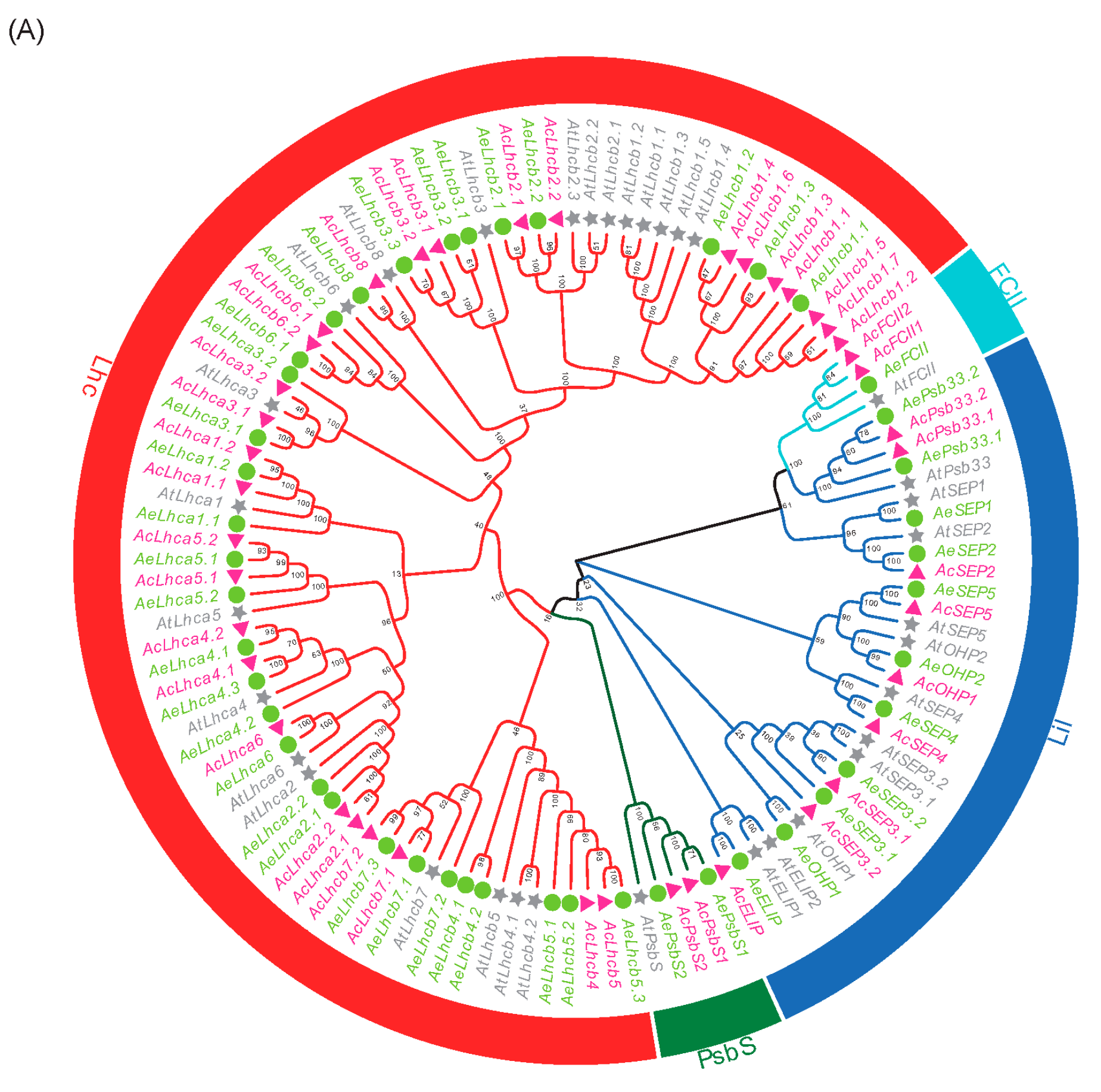
2.1. Genome-Wide Identification and Phylogenetic Analysis of Kiwifruit LHCs

We firstly retrieved sequences of AtLHC proteins, and those sequences were further used as a query to search Ac and Ae proteins using the Blastp tool [26]. Proteins identified in Ac and Ae were confirmed by the conserved domain analysis, and we identified 42 and 45 putative LHCs from Ac and Ae genomes, respectively (Table S2). The kiwifruit LHC proteins were named after AtLHC proteins.
Phylogenetic analyses were performed for 42 AcLHCs, 45 AeLHCs, and 34 AtLHCs proteins to explore the phylogenetic relationship and evolutionary pattern of kiwifruit LHC genes. Consistent with the classification of AtLHC proteins, kiwifruit LHC proteins were grouped into four distinct subfamilies, namely Lhc, Lil, PsbS, and FCII (Figure 1A). Our results show that the Lhc subfamily had the most members while FCII had the least (Figure S1). In the kiwifruit genome, the AcLHC genes were randomly distributed on 21 chromosomes (Figure 1B), of which chromosome 6 included the maximum gene number (five AcLHC genes), while gene numbers varied between 1 and 3 for the rest of the chromosomes (Figure S2A). Similarly, the AeLHC genes were unevenly distributed on 23 chromosomes (Figure 1C), and gene numbers varied between 1 and 4 for all chromosomes (Figure S2B).

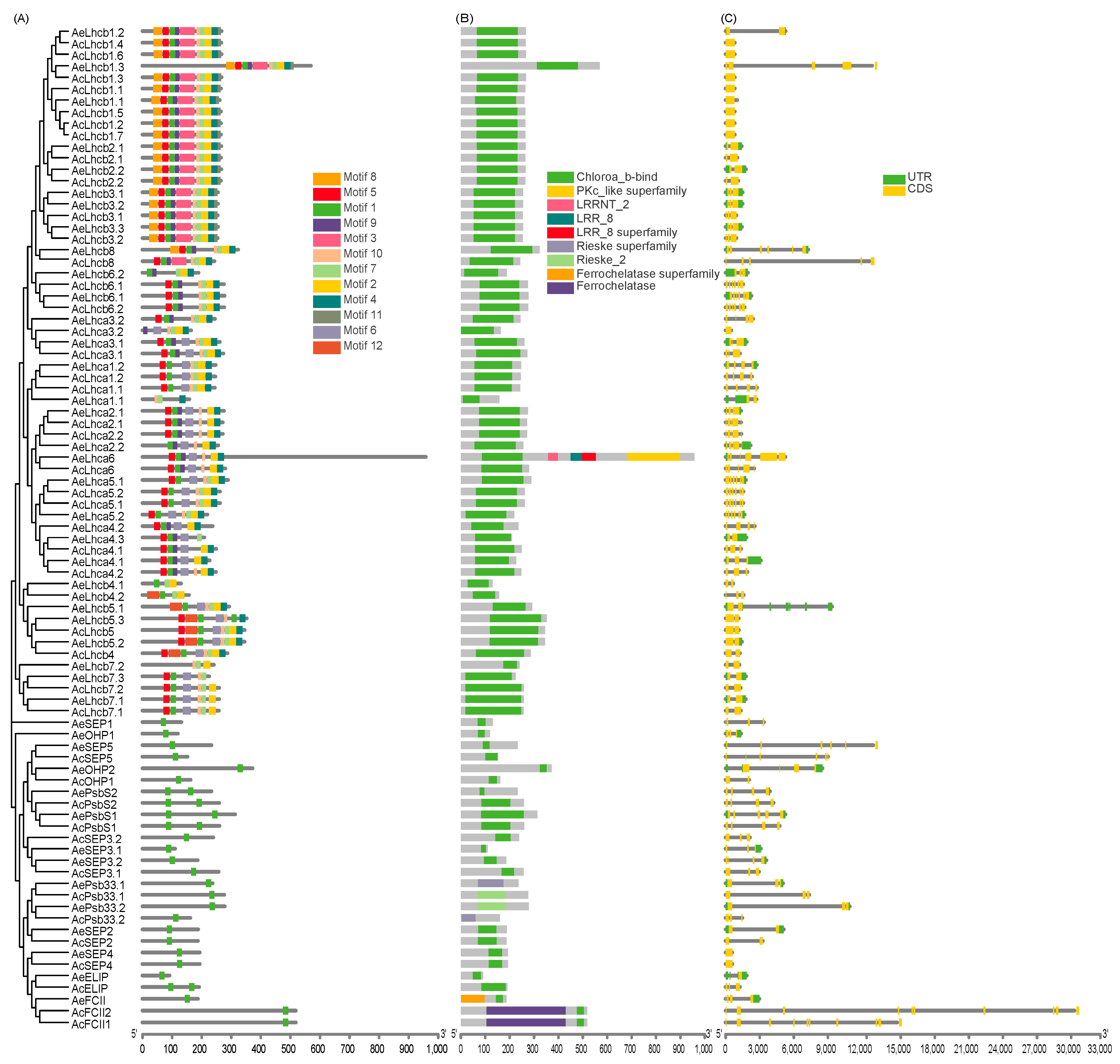
2.2. Multiple Sequence Alignment and Analysis of Kiwifruit LHCs Structure

The conserved motifs among kiwifruit LHCs were identified to infer structural variation and possible functional divergence. We identified 12 conserved motifs (designated as motifs 1–12) in kiwifruit LHCs (Figure 2A and Figure S3). The results show that kiwifruit LHC proteins with closer phylogenetic relationships had more similar motif architectures (Figure 2A). The Lhc subfamily contained the highest motif number compared to other subfamilies (Figure S4). Motif 1 was presented in all kiwifruit LHC proteins (Figure 2A) and located in the conserved Chloroa_b-bind domain (Figure 2B).
Intron numbers of kiwifruit LHCs varied from zero to nine, while corresponding exon numbers ranged from one to ten (Figure 2C). In addition, exon-intron structures and gene length of kiwifruit LHCs varied, especially for genes belonging to Lil, PsbS, and FCII subfamilies, indicating that gene structures might drive gene function divergence of kiwifruit LHCs (Figure 2C). The comparison of exon-intron structure in genes could provide insights into evolutionary mechanisms underlying the formation of gene families in plants [30].

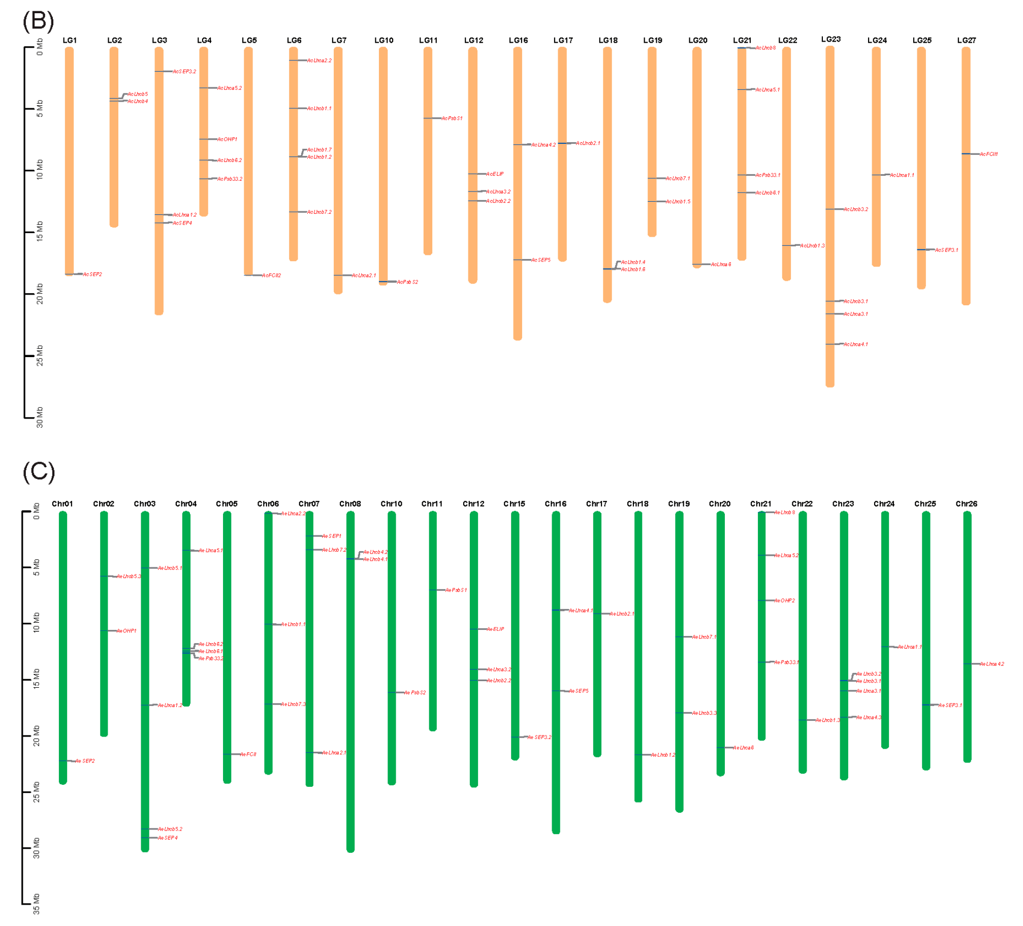
2.3. Synteny Analysis and cis-Element of Kiwifruit LHCs

The collinearity analysis was carried out to visualize the synteny relationships among homologous LHCs and infer gene duplication events. Eighteen duplicated gene pairs were identified in Ac and Ae (Figure S5). The Ka/Ks ratio for duplicated gene pairs varied from 0.02 to 0.47, with an average of 0.18 (Table S3), suggesting that all duplicated gene pairs underwent purifying selection. In addition, the Ks value of duplicated gene pairs varied from 0.10 to 1.37 (Table S3), suggesting that duplicated pairs have different evolution rates. The higher Ks value of dispersed duplication than whole-genome duplication (WGD) suggested that it may have happened earlier than the WGD event. Most duplicated gene pairs underwent WGD, indicating that WGD played a vital role in expanding LHCs in kiwifruit.
The potential cis-regulatory elements were predicted in the 2.0 kb upstream sequence of AcLHCs and AeLHCs. In total, 509 and 500 cis-elements were identified in promoter regions of AcLHCs and AeLHCs and further classified into nine types, including light-responsive element, auxin-responsive element, wound-responsive element, defense- and stress-responsive element, abscisic acid-responsive element, MeJA-responsive element, low-temperature-responsive element, gibberellin-responsive element, and salicylic acid-responsive element (Figure S6). Light-responsive cis-element and low temperature-responsive cis-element were the most abundant in both kiwifruit species, indicating that light and low temperature could significantly regulate LHCs expression patterns (Figure S6).

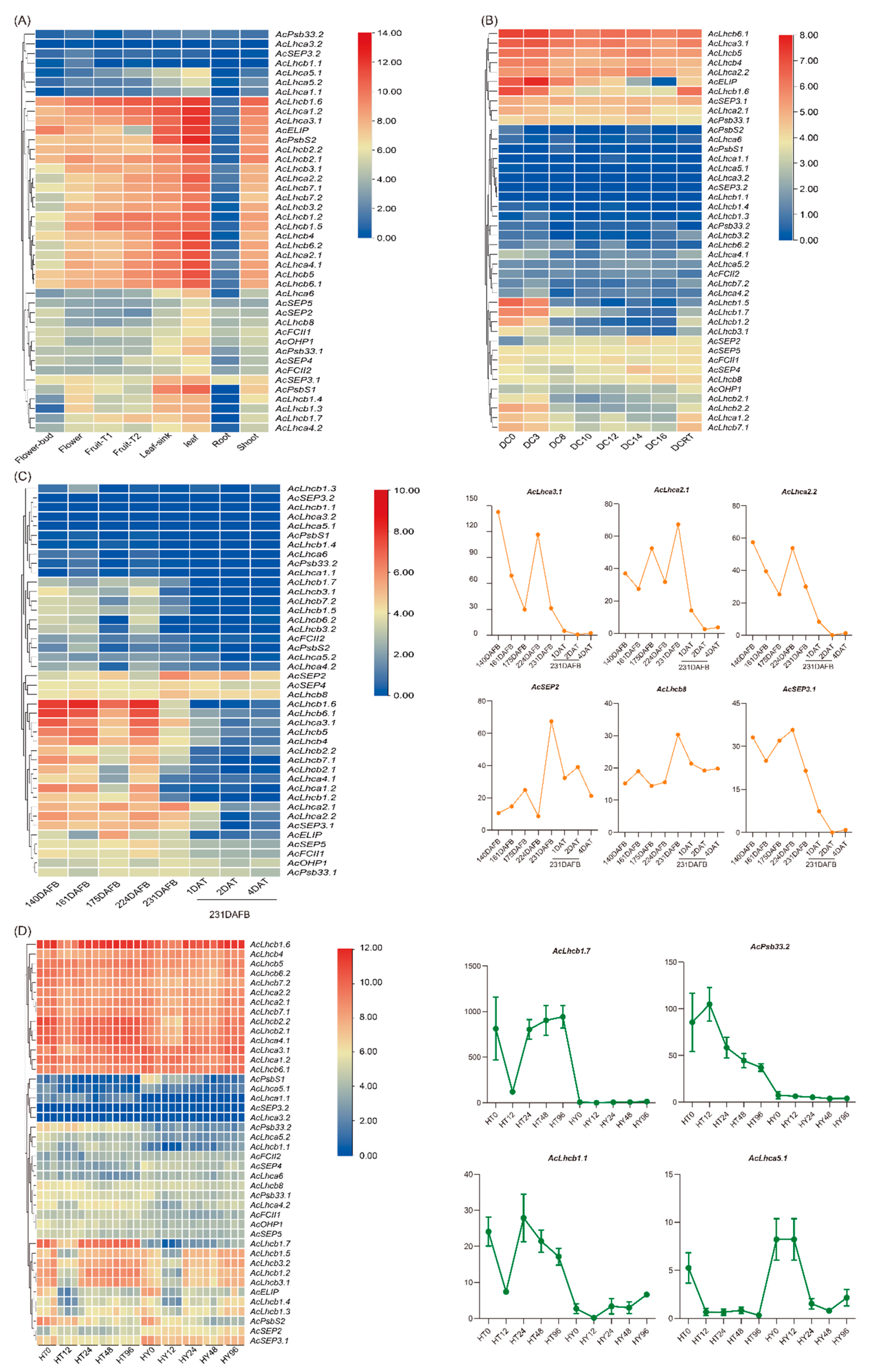
2.4. AcLHC Genes Regulated Kiwifruit Responses to Biotic and Abiotic Stresses

We used four different transcriptome datasets to assess the expression patterns of AcLHCs. To estimate the expression bias of AcLHCs in different kiwifruit tissues, we analyzed 42 genes in eight different tissues (flower bud, flower, fruit T1: no ethylene production, fruit T2: autocatalytic ethylene production, leaf sink, leaf, root, and shoot) (Figure 3A). Our results suggest that AcLHCs exhibited tissue-specific expression patterns (Figure 3A). Interestingly, AcLHCs were abundantly expressed in green tissues (such as leaves) compared to non-green tissues (such as roots) (Figure 3A). Low temperature affects the plant chlorophyll content [28], and we explored the effect of low temperature on AcLHCs expression levels (Figure 3B). The results reveal that AcLHC members respond to low-temperature regimes differently (Figure 3B). We identified three candidate genes (AcLhcb1.5, AcLhcb1.7, and AcLhcb1.2) as significantly responsive to low temperatures, and low-temperature responsive elements were identified in promoter regions of those three genes (Figure 3B and Figure S6).
To infer the potential functions of AcLHCs in regulating kiwifruit development and response to ethylene treatment, we re-analyzed the expression profiles of AcLHCs in different fruit developmental stages treated with or without ethylene (Figure 3C). The results reveal that the expression levels of five AcLHCs (AcLhca3.1/2.1/2.2, AcSEP2, and AcSEP3.1) were significantly down-regulated and one AcLHC (AcLhcb8) was down-regulated with the ethylene treatment (Table S4), verifying that those six AcLHCs could respond for kiwifruit degreening caused by the ethylene treatment [31].
A previous study suggested that chlorophyll content significantly affected plant resistance to bacterial pathogens [32]. We assessed expression profiles for AcLHCs after Pseudomonas syringae pv. actinidiae (Psa) inoculation in a resistant cultivar (Huate, HT) and a susceptible cultivar (Hongyang, HY), revealing expression levels of three genes (AcLhcb1.7, AcPsb33.2, and AcLhcb1.1) significantly up-regulated, but one gene (AcLhca5.1) significantly down-regulated in HT compared to HY. Our results suggest that these four genes played an essential role in the resistance of kiwifruit to Psa (Figure 3D). It will be interesting to investigate genome environment associations (GEAs) to identify adaptive variations between green and non-green flesh kiwifruit [33,34,35].

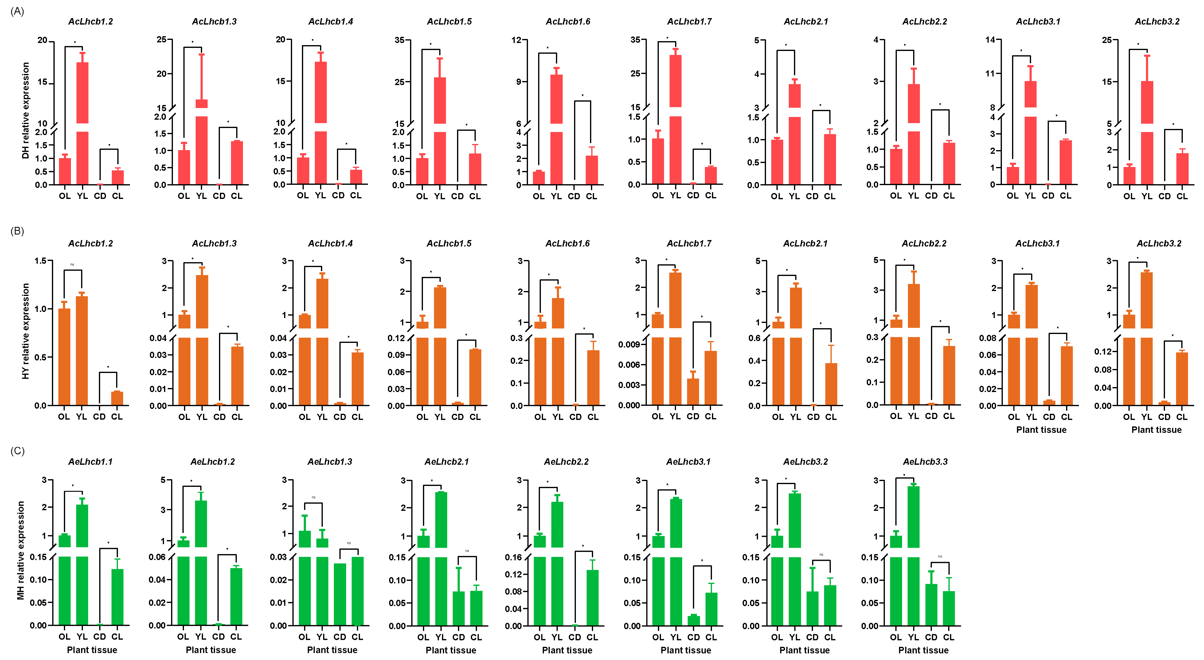
2.5. RT-qPCR Validation of Kiwifruit LHCs in Different Tissues

We selected ten AcLHCs and eight AeLHCs to perform RT-qPCR analysis in old and young leaves (OL, YL) and callus under light and dark conditions (CL, CD) (Figure S7). Plant samples used for this analysis were collected from Ac cultivars ‘Donghong’ (DH) and ‘Hongyang’ (HY) and Ae cultivar ‘Maohua no.1’ (MH) (Figure S7). Our results show that all the selected kiwifruit LHC genes were more expressed in leaves than calluses. The gene expressions were relatively higher in young leaves than in old leaves, except AeLhcb1.3 in MH, suggesting that those genes were primarily expressed in green tissues, consistent with the first transcriptome results (Figure 3A and Figure 4). Additionally, expression levels of all selected AcLHC genes in calluses under light conditions were higher than those in calluses under dark, suggesting that light induces higher expression of those AcLHC genes, which is confirmed by the identification of the light-responsive elements in promoter regions (Figure 4A,B and Figure S6). Four out of eight AeLHC genes were also induced by the light in calluses (Figure 4C). Interestingly, the relative expression levels of eight AcLHC genes (AcLhcb1.2/1.3/1.4/1.5/1.6/1.7/3.1/3.2) in DH were higher than those in HY, revealing significant differences among different cultivars (Figure 4A,B).

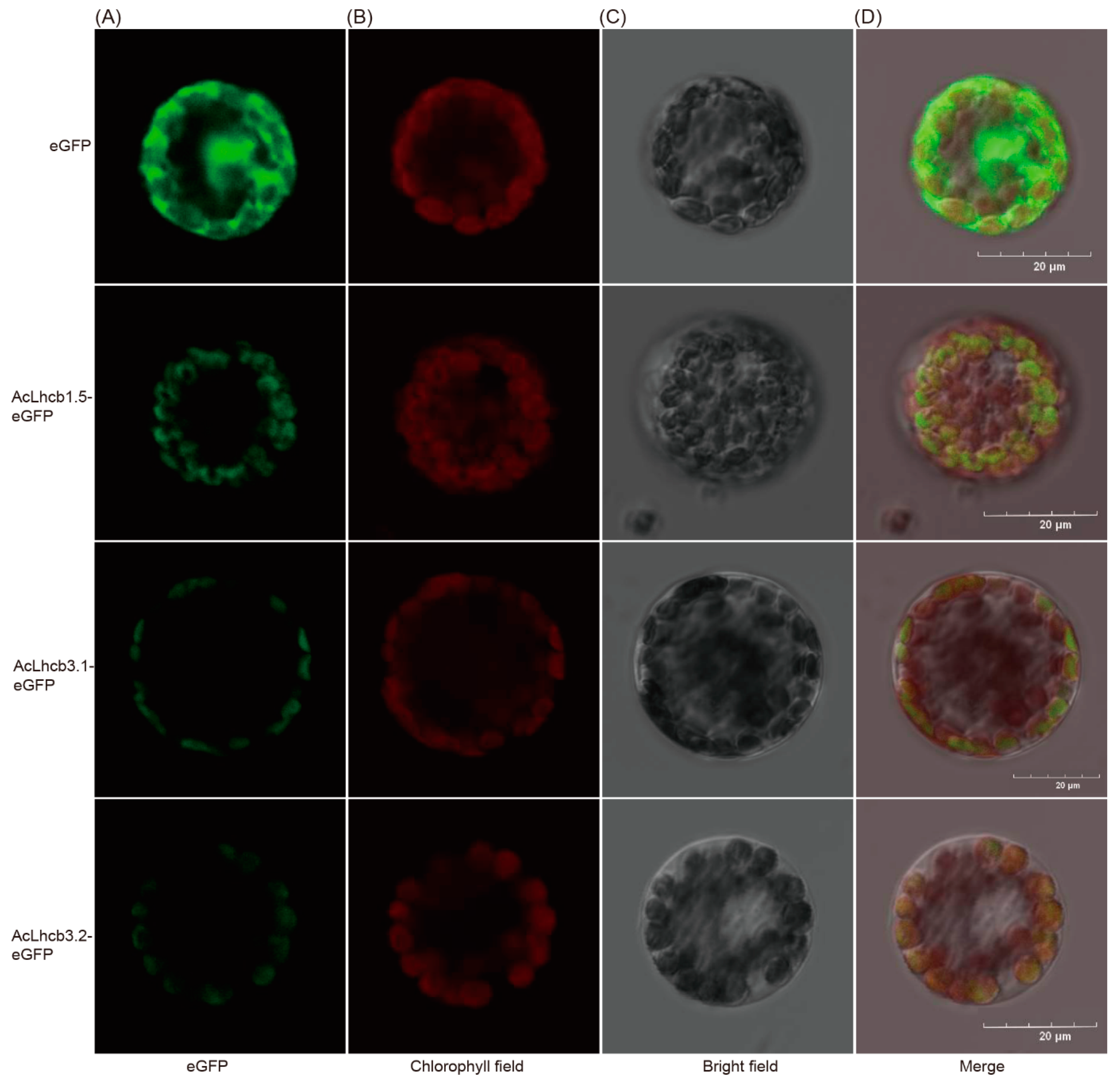
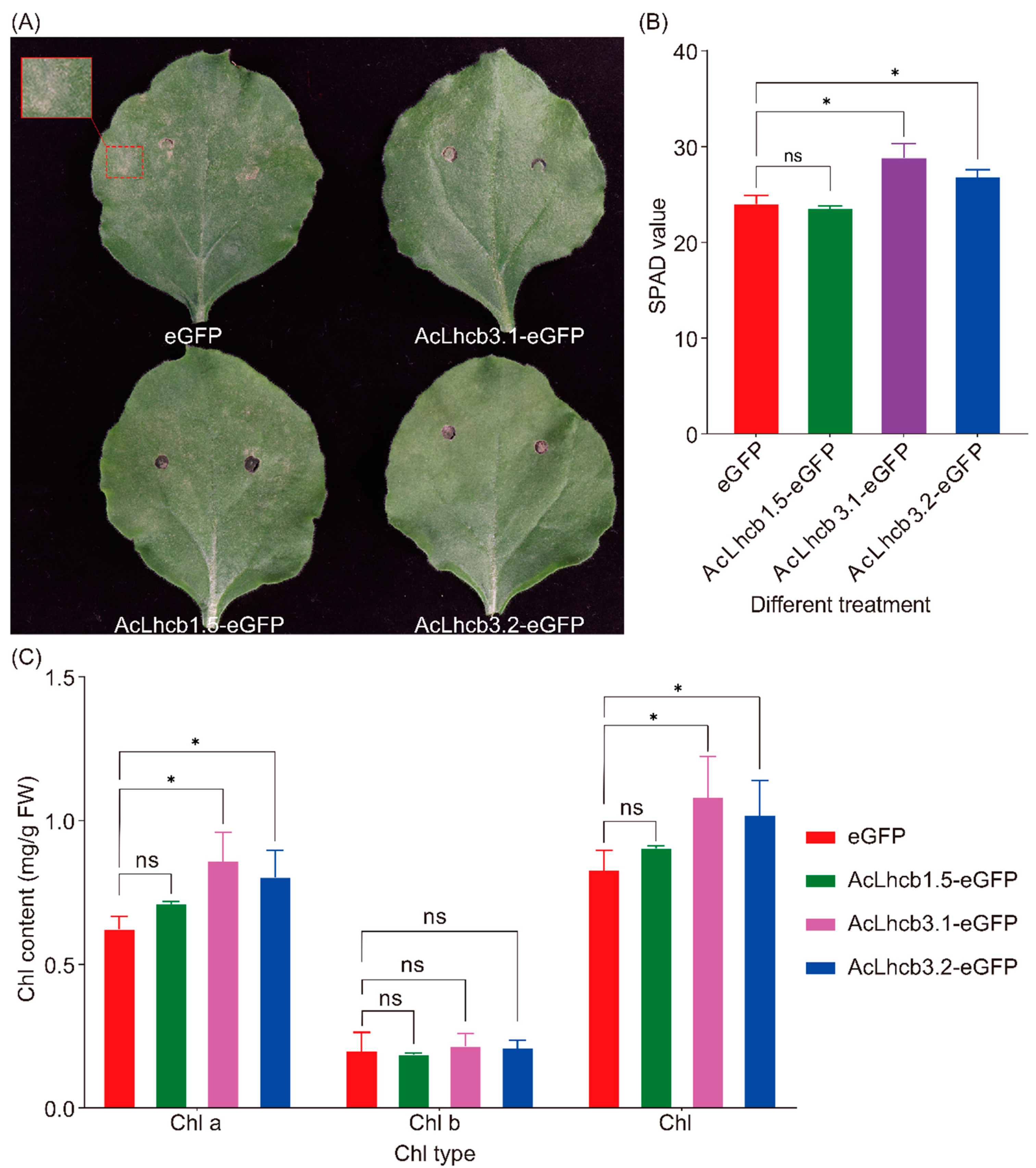
2.6. Subcellular Localization of Kiwifruit LHC and Transient Transformation of AcLhcb Genes in Tobacco Leaves

We selected three candidate genes (AcLhcb1.5/3.1/3.2) based on the difference in significance levels of mean differences for RT-qPCR data from leaves (OL and YL) and calluses (CL and CD). In-silico analysis for subcellular localization of AcLhcb1.5/3.1/3.2 predicted their presence in the chloroplast. Then, we further confirmed the localization of candidate gene products by performing transient transformation of Arabidopsis leaf protoplasts and tobacco leaves (Figure 5and Figure S8). We performed transient expression of AcLhcb1.5/3.1/3.2 in tobacco leaves to assess their potential role in chlorophyll content. Our results show that tobacco leaves which transiently expressed AcLhcb3.1/3.2 had significantly higher SPAD value (Soil and Plant Analyzer Development), chlorophyll a content, and total chlorophyll content. Still, no significant change was found in the content of chlorophyll b. This result is consistent with GhLhcb2.3 influence on the synthesis of chlorophyll a [9]. In contrast, AcLhcb1.5 did not affect chlorophyll content in tobacco leaves (Figure 6). The tobacco leaves transiently expressed AcLhcb1.5/3.1/3.2 had a higher value of Chl a/b than control (Figure S9).

3. Discussion

During photosynthesis, the chlorophyll-LHCs complexes located at PSI and PSII capture light to convert light energy into chemical energy required for various cellular processes in plants [36]. However, free chlorophylls and their metabolic intermediates can produce harmful reactive oxygen species even under normal conditions [2]. The dynamic balance of chlorophylls is closely related to the synthesis, degradation, maintenance of chlorophylls, and the dismantling and assembly of the PSI and PSII complexes [37]. A previous report suggested that the application of CO2 improved the chlorophyll content by upregulating chlorophyll-related genes, particularly LHCs, in cucumber plants [20]. The LHC family members have been identified in different plant species, i.e., 34 in Arabidopsis, 29 in Oryza sativa, 28 in Carica papaya, 55 in Gossypium hirsutum, 35 in Manihot esculenta, and 28 in Ricinus communis [5,7,8,9,10]. In the present study, we performed the genome-wide analysis of LHC gene family members in two different diploid kiwifruit species (Ac and Ae) and identified 42 LHC genes in Ac and 45 LHC genes in Ae (Table S1). Thus, Actinidia species have more LHCs than most reported plants. Interestingly, none of the kiwifruit LHCs genes was clustered with AtSEP1 (Figure 1). We speculate that the kiwifruit genome underwent loss of the SEP1 gene during the kiwifruit LHC family evolution process.
The WGD events were responsible for the expansion of kiwifruit LHC family members except for AcLhcb1.1, which could experience dispersed duplication events (Figure S5 and Table S3). Our finding confirms that both Ac and Ae had experienced three ancient WGD events [38]. The difference in the numbers of LHCs between A. thaliana or other plants and kiwifruit is probably due to the number of WGD events (two in A. thaliana and three in kiwifruit). However, the difference in the number of LHC members in the superfamily and subfamilies of Ac and Ae suggests that they might have experienced different evolution patterns. In addition, the Ka/Ks ratios of the 18 duplicated pairs in both AeLHCs and AcLHCs revealed that LHC family members had experienced purifying selection during the evolution process of both species.
The chlorophyll a/b-binding domain is highly conserved in the LHC family in different plants [5]. In the present study, all kiwifruit LHC family members have chlorophyll a/b-binding domain except AcPsb33.1, AcPsb33.2, AePsb33.1, and AePsb33.2, which possessed the Rieske domain similarly to AtPsb33 (Figure 2). It would be interesting to know if LHCs without the chlorophyll a/b-binding domain play a role identical to LHCs containing this domain. Kiwifruit LHC genes in the Lhc subfamily contained similar motifs and intron-exon structures, indicating that this subfamily had a highly conserved structural evolution (Figure 2). However, this phenomenon was not found in the other three subfamilies of LHC.
Our analysis of the promoter region of kiwifruit LHC genes found that kiwifruit LHCs differ greatly in the composition of cis-acting elements even within the same subfamily (Figure S6). The cis-element arrangements of the orthologous LHC gene pairs for both species of kiwifruit displayed a marked difference, suggesting that the orthologous LHC gene pairs possibly had different response mechanisms for biotic and abiotic stress in kiwifruit plants. In addition, the type and number of cis-element significantly varied in AcLHCs and AeLHCs, indicating that LHCs could function independently or synergistically to promote the normal growth of plants under different environmental conditions. It is reasonably speculated that within the same subfamily of LHC genes, expression was regulated through different transcriptional expression patterns, and our transcriptome analysis results are consistent with this speculation (Figure 3). All kiwifruit LHC genes had multiple light-responsive elements, suggesting a similar photoprotection function of the HvLhcb1 protein [39]. Additionally, we found a variety of hormone and stress-related elements in the promoter region of kiwifruit LHC genes. Previously, it has been reported that the expression of LHC genes was related to the hormone and abiotic stress. For instance, high salinity and low temperature reduced the expression of most ZmLhca and ZmLhcb genes [40]. Low concentration of ABA promoted AtLhcb1~6 expression, but high concentration inhibited AtLhcb1~6 expression [41]. The AtLhcb1~6 mutant had a poorer tolerance to drought and showed lower reactive oxygen species (ROS) after ABA treatment than the wild type [14]. Thus, we believe that cis-acting elements in the promoter region could be responsible for regulating kiwifruit LHCs expression.
Characterization of gene expressions in different plant organs under different conditions can be valuable for identifying candidate genes for desired breeding traits. In the present study, the kiwifruit LHC genes exhibited low expression in CL and almost no expression in CD, similar to low expression of LHC genes in esl mutant rice compared to wildtype [23]. Moreover, significant differences in kiwifruit LHC gene expression profiles, such as AcLhcb1.5/1.7, for different cultivars suggested that gene expression was influenced by the different cultivars’ genetic backgrounds.
The subcellular localization analysis of AcLhcb1.5/3.1/3.2 proteins in tobacco and Arabidopsis confirmed their presence in chloroplasts (Figure 5 and Figure S8), supporting the argument that these three proteins might be involved in photosynthesis. As expected, the transformation of AcLhcb3.1/3.2 proteins in tobacco leaves enhanced the SPAD value and chlorophyll a content.
It is essential to know the molecular regulation mechanism of crops to cope with the adverse effects of climate change on food safety and agricultural products [38]. Breeders have long been interested in the metabolism of chlorophylls because of their crucial role in plant growth and development and human health [2,42]. Similar to previous findings [9,43], the AcLhcb3.1/3.2 genes regulated chlorophyll content under biotic and abiotic stress. In tomatoes, researchers have reported that all the suffulta mutants for chlorophyll were segregated in the recessive mendelian manner in a reciprocal backcross with wild type plants [44]. Previously, researchers have incorporated genome-wide association study (GWAS) for connecting the desired trait to its underlying genetics [33,45]. It will be worthwhile to conduct GWAS for chlorophyll traits to improve our understanding of chlorophyll metabolism in plants [46].

4. Materials and Methods and Analysis

4.1. Genome-Wide Identification of LHC Genes in Kiwifruit

We retrieved whole genome sequences, coding sequences, and protein sequences for both kiwifruit species (Ac and Ae) from Kiwifruit Genome Database (http://kiwifruitgenome.org/ (accessed on 1 August 2021)). The protein sequences for AtLHCs were collected from the TAIR database (https://www.arabidopsis.org/ (accessed on 1 August 2021)). The local BLAST tool was used to construct the protein database of both kiwifruit species. The protein sequences of Arabidopsis LHC protein were used to query the kiwifruit protein database by the BLASTp. The candidates AcLHC and AeLHC were identified using a cutoff score of ≥100 and an e-value of ≤1 × e−10 for BLASTp. The Conserved Domain for kiwifruit LHCs was determined by utilizing the Conserved Domain Database (CDD) (https://www.ncbi.nlm.nih.gov/Structure/cdd/cdd.shtml (accessed on 3 August 2021)) and the simple modular architecture research tool (SMART) (http://smart.embl.de/ (accessed on 3 August 2021)). The protein sequences containing the Chloroa_b-bind domain were used in subsequent analysis.

4.2. Analysis of Kiwifruit LHC Protein Structure

The length of the protein, theoretical isoelectric point (pI), grand average of hydropathicity (GRAVY), and molecular weight (MW) of the kiwifruit LHC proteins in the kiwifruit species were computed by the ExPASy server (http://web.expasy.org/protparam/ (accessed on 5 August 2021)).

4.3. Gene Structure, Motif Features, and cis-Elements Analysis

The genome sequences and coding sequences of the kiwifruit LHC genes were used to investigate gene structures using the Gene Structure Display Server (GSDS 2.0, http://gsds.cbi.pku.edu.cn/ (accessed on 8 August 2021)). A maximum of 12 conserved motifs for kiwifruit LHC proteins were identified using MEME (http://meme-suite.org/tools/meme (accessed on 8 August 2021)) [47]. The cis-regulatory in 1500 bp upstream sequence of kiwifruit LHC genes were predicted with the PlantCARE database [48].

4.4. Phylogenetic Analysis of LHCs

The multiple sequence alignments of AtLHC, AcLHC, and AeLHC were performed using ClustalX with default parameters [49]. The phylogenetic tree was constructed by MEGA software (v7.0.26, download from https://www.megasoftware.net/ (accessed on 9 August 2021)) using the neighbor-joining (NJ) method with 1000 bootstrap replicates.

4.5. Chromosomal Location, Gene Duplication, and Synteny Analysis

The location of kiwifruit LHC genes was extracted from the corresponding GFF file using an in-house Perl script, and the genes on chromosomes were visualized using MapGene2 Chrome (http://mg2c.iask.in/mg2c_v2.0/ (accessed on 9 August 2021)). The MCScan software was employed to identify duplication patterns of kiwifruit LHCs using default parameters [50]. The synonymous (Ks) and nonsynonymous (Ka) mutation rates of the duplicated LHC gene pairs were calculated using TBtools software [51]. The syntenic blocks for kiwifruit LHCs were produced using the MCScanX software with default parameters [50], and gene pairs of kiwifruit LHCs were visualized by TBtools [51].

4.6. Expression Analysis of Kiwifruit LHCs

To investigate the expression profiles of kiwifruit LHCs, we collected four published RNA-seq data from NCBI (https://www.ncbi.nlm.nih.gov/ (accessed on 10 August 2021)) including samples from leaves, roots, different developmental stages of fruits, fruits treated with or without low temperature, leaves infected with pathogens (PRJNA691387, PRJNA277383, PRJNA514344, and PRJNA514180). All transcriptome datasets were re-analyzed using Actinidia chinensis, ‘Red5’ and A. eriantha ‘white’ cultivars as reference genomes [38,52]. The reads were aligned using the HISAT2 v2.0.1 [38,52]. The reads alignment was performed using the HISAT2 v2.0.1 [53], and the transcripts were assembled and quantified using the STRINGTIE v2.1.5 [54].

4.7. Plant Materials and Treatments

The fresh leaf samples for A. eriantha ‘Maohua no.1’ and A. chinensis ‘Hongyang’ ‘Donghong’ were collected from plants at the Germplasm Resources Nursery of Lushan Botanical Garden, Nanchang County, Jiangxi Province, China. A part of the samples was used for gene cloning, and the rest of the samples were used for callus induction [55]. The calluses were placed under light and dark conditions for subsequent study. The tobacco plants were grown under a 16/8-h-light/dark photoperiod, 250 μmmol photons m−2 s−1, 26 °C, and 55% humidity.

4.8. RNA Extraction and cDNA Synthesis

Total RNA was extracted with a Hipure Plant RNA Mini Kit (Magen, Shanghai, China), and cDNA was synthesized with a Trans Script One-Step gDNA Removal and cDNA Synthesis Super Mit (Transgen, Beijing, China) by following the manufacturer’s instructions.

4.9. Quantitative Real-Time PCR (qRT-PCR) Analysis

For RT-qPCR analysis, specific primers (Table S2) were designed based on the predicted sequences of the genes. The cDNA from kiwifruit leaves, callus, and fruit was used as a template to perform qRT-PCR. According to the manufacturer’s instructions, the reaction mixture was prepared using Perfect Start Green qPCR SuperMix (Transgen, Beijing, China). Each sample was replicated thrice to minimize inherent errors. Additionally, each biological replicate contained three technical replicates. The actin gene was used as an internal control to calculate the ΔCt values of target genes. The relative expression was calculated by the 2ΔΔCt method and the specificity of the amplification was determined from the melting curve [56].

4.10. Subcellular Localization of AcLHCs and Transformation

The coding sequences (CDS) of the AcLHCs without stop codon were ligated into the pGreen vector to construct an AcLHC-eGFP fusion protein expression vector. In contrast, the empty pGreen vector was used as a negative control. For subcellular localization, constructed vector was introduced into A. thaliana plant by Agrobacterium tumefaciens (strain EHA105) [12]. Transformation assays in cell suspension culture of Arabidopsis thaliana leaves were performed under a laser scanning confocal microscope (Olympus IX83, Olympus company, Beijing, China) as described previously [57,58].

4.11. Transient Over-Expression Analysis in Tobacco Leaf

For transient overexpression, we followed the same experimental procedure used for subcellular localization to construct the vectors. The experiment was performed in 6-weeks-old plants by selecting leaves at the fourth internode (counting from the shoot tip) for bacterial culture introduction. Chlorophyll contents and SPAD value were determined in transformed leaves according to previously described methods [59].

4.12. Statistical Analysis

All data were analyzed with GraphPad Prism 9 software (v9.0.0.121, purchased from https://www.graphpad.com/scientific-software/prism/ (accessed on 9 August 2021)). One-way ANOVA and two-way ANOVA were performed to check the significance level of the data. Tukey’s tests were used to compare the mean differences [60]. The mean differences were considered statistically significant at p < 0.05.

5. Conclusions

The LHC superfamily members are reported to participate in the plant photosynthesis process. The present study carried out the evolutionary gene relationships, protein motifs, structure, and expression profiles to characterize LHC superfamily members in Ac and Ae genomes. The subcellular localization analysis revealed that LHC genes were located in the chloroplast. Quantitative RT–qPCR analysis showed that LHC genes were preferentially expressed in the leaf. Additionally, the functional validation results showed that AcLhcb3.1/3.2 improved chlorophyll a content in tobacco leaves. Overall, our findings provided valuable insights into evolutionary patterns and functional diversity of LHC superfamily members and provided candidate genes for future breeding programs of kiwifruit. Predicting highly correlated loci for chlorophyll content in plant genomes through artificial intelligence techniques coupled with multi-omics studies can open new avenues for researchers to improve the understanding of chlorophyll metabolism in plants [61,62].

Supplementary Materials

The following supporting information can be downloaded at: https://www.mdpi.com/article/10.3390/ijms23126528/s1.

Author Contributions

Z.W. and P.G. conceived the research. J.L., M.A. and J.T. performed the experiments, analyzed the data, and wrote the manuscript. Z.W. and H.H. initiated the study ideas and revised the manuscript. All authors have read and agreed to the published version of the manuscript.

Funding

This work was supported by the National Key R&D Program of China (2019YFD100080) and the plant germplasm innovation program and biological resources programmer of the Chinese academy of sciences (KFJ-BRP-007-001).

Conflicts of Interest

The authors declare no conflict of interest.

References

  1. Kirilovsky, D.; Büchel, C. Chapter Nine—Evolution and function of light-harvesting antenna in oxygenic photosynthesis. Adv. Bot. Res. 2019, 91, 247–293. [Google Scholar] [CrossRef]
  2. Wang, P.; Grimm, B. Connecting Chlorophyll Metabolism with Accumulation of the Photosynthetic Apparatus. Trends Plant Sci. 2021, 26, 484–495. [Google Scholar] [CrossRef]
  3. De Bianchi, S.; Dall’Osto, L.; Tognon, G.; Morosinotto, T.; Bassi, R. Minor antenna proteins CP24 and CP26 affect the interactions between photosystem II subunits and the electron transport rate in grana membranes of Arabidopsis. Plant Cell 2008, 20, 1012–1028. [Google Scholar] [CrossRef]
  4. Chen, Y.E.; Ma, J.; Wu, N.; Su, Y.Q.; Zhang, Z.W.; Yuan, M.; Zhang, H.Y.; Zeng, X.Y.; Yuan, S. The roles of Arabidopsis proteins of Lhcb4, Lhcb5 and Lhcb6 in oxidative stress under natural light conditions. Plant Physiol. Biochem. 2018, 130, 267–276. [Google Scholar] [CrossRef]
  5. Zou, Z.; Li, M.; Jia, R.; Zhao, H.; He, P.; Zhang, Y.; Guo, A. Genes encoding light-harvesting chlorophyll a/b-binding proteins in papaya (Carica papaya L.) and insight into lineage-specific evolution in Brassicaceae. Gene 2020, 748, 144685. [Google Scholar] [CrossRef]
  6. Zhao, Y.; Kong, H.; Guo, Y.; Zou, Z. Light-harvesting chlorophyll a/b-binding protein-coding genes in jatropha and the comparison with castor, cassava and arabidopsis. Peer J. 2020, 8, e8465. [Google Scholar] [CrossRef]
  7. Zou, Z.; Yang, J. Genomics analysis of the light-harvesting chlorophyll a/b-binding (Lhc) superfamily in cassava (Manihot esculenta Crantz). Gene 2019, 702, 171–181. [Google Scholar] [CrossRef]
  8. Umate, P. Genome-wide analysis of the family of light-harvesting chlorophyll a/b-binding proteins in Arabidopsis and rice. Plant Signal. Behav. 2010, 5, 1537–1542. [Google Scholar] [CrossRef]
  9. Zhang, Q.; Ma, C.; Wang, X.; Ma, Q.; Fan, S.; Zhang, C. Genome-wide identification of the light-harvesting chlorophyll a/b binding (Lhc) family in Gossypium hirsutum reveals the influence of GhLhcb2.3 on chlorophyll a synthesis. Plant Biol. (Stuttg) 2021, 23, 831–842. [Google Scholar] [CrossRef]
  10. Zou, Z.; Huang, Q.; An, F.J.A.B. Genome-wide Identification, Classification and Expression Analysis of Lhc Supergene Family in Castor Bean (Ricinus communis L.). Agric. Biotechnol. 2013, 2, 44–49. [Google Scholar]
  11. Deng, Y.S.; Kong, F.Y.; Zhou, B.; Zhang, S.; Yue, M.M.; Meng, Q.W. Heterology expression of the tomato LeLhcb2 gene confers elevated tolerance to chilling stress in transgenic tobacco. Plant Physiol. Biochem. 2014, 80, 318–327. [Google Scholar] [CrossRef]
  12. Meng, L.; Fan, Z.; Zhang, Q.; Wang, C.; Gao, Y.; Deng, Y.; Zhu, B.; Zhu, H.; Chen, J.; Shan, W.; et al. BEL1-LIKE HOMEODOMAIN 11 regulates chloroplast development and chlorophyll synthesis in tomato fruit. Plant J. 2018, 94, 1126–1140. [Google Scholar] [CrossRef]
  13. Zhang, M.; Senoura, T.; Yang, X.; Chao, Y.; Nishizawa, N.K. Lhcb2 gene expression analysis in two ecotypes of Sedumalfredii subjected to Zn/Cd treatments with functional analysis of SaLhcb2 isolated from a Zn/Cd hyperaccumulator. Biotechnol. Lett. 2011, 33, 1865–1871. [Google Scholar] [CrossRef]
  14. Xu, Y.H.; Liu, R.; Yan, L.; Liu, Z.Q.; Jiang, S.C.; Shen, Y.Y.; Wang, X.F.; Zhang, D.P. Light-harvesting chlorophyll a/b-binding proteins are required for stomatal response to abscisic acid in Arabidopsis. J. Exp. Bot. 2012, 63, 1095–1106. [Google Scholar] [CrossRef]
  15. Wang, S.-S.; Song, Z.-B.; Sun, Z.; Zhang, J.; Mei, Y.; Nian, H.-J.; Li, K.-Z.; Chen, L.-M. Effects of Formaldehyde Stress on Physiological Characteristics and Gene Expression Associated with Photosynthesis in Arabidopsis thaliana. Plant Mol. Biol. Rep. 2012, 30, 1291–1302. [Google Scholar] [CrossRef]
  16. Peterson, R.B.; Schultes, N.P. Light-harvesting complex B7 shifts the irradiance response of photosynthetic light-harvesting regulation in leaves of Arabidopsis thaliana. J. Plant Physiol. 2014, 171, 311–318. [Google Scholar] [CrossRef]
  17. Pietrzykowska, M.; Suorsa, M.; Semchonok, D.A.; Tikkanen, M.; Boekema, E.J.; Aro, E.M.; Jansson, S. The light-harvesting chlorophyll a/b binding proteins Lhcb1 and Lhcb2 play complementary roles during state transitions in Arabidopsis. Plant Cell 2014, 26, 3646–3660. [Google Scholar] [CrossRef]
  18. Jiang, Q.; Xu, Z.S.; Wang, F.; Li, M.Y.; Ma, J.; Xiong, A.S. Effects of abiotic stresses on the expression of Lhcb1 gene and photosynthesis of Oenanthe javanica and Apium graveolens. Biol. Plant 2014, 58, 256–264. [Google Scholar] [CrossRef]
  19. Yadavalli, V.; Neelam, S.; Rao, A.S.V.C.; Reddy, A.R.; Subramanyam, R. Differential degradation of photosystem I subunits under iron deficiency in rice. J. Plant Physiol. 2012, 169, 753–759. [Google Scholar] [CrossRef]
  20. Song, H.; Li, Y.; Xu, X.; Zhang, J.; Zheng, S.; Hou, L.; Xing, G.; Li, M. Analysis of genes related to chlorophyll metabolism under elevated CO2 in cucumber (Cucumis sativus L.). Sci. Hortic. 2020, 261, 108988. [Google Scholar] [CrossRef]
  21. Zhao, Y.; Qiang, C.; Wang, X.; Chen, Y.; Deng, J.; Jiang, C.; Sun, X.; Chen, H.; Li, J.; Piao, W.; et al. New alleles for chlorophyll content and stay-green traits revealed by a genome wide association study in rice (Oryza sativa). Sci. Rep. 2019, 9, 2541. [Google Scholar] [CrossRef]
  22. Koziol, A.G.; Borza, T.; Ishida, K.; Keeling, P.; Lee, R.W.; Durnford, D.G. Tracing the evolution of the light-harvesting antennae in chlorophyll a/b-containing organisms. Plant Physiol. 2007, 143, 1802–1816. [Google Scholar] [CrossRef]
  23. Lin, W.; Guo, X.; Pan, X.; Li, Z. Chlorophyll Composition, Chlorophyll Fluorescence, and Grain Yield Change in esl Mutant Rice. Int. J. Mol. Sci. 2018, 19, 2945. [Google Scholar] [CrossRef]
  24. Chen, X.; Li, J.; Yu, Y.; Kou, X.; Periakaruppan, R.; Chen, X.; Li, X. STAY-GREEN and light-harvesting complex II chlorophyll a/b binding protein are involved in albinism of a novel albino tea germplasm ‘Huabai 1’. Sci. Hortic. 2022, 293, 110653. [Google Scholar] [CrossRef]
  25. Zhao, S.; Gao, H.; Luo, J.; Wang, H.; Dong, Q.; Wang, Y.; Yang, K.; Mao, K.; Ma, F. Genome-wide analysis of the light-harvesting chlorophyll a/b-binding gene family in apple (Malus domestica) and functional characterization of MdLhcb4.3, which confers tolerance to drought and osmotic stress. Plant Physiol. Biochem. 2020, 154, 517–529. [Google Scholar] [CrossRef]
  26. de Bianchi, S.; Betterle, N.; Kouril, R.; Cazzaniga, S.; Boekema, E.; Bassi, R.; Dall’Osto, L. Arabidopsis mutants deleted in the light-harvesting protein Lhcb4 have a disrupted photosystem II macrostructure and are defective in photoprotection. Plant Cell 2011, 23, 2659–2679. [Google Scholar] [CrossRef]
  27. Tao, J.; Jia, H.; Wu, M.; Zhong, W.; Jia, D.; Wang, Z.; Huang, C. Genome-wide identification and characterization of the TIFY gene family in kiwifruit. BMC Genom. 2022, 23, 179. [Google Scholar] [CrossRef]
  28. Drummond, L. Chapter Three—The Composition and Nutritional Value of Kiwifruit. Adv. Food Nutr. Res. 2013, 68, 33–57. [Google Scholar] [CrossRef]
  29. Tu, M.; Wu, Y.; Li, J.; Chen, D.; Jiang, G.; Song, H.; Yin, X.; Liu, X.; Li, M.; Sun, S. Transcriptome analysis reveals the roles of chlorophyll a/b-binding proteins (CABs) and stay-green (SGR) in chlorophyll degradation during fruit development in kiwifruit. N. Z. J. Crop Hortic. Sci. 2020, 49, 106–126. [Google Scholar] [CrossRef]
  30. Majoros, W.H.; Holt, C.; Campbell, M.S.; Ware, D.; Yandell, M.; Reddy, T.E. Predicting gene structure changes resulting from genetic variants via exon definition features. Bioinformatics 2018, 34, 3616–3623. [Google Scholar] [CrossRef]
  31. Salazar, J.; Zapata, P.; Silva, C.; González, M.; Pacheco, I.; Bastías, M.; Meneses, C.; Jorquera, C.; Moreno, I.; Shinya, P.; et al. Transcriptome analysis and postharvest behavior of the kiwifruit ‘Actinidia deliciosa’ reveal the role of ethylene-related phytohormones during fruit ripening. Tree Genet. Genomes 2021, 17, 8. [Google Scholar] [CrossRef]
  32. Cao, W.; Zhang, H.; Zhou, Y.; Zhao, J.; Lu, S.; Wang, X.; Chen, X.; Yuan, L.; Guan, H.; Wang, G.; et al. Suppressing chlorophyll degradation by silencing OsNYC3 improves rice resistance to Rhizoctonia solani, the causal agent of sheath blight. Plant Biotechnol. J. 2022, 20, 335–349. [Google Scholar] [CrossRef]
  33. Cortes, A.J.; Blair, M.W. Genotyping by Sequencing and Genome-Environment Associations in Wild Common Bean Predict Widespread Divergent Adaptation to Drought. Front. Plant Sci. 2018, 9, 128. [Google Scholar] [CrossRef]
  34. Wu, X.; Islam, A.; Limpot, N.; Mackasmiel, L.; Mierzwa, J.; Cortes, A.J.; Blair, M.W. Genome-Wide SNP Identification and Association Mapping for Seed Mineral Concentration in Mung Bean (Vigna radiata L.). Front. Genet. 2020, 11, 656. [Google Scholar] [CrossRef] [PubMed]
  35. Fernandez-Paz, J.; Cortes, A.J.; Hernandez-Varela, C.A.; Mejia-de-Tafur, M.S.; Rodriguez-Medina, C.; Baligar, V.C. Rootstock-Mediated Genetic Variance in Cadmium Uptake by Juvenile Cacao (Theobroma cacao L.) Genotypes, and Its Effect on Growth and Physiology. Front. Plant Sci. 2021, 12, 777842. [Google Scholar] [CrossRef]
  36. Tang, Y.; Wen, X.; Lu, C. Differential changes in degradation of chlorophyll–protein complexes of photosystem I and photosystem II during flag leaf senescence of rice. Plant Physiol. Biochem. 2005, 43, 193–201. [Google Scholar] [CrossRef]
  37. Woo, H.R.; Kim, H.J.; Lim, P.O.; Nam, H.G. Leaf Senescence: Systems and Dynamics Aspects. Annu. Rev. Plant Biol. 2019, 70, 347–376. [Google Scholar] [CrossRef]
  38. Tang, W.; Sun, X.; Yue, J.; Tang, X.; Jiao, C.; Yang, Y.; Niu, X.; Miao, M.; Zhang, D.; Huang, S.; et al. Chromosome-scale genome assembly of kiwifruit Actinidia eriantha with single-molecule sequencing and chromatin interaction mapping. Gigascience 2019, 8, giz027. [Google Scholar] [CrossRef]
  39. Saito, A.; Iino, T.; Sonoike, K.; Miwa, E.; Higuchi, K. Remodeling of the major light-harvesting antenna protein of PSII protects the young leaves of barley (Hordeum vulgare L.) from photoinhibition under prolonged iron deficiency. Plant Cell Physiol. 2010, 51, 2013–2030. [Google Scholar] [CrossRef]
  40. Kong, F.; Zhou, Y.; Sun, P.; Cao, M.; Li, H.; Mao, Y. Identification of light-harvesting chlorophyll a/b-binding protein genes of Zostera marina L. and their expression under different environmental conditions. J. Ocean Univ. China 2016, 15, 152–162. [Google Scholar] [CrossRef]
  41. Liu, R.; Xu, Y.H.; Jiang, S.C.; Lu, K.; Lu, Y.F.; Feng, X.J.; Wu, Z.; Liang, S.; Yu, Y.T.; Wang, X.F.; et al. Light-harvesting chlorophyll a/b-binding proteins, positively involved in abscisic acid signalling, require a transcription repressor, WRKY40, to balance their function. J. Exp. Bot. 2013, 64, 5443–5456. [Google Scholar] [CrossRef]
  42. Tanaka, A.; Tanaka, R. Chlorophyll metabolism. Curr. Opin. Plant Biol. 2006, 9, 248–255. [Google Scholar] [CrossRef] [PubMed]
  43. Rochaix, J.-D.; Bassi, R. LHC-like proteins involved in stress responses and biogenesis/repair of the photosynthetic apparatus. Biochem. J. 2019, 476, 581–593. [Google Scholar] [CrossRef] [PubMed]
  44. Forth, D.; Pyke, K.A. The suffulta mutation in tomato reveals a novel method of plastid replication during fruit ripening. J. Exp. Bot. 2006, 57, 1971–1979. [Google Scholar] [CrossRef]
  45. Blair, M.W.; Cortes, A.J.; Farmer, A.D.; Huang, W.; Ambachew, D.; Penmetsa, R.V.; Carrasquilla-Garcia, N.; Assefa, T.; Cannon, S.B. Uneven recombination rate and linkage disequilibrium across a reference SNP map for common bean (Phaseolus vulgaris L.). PLoS ONE 2018, 13, e0189597. [Google Scholar] [CrossRef] [PubMed]
  46. Lopez-Hernandez, F.; Cortes, A.J. Last-Generation Genome-Environment Associations Reveal the Genetic Basis of Heat Tolerance in Common Bean (Phaseolus vulgaris L.). Front. Genet. 2019, 10, 954. [Google Scholar] [CrossRef] [PubMed]
  47. Bailey, T.L.; Boden, M.; Buske, F.A.; Frith, M.; Grant, C.E.; Clementi, L.; Ren, J.; Li, W.W.; Noble, W.S. MEME SUITE: Tools for motif discovery and searching. Nucleic Acids Res. 2009, 37, w202–w208. [Google Scholar] [CrossRef]
  48. Lescot, M.; Déhais, P.; Thijs, G.; Marchal, K.; Moreau, Y.; Van de Peer, Y.; Rouzé, P.; Rombauts, S. PlantCARE, a database of plant cis-acting regulatory elements and a portal to tools for in silico analysis of promoter sequences. Nucleic Acids Res. 2002, 30, 325–327. [Google Scholar] [CrossRef]
  49. Larkin, M.A.; Blackshields, G.; Brown, N.P.; Chenna, R.; McGettigan, P.A.; McWilliam, H.; Valentin, F.; Wallace, I.M.; Wilm, A.; Lopez, R.; et al. Clustal W and Clustal X version 2.0. Bioinformatics 2007, 23, 2947–2948. [Google Scholar] [CrossRef]
  50. Wang, Y.; Tang, H.; Debarry, J.D.; Tan, X.; Li, J.; Wang, X.; Lee, T.H.; Jin, H.; Marler, B.; Guo, H.; et al. MCScanX: A toolkit for detection and evolutionary analysis of gene synteny and collinearity. Nucleic Acids Res. 2012, 40, e49. [Google Scholar] [CrossRef]
  51. Chen, C.; Chen, H.; Zhang, Y.; Thomas, H.R.; Frank, M.H.; He, Y.; Xia, R. TBtools: An Integrative Toolkit Developed for Interactive Analyses of Big Biological Data. Mol. Plant 2020, 13, 1194–1202. [Google Scholar] [CrossRef] [PubMed]
  52. Pilkington, S.M.; Crowhurst, R.; Hilario, E.; Nardozza, S.; Fraser, L.; Peng, Y.; Gunaseelan, K.; Simpson, R.; Tahir, J.; Deroles, S.C.; et al. A manually annotated Actinidia chinensis var. chinensis (kiwifruit) genome highlights the challenges associated with draft genomes and gene prediction in plants. BMC Genom. 2018, 19, 257. [Google Scholar] [CrossRef] [PubMed]
  53. Kim, D.; Paggi, J.M.; Park, C.; Bennett, C.; Salzberg, S.L. Graph-based genome alignment and genotyping with HISAT2 and HISAT-genotype. Nat. Biotechnol. 2019, 37, 907–915. [Google Scholar] [CrossRef] [PubMed]
  54. Pertea, M.; Pertea, G.M.; Antonescu, C.M.; Chang, T.C.; Mendell, J.T.; Salzberg, S.L. StringTie enables improved reconstruction of a transcriptome from RNA-seq reads. Nat. Biotechnol. 2015, 33, 290–295. [Google Scholar] [CrossRef]
  55. Wang, Z.; Wang, S.; Li, D.; Zhang, Q.; Li, L.; Zhong, C.; Liu, Y.; Huang, H. Optimized paired-sgRNA/Cas9 cloning and expression cassette triggers high-efficiency multiplex genome editing in kiwifruit. Plant Biotechnol. J. 2018, 16, 1424–1433. [Google Scholar] [CrossRef]
  56. Liu, X.; Yu, F.; Yang, G.; Liu, X.; Peng, S. Identification of TIFY gene family in walnut and analysis of its expression under abiotic stresses. BMC Genom. 2022, 23, 190. [Google Scholar] [CrossRef]
  57. Yuan, Y.; Mei, L.; Wu, M.; Wei, W.; Shan, W.; Gong, Z.; Zhang, Q.; Yang, F.; Yan, F.; Zhang, Q.; et al. SlARF10, an auxin response factor, is involved in chlorophyll and sugar accumulation during tomato fruit development. J. Exp. Bot. 2018, 69, 5507–5518. [Google Scholar] [CrossRef]
  58. Yoo, S.D.; Cho, Y.H.; Sheen, J. Arabidopsis mesophyll protoplasts: A versatile cell system for transient gene expression analysis. Nat. Protoc. 2007, 2, 1565–1572. [Google Scholar] [CrossRef]
  59. Tan, X.L.; Fan, Z.Q.; Kuang, J.F.; Lu, W.J.; Reiter, R.J.; Lakshmanan, P.; Su, X.G.; Zhou, J.; Chen, J.Y.; Shan, W. Melatonin delays leaf senescence of Chinese flowering cabbage by suppressing ABFs-mediated abscisic acid biosynthesis and chlorophyll degradation. J. Pineal. Res. 2019, 67, e12570. [Google Scholar] [CrossRef]
  60. Berkman, S.J.; Roscoe, E.M.; Bourret, J.C. Comparing self-directed methods for training staff to create graphs using Graphpad Prism. J. Appl. Behav. Anal. 2019, 52, 188–204. [Google Scholar] [CrossRef]
  61. Cortes, A.J.; Restrepo-Montoya, M.; Bedoya-Canas, L.E. Modern Strategies to Assess and Breed Forest Tree Adaptation to Changing Climate. Front. Plant Sci. 2020, 11, 583323. [Google Scholar] [CrossRef] [PubMed]
  62. Tong, H.; Nikoloski, Z. Machine learning approaches for crop improvement: Leveraging phenotypic and genotypic big data. J. Plant Physiol. 2021, 257, 153354. [Google Scholar] [CrossRef] [PubMed]
Figure 1. Phylogenetic tree of LHC proteins (A) and distribution of LHC genes in Ac (B) and Ae (C) chromosomes. The full-length LHC protein sequences from Arabidopsis (At, gray gene name and pentagram), A. chinensis (Ac, deep pink gene name and triangle), and A. eriantha (Ae, lawn green, and circle) were aligned using ClustalX 2.0 with default parameters. Then, the unrooted phylogenetic tree was constructed using MEGA X and the Neighbour-Joining method. The Lhc subfamily, PsbS subfamily, Lil subfamily, and FCII subfamily were highlighted using red, green, royal blue, and aqua sectors.
Ijms 23 06528 g001aIjms 23 06528 g001b
Figure 2. Gene structure and conserved motif architecture of LHC family in two kiwifruit species. (A) motifs contained in AcLHC and AeLHC proteins; (B) showed conserved domain distribution; (C) the exon-intron structure of kiwifruit LHC genes.
Ijms 23 06528 g002
Figure 3. Expression profiles of AcLHC genes in different tissues, fruit developmental stages, and under different treatments. The bar at the right of each heat map represents expression values. (A) Expression profiles of AcLHCs in eight different tissues (flower bud, flower, fruit T1: no ethylene production, fruit T2: autocatalytic ethylene production, leaf sink, leaf, root, and shoot). (B) Expression profiles of AcLHCs in mature fruit exposed to eight different temperatures in storage for two days. DC, degrees celsius; RT, room temperature. (C) Expression profiles of AcLHCs in the fruit developmental stages and samples treated with ethylene. DAFB, days after the full bloom of fruit; DAT, day after being treated with ethylene. (D) Expression profiles of AcLHCs in two kiwifruit cultivars infected with Psa. Huate (HT) and Hongyang (HY) represented resistant and susceptible cultivars, respectively. The number following the cultivar’s name showed hours post the Psa invasion.
Ijms 23 06528 g003
Figure 4. Expression analysis of AcLHC and AeLHC genes using RT-qPCR in different tissues. Actin was used as the internal standard for each gene. (A) for DH, (B) for HY, and (C) for MH. The results are shown as mean values and standard deviation of three biological replicates of different tissues and three technical replicates in each biological sample. The y-axis was the relative expression level. * indicated significant differences accorded to Tukey’s multiple range tests (*, p < 0.05). OL, old leaves; YL, young leaves; CD, callus tissues under dark condition; CL, callus tissues under light condition; DH, ‘Donghong’; HY, ‘Hongyang’; MH, ‘Maohua no.1’.
Ijms 23 06528 g004
Figure 5. Subcellular localization of the fusion protein 35S::AcLhcb1.5/3.1/3.2::eGFP in mesophyll protoplasts of Arabidopsis. Images were taken under (A) fluorescence, (B) chlorophyll field, and (C) bright field. (D) Merged images (AC) of representative cells expressing eGFP. The chlorophyll autofluorescence was used to localize chloroplasts. Bars = 20 μm.
Ijms 23 06528 g005
Figure 6. Transient expression of AcLhcb1.5/3.1/3.2 in tobacco leaves. (A) Growth phenotypes of 6-weeks-old soil-grown tobacco leaves of control (eGFP) and LHC overexpression (AcLhcb1.5-eGFP, AcLhcb3.1-eGFP, and AcLhcb3.2-eGFP) for 4 d. (B) Each treatment’s SPAD value and (C) chlorophyll content was measured on the fourth day after transformation. The results are shown as means and SDs from three independent experiments. For (B,C) * indicate significant differences between treatments, ns indicate no significance, according to two-way ANOVA and Tukey’s multiple range tests (*, p < 0.05).
Ijms 23 06528 g006
Publisher’s Note: MDPI stays neutral with regard to jurisdictional claims in published maps and institutional affiliations.

