Article

Improvement of Cognitive Function in Ovariectomized Rats by Human Neural Stem Cells Overexpressing Choline Acetyltransferase via Secretion of NGF and BDNF

1
Department of Biology Education, Korea National University of Education, Cheongju 28173, Korea
2
Department of Counseling, Health, and Kinesiology, College of Education and Human Development, Texas A&M University-San Antonio, One University Way, San Antonio, TX 78224, USA
*
Author to whom correspondence should be addressed.
Int. J. Mol. Sci. 2022, 23(10), 5560; https://doi.org/10.3390/ijms23105560
Submission received: 22 April 2022 / Revised: 15 May 2022 / Accepted: 15 May 2022 / Published: 16 May 2022

Abstract

:
Menopause is associated with memory deficits attributed to reduced serum estrogen levels. We evaluated whether an increase in brain-derived neurotrophic factor (BDNF) and nerve-growth factor (NGF) levels, through transplantation of choline acetyltransferase (ChAT)-overexpressing neural stem cells (F3.ChAT), improved learning and memory in ovariectomized rats. PD13 mouse neuronal primary culture cells were treated with estradiol or co-cultured with F3.ChAT cells; choline transporter1 (CHT1), ChAT, and vesicular acetylcholine transporter (VAChT) expression was evaluated using real-time PCR. The relationship between estrogen receptors (ERs) and neurotrophin family members was analyzed using immunohistochemistry. After the transplantation of F3.ChAT cells into OVx rats, we evaluated the memory, ACh level, and the expression of ER, neurotrophin family proteins, and cholinergic system. Estradiol upregulated CHT1, ChAT, and VAChT expression in ER; they were co-localized with BDNF, NGF, and TrkB. Co-culture with F3.ChAT upregulated CHT1, ChAT, and VAChT by activating the neurotrophin signalling pathway. Transplantation of F3.ChAT cells in OVX animals increased the ACh level in the CSF and improved memory deficit. In addition, it increased the expression of ERs, neurotrophin signaling, and the cholinergic system in the brains of OVX animals. Therefore, the estradiol deficiency induced memory loss by the down-regulation of the neurotrophin family and F3.ChAT could ameliorate the cognitive impairment owing to the loss or reduction of estradiol.

1. Introduction

Estrogen plays an important role in physiology and metabolism in women and estrogen deficiency induces various diseases and abnormalities, with cognitive dysfunction and memory disorders being the most common [1,2,3]. Estrogen influences neuronal connectivity through the formation of new synaptic connections in the brain and cognitive function by increasing the activity of the cholinergic system [4,5]. Estrogen deficiency induces memory impairment in ovariectomized animals [6]. In addition, estrogen upregulates the expression of neurotrophin family proteins, such as brain-derived neurotrophic factor (BDNF), in the hippocampus [7]. Estrogen influences memory-related processes and prevents a decline in neuronal function [8].
Neurotrophins are a family of proteins that regulate neural survival, development, function, and plasticity [9,10,11,12]. Nerve growth factor (NGF) is a member of the neurotrophin family that supports neural survival after brain damage [10]. BDNF supports neural survival, growth, and synaptic plasticity in the hippocampus and the cortex [9]. NGF and BDNF influence memory function and the increase in NGF and BDNF levels help recover learning and memory in Alzheimer’s animal models [11,12]. In addition, memory deficits can be restored by NGF and BDNF through the transplantation of stem cells [13,14,15].
Acetylcholine (ACh) is a key neurotransmitter in the memory process [16]. Both NGF and BDNF influence cholinergic neurons and increase ACh concentrations [17,18,19]. In addition, estrogen facilitates cholinergic neurotransmission by increasing the activity and mRNA levels of choline acetyltransferase (ChAT) [20]. Therefore, in menopausal women, estrogen replacement therapy increases cognitive function, and upregulation of NGF and BDNF improves cognitive function and menopausal symptoms [21,22].
In an earlier study, we developed neural stem cells overexpressing ChAT (F3.ChAT). F3.ChAT cells restore cognitive function via acetylcholine synthesis and amyloid beta elimination [23]. In addition, resistance exercises enhanced cognitive function in ovariectomized rats via the upregulation of BDNF and NGF mRNA expression [6,24,25,26]. Therefore, in this study, we aimed to demonstrate that an increase in the NGF and BDNF levels in the brain improves learning and memory function in ovariectomized rats using stem cells. In addition, we aimed to elucidate their signaling pathways by evaluating the relationship between estrogen receptors and the neurotrophin family.

2. Results

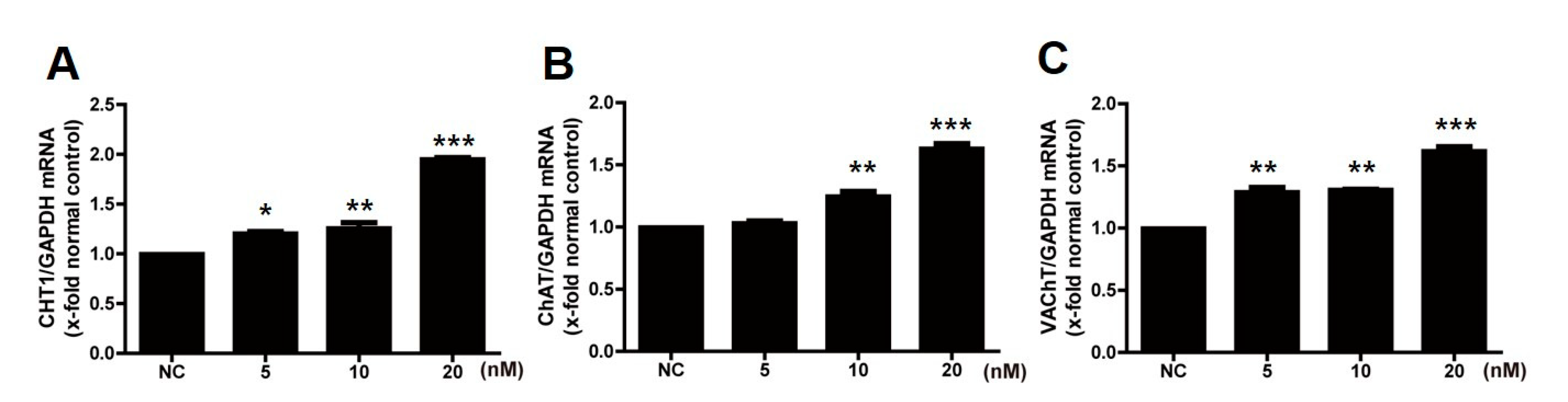
2.1. Treatment with Estradiol Upregulates the Cholinergic System

To investigate the effects of estradiol on the cholinergic system, mouse primary neuronal cells were treated with estradiol. Treatment with estradiol (0, 5, 10, and 20 nm) significantly increased the expression of cholinergic systems, such as choline transporter (CHT), ChAT, and vesicular acetylcholine transporter (VAChT) (Figure 1), in a dose-dependent manner.

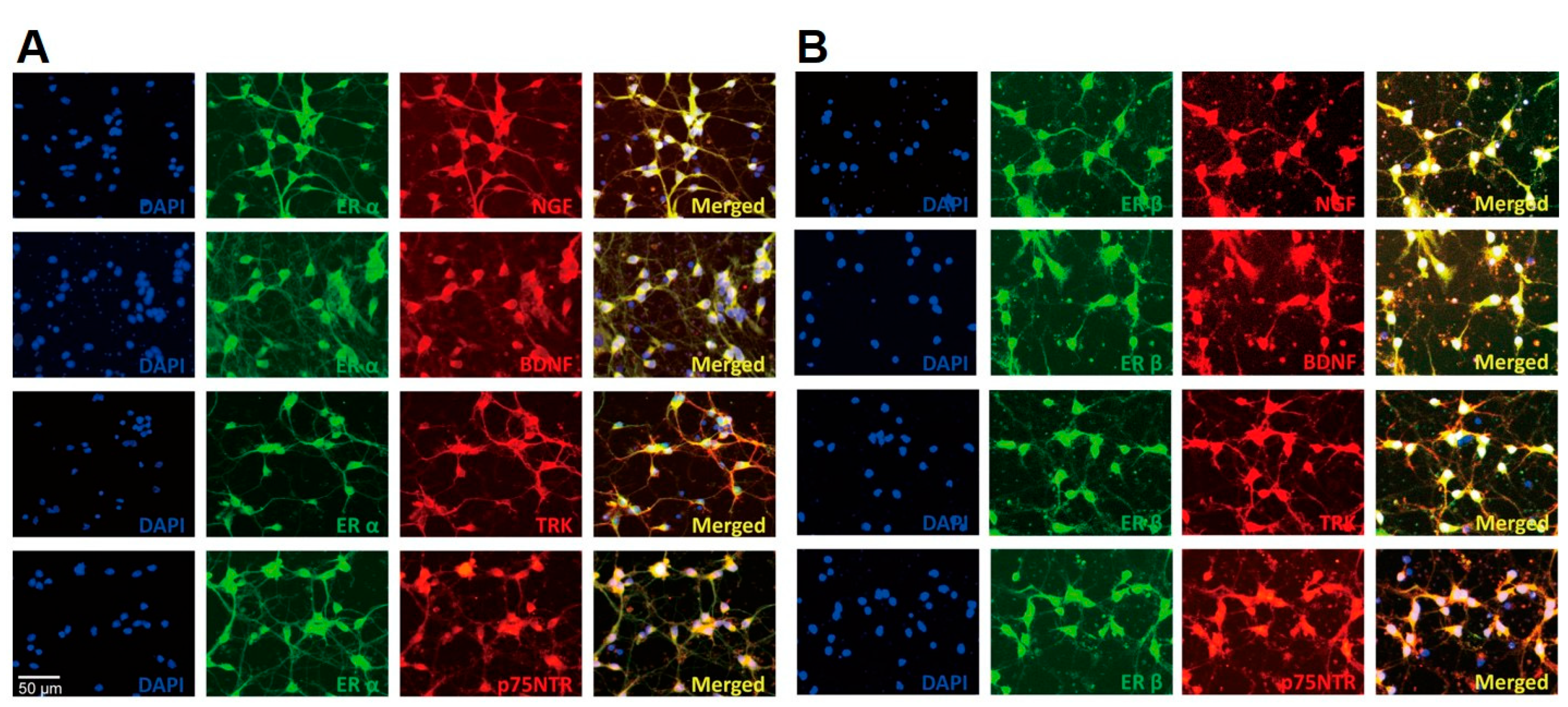
2.2. Estrogen Receptors Co-Localize with Neurotrophin Family

To check the localization of estrogen receptors and neurotrophin family proteins, a double immunocytochemistry analysis was performed with estrogen receptor alpha (ERα) and beta (ERβ) and the neurotrophin family members, NGF, BDNF, TRK, and p75NTR (Figure 2). Both ERα and ERβ were co-localized with neurotrophin family proteins.
The localization was analyzed using immunocytochemistry. ER α and ERβ were stained with Alexa Fluor 488 (green), and the neurotrophin family proteins were stained with Alexa Fluor 594 (red). The cells were counterstained with 4,6-diamino-2-phenylindole (DAPI) to confirm cellular nuclei. NGF, nerve growth factor; BDNF, brain-derived neurotrophic factor; TRK, tyrosine receptor kinase; p75NTR, p75 neurotrophin receptor. Scale bar = 50 μm, magnification ×200.

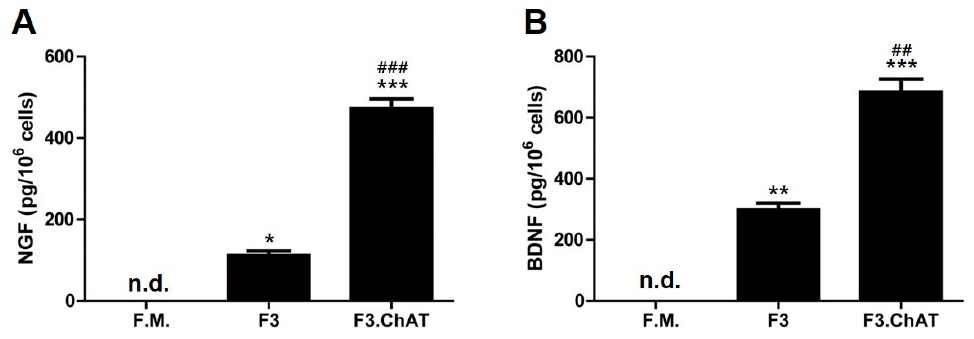
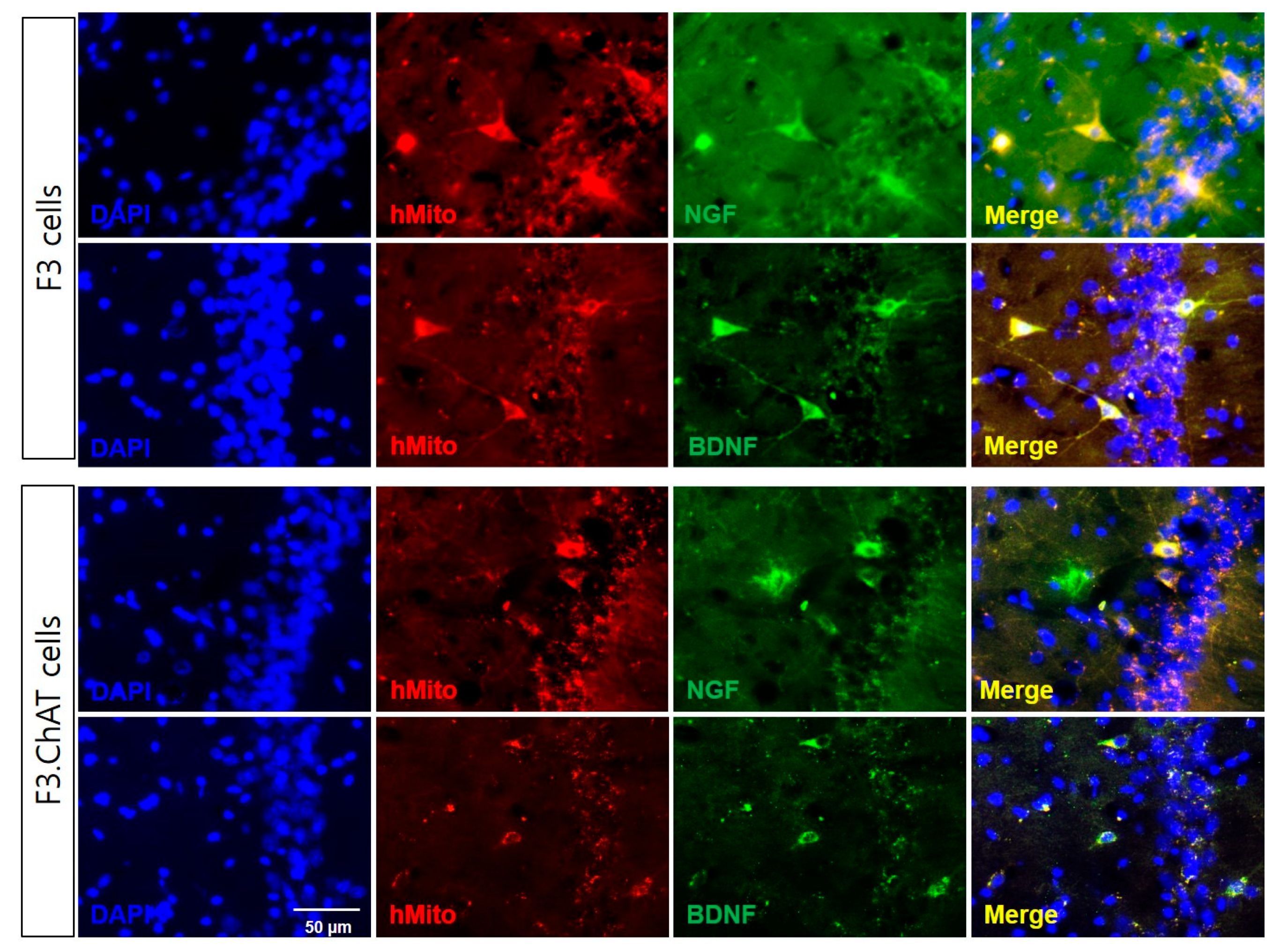
2.3. F3.ChAT Cells Release NGF and BDNF

To investigate whether NGF and BDNF increased the levels of cholinergic markers, F3 and F3.ChAT cells were used (Figure 3). To confirm the secretion of NGF and BDNF from F3 and F3.ChAT cells, their concentrations were measured in a conditioned medium. Both F3 and F3.ChAT release NGF and BDNF (Figure 3). The secretion of NGF and BDNF from F3.ChAT cells was higher than that from F3 cells.

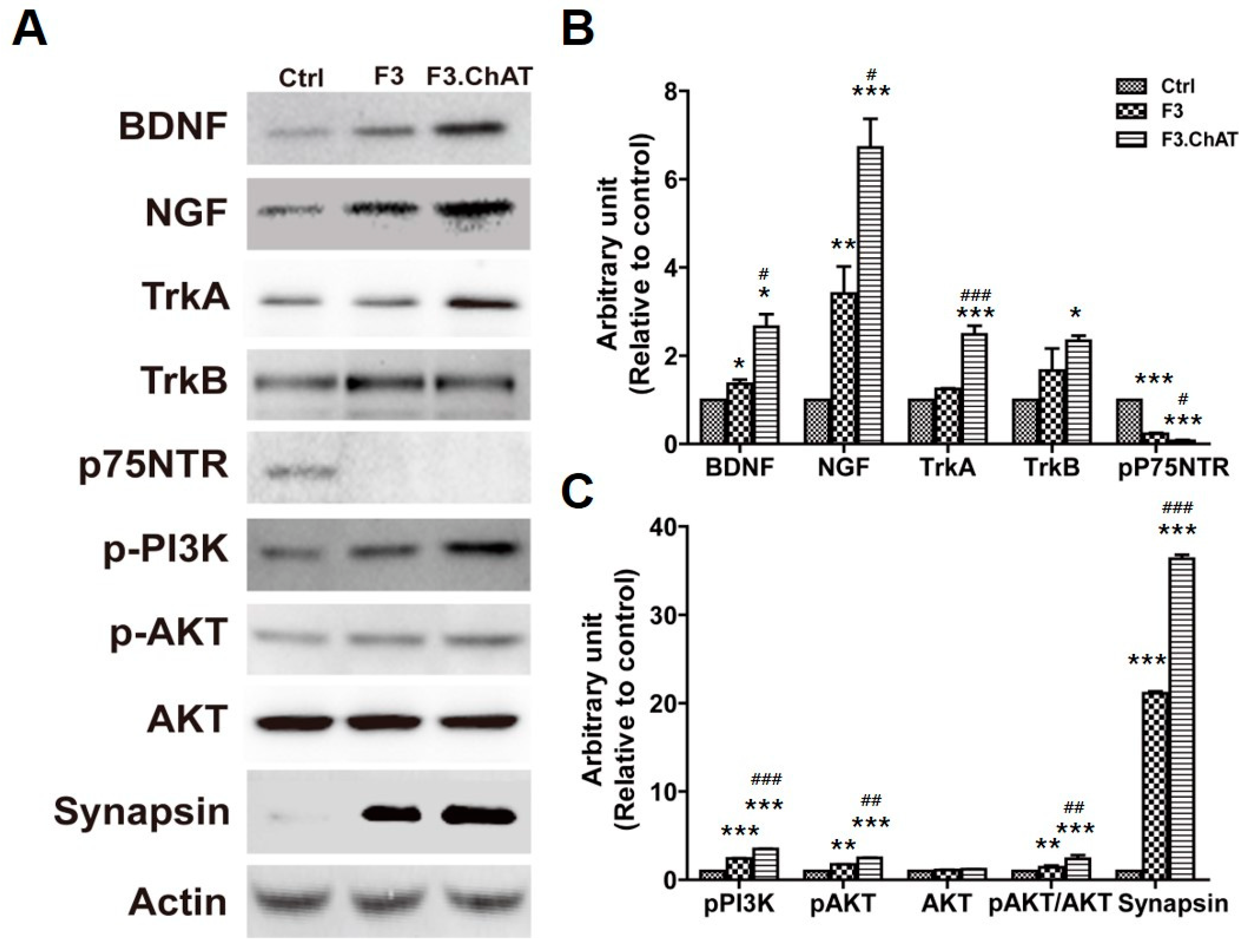
2.4. Co-Culture with F3.ChAT Cells Enhances Neurotrophin Signaling Pathway

F3 or F3.ChAT cells were co-cultured with primary cells in a transwell (pore 0.4 μm); the levels of the neurotrophin signaling pathway molecules were analyzed (Figure 4). The expression of NGF and BDNF in primary cells co-cultured with F3 or F3.ChAT cells was higher than that in the normal control. In addition, the receptors, Pan-Trk and TrkB, were upregulated; however, the expression of p75NTR was downregulated. This resulted in increased PI3k and AKT expression involved in the neurotrophin pathway, which induces cell proliferation [27,28]. Interestingly, synapsin, which influences synaptic plasticity, was upregulated. The expression of proteins in F3.ChAT-co-cultured cells was higher than that in the F3-co-cultured cells.

2.5. Co-Culture with F3.ChAT Cells Upregulates the Cholinergic System

In parallel to that of the neurotrophin signaling pathway, expression of CHT1, ChAT, and VAChT was significantly enhanced by co-culture with F3 and F3.ChAT cells, compared to that in the normal controls (Figure 5). Their expression in primary cells co-cultured with F3.ChAT cells were higher than that in the cells co-cultured with F3 cells.

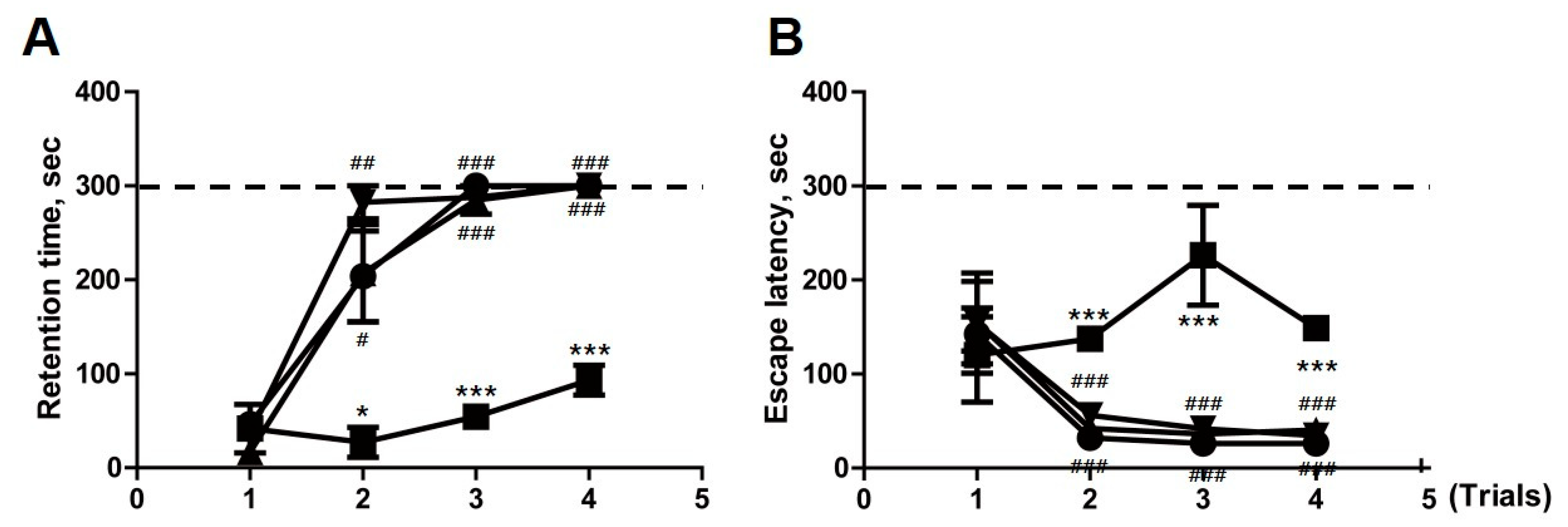
2.6. Transplantation of F3.ChAT Cells Improves Memory Deficit in Ovariectomized Rat

After confirming the in vitro effects of NGF and BDNF from the co-culture of F3 and F3.ChAT cells with primary cells, the in vivo cognitive improvement effect of F3 and F3.ChAT were investigated in an ovariectomized animal model. Three weeks after ovariectomy, animals were subjected to a passive avoidance test and water-maze tests to confirm memory deficits (data not shown). F3 and F3.ChAT cells were transplanted into the ovariectomized rats via the tail vein. Four weeks after the F3 and F3.ChAT transplantations, the rats in the OVx group (control) displayed profound impairments in memory acquisition and retention in both the passive avoidance test and water maze performance (Figure 6). However, the transplantation of F3 and F3.ChAT cells significantly improved memory acquisition in the passive avoidance test. Similar improvements in memory acquisition and retention were observed in the water-maze performance.

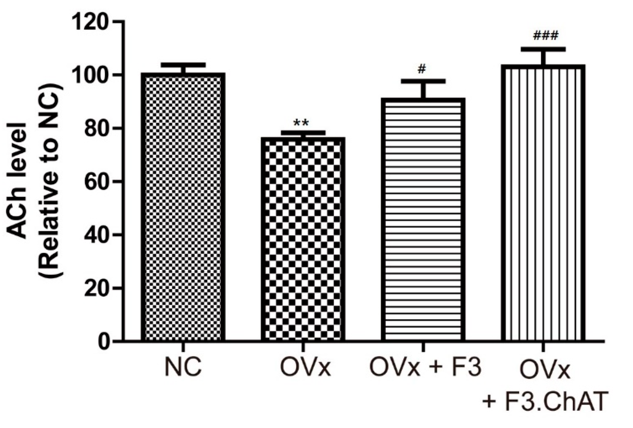
2.7. Transplantation of F3.ChAT Cells Restores ACh Concentration in CSF

To confirm the relationship between memory deficits and decreases in ACh release, we analyzed the ACh concentration (Figure 7). The ACh concentration in the CSF of ovariectomized rats decreased to 80%; this was reversed by the transplantation of F3 and F3.ChAT cells. Notably, the transplantation of F3.ChAT cells resulted in an almost full reversal of the ACh concentration to the normal control level.

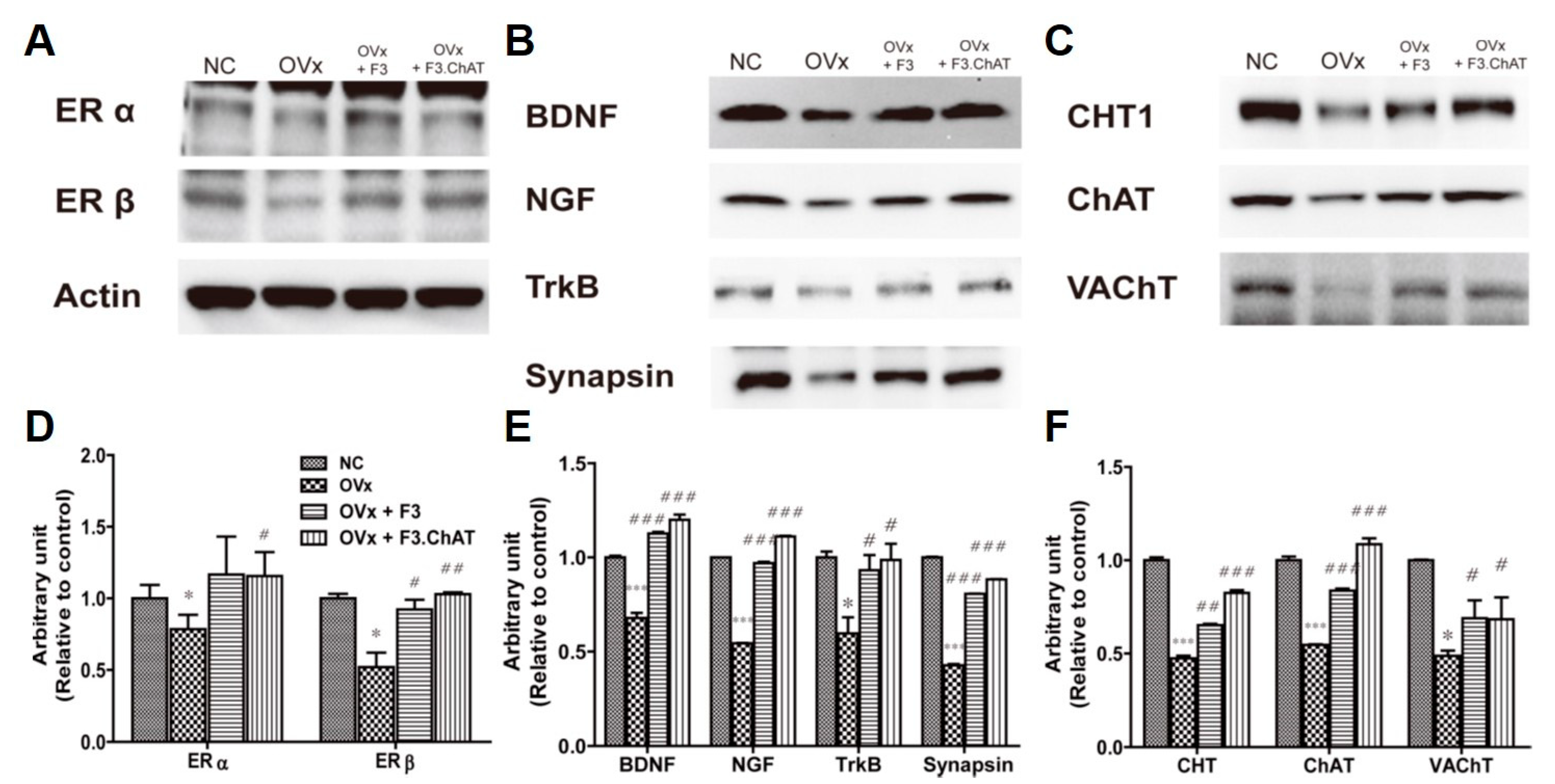
2.8. Transplantation of F3.ChAT Cells Enhances Expression of the Cholinergic System via Upregulation of NGF and BDNF

The expression of ERα and ERβ was markedly decreased in the brains of ovariectomized rats (Figure 8). The degenerative changes in estrogen receptors led to the decreased expression of the neurotrophin family proteins (NGF, BDNF, and Trk), resulting in decreased levels of cholinergic functional proteins such as CHT1, ChAT, and VAChT. Interestingly, transplantation of F3 and F3 ChAT cells restored the expression of the estrogen receptors, neurotrophin family proteins, and cholinergic functional proteins; F3.ChAT cells were more efficient than F3 cells.
The intravenous transplantation F3 and F3.ChAT cells were detected in the hippocampus of ovariectomized rats after four weeks (Figure 9). Double immunostaining indicated that the F3 and F3.ChAT cells (hMito-positive cells) expressed NGF and BDNF.

3. Discussion

In this study, we confirmed that estradiol had an influence on the cholinergic system; F3 and F3.ChAT cells improved cognitive function in ovariectomized rats via the neurotrophin signaling pathway. Estrogen receptors were co-localized with the neurotrophin receptor, and co-culture of primary cells and stem cells increased the concentrations of NGF and BDNF, resulting in the increased expression of CHT1, ChAT, and VAChT. In addition, transplantation of F3 and F3.ChAT cells in ovariectomized rats enhanced the levels of ACh, expression of estrogen receptors, NGF, and BDNF, and that of the cholinergic system, improving the memory dysfunction.
Estrogen deficiency during menopause induces diverse abnormal symptoms, including osteoporosis and weight gain [3,29]. In particular, it induces apoptosis, neuronal loss, and cognitive dysfunction in the hippocampus [30]. Therefore, several studies have focused on developing therapeutic strategies, such as hormone replacement, and understanding their mechanisms [31,32,33]. In this study, estradiol treatment increased the expression of the cholinergic system for ACh synthesis. The cholinergic system is an important site of action for estrogen in the brain. Estrogen facilitates cholinergic neurotransmission in the septal–hippocampal pathway, as evidenced by its ability to increase the activity and levels of ChAT [34]. Estrogen upregulates BDNF expression [7]. The increase in NGF and BDNF levels with exercise restores cognitive function in ovariectomized rats [35,36,37]. Therefore, estrogen receptors could modulate the expression of neurotrophin family proteins [38]. In this study, we confirmed that ERα and ERβ co-localized with neurotrophin family proteins. Therefore, estradiol treatment might upregulate the cholinergic system via the activation of the neurotrophin signaling pathway. Estrogen targets neurons in the basal forebrain, which expresses both NGF and ChAT [38,39,40].
Exogenous NGF and BDNF increased the expression of the cholinergic system. Stem cells facilitate the recovery of the damaged brain by the secretion of NGF and BDNF [13,14,41]. In this study, F3 and F3.ChAT cells secreted NGF and BDNF in the conditioned medium. When primary cells were co-cultured with F3 or F3.ChAT cells, the expression of endogenous BDNF, NGF, and their receptors increased. The PI3K/Akt signaling pathway involved in the neurotrophin pathway was upregulated [42]. These results are in line with those from earlier reports [43]. Exposure to neurotrophins increases ChAT activity and ACh release via Trk and calcium-dependent pathways [44,45]. Interestingly, co-culture with F3 and F3.ChAT cells decreased p75NTR levels; this could protect against apoptosis, because p75NTR signaling induces the apoptotic pathway [46,47]. The co-culture of primary cells with F3 and F3.ChAT increased the levels of CHT1, ChAT, and VAChT; the effect of F3.ChAT cells was higher than that of F3 cells. This could be attributed to the higher expression of NGF and BDNF in F3.ChAT cells compared to that in F3 cells.
Estradiol deficiency in ovariectomized rats decreases estrogen receptors, the uptake of high-affinity choline, ChAT activity, and ChAT mRNA levels [40,48]. NGF and BDNF mRNA levels were decreased after ovariectomy. In this study, we confirmed that the levels of the proteins associated with the cholinergic system (CHT, ChAT, and VAChT) and that of the neurotrophins (NGF and BDNF) decreased in the brain tissue of ovariectomized animals. In addition, the concentration of ACh in the CSF was reduced, resulting in memory deficits in behavioral tests. These effects could be reversed by exogenous estradiol, estradiol replacement therapy, and the upregulation of NGF and BDNF, either through exercise or the transplantation of F3 and F3.ChAT cells. In particular, F3.ChAT cells showed more potent effects than F3 cells in ovariectomized rats and Alzheimer’s animal models [23,49]. This is because F3.ChAT cells not only secrete more NGF and BDNF than F3 cells, but also produce more ACh by upregulating ChAT [14,49]. In this study, F3.ChAT cells showed a more potent effect than F3 cells in an in vitro experiment. However, in an in vivo experiment, F3.ChAT cells showed a mimic effect in a passive avoidance test and Morris water maze. It thought that most of stem cells are distributed to other peripheral organs and rapidly cleared after IV injection. After ICV injection, F3.ChAT cells showed more potent effect than F3 cells [14,23]. Interestingly, the levels of ERα and ERβ were also restored by transplantation of F3 and F3.ChAT cells. These could be a direct effect of replacing damaged cells by F3.ChAT cells, or an indirect effect of NGF and BDNF secreted from F3.ChAT cells. The estrogen receptors and BDNF were localized within the same cells, and estrogen significantly influenced the levels of BDNF mRNA and protein in a bidirectional manner within the adult rat hippocampus.
In general, hormone replacement therapy is performed for menopausal women. But it sometimes causes side effects such as breast tenderness, bloating, irritability and depression [50,51,52,53,54]. However, for memory deficiency, the up-regulation of NGF and BDNF using exercise or stem cells is thought to be a good alternative.

4. Materials and Methods

4.1. Primary Culture Cholinergic Neuron in Basal Forebrain

The septal region, including the basal forebrain, was obtained from BALB/c mice (Daehan Biolink, Eumseong, Korea) at embryonic day 15. Tissue was dissected in serum-free neurobasal medium (Gibco BRL, Grand Island, NY, USA) and treated with 0.03% trypsin in serum-free neurobasal medium for 3 min at 37 °C. After sieving through a 63 μm nylon mesh, cells were resuspended in serum-free neurobasal medium. The culture was initiated at 1 × 10⁵ cells per ml in 6 well plates coated with 10 μg/mL poly-D-lysine (PDL; Sigma-Aldrich, St. Louis, MO, USA) in neurobasal medium (Gibco) containing antibiotics (100 IU/mL penicillin and 100 ug/mL streptomycin) and 10% heat-inactivated fetal bovine serum (Gibco) at 37 °C in a 5% CO2/95% air atmosphere. To obtain glia-free neuronal cultures, cytosine arabinofuranoside (AraC, 1 μM, Sigma) was added to the cell culture media from days one to three to inhibit glial cell division.
To investigate the effect of estradiol on the cholinergic system, primary cells were washed twice with PBS, and then treated with 0, 5, 10, and 20 nM β-estradiol in culture medium (Sigma). After 24 h incubation, the cells were subjected to real-time PCR and western blotting analysis.

4.2. F3.ChAT Cell Culture

The establishment of F3 and F3.ChAT neural stem cell lines was described previously [14,49]. F3 (human neural stem cells) and F3.ChAT (ChAT overexpressing F3 cells) were cultured in Dulbecco’s modified Eagle’s medium (DMEM; Biowest, Nuaille’, Cholet, France) containing antibiotics (100 IU/mL penicillin and 100 μg/mL streptomycin) and 10% heat-inactivated fetal bovine serum (Biowest) at 37 °C in a 5% CO2/95% air atmosphere. In all experiments, cells were grown until more than 90% confluence and subjected to no more than 20 passages.
To investigate the effect of NGF and BDNF on the cholinergic system, primary cells were washed twice with PBS and were co-cultured with F3 and F3.ChAT cells. F3 and F3.ChAT cells were seeded at 1 × 10⁵ cells per ml in the upper chambers of 6-well transwell plates (pore 0.4 μm). After 24 h incubation, the cells were subjected to real-time PCR and western blotting analysis.

4.3. Assessing BDNF and NGF Levels

To measure NGF and BDNF levels in the medium, both F3 and F3.ChAT cells were seeded at 1 × 10⁶ cells/mL in six wells. After a 24 h incubation, BDNF and NGF levels in the medium were determined using enzyme-linked immunosorbent assay (ELISA). Human BDNF and NGF ELISA kits (Abcam, Cambridge, UK) were used according to the manufacturer’s instructions. O.D values were measured using an ELISA plate reader (MX2; Molecular Devices, Sunnyvale, CA, USA). The experiments were performed in triplicate, and the mean values are presented.

4.4. Immunocytochemistry of Estrogen Receptors and Neurophin Family in Primary Cells

To analyze the localization of the estrogen receptor and the neurotrophin family proteins, the primary cells were incubated in PDL coated 6 well plates and fixed in 10% neutral buffered formalin for 5 min at room temperature. After blocking for 10 min, the cells were incubated with primary antibodies specific for estrogen receptor α and β (1:100; mouse monoclonal, Abcam, Cambridge, UK) for overnight at 4 °C, followed by incubation with Alexa Fluor 488-conjugated anti-mouse IgG (Molecular Probes, Eugene, OR, USA) for 1 h at room temperature. The cells were washed thrice with PBS; for double immunostaining, the cells were incubated with primary antibodies specific for NGF, BDNF, TRK, and p75NTR (1:100; rabbit polyclonal, Abcam, Cambridge, UK) for 3 h at room temperature, followed by incubation with Alexa Fluor 594-cojugated anti-rabbit IgG (Molecular Probes) for 1 h at room temperature. Cells were counterstained with 4,6-diamino-2-phenylindole (DAPI; Sigma-Aldrich, St. Louis, MO, USA) to stain the nuclei and viewed under a fluorescence microscope (EVOS FL Auto2 Cell imaging system; Thermo Fisher Scientific, Waltham, MA, USA).

4.5. Quantitative Real Time PCR Analysis

Total RNA was isolated from primary cells using TRIzol Reagent (Invitrogen, Carlsbad, CA, USA), according to the manufacturer’s instructions. Quantitative real-time PCR was performed as described previously [55]. Glyceraldehyde 3-phosphate dehydrogenase (GAPDH) was used as the internal standard to normalize the expression of the target transcripts. The details of primers used to amplify the choline transporter (CHT), ChAT, and vesicular acetylcholine transporter (VAChT) are included in Table 1. Triplicate data were analyzed in three independent assays using the comparative Ct method [55].

4.6. Animals

Twelve-week-old female Sprague-Dawley rats were purchased from Daehan Biolink (Eumseong, Korea). Rats were housed in a controlled room with a constant temperature (23 ± 3 °C), relative humidity (50 ± 10%), and a 12-h light/dark cycle. Rats were fed a standard rodent diet and purified water ad libitum. All experimental procedures were approved and carried out in accordance with the Institutional Animal Care and Use Committee of the Korea National University of Education, Korea (#KNUE-202008-001-02).

4.7. Ovariectomy and Stem Cell Transplantation

Following the acclimation to the laboratory environment, the 12-week-old rats underwent bilateral ovariectomy, as previously described [6]. The rats were subjected to systemic anesthesia with ether and sterilized (10% povidone-iodine scrub followed by 70% alcohol wipe) before hair removal and treatment. A 1 cm incision was made in the center of the dorsal side of the experimental animal; the ovaries were ligated with suture threads, and ovarian resection was performed on both sides. Antibiotics (cafazolin 50 mg/kg) were administered through muscular injection.
Three weeks after recovery from surgery, 3 animals (normal and OVX rats) were subjected to passive avoidance and water maze tests to assess memory deficits. Other rats were randomly assigned to the following groups: normal group (NC; n = 10), OVX group (OVX; n = 10), OVX + F3 cells group (OVX-F3), and OVX + F3.ChAT cells group (OVX-F3.ChAT; n = 10). F3 or F3∙ChAT cells (1 × 10⁶ cells/rat) were transplanted into rats via tail vein injection.

4.8. Passive Avoidance Performance

Passive avoidance performance was assessed using a shuttle box (Med Associates, St. Albans, VT, USA) to evaluate memory acquisition and retention. The shuttle box apparatus consisted of light and dark compartments; a light chamber equipped with a lamp and a dark chamber with a steel-grid floor for electric shock. During the trials, an electric shock was delivered when the rats entered the dark compartment from the light room through a guillotine door. The latency time of remaining in a room with the light on following an electric shock (1 mA for 2 s) in a dark compartment was recorded. Four consecutive trials at 5-min intervals were performed with an electric shock when the rats entered the dark compartment. The endpoint was set to 300 s, denoting full memory acquisition.

4.9. Morris Water-Maze Performance

The Morris water-maze performance was assessed in a round water bath (180 cm in diameter; Panlab Technology, Barcelona, Spain) filled with water (27 cm in depth) maintained at 22 ± 2 °C to evaluate spatial memory. The bath was divided into four quadrants, and a hidden escape platform (10 cm in diameter, 25 cm in height) was submerged in the center of one quadrant, 2 cm below the water surface. The rats were subjected to four trials at 5 min intervals to find the hidden platform based on several cues external to the maze. The endpoint was set to 300 s if the animal failed to find the platform. The mean escape latency time, which was the time spent to escape from the platform during the trials, was recorded.

4.10. ACh Analysis in CSF

The rats were sacrificed at the end of the learning/memory testing, and CSF was collected to analyze ACh content [49]. The ACh concentration in the CSF was measured using the Amplex Red acetylcholine/acetylcholinesterase assay kit (Molecular Probes). In this assay, ACh is hydrolyzed by AChE to release choline, which is then oxidized by choline oxidase to betaine and H2O2. H2O2 interacts with Amplex Red (7-dihydroxyphenoxazine) in the presence of horseradish peroxidase to generate the highly fluorescent, resorufin. The resulting fluorescence was measured using a fluorescence microplate reader (MX2; Molecular Devices, Sunnyvale, CA, USA); excitation wavelength, 530–560 nm and emission wavelength, ~590 nm.

4.11. Western Blot Analysis in Primary Cells and Brain Tissues

Primary cells and brain tissue were homogenized in 10 volumes of RIPA buffer (Thermo Scientific, Waltham, MA, USA) containing protease inhibitors (Sigma-Aldrich, St. Louis, MO, USA) and phosphatase inhibitors (Sigma-Aldrich, St. Louis, MO, USA). Western blotting was performed as previously described. The membranes were immunoblotted with primary antibodies, followed by incubation with horseradish peroxidase-conjugated anti-rabbit and anti-mouse secondary antibodies (Jackson Laboratories). The antibodies used in this study are listed in Table 2. Band densities were measured using ImageJ software (NIH, Bethesda, MD, USA) and normalized to the density of the actin band.

4.12. Immunohistochemistry in Brain Sections

The brains of rats were perfusion-fixed with 10% paraformaldehyde solution and post-fixed in the same solution for 48 h, followed by cryoprotection in 30% sucrose for 72 h. Coronal cryosections of 30-μm thickness, 1.0 mm posterior to bregma were prepared and processed for double immunostaining of human mitochondria (hMito; for human cells), NGF, and BDNF using antibodies specific for hMito (1:200, mouse monoclonal, Chemicon), NGF (1:300, rabbit polyclonal, Abcam), and BDNF (1:300, rabbit polyclonal, Abcam). Brain sections were incubated with the primary antibodies overnight at 4 °C, followed by incubation with secondary antibodies conjugated with Alexa Fluor-488 or -594 (1:300, Molecular Probes) for 2 h at room temperature [6,7,8,9]. This was followed by staining with 4,6-diamidino-2-phenylindole (DAPI) for 30 min. All samples were evaluated immediately after staining and photographed using a fluorescence microscope (EVOS FL Auto2 Cell imaging system; Thermo Fisher Scientific, Waltham, MA, USA).

4.13. Statistical Analysis

Statistical comparisons between the groups were performed using one-way analysis of variance (ANOVA) followed by Tukey’s multiple comparison test. All analyses were conducted using Statistical Package for Social Sciences for Windows software (version 12.0; SPSS Inc., Chicago, IL, USA). Statistical significance was set at p < 0.05. All data are expressed as the mean ± SD.

5. Conclusions

Treatment with estradiol or with exogenous NGF and BDNF induced the cholinergic system. Transplantation of F3.ChAT cells restored memory dysfunction in ovariectomized rats via upregulation of NGF and BDNF. In addition, it restored the function of estrogen receptors. Upregulation of neurotrophins through stem cell transplantation or exercise improves abnormal symptoms in menopausal women, and this is a promising strategy for ameliorating the cognitive impairment caused by the loss or reduction of estradiol.

Author Contributions

Conceptualization, E.-J.Y. and D.P.; methodology, E.-J.Y. and Y.C.; formal analysis, E.-J.Y. and Y.C.; investigation, E.-J.Y.; writing—original draft preparation, E.-J.Y.; writing—review and editing, D.P.; visualization, E.-J.Y.; supervision, D.P.; project administration, E.-J.Y. and D.P.; funding acquisition, E.-J.Y. All authors have read and agreed to the published version of the manuscript.

Funding

This research was supported by the Basic Science Research Program through the National Research Foundation of Korea (NRF), funded by the Ministry of Education (NRF-2020R1A6A3A01100641).

Institutional Review Board Statement

All experimental procedures were approved and carried out in accordance with the Institutional Animal Care and Use Committee of the Korea National University of Education, Korea (#KNUE-202008-001-02).

Informed Consent Statement

Not applicable.

Data Availability Statement

All data generated from this study are contained within the article.

Conflicts of Interest

The authors declare that they have no conflict of interest.

References

  1. Genazzani, A.R.; Pluchino, N.; Luisi, S.; Luisi, M. Estrogen, cognition and female ageing. Hum. Reprod. Update 2007, 13, 175–187. [Google Scholar] [CrossRef] [PubMed] [Green Version]
  2. Sherwin, B.B. Estrogen and cognitive functioning in women. Endocr. Rev. 2003, 24, 133–151. [Google Scholar] [CrossRef] [PubMed] [Green Version]
  3. Mauvais-Jarvis, F.; Clegg, D.J.; Hevener, A.L. The role of estrogens in control of energy balance and glucose homeostasis. Endocr. Rev. 2013, 34, 309–338. [Google Scholar] [CrossRef] [Green Version]
  4. Newhouse, P.; Dumas, J. Estrogen–cholinergic interactions: Implications for cognitive aging. Horm. Behav. 2015, 74, 173–185. [Google Scholar] [CrossRef] [PubMed] [Green Version]
  5. Russell, J.K.; Jones, C.K.; Newhouse, P.A. The Role of Estrogen in Brain and Cognitive Aging. Neurotherapeutics 2019, 16, 649–665. [Google Scholar] [CrossRef]
  6. Yoon, E.-J.; Kim, K.-L.; Park, D. Effects of Resistance Exercise on mRNA Expression of Brain Neuroplasticity Related Factors in Hippocampus and Cognitive Function in Ovariectomized Rats. Brain Digit. Learn. 2021, 11, 577–587. [Google Scholar]
  7. Harte-Hargrove, L.C.; Maclusky, N.J.; Scharfman, H.E. Brain-derived neurotrophic factor–estrogen interactions in the hippocampal mossy fiber pathway: Implications for normal brain function and disease. Neuroscience 2013, 239, 46–66. [Google Scholar] [CrossRef] [Green Version]
  8. Spencer, J.L.; Waters, E.M.; Romeo, R.D.; Wood, G.E.; Milner, T.A.; McEwen, B.S. Uncovering the mechanisms of estrogen effects on hippocampal function. Front. Neuroendocr. 2008, 29, 219–237. [Google Scholar] [CrossRef] [Green Version]
  9. Cowansage, K.K.; LeDoux, J.E.; Monfils, M.-H. Brain-derived neurotrophic factor: A dynamic gatekeeper of neural plasticity. Curr. Mol. Pharmacol. 2010, 3, 12–29. [Google Scholar] [CrossRef]
  10. Korsching, S. The role of nerve growth factor in the CNS. Trends Neurosci. 1986, 9, 570–573. [Google Scholar] [CrossRef]
  11. Tapia-Arancibia, L.; Aliaga, E.; Silhol, M.; Arancibia, S. New insights into brain BDNF function in normal aging and Alzheimer disease. Brain Res. Rev. 2008, 59, 201–220. [Google Scholar] [CrossRef] [PubMed]
  12. Tuszynski, M.H.; Yang, J.H.; Barba, D.; U, H.-S.; Bakay, R.A.E.; Pay, M.M.; Masliah, E.; Conner, J.M.; Kobalka, P.; Roy, S.; et al. Nerve Growth Factor Gene Therapy. JAMA Neurol. 2015, 72, 1139. [Google Scholar] [CrossRef] [PubMed]
  13. Park, D.; Yang, G.; Bae, D.K.; Lee, S.H.; Yang, Y.-H.; Kyung, J.; Kim, D.; Choi, E.-K.; Choi, K.-C.; Kim, S.U.; et al. Human adipose tissue-derived mesenchymal stem cells improve cognitive function and physical activity in ageing mice. J. Neurosci. Res. 2013, 91, 660–670. [Google Scholar] [CrossRef] [PubMed]
  14. Park, D.; Yang, Y.H.; Bae, D.K.; Lee, S.H.; Yang, G.; Kyung, J.; Kim, D.; Choi, E.K.; Lee, S.W.; Kim, G.H.; et al. Improvement of cognitive function and physical activity of aging mice by human neural stem cells over-expressing choline acetyltransferase. Neurobiol. Aging 2013, 34, 2639–2646. [Google Scholar] [CrossRef]
  15. Pramanik, S.; Sulistio, Y.A.; Heese, K. Neurotrophin Signaling and Stem Cells-Implications for Neurodegenerative Diseases and Stem Cell Therapy. Mol. Neurobiol. 2017, 54, 7401–7459. [Google Scholar] [CrossRef]
  16. Blokland, A. Acetylcholine: A neurotransmitter for learning and memory? Brain Res. Rev. 1995, 21, 285–300. [Google Scholar] [CrossRef]
  17. Da Penha Berzaghi, M.; Cooper, J.; Castren, E.; Zafra, F.; Sofroniew, M.; Thoenen, H.; Lindholm, D. Cholinergic regulation of brain-derived neurotrophic factor (BDNF) and nerve growth factor (NGF) but not neurotrophin-3 (NT-3) mRNA levels in the developing rat hippocampus. J. Neurosci. 1993, 13, 3818–3826. [Google Scholar] [CrossRef] [Green Version]
  18. Tyler, W.J.; Pozzo-Miller, L.D. BDNF Enhances Quantal Neurotransmitter Release and Increases the Number of Docked Vesicles at the Active Zones of Hippocampal Excitatory Synapses. J. Neurosci. 2001, 21, 4249–4258. [Google Scholar] [CrossRef]
  19. Li, Y.-X.; Zhang, Y.; Lester, H.A.; Schuman, E.M.; Davidson, N. Enhancement of neurotransmitter release induced by brain-derived neurotrophic factor in cultured hippocampal neurons. J. Neurosci. 1998, 18, 10231–10240. [Google Scholar] [CrossRef] [Green Version]
  20. Gibbs, R.B. Fluctuations in relative levels of choline acetyltransferase mRNA in different regions of the rat basal forebrain across the estrous cycle: Effects of estrogen and progesterone. J. Neurosci. 1996, 16, 1049–1055. [Google Scholar] [CrossRef]
  21. Kaur, G.; Janik, J.; Isaacson, L.G.; Callahan, P. Estrogen regulation of neurotrophin expression in sympathetic neurons and vascular targets. Brain Res. 2007, 1139, 6–14. [Google Scholar] [CrossRef] [PubMed]
  22. Hara, Y.; Waters, E.M.; McEwen, B.S.; Morrison, J.H. Estrogen Effects on Cognitive and Synaptic Health Over the Lifecourse. Physiol. Rev. 2015, 95, 785–807. [Google Scholar] [CrossRef] [PubMed] [Green Version]
  23. Park, D.; Choi, E.K.; Cho, T.H.; Joo, S.S.; Kim, Y.B. Human Neural Stem Cells Encoding ChAT Gene Restore Cognitive Function via Acetylcholine Synthesis, Abeta Elimination, and Neuroregeneration in APPswe/PS1dE9 Mice. Int. J. Mol. Sci. 2020, 21, 3958. [Google Scholar] [CrossRef] [PubMed]
  24. Berchtold, N.C.; Kesslak, J.P.; Pike, C.J.; Adlard, P.A.; Cotman, C.W. Estrogen and exercise interact to regulate brain-derived neurotrophic factor mRNA and protein expression in the hippocampus. Eur. J. Neurosci. 2001, 14, 1992–2002. [Google Scholar] [CrossRef]
  25. Russo-Neustadt, A.; Ha, T.; Ramirez, R.; Kesslak, J.P. Physical activity–antidepressant treatment combination: Impact on brain-derived neurotrophic factor and behavior in an animal model. Behav. Brain Res. 2001, 120, 87–95. [Google Scholar] [CrossRef]
  26. Lu, J.; Xu, Y.; Hu, W.; Gao, Y.; Ni, X.; Sheng, H.; Liu, Y. Exercise ameliorates depression-like behavior and increases hippocampal BDNF level in ovariectomized rats. Neurosci. Lett. 2014, 573, 13–18. [Google Scholar] [CrossRef] [Green Version]
  27. Hua, Z.; Gu, X.; Dong, Y.; Tan, F.; Liu, Z.; Thiele, C.J.; Li, Z. PI3K and MAPK pathways mediate the BDNF/TrkB-increased metastasis in neuroblastoma. Tumour Biol. 2016, 37, 16227–16236. [Google Scholar] [CrossRef] [Green Version]
  28. Sanchez-Alegria, K.; Flores-Leon, M.; Avila-Munoz, E.; Rodriguez-Corona, N.; Arias, C. PI3K Signaling in Neurons: A Central Node for the Control of Multiple Functions. Int. J. Mol. Sci. 2018, 19, 3725. [Google Scholar] [CrossRef] [Green Version]
  29. Roman-Blas, J.A.; Castañeda, S.; Largo, R.; Herrero-Beaumont, G. Osteoarthritis associated with estrogen deficiency. Arthritis Res. Amp. Ther. 2009, 11, 241. [Google Scholar] [CrossRef] [Green Version]
  30. Pandey, R.; Shukla, P.; Anjum, B.; Gupta, H.P.; Pal, S.; Arjaria, N.; Gupta, K.; Chattopadhyay, N.; Sinha, R.A.; Bandyopadhyay, S. Estrogen deficiency induces memory loss via altered hippocampal HB-EGF and autophagy. J. Endocrinol. 2020, 244, 53–70. [Google Scholar] [CrossRef]
  31. Sullivan, S.D.; Sarrel, P.M.; Nelson, L.M. Hormone replacement therapy in young women with primary ovarian insufficiency and early menopause. Fertil. Steril. 2016, 106, 1588–1599. [Google Scholar] [CrossRef] [PubMed] [Green Version]
  32. Adu-Nti, F.; Gao, X.; Wu, J.-M.; Li, J.; Iqbal, J.; Ahmad, R.; Ma, X.-M. Osthole ameliorates estrogen deficiency-induced cognitive impairment in female mice. Front. Pharmacol. 2021, 12, 641909. [Google Scholar] [CrossRef] [PubMed]
  33. Szegő, É.M.; Csorba, A.; Janáky, T.; Kékesi, K.A.; Ábrahám, I.M.; Mórotz, G.M.; Penke, B.; Palkovits, M.; Murvai, Ü.; Kellermayer, M.S. Effects of estrogen on beta-amyloid-induced cholinergic cell death in the nucleus basalis magnocellularis. Neuroendocrinology 2011, 93, 90–105. [Google Scholar] [CrossRef] [PubMed]
  34. Daniel, J.M.; Dohanich, G.P. Acetylcholine mediates the estrogen-induced increase in NMDA receptor binding in CA1 of the hippocampus and the associated improvement in working memory. J. Neurosci. 2001, 21, 6949–6956. [Google Scholar] [CrossRef] [Green Version]
  35. Rashidy-Pour, A.; Bavarsad, K.; Miladi-Gorji, H.; Seraj, Z.; Vafaei, A.A. Voluntary exercise and estradiol reverse ovariectomy-induced spatial learning and memory deficits and reduction in hippocampal brain-derived neurotrophic factor in rats. Pharmacol. Biochem. Behav. 2019, 187, 172819. [Google Scholar] [CrossRef]
  36. Habibi, P.; Babri, S.; Ahmadiasl, N.; Yousefi, H. Effects of genistein and swimming exercise on spatial memory and expression of microRNA 132, BDNF, and IGF-1 genes in the hippocampus of ovariectomized rats. Iran. J. Basic Med. Sci. 2017, 20, 856. [Google Scholar]
  37. Chae, C.-H.; Kim, H.-T. Forced, moderate-intensity treadmill exercise suppresses apoptosis by increasing the level of NGF and stimulating phosphatidylinositol 3-kinase signaling in the hippocampus of induced aging rats. Neurochem. Int. 2009, 55, 208–213. [Google Scholar] [CrossRef]
  38. Toran-Allerand, C.D.; Miranda, R.C.; Bentham, W.D.; Sohrabji, F.; Brown, T.J.; Hochberg, R.B.; Maclusky, N.J. Estrogen receptors colocalize with low-affinity nerve growth factor receptors in cholinergic neurons of the basal forebrain. Proc. Natl. Acad. Sci. USA 1992, 89, 4668–4672. [Google Scholar] [CrossRef] [Green Version]
  39. Bora, S.H.; Liu, Z.; Kecojevic, A.; Merchenthaler, I.; Koliatsos, V.E. Direct, complex effects of estrogens on basal forebrain cholinergic neurons. Exp. Neurol. 2005, 194, 506–522. [Google Scholar] [CrossRef]
  40. McMillan, P.; Singer, C.; Dorsa, D. The effects of ovariectomy and estrogen replacement on trkA and choline acetyltransferase mRNA expression in the basal forebrain of the adult female Sprague-Dawley rat. J. Neurosci. 1996, 16, 1860–1865. [Google Scholar] [CrossRef] [Green Version]
  41. Park, D.; Lee, S.H.; Bae, D.K.; Yang, Y.-H.; Yang, G.; Kyung, J.; Kim, D.; Choi, E.-K.; Hong, J.T.; Shin, I.S.; et al. Transplantation of Human Adipose Tissue-Derived Mesenchymal Stem Cells Restores the Neurobehavioral Disorders of Rats with Neonatal Hypoxic-Ischemic Encephalopathy. Cell Med. 2013, 5, 17–28. [Google Scholar] [CrossRef] [PubMed] [Green Version]
  42. Madziar, B.; Lopez-Coviella, I.; Zemelko, V.; Berse, B. Regulation of cholinergic gene expression by nerve growth factor depends on the phosphatidylinositol-3’-kinase pathway. J. Neurochem. 2005, 92, 767–779. [Google Scholar] [CrossRef] [PubMed]
  43. Li, M.; Dai, F.-r.; Du, X.-p.; Yang, Q.-d.; Zhang, X.; Chen, Y. Infusion of BDNF into the nucleus accumbens of aged rats improves cognition and structural synaptic plasticity through PI3K-ILK-Akt signaling. Behav. Brain Res. 2012, 231, 146–153. [Google Scholar] [CrossRef] [PubMed]
  44. Scott-Solomon, E.; Kuruvilla, R. Mechanisms of neurotrophin trafficking via Trk receptors. Mol. Cell. Neurosci. 2018, 91, 25–33. [Google Scholar] [CrossRef] [PubMed]
  45. Mufson, E.J.; Ginsberg, S.D.; Ikonomovic, M.D.; Dekosky, S.T. Human cholinergic basal forebrain: Chemoanatomy and neurologic dysfunction. J. Chem. Neuroanat. 2003, 26, 233–242. [Google Scholar] [CrossRef]
  46. Miller, F.; Kaplan, D. Neurotrophin signalling pathways regulating neuronal apoptosis. Cell. Mol. Life Sci. CMLS 2001, 58, 1045–1053. [Google Scholar] [CrossRef] [PubMed]
  47. Roux, P.P.; Colicos, M.A.; Barker, P.A.; Kennedy, T.E. p75 Neurotrophin Receptor Expression Is Induced in Apoptotic Neurons After Seizure. J. Neurosci. 1999, 19, 6887–6896. [Google Scholar] [CrossRef] [Green Version]
  48. Qu, N.; Wang, L.; Liu, Z.C.; Tian, Q.; Zhang, Q. Oestrogen receptor alpha agonist improved long-term ovariectomy-induced spatial cognition deficit in young rats. Int. J. Neuropsychopharmacol. 2013, 16, 1071–1082. [Google Scholar] [CrossRef] [PubMed] [Green Version]
  49. Park, D.; Lee, H.J.; Joo, S.S.; Bae, D.K.; Yang, G.; Yang, Y.H.; Lim, I.; Matsuo, A.; Tooyama, I.; Kim, Y.B.; et al. Human neural stem cells over-expressing choline acetyltransferase restore cognition in rat model of cognitive dysfunction. Exp. Neurol. 2012, 234, 521–526. [Google Scholar] [CrossRef]
  50. MacLennan, A.H.; MacLennan, A.; Wenzel, S.; Chambers, H.M.; Eckert, K. Continuous low-dose oestrogen and progestogen hormone replacement therapy: A randomised trial. Med. J. Aust. 1993, 159, 102–106. [Google Scholar] [CrossRef]
  51. Clisham, P.R.; de Ziegler, D.; Lozano, K.; Judd, H.L. Comparison of continuous versus sequential estrogen and progestin therapy in postmenopausal women. Obs. Gynecol. 1991, 77, 241–246. [Google Scholar] [CrossRef] [PubMed]
  52. Hawthorn, R.J.; Spowart, K.; Walsh, D.; Hart, D.M. The endometrial status of women on long-term continuous combined hormone replacement therapy. Br. J. Obs. Gynaecol. 1991, 98, 939–940. [Google Scholar] [CrossRef]
  53. Williams, S.R.; Frenchek, B.; Speroff, T.; Speroff, L. A study of combined continuous ethinyl estradiol and norethindrone acetate for postmenopausal hormone replacement. Am. J. Obs. Gynecol. 1990, 162, 438–446. [Google Scholar] [CrossRef]
  54. Luciano, A.A.; Turksoy, R.N.; Carleo, J.; Hendrix, J.W. Clinical and metabolic responses of menopausal women to sequential versus continuous estrogen and progestin replacement therapy. Obs. Gynecol. 1988, 71, 39–43. [Google Scholar]
  55. Yon, J.-M.; Kim, Y.-B.; Park, D. The Ethanol Fraction of White Rose Petal Extract Abrogates Excitotoxicity-Induced Neuronal Damage In Vivo and In Vitro through Inhibition of Oxidative Stress and Proinflammation. Nutrients 2018, 10, 1375. [Google Scholar] [CrossRef] [PubMed] [Green Version]
Figure 1. Expression of CHT1 (A), ChAT (B), and VAChT (C) in primary forebrain neurons after treatment with estradiol. Gene expression was analyzed by real-time PCR. CHT1; choline transporter1, ChAT; choline acetyltransferase, VAChT; vesicular acetylcholine transporter. * Significantly different from normal control (p < 0.05). ** Significantly different from normal control (p < 0.005). *** Significantly different from normal control (p < 0.001).
Ijms 23 05560 g001
Figure 2. Localization of estrogen receptor (ER) α (A) and β (B) with NGF, BDNF, TRK, and p75NTR in primary forebrain neuron.
Ijms 23 05560 g002
Figure 3. Concentration of NGF (A) and BDNF (B) in F3 and F3.ChAT cells cultured in conditioned medium. The levels of NGF and BDNF were analyzed using an ELISA kit. The experiments were performed in triplicate, and mean values are presented. F.M.; Fresh mediumm, n.d.; not detected. * Significantly different from the fresh medium (p < 0.05). ** Significantly different from the fresh medium (p < 0.005). *** Significantly different from the fresh medium (p < 0.001). ## Significantly different from the F3 (p < 0.005). ### Significantly different from the F3 (p < 0.001).
Ijms 23 05560 g003
Figure 4. Expression of proteins related to the neurotrophin pathway. (A) Representative bands of protein related with the neurotrophin pathway. (B,C) The band densities were normalized to that of actin. * Significantly different from the normal control (p < 0.05). ** Significantly different from the normal control (p < 0.005). *** Significantly different from the normal control (p < 0.001). # Significantly different from the F3 (p < 0.05). ## Significantly different from the F3 (p < 0.005). ### Significantly different from the F3 (p < 0.001).
Ijms 23 05560 g004
Figure 5. Expression of CHT1 (A), ChAT (B), and VAChT (C) in primary forebrain neurons co-cultured with F3 and F3.ChAT cells. The F3 and F3.ChAT cells were seeded at 1 × 10⁵ cells per ml in the upper chambers of 6-well transwell plates (pore 0.4 μm) on the primary forebrain neuron. After a 24 h incubation, the cells were used for performing real time PCR. * Significantly different from normal control (p < 0.05). ** Significantly different from the normal control (p < 0.005). # Significantly different from the F3 (p < 0.05).
Ijms 23 05560 g005
Figure 6. Cognitive function of ovariectomized rats transplanted with F3 or F3.ChAT cells. Cognitive function was analyzed through passive avoidance performance (A) and Morri water-maze performance (B). The endopind was set to 300 s, if the animals stayed in light chamber in passive avoidance or failed to find the platform in Morris water-maze performance. ●; normal group, ■; Ovariectomy (OVx) group, ▲; OVx + F3 cells group, ▼; OVx + F3.ChAT cells group. * Significantly different from the normal control (p < 0.05). *** Significantly different from the normal control (p < 0.001). # Significantly different from the OVx (p < 0.05) group. ## Significantly different from the OVx (p < 0.05) group. ### Significantly different from the OVx (p < 0.05) group.
Ijms 23 05560 g006
Figure 7. Acetylcholine (ACh) level in the CSF of ovariectomized rat four weeks after transplantation with F3 or F3.ChAT cells. ACh levels were analyzed using an ELISA kit. ** Significantly different from the normal control (p < 0.005). # Significantly different from the OVx (p < 0.05) group. ### Significantly different from the OVx (p < 0.05) group.
Ijms 23 05560 g007
Figure 8. Expression of ER α and β (A), proteins related to the neurotrophin pathway (B), and the cholinergic system (C) in ovariectomized rats four weeks after transplantation of F3 or F3.ChAT cells. (AC) Representative bands of ER α and β (A), proteins related to the neurotrophin pathway (B), and the cholinergic system (C). (DF) The band densities were normalized to that of actin. * Significantly different from the normal control (p < 0.05). *** Significantly different from the normal control (p < 0.001). # Significantly different from the OVx (p < 0.05) group. ## Significantly different from the OVx (p < 0.05) group. ### Significantly different from the OVx (p < 0.05) group.
Ijms 23 05560 g008
Figure 9. Representative immunohistochemical images indicating the expression of NGF and BDNF in F3 and F3.ChAT cells. Human mitochondria (hMito) was used as the transplanted F3 and F3.ChAT cell markers. Expression of NGF (Upper panel) and BDNF (Bottom panel) in the hippocampus of ovariectomized rat four weeks after transplantation of F3 and F3.ChAT cells were analyzed using double immunostating. Scale bar = 50 μm, magnification × 200.
Ijms 23 05560 g009
Table 1. Sequences of the primers used in the current study.
Gene NameAccession No.Mouse Primer
ForwardReverse
CHTNM_022025.4TTCCAGATTCAGGCAGTAGACGGGGAGGGAAACTCCTATCTTGT
ChATNM_009891.2TGGGTCTCTGAATACTGGCTGAGGGCTAGAGTTGACTGGCAGG
VAChTNM_021712.3CCCTTTTGATGGCTGTGAGGGCTAGGGTACTCATTAGA
GAPdHXM_039103226GTCGGTGTGAACGGATTTGGCCACTTTGTCACAAGAGAAGGCA
Table 2. List of antibodies used in the current study.
EpitopeCompanyCat. NumberDilution2° Ab (IgG)
BDNFAbcamAb2268431:1000Rb
NGFAbcamAb61991:1000Rb
TrkAThermoFisherPa5-980181:1000Rb
TrkBAbcamAb189871:2000Rb
P75NTRAbcamAb529871:1000Rb
p-PI3KCell signaling#42281:1000Rb
p-AKTCell signaling#92711:1000Rb
AKTCell Signaling#92721:1000Rb
SynapsinAbcamAb645811:1000Rb
ActinCell Signal#51251:1000Rb
ER αAbcamAb661021:100Ms
ER βAbcamAb2881:100Ms
CHT1AbcamAb1541861:1000Rb
ChATAbcamAb1788501:1000Rb
VAChTSynaptic systems#1391031:500Rb
Publisher’s Note: MDPI stays neutral with regard to jurisdictional claims in published maps and institutional affiliations.

