Review

Neurofibromatosis Type 1 Gene Alterations Define Specific Features of a Subset of Glioblastomas

1
Department of Neurosurgery, Medical Faculty, Martin Luther University Halle-Wittenberg, Ernst-Grube-Straße 40, 06120 Halle, Germany
2
Department of Neuropathology, Institute of Pathology, Medical Faculty, Martin Luther University Halle-Wittenberg, Magdeburger Str. 14, 06112 Halle, Germany
3
Department of Research, National Neuroscience Institute, Singapore 308433, Singapore
4
Institute of Neuropathology, University Hospital Münster, 48149 Münster, Germany
5
Brandenburg Medical School Theodor Fontane, Faculty of Health Sciences, Joint Faculty of the Brandenburg University, 16816 Neuruppin, Germany
*
Author to whom correspondence should be addressed.
These authors contributed equally to this work.
Int. J. Mol. Sci. 2022, 23(1), 352; https://doi.org/10.3390/ijms23010352
Submission received: 25 October 2021 / Revised: 14 December 2021 / Accepted: 24 December 2021 / Published: 29 December 2021

Abstract

:
Neurofibromatosis type 1 (NF1) gene mutations or alterations occur within neurofibromatosis type 1 as well as in many different malignant tumours on the somatic level. In glioblastoma, NF1 loss of function plays a major role in inducing the mesenchymal (MES) subtype and, therefore defining the most aggressive glioblastoma. This is associated with an immune signature and mediated via the NF1–MAPK–FOSL1 axis. Specifically, increased invasion seems to be regulated via mutations in the leucine-rich domain (LRD) of the NF1 gene product neurofibromin. Novel targets for therapy may arise from neurofibromin deficiency-associated cellular mechanisms that are summarised in this review.

1. Molecular Subtypes of Glioblastomas

Glioblastoma, previously denominated glioblastoma multiforme (GBM), represents the most common malignant glial primary brain tumour [1]. The brain tumour classification of the 4th edition of the World Health Organization (WHO) distinguishes between four glioma grades. Among these, GBM belongs to grade 4, which is the most aggressive type [2]. Although the survival of GBM patients improved significantly during the last few decades, median survival of approximately 15 months is still poor [3,4]. There is little but growing knowledge on risk factors for GBM such as ionising radiation or hereditary cancer syndromes. As currently assessed, only a small subset of GBM is associated with hereditary syndromes, such as neurofibromatosis type 1 (NF1), Lynch syndrome, or Li–Fraumeni syndrome [5].
The clinical course of GBM is diverse and depends strongly on tumour localisation. Signs due to raised intracranial pressure, epileptic seizures, and focal neurological deficits are typical [5]. The combination of surgery, radiation, and chemotherapy with temozolomide (TMZ) represents the current standard therapy [6,7]. Surgical radicality is of paramount importance for the overall outcome. Resection of >95% of tumour volume as demonstrated by contrast-enhanced Magnetic Resonance Imaging (MRI) (“gross total resection”) has been shown to improve overall and progression-free survival significantly [8]. Nevertheless, gross total resection is not always achievable, even with integrating intraoperative imaging or 5-aminolevulinic acid fluorescence. Eloquent tumour localisation close to cortical or subcortical structures of major functional relevance such as the primary motor cortex or the deep tracts of the language system in the dominant hemisphere limit the extent of resection. Incomplete resection has been demonstrated to influence treatment outcome and negatively affect patient’s survival [9]. Recently, novel therapeutic approaches such as a combination of lomustine with TMZ as well as the use of Tumour-Treating Fields (TTFields) [4,10] are associated with improved survival. The effectiveness of these and possible future modalities is influenced by GBM molecular subclasses. Therefore, the subclasses are of high clinical importance and value.
GBM shows a diverse histological pattern that consists of necrosis, microvascular proliferation, increased mitotic activity, anaplasia, and invasion. Among these features, necrosis and microvascular proliferation conventionally distinguish GBM from high-grade astrocytoma [11]. In the last couple of years, the impact of molecular features on diagnosis increased dramatically. The combination of histological and molecular characteristics into an integrated diagnosis has become indispensable for appropriate diagnostic procedures and therapeutic planning [12,13]. For this purpose, next-generation-based genomic profiling entered clinical practice. Using specifically methylation profiling, GBM can be divided into six methylation subgroups [14]. For instance, gliomas that occur in NF1 patients are assigned to LGm6, which is a poorly defined methylation class subgroup [15].
At least two molecular markers have been established for a minimal clinical routine. Methylation of the O6-methylguanine-DNA methyltransferase (MGMT) promoter still serves as an important predictive marker and is associated with a better response to chemotherapy [6,7]. Additionally, hotspot mutations of the isocitrate dehydrogenase (IDH) gene that occur early in gliomagenesis are important diagnostic and prognostic markers in glioma subtypes [5]. GBM was classified into primary GBM, IDH-wild-type, and secondary GBM. According to the most recent 5th edition of the WHO classification diffuse astrocytomas are classified as IDH-wild-type GBM WHO grade 4 and IDH-mutant astrocytomas WHO grade 2, 3, or 4. Including now specific genetic events into diagnostics, microvascular proliferation or necrosis or one of the following genetic alterations such as telomerase reverse transcriptase (TERT) promoter mutation, epidermal growth factor receptor (EGFR) gene amplification, and +7/−10 chromosome copy number changes are sufficient to diagnose GBM [16,17].
Based on gene expression and genomic clustering, The Cancer Genome Atlas (TCGA) project established four GBM subclasses: classic (Cl), neural (N), proneural (PN), and mesenchymal (MES) [18]. Since recent studies did not identify the neural subtype securely, its existence is controversially discussed. Some authors claim contamination through normal neural tissue as an explanation [14,19,20] (Figure 1).
A high expression of angiogenesis or proliferation-associated genes, as well as poor median overall survival (about 14.7 months), is characteristic for the Cl subtype [5,21,22]. Particularly, molecular events comprise EGFR amplification or mutations and focal 9p21.3 homozygous deletions, including the cyclin-dependent kinase inhibitor 2A (CDKN2A) gene [18,21,23]. Amplification of chromosome 7 (+7), a loss of chromosome 10 (−10), and TERT promoter mutations, which are considered a characteristic event in GBM, are typically present in the Cl subtype. Additionally, loss of tumour protein 53 (TP53) and mutation in the Phosphatase and Tensin homolog (PTEN) gene is frequently observed in the Cl subtype [18,22]. The DNA methylation subtype “receptor tyrosine kinase (RTK) II” corresponds to the Cl subtype [24]. In contrast, the PN subtype corresponds to the RTK I methylation subtype. Patients with PN GBM subtype show a more favourable median survival time of approximately 17 months [5,22]. In contrast to the Cl subtype, there is a lack of PTEN and EGFR mutations. A specific alteration in the PN subtype is focal amplification of the 4q12 locus harbouring the platelet-derived growth factor receptor A (PDGFRA) gene [18,21]. In addition, the PN subtype can be found among different types of gliomas (WHO grade 2 and 3) and is often associated with IDH mutations [20,25]. TP53 mutations and loss of heterozygosity are frequent events, whereas chromosome 7 amplification paired with chromosome 10 loss is distinctly less prevalent [18,19]. The MES GBM subtype is considered most aggressive and is associated with the worst median overall survival of 11.5 months. MES tumours express mesenchymal markers such as chitinin-3-like protein (CHI3L/YKL40) and vimentin, and they downregulate proneural markers such as oligodendrocyte transcription factor 2 (Olig2), thus showing an upregulation of angiogenesis and proliferation-related genes [14,18,22]. The poor outcome of MES GBM patients that we similarly experience in our clinic (Figure 2) challenges research to identify specific therapies for this subtype.
One of the hallmarks of GBM is intra-tumour heterogeneity [5,26]. Recent studies demonstrated the presence of different GBM subtypes and progenitor cells in the same tumour [19,26,27]. Even in primary cell culture, this phenomenon was observed [28]. The shift from the PN to MES subtype is known as proneural–mesenchymal transition (PMT) and is characterised by an increased malignant behaviour [26,27]. PMT is associated with the downregulation of E-cadherin and the upregulation of N-cadherin, vimentin, and fibronectin [26]. Principally, the MES phenotype is a result of alternative processes, including intrinsic processes due to mutations and changes of the tumour microenvironment as well as extrinsic factors due to treatment [29]. Chemokines and cytokines secreted by other cellular components of the tumour microenvironment, as well as reactive oxygen species produced because of radiation and chemotherapy, were also shown to induce mesenchymal subtype transition. The recruitment of macrophages, stem cells, progenitor cells, the NF1 mutation, the cell of origin, and localisation and therapeutic effects due to chemo-, radio-, and antiangiogenic therapy are supposed to result in a mesenchymal transition [29]. Poor response to radiotherapy is associated with CD44 expression and NF-κB activation [30]. Important regulators of the proneural to mesenchymal transition in GBM are tumour-associated (M2) macrophages that produce growth factors and promote tumour growth and proliferation as well as neutrophils [19]. Tumour-infiltrating lymphocytes are enriched in MES GBM and are strongly associated with NF1 mutations [19,26,27,31].

2. The Neurofibromatosis Type 1 (NF1) Gene in Normal Tissue

The NF1 genomic DNA sequence is mapped on chromosome 17q11.2 [32,33], and its protein, neurofibromin, spans over a large size of 280 kb [34]. The NF1 gene comprises 57 constitutive and at least three alternatives spliced exons. NF1 pseudogenes (on chromosomes 2, 12, 14, 15, 18, 21 and 22) may complicate molecular diagnosis [35,36].
Many studies indicate the importance of NF1 splice variants, of which five are analysed on an experimental level [37,38,39,40]. In general, the gene product neurofibromin isoform type 2 (NP_000258.1, 2818 amino acids (aa)) is expressed ubiquitously and shows a 10 times higher Rat sarcoma GTPase activating protein (Ras-GAP) activity than isoform 1 (NP_001035957.1; 2839aa) [41]. It is preferentially expressed in differentiated cells [37,42]. Isoform 1 contains 21 additional amino acids encoding for the alternatively spliced exon 23a. The alternatively spliced exon 23a (exon 31 according to the new nomenclature) [43,44] is placed amid the GTPase-activating domain (GAP) related domain (GRD). Therefore, the Ras-GAP activity depends on 23a exon splicing. Isoform 1 represents the most abundant isoform [45] and is expressed in adult tissues of neural crest lineage [46]. Still, there is evidence for a tissue-specific accumulation of splice variants, the co-existence of different splice variants in the same cell type, and a correlation between the protein expression level and tissue type [39,47,48]. It was also shown that benign tumours and peripheral nerves share the same spliced RNA expression profile, indicating that in benign tumours, NF1 may be spliced identically. In the CNS, NF1 isoform 2 is preferentially expressed in pure glial cultures, while isoform 1 is predominantly expressed in neuronal cells [49]. Among the different splice variants, the National Center for Biotechnology Information (NCBI) reference sequence NM_000267.3 is most widely used for variant analysis. The accumulation and expression of splice variants are specific to developmental stage and tissue [50]: the splice variant resulting from alternative splicing of exon 9a adds ten amino acids to the protein sequence and is mainly located in the central nervous system. Studies in mice showed increased expression levels during the first postnatal week, suggesting a role for the maturation and differentiation of neurons [39,51,52]. The alternative spliced exon 10a-2 is located between exon 10a and 10b and adds fifteen additional amino acids. The resulting additional motive forms a transmembrane segment that does not appear in other variants. Although expression was detected in every human tissue, pointing to a housekeeping function [40]. Alternative splicing of exon 48a results in additional eighteen amino acids and is discussed to play a role in the differentiation of foetal and adult cardiac and skeletal muscle [38,53,54]. Interestingly, alternative spliced exons 29 and 30 lead to three different protein isoforms: ex29-, ex30-, and ex29-/30- [55]. Except for ex29-, which is only apparent in the brain, these variants are ubiquitously expressed, but no variant-specific function has been described so far.
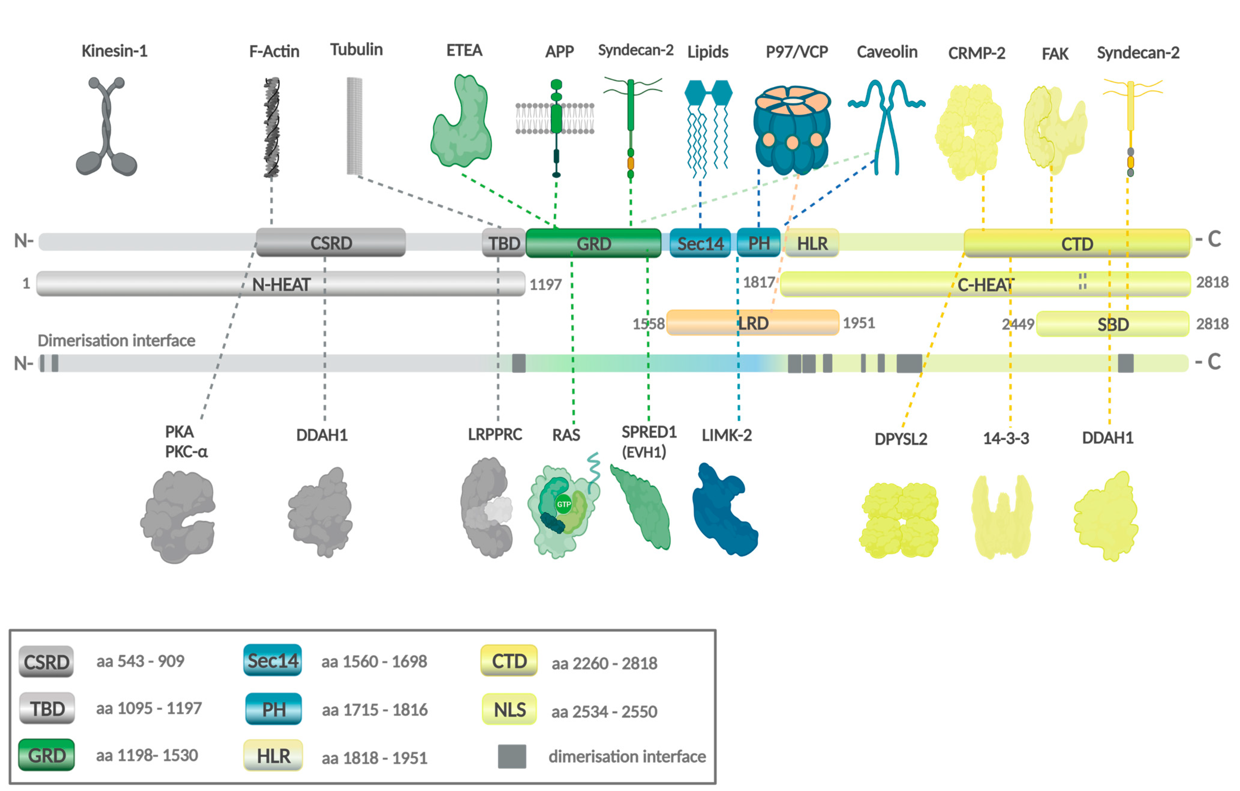
Structural and functional analysis of neurofibromin (Protein Data Bank P213599) revealed a complex domain architecture (Figure 3) [50]. While the precise role for many domains is still not fully understood, the GRD is well characterized. GRD promotes the hydrolysis of active Ras-GTP to the biologically inactive form of Ras-GDP [56], thereby negatively regulating the Ras/mitogen-activated protein kinase (Ras/MAPK) pathway. Interestingly, neurofibromin forms a high-affinity homodimer [57]. Mutant variants may dimerize with functional wild-type neurofibromin. A dysfunctional complex might be a target for proteasomal degradation and inhibit tumour-suppressor activity. Whether this plays a role in disease development or correlates with NF1 phenotypes remains to be investigated.
Upstream of neurofibromin, mainly transmembrane receptor tyrosine kinases (RTKs) regulate extracellular ligand binding and transduce signals into the cells. They regulate signalling cascades such as the RAS/ERK pathway and therefore interfere with neurofibromin. Therapies using RTK inhibitors may fail when NF1 mutations abrogate the effect on the cascade. Interestingly, Anaplastic Lymphoma Kinase (Alk) was shown to co-localise and interact with neurofibromin in Drosophila and was demonstrated to activate neurofibromin-regulated RAS signalling in the nervous system [70]. A direct interacting partner of neurofibromin is also the membrane-bound late endosomal/lysosomal adaptor and MAPK and mTOR1 activator (LAMTOR), which is a negative regulator of the mTOR pathway [71,72]. Although other interacting partners are very important, such as Sprouty-related and EVH1 domain-containing protein 1 (SPRED1), which recruits neurofibromin from the cytosol to facilitate the transport to the plasma membrane, they will not be discussed in detail here.

3. The Neurofibromatosis Type 1 (NF1) Gene in Neoplasia

Due to its large size and complexity, NF1 is one of the most frequently mutated genes in men and in cancers [73]. The Human Gene Mutation Database (HGMD® Professional 2021.2) currently lists 3084 NF1 germline mutations, and TCGA reports 1110 somatic mutations. The majority of these mutations lead to truncated neurofibromin, and about 30% of mutations lead to altered splicing [74]. Despite the high number of mutations, there are few mutation hotspots. NF1 patients with gliomas do not show the involvement of specific NF1 gene regions [15]. In fact, very few genotype–phenotype correlations exist, except for ahigher and more aggressive tumour load in patients with microdeletions [75].
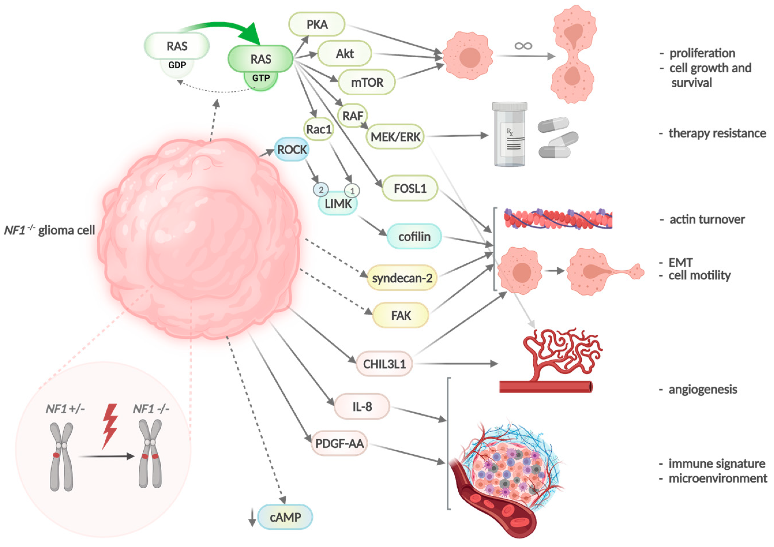
Neurofibromin regulates cell growth and survival through several downstream signalling effectors such as Ak strain transforming/protein kinase B (Akt), mammalian target of rapamycin (mTor), and protein kinase A (PKA) by accelerating the conversion of Ras hydrolysis via the catalytic central GRD [50,69]. Some of the Ras-induced proteins are involved in EMT and have been shown to be increasingly expressed in NF1-deficient malignant peripheral nerve sheath tumours (MPNST) [76,77]. Neurofibromin deficiency promotes not only EMT but also resistance to inhibitors along the MAPK pathway [78,79]. Therefore, in cancer, NF1 mutations act not only as drivers but contribute to therapy resistance [69]. B rat fibrosarcoma (BRAF) mutations, upregulation of EGFR, or activation of mitogen-activated protein kinase (MEK) are associated with resistance as reported for melanoma, neuroblastoma, lung cancer, and other lesions [80,81,82,83]. Loss of NF1 also activates cell motility by negative regulation of the Rho/Rho-associated coiled-coil-containing protein kinase (ROCK)/LIM domain kinase (LIMK), cofilin pathway, which induces the dynamic reorganisation and turnover of actin filaments [77,84]. Consequences of neurofibromin deficiency in tumours are schematically summarised in Figure 4.
Patients with autosomal dominantly inherited NF1 are prone to develop benign peripheral nerve tumours known as neurofibromas, which is the hallmark of the disease. Cutaneous neurofibromas arise due to mutations of both copies of the NF1 tumour suppressor gene in Schwann cells (biallelic inactivation). It is important to point out that only the Schwann cells are NF1 -/-, while other components within the microenvironment are NF1 +/- [85]. The development of NF1 and the subsequent reprogramming of Schwann cells have been extensively reviewed and are not the focus of this review [75,86,87,88,89,90,91]. Other NF1-associated tumours comprise plexiform neurofibromas (30–50%), optic pathway gliomas (15–20%), MPNST (10–15%), and others [73,92]. Recently, diagnostic criteria have been updated [93]. The mutational spectrum includes missense or nonsense mutations (33%), small deletions (26%), splicing substitutions (15%), small insertions/duplications (11%), and gross deletions (over 20 bp, 11%). About half of all NF1 patients display new mutations. In principle, NF1-associated benign lesions in NF1 patients acquire a somatic NF1 loss of heterozygosity (LOH) to be initiated, which accompanies the germline NF1 mutation. For the development of pre-malignant and malignant lesions, additional genetic hits are necessary. This genetically defined increased risk of NF1 patients to develop malignancies from their benign lesions still reduces life expectancy in NF1 [75].
In contrast, lesions that are independent of NF1 can develop when somatic NF1 loss of heterozygosity occurs. These lesions include not only GBM but breast cancer, uterine cancer, and melanoma, among others [94,95]. In these cancer types, NF1 is co-mutated with other tumour-suppressor genes such as p53, PTEN, and BRAF, and others. The frequency of mutations and copy number variation loss events of NF1 in different tumour entities is variable (Table 1). Attempts have been made to delineate the genotype–phenotype correlation of these mutations.

4. The Neurofibromatosis Type 1 (NF1) Gene in GBM

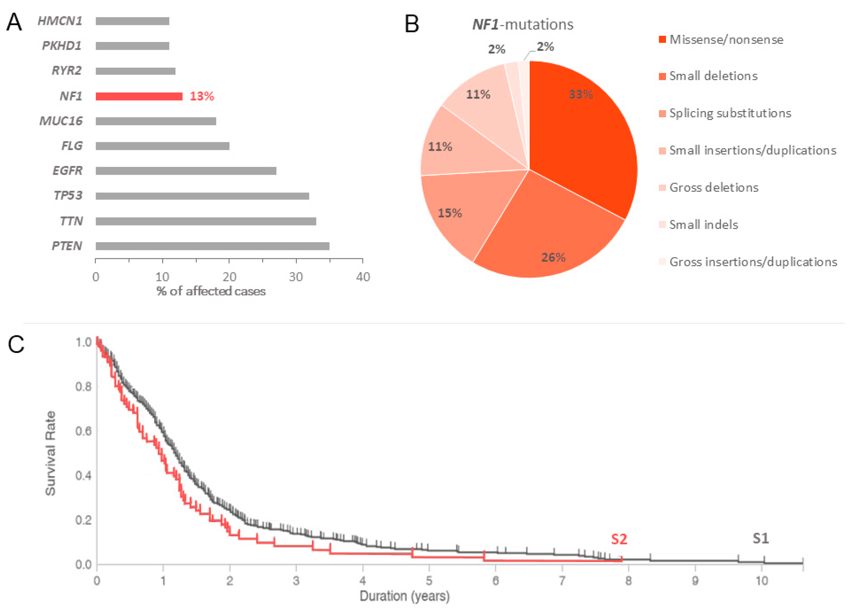
NF1 is mutated in approximately 13–14% of GBM patients according to the TCGA PanCancer Atlas GBM database [94,95]. Most (78%) of the pathogenic variants are generated by frameshifts, single nucleotide polymorphisms (SNP), or splice variants, resulting in truncation of the full-length neurofibromin and nonsense-mediated ribonucleic acid (RNA) decay. Although GBM shows an increased incidence of NF1 mutation, PTEN (35%), TTN (33%), TP53 (32%), EGFR (27%), FLG (20%), and MUC16 (18%) display higher mutation rates (Figure 5A). Patients with NF1-mutated GBM have a lower overall survival than those patients without (Figure 5C). Interestingly, 53% of the mesenchymal GBM subtype are NF1 mutated [15]. In an Nf1+/− mouse model, loss of NF1 function was shown to increase astrocyte proliferation [43]. In astrocytes, loss of neurofibromin causes the selective hyperactivation of KRAS rather than HRAS [44].

5. Mesenchymal Glioblastomas Accumulate Neurofibromatosis Type 1 (NF1) Gene Alterations

The MES molecular subtype accounts for approximately 35% of all adult high-grade gliomas [18,21,23] and is primarily characterised by the loss or deregulated expression of NF1 [19,30,96,97]. The MES subtype is also associated with mutations of TP53 and RB1 [22], enhanced activity of the tumour necrosis factor superfamily (TNF), and nuclear factor kappa-light-chain-enhancer of activated B cells (NF-κB) pathways with co-mutation of PTEN [18,20,21,94,95]. The upregulation of these genes is accompanied by higher overall necrosis and inflammatory infiltrates.
It is known that NF1 mutations correlate with high levels of leukocytes in different tumour types [98]. NF1 mutations lead to altered levels of cytokines, mast cells, macrophages, microglia, T and B cells, and they both directly affect immune cells and indirectly affect interactions between different NF1-mutated cells important for the tumour microenvironment [99,100,101]. In NF1-associated neurofibromas and MPNST, which are derived from peripheral glia, up to 30% of cells are macrophages. This finding led to the current hypothesis of neurofibroma formation in NF1: tumour initiation due to NF1 loss is followed by macrophage and mast cell recruitment, which is then followed by the recruitment of T and dendritic cells to enable tumour formation [102]. Half of NF1-associated low-grade gliomas were detected to harbour an immune signature, infiltrates of T cells, and increased neoantigens [15]. Therefore, the role of NF1 loss for microenvironment and tumour formation may well be adapted to the central nervous system-derived malignant glial tumours, the GBM, although the literature is sparse [99]. In a recent animal model, midkine being produced by NF1 mutant neurons activates T lymphocytes and maintains glioma growth [103].
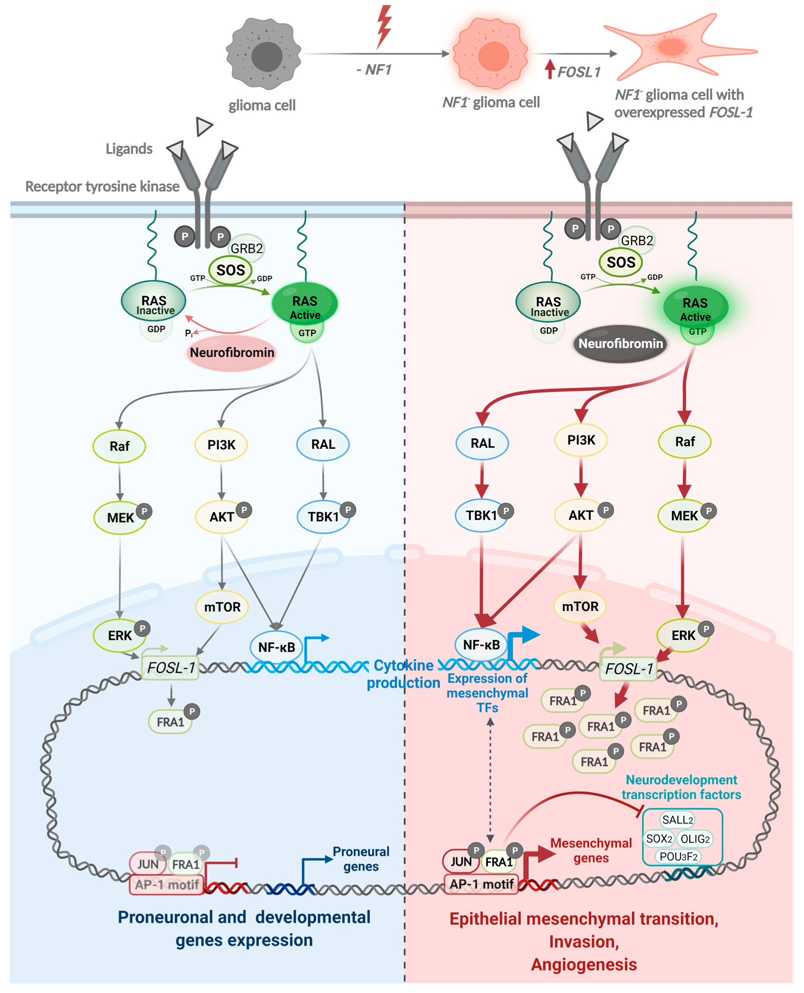
NF1-related tumours are associated with the abnormal secretion of chemokines such as C-C motif ligand (CCL) 15, CCL 2, and macrophage colony-stimulating factor (M-CSF), leading to an increase in tumour-associated macrophages (TAM) and microglia [99,104]. Especially in GBM, loss of neurofibromin is clearly associated with the attraction of macrophages (tumour-associated macrophages, TAM) or microglia [19]. Immunotherapy strategies targeting TAM have certain potential but have only been studied in mouse models and small clinical trials. CCL antibodies or M-CSF receptor inhibitors reduced glioma cell invasion and resulted in longer overall survival in glioblastoma mouse models [105,106,107]. In addition, the activation of immune cell response with immune checkpoint inhibitors and cytokine therapy (IL-2, IFN-ß) leads to prolonged patient survival [108,109,110,111,112]. Thus, numerous ongoing clinical trials are investigating the effect of PD-1/PD-L1 antibodies in glioma. Moreover, immunotherapies seem to be not only a promising strategy for mesenchymal gliomas, but they are also an important treatment option for NF1-related melanomas, lung carcinomas, or MPNST. Recently, loss of NF1 was shown to modulate FOS like 1, AP-1 transcription factor subunit (FOSL1) expression, which is a key regulator for stemness, mesenchymal shift, and plasticity [113]. Transcription factor FOSL1 is overexpressed in cancer and associated with worse outcomes and EMT as well as with glioma malignancy [113]. The authors demonstrated that FOSL1 depletion in NF1 mutant human brain tumour stem cells and KRAS mutant mouse neural stem cells resulted in the loss of the MES signature and a reduction in stem cell properties. They first proved that NF1 mutations act via the NF1–MAPK–FOSL1 axis in MES gliomas as they increase FOSL1 RNA and protein expression and therefore activate the expression of the MES gene signature and inhibit the non-MES gene signature [113].
The important role of NF1 to regulate FOSL1 expression explains the proneural to mesenchymal transition in tumours that acquire NF1 mutations such as MES GBM (Figure 6). This interaction otherwise hints to novel therapeutics against the FOSL1 axis, the immune system, and combined approaches against several cellular components.

6. NF1 Mutations and Glioma Invasiveness

Neurofibromin regulates the dynamic reorganisation and turnover of actin filaments through its interacting partners such as Ras-related C3 botulinum toxic substrate 1 (Rac1) [114,115], Lim kinases (LIMK1/2) [77,84,114,116], syndecan-2 [117], and focal adhesion kinase (FAK) [118,119,120] among others. Neurofibromin binding to syndecan-2 induces actin polymerisation and filopodia formation in dendrites [117]. Along the same line, neurofibromin interaction with FAK regulates cell migration [120]. Given its role as a modulator of cytoskeletal and focal adhesion as well as a negative regulator of RAS signalling, mutations, or loss of NF1 results in disruption of the extracellular matrix and induction of EMT. Indeed, we and others have shown that the deregulation of neurofibromin signalling enhanced cancer cells invasion and migration [76,100,121,122,123] with an increase in EMT markers such as vimentin and Chitinase-3-like protein 1 (CHI3L/YKL40) expression.
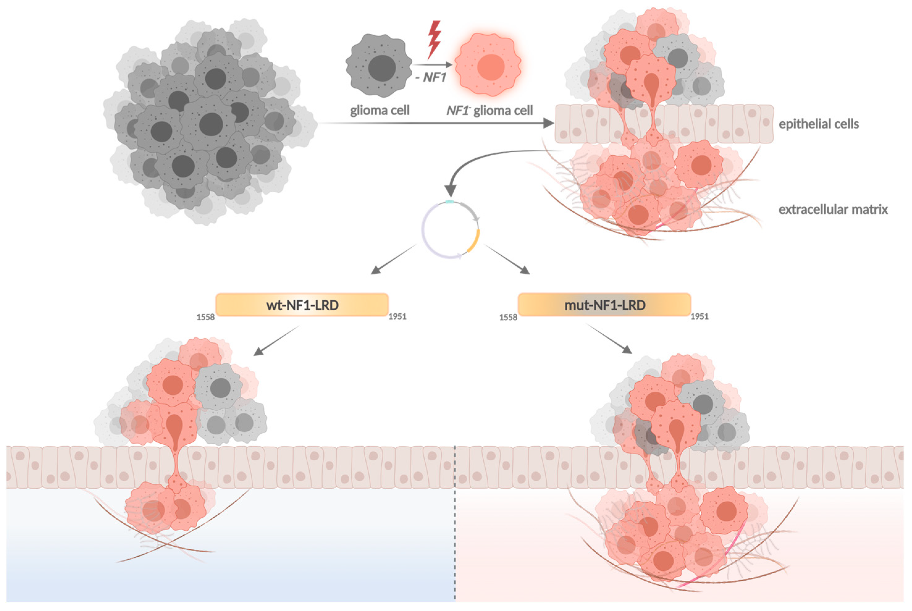
Our group recently showed that the leucine-rich domain (LRD, aa1558–1951, isoform 2) of neurofibromin, which consists of the Sec14-pleckstrin homology (PH) domain (aa 1558–1817) and part of the Heat-like repeat (HLR; aa1818–1951), inhibits NF1-loss induced cell invasion in human glioma stem cells (GSC) and orthotopic mouse glioma model independent of RAS [122] (Figure 7). Mutation screening performed on the TCGA PanCancer Atlas GBM database identified 10 mutations in the LRD of which three are located within the 1818–1951 HLR region (D1828N, W1931*, and R1947*) [94,95]. Unlike the wild-type (wt)-LRD that suppresses glioma cell invasion, the inhibitory effect is lost in both D1828N and W1931* pathogenic mutants. We further narrowed down the region critical for LRD function to a 42-aa peptide between 1818 and 1860. This 42-aa peptide suppresses glioma invasion to levels significantly lower than that of wt-LRD, suggesting a critical role of the 1818–1860 region in regulating glioma cell motility. It is not clear how this peptide mediates its function. The peptide may interact with protein(s) that regulate cell motility and ECM remodelling, since one of the roles of the HLR is protein–protein interaction. This hypothesis is consistent with Welti’s and Scheffzek’s findings that the Sec14-PH domain of LRD interacts with phospholipids for membrane localisation [68,124]. The D1828X and W1931X mutations are detected in patients with cutaneous melanoma, colon carcinoma, diffuse large B cells lymphoma [125], and infiltrative breast carcinoma (cBioportal TCGA database, Tumour suppressor gene database, NCBI dbSNP, ClinVar, and Human Proteome Variation Database). Additionally, the mutation W1931X nonsense variant has been previously reported to be associated with NF1 [126,127,128].
Other domains involved in cell invasion and migration include the GRD, Sec14-PH domain, pre-GRD NF11-1163, and the CTD. Both GRD and Sec14-PH domains mediate cell migration and invasion through LIMK2, which is a kinase in the Rho/ROCK/LIMK2/cofilin pathway. The overexpression of GRD has been shown to alter cellular morphology to inhibit cell invasion via LIMK2 dephosphorylation of cofilin [84]. Similarly, the interaction between LIMK2 and the Sec14-PH domain prevents the activation of LIMK2 by ROCK due to steric hindrance, thus resulting in actin depolymerisation via cofilin [77]. Interestingly, the Sec14-PH domain interacts with LIMK2 exclusively and does not bind to LIMK1. By contrast, the pre-GRD NF11-1163 domain does not bind to ROCK and Ras. Rather, it negatively regulates the Rac1/p21 Rac-activated kinase (Pak)1/LIMK1/cofilin pathway [114]. By inducing the depolymerisation of cofilin, the NF11-1163 that contains the cysteine-serine-rich domain (CSRD) inhibits cell migration and invasion. Neurofibromin also binds to the N-terminal of FAK [118] and syndecan-2 via the CTD domain. Whether mutations in these domains will affect the depolymerisation of cofilin is unknown; hence, cell invasion is unsolved. Since most of the alterations generate truncation mutants, it is conceivable that mutations observed in glioma patients will most likely abolish the interaction between the neurofibromin domains and their substrates and destabilise the actin filament organisation, thus affecting cell invasion. Of note, the NF11-1163 region is highly conserved. Mutations in this domain are found in a higher proportion of NF1 patients with optic pathway glioma [129,130]. Protein kinase C (PKC)-α phosphorylation on the serine residues within CSRD induces the association of neurofibromin with the actin cytoskeleton [60]. Thus, a mutation in the CSRD may affect the actin reorganisation.
It is important to note that although mutations identified from the cBioportal database may help to dissect the functional significance of the neurofibromin domains in GBM, some of these mutations are different from the mutations observed in NF1 patients since neurofibromin is a macromolecule without any mutational hotspot. In addition, most studies were done using specific neurofibromin domains in the absence of the entire NF1 gene; thus, they may not offer sufficient power to detect potential genotype–phenotype correlations.

7. Conclusions

Despite advances in surgery and molecular therapeutics, the prognosis for patients with GBM remains dismal. The highly infiltrative and heterogenous nature of the tumour is rendering standard therapeutic strategies ineffective. NF1 is one of the driver genes for MES GBM. In this review, we discussed the molecular characteristics of MES GBM, NF1 gene mutation, and dysregulation in NF1-associated and non-NF1 associated cancers, particularly GBM. However, many questions remain unanswered. MES GBM gene expression is influenced by dysregulated neurofibromin signalling and the tumour microenvironment [131]. In NF1-null or silenced MES GBM, the microenvironment is heterogenous with a hypoxic core and perivascular niche, each secreting different cytokines and chemokines that drive tumour malignancy. Given the complexity of the bi-directional interaction, the design of therapeutics must take into consideration the dynamic crosstalk among the various players such as glioma cells, immune cells (immunosuppressive versus pro-inflammatory), and endothelial cells, among others. Macrophages and microglia cells secrete factors that promote tumour growth. Are we able to re-educate these cells in the NF1-null microenvironment to achieve the anti-tumour function? Studies conducted by Pyonteck et al. using a brain-penetrant inhibitor of colony-stimulating factor 1 receptor (CSF-1R) showed a significant decrease in pro-tumourigenic tumour-associated macrophages [106], suggesting that blocking CSF-1R signalling may re-educate the immunosuppressive macrophage to pro-inflammatory cells. Another CSF-1R tyrosine kinase inhibitor, PLX3397, prevented the differentiation of monocytes into immunosuppressive macrophages [132]. Unfortunately, PLX3397 was ineffective in a phase II trial in treating recurrent GBM [107]. Thus, understanding the intricate relationship between these cells and their associated gene expression changes may help develop more effective immunotherapeutics. Given that GBM subtypes are not static, it is evident that multiprong therapy may afford a better therapeutic outcome. Previous publications have shown that CEBP-β, STAT3, NF-kB, and FOSL2 are some of the transcription factors (TFs) that play a role in NF1-loss-associated MES transition [133]. Among these TFs, STAT3, and CEBP-β have been shown to associate with the hypoxic microenvironment [29,134], which is enriched with immunosuppressive tumour-associated macrophages [135]. Gabrusiewicz et al. showed that GBM-derived exosomes triggered the release of STAT3 in monocytes and led to the upregulation of programmed death-ligand 1 (PD-L1) and a shift to the immunosuppressive phenotype [136]. Several STAT3 inhibitors are currently in clinical trials. These inhibitors were designed to be used concurrently with conventional radiation (NCT03514069) and chemotherapy (NCT02315534). Other inhibitors that target the molecules in the STAT3 pathway, such as JAK1/JAK2, are also being evaluated in phase I trial for patients with newly diagnosed GBM (NCT03514069). While we await the results from these trials, identifying other NF1-loss associated master regulators and their inhibitors may improve the treatment options for patients with MES GBM.

Author Contributions

Conceptualisation and supervision: A.H. and I.H. Writing and draft preparation: M.S., S.L., E.S., O.S., J.P., I.H. and A.H. O.S. revised figures using BioRender (BioRender.com), as she is holding a licence. All authors have read and agreed to the published version of the manuscript.

Funding

Publication fee of the review was funded by the Medical Faculty of the Martin Luther University Halle-Wittenberg.

Institutional Review Board Statement

Not applicable.

Informed Consent Statement

Not applicable.

Conflicts of Interest

The authors declare no conflict of interest.

References

  1. Ostrom, Q.T.; Gittleman, H.; Truitt, G.; Boscia, A.; Kruchko, C.; Barnholtz-Sloan, J.S. CBTRUS Statistical Report: Primary Brain and Other Central Nervous System Tumors Diagnosed in the United States in 2011–2015. Neuro-Oncollogy 2018, 20, iv1–iv86. [Google Scholar] [CrossRef] [Green Version]
  2. Wesseling, P.; Capper, D. WHO 2016 Classification of gliomas. Neuropathol. Appl. Neurobiol. 2018, 44, 139–150. [Google Scholar] [CrossRef] [PubMed]
  3. Delgado-López, P.D.; Corrales-García, E.M. Survival in glioblastoma: A review on the impact of treatment modalities. Clin. Transl. Oncol. 2016, 18, 1062–1071. [Google Scholar] [CrossRef] [PubMed]
  4. Stupp, R.; Taillibert, S.; Kanner, A.; Read, W.; Steinberg, D.; Lhermitte, B.; Toms, S.; Idbaih, A.; Ahluwalia, M.S.; Fink, K.; et al. Effect of Tumor-Treating Fields Plus Maintenance Temozolomide vs Maintenance Temozolomide Alone on Survival in Patients With Glioblastoma: A Randomized Clinical Trial. JAMA 2017, 318, 2306–2316. [Google Scholar] [CrossRef] [Green Version]
  5. Wirsching, H.G.; Galanis, E.; Weller, M. Glioblastoma. Handb. Clin. Neurol. 2016, 134, 381–397. [Google Scholar] [CrossRef] [PubMed]
  6. Stupp, R.; Mason, W.P.; van den Bent, M.J.; Weller, M.; Fisher, B.; Taphoorn, M.J.; Belanger, K.; Brandes, A.A.; Marosi, C.; Bogdahn, U.; et al. Radiotherapy plus concomitant and adjuvant temozolomide for glioblastoma. N. Engl. J. Med. 2005, 352, 987–996. [Google Scholar] [CrossRef] [PubMed]
  7. Lukas, R.V.; Wainwright, D.A.; Ladomersky, E.; Sachdev, S.; Sonabend, A.M.; Stupp, R. Newly Diagnosed Glioblastoma: A Review on Clinical Management. Oncology 2019, 33, 91–100. [Google Scholar] [PubMed]
  8. Brown, T.J.; Brennan, M.C.; Li, M.; Church, E.W.; Brandmeir, N.J.; Rakszawski, K.L.; Patel, A.S.; Rizk, E.B.; Suki, D.; Sawaya, R.; et al. Association of the Extent of Resection With Survival in Glioblastoma: A Systematic Review and Meta-analysis. JAMA Oncol. 2016, 2, 1460–1469. [Google Scholar] [CrossRef] [PubMed] [Green Version]
  9. Bette, S.; Barz, M.; Wiestler, B.; Huber, T.; Gerhardt, J.; Buchmann, N.; Combs, S.E.; Schmidt-Graf, F.; Delbridge, C.; Zimmer, C.; et al. Prognostic Value of Tumor Volume in Glioblastoma Patients: Size Also Matters for Patients with Incomplete Resection. Ann. Surg. Oncol. 2018, 25, 558–564. [Google Scholar] [CrossRef]
  10. Herrlinger, U.; Tzaridis, T.; Mack, F.; Steinbach, J.P.; Schlegel, U.; Sabel, M.; Hau, P.; Kortmann, R.D.; Krex, D.; Grauer, O.; et al. Lomustine-temozolomide combination therapy versus standard temozolomide therapy in patients with newly diagnosed glioblastoma with methylated MGMT promoter (CeTeG/NOA-09): A randomised, open-label, phase 3 trial. Lancet 2019, 393, 678–688. [Google Scholar] [CrossRef]
  11. Perry, A.; Wesseling, P. Histologic classification of gliomas. Handb. Clin. Neurol. 2016, 134, 71–95. [Google Scholar] [CrossRef] [PubMed]
  12. Louis, D.N.; Perry, A.; Burger, P.; Ellison, D.W.; Reifenberger, G.; von Deimling, A.; Aldape, K.; Brat, D.; Collins, V.P.; Eberhart, C.; et al. International Society Of Neuropathology-Haarlem consensus guidelines for nervous system tumor classification and grading. Brain Pathol. 2014, 24, 429–435. [Google Scholar] [CrossRef] [PubMed]
  13. Capper, D.; Jones, D.T.W.; Sill, M.; Hovestadt, V.; Schrimpf, D.; Sturm, D.; Koelsche, C.; Sahm, F.; Chavez, L.; Reuss, D.E.; et al. DNA methylation-based classification of central nervous system tumours. Nature 2018, 555, 469–474. [Google Scholar] [CrossRef] [PubMed]
  14. Ceccarelli, M.; Barthel, F.P.; Malta, T.M.; Sabedot, T.S.; Salama, S.R.; Murray, B.A.; Morozova, O.; Newton, Y.; Radenbaugh, A.; Pagnotta, S.M.; et al. Molecular Profiling Reveals Biologically Discrete Subsets and Pathways of Progression in Diffuse Glioma. Cell 2016, 164, 550–563. [Google Scholar] [CrossRef] [PubMed] [Green Version]
  15. D’Angelo, F.; Ceccarelli, M.; Tala; Garofano, L.; Zhang, J.; Frattini, V.; Caruso, F.P.; Lewis, G.; Alfaro, K.D.; Bauchet, L.; et al. The molecular landscape of glioma in patients with Neurofibromatosis 1. Nat. Med. 2019, 25, 176–187. [Google Scholar] [CrossRef]
  16. Brat, D.J.; Aldape, K.; Colman, H.; Holland, E.C.; Louis, D.N.; Jenkins, R.B.; Kleinschmidt-DeMasters, B.K.; Perry, A.; Reifenberger, G.; Stupp, R.; et al. cIMPACT-NOW update 3: Recommended diagnostic criteria for “Diffuse astrocytic glioma, IDH-wildtype, with molecular features of glioblastoma, WHO grade IV”. Acta Neuropathol. 2018, 136, 805–810. [Google Scholar] [CrossRef] [Green Version]
  17. Louis, D.N.; Wesseling, P.; Aldape, K.; Brat, D.J.; Capper, D.; Cree, I.A.; Eberhart, C.; Figarella-Branger, D.; Fouladi, M.; Fuller, G.N.; et al. cIMPACT-NOW update 6: New entity and diagnostic principle recommendations of the cIMPACT-Utrecht meeting on future CNS tumor classification and grading. Brain Pathol. 2020, 30, 844–856. [Google Scholar] [CrossRef] [PubMed]
  18. Verhaak, R.G.; Hoadley, K.A.; Purdom, E.; Wang, V.; Qi, Y.; Wilkerson, M.D.; Miller, C.R.; Ding, L.; Golub, T.; Mesirov, J.P.; et al. Integrated genomic analysis identifies clinically relevant subtypes of glioblastoma characterized by abnormalities in PDGFRA, IDH1, EGFR, and NF1. Cancer Cell 2010, 17, 98–110. [Google Scholar] [CrossRef] [PubMed] [Green Version]
  19. Wang, Q.; Hu, B.; Hu, X.; Kim, H.; Squatrito, M.; Scarpace, L.; deCarvalho, A.C.; Lyu, S.; Li, P.; Li, Y.; et al. Tumor Evolution of Glioma-Intrinsic Gene Expression Subtypes Associates with Immunological Changes in the Microenvironment. Cancer Cell 2017, 32, 42–56.e46. [Google Scholar] [CrossRef] [Green Version]
  20. Chen, R.; Smith-Cohn, M.; Cohen, A.L.; Colman, H. Glioma Subclassifications and Their Clinical Significance. Neurotherapeutics 2017, 14, 284–297. [Google Scholar] [CrossRef] [Green Version]
  21. Phillips, H.S.; Kharbanda, S.; Chen, R.; Forrest, W.F.; Soriano, R.H.; Wu, T.D.; Misra, A.; Nigro, J.M.; Colman, H.; Soroceanu, L.; et al. Molecular subclasses of high-grade glioma predict prognosis, delineate a pattern of disease progression, and resemble stages in neurogenesis. Cancer Cell 2006, 9, 157–173. [Google Scholar] [CrossRef] [PubMed] [Green Version]
  22. Abedalthagafi, M.; Barakeh, D.; Foshay, K.M. Immunogenetics of glioblastoma: The future of personalized patient management. NPJ Precis. Oncol. 2018, 2, 27. [Google Scholar] [CrossRef] [PubMed]
  23. Sturm, D.; Witt, H.; Hovestadt, V.; Khuong-Quang, D.A.; Jones, D.T.; Konermann, C.; Pfaff, E.; Tonjes, M.; Sill, M.; Bender, S.; et al. Hotspot mutations in H3F3A and IDH1 define distinct epigenetic and biological subgroups of glioblastoma. Cancer Cell 2012, 22, 425–437. [Google Scholar] [CrossRef] [PubMed] [Green Version]
  24. Wu, Y.; Fletcher, M.; Gu, Z.; Wang, Q.; Costa, B.; Bertoni, A.; Man, K.H.; Schlotter, M.; Felsberg, J.; Mangei, J.; et al. Glioblastoma epigenome profiling identifies SOX10 as a master regulator of molecular tumour subtype. Nat. Commun. 2020, 11, 6434. [Google Scholar] [CrossRef] [PubMed]
  25. Ozawa, T.; Riester, M.; Cheng, Y.K.; Huse, J.T.; Squatrito, M.; Helmy, K.; Charles, N.; Michor, F.; Holland, E.C. Most human non-GCIMP glioblastoma subtypes evolve from a common proneural-like precursor glioma. Cancer Cell 2014, 26, 288–300. [Google Scholar] [CrossRef] [PubMed] [Green Version]
  26. Fedele, M.; Cerchia, L.; Pegoraro, S.; Sgarra, R.; Manfioletti, G. Proneural-Mesenchymal Transition: Phenotypic Plasticity to Acquire Multitherapy Resistance in Glioblastoma. Int. J. Mol. Sci. 2019, 20, 2746. [Google Scholar] [CrossRef] [Green Version]
  27. Segerman, A.; Niklasson, M.; Haglund, C.; Bergström, T.; Jarvius, M.; Xie, Y.; Westermark, A.; Sönmez, D.; Hermansson, A.; Kastemar, M.; et al. Clonal Variation in Drug and Radiation Response among Glioma-Initiating Cells Is Linked to Proneural-Mesenchymal Transition. Cell Rep. 2016, 17, 2994–3009. [Google Scholar] [CrossRef] [Green Version]
  28. Patel, A.P.; Tirosh, I.; Trombetta, J.J.; Shalek, A.K.; Gillespie, S.M.; Wakimoto, H.; Cahill, D.P.; Nahed, B.V.; Curry, W.T.; Martuza, R.L.; et al. Single-cell RNA-seq highlights intratumoral heterogeneity in primary glioblastoma. Science 2014, 344, 1396–1401. [Google Scholar] [CrossRef] [Green Version]
  29. Behnan, J.; Finocchiaro, G.; Hanna, G. The landscape of the mesenchymal signature in brain tumours. Brain 2019, 142, 847–866. [Google Scholar] [CrossRef] [Green Version]
  30. Bhat, K.P.L.; Balasubramaniyan, V.; Vaillant, B.; Ezhilarasan, R.; Hummelink, K.; Hollingsworth, F.; Wani, K.; Heathcock, L.; James, J.D.; Goodman, L.D.; et al. Mesenchymal differentiation mediated by NF-kappaB promotes radiation resistance in glioblastoma. Cancer Cell 2013, 24, 331–346. [Google Scholar] [CrossRef] [Green Version]
  31. Rutledge, W.C.; Kong, J.; Gao, J.; Gutman, D.A.; Cooper, L.A.; Appin, C.; Park, Y.; Scarpace, L.; Mikkelsen, T.; Cohen, M.L.; et al. Tumor-infiltrating lymphocytes in glioblastoma are associated with specific genomic alterations and related to transcriptional class. Clin. Cancer Res. 2013, 19, 4951–4960. [Google Scholar] [CrossRef] [Green Version]
  32. Barker, D.; Wright, E.; Nguyen, K.; Cannon, L.; Fain, P.; Goldgar, D.; Bishop, D.T.; Carey, J.; Baty, B.; Kivlin, J.; et al. Gene for von Recklinghausen neurofibromatosis is in the pericentromeric region of chromosome 17. Science 1987, 236, 1100–1102. [Google Scholar] [CrossRef] [Green Version]
  33. Wallace, M.R.; Andersen, L.B.; Saulino, A.M.; Gregory, P.E.; Glover, T.W.; Collins, F.S. A de novo Alu insertion results in neurofibromatosis type 1. Nature 1991, 353, 864–866. [Google Scholar] [CrossRef] [PubMed]
  34. Viskochil, D.; Buchberg, A.M.; Xu, G.; Cawthon, R.M.; Stevens, J.; Wolff, R.K.; Culver, M.; Carey, J.C.; Copeland, N.G.; Jenkins, N.A.; et al. Deletions and a translocation interrupt a cloned gene at the neurofibromatosis type 1 locus. Cell 1990, 62, 187–192. [Google Scholar] [CrossRef]
  35. Luijten, M.; Wang, Y.; Smith, B.T.; Westerveld, A.; Smink, L.J.; Dunham, I.; Roe, B.A.; Hulsebos, T.J. Mechanism of spreading of the highly related neurofibromatosis type 1 (NF1) pseudogenes on chromosomes 2, 14 and 22. Eur. J. Hum. Genet. 2000, 8, 209–214. [Google Scholar] [CrossRef] [PubMed] [Green Version]
  36. Martín, Y.; Dopazo, A.; Hernández-Chico, C. Progress and challenges in developing a molecular diagnostic test for neurofibromatosis type 1. Expert Rev. Mol. Diagn. 2011, 11, 671–673. [Google Scholar] [CrossRef]
  37. Nishi, T.; Lee, P.S.; Oka, K.; Levin, V.A.; Tanase, S.; Morino, Y.; Saya, H. Differential expression of two types of the neurofibromatosis type 1 (NF1) gene transcripts related to neuronal differentiation. Oncogene 1991, 6, 1555–1559. [Google Scholar] [PubMed]
  38. Gutman, D.H.; Andersen, L.B.; Cole, J.L.; Swaroop, M.; Collins, F.S. An alternatively-spliced mRNA in the carboxy terminus of the neurofibromatosis type 1 (NF1) gene is expressed in muscle. Hum. Mol. Genet. 1993, 2, 989–992. [Google Scholar] [CrossRef] [PubMed]
  39. Danglot, G.; Regnier, V.; Fauvet, D.; Vassal, G.; Kujas, M.; Bernheim, A. Neurofibromatosis 1 (NF1) mRNAs expressed in the central nervous system are differentially spliced in the 5’ part of the gene. Hum. Mol. Genet. 1995, 4, 915–920. [Google Scholar] [CrossRef]
  40. Kaufmann, D.; Muller, R.; Kenner, O.; Leistner, W.; Hein, C.; Vogel, W.; Bartelt, B. The N-terminal splice product NF1-10a-2 of the NF1 gene codes for a transmembrane segment. Biochem. Biophys. Res. Commun. 2002, 294, 496–503. [Google Scholar] [CrossRef]
  41. Fagerberg, L.; Hallström, B.M.; Oksvold, P.; Kampf, C.; Djureinovic, D.; Odeberg, J.; Habuka, M.; Tahmasebpoor, S.; Danielsson, A.; Edlund, K.; et al. Analysis of the human tissue-specific expression by genome-wide integration of transcriptomics and antibody-based proteomics. Mol. Cell. Proteom. MCP 2014, 13, 397–406. [Google Scholar] [CrossRef] [PubMed] [Green Version]
  42. Andersen, L.B.; Ballester, R.; Marchuk, D.A.; Chang, E.; Gutmann, D.H.; Saulino, A.M.; Camonis, J.; Wigler, M.; Collins, F.S. A conserved alternative splice in the von Recklinghausen neurofibromatosis (NF1) gene produces two neurofibromin isoforms, both of which have GTPase-activating protein activity. Mol. Cell. Biol. 1993, 13, 487–495. [Google Scholar] [PubMed] [Green Version]
  43. Anastasaki, C.; Le, L.Q.; Kesterson, R.A.; Gutmann, D.H. Updated nomenclature for human and mouse neurofibromatosis type 1 genes. Neurol. Genet. 2017, 3, e169. [Google Scholar] [CrossRef] [Green Version]
  44. Messiaen, L.M. Molecular Diagnosis for NF1. In Multidisciplinary Approach to Neurofibromatosis Type 1; Tadini, G., Legius, E., Brems, H., Eds.; Springer International Publishing: Cham, Switzerland, 2020; pp. 15–34. [Google Scholar]
  45. Li, S.; Janosch, P.; Tanji, M.; Rosenfeld, G.C.; Waymire, J.C.; Mischak, H.; Kolch, W.; Sedivy, J.M. Regulation of Raf-1 kinase activity by the 14-3-3 family of proteins. EMBO J. 1995, 14, 685–696. [Google Scholar] [CrossRef] [PubMed]
  46. Hinman, M.N.; Sharma, A.; Luo, G.; Lou, H. Neurofibromatosis type 1 alternative splicing is a key regulator of Ras signaling in neurons. Mol. Cell. Biol. 2014, 34, 2188–2197. [Google Scholar] [CrossRef] [PubMed] [Green Version]
  47. Danglot, G.; Teinturier, C.; Duverger, A.; Bernheim, A. Tissue-specific alternative splicing of neurofibromatosis 1 (NF1) mRNA. Biomed. Pharmacother. 1994, 48, 365–372. [Google Scholar] [CrossRef]
  48. Vandenbroucke, I.; Vandesompele, J.; De Paepe, A.; Messiaen, L. Quantification of NF1 transcripts reveals novel highly expressed splice variants. FEBS Lett. 2002, 522, 71–76. [Google Scholar] [CrossRef] [Green Version]
  49. Gutmann, D.H.; Geist, R.T.; Rose, K.; Wright, D.E. Expression of two new protein isoforms of the neurofibromatosis type 1 gene product, neurofibromin, in muscle tissues. Dev. Dyn. 1995, 202, 302–311. [Google Scholar] [CrossRef]
  50. Bergoug, M.; Doudeau, M.; Godin, F.; Mosrin, C.; Vallée, B.; Bénédetti, H. Neurofibromin Structure, Functions and Regulation. Cells 2020, 9, 2365. [Google Scholar] [CrossRef]
  51. Gutmann, D.H.; Zhang, Y.; Hirbe, A. Developmental regulation of a neuron-specific neurofibromatosis 1 isoform. Ann. Neurol. 1999, 46, 777–782. [Google Scholar] [CrossRef]
  52. Geist, R.T.; Gutmann, D.H. Expression of a developmentally-regulated neuron-specific isoform of the neurofibromatosis 1 (NF1) gene. Neurosci. Lett. 1996, 211, 85–88. [Google Scholar] [CrossRef]
  53. Gutmann, D.H.; Cole, J.L.; Collins, F.S. Modulation of neurofibromatosis type 1 gene expression during in vitro myoblast differentiation. J. Neurosci. Res. 1994, 37, 398–405. [Google Scholar] [CrossRef] [PubMed] [Green Version]
  54. Skuse, G.R.; Cappione, A.J. RNA processing and clinical variability in neurofibromatosis type I (NF1). Hum. Mol. Genet. 1997, 6, 1707–1712. [Google Scholar] [CrossRef] [Green Version]
  55. Park, V.M.; Kenwright, K.A.; Sturtevant, D.B.; Pivnick, E.K. Alternative splicing of exons 29 and 30 in the neurofibromatosis type 1 gene. Hum. Genet. 1998, 103, 382–385. [Google Scholar] [CrossRef] [PubMed]
  56. Scheffzek, K.; Ahmadian, M.R.; Kabsch, W.; Wiesmuller, L.; Lautwein, A.; Schmitz, F.; Wittinghofer, A. The Ras-RasGAP complex: Structural basis for GTPase activation and its loss in oncogenic Ras mutants. Science 1997, 277, 333–338. [Google Scholar] [CrossRef] [Green Version]
  57. Sherekar, M.; Han, S.W.; Ghirlando, R.; Messing, S.; Drew, M.; Rabara, D.; Waybright, T.; Juneja, P.; O’Neill, H.; Stanley, C.B.; et al. Biochemical and structural analyses reveal that the tumor suppressor neurofibromin (NF1) forms a high-affinity dimer. J. Biol. Chem. 2020, 295, 1105–1119. [Google Scholar] [CrossRef]
  58. Lupton, C.J.; Bayly-Jones, C.; D’Andrea, L.; Huang, C.; Schittenhelm, R.B.; Venugopal, H.; Whisstock, J.C.; Halls, M.L.; Ellisdon, A.M. The cryo-EM structure of the neurofibromin dimer reveals the molecular basis for von Recklinghausen disease. Biorxiv 2021. [Google Scholar] [CrossRef]
  59. Tokuo, H.; Yunoue, S.; Feng, L.; Kimoto, M.; Tsuji, H.; Ono, T.; Saya, H.; Araki, N. Phosphorylation of neurofibromin by cAMP-dependent protein kinase is regulated via a cellular association of N G, N G-dimethylarginine dimethylaminohydrolase. FEBS Lett. 2001, 494, 48–53. [Google Scholar] [CrossRef] [Green Version]
  60. Mangoura, D.; Sun, Y.; Li, C.; Singh, D.; Gutmann, D.H.; Flores, A.; Ahmed, M.; Vallianatos, G. Phosphorylation of neurofibromin by PKC is a possible molecular switch in EGF receptor signaling in neural cells. Oncogene 2006, 25, 735–745. [Google Scholar] [CrossRef] [Green Version]
  61. Leondaritis, G.; Petrikkos, L.; Mangoura, D. Regulation of the Ras-GTPase activating protein neurofibromin by C-tail phosphorylation: Implications for protein kinase C/Ras/extracellular signal-regulated kinase 1/2 pathway signaling and neuronal differentiation. J. Neurochem. 2009, 109, 573–583. [Google Scholar] [CrossRef] [PubMed]
  62. Koliou, X.; Fedonidis, C.; Kalpachidou, T.; Mangoura, D. Nuclear import mechanism of neurofibromin for localization on the spindle and function in chromosome congression. J. Neurochem. 2016, 136, 78–91. [Google Scholar] [CrossRef] [PubMed]
  63. Feng, L.; Yunoue, S.; Tokuo, H.; Ozawa, T.; Zhang, D.; Patrakitkomjorn, S.; Ichimura, T.; Saya, H.; Araki, N. PKA phosphorylation and 14-3-3 interaction regulate the function of neurofibromatosis type I tumor suppressor, neurofibromin. FEBS Lett. 2004, 557, 275–282. [Google Scholar] [CrossRef] [Green Version]
  64. Gregory, P.; Gutmann, D.; Mitchell, A.; Park, S.; Boguski, M.; Jacks, T.; Wood, D.; Jove, R.; Collins, F. Neurofibromatosis type 1 gene product (neurofibromin) associates with microtubules. Somat. Cell Mol. Genet. 1993, 19, 265–274. [Google Scholar] [CrossRef] [PubMed] [Green Version]
  65. D’Angelo, I.; Welti, S.; Bonneau, F.; Scheffzek, K. A novel bipartite phospholipid-binding module in the neurofibromatosis type 1 protein. EMBO Rep. 2006, 7, 174–179. [Google Scholar] [CrossRef] [Green Version]
  66. Boyanapalli, M.; Lahoud, O.B.; Messiaen, L.; Kim, B.; Anderle de Sylor, M.S.; Duckett, S.J.; Somara, S.; Mikol, D.D. Neurofibromin binds to caveolin-1 and regulates ras, FAK, and Akt. Biochem. Biophys. Res. Commun. 2006, 340, 1200–1208. [Google Scholar] [CrossRef] [PubMed]
  67. Yang, K.; Du, J.; Shi, D.; Ji, F.; Ji, Y.; Pan, J.; Lv, F.; Zhang, Y.; Zhang, J. Knockdown of MSI2 inhibits metastasis by interacting with caveolin-1 and inhibiting its ubiquitylation in human NF1-MPNST cells. Cell Death Dis. 2020, 11, 489. [Google Scholar] [CrossRef] [PubMed]
  68. Scheffzek, K.; Welti, S. Pleckstrin homology (PH) like domains—Versatile modules in protein-protein interaction platforms. FEBS Lett. 2012, 586, 2662–2673. [Google Scholar] [CrossRef] [PubMed] [Green Version]
  69. Ratner, N.; Miller, S.J. A RASopathy gene commonly mutated in cancer: The neurofibromatosis type 1 tumour suppressor. Nat. Rev. Cancer 2015, 15, 290–301. [Google Scholar] [CrossRef]
  70. Gouzi, J.Y.; Moressis, A.; Walker, J.A.; Apostolopoulou, A.A.; Palmer, R.H.; Bernards, A.; Skoulakis, E.M. The receptor tyrosine kinase Alk controls neurofibromin functions in Drosophila growth and learning. PLoS Genet. 2011, 7, e1002281. [Google Scholar] [CrossRef]
  71. Li, X.; Gao, M.; Choi, J.M.; Kim, B.J.; Zhou, M.T.; Chen, Z.; Jain, A.N.; Jung, S.Y.; Yuan, J.; Wang, W.; et al. Clustered, Regularly Interspaced Short Palindromic Repeats (CRISPR)/Cas9-coupled Affinity Purification/Mass Spectrometry Analysis Revealed a Novel Role of Neurofibromin in mTOR Signaling. Mol. Cell. Proteomics 2017, 16, 594–607. [Google Scholar] [CrossRef] [Green Version]
  72. Nada, S.; Mori, S.; Takahashi, Y.; Okada, M. p18/LAMTOR1: A late endosome/lysosome-specific anchor protein for the mTORC1/MAPK signaling pathway. Methods Enzymol. 2014, 535, 249–263. [Google Scholar] [CrossRef] [PubMed]
  73. Philpott, C.; Tovell, H.; Frayling, I.M.; Cooper, D.N.; Upadhyaya, M. The NF1 somatic mutational landscape in sporadic human cancers. Hum. Genom. 2017, 11, 13. [Google Scholar] [CrossRef] [PubMed] [Green Version]
  74. Messiaen, L.; Callens, T.; Mortier, G.; Beysen, D.; Vandenbroucke, I.; Van Roy, N.; Speleman, F.; Paepe, A.D. Exhaustive mutation analysis of the NF1 gene allows identification of 95% of mutations and reveals a high frequency of unusual sloicng defects. Hum. Mutat. 2000, 15, 541–555. [Google Scholar] [CrossRef]
  75. Tamura, R. Current Understanding of Neurofibromatosis Type 1, 2, and Schwannomatosis. Int. J. Mol. Sci. 2021, 22, 5850. [Google Scholar] [CrossRef] [PubMed]
  76. Miller, S.J.; Rangwala, F.; Williams, J.; Ackerman, P.; Kong, S.; Jegga, A.G.; Kaiser, S.; Aronow, B.J.; Frahm, S.; Kluwe, L.; et al. Large-scale molecular comparison of human schwann cells to malignant peripheral nerve sheath tumor cell lines and tissues. Cancer Res. 2006, 66, 2584–2591. [Google Scholar] [CrossRef] [Green Version]
  77. Vallee, B.; Doudeau, M.; Godin, F.; Gombault, A.; Tchalikian, A.; de Tauzia, M.L.; Benedetti, H. Nf1 RasGAP inhibition of LIMK2 mediates a new cross-talk between Ras and Rho pathways. PLoS ONE 2012, 7, e47283. [Google Scholar] [CrossRef] [PubMed] [Green Version]
  78. Whittaker, S.R.; Theurillat, J.P.; Van Allen, E.; Wagle, N.; Hsiao, J.; Cowley, G.S.; Schadendorf, D.; Root, D.E.; Garraway, L.A. A genome-scale RNA interference screen implicates NF1 loss in resistance to RAF inhibition. Cancer Discov. 2013, 3, 350–362. [Google Scholar] [CrossRef] [PubMed] [Green Version]
  79. Nissan, M.H.; Pratilas, C.A.; Jones, A.M.; Ramirez, R.; Won, H.; Liu, C.; Tiwari, S.; Kong, L.; Hanrahan, A.J.; Yao, Z.; et al. Loss of NF1 in cutaneous melanoma is associated with RAS activation and MEK dependence. Cancer Res. 2014, 74, 2340–2350. [Google Scholar] [CrossRef] [Green Version]
  80. Maertens, O.; Johnson, B.; Hollstein, P.; Frederick, D.T.; Cooper, Z.A.; Messiaen, L.; Bronson, R.T.; McMahon, M.; Granter, S.; Flaherty, K.; et al. Elucidating distinct roles for NF1 in melanomagenesis. Cancer Discov. 2013, 3, 338–349. [Google Scholar] [CrossRef] [PubMed] [Green Version]
  81. Holzel, M.; Huang, S.; Koster, J.; Ora, I.; Lakeman, A.; Caron, H.; Nijkamp, W.; Xie, J.; Callens, T.; Asgharzadeh, S.; et al. NF1 is a tumor suppressor in neuroblastoma that determines retinoic acid response and disease outcome. Cell 2010, 142, 218–229. [Google Scholar] [CrossRef] [PubMed] [Green Version]
  82. de Bruin, E.C.; Cowell, C.; Warne, P.H.; Jiang, M.; Saunders, R.E.; Melnick, M.A.; Gettinger, S.; Walther, Z.; Wurtz, A.; Heynen, G.J.; et al. Reduced NF1 expression confers resistance to EGFR inhibition in lung cancer. Cancer Discov. 2014, 4, 606–619. [Google Scholar] [CrossRef] [Green Version]
  83. Harder, A. MEK inhibitors—Novel targeted therapies of neurofibromatosis associated benign and malignant lesions. Biomark. Res. 2021, 9, 26. [Google Scholar] [CrossRef]
  84. Ozawa, T.; Araki, N.; Yunoue, S.; Tokuo, H.; Feng, L.; Patrakitkomjorn, S.; Hara, T.; Ichikawa, Y.; Matsumoto, K.; Fujii, K.; et al. The neurofibromatosis type 1 gene product neurofibromin enhances cell motility by regulating actin filament dynamics via the Rho-ROCK-LIMK2-cofilin pathway. J. Biol. Chem. 2005, 280, 39524–39533. [Google Scholar] [CrossRef] [Green Version]
  85. Buchstaller, J.; McKeever, P.E.; Morrison, S.J. Tumorigenic cells are common in mouse MPNSTs but their frequency depends upon tumor genotype and assay conditions. Cancer Cell 2012, 21, 240–252. [Google Scholar] [CrossRef] [Green Version]
  86. Wilson, B.N.; John, A.M.; Handler, M.Z.; Schwartz, R.A. Neurofibromatosis type 1: New developments in genetics and treatment. J. Am. Acad. Dermatol. 2021, 84, 1667–1676. [Google Scholar] [CrossRef] [PubMed]
  87. Mazuelas, H.; Carrio, M.; Serra, E. Modeling tumors of the peripheral nervous system associated with Neurofibromatosis type 1: Reprogramming plexiform neurofibroma cells. Stem Cell Res. 2020, 49, 102068. [Google Scholar] [CrossRef] [PubMed]
  88. Hirbe, A.C.; Gutmann, D.H. Neurofibromatosis type 1: A multidisciplinary approach to care. Lancet Neurol. 2014, 13, 834–843. [Google Scholar] [CrossRef] [Green Version]
  89. Gutmann, D.H.; Ferner, R.E.; Listernick, R.H.; Korf, B.R.; Wolters, P.L.; Johnson, K.J. Neurofibromatosis type 1. Nat. Rev. Dis. Primers 2017, 3, 17004. [Google Scholar] [CrossRef] [PubMed]
  90. Anderson, J.L.; Gutmann, D.H. Neurofibromatosis type 1. Handb. Clin. Neurol. 2015, 132, 75–86. [Google Scholar] [CrossRef]
  91. Karaconji, T.; Whist, E.; Jamieson, R.V.; Flaherty, M.P.; Grigg, J.R.B. Neurofibromatosis Type 1: Review and Update on Emerging Therapies. Asia Pac. J. Ophthalmol. 2019, 8, 62–72. [Google Scholar] [CrossRef]
  92. Melloni, G.; Eoli, M.; Cesaretti, C.; Bianchessi, D.; Ibba, M.C.; Esposito, S.; Scuvera, G.; Morcaldi, G.; Micheli, R.; Piozzi, E.; et al. Risk of Optic Pathway Glioma in Neurofibromatosis Type 1: No Evidence of Genotype-Phenotype Correlations in A Large Independent Cohort. Cancers 2019, 11, 1838. [Google Scholar] [CrossRef] [Green Version]
  93. Legius, E.; Messiaen, L.; Wolkenstein, P.; Pancza, P.; Avery, R.A.; Berman, Y.; Blakeley, J.; Babovic-Vuksanovic, D.; Cunha, K.S.; Ferner, R.; et al. Revised diagnostic criteria for neurofibromatosis type 1 and Legius syndrome: An international consensus recommendation. Genet. Med. 2021, 23, 1506–1513. [Google Scholar] [CrossRef] [PubMed]
  94. Cerami, E.; Gao, J.; Dogrusoz, U.; Gross, B.E.; Sumer, S.O.; Aksoy, B.A.; Jacobsen, A.; Byrne, C.J.; Heuer, M.L.; Larsson, E.; et al. The cBio cancer genomics portal: An open platform for exploring multidimensional cancer genomics data. Cancer Discov. 2012, 2, 401–404. [Google Scholar] [CrossRef] [PubMed] [Green Version]
  95. Gao, J.; Aksoy, B.A.; Dogrusoz, U.; Dresdner, G.; Gross, B.; Sumer, S.O.; Sun, Y.; Jacobsen, A.; Sinha, R.; Larsson, E.; et al. Integrative analysis of complex cancer genomics and clinical profiles using the cBioPortal. Sci Signal 2013, 6, pl1. [Google Scholar] [CrossRef] [PubMed] [Green Version]
  96. Herting, C.J.; Chen, Z.; Pitter, K.L.; Szulzewsky, F.; Kaffes, I.; Kaluzova, M.; Park, J.C.; Cimino, P.J.; Brennan, C.; Wang, B.; et al. Genetic driver mutations define the expression signature and microenvironmental composition of high-grade gliomas. Glia 2017, 65, 1914–1926. [Google Scholar] [CrossRef]
  97. Neftel, C.; Laffy, J.; Filbin, M.G.; Hara, T.; Shore, M.E.; Rahme, G.J.; Richman, A.R.; Silverbush, D.; Shaw, M.L.; Hebert, C.M.; et al. An Integrative Model of Cellular States, Plasticity, and Genetics for Glioblastoma. Cell 2019, 178, 835–849.e821. [Google Scholar] [CrossRef]
  98. Thorsson, V.; Gibbs, D.L.; Brown, S.D.; Wolf, D.; Bortone, D.S.; Ou Yang, T.H.; Porta-Pardo, E.; Gao, G.F.; Plaisier, C.L.; Eddy, J.A.; et al. The Immune Landscape of Cancer. Immunity 2018, 48, 812–830.e814. [Google Scholar] [CrossRef] [Green Version]
  99. Wei, C.J.; Gu, S.C.; Ren, J.Y.; Gu, Y.H.; Xu, X.W.; Chou, X.; Lian, X.; Huang, X.; Li, H.Z.; Gao, Y.S.; et al. The impact of host immune cells on the development of neurofibromatosis type 1: The abnormal immune system provides an immune microenvironment for tumorigenesis. Neurooncol. Adv. 2019, 1, vdz037. [Google Scholar] [CrossRef]
  100. Wood, M.D.; Mukherjee, J.; Pieper, R.O. Neurofibromin knockdown in glioma cell lines is associated with changes in cytokine and chemokine secretion in vitro. Sci. Rep. 2018, 8, 5805. [Google Scholar] [CrossRef] [Green Version]
  101. Pan, Y.; Smithson, L.J.; Ma, Y.; Hambardzumyan, D.; Gutmann, D.H. Ccl5 establishes an autocrine high-grade glioma growth regulatory circuit critical for mesenchymal glioblastoma survival. Oncotarget 2017, 8, 32977–32989. [Google Scholar] [CrossRef] [Green Version]
  102. Fletcher, J.S.; Pundavela, J.; Ratner, N. After Nf1 loss in Schwann cells, inflammation drives neurofibroma formation. Neurooncol. Adv. 2020, 2, i23–i32. [Google Scholar] [CrossRef]
  103. Guo, X.; Pan, Y.; Xiong, M.; Sanapala, S.; Anastasaki, C.; Cobb, O.; Dahiya, S.; Gutmann, D.H. Midkine activation of CD8(+) T cells establishes a neuron-immune-cancer axis responsible for low-grade glioma growth. Nat. Commun. 2020, 11, 2177. [Google Scholar] [CrossRef]
  104. Xu, S.; Tang, L.; Li, X.; Fan, F.; Liu, Z. Immunotherapy for glioma: Current management and future application. Cancer Lett. 2020, 476, 1–12. [Google Scholar] [CrossRef]
  105. Zhu, X.; Fujita, M.; Snyder, L.A.; Okada, H. Systemic delivery of neutralizing antibody targeting CCL2 for glioma therapy. J. Neurooncol. 2011, 104, 83–92. [Google Scholar] [CrossRef] [Green Version]
  106. Pyonteck, S.M.; Akkari, L.; Schuhmacher, A.J.; Bowman, R.L.; Sevenich, L.; Quail, D.F.; Olson, O.C.; Quick, M.L.; Huse, J.T.; Teijeiro, V.; et al. CSF-1R inhibition alters macrophage polarization and blocks glioma progression. Nat. Med. 2013, 19, 1264–1272. [Google Scholar] [CrossRef] [Green Version]
  107. Butowski, N.; Colman, H.; De Groot, J.F.; Omuro, A.M.; Nayak, L.; Wen, P.Y.; Cloughesy, T.F.; Marimuthu, A.; Haidar, S.; Perry, A.; et al. Orally administered colony stimulating factor 1 receptor inhibitor PLX3397 in recurrent glioblastoma: An Ivy Foundation Early Phase Clinical Trials Consortium phase II study. Neuro-Oncolgy 2016, 18, 557–564. [Google Scholar] [CrossRef] [PubMed] [Green Version]
  108. Omuro, A.; Vlahovic, G.; Lim, M.; Sahebjam, S.; Baehring, J.; Cloughesy, T.; Voloschin, A.; Ramkissoon, S.H.; Ligon, K.L.; Latek, R.; et al. Nivolumab with or without ipilimumab in patients with recurrent glioblastoma: Results from exploratory phase I cohorts of CheckMate 143. Neuro-Oncolgy 2018, 20, 674–686. [Google Scholar] [CrossRef]
  109. Schalper, K.A.; Rodriguez-Ruiz, M.E.; Diez-Valle, R.; Lopez-Janeiro, A.; Porciuncula, A.; Idoate, M.A.; Inoges, S.; de Andrea, C.; Lopez-Diaz de Cerio, A.; Tejada, S.; et al. Neoadjuvant nivolumab modifies the tumor immune microenvironment in resectable glioblastoma. Nat. Med. 2019, 25, 470–476. [Google Scholar] [CrossRef] [PubMed]
  110. Cloughesy, T.F.; Mochizuki, A.Y.; Orpilla, J.R.; Hugo, W.; Lee, A.H.; Davidson, T.B.; Wang, A.C.; Ellingson, B.M.; Rytlewski, J.A.; Sanders, C.M.; et al. Neoadjuvant anti-PD-1 immunotherapy promotes a survival benefit with intratumoral and systemic immune responses in recurrent glioblastoma. Nat. Med. 2019, 25, 477–486. [Google Scholar] [CrossRef]
  111. Colombo, F.; Barzon, L.; Franchin, E.; Pacenti, M.; Pinna, V.; Danieli, D.; Zanusso, M.; Palu, G. Combined HSV-TK/IL-2 gene therapy in patients with recurrent glioblastoma multiforme: Biological and clinical results. Cancer Gene. Ther. 2005, 12, 835–848. [Google Scholar] [CrossRef] [PubMed]
  112. Wakabayashi, T.; Kayama, T.; Nishikawa, R.; Takahashi, H.; Hashimoto, N.; Takahashi, J.; Aoki, T.; Sugiyama, K.; Ogura, M.; Natsume, A.; et al. A multicenter phase I trial of combination therapy with interferon-beta and temozolomide for high-grade gliomas (INTEGRA study): The final report. J. Neuro-Oncol. 2011, 104, 573–577. [Google Scholar] [CrossRef]
  113. Marques, C.; Unterkircher, T.; Kroon, P.; Oldrini, B.; Izzo, A.; Dramaretska, Y.; Ferrarese, R.; Kling, E.; Schnell, O.; Nelander, S.; et al. NF1 regulates mesenchymal glioblastoma plasticity and aggressiveness through the AP-1 transcription factor FOSL1. Elife 2021, 10, e64846. [Google Scholar] [CrossRef]
  114. Starinsky-Elbaz, S.; Faigenbloom, L.; Friedman, E.; Stein, R.; Kloog, Y. The pre-GAP-related domain of neurofibromin regulates cell migration through the LIM kinase/cofilin pathway. Mol. Cell. Neurosci. 2009, 42, 278–287. [Google Scholar] [CrossRef] [PubMed]
  115. Larribere, L.; Cakrapradipta Wibowo, Y.; Patil, N.; Abba, M.; Tundidor, I.; Aguinon Olivares, R.G.; Allgayer, H.; Utikal, J. NF1-RAC1 axis regulates migration of the melanocytic lineage. Transl. Oncol. 2020, 13, 100858. [Google Scholar] [CrossRef] [PubMed]
  116. Manetti, F. LIM kinases are attractive targets with many macromolecular partners and only a few small molecule regulators. Med. Res. Rev. 2012, 32, 968–998. [Google Scholar] [CrossRef]
  117. Lin, Y.L.; Lei, Y.T.; Hong, C.J.; Hsueh, Y.P. Syndecan-2 induces filopodia and dendritic spine formation via the neurofibromin-PKA-Ena/VASP pathway. J. Cell Biol. 2007, 177, 829–841. [Google Scholar] [CrossRef] [PubMed] [Green Version]
  118. Kweh, F.; Zheng, M.; Kurenova, E.; Wallace, M.; Golubovskaya, V.; Cance, W.G. Neurofibromin physically interacts with the N-terminal domain of focal adhesion kinase. Mol. Carcinog. 2009, 48, 1005–1017. [Google Scholar] [CrossRef] [Green Version]
  119. Tsai, P.I.; Wang, M.; Kao, H.H.; Cheng, Y.J.; Walker, J.A.; Chen, R.H.; Chien, C.T. Neurofibromin mediates FAK signaling in confining synapse growth at Drosophila neuromuscular junctions. J. Neurosci. 2012, 32, 16971–16981. [Google Scholar] [CrossRef]
  120. Errico, A.; Stocco, A.; Riccardi, V.M.; Gambalunga, A.; Bassetto, F.; Grigatti, M.; Ferlosio, A.; Tadini, G.; Garozzo, D.; Ferraresi, S.; et al. Neurofibromin Deficiency and Extracellular Matrix Cooperate to Increase Transforming Potential through FAK-Dependent Signaling. Cancers 2021, 13, 2329. [Google Scholar] [CrossRef] [PubMed]
  121. Arima, Y.; Hayashi, H.; Kamata, K.; Goto, T.M.; Sasaki, M.; Kuramochi, A.; Saya, H. Decreased expression of neurofibromin contributes to epithelial-mesenchymal transition in neurofibromatosis type 1. Exp. Dermatol. 2010, 19, e136–e141. [Google Scholar] [CrossRef]
  122. Fadhlullah, S.F.B.; Halim, N.B.A.; Yeo, J.Y.T.; Ho, R.L.Y.; Um, P.; Ang, B.T.; Tang, C.; Ng, W.H.; Virshup, D.M.; Ho, I.A.W. Pathogenic mutations in neurofibromin identifies a leucine-rich domain regulating glioma cell invasiveness. Oncogene 2019, 38, 5367–5380. [Google Scholar] [CrossRef] [PubMed] [Green Version]
  123. Zhang, Y.; Zhou, R.; Qu, Y.; Shu, M.; Guo, S.; Bai, Z. Lipoamide Inhibits NF1 Deficiency-induced Epithelial-Mesenchymal Transition in Murine Schwann Cells. Arch. Med. Res. 2017, 48, 498–505. [Google Scholar] [CrossRef] [PubMed]
  124. Welti, S.; Fraterman, S.; D’Angelo, I.; Wilm, M.; Scheffzek, K. The sec14 homology module of neurofibromin binds cellular glycerophospholipids: Mass spectrometry and structure of a lipid complex. J. Mol. Biol. 2007, 366, 551–562. [Google Scholar] [CrossRef] [PubMed]
  125. Lacy, S.E.; Barrans, S.L.; Beer, P.A.; Painter, D.; Smith, A.G.; Roman, E.; Cooke, S.L.; Ruiz, C.; Glover, P.; Van Hoppe, S.J.L.; et al. Targeted sequencing in DLBCL, molecular subtypes, and outcomes: A Haematological Malignancy Research Network report. Blood 2020, 135, 1759–1771. [Google Scholar] [CrossRef]
  126. Griffiths, S.; Thompson, P.; Frayling, I.; Upadhyaya, M. Molecular diagnosis of neurofibromatosis type 1: 2 years experience. Fam. Cancer 2007, 6, 21–34. [Google Scholar] [CrossRef] [PubMed]
  127. Stenson, P.D.; Mort, M.; Ball, E.V.; Shaw, K.; Phillips, A.; Cooper, D.N. The Human Gene Mutation Database: Building a comprehensive mutation repository for clinical and molecular genetics, diagnostic testing and personalized genomic medicine. Hum. Genet. 2014, 133, 1–9. [Google Scholar] [CrossRef] [Green Version]
  128. Mao, B.; Chen, S.; Chen, X.; Yu, X.; Zhai, X.; Yang, T.; Li, L.; Wang, Z.; Zhao, X.; Zhang, X. Clinical characteristics and spectrum of NF1 mutations in 12 unrelated Chinese families with neurofibromatosis type 1. BMC Med. Genet. 2018, 19, 1–9. [Google Scholar] [CrossRef]
  129. Xu, M.; Xiong, H.; Han, Y.; Li, C.; Mai, S.; Huang, Z.; Ai, X.; Guo, Z.; Zeng, F.; Guo, Q. Identification of Mutation Regions on NF1 Responsible for High- and Low-Risk Development of Optic Pathway Glioma in Neurofibromatosis Type I. Front. Genet. 2018, 9, 270. [Google Scholar] [CrossRef]
  130. Anastasaki, C.; Gao, F.; Gutmann, D.H. Commentary: Identification of Mutation Regions on NF1 Responsible for High- and Low-Risk Development of Optic Pathway Glioma in Neurofibromatosis Type I. Front. Genet. 2019, 10, 115. [Google Scholar] [CrossRef] [Green Version]
  131. Schmitt, M.J.; Company, C.; Dramaretska, Y.; Barozzi, I.; Gohrig, A.; Kertalli, S.; Grossmann, M.; Naumann, H.; Sanchez-Bailon, M.P.; Hulsman, D.; et al. Phenotypic Mapping of Pathologic Cross-Talk between Glioblastoma and Innate Immune Cells by Synthetic Genetic Tracing. Cancer Discov. 2021, 11, 754–777. [Google Scholar] [CrossRef]
  132. Sa, J.K.; Chang, N.; Lee, H.W.; Cho, H.J.; Ceccarelli, M.; Cerulo, L.; Yin, J.; Kim, S.S.; Caruso, F.P.; Lee, M.; et al. Transcriptional regulatory networks of tumor-associated macrophages that drive malignancy in mesenchymal glioblastoma. Genome Biol. 2020, 21, 216. [Google Scholar] [CrossRef]
  133. Carro, M.S.; Lim, W.K.; Alvarez, M.J.; Bollo, R.J.; Zhao, X.; Snyder, E.Y.; Sulman, E.P.; Anne, S.L.; Doetsch, F.; Colman, H.; et al. The transcriptional network for mesenchymal transformation of brain tumours. Nature 2010, 463, 318–325. [Google Scholar] [CrossRef]
  134. Cooper, L.A.; Gutman, D.A.; Chisolm, C.; Appin, C.; Kong, J.; Rong, Y.; Kurc, T.; Van Meir, E.G.; Saltz, J.H.; Moreno, C.S.; et al. The tumor microenvironment strongly impacts master transcriptional regulators and gene expression class of glioblastoma. Am. J. Pathol. 2012, 180, 2108–2119. [Google Scholar] [CrossRef] [PubMed] [Green Version]
  135. Wu, A.; Wei, J.; Kong, L.Y.; Wang, Y.; Priebe, W.; Qiao, W.; Sawaya, R.; Heimberger, A.B. Glioma cancer stem cells induce immunosuppressive macrophages/microglia. Neuro. Oncol. 2010, 12, 1113–1125. [Google Scholar] [CrossRef] [PubMed]
  136. Gabrusiewicz, K.; Li, X.; Wei, J.; Hashimoto, Y.; Marisetty, A.L.; Ott, M.; Wang, F.; Hawke, D.; Yu, J.; Healy, L.M.; et al. Glioblastoma stem cell-derived exosomes induce M2 macrophages and PD-L1 expression on human monocytes. Oncoimmunology 2018, 7, e1412909. [Google Scholar] [CrossRef] [PubMed]
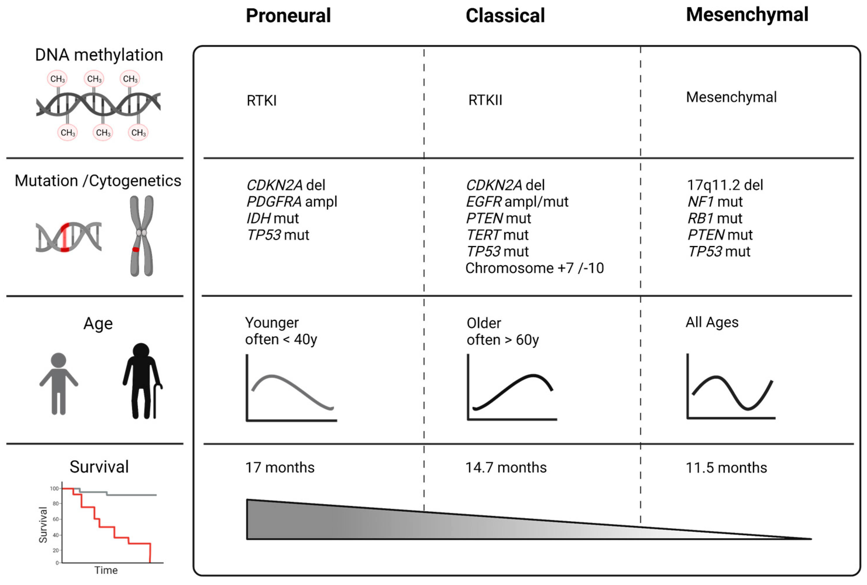
Figure 1. GBM subclasses based on The Cancer Genome Atlas (TCGA) project and Verhaak classification with the most prevalent genetic abnormalities [18]. Novel WHO classification and methylation profiling differentiate between more subgroups of malignant astrocytoma, but in comparison, the MES subtype is still associated with very poor survival. Created with BioRender (ED2349TVGA, 25.10.2021).
Ijms 23 00352 g001
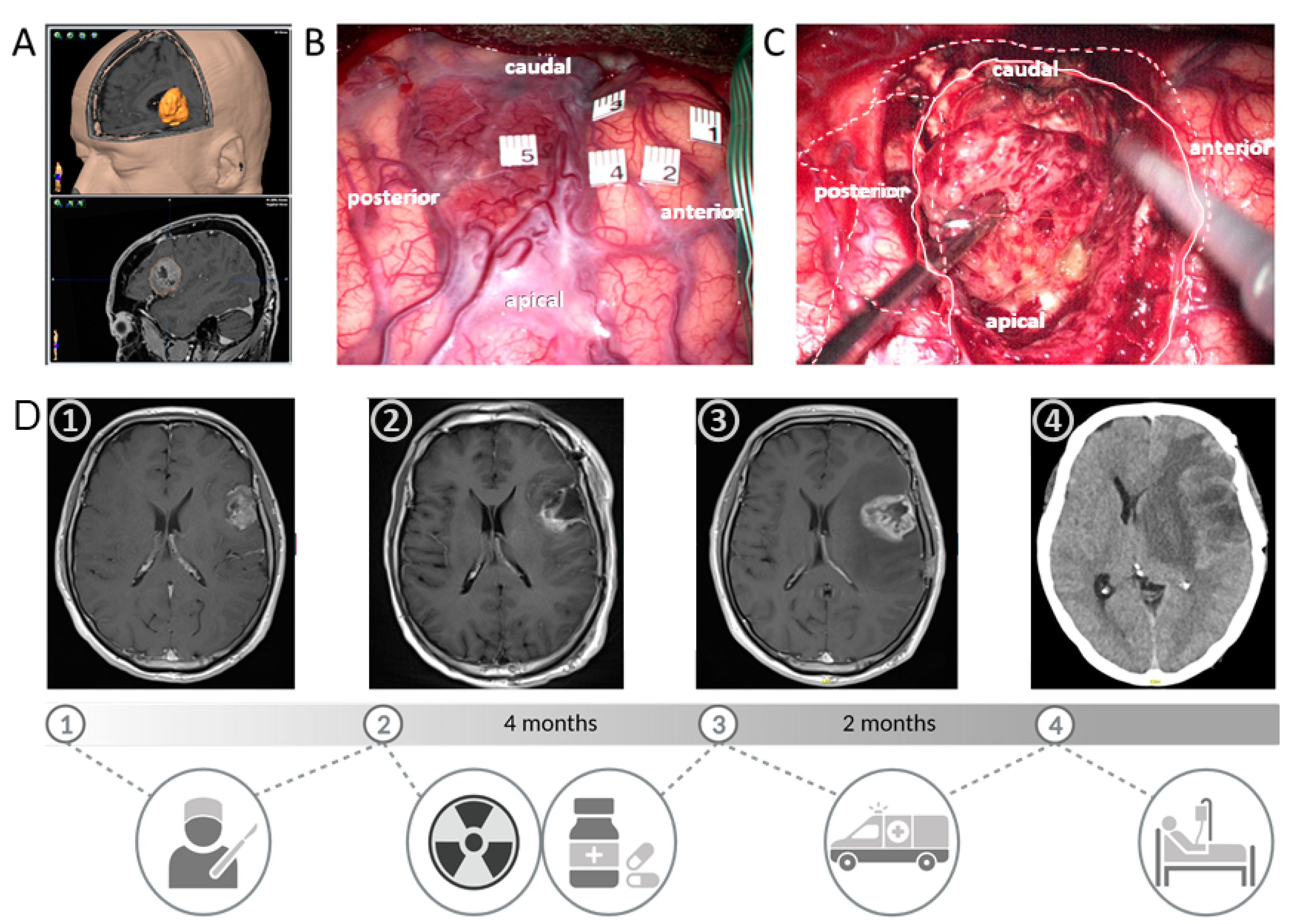
Figure 2. This case of a patient with a mesenchymal GBM highlights the unfavourable clinical course compared to other subtypes impressively. A 42-year-old male patient presenting with seizures demonstrated a contrast-enhancing lesion in Broca’s left-sided area on MRI (A,D) and bilateral activation by functional MRI. Awake craniotomy with cortical (B) and subcortical mapping was scheduled due to the highly eloquent localisation of the tumour, which was both clearly visible on the brain surface (B) and subcortically (C) with typical necrotic tissue and pathological vessels. Here, 86% of the contrast-enhancing lesion was resected (D, first to the second picture), as quantified by MRI volumetry, while a total resection was not feasible due to close eloquent subcortical pathways as demonstrated by bipolar electrical stimulation intraoperatively. Postoperatively, the patient suffered from dysphasia. Radiotherapy (60 Gy) and TMZ therapy were simultaneously administered and discontinued after radiotherapy by the patient himself. Only 4 months after surgery, the patient developed progressive dysphasia and hemiparesis (D, third picture). A second surgery was not considered to be feasible, and chemotherapy, according to Stupp’s protocol, was administered together with TTFields. However, the patient again presented an emergency admission with rapid clinical and radiological progression 2 months later (D, fourth picture). Lacking alternatives, palliative care was initiated. Created with BioRender (OI2349TKM9, 25 October 2021).
Ijms 23 00352 g002
Figure 3. Domain architecture of neurofibromin: The lemniscate NF1 complex is formed by a head-to-tail dimer of an N-terminal HEAT domain (N-HEAT) and a C-terminal HEAT domain (C-HEAT) [58]. The GRD domain possesses a Ras-GAP function. The cysteine and serine-rich domain/Ras-GTPase activating protein domain (CSRD) and the C-terminal domain (CTD) harbour phosphorylation sites, and they are anticipated to regulate GAP-activity when phosphorylated by protein kinase C (PKC) and cAMP-dependent protein kinase A (PKA) [59]. Phosphorylation at the CSRD domain potentiates the Ras-GAP activity [60], while phosphorylation at multiple serine residues in the CTD prolongs activation of the Ras/extracellular signal-regulated kinase (ERK) pathway [61], mediates nuclear import of neurofibromin during the cell cycle [62], and facilitates neurofibromin interaction with 14-3-3 that negatively regulates the GAP-activity [63]. CTD includes a nuclear localisation signal (NLS). Aside from binding to Ras, the GRD also interacts with tubulin via its tubulin-binding domain (TBD) motive [64]. Bipartite module Sec14-homologous segment and pleckstrin homology (PH)-like domain binds phospholipids and is structurally well characterised [65]. Caveolin (CAV1) binding sites are spread over the GRD and the Sec14/PH-domain [66]. CAV1 interacts with Musashi-2 (MSI2), and knockdown of MSI2 elevates the CAV1 protein expression, inhibiting the ubiquitinylation of CAV1 [67]. The leucine-rich domain (LRD) consists of 393 amino acids and includes SEC-PH and Heat-like repeat (HLR) domains. It is involved in membrane localisation through the binding with lipids, actin remodelling through the Rho–ROCK pathway, and dendritic spine formation through VCP. As a neurofibromin creates a high-affinity dimer, on the bottom with the gray colour are shown primary dimerisation interfaces [58]. The figure was created with BioRender.com (XR2390NUCR, 27.11.2021) and adapted from [68,69].
Ijms 23 00352 g003
Figure 4. A consequence of NF1 loss of function in cancer: NF1 deficiency prevents inactivation of Ras through GTP hydrolysis and leads to upregulation of Ras signalling. It is associated with increased tumour proliferation, EMT, invasion, cell motility, and therapy resistance. Moreover, the hyperphosphorylation of LIMK1 in Ras- or LIMK2 in a ROCK-dependent manner results in the activation of cofilin pathways, which leads to changes in actin cytoskeleton and cell motility. The inability of the C-terminal domain of neurofibromin to bind to focal adhesion kinase and syndecan-2 induces cell detachment and might facilitate the epithelial to mesenchymal transition. This process is supported by the upregulation of FOSL1 and increased secretion of chitinase-3-like protein 1 (CHI3L1). Loss of NF1 changes also the tumour microenvironment and angiogenesis by the enhanced secretion of platelet-derived growth factor AA (PDGF-AA) and interleukin-8 (IL-8). In patients with NF1, the somatic NF1 hit accompanies a germline mutation (NF1 -/-), which corresponds to a complete loss of function of neurofibromin, a classical tumour suppressor. In non-NF1-associated NF1-altered GBM, the somatic NF1 hit accompanies other primary genetic alterations that might act similar, e.g., affect the MAPK pathway. Created with BioRender (ZE2345L4TH, 24 October 2021).
Ijms 23 00352 g004
Figure 5. NF1 mutations in glioblastoma cases (not associated with NF1). (A,B): Distribution of most frequently mutated genes in glioblastoma and spectrum of mutations. (C): Overall survival plot of patients with NF1 mutated glioblastoma (S2; n = 79) compared with NF1 wild-type cases (S1; n = 516). Log-Rank test p-value 0.017. Data were derived from the Genomic Data Commons Data Portal (TCGA database).
Ijms 23 00352 g005
Figure 6. FOSL1 is regulated by neurofibromin and its role for proneural to mesenchymal transition. Created with BioRender.com (SY2390LZAW, 27 November 2021).
Ijms 23 00352 g006
Figure 7. NF1 alteration of the leucine-rich domain (LRD) of neurofibromin promotes glioma invasiveness. Created with BioRender (UE233WMPOB, 23 October 2021).
Ijms 23 00352 g007
Table 1. Loss of NF1 in a spectrum of neoplasms underlines its tumour suppressor function (mutation frequencies of NF1 according to the Genomic Data Commons Data Portal).
Tumour EntityFrequency of Somatic MutationFrequency of CNV Loss Events
Uterine Corpus Endometrial Carcinoma19.62%5.69%
Melanoma16.63%3.63%
Glioblastoma multiforme12.98%3.04%
Lung Squamous Cell Carcinoma12.73%5.78%
Lung Adenocarcinoma12.52%3.31%
Angiosarcoma11.11%1.76%
Cervical Squamous Cell Carcinoma and Endocervical Adenocarcinoma10.03%2.78%
Adrenocortical Carcinoma9.78%3.33%
Stomach Adenocarcinoma9.55%1.16%
Paragangliomas and Glomus Tumours9.50%10.83%
Bladder Urothelial Carcinoma8.98%2.94%
Ovarian Serous Cystadenocarcinoma7.57%14.36%
Sarcoma7.17%17.31%
Breast Invasive Carcinoma5.58%6.06%
Publisher’s Note: MDPI stays neutral with regard to jurisdictional claims in published maps and institutional affiliations.