Review

Cannabidiol and Other Cannabinoids in Demyelinating Diseases

1
Emerald Health Pharmaceuticals, San Diego, CA 92121, USA
2
Instituto Maimónides de Investigación Biomédica de Córdoba, 14004 Córdoba, Spain
3
Departamento de Biología Celular, Fisiología e Inmunología, Universidad de Córdoba, 14071 Córdoba, Spain
4
Hospital Universitario Reina Sofía, 14004 Córdoba, Spain
*
Author to whom correspondence should be addressed.
Int. J. Mol. Sci. 2021, 22(6), 2992; https://doi.org/10.3390/ijms22062992
Submission received: 27 February 2021 / Revised: 9 March 2021 / Accepted: 10 March 2021 / Published: 15 March 2021

Abstract

:
A growing body of preclinical evidence indicates that certain cannabinoids, including cannabidiol (CBD) and synthetic derivatives, may play a role in the myelinating processes and are promising small molecules to be developed as drug candidates for management of demyelinating diseases such as multiple sclerosis (MS), stroke and traumatic brain injury (TBI), which are three of the most prevalent demyelinating disorders. Thanks to the properties described for CBD and its interesting profile in humans, both the phytocannabinoid and derivatives could be considered as potential candidates for clinical use. In this review we will summarize current advances in the use of CBD and other cannabinoids as future potential treatments. While new research is accelerating the process for the generation of novel drug candidates and identification of druggable targets, the collaboration of key players such as basic researchers, clinicians and pharmaceutical companies is required to bring novel therapies to the patients.

1. Introduction

Cannabis sativa, which contains about 545 natural compounds of different chemical structures known as cannabinoids, and its use for medicinal purposes, is centuries old [1]. The contemporary history of use of medical cannabis begins in the 19th century when an Irish physician, William Brooke O’Shaughnessy, introduced the cannabis plant into Western medicine for its analgesic, anti-inflammatory and anticonvulsant properties [2]. The most advanced characterization of different compounds extracted from the cannabis plant, termed phytocannabinoids, was undertaken during 1960s by the Israeli researcher Dr. Ralph Mechoulam, whose group isolated and reported among others on the chiral cannabidiol [3] and the psychotropic Δ9-tetrahydrocannabinol (Δ9-THC), two of the main bioactive compounds in the plant [4,5].
In 1985, the Food and Drug Administration (FDA) approved the first two cannabinoid derivatives for clinical use named dronabinol and nabilone. Dronabinol contains the trans isomer of Δ9-THC (synthetically derived) dosed in a gelatin capsule. This drug was approved for two indications: 1) chemotherapy-induced nausea and vomiting; and 2) anorexia in acquired immunodeficiency syndrome (AIDS) patients [6]. The second, nabilone, is a synthetic cannabinoid that mimics the activity of Δ9-THC. This drug was approved by the FDA to treat chemotherapy-induced nausea [7]. Both drugs are available only as oral capsules. In 2005, the authorization of Sativex, a mixture of Δ9-THC and CBD indicated to treat pain and spasticity in MS, supposed a milestone in cannabinoids research [7,8]. Furthermore, cannabidiol oral solution named Epidiolex, which presents beneficial effects for treatment of severe childhood epilepsy, has been recently approved by the FDA as a non-controlled substance.
Considerable interest in CBD is emerging due to its beneficial antiepileptic [8], neuroprotective in hypoxia-ischemia [9], anxiolytic, antipsychotic [10], anti-inflammatory [11] and anticancer properties [12], among others (Table 1). In the past, CBD has received less attention as a potential drug candidate than Δ9-THC although it has been commonly used in cannabis-based formulations.
Table 1. Therapeutic potential of CBD, analogs, and derivatives.
CompoundsTherapeutic PotentialReferences
CBDInflammationMecha et al., 2012 [13]
EpilepsyBurstein et al., 2015 [11]
CancerMori et al., 2017 [9]
AnxietyKis et al., 2019 [12]
NeuroprotectionGarcía-Gutiérrez et al., 2020 [10]
MyelinationLi et al., 2020 [8]
CBDAInflammationPellati et al., 2018 [14]
Cancer
Antimicrobial
CBDVA-C3ConvulsionAnderson et al., 2019 [15]
CBDVConvulsionZamberletti et al., 2019 [16]
EpilepsyMorano et al., 2020 [17]
Autism spectrum disorder
H2-CBDInflammationBen-Shabat et al., 2006 [18]
H4-CBDInflammationBen-Shabat et al., 2006 [18]
HU-446InflammationKozela et al., 2016 [19]
HU-465InflammationKozela et al., 2016 [19]
DMH-CBDInflammation
Cancer
Pain
Neuroprotection
Burstein et al., 2015 [11]
Juknat et al., 2016 [20]
HU-330InflammationSumariwalla et al., 2004 [21]
Immunosuppresion
HU-410InflammationMechoulam et al., 2008 [22]
HU-427InflammationMechoulam et al., 2008 [22]
HU-432InflammationMechoulam et al., 2008 [22]
HU-331CancerKogan et al., 2003 [23]
VCE-004.8/EHP-101Inflammation
Fibrosis
Neuroprotection
Remyelination
Del Rio et al., 2016 [24]
Navarrete et al., 2018 [25]
García-Martin et al., 2018 [26]
García-Martin et al., 2019 [27]
Navarrete et al., 2020 [28]
Now, due to its beneficial properties, the business surrounding the use of CBD in different products is increasing. In addition, CBD scaffolds have attracted increasing consideration for medicinal chemists. Therefore, CBD constitutes one of the most studied cannabinoids in neurodegenerative and demyelinating diseases where CBD has shown benefits in preclinical studies, warranting further investigation.

2. Cannabidiol: General Pharmacology and Therapeutic Profile

The understanding of cannabinoid pharmacology is continuously increasing, and the therapeutic effects of agonists and antagonists of the cannabinoid receptors type 1 and 2 (CB1R and CB2R) have been proposed for the treatment of several human disorders. This has been the result of several preclinical and clinical observations in which interactions with the cannabinoid receptors seem to alter molecular pathways that are responsible for the development of the diseases [29]. CBD is a potential candidate for clinical use thanks to its notable lack of psychotropic action and to its remarkable tolerability profile in humans [30].
CBD was identified by Adams et al. at the University of Illinois in 1940 but its structure was not completely clarified until the 1960s by Mechoulam et al. [3]. Up until now, the mechanisms of action of CBD are not totally known. It has been determined that CBD modulates central nervous system (CNS) receptors such as CB1R (negative allosteric modulator), CB2R, peroxisome proliferator-activated receptor-gamma (PPARγ), serotonin 1A receptor (5-HT1A), transient receptor potential cation channel subfamily V member 1 (TRPV1) and G protein-coupled receptor 55 (GPR55). CBD may antagonize CB1R receptor function by negative allosteric modulation of the orthosteric receptor site [31]. Regarding CB2R receptor, although CBD is a weak agonist of this receptor [32] it has been described that its activation could provide an anti-inflammatory and anti-oxidative effects [33]. Furthermore, CBD may act as an inverse agonist that could explain in part its anti-inflammatory properties inhibiting immune cell migration [34,35]. In vitro assays have shown that CBD is a weak agonist of PPARγ but in vivo assays demonstrated that some CBD biological activities can be blocked by pharmacological inhibition of PPARγ, suggesting that some metabolites of CBD may account for its activity of this nuclear receptor [36]. Furthermore, it has been described that CBD causes analgesia in a TRPV1-dependent manner and ameliorates anxiety through 5-HT1A receptor [37]. Besides, 5-HT1A receptor activation is also involved in CBD neuroprotection in in vitro adult and rat newborn models of the acute hypoxic-ischemic brain [38]. Likewise, CBD has been described as functional antagonist of the GPR55 receptor that can be relevant to explain the anticonvulsant activity of CBD [39].
Clearly, the impact of CBD on the provides many health benefits. Unfortunately, most of this evidence to date comes from animal studies and anecdotal human experience, since very few well-controlled human studies have been conducted with CBD, although this tendency is changing.

The Endocannabinoid System

Due to the interest in recreational and medical uses of marijuana, efforts were made early in the sixties to identify the major cannabinoids in the cannabis plant [40,41]. These attempts resulted in the discovery of Δ9-THC, CBD, and cannabinol (a processing product of Δ9-THC) [3,42,43]. Then, using a synthetic radiolabeled Δ9-THC analogue, high-affinity binding sites for Δ9-THC in the brain were discovered as the CB1R [44], a G protein-coupled receptor (GPCR). Afterwards, a second G protein-coupled receptor (GPCR) called CB2R was identified outside the CNS, mainly in the immune system [45]. As a result, the cloning and identification of the two main cannabinoid receptors led to isolation of endogenous CB1R and CB2R ligands and the discovery of the endocannabinoid system (ECS). The lipids anandamide (N-arachidonoylethanolamide or AEA) and 2-arachidonoylglycerol (2-AG), identified in the brain and intestinal tissues, were shown to activate both receptors with high affinity and as consequence these lipids were named endocannabinoids. The levels of these endocannabinoids is regulated by enzymes, including fatty acid amide hydrolase (FAAH) [46] and monoacylglycerol lipase (MAGL) [47], that metabolize AEA and 2-AG, respectively. To partly explain their multipronged bioactivities, exogenous and endogenous cannabinoids also interact with non-cannabinoid receptors as described above for CBD [48].
Modifications in the ECS are frequently observed in neurological diseases [49] and genetic and pharmacological changes of this system in animal models suggest a major role for this system in neurodegenerative disorders and demyelinating diseases [50,51]. The ECS is a complex system due to the promiscuity of mediators and its interactions with other metabolic pathways. The regulation of the ECS components alters the endocannabinoid-related system, known as the endocannabidiome (eCBome). This complicated system presents a challenge for the discovery of novel bioactive molecules inspired in endocannabinoid and also suggests new chances for the utilization of non-psychotropic cannabinoids such as CBD and derivatives of CBD, which frequently modulate several eCBome proteins. In addition, lifestyle, including the lipid dietary component, habits, and environment are suggested to have an impact on the eCBome, which seems to be relevant in many physiological and pathological conditions [29,52].

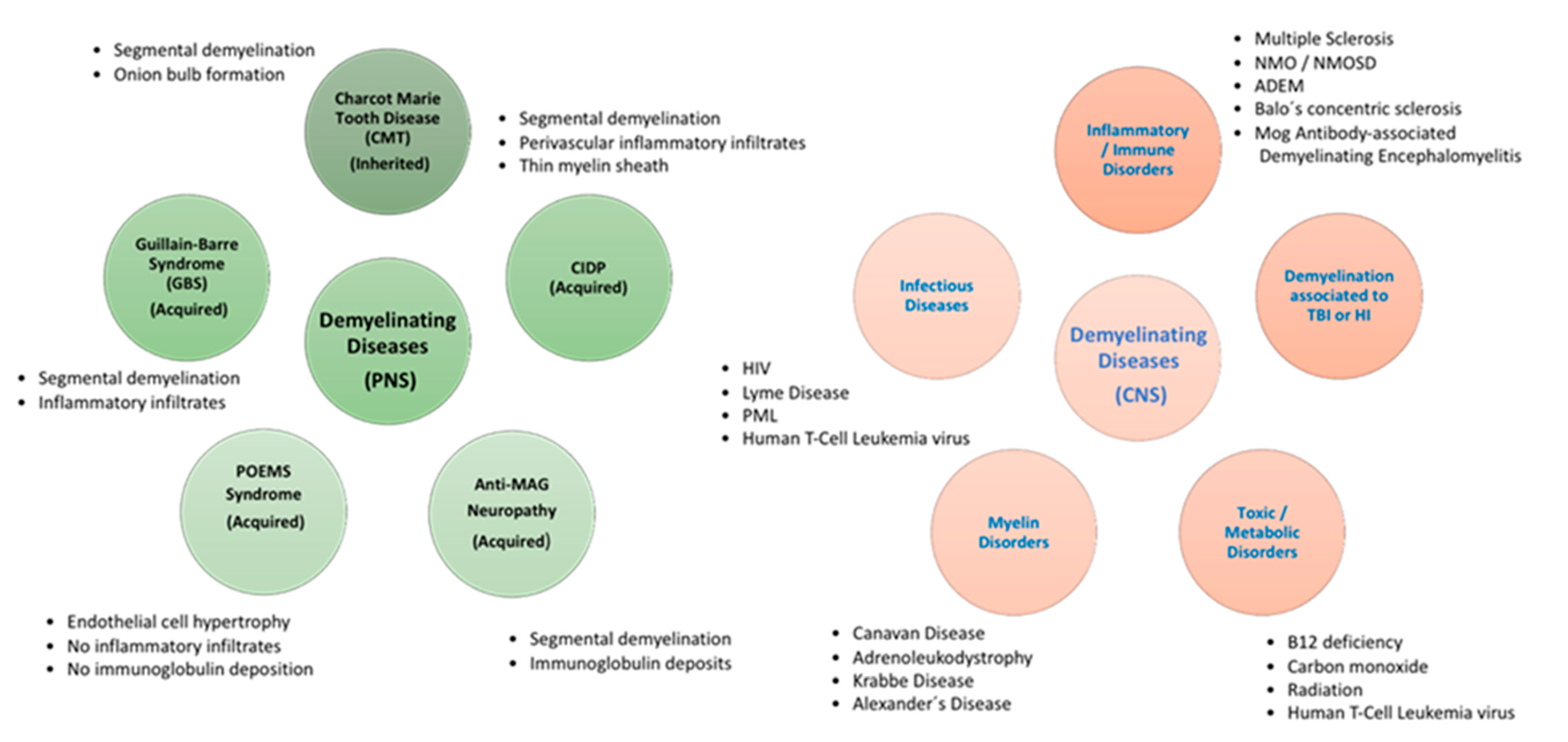
3. Demyelinating Diseases

Myelin is believed to be generated in early gnathostomes by a glial precursor, which later produce the different Schwann cell (SC) and oligodendrocyte lineages [53,54]. In fact, the global organization of myelinated axons is similar in the central nervous system (CNS) and peripheral nervous system (PNS), regarding their functions in saltatory transmission. However, Schwann cells and oligodendrocytes present considerable variations in the development and formation of myelin. In line with this, demyelinating diseases are limited to those involving PNS myelinated fibers or CNS fibers (Figure 1).
The myelin disorders can be categorized into several categories according to their etiologies: demyelination associated to inflammation, demyelination associated to virus, loss of myelin produced by metabolic imbalances, loss of myelin due to hypoxic-ischemic conditions and demyelination caused by brain injury. Most of the different categories overlap in pathogenesis but this organization may be helpful to establish a diagnosis. The prognosis of these diseases is generally difficult, and no curative treatment is currently available.
Several diseases involving significant injury to axons and glial cells, especially SC in the PNS, are classified as peripheral demyelinating diseases (PDD) [55]. Schwann cells, which are derived from the neural crest, represent the main glial cells in peripheral nerves. The development of SC happens through different embryonic and postnatal periods, which are strictly controlled by several cellular signaling pathways. Initially, the undifferentiated SC matures into either myelinating or non-myelinating SC and covers around axons, thus constituting the process named myelination [56]. The myelin sheath is composed of various coats of lipids and lipoprotein plasma membranes of SC which are arranged around the axon of neurons [57]. In PNS, the demyelination process involves the damage of the myelin sheath due to the injury on SC [58]. Currently, there are no consistent biomarkers for PNS-associated disorders and the diagnosis is based on several studies such as electrophysiological and cerebrospinal fluid (CSF) analysis. PDD can be classified in two main groups: Acquired Demyelinating Diseases and Inherited Demyelinating Diseases (Figure 1).
The first group comprises four main type of disorders such as Guillain-Barre Syndrome (GBS), chronic inflammatory demyelinating polyradiculoneuropathy (CIDP), anti-myelin associated glycoprotein (MAG) neuropathy and polyneuropathy, organomegaly, endocrinopathy, m protein and skin changes (POEMS) syndrome. GBS is a severe idiopathic autoimmune demyelinating disease associated to acute ascending neuromuscular palsy [59]. A high percentage of GBS cases have been related to autoantibodies related with several bacterial and viral infections [60,61,62]. Emerging information suggests that acute respiratory syndrome coronavirus-2 (SARS-CoV-2 or COVID-19) can cause GBS and several neurological autoimmunity-related diseases requiring attention for quick diagnostic and treatment [63]. MAG neuropathy is caused by circulating monoclonal antibodies towards the human natural killer-1 epitope. This epitope is expressed on adhesion molecules present in peripheral nerves such as the glycoprotein MAG. A low expression of MAG affects the myelin sheath structure and axonal function. This progressive disease causes mild to moderate distal muscle fragility, with gradual sensory ataxia and recurrent tremors [64]. CIDP is associated with a gradual loss of sensorimotor functions [65]. There are several effective treatments based on immunoglobulin, corticosteroids, and plasma exchange treatments, but long-lasting treatments are needed. Regarding POEMS syndrome, this is an unusual paraneoplastic syndrome with demyelinating neuropathy produced by a disorder related to plasma cell proliferation. The association of vascular endothelial growth factor (VEGF) with POEMS syndrome is very effective in clinical diagnosis as accurate biomarker and monitoring responses to treatment. POEMS patients present high serum level of VEGF, although low levels are described upon effective treatment [66].
The second group of hereditary demyelinating diseases include Charcot Marie tooth disease (CMT). Although CMT is an uncommon inherited neurological disease, it is the major disorder that affects the peripheral nerves. CMT patients, despite their genetic heterogeneity, typically present an indolent, length-dependent, sensorimotor polyneuropathy [67].
Currently, synthetic drugs and natural products are used for the management of PDD. Nevertheless, these diseases remain misdiagnosed due to the absence of solid biomarkers and disease safe-diagnostic criteria. Therefore, the search for new therapies and accurate biomarkers are essential to address this type of neuropathic disease [68].
Demyelinating disorders of the CNS have different etiologies and are divided into primary, such as MS and other idiopathic inflammatory-demyelinating diseases (IIDDs), and secondary, such as infective, ischemic, metabolic, or toxic diseases (Figure 1). These CNS demyelinating diseases comprise MS and its acute variant Marburg disease, also neuromyelitis optica (NMO), Balo’s concentric sclerosis, acute disseminated encephalomyelitis (ADEM), and ADEM’s hyperacute variant, acute hemorrhagic leukoencephalitis (AHL) (Figure 1). The term IIDD includes several CNS disorders that are classified according to their severity, clinical progression, and lesioned zone, as well as their pathological outcomes. The spectrum of diseases involves monophasic, multiphasic, and progressive disorders. Aggressive types of IIDD include a plethora of disorders that share the symptomatology, an acute clinical course, and atypical outcomes on magnetic resonance imaging (MRI). Marburg disease is the classic fulminant IIDD, but it is extremely rare. Baló’s concentric sclerosis, which is considered a variant of MS and ADEM, can also appear with acute attacks [69].
MS is characterized by inflammation-related injury, principally to myelin structure and composition from nerves in the brain (including optic nerves) and spinal cord, causing axonal damage and neurodegeneration. The most frequent forms of MS are the relapsing-remitting (RR) and secondary progressive (SP) forms, although it can also present a progression from onset (primary progressive (PP). The presentation of demyelinating lesions distributed in time and space are critical in the clinical diagnosis of MS. In addition to the neurological symptoms, lesions consistent with MS determined by MRI, along with presentation of oligoclonal bands in CSF and findings of abnormal visual evoked potentials, are proposed to provide an accurate diagnosis [70]. Neuromyelitis optica spectrum disorder (NMOSD) and myelin oligodendrocyte glycoprotein (MOG) antibody (Ab)-associated disease are also inflammatory CNS demyelinating disorders although clinically and pathologically they differ from MS and are far less common. NMO has been identified as a disease distinct from MS due to the identification of an NMO-specific autoantibody directed against aquaporin-4 (AQP4-Ab), the major water channel in the CNS [71].
Among the infectious inflammatory demyelinating disorders, progressive multifocal leukoencephalopathy (PML) is an aggressive CNS infection caused by JC virus (JCV). The disease is triggered by a JCV that selectively damages the oligodendrocytes, causing demyelination. Therapy with monoclonal antibody treatment or other immunomodulatory drugs, generally applied to MS patients, has also been used for PML treatment [72].
Traumatic brain injury (TBI) and stroke are major pathologies which result in demyelination. The neurovascular unit (NVU) is constituted by neurons, endothelial cells, smooth muscle cells, pericytes, astrocytes, and microglia [73]. This NVU is affected by secondary injuries after TBI and suffer alterations such as a reduction of blood–brain perfusion with adverse effects on the correct function of the neurons [74]. During the past decades, cerebrovascular dysfunction has been associated with a poor prognostic outcome. In addition, alterations in the structure of the blood-brain barrier (BBB) trigger edema generation with interference in the brain homeostasis. This alteration exacerbates the secondary injury processes including excitotoxicity and inflammation. For these reasons, TBI has been considered a chronic brain disease with molecular alterations in the BBB after the initial injury [75]. This long-term alteration of the blood-brain barrier could be responsible of premature aging of the brain after TBI [75,76]. Research on animal models have shown that cannabinoids targeting CB2R after TBI improve neurobehavioral manifestations and memory tests and the neurological insufficiency, and diminish motor deficit through downregulation of proinflammatory markers. Furthermore, the modulation of cannabinoid system reduces oedema generation and BBB permeability, avoiding neuronal cell death and upregulating the levels of adherence junction proteins (reviewed in [77]). Moreover, PPARγ seems to be another interesting target to prevent neuroinflammation and demyelination in TBI [78].
At present, advancement has been made in identifying the pathogenesis of demyelinating disorders, but we have to discover their origin or a therapeutic treatment for these debilitating diseases that affect millions of young adults around the world. The development of new therapies for the treatment of these diseases remains a challenge. Indeed, the support of the beneficial potential of cannabinoids, especially CBD, for the control of pathological events related to these diseases is increasing.

4. Cannabidiol and Demyelinating Diseases

During the past decade, the therapeutic potential of cannabinoids for treating demyelinating diseases, specifically MS, has been well-studied. It is now established that CBD and various CBD-derivatives confer neuroprotective effects and attenuate the inflammatory process in several demyelinating animal models [19,20,25,79]. For instance, the impact of CBD has been determined in hypoxic-ischemic immature brain. CBD, at micromolar concentrations, reduces the levels of inflammatory markers such as interleukin-6 (IL-6), tumor necrosis factor-α (TNF-α), cyclooxygenase-2 (COX-2), and inducible nitric oxide synthase (iNOS), through activation of CB2R and A2A receptors [80,81]. Regarding A2A receptors, it has been described that CBD increases adenosine signaling inhibiting adenosine uptake [82].
Currently the search for new remyelinating therapies is focused principally on identifying the factors that promote repair of the myelin sheath. Demyelinating diseases share as a common feature the principal pathogenic process that targets the myelin sheath. This neuronal covering allows proper conduction of the nerve impulses. MS is characterized in particular by a decrease in the number of oligodendrocytes producing myelin in the CNS and by progressive axonal deterioration. As it occurs in most demyelinating diseases, damage of the myelin sheath triggers regeneration mechanisms, a process named remyelination. Oligodendrocytes responsible for production of mature myelin are derived during postnatal development from immature cells named oligodendrocyte progenitor cells (OPCs). OPCs stay in the adult brain and produce new mature oligodendrocytes when the myelin is injured. The beneficial role of CBD against injury to OPCs mediated by the immune system has been described. Indeed, cells treated with CBD present less oxidative stress avoiding the generation of reactive oxygen species. Furthermore, the treatment of OPC with CBD prevented apoptosis by mechanisms independent from CB1R, CB2R, TRPV1 or PPARγ receptors [13]. OPCs have remarkable metabolic conditions during development because they can differentiate to generate myelin segments, indicating they need a satisfactory blood supply. However, processes that coordinate myelination and angiogenesis must be better defined. It has been described that Hypoxia-Inducible Factor (HIF) is an important regulator of postnatal myelination. HIF-1α activation may play a role in the inflammatory and the remitting phases of MS [83]. Moreover, activation of the HIF pathway may also be associated to neuronal protection and remyelination [84]. Thus, we have described that VCE-004.8, a synthetic aminoquinone derivative of cannabidiol, could be a potential drug to treat MS by regulating the immune response and supporting neuroprotection and axonal regeneration through activation of the hypoxia-inducible factor pathway. Furthermore, this novel synthetic cannabinoid, which also acts as a dual PPARγ and CB2R agonist, presents potent anti-inflammatory activity [25].
Neonatal hypoxia-ischemia (HI), which causes myelination disorders and is related to cerebral palsy, presents a complex pathophysiology which includes oxidative stress, excitotoxicity, and severe inflammatory response [85,86]. Due to this altered environment, oligodendrocyte progenitors are particularly sensitive. Thus, OPC injury is increased in the brain, both in animal models and human newborns after HI damage [87] which eventually leads to hypomyelination [88]. The protective role of CBD after HI injury in newborn animals has been described in neuronal and glial cells [89].
MS, as well as other demyelinating diseases, presents many of the characteristics of autoimmune disorder with rupture of the BBB. The BBB is an extraordinary composition of endothelial cells, pericytes, which are enclosed and supported by astrocytes and perivascular macrophages. In pathological circumstances, peripheral lymphocytes are activated and infiltrate the CNS to trigger an immune response injuring myelin and axons. In an experimental model of MS, the Theiler’s murine encephalomyelitis virus-induced demyelinating disease, it has been demonstrated that CBD ameliorates the symptomatology of the disease. It has been shown that intraperitoneal treatment with CBD reduces the extravasation of leukocytes from the systemic circulation by downregulating the expression of several chemokines as well as by decreasing microglia activation [90]. Furthermore, the effect of CBD on BBB permeability has been determined by using human brain microvascular endothelial cells and human astrocyte co-cultures as a BBB model. In this model, CBD restored the BBB permeability produced by oxygen-glucose deprivation (OGD). Treatment was more efficient when it was administered prior to OGD, but positive results were detected up to two hours into reperfusion. The protective effect was dependent on PPARγ and relatively reduced by a 5-HT1A receptor antagonist but was independent of CB1R, CB2R, TRPV1 or Adenosine A2A receptors [91].
During the past few decades, our understanding about the loss of myelin after stroke and TBI has increased. Previous research on these pathologies highlighted changes in neuronal cells within the gray matter. Recently, several studies have shown the same importance of the white matter integrity in long-term recovery. Demyelination following brain injury causes long-term sensory, motor, and cognitive insufficiencies due to the adult brain’s low capability to regenerate oligodendrocyte cells and the restoration of axonal myelin. New molecules that control the process of remyelination may offer novel therapies to restore white matter integrity and improve long-term neurological improvement in stroke and TBI patients. In addition, it has been shown that treatment with oral CBD oil restored behavioral dysfunctions and normalized the cortical biochemical changes associated with TBI. Therefore, CBD has been proposed as a pharmacological tool to improve neurological dysfunctions triggered by the trauma [92]. Furthermore, several studies have shown that short-term treatment with CBD in a mouse model of brain injury, such as bilateral common carotid artery occlusion (BCCAO), is capable of improving motor and cognitive disability by activating a complex mechanism related with an increase in hippocampal levels of brain-derived neurotrophic factor (BDNF) and microtubule-associated protein 2 (MAP-2) proteins, resulting in stimulation of neurogenesis. Because of these effects, the treatment with CBD ameliorated neuroinflammation and neuronal death in the hippocampal zone [9,93].

5. Medicinal Chemistry of Synthetic and Natural Derivatives of Cannabidiol

Of the over one hundred phytocannabinoids discovered in Cannabis sativa, seven have been categorized as CBD-type compounds, including CBD [94,95]. All of them present the same configuration as CBD. Among these natural analogs, cannabidiolic acid (CBDA) and cannabidivarinic acid (CBDVA-C3), which are C30-carboxylic derivatives, have been isolated. Moreover, cannabidiorcol (CBD-C1), cannabidiol-C4 and cannabidivarin (CBDV), which vary from CBD by the length of their C40-side chain, have been identified. Finally, cannabidiol monomethyl ether (CBDM), the C60-methoxy CBD analog, has also been isolated from the plant. Although these natural CBD derivatives present potential therapeutic benefits (Table 1), only a few pharmacological studies have been described [14,15,16,17].
Because of the favorable therapeutic benefits of CBD in a variety of diseases, synthetic CBD derivatives have also been taken into consideration by drug discovery projects, with the purpose of improving the potency, efficacy, and/or pharmacokinetic properties of this natural cannabinoid. To obtain new synthetic analogs, a series of structural modifications such as hydrogenation of CBD produced the dihydro and tetrahydrocannabidiol derivatives H2-CBD and H4-CBD [18]. These molecules have been attributed anti-inflammatory properties because of their effects on the generation of reactive oxygen species, nitric oxide, and tumor necrosis factor. Furthermore, the hydroxy-CBD enantiomers, named HU-446 and HU-465, have shown potential anti-inflammatory effects in a proinflammatory model of encephalitogenic T cells (Table 1). Specifically, both HU-446 and HU-465 prevented the production of IL-17, a crucial autoimmune cytokine, from MOG35-55-stimulated T(MOG) cells. These data indicated that both CBD derivatives have anti-inflammatory effects in autoimmune diseases [19].
The synthesis of dimethylheptyl (DMH) CBD derivatives such as DMH-CBD, HU-320 [21], and 7-OH-DMH-CBD have been described by Mechoulam and colleagues [96]. In the case of DMH-CBD, it was reported that this derivative abolishes the production of proinflammatory cytokines and prevents microglia reactivation by generating an adaptive cellular response, thus avoiding inflammation and oxidative injury (Table 1). In addition, DMH-CBD reduced the proliferation of pathogenic activated TMOG cells [20]. For such derivative, remarkable benefits such as anti-inflammatory, analgesic, neuroprotective or antitumor effects have been described, and it has been used as a pharmacological tool in many cannabinoid studies supporting the progress in this field [11].
Finally, other interesting modifications have consisted of changes in the C40-alkyl chain with the purpose of improving oral bioavailability, modifications of the resorcinol hydroxyl groups, thus generating new molecules (named HU-410, HU-427, and HU-432) that present anti-inflammatory activities as reported in the patent literature (Table 1) [22]. The development of new quinone derivatives of CBD has also been investigated.
The first quinone derivative of CBD, named HU331, was described by Mechoulam et al. by oxidation of CBD [97] and its antineoplastic activity was reported (Table 1) [23]. Quinone-based drugs causing anti-infective and antitumoral effects are frequently applied in clinical practice, but their use for chronic therapies is not recommended due to their reactivity and toxicity. HU-331 is a thiol-trapping compound that generates reactive oxygen species (ROS), affects the mitochondria transmembrane potential and causes cytotoxicity in primary and transformed cells in vitro [98].
We have generated a non-thiophilic and chemically stable derivative of a CBD aminoquinone (VCE-004.8) that acts as a dual agonist of PPARγ and CB2R. VCE-004.8 does not have affinity for the CB1R receptor and presents potent antifibrotic activity in vitro and in vivo (Table 1) [24]. Furthermore, we have described that VCE-004.8 also activates the HIF pathway. In fact, we have reported that VCE-004.8 stabilizes and activates HIF-1α and HIF-2α in human microvascular endothelial cells, oligodendrocytes, and microglia cells [25]. VCE-004.8 ameliorated neuroinflammation and prevented myelin loss in several murine models of MS, such as Experimental autoimmune encephalomyelitis (EAE) and Theiler’s virus-induced demyelinating disease [25]. Recently, we have reported that EHP-101, which is an oral lipidic formulation of VCE-004.8, also had efficacy in EAE and induced remyelination in two demyelination models induced by cuprizone. [28]. Hence, EHP-101 could be a promising cannabinoid-derived drug candidate for the treatment of different forms of MS. In addition, EHP-101 also demonstrated efficacy in a murine model of systemic sclerosis (SSc) [26,27]. EHP-101 is now under evaluation in a Phase II study in SSc patients (ClinicalTrials.gov: NCT04166552) and the initiation of a Phase II study in MS patients is underway.

6. Clinical Trials of Cannabidiol Focused on Demyelinating Disorders

Currently, medicine may be focused on CBD as a new treatment for patients with reduced conventional options and medical professionals are often asked about CBD products by patients, family, and patient associations. Due to its minimal toxicity in humans, an interesting number of trials have been performed to determine the clinical efficacy of CBD in different pathologies. Numerous CBD formulations have been assessed in preclinical studies for various pharmaceutical properties, such as anti-nausea, anti-emetic, anti-tumor, anti-inflammatory, antidepressant, anti-psychotic, and anti-anxiolytic [10,11,12,99] benefits. Nevertheless, the variation in CBD quality, the type of drug formulations applied, and the minimal sample sizes compromise the development of these preclinical studies.
As we have previously indicated in this review, to date the FDA has approved three CBD- and Δ9-THC-based medicines. Dronabinol (Marinol, Syndros) which is a synthetic form of THC in an oily base, administered to stimulate appetite in AIDS patients and for the improvement of nausea and vomiting associated with cancer chemotherapy. Nabilone (Cesamet) is another Δ9-THC analog for the treatment of nausea in patients undergoing chemotherapy. Cannabidiol oral solution (Epidiolex) is authorized in the USA as therapy of two severe rare childhood epilepsy disorders (Dravet syndrome and Lennox-Gastault syndrome). A fourth medication, nabiximols (Sativex), a combination of Δ9-THC and CBD, is sold legally in more than ten countries including Canada, Mexico, and parts of Europe, for the treatment of muscle spasticity and neuropathic pain in multiple sclerosis, and the FDA recently recognized an Investigational New Drug application for nabiximols. This aromatized water-ethanol oral-mucosal spray was created to offer a simple delivery system. Specifically, this method of dispensation allows rapid entry to the circulation through the oral mucosa with an extremely rapid plateau of plasma concentration, preventing the complications of the gastrointestinal route. Furthermore, it has been shown that co-administration of CBD to Δ9-THC counteracts the undesirable effects of Δ9-THC alone [100].
Until now there are about 2,500 clinical trials focused on demyelinating disorders and among them only 30 studies have been related to the benefits of cannabis or cannabinoids. In fact, there are 19 clinical trials that address the effect of CBD in demyelinating disorders, specifically in MS. Medicinal cannabis has been researched as potential therapy for multiple sclerosis symptomatology, such as pain and spasticity [101]. During the first 10 years of initial MS diagnosis, up to 80 percent of patients are affected by moderate spasticity and the numbers of affected patients rises over time [102,103]. The study called “The Cannabinoids for Treatment of Spasticity and Other Symptoms Related to Multiple Sclerosis (CAMS)” was a major randomized trial that explored spasticity in more than 600 MS patients. This study did not observe changes in the Ashworth Spasticity Scale between either oral cannabis extract contrasted with placebo after 15 weeks. However, objective improvement in mobility and pain suggested cannabinoids might be clinically beneficial [104].
Many studies have investigated the pharmacological properties of cannabinoids in demyelinating diseases such as MS. In fact, most of the trials have been executed using Sativex which is suggested as a second line therapy for spasticity in MS patients who do not respond to other anti-spasticity treatment and who experienced clinically remarkable improvement in symptoms associated to spasticity during the onset of the trial. Since the first reported study in 2003, when an initial controlled study determined that cannabis extracts could improve intractable neurogenic symptoms [105,106], and during the last decade, several clinical trials have evaluated the efficacy of Sativex as a supplementary treatment for symptomatology recovery in patients with MS-related spasticity and neuropathic pain.
In 2010, a meta-analysis of the effectiveness and security of nabiximols on spasticity in 666 MS patients showed remarkable superior percentage of treated patients as responders and treated patients also reported remarkable improvements [107]. In the same year, a double-blind, randomized, placebo-controlled, parallel-group study of nabiximols was performed in subjects with symptoms of spasticity due to MS. This randomized controlled trial studied the treatment effects in 337 subjects for 15 weeks and no important improvement in the mean spasticity numerical rate scale (NRS) was observed in intent-to-treat (ITT) analysis but responder per protocol (PP) analyses confirmed that nabiximols treatment caused an important decrease in treatment-resistant spasticity, in patients with progressive MS and severe spasticity [108]. Later, Novotna et al., published the results derived from a randomized, double-blind, placebo-controlled, parallel-group, enriched-design study of nabiximols, as add-on therapy, in MS subjects with refractory spasticity. In this trial, 241 randomized patients received treatment with nabiximols, as add-on therapy, in a single-blind manner for 28 days, after which those showing positive effects in spasticity of ≥ 20% progressed to 84-days randomized, placebo-controlled phase. ITT analysis showed an important significant variation positive for nabiximols treatment [109]. In 2014, the results derived from the first Phase III placebo-controlled study of the efficacy of the nabiximols to improve central neuropathic pain (CNP), which occurs in many MS patients, were published. More than 300 subjects were randomized to Phase A (167 received nabaximols and 172 received placebo). Of those who finished Phase A, 58 started the randomized-withdrawal phase. The results of this research were ambiguous, with contradictory conclusions in the study [110]. In 2014, a multicenter, non-interventional study called MOVE-2 showed the effect of nabiximols in 276 patients [111]. After one month, nabiximols improved resistant MS spasticity (MSS) in 74.6% of patients. After three months, 55.3% of subjects had persisted to receive nabiximols and the mean NRS score had decreased by 25% from baseline. This study MOVE-2 was prolongated for 12 months and 52 patients were incorporated to the effectiveness analysis. The mean spasticity NRS reduced considerably from 6.0 ± 1.8 at baseline to 4.8 ± 1.9 after the first 30 days and remained on 4.5 ± 2.0 after 12 months [112]. These data confirmed the long-term efficacy and tolerability of nabiximols for the therapy of resistant MSS in clinical.
Recently, Marinelli et al. proposed a novel study to identify if nabiximols could be useful ameliorating spasticity in stroke and to investigate its tolerability and security by accurate cardiovascular monitoring. The study will recruit 50 patients with spasticity following stroke to be dosed with nabiximols in a double-blind placebo-controlled cross-over study [113]. Finally, in 2019 the results derived from the study named SAVANT were published, evaluating the effects of nabiximols as add-on therapy versus optimized first line antispastics in resistant MS spasticity. In this double-blind, placebo-controlled randomized clinical trial of 191 patients who entered Phase A, 106 were randomized in Phase B to receive add-on nabiximols spray (n = 53) or placebo (n = 53). The percentage of clinically relevant responders after 12 weeks was significantly superior with nabiximols than placebo [114].
At present, data indicates that cannabidiol and some derivatives have a remarkable role in the modulation of myelinating processes, and it has been suggested as a promising approach in the treatment of demyelinating diseases. Although serious advances are being made in the development of new cannabidiol derivative drugs and therapeutic targets, the collaboration of researchers and pharmaceutical companies is needed to achieve successful outcomes.

Author Contributions

All authors have contributed to the manuscript. All authors have read and agreed to the published version of the manuscript.

Funding

This review received no external funding.

Institutional Review Board Statement

Not applicable.

Informed Consent Statement

Not applicable.

Data Availability Statement

Not applicable.

Conflicts of Interest

The authors declare no conflict of interest.

Abbreviations

2-AG2-arachidonoylglycerol
ADEMAcute Disseminated Encephalomyelitis
AEAAnandamide
AHLAcute Hemorrhagic Leukoencephalitis
AIDSAcquired Immunodeficiency Syndrome
BBBBlood-Brain Barrier
BCCAOBilateral Common Carotid Artery Occlusion
BDNFBrain-Derived Neurotrophic Factor
CB1RCannabinoid receptor type 1
CB2RCannabinoid receptor type 2
CBDCannabidiol
CBDACannabidiolic Acid
CBDMCannabidiol Monomethyl Ether
CBDVCannabidivarin
CBDVACannabidivarinic Acid
CIDPChronic Inflammatory Demyelinating Polyradiculoneuropathy
CMTCharcot Marie Tooth Disease
CNPCentral Neuropathic Pain
CNSCentral Nervous System
COX-2Cyclooxygenase-2
CSFCerebrospinal Fluid
EAEExperimental Autoimmune Encephalomyelitis
eCBomeEndocannabidiome
ECSEndocannabinoid System
FAAHFatty Acid Amide Hydrolase
FDAFood and Drug Administration
GBSGuillain-Barre Syndrome
GPCRG protein-coupled receptor
GPR55G protein-coupled receptor 55
HIHypoxia-Ischemia
HIFHypoxia-Inducible Factor
IIDDsIdiopathic Inflammatory-Demyelinating Diseases
IL-6Interleukin-6
iNOSInducible Nitric Oxide Synthase
MAGAnti-Myelin Associated Glycoprotein
MAGLMonoacylglycerol Lipase
MAP-2Microtubule-Associated Protein 2
MOGMyelin Oligodendrocyte Glycoprotein
MRIMagnetic Resonance Imaging
MSMultiple Sclerosis
NMONeuromyelitis Optica
NMOSDNeuromyelitis Optica Spectrum Disorder
NRSNumerical Rate Scale
NVUNeurovascular Unit
OGDOxygen-Glucose Deprivation
OPCsOligodendrocyte Progenitor Cells
PDDPeripheral Demyelinating Diseases
PMLProgressive Multifocal Leukoencephalopathy
PNSPeripheral Nervous System
POEMSPolyneuropathy, Organomegaly, Endocrinopathy, M protein and Skin changes Syndrome
PPPrimary Progressive
PPARγPeroxisome Proliferator-Activated Receptor-Gamma
ROSReactive Oxygen Species
RRRelapsing-Remitting
SCSchwann Cell
SPSecondary Progressive
TBITraumatic Brain Injury
TNF-αTumor Necrosis Factor-α
TRPV1Transient Receptor Potential Cation Channel Subfamily V Member 1
VEGFVascular Endothelial Growth Factor
Δ9-THCΔ9-tetrahydrocannabinol

References

  1. Hanuš, L.O.; Meyer, S.M.; Muñoz, E.; Taglialatela-Scafati, O.; Appendino, G. Phytocannabinoids: A unified critical inventory. Nat. Prod. Rep. 2016, 33, 1357–1392. [Google Scholar] [CrossRef] [PubMed] [Green Version]
  2. O’Shaughnessy, W.B. On the Preparations of the Indian Hemp, or Gunjah (Cannabis indica), Their Effects on the Animal System in Health, and Their Utility in the Treatment of Tetanus and Other Convulsive Diseases. Br. Foreign Med. Rev. 1840, 10, 225–228. [Google Scholar]
  3. Mechoulam, R.; Shvo, Y. Hashish. I. The structure of cannabidiol. Tetrahedron 1963, 19, 2073–2078. [Google Scholar] [CrossRef]
  4. Gaoni, Y.; Mechoulam, R. Isolation and structure of DELTA+−tetrahydrocannabinol and other neutral cannabinoids from hashish. J. Am. Chem. Soc. 1971, 93, 217–224. [Google Scholar] [CrossRef]
  5. Mechoulam, R.; Gaoni, Y. The absolute configuration of δ1-tetrahydrocannabinol, the major active constituent of hashish. Tetrahedron Lett. 1967, 12, 1109–1111. [Google Scholar] [CrossRef]
  6. Borgelt, L.M.; Franson, K.L.; Nussbaum, A.M.; Wang, G.S. The pharmacologic and clinical effects of medical cannabis. Pharmacotherapy 2013, 33, 195–209. [Google Scholar] [CrossRef]
  7. Davis, M.P. Oral nabilone capsules in the treatment of chemotherapy-induced nausea and vomiting and pain. Expert Opin. Investig. Drugs 2008, 17, 85–95. [Google Scholar] [CrossRef]
  8. Li, H.; Liu, Y.; Tian, D.; Tian, L.; Ju, X.; Qi, L.; Wang, Y.; Liang, C. Overview of cannabidiol (CBD) and its analogues: Structures, biological activities, and neuroprotective mechanisms in epilepsy and Alzheimer’s disease. Eur. J. Med. Chem. 2020, 192, 112163. [Google Scholar] [CrossRef]
  9. Mori, M.A.; Meyer, E.; Soares, L.M.; Milani, H.; Guimarães, F.S.; de Oliveira, R.M.W. Cannabidiol reduces neuroinflammation and promotes neuroplasticity and functional recovery after brain ischemia. Prog. Neuro Psychopharmacol. Biol. Psychiatry 2017, 75, 94–105. [Google Scholar] [CrossRef] [PubMed]
  10. García-Gutiérrez, M.S.; Navarrete, F.; Gasparyan, A.; Austrich-Olivares, A.; Sala, F.; Manzanares, J. Cannabidiol: A Potential New Alternative for the Treatment of Anxiety, Depression, and Psychotic Disorders. Biomolecules 2020, 10, 1575. [Google Scholar] [CrossRef] [PubMed]
  11. Burstein, S. Cannabidiol (CBD) and its analogs: A review of their effects on inflammation. Bioorganic. Med. Chem. 2015, 23, 1377–1385. [Google Scholar] [CrossRef] [PubMed]
  12. Kis, B.; Ifrim, F.C.; Buda, V.; Avram, S.; Pavel, I.Z.; Antal, D.; Paunescu, V.; Dehelean, C.A.; Ardelean, F.; Diaconeasa, Z.; et al. Cannabidiol—From Plant to Human Body: A Promising Bioactive Molecule with Multi-Target Effects in Cancer. Int. J. Mol. Sci. 2019, 20, 5905. [Google Scholar] [CrossRef] [Green Version]
  13. Mecha, M.; Torrao, A.S.; Mestre, L.; Carrillo-Salinas, F.J.; Mechoulam, R.; Guaza, C. Cannabidiol protects oligodendrocyte progenitor cells from inflammation-induced apoptosis by attenuating endoplasmic reticulum stress. Cell Death Dis. 2012, 3, e331. [Google Scholar] [CrossRef] [Green Version]
  14. Pellati, F.; Borgonetti, V.; Brighenti, V.; Biagi, M.; Benvenuti, S.; Corsi, L. Cannabis sativa L. and Nonpsychoactive Cannabinoids: Their Chemistry and Role against Oxidative Stress, Inflammation, and Cancer. Biomed Res. Int. 2018, 2018, 1691428. [Google Scholar] [CrossRef] [PubMed] [Green Version]
  15. Anderson, L.L.; Low, I.K.; Banister, S.D.; McGregor, I.S.; Arnold, J.C. Pharmacokinetics of Phytocannabinoid Acids and Anticonvulsant Effect of Cannabidiolic Acid in a Mouse Model of Dravet Syndrome. J. Nat. Prod. 2019, 82, 3047–3055. [Google Scholar] [CrossRef] [Green Version]
  16. Zamberletti, E.; Gabaglio, M.; Woolley-Roberts, M.; Bingham, S.; Rubino, T.; Parolaro, D. Cannabidivarin Treatment Ameliorates Autism-Like Behaviors and Restores Hippocampal Endocannabinoid System and Glia Alterations Induced by Prenatal Valproic Acid Exposure in Rats. Front. Cell. Neurosci. 2019, 13, 367. [Google Scholar] [CrossRef] [PubMed] [Green Version]
  17. Morano, A.; Fanella, M.; Albini, M.; Cifelli, P.; Palma, E.; Giallonardo, A.T.; Di Bonaventura, C. Cannabinoids in the Treatment of Epilepsy: Current Status and Future Prospects. Neuropsychiatr. Dis. Treat. 2020, 16, 381–396. [Google Scholar] [CrossRef] [PubMed] [Green Version]
  18. Ben-Shabat, S.; Hanus, L.O.; Katzavian, G.; Gallily, R. New cannabidiol derivatives: Synthesis, binding to cannabinoid receptor, and evaluation of their antiinflammatory activity. J. Med. Chem. 2006, 49, 1113–1117. [Google Scholar] [CrossRef] [PubMed]
  19. Kozela, E.; Haj, C.; Hanus, L.; Chourasia, M.; Shurki, A.; Juknat, A.; Kaushansky, N.; Mechoulam, R.; Vogel, Z. HU-446 and HU-465, Derivatives of the Non-psychoactive Cannabinoid Cannabidiol, Decrease the Activation of Encephalitogenic T Cells. Chem. Biol. Drug Des. 2016, 87, 143–153. [Google Scholar] [CrossRef] [PubMed]
  20. Juknat, A.; Kozela, E.; Kaushansky, N.; Mechoulam, R.; Vogel, Z. Anti-inflammatory effects of the cannabidiol derivative dimethylheptyl-cannabidiol—Studies in BV-2 microglia and encephalitogenic T cells. J. Basic Clin. Physiol Pharmcol. 2016, 27, 289–296. [Google Scholar] [CrossRef] [Green Version]
  21. Sumariwalla, P.F.; Gallily, R.; Tchilibon, S.; Fride, E.; Mechoulam, R.; Feldmann, M. A novel synthetic, nonpsychoactive cannabinoid acid (HU-320) with antiinflammatory properties in murine collagen-induced arthritis. Arthritis Rheum. 2004, 50, 985–998. [Google Scholar] [CrossRef]
  22. Mechoulam, R.; Kogan, N.; Gallily, R.; Breuer, A. Novel Cannabidiol Derivatives and Their Use as Anti-inflammatory Agents. WO Patent 2008/107879 A1, 5 March 2008. [Google Scholar]
  23. Kogan, N.M.; Rabinowitz, R.; Levi, P.; Gibson, D.; Sandor, P.; Schlesinger, M.; Mechoulam, R. Synthesis and antitumor activity of quinonoid derivatives of cannabinoids. J. Med. Chem. 2004, 47, 3800–3806. [Google Scholar] [CrossRef]
  24. Del Río, C.; Navarrete, C.; Collado, J.A.; Bellido, M.L.; Gómez-Cañas, M.; Pazos, M.R.; Fernández-Ruiz, J.; Pollastro, F.; Appendino, G.; Calzado, M.A.; et al. The cannabinoid quinol VCE-004.8 alleviates bleomycin-induced scleroderma and exerts potent antifibrotic effects through peroxisome proliferator-activated receptor-γ and CB2 pathways. Sci. Rep. 2016, 6, 21703. [Google Scholar] [CrossRef]
  25. Navarrete, C.; Carrillo-Salinas, F.; Palomares, B.; Mecha, M.; Jiménez-Jiménez, C.; Mestre, L.; Feliú, A.; Bellido, M.L.; Fiebich, B.L.; Appendino, G.; et al. Hypoxia mimetic activity of VCE-004.8, a cannabidiol quinone derivative: Implications for multiple sclerosis therapy. J. Neuroinflamm. 2018, 15, 64. [Google Scholar] [CrossRef]
  26. García-Martín, A.; Garrido-Rodríguez, M.; Navarrete, C.; Del Río, C.; Bellido, M.L.; Appendino, G.; Calzado, M.A.; Muñoz, E. EHP-101, an oral formulation of the cannabidiol aminoquinone VCE-004.8, alleviates bleomycin-induced skin and lung fibrosis. Biochem. Pharm. 2018, 157, 304–313. [Google Scholar] [CrossRef]
  27. García-Martín, A.; Garrido-Rodríguez, M.; Navarrete, C.; Caprioglio, D.; Palomares, B.; DeMesa, J.; Rollland, A.; Appendino, G.; Muñoz, E. Cannabinoid derivatives acting as dual PPARγ/CB2 agonists as therapeutic agents for systemic sclerosis. Biochem. Pharmcol. 2019, 163, 321–334. [Google Scholar] [CrossRef] [PubMed]
  28. Navarrete, C.; García-Martin, A.; Garrido-Rodríguez, M.; Mestre, L.; Feliú, A.; Guaza, C.; Calzado, M.A.; Muñoz, E. Effects of EHP-101 on inflammation and remyelination in murine models of Multiple sclerosis. Neurobiol. Dis. 2020, 143, 104994. [Google Scholar] [CrossRef] [PubMed]
  29. Di Marzo, V. Targeting the endocannabinoid system: To enhance or reduce? Nat. Rev. Drug Discov. 2008, 7, 438–455. [Google Scholar] [CrossRef] [PubMed]
  30. Pertwee, R.G. Cannabidiol as a potential medicine. In Cannabinoids as Therapeutics, Milestones in Drug Therapy MDT; Mechoulam, R., Ed.; Birkhäuser: Basel, Switzerland, 2005. [Google Scholar]
  31. Laprairie, R.B.; Bagher, A.M.; Kelly, M.E.; Denovan-Wright, E.M. Cannabidiol is a negative allosteric modulator of the cannabinoid CB1 receptor. Br. J. Pharm. 2015, 172, 4790–4805. [Google Scholar] [CrossRef] [Green Version]
  32. Muller, C.; Morales, P.; Reggio, P.H. Cannabinoid Ligands Targeting TRP Channels. Front. Mol. Neurosci. 2018, 11, 487. [Google Scholar] [CrossRef] [PubMed]
  33. Ashton, J.C.; Glass, M. The cannabinoid CB2 receptor as a target for inflammation-dependent neurodegeneration. Curr. Neuropharmacol. 2007, 5, 73–80. [Google Scholar] [CrossRef] [PubMed] [Green Version]
  34. Thomas, A.; Baillie, G.L.; Phillips, A.M.; Razdan, R.K.; Ross, R.A.; Pertwee, R.G. Cannabidiol displays unexpectedly high potency as an antagonist of CB1 and CB2 receptor agonists in vitro. Br. J. Pharmcol. 2007, 150, 613–623. [Google Scholar] [CrossRef] [Green Version]
  35. Lunn, C.A.; Fine, J.S.; Rojas-Triana, A.; Jackson, J.V.; Fan, X.; Kung, T.T.; Gonsiorek, W.; Schwarz, M.A.; Lavey, B.; Kozlowski, J.A.; et al. A novel cannabinoid peripheral cannabinoid receptor-selective inverse agonist blocks leukocyte recruitment in vivo. J. Pharmacol. Exp. Ther. 2006, 316, 780–788. [Google Scholar] [CrossRef] [PubMed]
  36. O’Sullivan, S.E. An update on PPAR activation by cannabinoids. Br. J. Pharmcol. 2016, 173, 1899–1910. [Google Scholar] [CrossRef] [PubMed] [Green Version]
  37. De Gregorio, D.; McLaughlin, R.J.; Posa, L.; Ochoa-Sanchez, R.; Enns, J.; Lopez-Canul, M.; Aboud, M.; Maione, S.; Comai, S.; Gobbi, G. Cannabidiol modulates serotonergic transmission and reverses both allodynia and anxiety-like behavior in a model of neuropathic pain. Pain 2019, 160, 136–150. [Google Scholar] [CrossRef]
  38. Franco, R.; Villa, M.; Morales, P.; Reyes-Resina, I.; Gutierrez-Rodriguez, A.; Jimenez, J.; Jagerovic, N.; Martinez-Orgado, J.; Navarro, G. Increased expression of cannabinoid CB2 and serotonin 5-HT1A heteroreceptor complexes in a model of newborn hypoxic-ischemic brain damage. Neuropharmacology 2019, 152, 58–66. [Google Scholar] [CrossRef] [PubMed]
  39. Whalley, B.J.; Bazelot, M.; Rosenberg, E.; Tsien, R. A role of GPR55 in the antiepileptic properties of cannabidiol (CBD). Neurology 2018, 90 (Suppl. 15), P2.277. [Google Scholar]
  40. Lucas, C.J.; Galettis, P.; Schneider, J. The pharmacokinetics and the pharmacodynamics of cannabinoids. Br. J. Clin. Pharmcol. 2018, 84, 2477–2482. [Google Scholar] [CrossRef] [PubMed] [Green Version]
  41. Stetten, N.; Pomeranz, J.; Moorhouse, M.; Yurasek, A.; Blue, A.V. The level of evidence of medical marijuana use for treating disabilities: A scoping review. Disabil. Rehabil. 2020, 42, 1190–1201. [Google Scholar] [CrossRef] [PubMed]
  42. Mechoulam, R.; Shani, A.; Edery, H.; Grunfeld, Y. Chemical basis of hashish activity. Science 1970, 169, 611–612. [Google Scholar] [CrossRef] [PubMed]
  43. Mechoulam, R.; Gaoni, Y. A Total Synthesis of dl-Δ1-Tetrahydrocannabinol, the Active Constituent of Hashish. J. Am. Chem. Soc. 1965, 87, 3273–3275. [Google Scholar] [CrossRef] [PubMed]
  44. Devane, W.A.; Dysarz, F.A., III; Johnson, M.R.; Melvin, L.S.; Howlett, A.C. Determination and characterization of a cannabinoid receptor in rat brain. Mol. Pharmcol. 1988, 34, 605–613. [Google Scholar]
  45. Munro, S.; Thomas, K.L.; Abu-Shaar, M. Molecular characterization of a peripheral receptor for cannabinoids. Nature 1993, 365, 61–65. [Google Scholar] [CrossRef]
  46. Cravatt, B.F.; Giang, D.K.; Mayfield, S.P.; Boger, D.L.; Lerner, R.A.; Gilula, N.B. Molecular characterization of an enzyme that degrades neuromodulatory fatty-acid amides. Nature 1996, 384, 83–87. [Google Scholar] [CrossRef]
  47. Dinh, T.P.; Freund, T.F.; Piomelli, D. A role for monoglyceride lipase in 2-arachidonoylglycerol inactivation. Chem. Phys. Lipids 2002, 121, 149–158. [Google Scholar] [CrossRef] [Green Version]
  48. Di Marzo, V. New approaches and challenges to targeting the endocannabinoid system. Nat. Rev. Drug Discov. 2018, 17, 623–639. [Google Scholar] [CrossRef]
  49. Di Iorio, G.; Lupi, M.; Sarchione, F.; Matarazzo, I.; Santacroce, R.; Petruccelli, F.; Martinotti, G.; Di Giannantonio, M. The endocannabinoid system: A putative role in neurodegenerative diseases. Int. J. High Risk Behav. Addict. 2013, 2, 100–106. [Google Scholar] [CrossRef] [Green Version]
  50. Aymerich, M.S.; Aso, E.; Abellanas, M.A.; Tolon, R.M.; Ramos, J.A.; Ferrer, I.; Romero, J.; Fernandez-Ruiz, J. Cannabinoid pharmacology/therapeutics in chronic degenerative disorders affecting the central nervous system. Biochem. Pharmcol. 2018, 157, 67–84. [Google Scholar] [CrossRef] [PubMed] [Green Version]
  51. Ilyasov, A.A.; Milligan, C.E.; Pharr, E.P.; Howlett, A.C. The Endocannabinoid System and Oligodendrocytes in Health and Disease. Front. Neurosci. 2018, 12, 733. [Google Scholar] [CrossRef] [PubMed] [Green Version]
  52. Di Marzo, V.; Silvestri, C. Lifestyle and Metabolic Syndrome: Contribution of the Endocannabinoidome. Nutrients 2019, 11, 1956. [Google Scholar] [CrossRef] [PubMed] [Green Version]
  53. Miron, V.E.; Kuhlmann, T.; Antel, J.P. Cells of the oligodendroglial lineage, myelination, and remyelination. Biochim. Biophys. Acta 2011, 1812, 184–193. [Google Scholar] [CrossRef] [Green Version]
  54. Muppirala, A.N.; Limbach, L.E.; Bradford, E.F.; Petersen, S.C. Schwann cell development: From neural crest to myelin sheath. Wiley Interdiscip. Rev. Dev. Biol. 2020, e398. [Google Scholar] [CrossRef] [PubMed]
  55. Mehndiratta, M.M.; Gulati, N.S. Central and peripheral demyelination. J. Neurosci. Rural Pract. 2014, 5, 84–86. [Google Scholar] [CrossRef] [PubMed]
  56. Jessen, K.R.; Mirsky, R. The origin and development of glial cells in peripheral nerves. Nat. Rev. Neurosci. 2005, 6, 671–682. [Google Scholar] [CrossRef]
  57. Stassart, R.M.; Möbius, W.; Nave, K.A.; Edgar, J.M. The Axon-Myelin Unit in Development and Degenerative Disease. Front. Neurosci. 2018, 12, 467. [Google Scholar] [CrossRef] [Green Version]
  58. Love, S. Demyelinating diseases. J. Clin. Pathol. 2006, 59, 1151–1159. [Google Scholar] [CrossRef]
  59. Leonhard, S.E.; Mandarakas, M.R.; Gondim, F.A.A.; Bateman, K.; Ferreira, M.L.B.; Cornblath, D.R.; van Doorn, P.A.; Dourado, M.E.; Hughes, R.A.C.; Islam, B.; et al. Diagnosis and management of Guillain-Barré syndrome in ten steps. Nat. Rev. Neurol. 2019, 15, 671–683. [Google Scholar] [CrossRef]
  60. Orlikowski, D.; Porcher, R.; Sivadon-Tardy, V.; Quincampoix, J.-C.; Raphaël, J.-C.; Durand, M.-C.; Sharshar, T.; Roussi, J.; Caudie, C.; Annane, D.; et al. Guillain-Barré Syndrome following Primary Cytomegalovirus Infection: A Prospective Cohort Study. Clin. Infect. Dis. 2011, 52, 837–844. [Google Scholar] [CrossRef] [Green Version]
  61. Islam, B.; Islam, Z.; GeurtsvanKessel, C.H.; Jahan, I.; Endtz, H.P.; Mohammad, Q.D.; Jacobs, B.C. Guillain-Barré syndrome following varicella-zoster virus infection. Eur. J. Clin. Microbiol. Infect. Dis. Off. Publ. Eur. Soc. Clin. Microbiol. 2018, 37, 511–518. [Google Scholar] [CrossRef] [Green Version]
  62. Tam, C.C.; O’Brien, S.J.; Petersen, I.; Islam, A.; Hayward, A.; Rodrigues, L.C. Guillain-Barré syndrome and preceding infection with campylobacter, influenza and Epstein-Barr virus in the general practice research database. PLoS ONE 2007, 2, e344. [Google Scholar] [CrossRef] [PubMed] [Green Version]
  63. Dalakas, M.C. Guillain-Barré syndrome: The first documented COVID-19-triggered autoimmune neurologic disease: More to come with myositis in the offing. Neurol. Neuroimmunol. Neuroinflamm. 2020, 7. [Google Scholar] [CrossRef] [PubMed]
  64. Aliu, B.; Demeestere, D.; Seydoux, E.; Boucraut, J.; Delmont, E.; Brodovitch, A.; Oberholzer, T.; Attarian, S.; Théaudin, M.; Tsouni, P.; et al. Selective inhibition of anti-MAG IgM autoantibody binding to myelin by an antigen-specific glycopolymer. J. Neurochem. 2020, 154, 486–501. [Google Scholar] [CrossRef] [PubMed]
  65. Bunschoten, C.; Jacobs, B.C.; Van den Bergh, P.Y.K.; Cornblath, D.R.; van Doorn, P.A. Progress in diagnosis and treatment of chronic inflammatory demyelinating polyradiculoneuropathy. Lancet Neurol. 2019, 18, 784–794. [Google Scholar] [CrossRef] [Green Version]
  66. Brown, R.; Ginsberg, L. POEMS syndrome: Clinical update. J. Neurol. 2019, 266, 268–277. [Google Scholar] [CrossRef] [PubMed] [Green Version]
  67. Morena, J.; Gupta, A.; Hoyle, J.C. Charcot-Marie-Tooth: From Molecules to Therapy. Int. J. Mol. Sci. 2019, 20, 3419. [Google Scholar] [CrossRef] [Green Version]
  68. Kamil, K.; Yazid, M.D.; Idrus, R.B.H.; Das, S.; Kumar, J. Peripheral Demyelinating Diseases: From Biology to Translational Medicine. Front. Neurol. 2019, 10, 87. [Google Scholar] [CrossRef]
  69. Cañellas, A.R.; Gols, A.R.; Izquierdo, J.R.; Subirana, M.T.; Gairin, X.M. Idiopathic inflammatory-demyelinating diseases of the central nervous system. Neuroradiology 2007, 49, 393–409. [Google Scholar] [CrossRef] [PubMed]
  70. Thompson, A.J.; Banwell, B.L.; Barkhof, F.; Carroll, W.M.; Coetzee, T.; Comi, G.; Correale, J.; Fazekas, F.; Filippi, M.; Freedman, M.S.; et al. Diagnosis of multiple sclerosis: 2017 revisions of the McDonald criteria. Lancet Neurol. 2018, 17, 162–173. [Google Scholar] [CrossRef]
  71. Rosenthal, J.F.; Hoffman, B.M.; Tyor, W.R. CNS inflammatory demyelinating disorders: MS, NMOSD and MOG antibody associated disease. J. Investig. Med. Off. Publ. Am. Fed. Clin. Res. 2020, 68, 321–330. [Google Scholar] [CrossRef] [PubMed]
  72. Cortese, I.; Reich, D.S.; Nath, A. Progressive multifocal leukoencephalopathy and the spectrum of JC virus-related disease. Nat. Rev. Neurol. 2021, 17, 37–51. [Google Scholar] [CrossRef]
  73. Bell, A.H.; Miller, S.L.; Castillo-Melendez, M.; Malhotra, A. The Neurovascular Unit: Effects of Brain Insults During the Perinatal Period. Front. Neurosci. 2020, 13, 1452. [Google Scholar] [CrossRef]
  74. Jang, H.; Huang, S.; Hammer, D.X.; Wang, L.; Rafi, H.; Ye, M.; Welle, C.G.; Fisher, J.A.N. Alterations in neurovascular coupling following acute traumatic brain injury. Neurophotonics 2017, 4, 045007. [Google Scholar] [CrossRef]
  75. Jullienne, A.; Obenaus, A.; Ichkova, A.; Savona-Baron, C.; Pearce, W.J.; Badaut, J. Chronic cerebrovascular dysfunction after traumatic brain injury. J. Neurosci. Res. 2016, 94, 609–622. [Google Scholar] [CrossRef] [Green Version]
  76. Pop, V.; Badaut, J. A neurovascular perspective for long-term changes after brain trauma. Transl. Stroke Res. 2011, 2, 533–545. [Google Scholar] [CrossRef] [Green Version]
  77. Calina, D.; Buga, A.M.; Mitroi, M.; Buha, A.; Caruntu, C.; Scheau, C.; Bouyahya, A.; El Omari, N.; El Menyiy, N.; Docea, A.O. The Treatment of Cognitive, Behavioural and Motor Impairments from Brain Injury and Neurodegenerative Diseases through Cannabinoid System Modulation—Evidence from In Vivo Studies. J. Clin. Med. 2020, 9, 2395. [Google Scholar] [CrossRef] [PubMed]
  78. Wen, L.; You, W.; Wang, H.; Meng, Y.; Feng, J.; Yang, X. Polarization of Microglia to the M2 Phenotype in a Peroxisome Proliferator-Activated Receptor Gamma—Dependent Manner Attenuates Axonal Injury Induced by Traumatic Brain Injury in Mice. J. Neurotrauma 2018, 35, 2330–2340. [Google Scholar] [CrossRef] [PubMed]
  79. Kozela, E.; Lev, N.; Kaushansky, N.; Eilam, R.; Rimmerman, N.; Levy, R.; Ben-Nun, A.; Juknat, A.; Vogel, Z. Cannabidiol inhibits pathogenic T cells, decreases spinal microglial activation and ameliorates multiple sclerosis-like disease in C57BL/6 mice. Br. J. Pharmcol. 2011, 163, 1507–1519. [Google Scholar] [CrossRef] [Green Version]
  80. Castillo, A.; Tolón, M.R.; Fernández-Ruiz, J.; Romero, J.; Martinez-Orgado, J. The neuroprotective effect of cannabidiol in an in vitro model of newborn hypoxic-ischemic brain damage in mice is mediated by CB2 and adenosine receptors. Neurobiol. Dis. 2010, 37, 434–440. [Google Scholar] [CrossRef]
  81. Pazos, M.R.; Mohammed, N.; Lafuente, H.; Santos, M.; Martínez-Pinilla, E.; Moreno, E.; Valdizan, E.; Romero, J.; Pazos, A.; Franco, R.; et al. Mechanisms of cannabidiol neuroprotection in hypoxic-ischemic newborn pigs: Role of 5HT1A and CB2 receptors. Neuropharmacology 2013, 71, 282–291. [Google Scholar] [CrossRef]
  82. Carrier, E.J.; Auchampach, J.A.; Hillard, C.J. Inhibition of an equilibrative nucleoside transporter by cannabidiol: A mechanism of cannabinoid immunosuppression. Proc. Natl. Acad. Sci. USA 2006, 103, 7895–7900. [Google Scholar] [CrossRef] [Green Version]
  83. Girolamo, F.; Coppola, C.; Ribatti, D.; Trojano, M. Angiogenesis in multiple sclerosis and experimental autoimmune encephalomyelitis. Acta Neuropathol. Commun. 2014, 2, 84. [Google Scholar] [CrossRef] [PubMed]
  84. Yao, S.Y.; Soutto, M.; Sriram, S. Preconditioning with cobalt chloride or desferrioxamine protects oligodendrocyte cell line (MO3.13) from tumor necrosis factor-α-mediated cell death. J. Neurosci. Res. 2008, 86, 2403–2413. [Google Scholar] [CrossRef]
  85. Fatemi, A.; Wilson, M.A.; Johnston, M.V. Hypoxic-ischemic encephalopathy in the term infant. Clin. Perinatol. 2009, 36, 835–858, vii. [Google Scholar] [CrossRef] [Green Version]
  86. Hagberg, H.; Mallard, C.; Ferriero, D.M.; Vannucci, S.J.; Levison, S.W.; Vexler, Z.S.; Gressens, P. The role of inflammation in perinatal brain injury. Nat. Rev. Neurol. 2015, 11, 192–208. [Google Scholar] [CrossRef] [PubMed]
  87. Baldassarro, V.A.; Marchesini, A.; Giardino, L.; Calzà, L. Differential effects of glucose deprivation on the survival of fetal versus adult neural stem cells-derived oligodendrocyte precursor cells. Glia 2020, 68, 898–917. [Google Scholar] [CrossRef] [PubMed]
  88. Janowska, J.; Sypecka, J. Therapeutic Strategies for Leukodystrophic Disorders Resulting from Perinatal Asphyxia: Focus on Myelinating Oligodendrocytes. Mol. Neurobiol. 2018, 55, 4388–4402. [Google Scholar] [CrossRef] [PubMed] [Green Version]
  89. Ceprián, M.; Vargas, C.; García-Toscano, L.; Penna, F.; Jiménez-Sánchez, L.; Achicallende, S.; Elezgarai, I.; Grandes, P.; Hind, W.; Pazos, M.R.; et al. Cannabidiol Administration Prevents Hypoxia-Ischemia-Induced Hypomyelination in Newborn Rats. Front. Pharmacol. 2019, 10, 1131. [Google Scholar] [CrossRef] [Green Version]
  90. Mecha, M.; Feliú, A.; Iñigo, P.M.; Mestre, L.; Carrillo-Salinas, F.J.; Guaza, C. Cannabidiol provides long-lasting protection against the deleterious effects of inflammation in a viral model of multiple sclerosis: A role for A2A receptors. Neurobiol. Dis. 2013, 59, 141–150. [Google Scholar] [CrossRef] [PubMed]
  91. Hind, W.H.; England, T.J.; O’Sullivan, S.E. Cannabidiol protects an in vitro model of the blood-brain barrier from oxygen-glucose deprivation via PPARγ and 5-HT1A receptors. Br. J. Pharm. 2016, 173, 815–825. [Google Scholar] [CrossRef] [Green Version]
  92. Belardo, C.; Iannotta, M.; Boccella, S.; Rubino, R.C.; Ricciardi, F.; Infantino, R.; Pieretti, G.; Stella, L.; Paino, S.; Marabese, I.; et al. Oral Cannabidiol Prevents Allodynia and Neurological Dysfunctions in a Mouse Model of Mild Traumatic Brain Injury. Front. Pharmacol. 2019, 10, 352. [Google Scholar] [CrossRef]
  93. Pazos, M.R.; Cinquina, V.; Gómez, A.; Layunta, R.; Santos, M.; Fernández-Ruiz, J.; Martínez-Orgado, J. Cannabidiol administration after hypoxia-ischemia to newborn rats reduces long-term brain injury and restores neurobehavioral function. Neuropharmacology 2012, 63, 776–783. [Google Scholar] [CrossRef]
  94. Elsohly, M.A.; Slade, D. Chemical constituents of marijuana: The complex mixture of natural cannabinoids. Life Sci. 2005, 78, 539–548. [Google Scholar] [CrossRef]
  95. Aizpurua-Olaizola, O.; Soydaner, U.; Öztürk, E.; Schibano, D.; Simsir, Y.; Navarro, P.; Etxebarria, N.; Usobiaga, A. Evolution of the Cannabinoid and Terpene Content during the Growth of Cannabis sativa Plants from Different Chemotypes. J. Nat. Prod. 2016, 79, 324–331. [Google Scholar] [CrossRef] [PubMed]
  96. Bisogno, T.; Hanus, L.; De Petrocellis, L.; Tchilibon, S.; Ponde, D.E.; Brandi, I.; Moriello, A.S.; Davis, J.B.; Mechoulam, R.; Di Marzo, V. Molecular targets for cannabidiol and its synthetic analogues: Effect on vanilloid VR1 receptors and on the cellular uptake and enzymatic hydrolysis of anandamide. Br. J. Pharmcol. 2001, 134, 845–852. [Google Scholar] [CrossRef] [PubMed]
  97. Mechoulam, R.; Ben-Zvi, Z.; Gaoni, Y. Hashish-13. On the nature of the Beam test. Tetrahedron 1968, 24, 5615–5624. [Google Scholar] [CrossRef]
  98. Wu, H.Y.; Jan, T.R. Cannabidiol hydroxyquinone-induced apoptosis of splenocytes is mediated predominantly by thiol depletion. Toxicol. Lett. 2010, 195, 68–74. [Google Scholar] [CrossRef] [PubMed]
  99. Rock, E.M.; Sullivan, M.T.; Collins, S.A.; Goodman, H.; Limebeer, C.L.; Mechoulam, R.; Parker, L.A. Evaluation of repeated or acute treatment with cannabidiol (CBD), cannabidiolic acid (CBDA) or CBDA methyl ester (HU-580) on nausea and/or vomiting in rats and shrews. Psychopharmacology 2020, 237, 2621–2631. [Google Scholar] [CrossRef]
  100. Perez, J. Combined cannabinoid therapy via an oromucosal spray. Drugs Today 2006, 42, 495–503. [Google Scholar] [CrossRef] [PubMed]
  101. Rice, J.; Cameron, M. Cannabinoids for Treatment of MS Symptoms: State of the Evidence. Curr. Neurol. Neurosci. Rep. 2018, 18, 50. [Google Scholar] [CrossRef]
  102. Patejdl, R.; Zettl, U.K. Spasticity in multiple sclerosis: Contribution of inflammation, autoimmune mediated neuronal damage and therapeutic interventions. Autoimmun. Rev. 2017, 16, 925–936. [Google Scholar] [CrossRef] [PubMed]
  103. Flachenecker, P.; Henze, T.; Zettl, U.K. Spasticity in patients with multiple sclerosis—Clinical characteristics, treatment and quality of life. Acta Neurol. Scand. 2014, 129, 154–162. [Google Scholar] [CrossRef]
  104. Zajicek, J.; Fox, P.; Sanders, H.; Wright, D.; Vickery, J.; Nunn, A.; Thompson, A. Cannabinoids for treatment of spasticity and other symptoms related to multiple sclerosis (CAMS study): Multicentre randomised placebo-controlled trial. Lancet 2003, 362, 1517–1526. [Google Scholar] [CrossRef]
  105. Pharma, G.W. Cannabis-based medicines—GW pharmaceuticals: High CBD, high THC, medicinal cannabis—GW pharmaceuticals, THC:CBD. Drugs R&D 2003, 4, 306–309. [Google Scholar]
  106. Wade, D.T.; Robson, P.; House, H.; Makela, P.; Aram, J. A preliminary controlled study to determine whether whole-plant cannabis extracts can improve intractable neurogenic symptoms. Clin. Rehabil. 2003, 17, 21–29. [Google Scholar] [CrossRef] [PubMed]
  107. Wade, D.T.; Collin, C.; Stott, C.; Duncombe, P. Meta-analysis of the efficacy and safety of Sativex (nabiximols), on spasticity in people with multiple sclerosis. Mult. Scler. J. 2010, 16, 707–714. [Google Scholar] [CrossRef]
  108. Collin, C.; Ehler, E.; Waberzinek, G.; Alsindi, Z.; Davies, P.; Powell, K.; Notcutt, W.; O’Leary, C.; Ratcliffe, S.; Nováková, I.; et al. A double-blind, randomized, placebo-controlled, parallel-group study of Sativex, in subjects with symptoms of spasticity due to multiple sclerosis. Neurol. Res. 2010, 32, 451–459. [Google Scholar] [CrossRef]
  109. Novotna, A.; Mares, J.; Ratcliffe, S.; Novakova, I.; Vachova, M.; Zapletalova, O.; Gasperini, C.; Pozzilli, C.; Cefaro, L.; Comi, G.; et al. A randomized, double-blind, placebo-controlled, parallel-group, enriched-design study of nabiximols* (Sativex®), as add-on therapy, in subjects with refractory spasticity caused by multiple sclerosis. Eur. J. Neurol. 2011, 18, 1122–1131. [Google Scholar] [CrossRef]
  110. Langford, R.M.; Mares, J.; Novotna, A.; Vachova, M.; Novakova, I.; Notcutt, W.; Ratcliffe, S. A double-blind, randomized, placebo-controlled, parallel-group study of THC/CBD oromucosal spray in combination with the existing treatment regimen, in the relief of central neuropathic pain in patients with multiple sclerosis. J. Neurol. 2013, 260, 984–997. [Google Scholar] [CrossRef]
  111. Flachenecker, P.; Henze, T.; Zettl, U.K. Nabiximols (THC/CBD oromucosal spray, Sativex®) in clinical practice—Results of a multicenter, non-interventional study (MOVE 2) in patients with multiple sclerosis spasticity. Eur. Neurol. 2014, 71, 271–279. [Google Scholar] [CrossRef] [PubMed]
  112. Flachenecker, P.; Henze, T.; Zettl, U.K. Long-term effectiveness and safety of nabiximols (tetrahydrocannabinol/cannabidiol oromucosal spray) in clinical practice. Eur. Neurol. 2014, 72, 95–102. [Google Scholar] [CrossRef]
  113. Marinelli, L.; Balestrino, M.; Mori, L.; Puce, L.; Rosa, G.M.; Giorello, L.; Currà, A.; Fattapposta, F.; Serrati, C.; Gandolfo, C.; et al. A randomised controlled cross-over double-blind pilot study protocol on THC:CBD oromucosal spray efficacy as an add-on therapy for post-stroke spasticity. BMJ Open 2017, 7, e016843. [Google Scholar] [CrossRef] [PubMed] [Green Version]
  114. Markovà, J.; Essner, U.; Akmaz, B.; Marinelli, M.; Trompke, C.; Lentschat, A.; Vila, C. Sativex® as add-on therapy vs. further optimized first-line ANTispastics (SAVANT) in resistant multiple sclerosis spasticity: A double-blind, placebo-controlled randomised clinical trial. Int. J. Neurosci. 2019, 129, 119–128. [Google Scholar] [CrossRef] [PubMed]
Figure 1. Demyelinating diseases.
Ijms 22 02992 g001
Publisher’s Note: MDPI stays neutral with regard to jurisdictional claims in published maps and institutional affiliations.

