Article

A Proteomics Analysis of Calmodulin-Binding Proteins in Dictyostelium discoideum during the Transition from Unicellular Growth to Multicellular Development

by
William D. Kim
1,†,
Shyong Q. Yap
1,† and
Robert J. Huber
2,*,†
1
Environmental and Life Sciences Graduate Program, Trent University, Peterborough, ON K9L 0G2, Canada
2
Department of Biology, Trent University, Peterborough, ON K9L 0G2, Canada
*
Author to whom correspondence should be addressed.
All authors contributed equally to this paper.
Int. J. Mol. Sci. 2021, 22(4), 1722; https://doi.org/10.3390/ijms22041722
Submission received: 16 December 2020 / Revised: 22 January 2021 / Accepted: 5 February 2021 / Published: 9 February 2021
(This article belongs to the Special Issue Calmodulin Binding Proteins)

Abstract

:
Calmodulin (CaM) is an essential calcium-binding protein within eukaryotes. CaM binds to calmodulin-binding proteins (CaMBPs) and influences a variety of cellular and developmental processes. In this study, we used immunoprecipitation coupled with mass spectrometry (LC-MS/MS) to reveal over 500 putative CaM interactors in the model organism Dictyostelium discoideum. Our analysis revealed several known CaMBPs in Dictyostelium and mammalian cells (e.g., myosin, calcineurin), as well as many novel interactors (e.g., cathepsin D). Gene ontology (GO) term enrichment and Search Tool for the Retrieval of Interacting proteins (STRING) analyses linked the CaM interactors to several cellular and developmental processes in Dictyostelium including cytokinesis, gene expression, endocytosis, and metabolism. The primary localizations of the CaM interactors include the nucleus, ribosomes, vesicles, mitochondria, cytoskeleton, and extracellular space. These findings are not only consistent with previous work on CaM and CaMBPs in Dictyostelium, but they also provide new insight on their diverse cellular and developmental roles in this model organism. In total, this study provides the first in vivo catalogue of putative CaM interactors in Dictyostelium and sheds additional light on the essential roles of CaM and CaMBPs in eukaryotes.

1. Introduction

Calcium is an essential ion within eukaryotic cells that is housed in all organelles but is most prominently found in the endoplasmic reticulum [1,2]. The mechanistic capabilities of calcium are mediated by its binding to the EF-hand regions of calcium-binding proteins [3]. Calmodulin (CaM) is an essential calcium-binding protein that is conserved across eukaryotes [4]. CaM binds to CaM-binding proteins (CaMBPs) in a calcium-dependent or calcium-independent manner via calmodulin-binding domains (CaMBDs) that include hydrophobic amino acid calcium-dependent motifs or calcium-independent IQ motifs [5,6].
The functions of CaMBPs are diverse and include, but are not limited to, kinases/phosphatases, heat-shock proteins, and cytoskeletal proteins [7,8]. As a result, CaM and CaMBPs are associated with several essential processes in eukaryotic cells such as autophagy, apoptosis, cell cycle progression, cell proliferation, cell differentiation, and cytoskeletal organization [8]. Not surprisingly, CaM and CaMBPs have been linked to several human diseases including Alzheimer’s disease, heart disease, and the neuronal ceroid lipofuscinoses (NCLs, commonly known as Batten disease) [9,10,11].
CaM-dependent events have been well studied in a variety of organisms. One such organism is the social amoeba Dictyostelium discoideum [6,12]. Dictyostelium has a 24-h life cycle that consists of unicellular and multicellular phases [13]. In the unicellular growth phase, Dictyostelium cells are in a nutrient-rich condition and undergo cellular division via mitosis. When nutrients are scarce or depleted, Dictyostelium cells centralize into a single mound via cyclic adenosine monophosphate (cAMP) chemoattractant signalling. Through a series of multicellular structural changes, a fruiting body is formed that is comprised of a droplet of spores that is held atop a slender stalk. When introduced into an environment containing nutrients, the spores germinate, and the life cycle restarts. Dictyostelium is an exceptional model system for studying conserved cellular and developmental processes as well as the functions of proteins associated with human diseases [13,14]. In Dictyostelium, CaM (also known as CalA), and its associated CaMBPs are linked to a variety of cellular and developmental processes [6,12,15].
Historically, CaMBPs in Dictyostelium have been revealed through directed studies aimed at confirming whether a suspected protein binds CaM [16,17,18,19,20,21,22,23,24,25,26]. In addition, the CaM-binding overlay technique (CaMBOT), which involves separating proteins by SDS-PAGE and then performing a gel overlay with recombinant radiolabelled CaM (35[S]-CaM), has been useful for identifying putative CaMBPs in a biological sample [27]. While these approaches have been useful for confirming putative CaMBPs and revealing novel interactors (e.g., CaMBOT), a global in vivo analysis of CaM interactors has not previously been performed in Dictyostelium. Here, we used immunoprecipitation (IP) coupled with liquid chromatography-mass spectrometry/mass spectrometry (LC-MS/MS) to reveal over 500 putative CaM interactors in growth-phase cells and cells starved for 6 h (representing the early stages of Dictyostelium development). The proteins we identified may bind CaM directly or may interact with CaMBPs that were pulled down with CaM in the IP. Our study not only confirms CaMBPs previously identified through in vitro methods, but it also identifies novel interactors that extend our understanding of CaM and CaMBP function in Dictyostelium.

2. Results

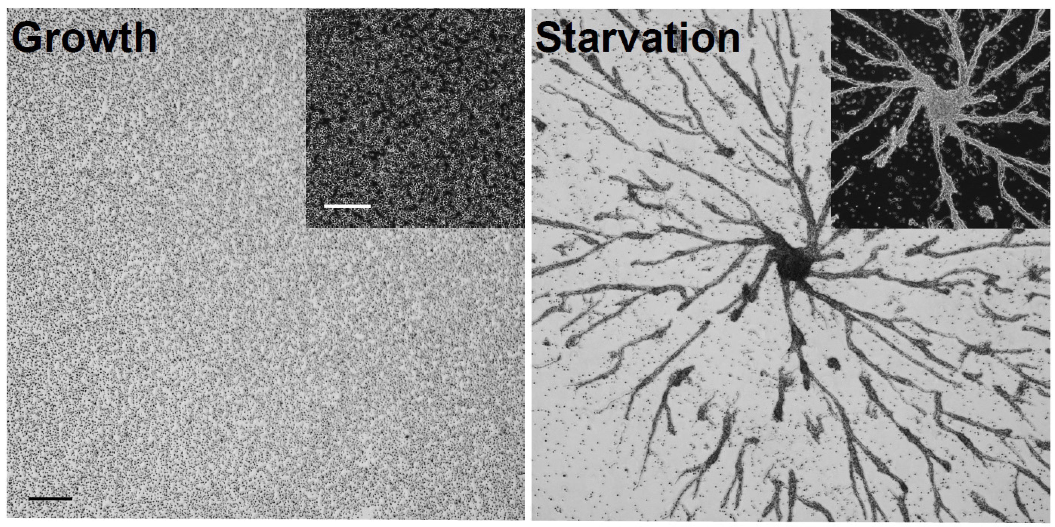
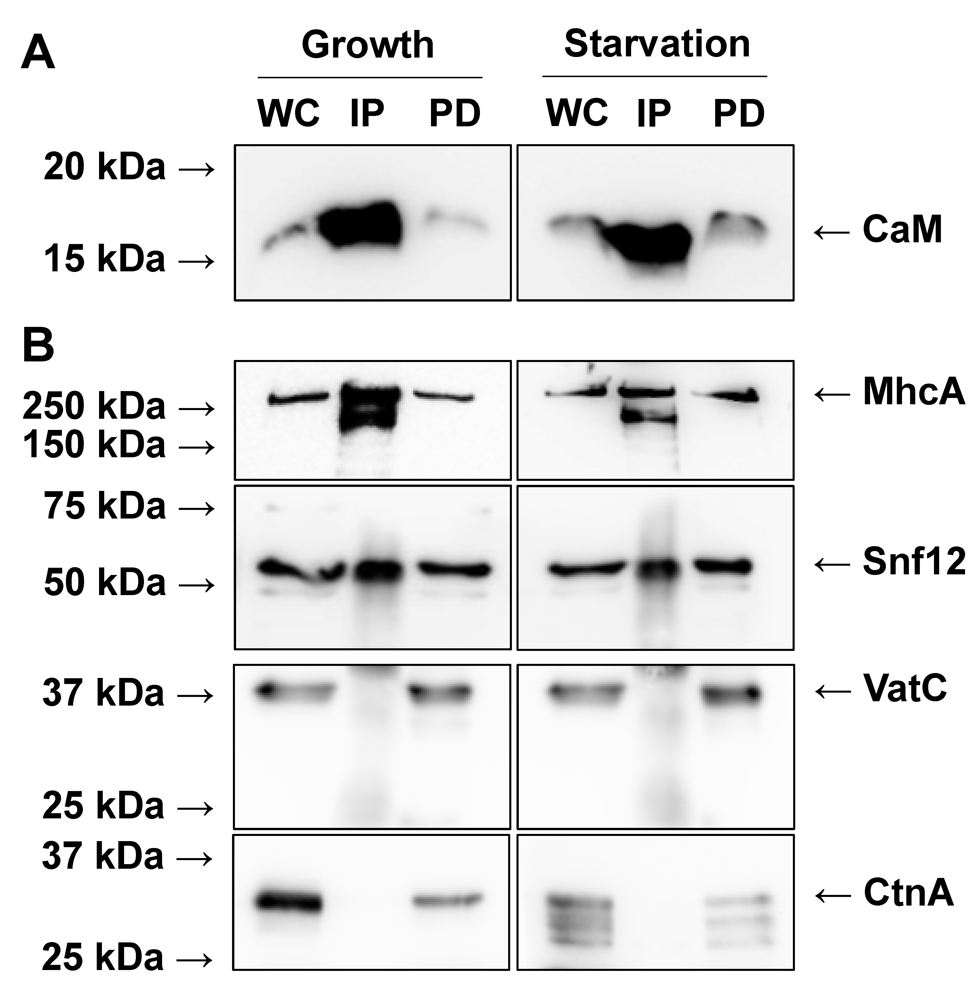
2.1. Immunoprecipitation of CaM from Growth-Phase and Starved Dictyostelium Cells

To determine putative CaM interactors during growth and the early stages of Dictyostelium development, we deposited cells in Petri dishes and submerged them in HL5 growth medium overnight at room temperature. The following day, confluent growth-phase cells were harvested (Figure 1). Cells were also starved for 6 h in KK2 buffer and harvested (Figure 1). After 6 h, cells were aggregating into multicellular mounds, which was consistent with the normal timing of Dictyostelium development [28]. A total of three biological replicates were harvested for both growth and starved conditions. CaM was immunoprecipitated from each biological replicate using a well-established antibody that was previously generated against Dictyostelium CaM [29]. Western blotting confirmed the immunoprecipitation of CaM from growth-phase and starved cells (Figure 2A).

2.2. Mass Spectrometry Reveals CaM Interactors during Growth and Starvation

CaM IP samples from growth-phase and starved cells were analyzed by mass spectrometry (LC-MS/MS), which revealed 517 putative CaM interactors during growth and 521 putative interactors during starvation (Supplementary Materials Table S1). 374 CaM interactors were common to both conditions, with 143 proteins identified only in growth samples, and 147 proteins unique to starved samples (Table S2). Mass spectrometry detected CaM in all biological replicates that were analyzed (Table S1). The Dictyostelium genome also encodes a calcium-binding CaM-like protein called CalB, that has about 50% similarity to other CaMs [30]. Mass spectrometry did not detect CalB in CaM IP fractions. To validate the mass spectrometry results, we confirmed that proteins present in our dataset could be detected via western blotting (positive controls). Conversely, we also validated that proteins absent in our dataset could not be detected (negative controls). For our positive controls, we confirmed the presence of myosin II heavy chain (MhcA) and SWI/SNF protein 12 (Snf12) in CaM IP fractions from growth and starved samples (Figure 2B). MhcA is a known CaMBP in Dictyostelium [31]. Previous work showed that Snf12, which is a homolog of yeast SNF12 and human SMARCD1 (involved in chromatin remodelling), localizes to the nucleolus and is a potential binding partner of nucleomorphin A (NumA), a previously identified CaMBP [18,32]. For our negative controls, we confirmed the absence of the catalytic subunit of V-ATPase (VatC) and countin (CtnA) in CaM IP samples since neither of these proteins were revealed in our mass spectrometry analysis or are predicted to bind to CaM (Figure 2B). In total, these results support the specificity of the CaM IP and the catalogue of putative interactors revealed by mass spectrometry (i.e., the presence or absence of specific proteins from the dataset were validated by western blotting).

2.3. Mass Spectrometry Reveals Previously Known CaMBPs in Dictyostelium

Several of the proteins identified by mass spectrometry have the gene ontology (GO) term identifier “calmodulin-binding”. These include known or predicted CaMBPs in Dictyostelium including calcineurin (CanA), WW domain-containing protein A (DwwA), histone H1 (H1), proteins in the myosin heavy chain family (MhcA, MyoA, MyoE, MyoJ), Ras GTPase-activating-like protein (RgaA), and 60S ribosomal protein L19 (Rpl19) (Table 1, Table S1) [16,17,21,31,33]. All these proteins were detected in growth-phase and starved cells except CanA and RgaA, which were present only in starved samples (Table 1, Table S1). The online bioinformatics resource for the Dictyostelium research community, dictyBase, provides accurate and updated annotations for curated genes and proteins [34]. Three proteins with the descriptor “calmodulin” or “calmodulin-binding” were revealed by mass spectrometry and included H1 (discussed above), guanine exchange factor for rac II (GxcII), and guanine exchange factor for rac JJ (GxcJJ) (Table 1, Table S1). All three proteins were present in growth and starved samples except GxcJJ, which was only detected in CaM IP fractions isolated from starved cells (Table 1, Table S1). In addition to the proteins described above, several known or predicted CaMBPs reviewed by O’Day et al [6] were detected by mass spectrometry including myosin heavy chain kinase B (MhkB) (starved only), the V-ATPase membrane subunit (VatM) (growth only), and additional members of the myosin heavy chain family (MyoB, MyoC, MyoD, MyoI) (Table 2, Table S1). MyoB, MyoD, and MyoI were detected in CaM IP fractions from both growth-phase and starved cells, while MyoC was detected only in growth samples (Table 2, Table S1). Finally, mass spectrometry also revealed homologs of known CaMBPs in mammals such as casein kinase I and II (Cak1-1, Cak1-2, CasK, Csnk2b), which were identified in both growth and starved samples [35]. Together, these findings show that mass spectrometry identified established CaMBPs in Dictyostelium, which provides additional evidence supporting the list of CaM interactors.

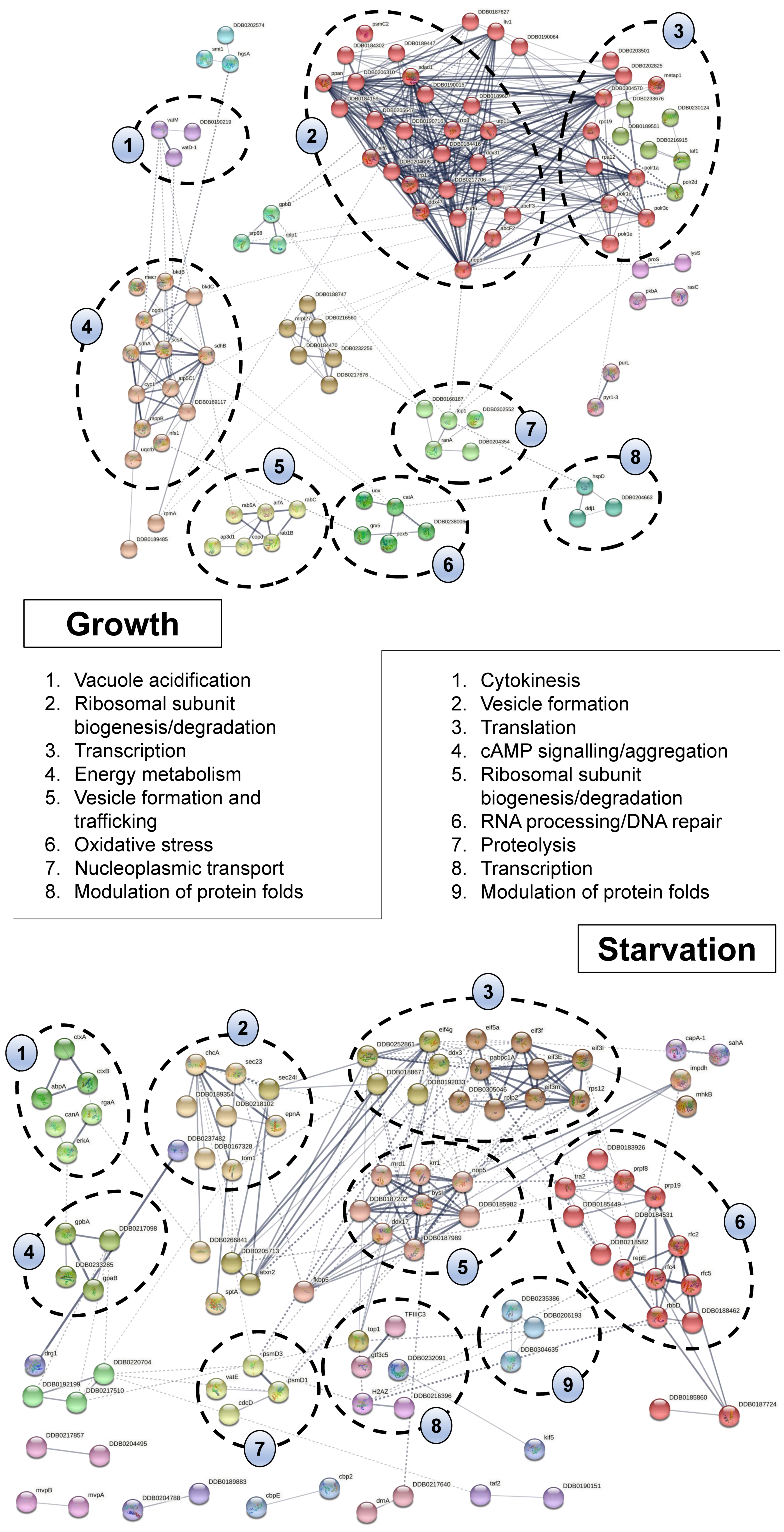
2.4. Bioinformatic Analyses Highlight the Diverse Cellular Roles of CaM Interactors in Dictyostelium

To further analyze our list of putative CaM interactors (Table S1), we performed a GO term enrichment analysis (Tables S3 and S4). When assessing “biological process”, 65% (337/517) of proteins in growth samples and 61% (318/517) of proteins in starved samples are associated with metabolic processes (Table 3). Other highly enriched GO term categories included gene expression (45% growth, 44% starved), biosynthetic process (42% growth, 39% starved), translation (28% growth, 27% starved), cytoskeleton organization (10% growth, 12% starved), endocytosis (6% growth, 6% starved), and cytokinesis (3% growth, 4% starved) (Table 3).
We also examined the “molecular function” of the CaM interactors. Most of the proteins from both growth and starved samples are involved in binding (67% growth, 68% starved) (e.g., nucleic acid, ion, RNA, anion, carbohydrate-derivative, ATP) (Table 4). In growth samples, 39% (202/517) of the proteins have catalytic activity (Table 4). Interestingly, this GO term category was not enriched in starved samples. However, CaM interactors in both growth and starved samples have hydrolase activity (14% growth, 12% starved), pyrophosphatase activity (14% growth, 12% starved), and ATPase activity (7% growth, 7% starved) (Table 4). Like catalytic activity, GTP binding and GTPase activity were only enriched for proteins identified in growth samples (Table 4). In contrast, calcium-binding activity was only enriched in starved samples (Table 4).
The localizations of proteins identified in growth and starved samples were diverse. The largest proportion of proteins in both samples localize to the nucleus (28% growth, 30% starved) (Table 5). The next highly enriched GO term category was cytosol, which includes the fluid enclosed within the plasma membrane (24% growth, 23% starved) (Table 5). Other highly enriched GO term categories included ribosome (24% growth, 22% starved), vesicle (20% growth, 20% starved), mitochondrion (17% growth, 12% starved), extracellular region (13% growth, 13% starved), and cytoskeleton (9% growth, 10% starved) (Table 5). In addition, some GO terms were only associated with either growth or starved samples. For example, mitochondrial inner membrane and mitochondrial respiratory chain were only enriched in growth samples (Table 5). In contrast, cell periphery was only enriched for proteins in starved samples (Table 5).
To gain additional insight into the pathways that the putative CaM interactors belong to, the proteins unique to each condition (Table S2) were analyzed using the Search Tool for the Retrieval of Interacting proteins (STRING) database [36]. STRING predicts protein associations based on direct interactions or participation in common biological pathways. STRING analysis on the list of CaM interactors unique to growth revealed eight processes including vacuolar acidification, energy metabolism, vesicle formation and trafficking, oxidative stress, nucleoplasmic transport, protein-fold regulation, transcription, and ribosomal subunit synthesis and degradation (Figure 3). Nine processes were identified for proteins unique to starvation including cytokinesis, cAMP signalling, vesicle formation, proteolysis, translation, ribosomal subunit synthesis and degradation, transcription, modulation of protein folds, and RNA processing/DNA repair responses (Figure 3). In total, these results highlight the diverse localizations and functions of the putative CaM interactors in Dictyostelium.

2.5. Mass Spectrometry Reveals Novel CaM Interactors in Dictyostelium with Biomedical Signficance

Mass spectrometry revealed novel CaM interactors with biomedical significance. One example was the Dictyostelium homolog of human cathepsin D (human: CTSD; Dictyostelium: CtsD), which functions as a lysosomal aspartyl protease, and was present in both growth and starved samples. Mutations in CTSD are associated with several human diseases including breast cancer, Alzheimer’s disease, Parkinson’s disease, and a subtype of NCL (Batten disease) [37,38,39,40]. In addition, the Dictyostelium homolog human hexosaminidase A (HEXA), N-acetylglucosaminidase A (NagA), was detected in growth samples. Mutations in HEXA cause Tay-Sachs disease in humans, which is a neurological disorder characterized by an accumulation of GM2 gangliosides in neurons [41]. Combined, these findings provide additional evidence supporting the biomedical significance of CaM and CaMBPs.

3. Discussion

In this study, we used a highly specific and well-characterized antibody to immunoprecipitate CaM (also known as CalA) from growth-phase and starved Dictyostelium cells. We then used mass spectrometry (LC-MS/MS) to reveal over 500 putative CaM interactors in IP fractions. CaM was detected in all IP fractions that were analyzed by mass spectrometry. CalB, a CaM-like protein that shares about 50% similarity with other CaMs was not, indicating that the anti-CaM antibody was specific for CalA. We used western blotting to validate the accuracy of the mass spectrometry analysis. Specifically, we confirmed the presence of MhcA and Snf12 in CaM IP fractions and the absence of VatC and CtnA. The list of proteins was further validated by the presence of known CaMBPs in the dataset including CanA, H1, and proteins belonging to the myosin family, among others. In addition, our analysis revealed several known CaMBPs as well as homologs of proteins associated with human diseases. Finally, bioinformatic analyses highlighted key processes mediated by the putative CaM interactors, thus enhancing our understanding of CaM-mediated signal transduction in Dictyostelium.
In Dictyostelium, CaM and CaMBPs have been linked to a variety of cellular and developmental processes including mitosis, phagocytosis, autophagy, osmoregulation, cell motility and chemotaxis, cell differentiation, and spore germination [12]. Here, GO term enrichment and STRING analyses linked the putative CaM interactors to a diversity of cellular processes including, but not limited to, metabolism, gene expression, biosynthetic processes, cytoskeleton organization, endocytosis, oxidative stress, and cytokinesis. Many ribosomal proteins were also detected in growth and starved samples, which is consistent with similar large-scale studies on putative CaMBPs in human cells [7]. GO term enrichment analyses also revealed that the putative CaM interactors localize throughout the cell, especially in nuclei, ribosomes, vesicles, mitochondria, and the cytoskeleton. In addition, 13% of the putative CaM interactors localize outside the cell. Consistent with this finding, extracellular CaM has been reported in Dictyostelium where it regulates growth and cAMP-mediated chemotaxis [24,42]. STRING analysis of proteins unique to starvation revealed 6 proteins linked to cytokinesis, which aligns with previous findings showing that a round of cytokinesis occurs early in development to allow multinucleated cells to complete their final growth-stage cell cycle [43,44]. Combined, these findings are consistent with the previously reported localizations of CaMBPs in Dictyostelium and the cellular and developmental roles of CaM and CaMBPs in Dictyostelium [6,12].
The functions of CaM and CaMBPs in mitosis and cytokinesis in Dictyostelium have been well documented [12,45]. These roles are consistent with the nuclear localization of CaM and CaMBPs such as Snf12 and Cbp4a, which were detected in CaM IP fractions [26,32,46]. In this study, mass spectrometry revealed DwwA as a CaM interactor in both growth and starved cells. DwwA contains domains that act as scaffolds for proteins involved in cytokinesis [21]. Previous work also demonstrated the importance of CaM-dependent phosphorylation and CaMBPs in regulating folic acid and cAMP-mediated chemotaxis in Dictyostelium [47]. Not surprisingly, proteins with catalytic or hydrolase activity were identified by mass spectrometry including phospholipase C, phosphatidylinositol-3-phosphatase, 3-hydroxyisobutyryl-CoA hydrolase, alpha mannosidase, and Ras proteins, among others. Cytoskeletal proteins (e.g., members of the myosin family), which are essential for cell motility and chemotaxis, were also identified by mass spectrometry. For example, annexin A7 was identified as a putative CaM interactor in starved cells. In mice, annexin A7 has been linked to the inflammatory response and calcium homeostasis in the brain [48,49]. In humans, annexin A7 appears to have a tumour-suppressive role in some forms of cancer (e.g., glioblastoma), but in others, it seems to promote malignancy (e.g., gastric cancer) [50]. Recently, CaM and annexin were shown to regulate wound repair in Dictyostelium by facilitating the accumulation of actin at the wound site [51]. In total, these observations highlight the essential roles of CaMBPs in processes regulated by the cytoskeleton.
The Dictyostelium homologs of human HEXA (NagA) and CTSD (CtsD) were identified in CaM IP fractions. Mutations in HEXA cause Tay-Sachs disease, while mutations in CTSD are associated with breast cancer, Alzheimer’s disease, Parkinson’s disease, and a subtype of NCL called CLN10 disease [37,38,39,40,41]. The identification of NagA and CtsD in Dictyostelium CaM IP fractions suggests that CaM-mediated signalling may play an important role in the pathology underlying both diseases. Consistent with these findings, previous work identified two regions in human CTSD that contain putative CaMBDs [11]. In addition, CTSD function has been linked to calcineurin, a known CaMBP in humans [52]. CTSD, together with calcineurin, have been shown to protect against alpha-synuclein toxicity in a yeast model of Parkinson’s disease [53]. Calcineurin has also been linked to autoimmune diseases (e.g., rheumatoid arthritis), psychiatric diseases (e.g., schizophrenia, bipolar disorder), and hypertension [54,55,56]. Importantly, the Dictyostelium homolog of human calcineuin, CanA, was identified by mass spectrometry in starved cells. Mass spectrometry also identified the Dictyostelium homologs of casein kinase I and II in CaM IP fractions. In humans, mutations in casein kinase I and II have been associated with abnormal circadian rythym and many forms of cancer [57,58]. In total, these results provide additional evidence linking CaM and CaMBPs to human diseases.
Mass spectrometry has also been used to reveal putative CaM interactors in human samples. For example, 297 CaM interactors were identified in various human tissues (brain, heart, spleen, thymus, and muscle) [7]. Among the identified proteins were DEAD box proteins, ribosomal proteins, and proteasomal subunits, which intriguingly, were all identified as putative CaM interactors in Dictyostelium (Table S1). In addition, mass spectrometry performed on proteins pulled down with affinity-tagged CaM identified 489 CaM interactors in HEK293 cells [59]. Combined, these findings show that our detection of over 500 putative CaM interactors in Dictyostelium is in line with studies in humans.
It is important to acknowledge that our dataset may not include all CaMBPs in Dictyostelium. For example, mass spectrometry did not reveal previously known CaMBPs such as the well characterized regulator of nuclear number, NumA [18]. However, the NumA-interactors, calcium-binding protein 4a (Cbp4a) (growth) and Snf12 (growth and starvation) were identified, suggesting that the interaction between NumA and CaM may have been sensitive to our IP protocol [19,32,46]. Another explanation for the exclusion of NumA and other proteins previously shown to bind CaM from our dataset (e.g., Cdk5) could be that those proteins bind to a region of CaM where the antibody binds. Therefore, our IP may have pulled down NumA-free CaM. Unfortunately, since the epitope recognized by the CaM antibody is not known, it is not possible for us to confirm this hypothesis. Technical limitations could also explain anomalies. For example, we found that we needed to extensively wash IP fractions to ensure a strong detection of CaM by LC-MS/MS. As a result, our list of putative CaMBPs may exclude weak interactors that were removed during washing. Our list may also exclude those proteins that are weakly expressed during growth and starvation.
A limitation of this study was the inability to assess whether the interactions with CaM were calcium-dependent or calcium-independent, as is done with the CaMBOT [27]. However, it is important to note that the culture medium, starvation buffer, and lysis buffer used in this study did not contain free calcium. Therefore, we speculate that putative interactors identified in CaM IP fractions were likely bound to CaM prior to cell lysis. One major benefit of our analysis was that we revealed interactions that occur in vivo under standard culturing conditions. This may explain why some CaMBPs that were previously identified in in vitro methods were not detected in our analysis. Finally, it is important to acknowledge that the proteins we revealed may not all bind CaM directly. Instead, they may interact with CaMBPs that were pulled down with CaM in the IP. In addition, some of the putative interactors could function as scaffolds that recruit non-CaMBPs (e.g., DwwA). However, since our analysis revealed previously verified CaMBPs (e.g., MhcA, CanA), and we extensively washed our IP fractions prior to LC-MS/MS, we are confident our dataset provides a thorough catalogue of putative CaMBPs in Dictyostelium. Finally, we cannot rule out the possibility that some of the proteins we identified were false positives. For that reason, we encourage any researcher using this catalogue of putative CaM interactors in Dictyostelium to independently validate proteins of interest. On the other hand, since we did not detect some proteins that were previously identified as CaMBPs in Dictyostelium, our catalogue might in fact underrepresent the number of CaM interactors in Dictyostelium during growth and starvation.
In summary, this study provides the first catalogue of putative CaM interactors in Dictyostelium, which provides a valuable resource for those interested in studying the functions of CaM and CaMBPs in this model organism. Intriguingly, we did not observe a dramatic difference in the list of CaM interactors between growth and starvation, despite the significant shift in the life cycle stages (i.e., 374 CaM interactors were shared between growth and starvation). These findings indicate that CaM mediates events essential to both, and possibly all stages of the Dictyostelium life cycle. Along the same lines, some of the putative CaM interactors were identified in either growth or starved samples, but not both, suggesting that the binding of CaM to specific CaMBPs is dynamic, and that CaM may bind to different proteins at different stages of the Dictyostelium life cycle. Finally, GO term enrichment analyses revealed that only 7 of the 517 proteins identified during growth, and 9 of the 521 proteins identified during starvation, have the GO term “calmodulin-binding” associated with them suggesting that much remains to be learned about the diverse cellular and developmental roles of CaM and CaMBPs in Dictyostelium.

4. Materials and Methods

4.1. Cell Culture, Media, and Antibodies

AX3 cells were grown and maintained on SM/2 agar with Klebsiella aerogenes [28]. Cells were also grown axenically in HL5 medium at room temperature and 150 rpm. For all experiments, cells were harvested in the mid-log phase of growth (1−5 × 10⁶ cells/mL). Cultures were supplemented with 100 μg/mL ampicillin and 300 μg/mL streptomycin sulfate to prevent bacterial growth. HL5 was purchased from Formedium (Hunstanton, Norfolk, UK). KK2 buffer was composed of 0.7 g/L K2HPO4 and 2.2 g/L KH2PO4, pH 6.5. Mouse monoclonal anti-CaM (clone 6D4, C3545) was purchased from Sigma Aldrich Canada (Oakville, ON, Canada). Mouse monoclonal anti-MhcA (56-396-5) and mouse monoclonal anti-VatC (224-256-2) were purchased from the Developmental Studies Hybridoma Bank (University of Iowa, Iowa City, IA, USA) [60,61]. Rabbit polyclonal anti-Snf12 was generously provided as a gift by Dr. Danton O’Day (University of Toronto, Toronto, ON, Canada) [32]. Rabbit polyclonal anti-CtnA was generously provided as a gift by Dr. Richard Gomer (Texas A&M University, College Station, TX, USA) [62]. Goat anti-rabbit and horse anti-mouse IgG linked to HRP were purchased from New England Biolabs Canada (Whitby, ON, Canada).

4.2. Immunoprecipitation

Growth-phase cells were deposited into tissue culture dishes and grown overnight in HL5 to confluency. Cells were washed two times with KK2 buffer and then lysed with NP40 lysis buffer (50 mM Tris HCl, 150 mM NaCl, 0.1% NP40, pH 8.0) supplemented with a protease inhibitor tablet (Fisher Scientific Company, Ottawa, ON, Canada). Cells were also starved for 6 h in KK2 buffer prior to being lysed with NP40 lysis buffer. CaM was immunoprecipitated from whole cell lysates using SureBeads Protein G Magnetic Beads according to the manufacturer’s instructions (Bio-Rad Laboratories Limited, Mississauga, ON, Canada). Briefly, lysate (2 mg) was incubated with anti-CaM (10 µL) overnight at 4 °C on a tube rotator (head-over-tail rotation). The following day, the antibody/lysate solution was incubated with 50 μL of SureBeads pre-washed with NP40 lysis buffer (1 mL total volume) for 4 h at 4 °C on a tube rotator (head-over-tail rotation). After 4 h, a magnetic rack was used to separate the SureBeads from the supernatant (i.e., protein-depleted fraction). SureBeads were washed three times with 1 mL of NP40 lysis buffer and then once with 1 mL of buffer containing 50 mM Tris, 150 mM NaCl, pH 8.0. Isolated SureBeads were stored at −80 °C for future use.

4.3. SDS-PAGE and Western Blotting

CaM IP fractions were isolated by resuspending SureBeads in Laemmli sample buffer [63]. Samples were then heated at 95 °C for 5 min, after which time the supernatants were removed. Protein quantification was performed using a Qubit 2.0 Fluorometer (Fisher Scientific Company, Ottawa, ON, Canada). SDS-PAGE and western blotting for CaM was performed using a method described elsewhere [29]. SDS-PAGE and western blotting for all other proteins was performed using standard methods (2-h incubation at room temperature for primary and secondary antibodies in 5% milk/TBST). A 1:1000 dilution was used for all primary antibodies and a 1:2000 dilution for secondary antibodies. Immunoblots were digitally scanned using the ChemiDoc MP Imaging System (Bio-Rad Laboratories Limited, Mississauga, ON, Canada).

4.4. Mass Spectrometry and Bioinformatic Analyses

CaM IP fractions were submitted for analysis to the Mass Spectrometry Facility of the SPARC BioCentre at the Hospital for Sick Children (Toronto, ON, Canada). Briefly, samples (i.e., washed and pelleted SureBeads) were reduced with 10 mM DTT for 1 h at 60 °C, alkylated with 20 mM iodoacetamide for 45 min in the dark, and digested with trypsin (2 µg per sample) overnight at 37 °C. Samples were then lyophilized using a Speedvac vacuum concentrator, desalted using a C18 ziptip, and resuspended in 0.1% formic acid. Mass spectrometry (LC-MS/MS) was performed using the EASY-nLC 1200 nano-LC system and a Thermo Scientific Q Exactive HF-X mass spectrometer (Fisher Scientific Company, Ottawa, ON, Canada). Protein identification and quantification were performed using Proteome Discoverer (MS Amanda and Sequest HT) and Scaffold (X! Tandem), which were setup to search the Uniprot_UP000002195_Dictyostelium_discoideum_15092020 database according to the following parameters: parent mass error tolerance, 10 ppm; fragment mass error tolerance, 0.02 Da; enzyme, trypsin; max missed cleavages, 3; fixed modifications, carbamidomethylation (C +57.02); variable modifications, oxidation (M +15.99), deamidation (N,Q +0.98), acetylation (peptide N-term +42.01). Scaffold version 4.11.1 (Proteome Software Inc., Portland, OR, USA) was used to validate MS/MS based peptide and protein identifications. Peptide and protein identifications were accepted if they could be established at greater than 95% probability. Protein identifications were further accepted if they contained at least 2 identified peptides and were present in at least 2 of the 3 biological replicates analyzed for each treatment (growth and 6-h starvation). GO term enrichment analyses were performed using LAGO (A Logically Accelerated GO Term Finder) [64]. A p-value cut-off of 0.05 and the Bonferroni correction were applied for all GO term enrichment analyses. Putative CaM interactors were also analyzed using STRING database version 11, which generates a network of predicted associations for a particular group of proteins [36].

Supplementary Materials

Supplementary materials can be found at https://www.mdpi.com/1422-0067/22/4/1722/s1.

Author Contributions

Conceptualization: R.J.H.; Data curation: W.D.K.; S.Q.Y. and R.J.H.; Formal analysis: W.D.K.; S.Q.Y. and R.J.H.; Funding acquisition: R.J.H.; Investigation: W.D.K. and S.Q.Y. Methodology: R.J.H. Project administration: R.J.H. Supervision: R.J.H. Writing—original draft: W.D.K.; S.Q.Y. and R.J.H. Writing—review & editing: W.D.K. and R.J.H. Writing—revisions for resubmission: R.J.H. All authors have read and agreed to the published version of the manuscript.

Funding

This study was supported by a Discovery Grant from the Natural Sciences and Engineering Research Council of Canada (RGPIN-2018–04855 to R.J.H.). W.D.K. was supported by a Queen Elizabeth II Graduate Scholarship in Science and Technology. S.Q.Y. was supported by an Ontario Graduate Scholarship.

Acknowledgments

The authors would like to thank Danton O’Day (University of Toronto) for critically reviewing this paper prior to submission and for generously providing antibodies against Snf12. Richard Gomer (Texas A&M University) is thanked for generously providing antibodies against CtnA.

Conflicts of Interest

The authors declare no conflict of interest.

Abbreviations

CaMcalmodulin
CaMBPCaM-binding protein
CaMBDCaM-binding domain
CaMBOTCaM-binding overlay technique
cAMPcyclic adenosine monophosphate
CanAcalcineurin A
CtnAcountin
CTSDcathepsin D
DwwAWW domain-containing protein A
GOGene ontology
GxcJJguanine exchange factor for rac JJ
H1histone H1
LC-MS/MSliquid chromatography-mass spectrometry/mass spectrometry
HEXAhexosaminidase A
IPimmunoprecipitation
MhcAmyosin II heavy chain
NagAN-acetylglucosaminidase A
NCLneuronal ceroid lipofuscinosis
NumAnucleomorphin A
RgaARas GTPase-activating-like protein
Snf12SWI/SNF protein 12
VatCV-ATPase catalytic subunit

References

  1. Krause, K.H. Ca2+-storage organelles. FEBS Lett. 1991, 285, 225–229. [Google Scholar] [CrossRef] [Green Version]
  2. Arruda, A.P.; Hotamisligil, G.S. Calcium homeostasis and organelle function in the pathogenesis of obesity and diabetes. Cell Metab. 2015, 22, 381–397. [Google Scholar] [CrossRef] [Green Version]
  3. Heizmann, C.W. Ca2+-Binding proteins of the EF-hand superfamily: Diagnostic and prognostic biomarkers and novel therapeutic targets. Adv. Struct. Saf. Stud. 2019, 1929, 157–186. [Google Scholar] [CrossRef]
  4. Zielinski, R.E. Calmodulin and calmodulin-binding proteins in plants. Annu. Rev. Plant. Biol. 1998, 49, 697–725. [Google Scholar] [CrossRef] [Green Version]
  5. Hoeflich, K.P.; Ikura, M. Calmodulin in action: Diversity in target recognition and activation mechanisms. Cell 2002, 108, 739–742. [Google Scholar] [CrossRef] [Green Version]
  6. O’Day, D.H.; Taylor, R.J.; Myre, M.A. Calmodulin and calmodulin binding proteins in Dictyostelium: A primer. Int. J. Mol. Sci. 2020, 21, 1210. [Google Scholar] [CrossRef] [Green Version]
  7. Shen, X.; Valencia, C.A.; Szostak, J.W.; Dong, B.; Liu, R. Scanning the human proteome for calmodulin-binding proteins. Proc. Natl. Acad. Sci. USA 2005, 102, 5969–5974. [Google Scholar] [CrossRef] [PubMed] [Green Version]
  8. Berchtold, M.W.; Villalobo, A. The many faces of calmodulin in cell proliferation, programmed cell death, autophagy, and cancer. Biochim. Biophys. Acta. Mol. Cell Res. 2014, 1843, 398–435. [Google Scholar] [CrossRef]
  9. O’Day, D.H. Calmodulin binding proteins and Alzheimer’s disease: Biomarkers, regulatory enzymes and receptors that are regulated by calmodulin. Int. J. Mol. Sci. 2020, 21, 7344. [Google Scholar] [CrossRef] [PubMed]
  10. Chazin, W.J.; Johnson, C.N. Calmodulin mutations associated with heart arrhythmia: A status report. Int. J. Mol. Sci. 2020, 21, 1418. [Google Scholar] [CrossRef] [PubMed] [Green Version]
  11. Mathavarajah, S.; O’Day, D.H.; Huber, R.J. Neuronal ceroid lipofuscinoses: Connecting calcium signalling through calmodulin. Cells 2018, 7, 188. [Google Scholar] [CrossRef] [Green Version]
  12. O’Day, D.H.; Mathavarajah, S.; Myre, M.A.; Huber, R.J. Calmodulin-mediated events during the life cycle of the amoebozoan Dictyostelium discoideum. Biol. Rev. 2019, 95, 472–490. [Google Scholar] [CrossRef] [Green Version]
  13. Mathavarajah, S.; Flores, A.; Huber, R.J. Dictyostelium discoideum: A model system for cell and developmental biology. Curr. Protoc. Essent. Lab. Tech. 2017, 15, 14.1.1–14.1.19. [Google Scholar] [CrossRef]
  14. Myre, M.A.; Huber, R.J.; O’Day, D.H. Chapter 21. Functional analysis of proteins involved in neurodegeneration using the model organism Dictyostelium: Alzheimer’s, Huntington’s and Batten disease. In Molecular-Genetic and Statistical Techniques for Behavioral and Neural Research, 1st ed.; Gerlai, R.T., Ed.; Academic Press Elsevier: San Diego, CA, USA, 2018; pp. 491–518. [Google Scholar]
  15. Clarke, M.; Bazari, W.L.; Kayman, S.C. Isolation and properties of calmodulin from Dictyostelium discoideum. J. Bacteriol. 1980, 141, 397–400. [Google Scholar] [CrossRef] [Green Version]
  16. Sonnemann, J.; Bäuerle, A.; Winckler, T.; Mutzel, R. A ribosomal calmodulin-binding protein from Dictyostelium. J. Biol. Chem. 1991, 266, 23091–23096. [Google Scholar] [CrossRef]
  17. Dammann, H.; Hellstern, S.; Husain, Q.; Mutzel, R. Primary structure, expression and developmental regulation of a Dicty-ostelium calcineurin A homologue. Eur. J. Biochem. 1996, 238, 391–399. [Google Scholar] [CrossRef] [PubMed]
  18. Myre, M.A.; O’Day, D.H. Nucleomorphin. A novel, acidic, nuclear calmodulin-binding protein from Dictyostelium that regu-lates nuclear number. J. Biol. Chem. 2002, 277, 19735–19744. [Google Scholar] [CrossRef] [Green Version]
  19. Myre, M.A.; O’Day, D.H. Dictyostelium calcium-binding protein 4a interacts with nucleomorphin, a BRCT-domain protein that regulates nuclear number. Biochem. Biophys. Res. Commun. 2004, 322, 665–671. [Google Scholar] [CrossRef]
  20. Myre, M.A.; O’Day, D.H. Calmodulin binds to and inhibits the activity of phosphoglycerate kinase. Biochim. Biophys. Acta Mol. Cell Res. 2004, 1693, 177–183. [Google Scholar] [CrossRef] [Green Version]
  21. Nagasaki, A.; Uyeda, T.Q. DWWA, a novel protein containing two WW domains and an IQ motif, is required for scission of the residual cytoplasmic bridge during cytokinesis in Dictyostelium. Mol. Biol. Cell 2004, 15, 435–446. [Google Scholar] [CrossRef] [Green Version]
  22. O’Day, D.H.; Chatterjee-Chakraborty, M.; Wagler, S.; Myre, M.A. Isolation and characterization of Dictyostelium thymidine kinase 1 as a calmodulin-binding protein. Biochem. Biophys. Res. Commun. 2005, 331, 1494–1502. [Google Scholar] [CrossRef]
  23. O’Day, D.H.; Suhre, K.; Myre, M.A.; Chatterjee-Chakraborty, M.; Chavez, S.E. Isolation, characterization, and bioinformatic analysis of calmodulin-binding protein cmbB reveals a novel tandem IP22 repeat common to many Dictyostelium and Mim-ivirus proteins. Biochem. Biophys. Res. Commun. 2006, 346, 879–888. [Google Scholar] [CrossRef]
  24. Suarez, A.; Huber, R.J.; Myre, M.A.; O’Day, D.H. An extracellular matrix, calmodulin-binding protein from Dictyostelium with EGF-like repeats that enhance cell motility. Cell. Signal. 2011, 23, 1197–1206. [Google Scholar] [CrossRef]
  25. Sriskanthadevan, S.; Brar, S.K.; Manoharan, K.; Siu, C.H. Ca(2+)-calmodulin interacts with DdCAD-1 and promotes DdCAD-1 transport by contractile vacuoles in Dictyostelium cells. FEBS J. 2013, 280, 1795–1806. [Google Scholar] [CrossRef]
  26. Huber, R.J.; Catalano, A.; O’Day, D.H. Cyclin-dependent kinase 5 is a calmodulin-binding protein that associates with puromycin-sensitive aminopeptidase in the nucleus of Dictyostelium. Biochim. Biophys. Acta Mol. Cell Res. 2013, 1833, 11–20. [Google Scholar] [CrossRef] [Green Version]
  27. O’Day, D.H. CaMBOT: Profiling and characterizing calmodulin-binding proteins. Cell. Signal. 2003, 15, 347–354. [Google Scholar] [CrossRef]
  28. Fey, P.; Kowal, A.S.; Gaudet, P.; Pilcher, K.E.; Chisholm, R.L. Protocols for growth and development of Dictyostelium discoideum. Nat. Protoc. 2007, 2, 1307–1316. [Google Scholar] [CrossRef] [PubMed]
  29. Hulen, D.; Baron, A.T.; Salisbury, J.; Clarke, M. Production and specificity of monoclonal antibodies against calmodulin from Dictyostelium discoideum. Cell Motil. Cytoskelet. 1991, 18, 113–122. [Google Scholar] [CrossRef] [PubMed]
  30. Rosel, D.; Půta, F.; Blahůšková, A.; Smykal, P.; Folk, P. Molecular characterization of a calmodulin-like Dictyostelium protein CalB. FEBS Lett. 2000, 473, 323–327. [Google Scholar] [CrossRef] [Green Version]
  31. Catalano, A.; O’Day, D.H. Calmodulin-binding proteins in the model organism Dictyostelium: A complete & critical review. Cell. Signal. 2008, 20, 277–291. [Google Scholar] [CrossRef]
  32. Catalano, A.; O’Day, D.H. Nucleoplasmic/nucleolar translocation and identification of a nuclear localization signal (NLS) in Dictyostelium BAF60a/SMARCD1 homologue Snf12. Histochem. Cell Biol. 2012, 138, 515–530. [Google Scholar] [CrossRef]
  33. O’Day, D.H. Calmodulin-mediated signaling in Dictyostelium discoideum: CaMBOT isolation and characterization of the novel poly-domain protein nucleomorphin and other calmodulin binding proteins. In Focus on Cellular Signalling Research; Leeds, D., Ed.; Nova Science Publishers, Inc.: New York, NY, USA, 2006; pp. 131–153. [Google Scholar]
  34. Fey, P.; Dodson, R.J.; Basu, S.; Hartline, E.C.; Chisholm, R.L. dictyBase and the Dicty Stock Center (version 2.0)—A progress report. Int. J. Dev. Biol. 2019, 63, 563–572. [Google Scholar] [CrossRef] [Green Version]
  35. Sacks, D.B.; Davis, H.W.; Williams, J.P.; Sheehan, E.L.; Garcia, J.G.N.; McDonald, J.M. Phosphorylation by casein kinase II alters the biological activity of calmodulin. Biochem. J. 1992, 283, 21–24. [Google Scholar] [CrossRef] [PubMed] [Green Version]
  36. Szklarczyk, D.; Gable, A.L.; Lyon, D.; Junge, A.; Wyder, S.; Huerta-Cepas, J.; Simonovic, M.; Doncheva, N.T.; Morris, J.H.; Bork, P.; et al. STRING v11: Protein–protein association networks with increased coverage, supporting functional discovery in genome-wide experimental datasets. Nucleic Acids Res. 2019, 47, D607–D613. [Google Scholar] [CrossRef] [Green Version]
  37. Briozzo, P.; Morisset, M.; Capony, F.; Rougeot, C.; Rochefort, H. In vitro degradation of extracellular matrix with Mr 52,000 cathepsin D secreted by breast cancer cells. Cancer Res. 1988, 48, 3688–3692. [Google Scholar] [PubMed]
  38. Bernstein, H.G.; Bruszis, S.; Schmidt, D.; Wiederanders, B.; Dorn, A. Immunodetection of cathepsin D in neuritic plaques found in brains of patients with dementia of Alzheimer type. J. Hirnforsch. 1989, 30, 613–618. [Google Scholar]
  39. Cullen, V.; Lindfors, M.; Ng, J.; Paetau, A.; Swinton, E.; Kolodziej, P.; Boston, H.; Saftig, P.; Woulfe, J.; Feany, M.B.; et al. Cathepsin D expression level affects alpha-synuclein processing, aggregation, and toxicity in vivo. Mol. Brain 2009, 2, 5. [Google Scholar] [CrossRef] [Green Version]
  40. Koike, M.; Nakanishi, H.; Saftig, P.; Ezaki, J.; Isahara, K.; Ohsawa, Y.; Schulz-Schaeffer, W.; Watanabe, T.; Waguri, S.; Kametaka, S.; et al. Cathepsin D deficiency induces lysosomal storage with ceroid lipofuscin in mouse CNS neurons. J. Neurosci. 2000, 20, 6898–6906. [Google Scholar] [CrossRef]
  41. Okada, S.; O’Brien, J.S. Tay-Sachs disease: Generalized absence of a beta-D-N-acetylhexosaminidase component. Science 1969, 165, 698–700. [Google Scholar] [CrossRef]
  42. O’Day, D.H.; Huber, R.J.; Suarez, A. Extracellular calmodulin regulates growth and cAMP-mediated chemotaxis in Dictyoste-lium discoideum. Biochem. Biophys. Res. Commun. 2012, 425, 750–754. [Google Scholar] [CrossRef] [PubMed]
  43. Zada-Hames, I.M.; Ashworth, J.M. The cell cycle and its relationship to development in Dictyostelium discoideum. Dev. Biol. 1978, 63, 307–320. [Google Scholar] [CrossRef]
  44. Chen, G.; Shaulsky, G.; Kuspa, A. Tissue-specific G1-phase cell-cycle arrest prior to terminal differentiation in Dictyostelium. Development 2004, 131, 2619–2630. [Google Scholar] [CrossRef] [Green Version]
  45. Liu, T.; Williams, J.G.; Clarke, M. Inducible expression of calmodulin antisense RNA in Dictyostelium cells inhibits the com-pletion of cytokinesis. Mol. Biol. Cell 1992, 3, 1403–1413. [Google Scholar] [CrossRef] [Green Version]
  46. Catalano, A.; O’Day, D.H. Rad53 homologue forkhead-associated kinase A (FhkA) and Ca2+-binding protein 4a (CBP4a) are nucleolar proteins that differentially redistribute during mitosis in Dictyostelium. Cell Div. 2013, 8, 4. [Google Scholar] [CrossRef]
  47. Gauthier, M.L.; O’Day, D.H. Detection of calmodulin-binding proteins and calmodulin-dependent phosphorylation linked to calmodulin-dependent chemotaxis to folic and cAMP in Dictyostelium. Cell. Signal. 2001, 13, 575–584. [Google Scholar] [CrossRef]
  48. Lang, P.A.; Kasinathan, R.S.; Brand, V.B.; Duranton, C.; Lang, C.; Koka, S.; Shumilina, E.; Kempe, D.S.; Tanneur, V.; Akel, A.; et al. Accelerated clearance of Plasmodium-infected erythrocytes in sickle cell trait and annexin-A7 deficiency. Cell. Physiol. Biochem. 2009, 24, 415–428. [Google Scholar] [CrossRef] [Green Version]
  49. Watson, W.; Srivastava, M.; Leighton, X.; Glasman, M.; Faraday, M.; Fossam, L.; Pollard, H.; Verma, A. Annexin 7 mobilizes calcium from endoplasmic reticulum stores in brain. Biochim. Biophys. Acta Mol. Cell Res. 2004, 1742, 151–160. [Google Scholar] [CrossRef] [PubMed] [Green Version]
  50. Guo, C.; Liu, S.; Greenaway, F.; Sun, M.-Z. Potential role of annexin A7 in cancers. Clin. Chim. Acta 2013, 423, 83–89. [Google Scholar] [CrossRef]
  51. Talukder, S.U.; Pervin, M.S.; Tanvir, I.O.; Fujimoto, K.; Tanaka, M.; Itoh, G.; Yumura, S. Ca2+–calmodulin dependent wound repair in Dictyostelium cell membrane. Cells 2020, 9, 1058. [Google Scholar] [CrossRef] [PubMed] [Green Version]
  52. Klee, C.B.; Crouch, T.H.; Krinks, M.H. Calcineurin: A calcium- and calmodulin-binding protein of the nervous system. Proc. Natl. Acad. Sci. USA 1979, 76, 6270–6273. [Google Scholar] [CrossRef] [Green Version]
  53. Aufschnaiter, A.; Habernig, L.; Kohler, V.; Diessl, J.; Carmona-Gutierrez, D.; Eisenberg, T.; Keller, W.; Büttner, S. The coor-dinated action of calcineurin and cathepsin D protects against alpha-synuclein toxicity. Front. Mol. Neurosci. 2017, 10, 207. [Google Scholar] [CrossRef] [Green Version]
  54. Park, Y.J.; Yoo, S.A.; Kim, M.; Kim, W.U. The role of calcium-calcineurin-NFAT signaling pathway in health and auto-immune diseases. Front. Immunol. 2020, 11, 195. [Google Scholar] [CrossRef]
  55. Kunii, Y.; Hino, M.; Matsumoto, J.; Nagaoka, A.; Nawa, H.; Kakita, A.; Akatsu, H.; Hashizume, Y.; Yabe, H. Differential protein expression of DARPP-32 versus calcineurin in the prefrontal cortex and nucleus accumbens in schizophrenia and bipolar disorder. Sci. Rep. 2019, 9, 14877–14879. [Google Scholar] [CrossRef]
  56. Hamrahian, S.M.; Fülöp, T. Hyperkalemia and hypertension post organ transplantation—A management challenge. Am. J. Med. Sci. 2021, 361, 106–110. [Google Scholar] [CrossRef] [PubMed]
  57. Xu, Y.; Padiath, Q.S.; Shapiro, R.E.; Jones, C.R.; Wu, S.C.; Saigoh, N.; Saigoh, K.; Ptácek, L.J.; Fu, Y.H. Functional consequences of a CKIdelta mutation causing familial advanced sleep phase syndrome. Nature 2005, 434, 640–644. [Google Scholar] [CrossRef]
  58. Rabalski, A.J.; Gyenis, L.; Litchfield, D.W. Molecular pathways: Emergence of protein kinase CK2 (CSNK2) as a potential target to inhibit survival and DNA damage response and repair pathways in cancer cells. Clin. Cancer Res. 2016, 22, 2840–2847. [Google Scholar] [CrossRef] [Green Version]
  59. Black, D.J.; Tran, Q.-K.; Keightley, A.; Chinawalkar, A.; McMullin, C.; Persechini, A. Evaluating calmodulin–protein interactions by rapid photoactivated cross-linking in live cells metabolically labeled with photo-methionine. J. Proteome Res. 2019, 18, 3780–3791. [Google Scholar] [CrossRef] [PubMed]
  60. Pagh, K.; Gerisch, G. Monoclonal antibodies binding to the tail of Dictyostelium discoideum myosin: Their effects on antiparallel and parallel assembly and actin-activated ATPase activity. J. Cell Biol. 1986, 103, 1527–1538. [Google Scholar] [CrossRef] [PubMed] [Green Version]
  61. Journet, A.; Chapel, A.; Jehan, S.; Adessi, C.; Freeze, H.; Klein, G.; Garin, J. Characterization of Dictyostelium discoideum cathepsin D. J. Cell Sci. 1999, 112, 3833–3843. [Google Scholar] [PubMed]
  62. Brock, D.A.; Gomer, R.H. A cell-counting factor regulating structure size in Dictyostelium. Genes Dev. 1999, 13, 1960–1969. [Google Scholar] [CrossRef] [PubMed]
  63. Laemmli, U.K. Cleavage of structural proteins during the assembly of the head of bacteriophage T4. Nature 1970, 227, 680–685. [Google Scholar] [CrossRef] [PubMed]
  64. Boyle, E.I.; Weng, S.; Gollub, J.; Jin, H.; Botstein, D.; Cherry, J.M.; Sherlock, G. GO::TermFinder—open source software for accessing Gene Ontology information and finding significantly enriched Gene Ontology terms associated with a list of genes. Bioinformatics 2004, 20, 3710–3715. [Google Scholar] [CrossRef] [PubMed] [Green Version]
Figure 1. Growth and starvation of Dictyostelium cells. Cells were grown overnight in Petri dishes to confluency, after which time, growth-phase cells and cells starved for 6 h in KK2 buffer were harvested. The images shown are representative of three biological replicates. Scale bar = 250 µm.
Ijms 22 01722 g001
Figure 2. Validation of mass spectrometry results by western blotting. Calmodulin (CaM) immunoprecipitation (IP) fractions from growth-phase and starved cells were separated by SDS-PAGE and analyzed by western blotting. (A) Western blots probed with anti-CaM. (B) Western blots probed with anti-MhcA (myosin II heavy chain, positive control), anti-Snf12 (SWI/SNF protein 12, positive control), anti-VatC (catalytic subunit of V-ATPase, negative control), and anti-CtnA (countin, negative control). Molecular weight markers (in kDa) are shown to the left of each blot. WC, whole cell lysate (7 µg total protein). IP, CaM IP fraction (11 µL). PD, protein-depleted fraction (7 µg total protein).
Ijms 22 01722 g002
Figure 3. Search Tool for the Retrieval of Interacting proteins (STRING) analysis of putative calmodulin (CaM) interactors unique to growth and starvation. The lines between protein nodes represent interactions under a threshold of medium interacting confidence score (0.400). The edges represent the predicted functional associations. Clustering was performed with an MCL inflation parameter value of 2 with some protein nodes moved to other clusters due to the protein description matching the identified pathway. Clustered proteins are shown in numbered groups.
Ijms 22 01722 g003
Table 1. List of proteins revealed in calmodulin (CaM) immunoprecipitation (IP) fractions with the gene ontology (GO) term identifier “calmodulin-binding”.
GeneProtein NameUniprot IDdictyBase Gene IDdictyBase Protein IDGrowthStarvation
canASerine/threonine-protein phosphatase 2B catalytic subunitQ7YSW8DDB_G0276883DDB0185021NoYes
dwwAWW domain-containing protein AQ54T86DDB_G0281827DDB0216188YesYes
H1Histone H1P54671DDB_G0285319DDB0191459YesYes
mhcAMyosin-2 heavy chainP08799DDB_G0286355DDB0191444YesYes
myoAMyosin IA heavy chainP22467DDB_G0280039DDB0215392YesYes
myoEMyosin IE heavy chainQ03479DDB_G0288679DDB0216200YesYes
myoJMyosin-J heavy chainP54697DDB_G0272112DDB0185050YesYes
rgaARas GTPase-activating-like protein rgaAQ54K32DDB_G0287585DDB0191437NoYes
rpl1960S ribosomal protein L19P14329DDB_G0281565DDB0214854YesYes
gxcII *DH domain-containing proteinQ54XW8DDB_G0278703DDB0233473YesYes
gxcJJ *Rac guanine nucleotide exchange factor JJQ553D3DDB_G0275679DDB0233356NoYes
* Proteins that do not have the GO term identifier “calmodulin-binding” but they do have the gene descriptor “calmodulin-binding” in dictyBase.
Table 2. List of proteins revealed in calmodulin (CaM) immunoprecipitation (IP) fractions with identified CaM-binding domains (CaMBDs).
CaMBPs with Canonical CaMBDs [6]
GeneProtein NameUniprot IDdictyBase Gene IDdictyBase Protein IDGrowthStarvation
canASerine/threonine-protein phosphatase 2B catalytic subunitQ7YSW8DDB_G0276883DDB0185021NoYes
mhkBMyosin heavy chain kinase BP90648DDB_G0289115DDB0191333NoYes
rgaARas GTPase-activating-like protein rgaAQ54K32DDB_G0287585DDB0191437NoYes
rpl1960S ribosomal protein L19P14329DDB_G0281565DDB0214854YesYes
CaMBPs with Non-Canonical CaMBDs [6]
GeneProtein NameUniprot IDdictyBase Gene IDdictyBase Protein IDGrowthStarvation
vatMVacuolar proton translocating ATPase 100 kDa subunitQ54E04DDB_G0291858DDB0216215YesNo
Full IQ Motifs [6]
GeneProtein NameUniprot IDdictyBase Gene IDdictyBase Protein IDGrowthStarvation
mhcAMyosin-2 heavy chainP08799DDB_G0286355DDB0191444YesYes
myoEMyosin IE heavy chainQ03479DDB_G0288679DDB0216200YesYes
myoJMyosin-J heavy chainP54697DDB_G0272112DDB0185050YesYes
IQ-Like Motifs [6]
GeneProtein NameUniprot IDdictyBase Gene IDdictyBase Protein IDGrowthStarvation
dwwAWW domain-containing protein AQ54T86DDB_G0281827DDB0216188YesYes
myoAMyosin IA heavy chainP22467DDB_G0280039DDB0215392YesYes
myoBMyosin IB heavy chainP34092DDB_G0289117DDB0191351YesYes
myoCMyosin IC heavy chainP42522DDB_G0276617DDB0215355YesNo
myoIMyosin-I heavy chainQ9U1M8DDB_G0274455DDB0185049YesYes
Non-IQ Motifs [6]
GeneProtein NameUniprot IDdictyBase Gene IDdictyBase Protein IDGrowthStarvation
myoDMyosin ID heavy chainP34109DDB_G0275447DDB0191347YesYes
Table 3. Gene ontology (GO) term enrichment analysis summary (biological process) for proteins identified in calmodulin (CaM) immunoprecipitation (IP) fractions. Displaying only those results with p < 0.05.
GOIDTermGrowth (# of Proteins)Starved (# of Proteins)
GO:0008152metabolic process 337318
GO:0010467gene expression231229
GO:0009058biosynthetic process217203
GO:0006412translation143141
GO:0050896response to stimulus98109
GO:0042254ribosome biogenesis8670
GO:0006396RNA processing6262
GO:0042221response to chemical5359
GO:0007010cytoskeleton organization5261
GO:0006950 *response to stress056
GO:0007049 *cell cycle036
GO:0006897endocytosis2931
GO:0006909phagocytosis2827
GO:0042255ribosome assembly2723
GO:0009617response to bacterium2227
GO:0000910cytokinesis1822
GO:0043327 *chemotaxis to cAMP016
GO:0010038response to metal ion1415
GO:0140053mitochondrial gene expression109
GO:0099518 *vesicle cytoskeletal trafficking90
GO:0006119 *oxidative phosphorylation90
GO:0030050vesicle transport along actin filament86
GO:0042026 *protein refolding06
* GOIDs that were enriched in either growth or starved samples, not both.
Table 4. Gene ontology (GO) term enrichment analysis summary (molecular function) for proteins identified in calmodulin (CaM) immunoprecipitation (IP) fractions. Displaying only those results with p < 0.05.
GOIDTermGrowth (# of Proteins)Starved (# of Proteins)
GO:0005488binding345354
GO:0003824 *catalytic activity2020
GO:0003676nucleic acid binding176188
GO:0043167ion binding168173
GO:0003723RNA binding138149
GO:0043168anion binding127117
GO:0097367carbohydrate derivative binding122107
GO:0003735structural constituent of ribosome115109
GO:0016787 *hydrolase activity1010
GO:0005524ATP binding8881
GO:0016462pyrophosphatase activity7365
GO:0016818hydrolase activity, acting on acid anhydrides, in phosphorus-containing anhydrides7365
GO:0008092cytoskeletal protein binding3543
GO:0016887ATPase activity3434
GO:0005525 *GTP binding280
GO:0003924 *GTPase activity220
GO:0005509 *calcium ion binding021
GO:0004386helicase activity2019
GO:0003743translation initiation factor activity1721
GO:0016779 *nucleotidyltransferase activity160
GO:0003899 *DNA-directed 5’–3’ RNA polymerase activity130
GO:0034062 *5’–3’ RNA polymerase activity130
GO:0097747 *RNA polymerase activity130
GO:0003682 *chromatin binding011
GO:0046983protein dimerization activity1110
GO:0003774motor activity98
GO:0043022 *ribosome binding80
GO:0005516calmodulin binding79
* GOIDs that were enriched in either growth or starved samples, not both.
Table 5. Gene ontology (GO) term enrichment analysis summary (cellular component) for proteins identified in calmodulin (CaM) immunoprecipitation (IP) fractions. Displaying only those results with p < 0.05.
GOIDTermGrowth (# of Proteins)Starved (# of Proteins)
GO:0005737cytoplasm326325
GO:0005634nucleus147155
GO:0005829cytosol123119
GO:0005840ribosome122115
GO:0031982vesicle104105
GO:0005739mitochondrion9064
GO:0030139endocytic vesicle8989
GO:0045335phagocytic vesicle8888
GO:0005576extracellular region6970
GO:0005730nucleolus6755
GO:0005856cytoskeleton4753
GO:0071944 *cell periphery050
GO:0031090 *organelle membrane430
GO:0005759mitochondrial matrix4031
GO:0005761mitochondrial ribosome2923
GO:0005938cell cortex2936
GO:0005694chromosome2628
GO:0031966mitochondrial membrane2323
GO:0005811lipid droplet2214
GO:0005743 *mitochondrial inner membrane220
GO:0000785chromatin2018
GO:0031143pseudopodium1521
GO:0005746 *mitochondrial respiratory chain100
GO:0030666endocytic vesicle membrane1010
GO:0005905clathrin-coated pit710
GO:0012506 *vesicle membrane015
GO:0070938 *contractile ring04
* GOIDs that were enriched in either growth or starved samples, not both.
Publisher’s Note: MDPI stays neutral with regard to jurisdictional claims in published maps and institutional affiliations.

