Article

Cyclophilin BcCyp2 Regulates Infection-Related Development to Facilitate Virulence of the Gray Mold Fungus Botrytis cinerea

1
College of Plant Sciences, Key Laboratory of Zoonosis Research, Ministry of Education, Jilin University, Changchun 130062, China
2
College of Plant Sciences, Jilin University, Changchun 130062, China
3
Key Laboratory of Symbolic Computation and Knowledge Engineering, Ministry of Education, College of Computer Science and Technology, Jilin University, Changchun 130012, China
4
College of Forestry, Beihua University, Jilin 132013, China
*
Authors to whom correspondence should be addressed.
Int. J. Mol. Sci. 2021, 22(4), 1694; https://doi.org/10.3390/ijms22041694
Submission received: 5 January 2021 / Revised: 1 February 2021 / Accepted: 4 February 2021 / Published: 8 February 2021
(This article belongs to the Section Molecular Microbiology)

Abstract

:
Cyclophilin (Cyp) and Ca2+/calcineurin proteins are cellular components related to fungal morphogenesis and virulence; however, their roles in mediating the pathogenesis of Botrytis cinerea, the causative agent of gray mold on over 1000 plant species, remain largely unexplored. Here, we show that disruption of cyclophilin gene BcCYP2 did not impair the pathogen mycelial growth, osmotic and oxidative stress adaptation as well as cell wall integrity, but delayed conidial germination and germling development, altered conidial and sclerotial morphology, reduced infection cushion (IC) formation, sclerotial production and virulence. Exogenous cyclic adenosine monophosphate (cAMP) rescued the deficiency of IC formation of the ∆Bccyp2 mutants, and exogenous cyclosporine A (CsA), an inhibitor targeting cyclophilins, altered hyphal morphology and prevented host-cell penetration in the BcCYP2 harboring strains. Moreover, calcineurin-dependent (CND) genes are differentially expressed in strains losing BcCYP2 in the presence of CsA, suggesting that BcCyp2 functions in the upstream of cAMP- and Ca2+/calcineurin-dependent signaling pathways. Interestingly, during IC formation, expression of BcCYP2 is downregulated in a mutant losing BcJAR1, a gene encoding histone 3 lysine 4 (H3K4) demethylase that regulates fungal development and pathogenesis, in B. cinerea, implying that BcCyp2 functions under the control of BcJar1. Collectively, our findings provide new insights into cyclophilins mediating the pathogenesis of B. cinerea and potential targets for drug intervention for fungal diseases.

Graphical Abstract

1. Introduction

Botrytis cinerea is a typical necrotrophic fungal pathogen that causes gray mold on over 1000 plant species, including almost all vegetable and fruit crops [1], and annually causes economic losses of USD 10 to 100 billion worldwide [2]. Gray mold of grape, tomato and strawberry is commonly seen. The pathogen mainly infects the remaining stigmas or petals, and then slowly spreads to fruits, causing grayish-white and later a thick layer of gray mold on the infected fruits. The symptoms of leaves often start from leaf-tips, and the lesions spread in a “V” shape along the veins and appear water-soaked [1,3].
In natural conditions, the main infection source of the pathogen is conidia that attach on the surface of plants, germinate and form appressoria or appressorium-like infection structures to facilitate host penetration [4]. Mycelia of the pathogen can form infection cushions (ICs), another kind of infection structure with a special morphology of highly melanized “specialized hyphal networks” or “clumps of hyphae”, on the plant surfaces to invade host cells/tissues [5,6]. Besides appressoria and ICs, germ tube apices of the pathogen also occasionally directly penetrate host cells [7]. During interactions with its hosts, the pathogen employs diverse virulence-associated factors, including cell wall degrading enzymes, cutinases, toxins, hormones, small RNAs and other pathogenesis-related factors, to facilitate host infection. These factors function coordinately to enable the pathogen to induce the silencing of host immune response genes, kill host cells, break down the dead host tissues and assimilate nutrients from the killed host cells to support its growth [2,8,9]. When the nutrient of hosts is exhausted, the pathogen forms sclerotia to survive in unfavorable environments for long periods. When the environment is suitable, the sclerotia germinate, form mycelia and conidiophores, and produce a large number of conidia; with the aids of wind, rain, irrigation water and farming tools, conidia land on plant surfaces and start a new round of infection [3].
Cyclophilins (Cyps) are a conserved family of proteins named after their ability of binding to cyclosporin A (CsA), an immunosuppressant used to suppress rejection after internal organ transplants [10,11,12]. The cytosolic cyclophilin A is an intracellular receptor of CsA and mediates CsA function in immunosuppression [13]. Cyclophilin has the activity of peptidyl-prolyl cis-trans isomerase (PPIase), a rate-limiting enzyme that plays an important role in protein folding and assembly [14,15,16]. During the interaction of cyclophilin and CsA, CsA binds to cyclophilin via contact within the hydrophobic pockets and inhibits the PPIase activity [11]. The resulting cyclophilin/CsA complex is a strong inhibitor of calcineurin (CN, also known as protein phosphatase 2B) [17,18]. Calcineurin is a Ca2+/calmodulin-dependent serine/threonine protein phosphatase and a crucial mediator of intracellular signaling that couples Ca2+ signals to many cellular responses or physiological processes, including hyphal branching and morphology, formation of infection structures, sexual development, environmental stress adaptation (e.g., alkaline pH, high concentrations of NaCl, CaCl2, and MgCl2), and virulence in pathogenic fungi [17,18,19,20,21].
Cyclophilins are involved in a variety of cellular processes, including response to environmental stresses, regulation of cell cycle, calcium signaling, and transcriptional repression [22]. The processes are also associated with human pathologies such as neurodegenerative diseases, infectious diseases and cancer. Therefore, cyclophilins are expected to become new targets for the cure of these diseases [23,24,25,26,27]. In pathogenic fungi, cyclophilins play a crucial role in vegetative growth and virulence. Beauveria bassiana, a filamentous insect-pathogenic fungus, contains 11 cyclophilins, among them BbCypA has the highest expression level during growth and shows a sensitive PPIase activity when expressed in vitro. Except for the BbCYPA deletion mutant, strains with deletion or overexpression of any other CYPs exhibit a certain degree of temperature-sensitive phenotype. Loss of BbCYPE and BbCYP6 impairs the virulence of the mutant strains [28]. MoCyp1 in the rice blast fungus Magnaporthe oryzae is a virulence-associated factor that mediates appressorium development and virulence by regulating Ca2+ signaling [29]. In B. cinerea, loss of BCP1, a gene that encodes a cyclophilin protein Bcp1, reduces virulence of the mutant strain [17]. Cryphonectria parasitica, the chestnut blight fungus, relies on cyclophilin Cyp1 to sense signals in the environment, and this protein plays an important role in the virulence of the pathogen [30].
The genome of B. cinerea contains 16 putative cyclophilins (Table S1). Although the cyclophilin A protein Bcp1 encoded by BCP1 has been characterized [17], the functions of the remaining cyclophilins in the mediation of the fungus development and pathogenesis remain unexplored. In this study, using a gene disruption strategy, we demonstrate that a putative cyclophilin protein, BcCyp2, is a novel virulence-associated factor. We found that the BcCYP2 deletion mutant ∆Bccyp2 was less sensitive to CsA, suggesting that BcCyp2 functions as a cyclophilin protein and is the target of CsA. Disruption of BcCYP2 impairs the pathogen IC formation and virulence. Furthermore, exogenous cAMP (cyclic adenosine monophosphate) rescues the defect in the ability to form ICs of the mutants. Our findings indicate that cyclophilin BcCyp2 is a virulence-associated factor of the gray mold fungus and provide new insights into the pathogenesis of B. cinerea.

2. Results

2.1. BcCYP2 Is a Virulence-Associated Gene in B. cinerea

To identify virulence-associated factors that mediate pathogenesis of B. cinerea, and to understand molecular mechanisms underlying the interactions between B. cinerea and its hosts, we screened pathogenicity-attenuated mutants from a B. cinerea library that contains ~50,000 transformants generated by Agrobacterium tumefaciens-mediated transformation (ATMT) approach [31,32,33] and identified pathogenicity-reduced (on both detached tomato and strawberry leaves) mutant strain M331. Thermal asymmetric interlaced PCR (TAIL-PCR) and sequencing analysis of the strain indicated that a T-DNA was inserted into the position of 800 bp coding region of an ORF (Figure S1a) that has been previously annotated as a gene encoding a hypothetical protein BCIN_05g03080 [34]. Bioinformatics analysis suggests that the hypothetical protein is a cyclophilin_ABH_like protein (Figure S2) and shares high homology with the B. cinerea Cyp protein Bcp1 [17]. Therefore, the T-DNA tagged gene was designated as BcCYP2.
To investigate the roles of BcCYP2 in the pathogen growth and virulence, we generated B. cinerea BcCYP2 knockout (KO) mutant ∆Bccyp2 with the illustrated strategy (Figure S1b) and its complemented strain ∆Bccyp2-C as previously described [6,35,36]. The deletion of BcCYP2 in the mutant strains was confirmed by PCR detection and quantitative reverse transcription PCR (qRT-PCR) analysis (Figure S1c). We then performed pathogenicity assays for the wild-type (WT), the T-DNA tagged mutant M331, ∆Bccyp2, and the complemented ∆Bccyp2-C strains using conidium–inoculation approach. Consistent with the attenuated pathogenicity in the T-DNA tagged mutant strain M331, loss of BcCYP2 significantly reduced virulence of the ∆Bccyp2 mutants. Complementation of the mutant strain ∆Bccyp2-1 with the B. cinerea WT BcCYP2 locus rescued the pathogenicity defect of the mutant (Figure S1d,e). The results indicate that BcCYP2 is a virulence-associated gene in B. cinerea.

2.2. BcCYP2 Mediates B. cinerea Conidial Development and Morphogenesis but Is Dispensable for Conidiation and Radial Growth of Mycelia

To test whether BcCYP2 regulates the growth of B. cinerea, we determined the mycelial growth rate of the WT, ∆Bccyp2 and ∆Bccyp2-C strains growing on complete medium (CM) plates. Our results demonstrated that all the tested strains failed to display a significant difference in mycelial radial growth during a time course of 3 (for mycelial plugs) or 4 (for conidia) days of incubation (Figure S3). However, conidial development assay in a time course of 6 h indicated that loss of BcCYP2 in B. cinerea delayed conidial germination and germling development (Figure S4). Together, these data indicate that BcCYP2 is required for B. cinerea conidial germination and germling development but is dispensable for radial growth of mycelia.
To investigate whether BcCYP2 plays a role in asexual reproduction and conidial morphogenesis, we inoculated conidial suspensions of the test strains on CM plates, and then determined their sporulation ability and conidial morphology at 10 days post-incubation/inoculation (dpi). Our data indicated that all the tested strains displayed similar conidiation abilities (Figure 1a,b). The morphological analysis of conidia produced by the tested strains showed that the conidial length of the ∆Bccyp2 mutant (8.81 ± 1.08 μm) was significantly shorter than that of the WT and complemented conidia (11.5 ± 1.11 μm and 11.39 ± 1.18 μm, respectively). However, the conidial width of all tested strains (7.05 ± 1.35 μm, 7.06 ± 1.42 μm, and 7.05 ± 1.36 μm for the WT, ∆Bccyp2 and complemented strains, respectively) was similar. The length/width ratio of the mutant conidia was 1.22 (1.63, 1.61 for the WT and complemented conidia, respectively), which made the ∆Bccyp2 mutant conidia smaller, more globose and less elliptical than controls (Figure 1c–e). These data demonstrate that BcCYP2 is dispensable for B. cinerea conidiation and mycelial growth but plays an important role in conidial morphogenesis.

2.3. BcCYP2 Is Dispensable for B. cinerea Cell Wall Integrity and Stress Adaptation

To test whether loss of BcCYP2 affects B. cinerea adaptation to infection-related stresses, we compared the radial growth rates of the WT, ΔBccyp2, and complemented strains on CM containing the osmotic stress agents NaCl and KCl, the oxidative stress agent H2O2, or the cell wall disturbing agents sodium dodecyl sulfate (SDS) and Congo red [6,31,32,36]. Our results showed that all the tested strains, no matter inoculated with conidia (Figure S5a) or mycelia (Figure S5b), displayed similar mycelial growth rates on CM supplemented with the indicated stress-mimic agents (Figure S5). These data suggest that BcCYP2 is dispensable for the pathogen mycelial osmotic- and oxidative-stress adaptation as well as cell wall integrity.

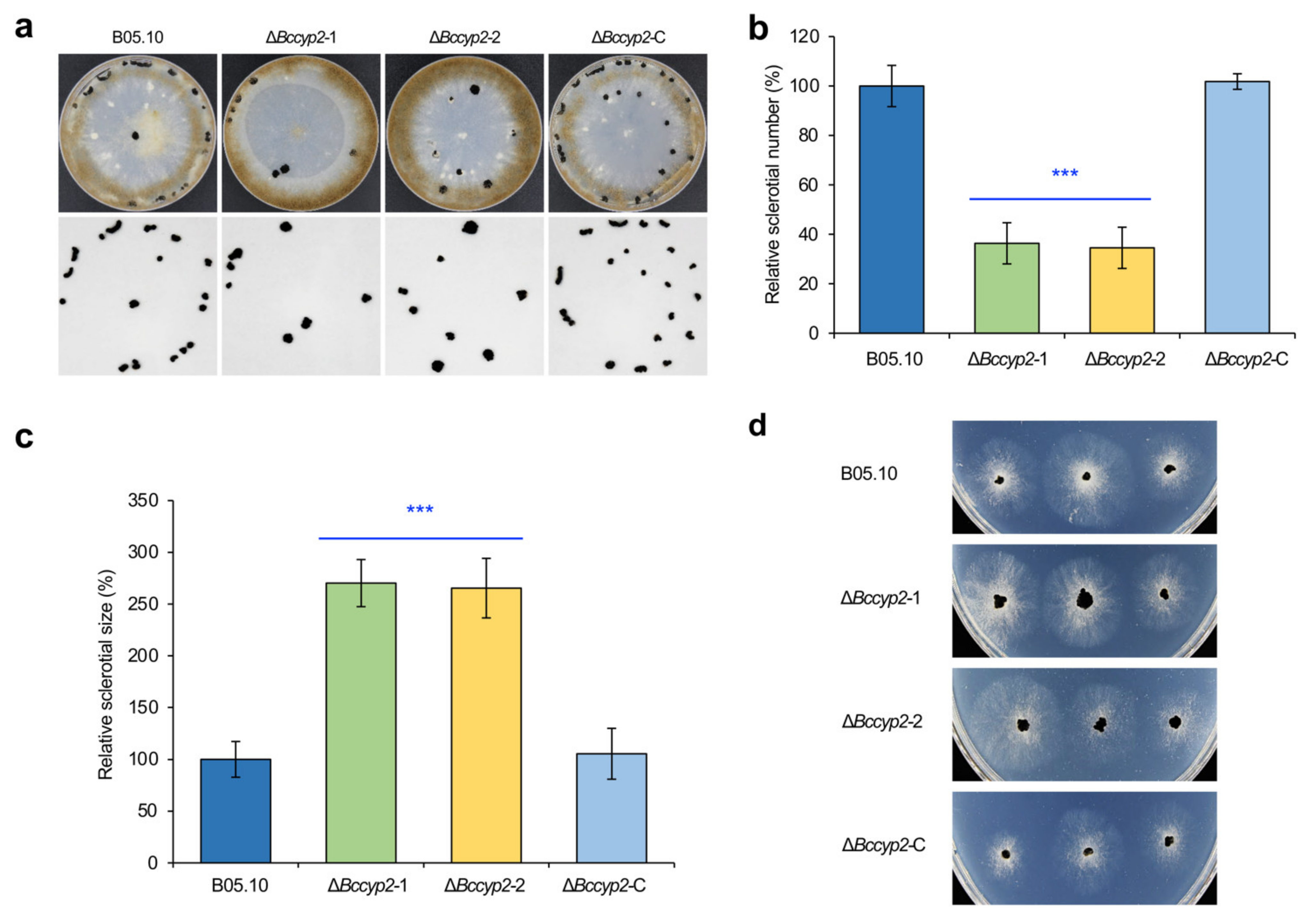
2.4. BcCYP2 Regulates B. cinerea Sclerotium Production and Morphogenesis

To determine the role of BcCYP2 in sclerotium production, we inoculated conidia of the WT, ΔBccyp2, and ΔBccyp2-C on CM plates, incubated these plates at 20 °C in dark condition for four weeks and analyzed sclerotium formation. We found that the number of sclerotia formed by the ΔBccyp2 mutant strains was only about 35% of that of the WT or ΔBccyp2-C strains (Figure 2a,b). However, the size of the mutant sclerotia was bigger than that of sclerotia produced by the WT or complemented strains (Figure 2a,c). Sclerotial germination assays indicated that germination rates of the normal and abnormal sclerotia produced by the WT or ΔBccyp2-C, and ΔBccyp2 strains, respectively, were not significantly different (Figure 2d). These findings suggest that BcCYP2 mediates B. cinerea sclerotium production and morphogenesis but is dispensable for sclerotial germination.

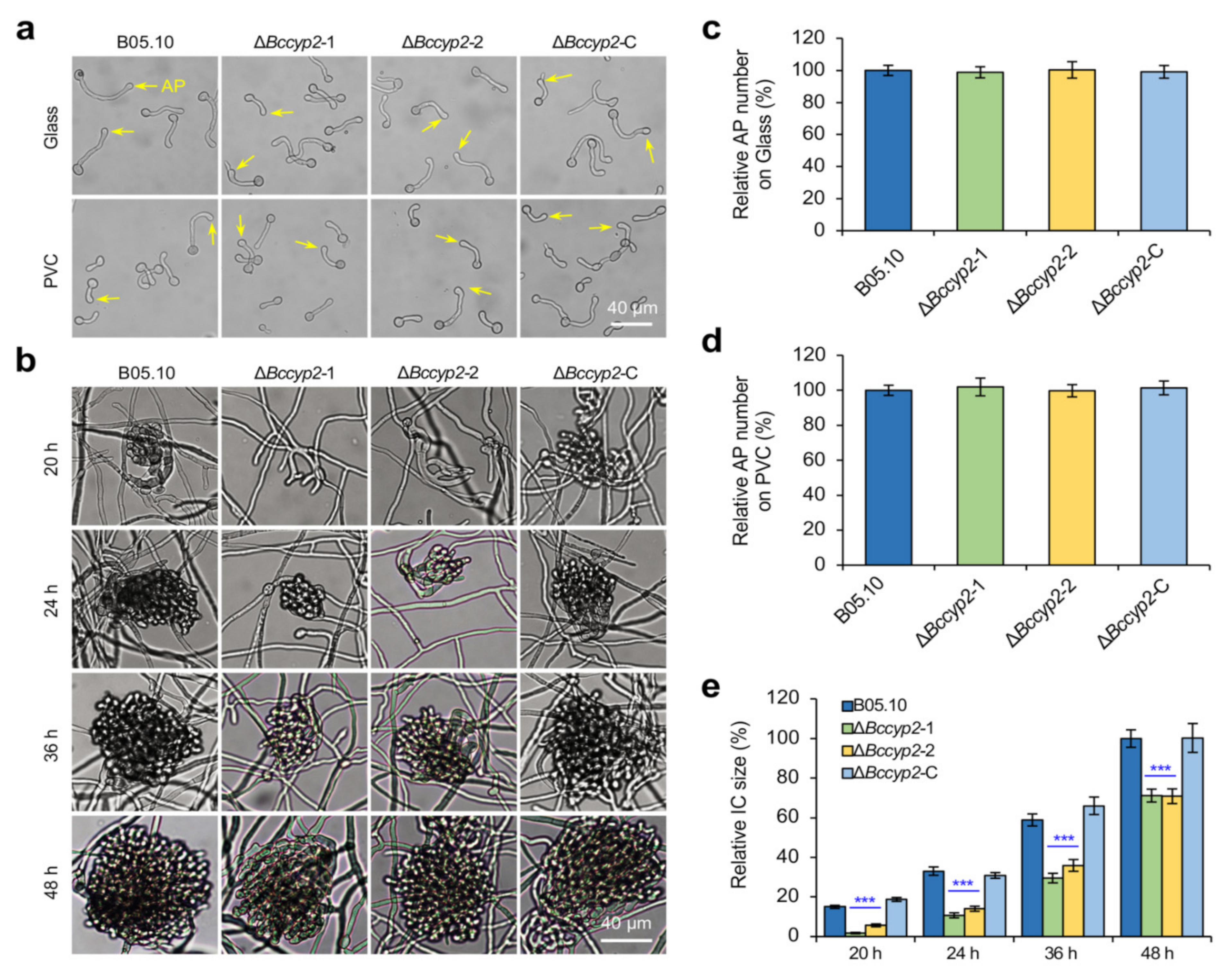
2.5. BcCYP2 Regulates IC Formation but Is Dispensable for Appressorium Formation

To evaluate the effect of BcCYP2 on infection structure formation, we inoculated the WT, ΔBccyp2, and ΔBccyp2-C strains on glass slides or PVC (Polyvinyl chloride) sheets and determined the formation of appressoria and ICs. Our results indicated that all the tested strains produced similar numbers of appressoria on the glass slides or PVC sheets at 7 h post-inoculation/incubation (hpi) (Figure 3a,c,d), suggesting that BcCYP2 is dispensable for the pathogen appressorium formation.
The initial formation of ICs was observed at ~16 hpi in the WT and complemented strains; however, for ΔBccyp2 mutants, the ICs were firstly observed until 20 hpi (Figure 3b). In addition, the ICs produced by the mutants were significantly smaller in size than those of the WT and complemented strains at 24, 36 or 48 hpi (Figure 3b,e). The results indicate that BcCYP2 mediates the development of ICs in B. cinerea.

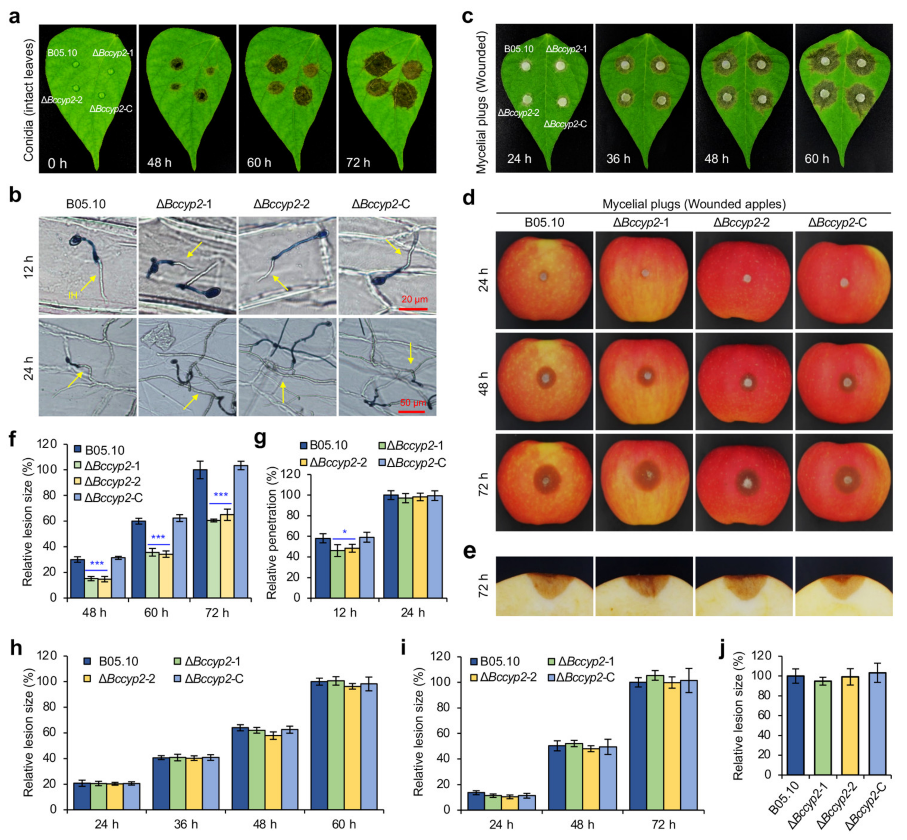
2.6. BcCYP2 Is Required for Full Virulence of B. cinerea

To investigate the role of BcCYP2 in B. cinerea virulence, we inoculated green bean leaves with conidial suspensions (2 × 10⁵ conidia/mL, 5 μL) of the WT, ΔBccyp2, and ΔBccyp2-C strains and determined their pathogenicity. Our result showed that the ΔBccyp2 mutants could induce lesions on the host leaves; however, the lesion size induced by the mutant strains was smaller than that induced by the WT or complemented strain (Figure 4a,f). The results indicate that BcCYP2 is required for B. cinerea full virulence.
To further analyze the role of BcCYP2 in host penetration, we inoculated onion epidermal cells with conidia of the tested B. cinerea strains and observed the infection and development of the pathogen. The results indicate that the mutant has the ability of host penetration, but its penetration rate was lower than that of the WT or complemented strains at 12 hpi and the penetration rates were similar at 24 hpi (Figure 4b,g). The results indicate that BcCYP2 plays a role in the initial host penetration of B. cinerea.
To determine whether BcCYP2 is involved in the pathogen expansion in host tissue upon penetration, we inoculated host leaves or apple fruits with mycelial plugs of the tested strains by a non-wound- or wound-inoculation method, the latter allows the pathogen to enter host cells through an infection structure-independent manner. Our findings indicated that the lesion sizes induced by all the tested strains failed to display a significant difference in the wound-inoculation assay (Figure 4c–e,h–j), suggesting that BcCYP2 plays a limited role, if any, in the pathogen expansion in planta. However, in the non-wound-inoculation, the mutant strains displayed a reduction in lesion expansion compared to the WT or complemented controls (Figure S6). Taken together, these data suggest that BcCYP2 is required for the initiation of host penetration but plays a limited role in invasive hyphal expansion in planta.

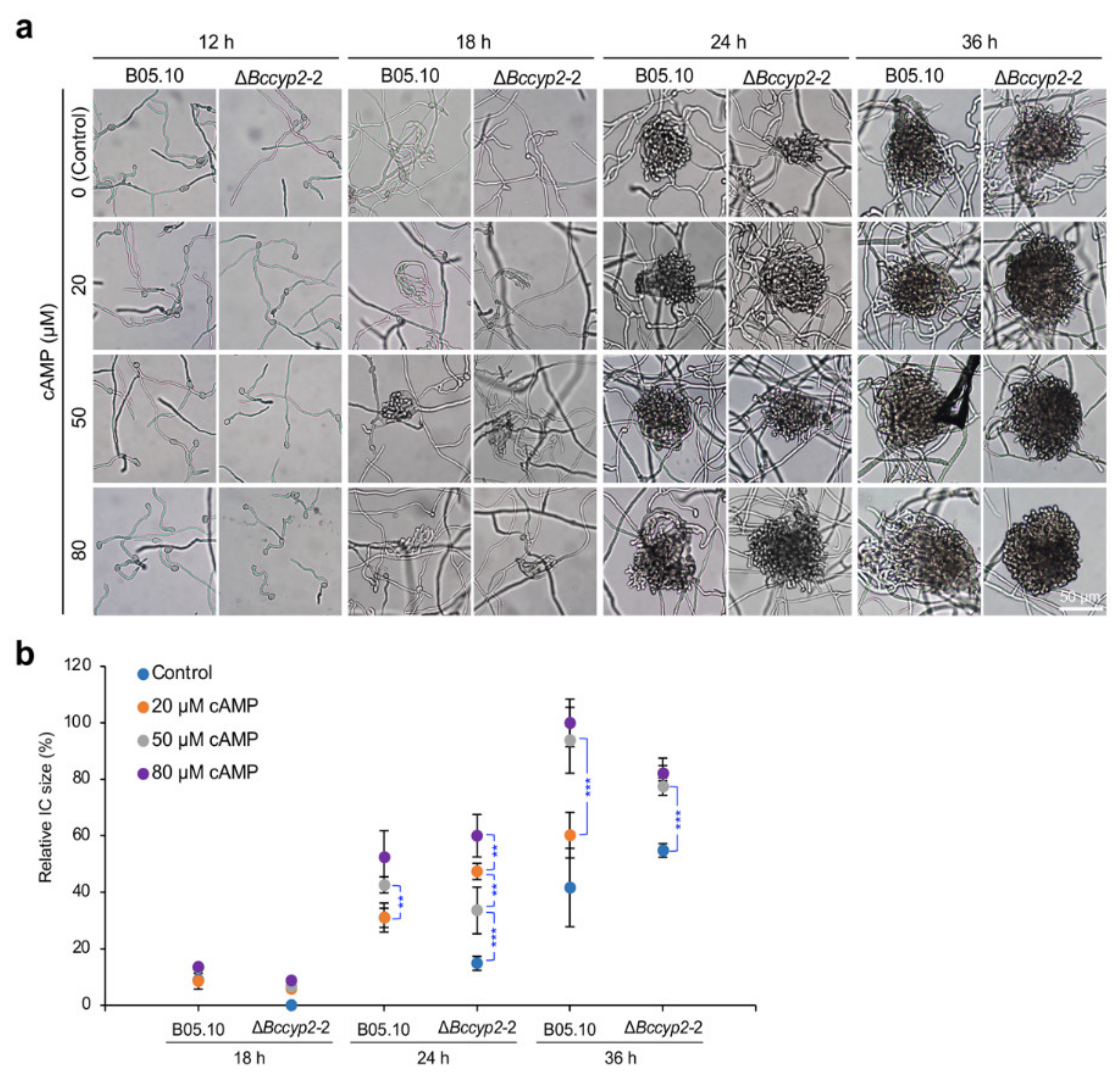
2.7. Exogenous cAMP Restores the Ability of BcCyp2-Deficient Mutants to Form ICs

cAMP can promote the formation of appressoria and ICs in phytopathogenic fungi, including B. cinerea [6,36]. To determine whether a delay in IC formation in the ΔBccyp2 mutants is regulated by cAMP signaling, we inoculated conidia (1 × 10⁵ conidia/mL, 10 µL) of the tested strains supplemented with exogenous cAMP (0, 20, 50, 80 μM) on glass and determined infection structure formation at the indicated hpi. Our findings demonstrated that exogenous cAMP restored the ability of IC formation of the ΔBccyp2 mutants to that of the WT or complemented strain without cAMP treatment (Figure 5). The result demonstrated that cAMP could rescue the defect of infection-related morphogenesis of the mutants losing BcCYP2, and suggests that BcCyp2 likely functions upstream of cAMP signaling.

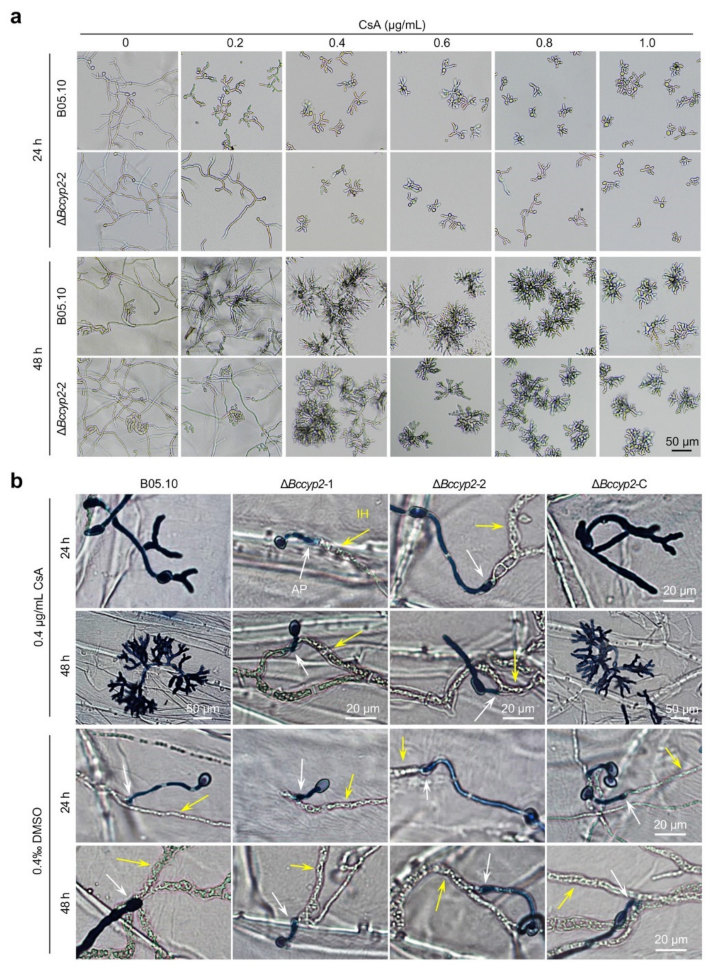
2.8. CsA Suppresses Conidial Germination, Hyphal Morphogenesis and Host Penetration of B. cinerea Strains Harboring CYP2

To determine whether BcCyp2 is a target of CsA, an immunosuppressive agent that binds to the cyclophilin and then inhibits calcineurin [11,17], we tested the sensitivity of the mutant strain ΔBccyp2 to CsA. Our results demonstrated that conidial germination and hyphal development of the WT strain were dramatically suppressed. Importantly, hyphal branching of the WT strain was also severely altered (Figure 6a; Figure S7). Compared to the WT strain, conidial germination and hyphal development of the ΔBccyp2 strain displayed less inhibition by low concentration of CsA (0.2~0.4 μg/mL). The suppression of conidial germination showed a CsA concentration-dependent fashion, and the inhibition phenotype of the mutant tended to be the same as the WT strain when higher doses of CsA were used (Figure 6a; Figure S7). The results suggest that BcCyp2, as a functional cyclophilin protein, is one of the targets of CsA.
To test the effect of CsA on fungal infection-related development and host-cell penetration, we then inoculated onion epidermal cells with conidial droplets (containing 0.4 μg/mL CsA) of the tested strains and then analyzed the effects of CsA on ΔBccyp2 infection. Our results showed that at 24 hpi, the CsA-treated WT and complemented strains failed to invade the onion epidermis; however, penetration of the onion epidermis by the CsA-treated mutant strains was not affected (Figure 6b). At 48 hpi, host penetration by the WT and ΔBccyp2-C strains was still hardly detected (Figure 6b). These findings indicate that suppression of fungal infection-related development and host-cell invasion by the immunosuppressant CsA is in a BcCyp2-dependant fashion; and imply that CsA functions via interaction with BcCyp2 which results in blocking host invasion by the pathogen.

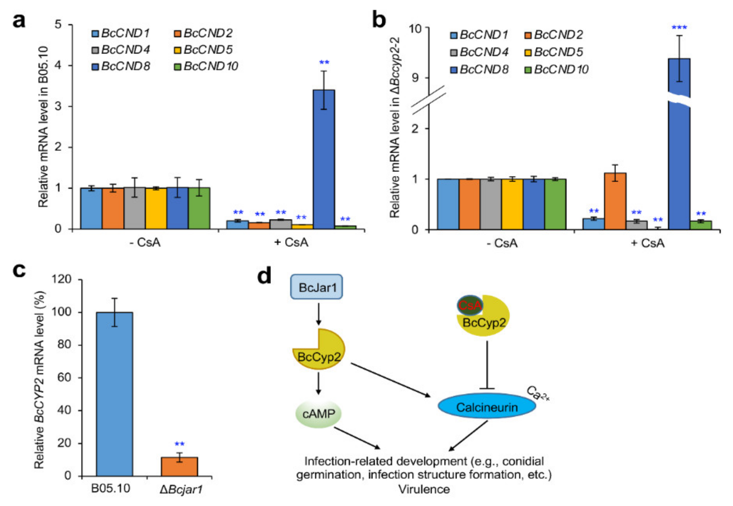
2.9. BcCyp2 Mediates CsA-Regulation of Calcineurin Dependent (CND) Genes

The less hyphal morphological alteration and the unaffected infection structure formation in strains lacking BcCYP2 in the presence of CsA suggest that BcCYP2 may be associated with Ca2+/calmodulin-dependent phosphatase calcineurin. To test this possibility, we cultured the WT, mutant, and complemented strains with media supplemented with 10 µg/mL CsA and profiled the expression of genes (including BcCND1, BcCND2, BcCND4, BcCND5, BcCND8 and BcCND10) related to Ca2+/calcineurin-signal [17,37]. Our results indicated that in the presence of CsA, the expression levels of the tested CND genes in the WT strain were downregulated except for that of BcCND8, compared to that in the absence of CsA (Figure 7a). Interestingly, in the mutant ΔBccyp2, BcCND2 was not suppressed by CsA, suggesting that the suppression of this CND gene is in a BcCyp2-dependent fashion. In addition, BcCyp2 is likely involved in the upregulation of BcCND8 by CsA, since disruption of BcCYP2 increased the degree of its upregulation (Figure 7b). Our data demonstrate that BcCyp2 is involved in mediating the expression of CND genes regulated by CsA.

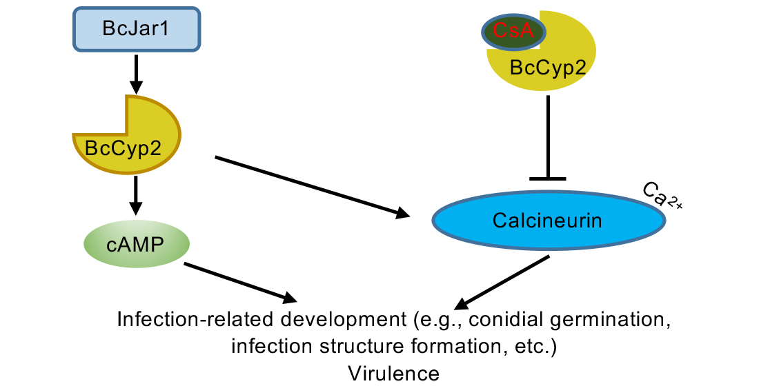
2.10. BcCyp2 Functions under the Control of Histone 3 Lysine 4 (H3K4) Demethylase BcJar1

H3K4 demethylase BcJar1 regulates B. cinerea infection-related development, including IC formation, and virulence via orchestrating genome-wide expression of the pathogenesis-related gene [36]. To determine whether the expression of BcCYP2 was regulated by H3K4 demethylase BcJar1 during IC formation, we cultured the WT and ΔBcjar1 mutant strains and compared the expression level of BcCYP2 in the tested strains during IC formation. Our result indicated that the expression level of BcCYP2 in the mutant strain dramatically reduced (Figure 7c), suggesting that BcCyp2 functions under the control of BcJar1 in mediating IC formation.

3. Discussion

In this work, we screened and identified a virulence-attenuated mutant from our B. cinerea T-DNA insertion library containing ~50,000 transformants [31,32,33]. Bioinformatics analysis indicates that the identified hypothetical protein BcCyp2 is a member of the cyclophilin_ABH_like protein family that contains cyclophilin A, B and H-like cyclophilin-type PPIase domain (Table S1; Figure S2). The family contains cyclophilin proteins similar to human cyclophilins CypA, CypB (hCyP-19) and CypH implicated in protein folding processes, which depend on their catalytic/chaperone-like activities. Human CypA, CypB, Saccharomyces cerevisiae Cpr1 and Caenorhabditis elegans Cyp-3, are inhibited by the immunosuppressant CsA (https://www.ncbi.nlm.nih.gov/Structure/cdd/cddsrv.cgi (accessed on 1 January 2021)). Our data of the strong inhibition of hyphal morphogenesis (e.g., hyphal branching, infection structure formation) and virulence of the strains harboring BcCYP2 by CsA (Figure 6) support the results from bioinformatics analysis.
Cyclophilins play diverse roles in the development and pathogenesis of pathogenic fungal pathogens. In the human fungal pathogen Cryptococcus neoformans, cyclophilin A proteins Cpa1 and Cpa2 play an overlapping role in cell growth, mating, virulence and CsA toxicity. Cpa1 and Cpa2 also have divergent functions. The cpa1 mutants are inviable at high temperatures and virulence-attenuated, whereas the cpa2 mutants are viable at 39 °C and fully virulent. The cpa1 cpa2 double mutants are severely virulence-attenuated and inviable at high temperatures. Both Cpa1 and Cpa2 mediate sensitivity to CsA, suggesting that either protein can form a CsA/CypA complex and inactivate calcineurin [38]. MoCyp1 is a virulence-associated factor in M. oryzae. The cyclophilin MoCyp1 regulates infection-related functions, e.g., penetration peg formation and appressorium turgor generation, and also is required for efficient conidiation [29]. In the chestnut blight fungus C. parasitica, CpCyp1 is a hypovirus-regulated cyclophilin and is a virulence-associated factor. Loss of CpCYP1 reduces transcript levels for genes encoding key components of the heterotrimeric guanosine triphosphate (GTP)-binding protein (G-protein) signaling pathway that is essential for sensing environmental cues and are involved in C. parasitica development and virulence [30]. Cyclophilins in the insect pathogenic fungus B. bassiana display common and unique roles in temperature sensitivity, hyphal growth, conidiation, cyclosporine resistance, and virulence [28].
Loss of cyclophilin Bcp1 in B. cinerea does not affect appressorium formation, but reduces the virulence of the pathogen [17]. Similar to these findings, we found that disruption of BcCYP2 did not affect appressorium formation, vegetative growth, and asexual reproduction, but delayed IC formation (Figure 3). The delay of IC formation may be an important reason in virulence reduction of the BcCYP2 deletion mutants (inefficient penetration is another reason). Many phytopathogenic fungi can recognize hydrophobic surface to initiate appressorium formation via the cAMP signaling pathway, and the differentiation of appressoria and invasive hyphae is mediated by the Pmk1-MAPK cascade [39,40,41,42]. Exogenous cAMP restored the ability of the ΔBccyp2 mutant strain to form ICs to the level of the WT and/or complemented strains without supplemented with cAMP, suggesting that loss of cyclophilin BcCyp2 in B. cinerea does not impair the cellular response to cAMP during IC formation, and that BcCyp2 is located in the upstream of the cAMP pathway. Interestingly, during IC formation, the expression level of BcCYP2 was regulated by H3K4 demethylase BcJar1 (Figure 7c), a pathogen factor that regulates fungal infection-related development and virulence via controlling the expression of pathogenesis-related genes [36]. The association of BcJar1 with BcCyp2 in the mediation of IC formation remains to be characterized.
Calmodulin-dependent signaling is involved in infection structure formation in some phytopathogenic fungi. In M. oryzae, CYP1 may encode a cellular target cyclophilin for CsA since the loss of the gene provides high-level resistance to CsA. CsA-mediated calcineurin inhibition greatly affects hyphal development and impairs appressorium formation, indicating that calcineurin is required for appressorium morphogenesis [29]. Our data of the ΔBccyp2 mutants displaying less affected hyphal development and infection structure formation in the presence of CsA (Figure 6) support the view.
CsA (in low concentrations) altered hyphal morphology and inhibited infection structure formation of the strains harboring BcCYP2, but less affected these development processes in strains lacking BcCYP2, suggesting that BcCYP2 is a receptor of CsA and their interaction may also inhibit the activity of calcineurin, a crucial regulator of many cellular responses or physiological processes, including hyphal morphology, infection structure formation, stress adaptation, and virulence in B. cinerea [17,21]. However, in the presence of higher concentration (>0.4 µg/mL) of CsA, the strain harboring or lacking BcCYP2 displayed similar levels in hyphal morphological alteration, infection structure formation, and virulence (Figure 6), suggesting that in B. cinerea, there may be other cyclophilins serving as CsA receptors. Our identification of 16 cyclophilin-like proteins in B. cinerea (Table S1) and the reported Bcp1 [17,21] supports the view. Among the identified 16 cyclophilin-like proteins, XP_001559584.2 (Bcp1), XP_001559969.1 (BcCyp2), AAQ16573.1, EMR81994.1, XP_001547831.1, XP_001556077.1, XP_001559491.1, and XP_024549591.1 belong to the cyclophilin ABH_like protein family. These putative cyclophilins, including the reported Bcp1 and BcCyp2 described here (Table S1), may serve as candidate CsA receptors. The observation of morphological alteration in the ΔBccyp2 mutants in the presence of a higher concentration of CsA may thus result from functional redundancy in the CsA receptor of other cyclophilins in the gray mold fungus.
Based on our findings, we proposed a model to describe cyclophilin BcCyp2 mediation of infection-related development and virulence in B. cinerea (Figure 7d). Several lines of evidence support the working model. First, the cyclophilin BcCyp2 regulates infection-related development (e.g., IC formation) and virulence. These processes may result from the interaction of the cyclophilin with calcineurin, as evidenced by our finding that BcCYP2 mediates the expression of CND genes in the absence of CsA (Figure S8), which may ultimately alter calcineurin function. Cyclophilins serve as virulence-associated factors and fulfill infection-related functions in many pathogenic fungi [17,28,29,38]. Our data indicate that loss of BcCYP2 does not impair hyphal development and appressorium formation, indicating that in B. cinerea, regulation of hyphal development and appressorium formation are not natural functions of cyclophilin. Second, cyclophilins including BcCyp2 (this study) and Bcp1 [17] are necessary for CsA inhibition of infection-related development and virulence. In the presence of CsA, the formation of a CsA/cyclophilin complex targets calcineurin and prevents its activity in the regulation of fungal morphogenesis, e.g., hyphal branching/development, infection structure formation [11,12,17,21,29]. Our findings on the differential inhibition of hyphal development, infection structure formation, and host infection in the strains harboring or lacking BcCYP2 in the presence of CsA support this view (Figure 6). Third, cyclophilin BcCyp2 functions in the upstream of cAMP-signaling that regulates the infection-related development of phytopathogenic fungi. Deletion of the cyclophilin in B. cinerea results in the delay of IC formation, and exogenous cAMP rescues the defect in IC formation of the mutant strains (Figure 5). Finally, cyclophilin BcCyp2 functions in morphogenesis and virulence may be through direct regulation of CND gene expression. Similar to the previous findings on Bcp1 or CsA mediation of CND gene expression [17,21], in the presence of CsA, most tested CND genes in both the WT and mutant strains were downregulated, except for CND2, whose suppression by CsA is specifically dependent on BcCyp2 (Figure 7a,b). These findings suggest that BcCyp2 functions in the upstream of Ca2+/calmodulin-dependent phosphatase calcineurin signaling.
Collectively, we identified BcCyp2 as a functional cyclophilin in B. cinerea and demonstrated that BcCyp2 is a virulence-associated factor that functions in fungal morphogenesis and virulence under the control of H3K4 demethylase Jar1 as well as in the upstream of cAMP- and Ca2+/calmodulin-dependent phosphatase calcineurin signaling pathways. CsA reduced virulence of the BcCYP2-harboring strains via impairment of their infection structure formation and host penetration. Our work reveals fundamental roles for cyclophilins in phytopathogenic fungi and provides new insights into the pathogenesis of B. cinerea.

4. Materials and Methods

4.1. Fungal Strains and Culture Conditions

B. cinerea WT strain B05.10 and its derived strains, including mutants ΔBccyp2-1, ΔBccyp2-2, and the ΔBccyp2-1 complemented strain ΔBccyp2-C, were cultured on potato dextrose agar (PDA) medium or CM plates as the methods previously described [6,31,32,33].

4.2. Identification of BcCYP2 as a Pathogenicity-Related Gene

A virulence-attenuated mutant strain M331 was identified from a B. cinerea T-DNA insertion library containing ~50,000 transformants via screening for pathogenicity-attenuated mutants using detached tomato and strawberry leaves as hosts [31,32,33]. The thermal asymmetric interlaced PCR approach [31,32,33] was used to analyze T-DNA insertion region in the genome of M331 with the primers listed in Table S2. T-DNA flanking sequences were identified by using BlastN analysis in B. cinerea database (http://fungi.ensembl.org/Botrytis_cinerea/Info/Index (accessed on 1 January 2021)). Sequence alignment of homologous proteins was performed by using GENEDOC (http://nrbsc.org/gfx/genedoc (accessed on 1 January 2021)).

4.3. Generation of Gene Deletion and the Corresponding Genetic Complemented Strains

The generation of BcCYP2 deletion mutants was performed using the gene replacement method described previously [6,31,32,33,43]. The vector pXEH containing the hygromycin phosphotransferase gene (HPH) was used to replace the target gene [6,31,32,36]. The 5′- and 3′- homologous flanks of the target gene were amplified and cloned into pXEH in the upstream and downstream of HPH, respectively. The resultant KO vector was transformed into A. tumefaciens strain AGL-1 as previously described [32]. The ATMT method was used to obtain fungal transformants [44]. The gene deleted transformants of B. cinerea were screened on PDA supplemented with 100 μg/mL hygromycin.
The vector pSULPH, resistant to chlorimuron ethyl, was used to generate the complemented strain ΔBccyp2-C [6,35,36]. The fragment containing 1156 bp upstream and 1070 bp downstream of BcCYP2 coding region was amplified by PCR and cloned into pSULPH to generate the complementary vector. The complementary vector was transformed into A. tumefaciens strain AGL-1 via the ATMT approach. The resultant transformants of B. cinerea were screened on a defined complex medium (DCM) containing 100 μg/mL chlorimuron ethyl.
The transformants were screened by PCR amplification. The BcCYP2 gene deletion mutants and complemented strains were further confirmed by qRT-PCR [2]. The primers used in these experiments are listed in Table S2. DNA and RNA were extracted as previously described [45,46].

4.4. Fungal Developmental Assays

The growth of the tested B. cinerea strains was determined by measuring the radial diameter of colonies on CM as previously described [6,31,32,33,36]. Fresh conidia of the WT, ΔBccyp2 and ΔBccyp2-C strains were harvested from PDA or CM plates. For the measurement of conidial morphology and germination, the concentration of the conidial suspension was adjusted to 1 × 10⁶ conidia/mL. For determination of IC formation, liquid CM droplets (18 μL) were placed on the surface of glass slides, then conidial suspension (1 × 10⁶ conidia/mL, 2 μL) was added and quickly mixed. The inoculated slides were then incubated in a wetting chamber at 20 °C. At 24 hpi, formation of IC was observed and imaged with a microscope (Nikon Eclipse 80i Fluorescence Microscope System, Nikon, Japan). For each sample, IC formation was quantitatively determined via a comparison of the total number of ICs from five randomly selected fields of view under the microscope. Conidial germination, appressorium formation and other phenotypic assays were also analyses with the microscopy system. The experiments were repeated at least three times.

4.5. Plant Infection and Cytological Assays

Conidia were harvested from 10-day old cultures on PDA at 20 °C. Conidial droplets (2 × 10⁵ conidia/mL in ½ liquid CM, 5 µL) of the WT, ΔBccyp2 and ΔBccyp2-C strains were inoculated on green bean leaves [32,33,35,36]. For pathogenicity assays using mycelia, mycelial plugs (5 mm in diameter) from 3-day old culture of the tested strains were inoculated on surfaces of the related plant materials, including green bean leaves and apple fruits. All the inoculated materials were incubated in plastic containers with a sheet of plastic film sealed on the top of each container to maintain a high humidity of infection conditions. B. cinerea inoculated leaves (at 0, 48, 60, and 72 hpi for inoculation with conidial suspension; at 24, 48, and 72 hpi for inoculation with mycelial plugs), apple fruits (at 24, 48 and 72 hpi for inoculation with mycelial plugs) were photographically documented at the indicated time points post-inoculation in the parentheses. Quantification of lesion size was analyzed using ImageJ software (https://imagej.nih.gov/ij/download.html (accessed on 1 January 2021)).
Hydrophobic sides of onion epidermal cells were inoculated with conidial suspension droplets (2 × 10⁵ conidia/mL, 5 µL) and cultured at 20 °C in a humid environment. At 12 and 24 hpi, the inoculated epidermal cells were stained with lactophenol aniline blue for host penetration assay with the microscopy system.

4.6. Stress Adaptation Assays

The sensitivity of the test strains to different stresses was analyzed as previously described [6,31,32,36]. Conidial droplets (1 × 10⁶ conidia/mL, 1 μL) or mycelial plugs (5 mm in diameter) of the WT, BcCYP2 deletion mutant and complemented strains were inoculated on CM supplemented with different stress agents including osmotic stress agents NaCl and KCl (1 M for each), oxidative stress agent H2O2 (5 mM), cell wall disturbing agents SDS (0.005%) and Congo Red (300 μg/mL). All the inoculated plates were incubated at 20 °C in the dark, and the adaptabilities of the strains to these stress agents were evaluated by comparison of their colony diameters. At least three independent experiments were performed, and triple plates of each strain were measured in each test.

4.7. CsA Sensitivity Assay

CsA was dissolved in dimethyl sulfoxide (DMSO). Liquid CM containing CsA was mixed with conidial suspension (1 × 10⁵ conidia/mL, vol:vol = 1:1) to make the final CsA concentration of 0.2, 0.4, 0.6, 0.8 and 1.0 μg/mL, respectively, and 200 μL of the mixture was placed in 96-well plates and cultured at 20 °C in the dark. At 24 or 48 hpi, conidial germination and mycelial morphology were observed and imaged using the microscopy system. For the effects of CsA on infection structure formation and host penetration of the tested strains, the droplets (5 × 10⁴ conidia/mL, 20 μL) of the mixture of 0.4 μg/mL CsA with the conidia suspension were inoculated on onion epidermis and incubated at 20 °C in the dark. The inoculation treated with DMSO was used as a control, since CsA was dissolved in DMSO. At 24 or 48 hpi, the appressorium formation and host penetration were determined and compared. All the CsA inhibition experiments were repeated three times.

4.8. cAMP Sensitivity Assay

The effect of cAMP on infection structure formation of the WT, ΔBccyp2, and complemented strains was evaluated as we previously described [6]. Briefly, conidial suspensions of the indicated strains, supplemented with cAMP at the concentration of 0, 20, 50, or 80 μM, were inoculated on glass slides and incubated in the dark for 12, 18, 24 or 36 hpi. At the indicated hpi, the size of ICs was determined and compared.

4.9. Gene Expression Profiling Assays

Conidia of the WT and ΔBccyp2 strains were grown in liquid YG medium [yeast extract, 2 g/L; glucose,10 g/L; KH2PO4, 2 g/L; K2HPO4, 1.5 g/L; (NH4)2SO4, 1 g/L; MgSO4·7H2O, 0.5 g/L] for 60 h and then moved to the same medium without the nitrogen source. After 4 h of cultivation, CsA (dissolved in 1% DMSO and 99% absolute ethanol) was added to make the final concentration of 10 μg/mL. Following treatment with CsA for 3 h, the mycelia of the tested strains were collected and subjected to RNA extraction using the previously described method [45,46]. The mRNA levels of Ca2+/calmodulin signaling genes were determined by the previously described qRT-PCR approach [31] with the primers listed in Table S2.

4.10. Statistical Analysis

All quantitative data in this study are derived from the results of at least three independent experiments with triplicate samples examined for each strain in each experiment. The data of the control including mycelial growth, lesion size, conidia germination, or other tested phenotypes were standardized to 100% to make the results from different independent experiments comparable. The Student’s t-test was used to analyze the data between control (or WT) and treatment (or mutant) groups and p < 0.05 was considered a significant difference.

Supplementary Materials

The following are available online at https://www.mdpi.com/1422-0067/22/4/1694/s1, Figure S1: Botrytis cinerea BcCYP2 is a virulence-associated gene. Figure S2: Sequence alignment and phylogenetic analyses of cyclophilin (Cyp) proteins from divergent organisms. Figure S3: BcCYP2 is dispensable for B. cinerea vegetative growth. Figure S4: BcCYP2 is required for B. cinerea conidial germination and germling development. Figure S5: BcCYP2 is dispensable for B. cinerea stress adaptation and cell wall integrity. Figure S6: Loss of BcCYP2 in B. cinerea impairs virulence of the pathogen. Figure S7: Dimethyl sulfoxide (DMSO) does not affect B. cinerea growth at the concentration that dissolved CsA. Figure S8: BcCyp2 regulates expression of calcineurin-dependent (CND) genes. Table S1: Subcellular localization and domains of putative cyclophilins in B. cinerea. Table S2: Primers used in this study.

Author Contributions

Conceptualization, Q.-M.Q., G.-H.L. and J.S.; formal analysis, G.-H.L., J.S., Q.-M.Q., H.-W.C., Y.L., M.-Z.Z. and H.Z.; investigation, J.S., C.-H.S., S.Y., Y.L. and J.H.; resources, Q.-M.Q.; writing—original draft preparation, Q.-M.Q. and J.S.; writing—review and editing, Q.-M.Q., J.S., G.-H.L. and Y.L.; visualization, J.S. and Q.-M.Q.; supervision, Q.-M.Q.; project administration, Q.-M.Q. and G.-H.L.; funding acquisition, Q.-M.Q. All authors have read and agreed to the published version of the manuscript.

Funding

This work was supported by the National Natural Science Foundation of China (Grant #: 31871913) and the National Key R&D Program of China (2019YFD1002000).

Institutional Review Board Statement

Not applicable.

Informed Consent Statement

Not applicable.

Data Availability Statement

Data is contained within the article and supplementary material.

Conflicts of Interest

The authors declare no conflict of interest. The funders had no role in the design of the study; in the collection, analyses, or interpretation of data; in the writing of the manuscript, or in the decision to publish the results.

References

  1. Fillinger, S.; Elad, Y. Botrytis—The Fungus, the Pathogen and Its Management in Agricultural Systems; Springer International Publishing: Cham, Switzerland, 2016. [Google Scholar]
  2. Weiberg, A.; Wang, M.; Lin, F.M.; Zhao, H.W.; Zhang, Z.H.; Kaloshian, I.; Huang, H.D.; Jin, H.L. Fungal small RNAs suppress plant immunity by hijacking host RNA interference pathways. Science 2013, 342, 118–123. [Google Scholar] [CrossRef] [PubMed] [Green Version]
  3. Williamson, B.; Tudzynski, B.; Tudzynski, P.; Van Kan, J.A. Botrytis cinerea: The cause of grey mould disease. Mol. Plant Pathol. 2010, 8, 561–580. [Google Scholar] [CrossRef] [PubMed]
  4. Gourgues, M.; Brunet-Simon, A.; Lebrun, M.H.; Levis, C. The tetraspanin BcPls1 is required for appressorium-mediated penetration of Botrytis cinerea into host plant leaves. Mol. Microbiol. 2010, 51, 619–629. [Google Scholar] [CrossRef] [PubMed]
  5. Marschall, R.; Tudzynski, P. Reactive oxygen species in development and infection processes. Semin. Cell Dev. Biol. 2016, 57, 138–146. [Google Scholar] [CrossRef]
  6. Feng, H.Q.; Li, G.H.; Du, S.W.; Yang, S.; Li, X.Q.; de Figueiredo, P.; Qin, Q.M. The septin protein Sep4 facilitates host infection by plant fungal pathogens via mediating initiation of infection structure formation. Environ. Microbiol. 2017, 19, 1730–1749. [Google Scholar] [CrossRef]
  7. Heuvel, J.V.D.; Waterreus, L.P. Conidial concentration as an important factor determining the type of prepenetration structures formed by Botrytis cinerea on leaves of French bean (Phaseolus vulgaris). Plant Pathol. 1983, 32, 263–272. [Google Scholar] [CrossRef]
  8. González-Fernández, R.; Valero-Galván, J.; Gómez-Gálvez, F.J.; Jorrín-Novo, J.V. Unraveling thein vitrosecretome of the phytopathogenBotrytis cinereato understand the interaction with its hosts. Front. Plant Sci. 2015, 6. [Google Scholar] [CrossRef] [Green Version]
  9. Huang, C.Y.; Wang, H.; Hu, P.; Hamby, R.; Jin, H. Small RNAs–Big Players in Plant.-Microbe Interactions. Cell Host Microbe 2019, 26, 173–182. [Google Scholar] [CrossRef] [Green Version]
  10. Stamnes, A.M.; Rutherford, S.L.; Zuker, C.S. Cyclophilins: A new family of proteins involved in intracellular folding. Trends Cell Biol. 1992, 2, 272–276. [Google Scholar] [CrossRef]
  11. Idris, M.; Idris, M.; Adeola, F.; Mensah Sedzro, D. Cyclophilins: The Structure and Functions of an Important Peptidyl-prolyl Isomerase. Int. J. Biochem. Biophys. Mol. Biol. 2019, 4, 1–6. [Google Scholar] [CrossRef]
  12. Barbosa dos Santos, I.; Park, S.-W. Versatility of Cyclophilins in Plant. Growth and Survival: A Case Study in Arabidopsis. Biomolecules 2019, 9, 20. [Google Scholar] [CrossRef] [Green Version]
  13. Marks, A.R. Cellular functions of immunophilins. Physiol. Rev. 1996, 76, 631–649. [Google Scholar] [CrossRef] [PubMed]
  14. Fischer, G.; Wittmann-Liebold, B.; Lang, K.; Kiefhaber, T.; Schmid, F.X. Cyclophilin and peptidyl-prolyl cis-trans isomerase are probably identical proteins. Nature 1989, 337, 476–478. [Google Scholar] [CrossRef]
  15. Lodish, F.H.; Kong, N. Cyclosporin A inhibits an initial step in folding of transferrin within the endoplasmic reticulum. J. Biol. Chem. 1991, 266, 14835–14838. [Google Scholar] [CrossRef]
  16. Takahashi, N.; Hayano, T.; Suzuki, M. Peptidyl-prolyl cis-trans isomerase is the cyclosporin A-binding protein cyclophilin. Nature 1989, 337, 473–475. [Google Scholar] [CrossRef] [PubMed]
  17. Viaud, M.; Brunet-Simon, A.; Brygoo, Y.; Pradier, J.M.; Levis, C. Cyclophilin A and calcineurin functions investigated by gene inactivation, cyclosporin A inhibition and cDNA arrays approaches in the phytopathogenic fungus Botrytis cinerea. Mol. Microbiol. 2003, 50, 1451–1465. [Google Scholar] [CrossRef]
  18. Fox, D.S.; Cruz, M.C.; Sia, R.A.L.; Ke, H.; Cox, G.M.; Cardenas, M.E.; Heitman, J. Calcineurin regulatory subunit is essential for virulence and mediates interactions with FKBP12–FK506 in Cryptococcus neoformans. Mol. Microbiol. 2001, 39, 835–849. [Google Scholar] [CrossRef] [PubMed]
  19. Juvvadi, P.R.; Kuroki, Y.; Arioka, M.; Nakajima, H.; Kitamoto, K. Functional analysis of the calcineurin-encoding gene cnaA from Aspergillus oryzae: Evidence for its putative role in stress adaptation. Arch. Microbiol. 2003, 179, 416–422. [Google Scholar] [CrossRef] [PubMed]
  20. Fox, S.D.; Heitman, J. Good fungi gone bad: The corruption of calcineurin. Bioessays 2002, 24, 894–903. [Google Scholar] [CrossRef] [PubMed]
  21. Harren, K.; Schumacher, J.; Tudzynski, B. The Ca2+/calcineurin-dependent signaling pathway in the gray mold Botrytis cinerea: The role of calcipressin in modulating calcineurin activity. PLoS ONE 2012, 7, e41761. [Google Scholar] [CrossRef] [PubMed] [Green Version]
  22. Trivedi, D.K.; Yadav, S.; Vaid, N.; Tuteja, N. Genome wide analysis of Cyclophilin gene family from rice and Arabidopsis and its comparison with yeast. Plant Signal. Behav. 2012, 7, 1653–1666. [Google Scholar]
  23. Blair, L.J.; Baker, J.D.; Sabbagh, J.J.; Dickey, C.A. The emerging role of peptidyl-prolyl isomerase chaperones in tau oligomerization, amyloid processing, and Alzheimer’s disease. J. Neurochem. 2015, 133, 1–13. [Google Scholar] [CrossRef] [PubMed] [Green Version]
  24. Davis, T.L.; Walker, J.R.; Campagna-Slater, V.; Finerty, P.J.; Paramanathan, R.; Bernstein, G.; Mackenzie, F.; Tempel, W.; Ouyang, H.; Lee, W.H.; et al. Structural and biochemical characterization of the human cyclophilin family of peptidyl-prolyl isomerases. PLoS Biol. 2010, 8, e1000439. [Google Scholar]
  25. Satoh, K.; Fukumoto, Y.; Sugimura, K.; Miura, Y.; Aoki, T.; Nochioka, K.; Tatebe, S.; Miyamichi Yamamoto, S.; Shimizu, T.; Osaki, S. Plasma cyclophilin A is a novel biomarker for coronary artery disease. Circ. J. 2013, 77, 447–455. [Google Scholar] [CrossRef] [Green Version]
  26. Schmidt, B.; Tradler, T.; Rahfeld, J.U.; Ludwig, B.; Jain, B.; Mann, K.; Rücknagel, K.P.; Janowski, B.; Schierhorn, A.; Küllertz, G. A cyclophilin-like peptidyl-prolyl cis/trans isomerase from Legionella pneumophila--characterization, molecular cloning and overexpression. Mol. Microbiol. 2010, 21, 1147–1160. [Google Scholar]
  27. Surichan, S.; Arroo, R.R.; Ruparelia, K.; Tsatsakis, A.M.; Androutsopoulos, V.P. Nobiletin bioactivation in MDA-MB-468 breast cancer cells by cytochrome P450 CYP1 enzymes. Food Chem. Toxicol. 2018, 113, 228–235. [Google Scholar] [CrossRef]
  28. Zhou, Y.; Keyhani, N.O.; Zhang, Y.; Luo, Z.; Fan, Y.; Li, Y.; Zhou, Q.; Chen, J.; Pei, Y. Dissection of the contributions of cyclophilin genes to development and virulence in a fungal insect pathogen. Environ. Microbiol. 2016, 18, 3812–3826. [Google Scholar] [CrossRef] [PubMed]
  29. Viaud, C.M.; Balhadère, P.V.; Talbot, N.J. A Magnaporthe grisea cyclophilin acts as a virulence determinant during plant infection. Plant Cell 2002, 14, 917–930. [Google Scholar] [PubMed] [Green Version]
  30. Chen, M.M.; Jiang, M.; Shang, J.; Lan, X.; Yang, F.; Huang, J.; Nuss, D.L.; Chen, B. CYP1, a hypovirus-regulated cyclophilin, is required for virulence in the chestnut blight fungus. Mol. Plant Pathol. 2011, 12, 239–246. [Google Scholar]
  31. Cao, S.N.; Yuan, Y.; Qin, Y.H.; Zhang, M.Z.; de Figueiredo, P.; Li, G.H.; Qin, Q.M. The pre-rRNA processing factor Nop53 regulates fungal development and pathogenesis via mediating production of reactive oxygen species. Environ. Microbiol. 2018, 20, 1531–1549. [Google Scholar]
  32. Liu, J.K.; Chang, H.W.; Liu, Y.; Qin, Y.; Ding, Y.H.; Wang, L.; Zhao, Y.; Zhang, M.Z.; Cao, S.N.; Li, L.T. The key gluconeogenic gene PCK1 is crucial for virulence of Botrytis cinerea via initiating its conidial germination and host penetration. Environ. Microbiol. 2018, 20, 1794–1814. [Google Scholar] [CrossRef]
  33. Liu, Y.; Liu, J.K.; Li, G.H.; Zhang, M.Z.; Zhang, Y.Y.; Wang, Y.Y.; Hou, J.; Yang, S.; Sun, J.; Qin, Q.M. A novel Botrytis cinerea-specific gene BcHBF1 enhances virulence of the grey mould fungus via promoting host penetration and invasive hyphal development. Mol. Plant Pathol. 2019, 20, 731–747. [Google Scholar] [CrossRef] [Green Version]
  34. Van Kan, J.A.; Stassen, J.H.; Mosbach, A.; Van Der Lee, T.A.; Faino, L.; Farmer, A.D.; Papasotiriou, D.G.; Zhou, S.; Seidl, M.F.; Cottam, E. A gapless genome sequence of the fungus Botrytis cinerea. Mol. Plant Pathol. 2017, 18, 75–89. [Google Scholar] [CrossRef] [Green Version]
  35. Zhang, M.Z.; Sun, C.H.; Liu, Y.; Feng, H.Q.; Chang, H.W.; Cao, S.N.; Li, G.H.; Yang, S.; Hou, J.; Zhu-Salzman, K.; et al. Transcriptome analysis and functional validation reveal a novel gene, BcCGF1, that enhances fungal virulence by promoting infection-related development and host penetration. Mol. Plant Pathol. 2020, 21, 834–853. [Google Scholar] [CrossRef] [Green Version]
  36. Hou, J.; Feng, H.Q.; Chang, H.W.; Liu, Y.; Li, G.H.; Yang, S.; Sun, C.H.; Zhang, M.Z.; Yuan, Y.; Sun, J.; et al. The H3K4 demethylase Jar1 orchestrates ROS production and expression of pathogenesis-related genes to facilitate Botrytis cinerea virulence. New Phytol. 2020, 225, 930–947. [Google Scholar] [CrossRef] [PubMed]
  37. Zhou, X.Y.; Zhang, H.F.; Li, G.T.; Shaw, B.; Xu, J.R. The Cyclase-associated protein Cap1 is important for proper regulation of infection-related morphogenesis in Magnaporthe oryzae. PLoS Pathog. 2012, 8, e1002911. [Google Scholar] [CrossRef] [PubMed] [Green Version]
  38. Wang, P.; Cardenas, M.E.; Cox, G.M.; Perfect, J.R.; Heitman, J. Two cyclophilin A homologs with shared and distinct functions important for growth and virulence of Cryptococcus neoformans. Embo Rep. 2001, 2, 511–518. [Google Scholar] [CrossRef] [Green Version]
  39. Xu, R.J.; Hamer, J.E. MAP kinase and cAMP signaling regulate infection structure formation and pathogenic growth in the rice blast fungus Magnaporthe grisea. Genes Dev. 1996, 10, 2696–2706. [Google Scholar] [CrossRef] [PubMed] [Green Version]
  40. Ding, S.L.; Zhou, X.Y.; Zhao, X.H.; Xu, J.R. The PMK1 MAP Kinase Pathway and Infection-Related Morphogenesis. In Advances in Genetics, Genomics and Control of Rice Blast Disease; Springer: Dordrecht, The Netherlands, 2009; pp. 13–21. [Google Scholar]
  41. Leroch, M.; Mueller, N.; Hinsenkamp, I.; Hahn, M. The signalling mucin Msb2 regulates surface sensing and host penetration via BMP1 MAP kinase signalling in B otrytis cinerea. Mol. Plant Pathol. 2015, 16, 787–798. [Google Scholar] [CrossRef]
  42. Schumacher, J.; Kokkelink, L.; Huesmann, C.; Jimenez-Teja, D.; Collado, I.G.; Barakat, R.; Tudzynski, P.; Tudzynski, B. The cAMP-dependent signaling pathway and its role in conidial germination, growth, and virulence of the gray mold Botrytis cinerea. Mol. Plant-Microbe Interact. 2008, 21, 1443–1459. [Google Scholar] [CrossRef] [Green Version]
  43. Giesbert, S.; Schumacher, J.; Kupas, V.; Espino, J.; Segmüller, N.; Haeuser-Hahn, I.; Schreier, P.H.; Tudzynski, P. Identification of pathogenesis-associated genes by T-DNA–mediated insertional mutagenesis in Botrytis cinerea: A Type 2A phosphoprotein phosphatase and an SPT3 transcription factor have significant impact on virulence. Mol. Plant-Microbe Interact. 2012, 25, 481–495. [Google Scholar] [CrossRef] [Green Version]
  44. Mullins, E.D.; Chen, X.; Romaine, P.; Raina, R.; Geiser, D.M.; Kang, S. Agrobacterium-mediated transformation of Fusarium oxysporum: An efficient tool for insertional mutagenesis and gene transfer. Phytopathology 2001, 91, 173–180. [Google Scholar] [CrossRef] [PubMed] [Green Version]
  45. Cenis, J.L. Rapid extraction of fungal DNA for PCR amplification. Nucleic Acids Res. 1992, 20, 2380. [Google Scholar] [CrossRef] [PubMed]
  46. Lecellier, G.; Silar, P. Rapid methods for nucleic acids extraction from Petri dish-grown mycelia. Curr. Genet. 1994, 25, 122–123. [Google Scholar] [CrossRef] [PubMed]
Figure 1. BcCYP2 is required for B. cinerea conidial morphogenesis but dispensable for conidiation. (a) Conidiation of the indicated wild-type (WT), ΔBccyp2 and complemented strains of B. cinerea on complete medium (CM) plates at 10 days post-inoculation/incubation (dpi). (b) Quantification of the relative conidiation of the indicated strains shown in (a). (c) Alteration of conidial morphology in the ΔBccyp2 mutants. (d) Quantification of sizes (length and width) of conidia produced by the indicated strains. Conidia were harvested from 10-day old CM plates and conidia (>300) of each strain were measured under a microscope. (e) Loss of BcCYP2 increases the number of globose and less elliptical conidia (closer to the dash line). Conidia (>300) of each strain were measured under a microscope in each independent experiment. The representative images are from one of the experiments, three independent experiments were performed and resulted in similar results. Data represent the means ± standard deviations (SD) from three independent experiments with triplicate plates examined for each treatment. **: significance at p < 0.01.
Ijms 22 01694 g001
Figure 2. BcCYP2 mediates B. cinerea sclerotium production and morphogenesis. (a) Deletion of BcCYP2 in B.cinerea reduces sclerotium production. Conidia of the wild-type (WT), ΔBccyp2, and complemented strains were inoculated on CM plates and cultured at 20 °C in the dark. Sclerotium production was photographically documented at four weeks post-incubation. (b) Quantification of sclerotium production. (c) Quantitative analysis of sclerotial sizes of the indicated strains via ImageJ software (https://imagej.nih.gov/ij/download.html (accessed on 1 January 2021)). (d) Germination of sclerotia produced by the tested strains. The representative images are from one of the experiments, at least three independent experiments were performed, and all the experiments resulted in similar results. Data represent means ± SD from at least three independent experiments. ***: significance at p < 0.001.
Ijms 22 01694 g002
Figure 3. BcCYP2 plays an important role in infection cushion (IC) formation. (a) BcCYP2 is dispensable for B. cinerea appressorium formation. Conidia (5 × 10⁵ conidia/mL in ½ liquid CM, 20 µL) of the indicated strains were inoculated on glass slides or PVC sheets and cultured at 20 °C. At 7 h post-inoculation/incubation (hpi), appressorium formation by the tested strains was observed and documented. (b) Loss of BcCYP2 delays the development of ICs. Conidia (1 × 10⁵ conidia/mL in ½ liquid CM, 20 µL) of the indicated strains were inoculated on glass slides and cultured at 20 °C. ICs were photographically documented at 20, 24, 36, and 48 hpi. Images from a single representative experiment are presented. (c,d) Quantitative analysis of the relative number of appressoria produced by the tested strains on glass slides (c) or PVC sheets (d). (e) Quantification of the sizes of ICs produced by the tested strains. Data represent means ± SD from three independent experiments in which triplicate slides were examined for each strain in each experiment. ***: significance at p < 0.001. AP: Appressorium; PVC: Polyvinyl chloride sheet.
Ijms 22 01694 g003
Figure 4. Loss of BcCYP2 in B. cinerea impairs virulence of the pathogen. (a) Deletion of BcCYP2 reduces the virulence of B. cinerea. Conidial suspensions (2 × 10⁵ conidia/mL in ½ liquid CM, 5 µL) of the WT, ΔBccyp2, and complement strains were inoculated on intact green bean leaves. At 0, 48, 60, and 72 hpi, the inoculated leaves were photographically documented. (b) Penetration assay of the test strains. Conidia (2 × 10⁵ conidia/mL, 5 μL) of the strains were inoculated on onion epidermis and cultured at 20 °C in the dark. At 12 and 24 hpi, the inoculated epidermal cells were stained with lactophenol aniline blue and then observed and photographically documented. (c,d) Deletion of BcCYP2 does not affect invasive hyphal expansion in planta. Mycelial plugs of each strain were inoculated on artificially wounded green bean leaves (c) or apple fruits (d) and the lesions were photographically documented at the indicated hpi. (e) The cross-section of lesions on wounded apple fruits. (f) Quantification of lesion sizes caused by the indicated strains on green bean leaves shown in (a). (g) Quantification of penetration by the indicated strains on onion epidermal cells shown in (b). (hi) Quantification of lesion sizes caused by the indicated strains on wounded green bean leaves or apple fruit shown in (c,d), respectively. (j) Quantification of lesion sizes caused by the indicated strains on apple fruits shown in (e). Data represent means ± SD from three independent experiments. *, ***: significance at p < 0.05 and 0.001, respectively. IH: invasive hyphae.
Ijms 22 01694 g004
Figure 5. Cyclic adenosine monophosphate (cAMP) restores IC formation of the BcCYP2 deletion mutants. (a) Exogenous cAMP rescues defects of IC formation of the ΔBccyp2 mutants. Conidial droplets (10 µL) supplemented with cAMP (0, 20, 50, 80 µM) were inoculated on glass slides and incubated in the dark for 12, 18, 24 or 36 h; the development of ICs was then photographically documented. (b) Quantitative analysis of the sizes of ICs produced by the tested strains. IC: Infection cushion. **, ***: significance at p < 0.01 and 0.001, respectively.
Ijms 22 01694 g005
Figure 6. Effects of CsA on infection-related morphogenesis as well as host penetration and growth of invasive hyphae. (a) Suppression of B. cinerea mycelial growth by CsA. Conidia (5 × 10⁴ conidia/mL in ½ liquid CM, 200 µL) of the indicated strains mixed with CsA (0, 0.2, 0.4, 0.6, 0.8, or 1.0 µg/mL) were inoculated in 96-well plates and cultured at 20 °C. Mycelial morphogenesis of the tested strains was observed and photographically documented at 24 and 48 hpi. (b) Suppression of B. cinerea infection by CsA. Conidia of each strain (5 × 10⁴ conidia/mL, 20 µL) mixed with or without CsA were inoculated on onion epidermis and cultured at 20 °C. The solvent (DMSO) treated inoculation was used as a control. At 24 or 48 hpi, the inoculated epidermal cells were performed lactophenol aniline blue staining, microscopy observation and imaging. AP: appressorium, IH: invasive hyphae. Representative images are from one of the experiments, at least three independent experiments were performed, and all the experiments resulted in similar results.
Ijms 22 01694 g006
Figure 7. BcCyp2 regulates expression of calcineurin-dependent (CND) genes and is regulated by H3K4 demethylase BcJar1. (a,b) Relative transcriptional level of CND genes in the WT B05.10 (a) and mutant ΔBccyp2-2 (b) strains. Conidia of the two strains were grown in liquid YG medium for 60 h and then moved to the same medium without the nitrogen source (YG-N) and incubated for 4 h, the mycelia of the tested strains were further incubated with YG-N medium containing 10 μg/mL CsA for 3 h and then collected for RNA extraction. The mRNA levels of Ca2+/calmodulin signaling genes were determined by quantitative reverse transcription PCR (qRT-PCR). (c) Loss of BcJAR1 [36] downregulates BcCYP2 expression level. Mycelial plugs of B05.10 or ΔBcjar1 strain were induced for IC formation. At 36 hpi (during IC formation), mycelia of the tested strains were then collected and subjected to RNA extraction. The mRNA levels of BcCYP2 were determined by qRT-PCR. (d) Proposed models describing BcCyp2 regulation of fungal infection-related development and virulence as well as CsA-mediated functional blocking. Data represent means ± SD from three independent experiments. **, ***: significance at p < 0.01 and 0.001, respectively.
Ijms 22 01694 g007
Publisher’s Note: MDPI stays neutral with regard to jurisdictional claims in published maps and institutional affiliations.

