Article

Design of PG-Surfactants Bearing Polyacrylamide Polymer Chain to Solubilize Membrane Proteins in a Surfactant-Free Buffer

1
Department of Life Science and Applied Chemistry, Graduate School of Engineering, Nagoya Institute of Technology, Gokiso-cho, Showa-ku, Nagoya, Aichi 466-8555, Japan
2
Research Center for Advanced Science & Technology, The University of Tokyo, 4-6-1 Komaba, Meguro-ku, Tokyo 153-8904, Japan
3
Department of Nanopharmaceutical Sciences, Graduate School of Engineering, Nagoya Institute of Technology, Gokiso-cho Showa-ku, Nagoya, Aichi 466-8555, Japan
4
The OCU Advanced Research Institute for Natural Science & Technology (OCARINA), Osaka City University, 3-3-138 Sugimoto-cho, Sumiyoshi, Osaka 558-8585, Japan
*
Author to whom correspondence should be addressed.
Present address: Biostructural Mechanism Laboratory, RIKEN SPring-8 Center, 1-1-1 Kouto, Sayo, Hyogo 679-5148, Japan.
Int. J. Mol. Sci. 2021, 22(4), 1524; https://doi.org/10.3390/ijms22041524
Submission received: 4 December 2020 / Revised: 26 January 2021 / Accepted: 1 February 2021 / Published: 3 February 2021

Abstract

:
The development of techniques capable of using membrane proteins in a surfactant-free aqueous buffer is an attractive research area, and it should be elucidated for various membrane protein studies. To this end, we examined a method using new solubilization surfactants that do not detach from membrane protein surfaces once bound. The designed solubilization surfactants, DKDKC12K-PAn (n = 5, 7, and 18), consist of two parts: one is the lipopeptide-based solubilization surfactant part, DKDKC12K, fand the other is the covalently connected linear polyacrylamide (PA) chain with different Mw values of 5, 7, or 18 kDa. Intermolecular interactions between the PA chains in DKDKC12K-PAn concentrated on the surfaces of membrane proteins via amphiphilic binding of the DKDKC12K part to the integral membrane domain was observed. Therefore, DKDKC12K-PAn (n = 5, 7, and 18) could maintain a bound state even after removal of the unbound by ultrafiltration or gel-filtration chromatography. We used photosystem I (PSI) from Thermosynecoccus vulcanus as a representative to assess the impacts of new surfactants on the solubilized membrane protein structure and functions. Based on the maintenance of unique photophysical properties of PSI, we evaluated the ability of DKDKC12K-PAn (n = 5, 7, and 18) as a new solubilization surfactant.

Graphical Abstract

1. Introduction

One-third of natural proteins are classified as membrane proteins, and these have crucial roles in various biological events occurring at cell membranes. Photosynthesis is one such membrane-mediated event that participates in different unique membrane proteins. In higher plants and cyanobacteria, photosystem I (PSI) and photosystem II (PSII), integrated in thylakoid membranes, play pivotal roles in generating reductive electrons from water using sunlight [1]. In natural thylakoid membranes, the generated reductive electrons are used for NADPH synthesis via ferredoxin NADP+ oxidoreductase (FNR) [2], but exchanging this reductive enzyme to other reductive enzymes or catalysts the artificial photosynthesis systems in vitro and in vivo were recently examined [3]. If the reductive electrons are directly poured into the electrode, it leads to solar cell construction [4]. To combine PSI with reductive enzymes or catalysts, such as molecular metal catalysts [5], Pt nanoparticles [6], or hydrogenases [7] in vitro, direct modification via adsorption or chemical bonds [8] and indirect electrochemical connection via electrodes or metal nanoparticles [9] have been studied. However, the construction of more sophisticated artificial photosynthesis systems [10] consisting of PSI, reductive catalysts, and electron transfer mediators, and sacrificial reductants using solubilization surfactants for membrane proteins to treat PSI in an aqueous buffer could become a decisive hindrance; if PSI could be treated in an aqueous buffer similar to water-soluble proteins, it would allow flexible design of molecular circuits using PSI. The techniques able to treat membrane proteins in an aqueous buffer, identical to water-soluble proteins, such as the construction of various semi-artificial molecular sensors and drug screening samples consisting of different natural membrane proteins is also an interesting research area [11].
Some methods capable of treating membrane proteins in a surfactant-free buffer have been reported so far. Its developments have particular relevance to structural analytical techniques of membrane proteins using cryo-microscopy [12] and multi-dimensional NMR [13]. For structural analysis by cryo-microscopy, sample preparation of membrane proteins with fewer contaminants is indispensable for obtaining high-quality TEM images for structural analysis. Some new surfactants such as lauryl maltose neopentyl glycol [14] and amphipols [15] have a high affinity for membrane proteins; therefore, the unbound surfactants are known to separate without precipitation by dialysis or ultrafiltration. On the other hand, some amphiphilic block copolymers are known to extract membrane proteins directly from bio-membranes [16,17]. By using the deuterated membrane proteins expressed in E. coli cell membranes, structural analysis of membrane proteins solubilized with these copolymers using multi-dimensional NMR has been reported [18]. Considering these situations, further developments of new surfactants or techniques capable of treating membrane proteins in surfactant-free buffers are expected. However, developments in these techniques are still limited to date.
Meanwhile, we recently studied Gemini-type peptide surfactants, PG-surfactants [19,20,21,22,23]. The basic molecular scaffold of PG-surfactants consists of three constituents: the linker peptide from to 3–5 residues (X), two alkylamidomethyl-modified Cys residues at both sides of the linker peptide, and the peripheral peptides (Y and Z) at the N- and C-terminal sides of the alkylamidomethyl-modified Cys residues (Figure 1a). From a set of screenings on hydrophilic peptide sequences at X, Y, and Z, we found that two PG-surfactants, DKDKC12K and DKDKC12D, were able to function as solubilization surfactants for membrane proteins [20]. Furthermore, by the tandem connection of the -Cys(C12)-Asp-Lys-Asp-Lys-Cys(C12)- units in DKDKC12K and DKDKC12D via flexible (Gly)4 linkers in one molecule, we succeeded in designing the high molecular weight (MW) solubilization surfactants, Bis-D3-DKDKC12 (MW ~3 kDa), Bis-K3-DKDKC12 (MW ~3 kDa), and Tris-D3-DKDKC12 (MW ~4.3 kDa) [21]. Interestingly, an increase in Mw enhanced the affinity between membrane proteins and surfactants, thereby allowing effective solubilization, even for lower concentration ranges (<0.0005 wt%). In this study, by conjugating a hydrophilic polymer unit with DKDKC12K, we designed different types of high-Mw surfactants, which can function as a solubilization surfactant for membrane proteins (Figure 1b). We hypothesized that due to the high affinity between polymer chains concentrated on membrane surfaces, these surfactants would not detach once bound to membrane protein surfaces, thereby generating a method to treat membrane proteins in the surfactant-free buffer. In this case, because the attached polymer chains would also interact with membrane protein surfaces, to choose hydrophilic polymers, giving less impact on tertiary structure of membrane proteins, is necessary. Therefore, as a hydrophilic polymer, in this study, we chose poly(acrylamide) (PA). We synthesized PA with different MW (Mn = 5, 7, and 18 kDa) and characterized the solubilization ability of the conjugates with DKDKC12K. DKDKC12K-PAn (n = 5, 7, and 18, Figure 1b), and the PA units were introduced at the Y position of DKDKC12K.

2. Results and Discussion

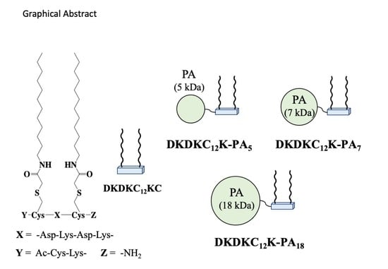
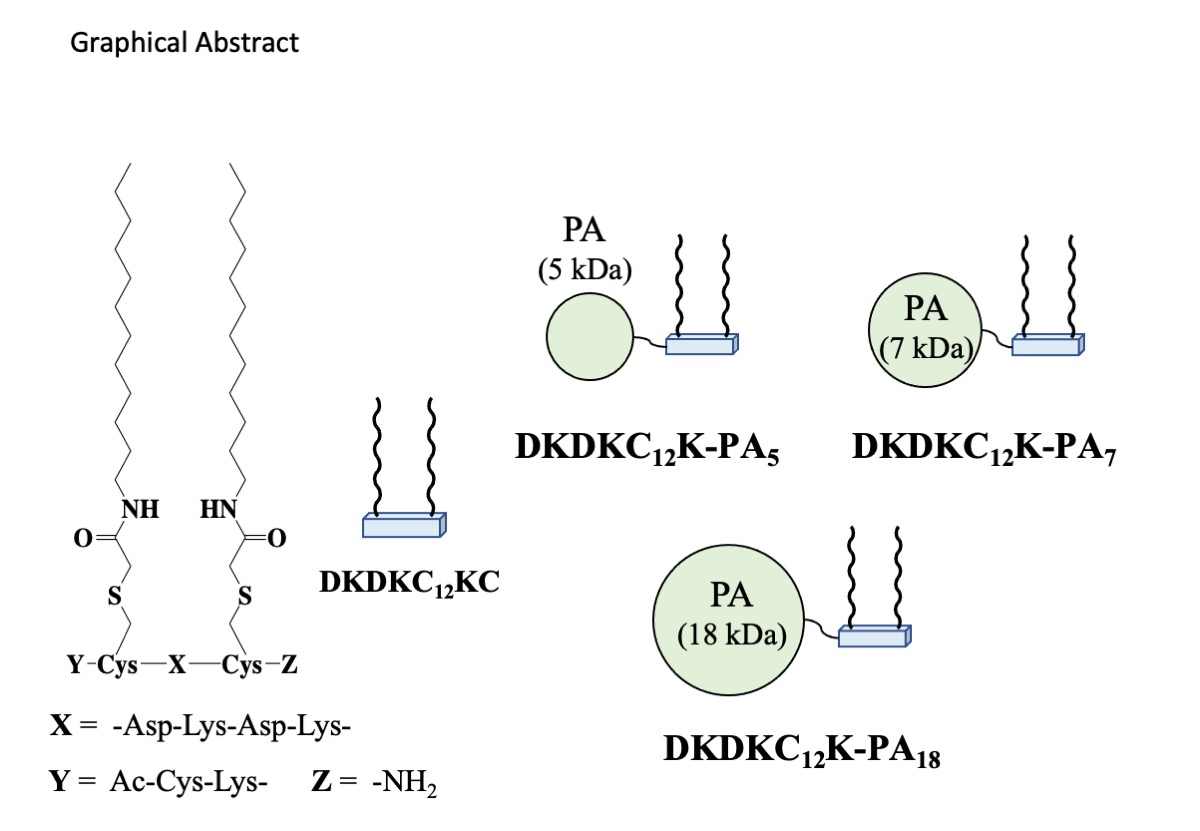
2.1. Design of Solubilization Surfactants Bearing Linear Polyacrylamide (PA) Polymer Chain

To synthesize the polymer-appended DKDKC12K, we separately synthesized the hydrophilic polymer and the surfactant parts and then joined them. For the surfactant part, we chose the PG-surfactant DKDKC12KC, in which one Cys residue was added to the N-terminal side of DKDKC12K (Figure S1). Because DKDKC12K has a higher molecular weight (MW 1330) than the general low MW solubilization surfactants such as n-octyl-β-d-glucopyranoside (β-OG, MW 292) and n-dodecyl-β-d-maltopyranoside (β-DDM, MW 511), it was tolerant of introducing other functional groups to the N- or C-terminal of the same molecule without losing its original solubilization function for membrane proteins [20,23]. We expected that DKDKC12K would also conjugate hydrophilic polymers without losing the original solubilization function. An additional Cys residue was used for conjugation with the hydrophilic polymer part.
For the hydrophilic polymer part, we chose polyacrylamide (PA) in this study. As PA is used as a gel material for protein electrophoresis [24] and immobilization substrates for proteins [25], choosing it as a polymer for solubilization surfactants would be reasonable to reduce structural damage to membrane proteins. To examine the impact of Mn on the solubilization functions of polymer-appended DKDKC12K, we synthesized PA with different Mn values of 5, 7, and 18 kDa by reversible addition-fragmentation chain transfer (RAFT) polymerization [26]. Further, to conjugate with DKDKC12KC, the RAFT initiator containing the dithiopyridyl (DTP) group, BSTP pyridyl disulfide [27] was used for the synthesis of PA bearing the DTP groups at the terminal, PAn-DTP (n = 5, 7, and 18, Scheme 1). The synthesized PAn-DTPs were subjected to gel permeation chromatography (GPC), and the calculated Mns and polydispersity indexes (PDI) are listed in Table 1.
Joining of DKDKC12KC and PAn-DTA via S-S bond (Scheme 2) was performed according to Scheme 2, and the target conjugates were purified by reverse-phase high-performance liquid chromatography (RP-HPLC, Figures S2–S4). Compound identification was performed by dividing the PA and DKDKC12KC units again via reducing the isolated conjugates using DTT, and RP-HPLC identified each divided unit. Hereafter, the polymer-appended DKDKC12Ks were named DKDKC12K-PAn (n = 5, 7, 18).
Before studies on PSI solubilization using new surfactants, we first characterized the fundamental micelle formation properties of DKDKC12K-PAn based on dynamic light scattering (DLS) measurements and critical aggregation concentrations (CACs) (Table 2). With an increase in surfactant concentration, the fluorescence intensity of ANS in a buffer increased with a single inflection point. The plots of F478 vs. surfactant concentration are shown in Figure S5. Since the concentrations at this inflection point, evaluated from the cross point of double linear-fittings, correspond to the CAC of the amphiphilic molecules, we could determine the CACs (mol/L) of DKDKC12K-PA5, DKDKC12K-PA7, and DKDKC12K-PA18 and they were found to be 51, 46, and 47 µM, respectively; the CACs with mol/L as a unit were calculated from CAC with wt% as a unit by the assumption of MW of DKDKC12K-PA5, DKDKC12K-PA7, and DKDKC12K-PA18, as 5900, 8300, and 19,200, respectively. The modification of PA to DKDKC12K hampered micelle formation between the DKDKC12K moieties, but the maintenance of micelle formation property was observed for all DKDKC12K-PAn. The DLS profiles of 0.1 wt% DKDKC12K-PA5, DKDKC12K-PA7, and DKDKC12K-PA18 in phosphate buffer gave a single peak at 13, 25, and 29 nm, respectively, and an apparent increase in micelle diameters was observed with the increase in Mn of the PA unit. These data also support the idea that DKDKC12K units in DKDKC12K-PAn could form micelles similar to those of DKDKC12K [20] even after the introduction of PA units.

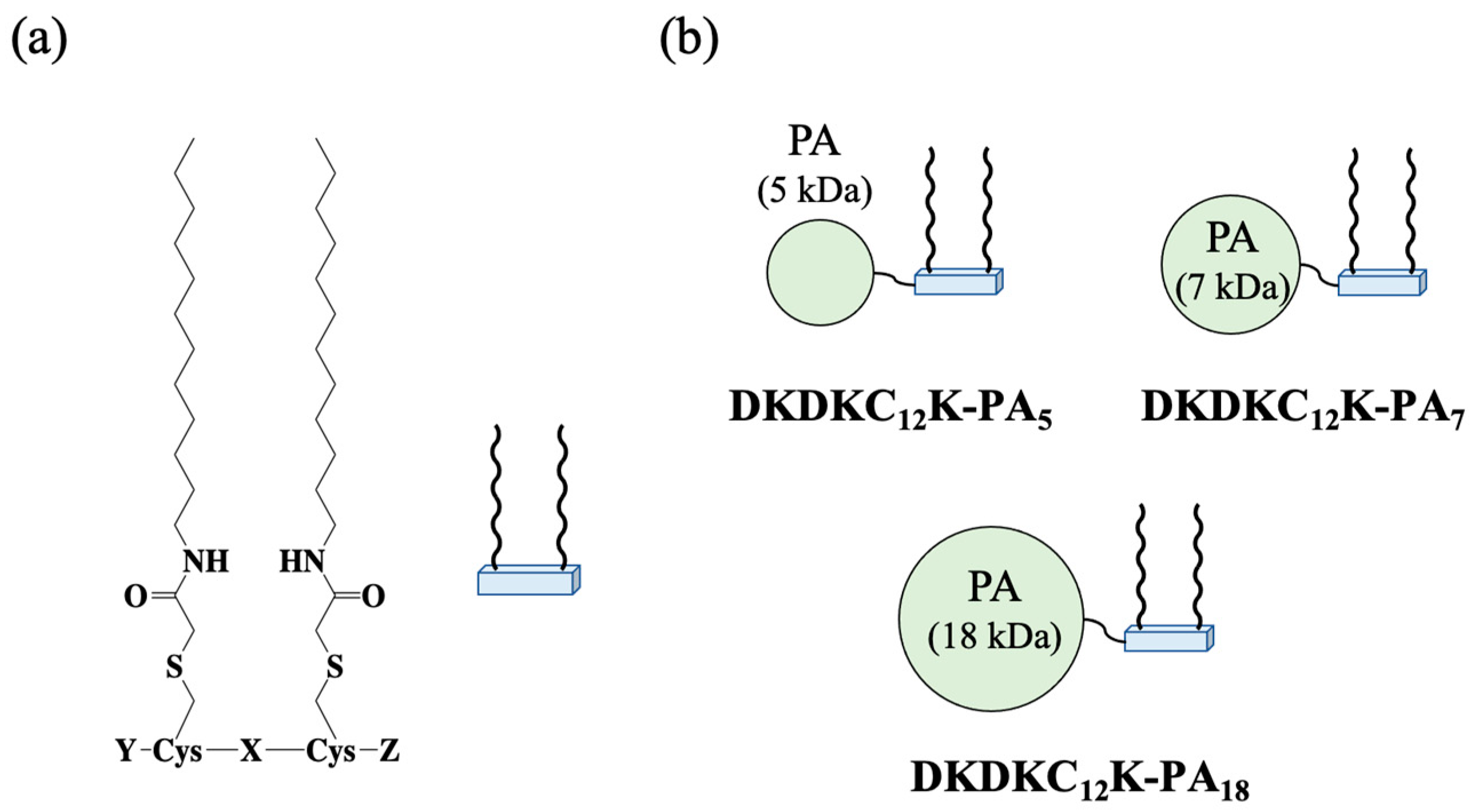
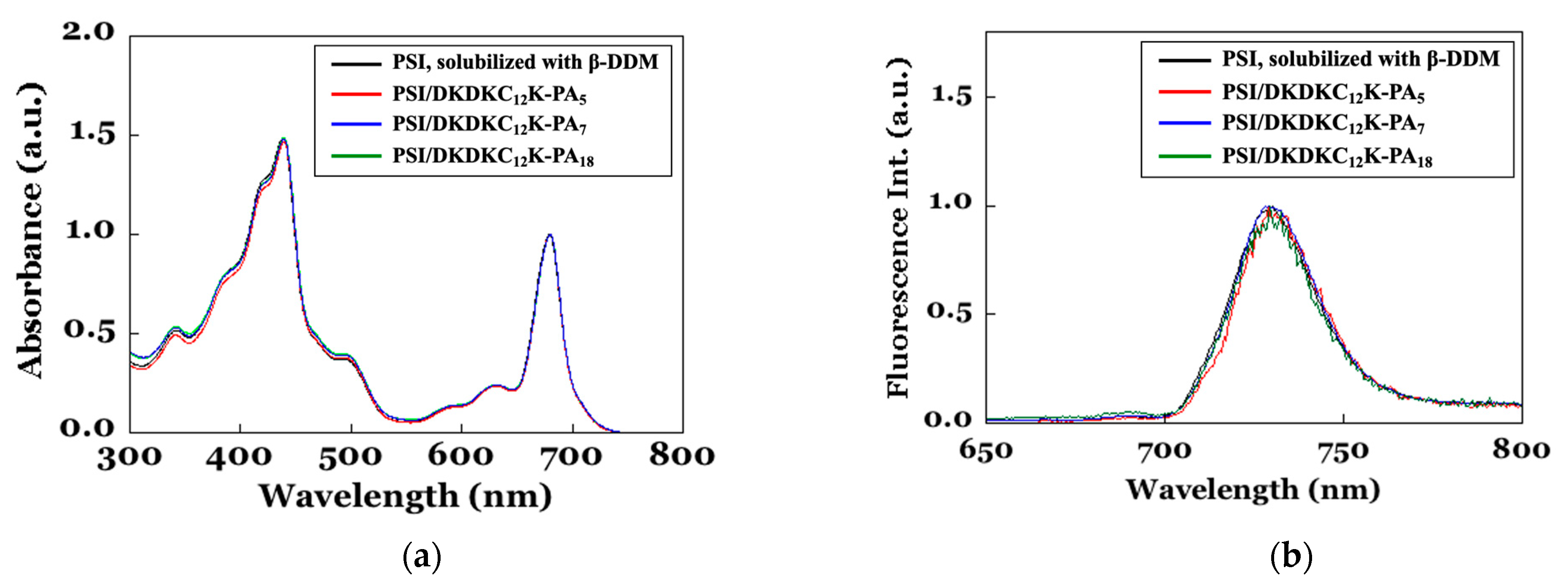
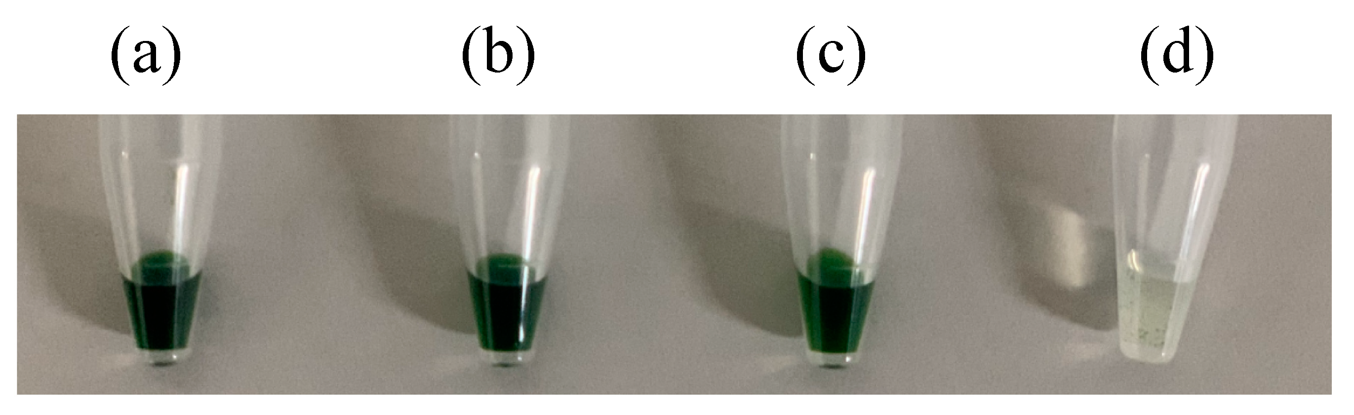
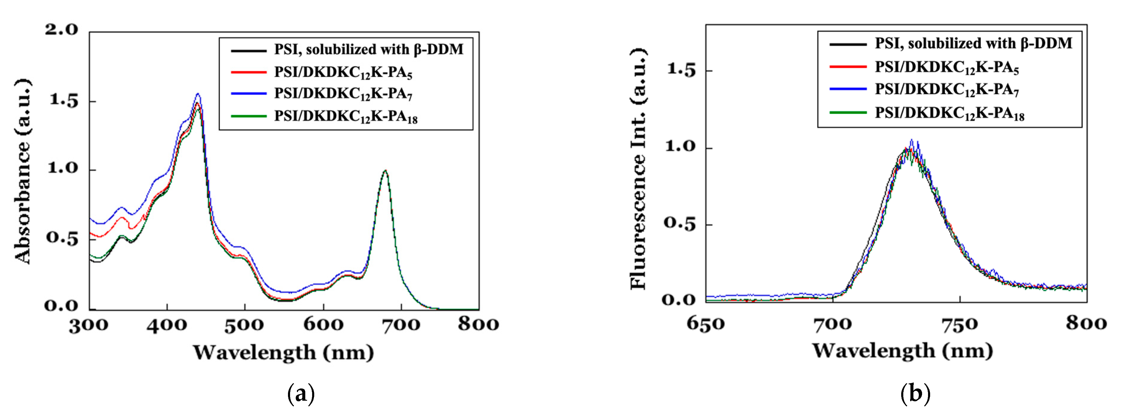
2.2. Solubilization of Photosystem I from Thermosynecoccus (T.) Vulcanus by DKDKC12K-PAn (n = 5, 7, and 18)

To evaluate the solubilization ability of DKDKC12K-PAn for membrane proteins, in this study, we used PSI from T. vulcanus as a representative membrane protein. PSI is a trimeric supramolecular pigment-protein complex (total MW of 1068 kDa); each PSI unit includes 12 protein subunits, 96 chlorophyll a (Chl a) molecules, and 3 [4Fe-4S] clusters [28]. In higher plants and cyanobacteria, it exists in thylakoid membranes and participates in the reductive side light reaction. Because surfactants have unique photophysical properties derived from the hierarchical supramolecular organization of these components, this protein could be used as a probe to evaluate impacts of surfactants on protein structure and functions from impacts on their photophysical properties [20,21]. The PSI samples solubilized with the buffer K2 (40 mM HEPES−NaOH (pH 7.8), 100 mM NaCl, 15 mM CaCl2, and 15 mM MgCl2) containing 0.1 wt% DKDKC12K-PAn were prepared by the surfactant-exchange method as previously reported [20]. The observed absorption spectra at 298 K and the fluorescence spectra at 77 K are shown in Figure 2. Absorption spectra in the 300–800 nm range mainly originated from 96 molecules of antenna Chl a, coordinated in PsaA and PsaB subunits at PSI integral membrane domain. Therefore, maintenance of the absorption spectrum could strongly suggest maintenance of the tertiary structure, especially at the integral membrane domain. The fluorescence spectrum of PSI in the 650–800 nm range at 77 K corresponds to the formation of the red chlorophyll state by Chl a molecules in the PsaA and PsaB scaffolds. This fluorescence is also a unique characteristic of Chl a molecules in native PSI. Therefore, maintenance of the fluorescence spectrum also supports the maintenance of the tertiary structure at the integral membrane domain of PSI. As shown in Figure 2, all PSI samples, solubilized by 0.1 wt% DKDKC12K-PAn, showed similar spectra using 0.1 wt% β-DDM as a solubilization surfactant (control of the native state), suggesting that all DKDKC12K-PAn showed successful solubilization of PSI without denaturation, similar to the parent PG-surfactants, DKDKC12K.
Upon using general low MW solubilization surfactants, membrane proteins are solubilized in a buffer by covering the hydrophobic surfaces of the membrane integral domain with surfactant micelles. Therefore, if the surfactant concentration is less than the critical micelle concentration (CMC), most surfactants cannot form micelles and cannot solubilize membrane proteins in a buffer. However, if the affinity of surfactants to membrane proteins or between surfactants concentrated onto membrane protein surfaces was sufficiently high, the necessary number of surfactant molecules able to solubilize was not governed under surfactant CMCs. In short, if the affinity between DKDKC12K-PAn and PSI or intermolecular interactions between the introduced PA chains in DKDKC12K-PAn is high enough, the isolation of the conjugates of PSI and DKDKC12K-PAn was probable. Therefore, we attempted to isolate the conjugates by separation of unbound DKDKC12K-PAn from the PSI sample. Using an ultrafiltration unit (cut-off MW: 100 kDa), we removed the unbound DKDKC12K-PAn from the PSI samples (theoretical concentration of DKDKC12K-PAn after ultrafiltration was less than 0.00001 wt%), prepared in the buffer K2 with 0.1 wt% DKDKC12K-PAn. As surfactant concentrations decreased to less than 0.00001 wt%, the reference PSI sample solubilized with 0.1 wt% β-DDM became insoluble (Figure 3d). However, those with DKDKC12K-PAn maintained good solubility. These samples were further subjected to gel permeation chromatography (GPC) using buffer K2 without including any solubilization surfactants, and the DKDKC12K-PAn-bound PSIs were finally obtained as a water-soluble conjugate in a surfactant-free buffer (Figure 3a–c). Upon this surfactant-free condition, the residual concentrations of DKDKC12K-PAns in PSI samples were quite less than their CMCs. With considering that hydrophobic interaction of DKDKC12K-PAns to the hydrophobic surfaces of PSI is mainly originated only from two C12 chains in DKDKC12K-PAns, this stable binding of DKDKC12K-PAns to PSI molecules could be occurred by effective intermolecular interactions between the PA chains in DKDKC12K-PAns.
To characterize the conjugated structure and properties of the soluble state of PSI with DKDKC12K-PAn, we first analyzed the photophysical properties by UV-vis and fluorescence spectroscopy (Figure 4). As shown in Figure 4, all conjugates with DKDKC12K-PA5, DKDKC12K-PA7, and DKDKC12K-PA18 showed typical absorption peaks of PSI at 423 and 680 nm in Figure 4a, similar to the case of solubilizing in a buffer with 0.1 wt% β-DDM (control of the native state) [20,21]. On the other hand, an increase in baseline, less than 600 nm region was also observed, which was different from that of solubilizing in a buffer with 0.1 wt% DKDKC12K-PAn, especially for the conjugates with DKDKC12K-PA5 and DKDKC12K-PA7 (Figure 2a). This suggests the formation of PSI aggregations. The fluorescence spectral peak at 720 nm, derived from the red-chlorophyll state, was retained for all PSI samples. If the elimination of Chl a from the PSI scaffold occurred, its fluorescence peak should be observed at 680 nm. However, no fluorescent peak at 680 nm was observed, suggesting that PSI maintained the native state in the conjugates.
The light-induced electron transfer activity of PSI is a useful probe to assess them and can be evaluated by concentration change in solubilized oxygen using an oxygen electrode. In the presence of MV2+ molecules in a solution, the electrons generated by photoexcitation at the P700 special pair first migrate following the potential gradients through phylloquinone and the [4Fe-4S] cluster sites (Fx, FA, and FB) and are finally trapped in MV2+ molecules in a solution. Because the dissolved oxygen immediately quenches the one-electron reductant of MV2+, the initial oxygen consumption rate can be considered identical to the initial rate of light-induced electron transfer in PSI [20,29]. After reducing the resultant hole at the special pair (P700+•) by sodium ascorbate assisted by dichloroindophenol (DCIP), one sequential electron transfer process initiated by light irradiation is completed. If protein denaturation occurred, the passage of electron migration was also damaged. As a result, the electron transfer rate could be reduced. The electron transfer rates per single PSI unit were calculated from the background-subtracted oxygen consumption data, and the obtained initial rates are summarized in Table 3. Although covering the PSI surface with PA units might hamper electron transfer to the MV2+ and DCIP in a buffer, comparable electron transfer rates of those in a buffer with 0.1 wt% β-DDM (control of the native state). This data also supported that PSI in the PSI/DKDKC12K-PAn conjugates maintain native characteristics, suggesting the maintenance of the tertiary structure of the membrane integral and the extracellular domains.
In order to examine the morphologies of the conjugates of PSI and DKDKC12K-PAn, we performed TEM observations. Each TEM sample was prepared by short adsorption of the conjugates onto the polyvinyl alcohol (PVA) layer-coated TEM grids and staining with sodium phosphotungstate. In the case of conjugates with DKDKC12K-PA5 and DKDKC12K-PA7, the formation of plate-type 2D aggregations (less than 100 nm of wide and ~7 nm thickness), orienting horizontally (blue enclosing regions and arrows) or perpendicularly (red enclosing regions and arrows) to the surface of the PVA layer was observed, as shown in the left and center low TEM images of Figure 5. On the other hand, the conjugates with DKDKC12K-PA18 showed homogeneously dispersed spherical morphologies, having ~20 nm diameter (purple enclosing regions and arrows) in the right low TEM images of Figure 5. From X-ray structural analysis, PSI is known to have a low columnar structure and a diameter of about 20 nm [29]. The reference TEM image of the PSI sample, prepared from the PSI solution in a buffer with 0.1 wt% β-DDM, gave circular morphologies with a diameter of ~20 nm (data not shown). This meant that the circular morphologies in the right low TEM images of Figure 5 correspond to the conjugate PSI portion. Upon solubilizing with DKDKC12K-PA18, DKDKC12K-PA18 molecules bind the integral membrane domain of PSI and locate around PSI molecules. Therefore, the bound DKDKC12K-PA18 was expected to be located between the circular ~20 nm morphologies with a lower height than the PSI.
Based on the morphologies and rigorous assessments of the photophysical properties, we found that DKDKC12K-PA18 is the best PA-appended PG-surfactant among the other DKDKC12K-PAn studied, solubilizing PSI in a surfactant-free buffer without inducing PSI aggregation and denaturation.

3. Materials and Methods

3.1. Materials

Rink-amide AM resin (200–400 mesh) was purchased from Merck Biosciences (Darmstadt, Germany). N-(9-fluorenylmethoxycarbonyl) (Fmoc)-protected-amino acids, 1-hydroxybenzotriazole (HOBT), 2-(1H-benzotriazole-1-yl)-1,1,3,3-tetramethyluronium hexafluorophosphate (HBTU), N,N-diisopropylethylamine (DIEA), piperidine, trifluoroacetic acid (TFA), and N-methyl pyrrolidone (NMP) were purchased from Watanabe Chemical Industries (Hiroshima, Japan). Acrylamide and VA-057 were purchased from Wako Pure Chemical Industries, Ltd. (Osaka, Japan). Poly(ethylene glycol) average Mn = 1500 g/mol (PEG1500), n-dodecyl-β-d-maltopyranoside (β-DDM), dichloroindophenol (DCIP), L(+)-Ascorbic Acid Sodium Salt, and methyl viologen dichloride dihydrate (MV2+) were purchased from Sigma-Aldrich (St. Louis, MO, USA). The RAFT initiator containing the dithiopyridyl (DTP) group, BSTP pyridyl disulfide, was synthesized according to previous study [27]. PSI derived from T. vulcanus was prepared similar to the previous study [21]. Unless otherwise stated, other chemicals and reagents were obtained commercially and used without further purification. BSTP disulfide was synthesized as following the previous study.

3.2. Synthesis of PG-Surfactant, DKDKC12KC

PG-surfactant, DKDKC12KC, was synthesized on a Rink-amide AM resin using commercially available Fmoc-protected amino acids and our synthesized Fmoc-Cys(C12)-OH [19]. For condensation onto the resin, standard coupling reagents (HOBT/HBTU/DIEA) were used. The N-terminus of PG-surfactants was end-capped with Ac2O. After cleavage of the synthesized PG-surfactants from the resin using TFA/H2O (95/5), the crude PG-surfactants were purified by reversed-phase high-performance liquid chromatography (RP-HPLC) with a core-shell-type ODS column (Kinetex, Shimadzu, Japan). A linear-gradient of CH3CN and H2O, both including 0.1 vol% TFA, was utilized as eluent (Figure S1a). Product identification was checked by high-resolution ESI-TOF (electrospray ionization time-of-flight) mass spectroscopy (Figure S1b).
DKDKC12KC: HRMS (ESI-TOF, [M + H]+): calcd. for C68H128N15O15S2, 1433.8298; found, 1433.8282.

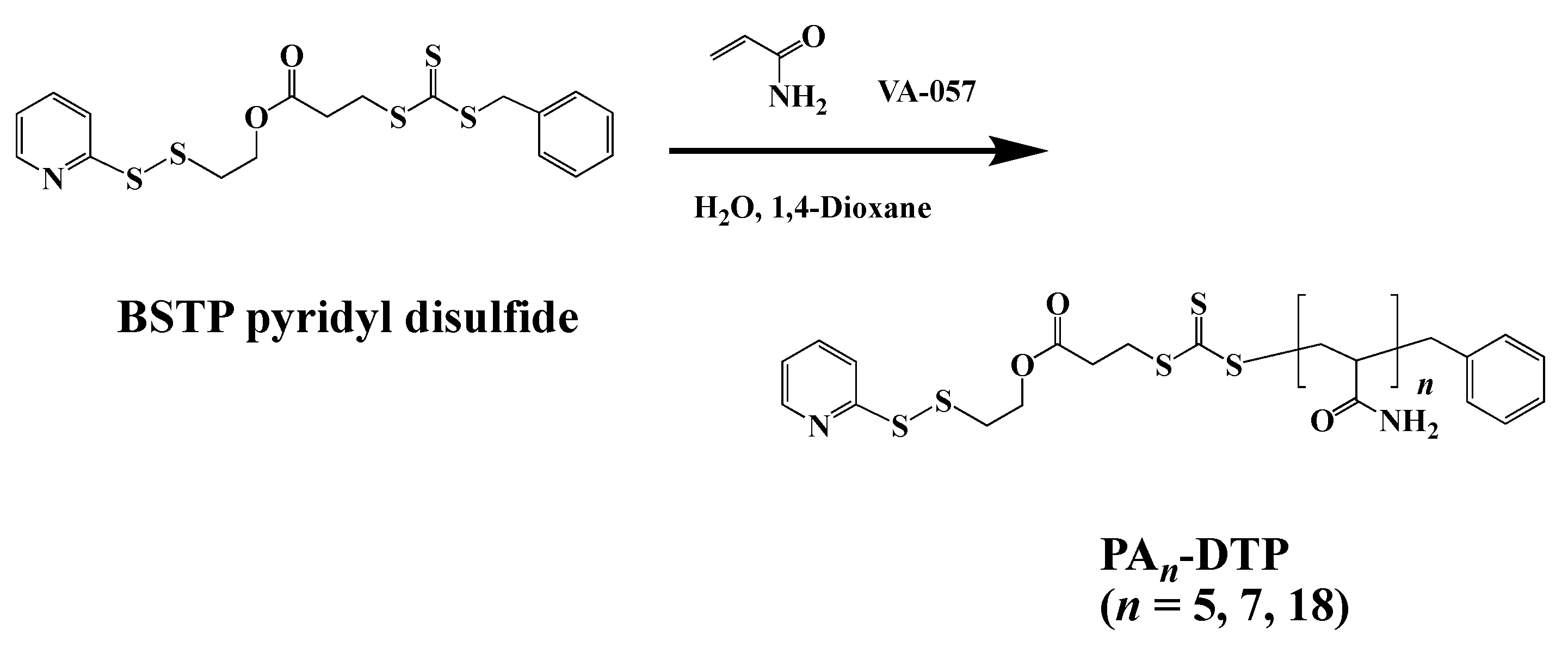
3.3. Synthesis of Polyacrylamide Having Dithiopyridyl Terminal Group (PAn-DTP, n = 5, 7, 18) by RAFT Polymerization

PAn-DTP (n = 5, 7, 18, Mn = 5, 7, 18 kDa), having 2-pyridyldisulfide groups at the terminal, were synthesized by the RAFT technique with molar ratio of monomer/initiator/BSTP pyridyl disulfide [27] of 1124/1/2 for PA (18 kDa), 562/1/2 for PA (7 kDa), and 281/1/2 for PA (5 kDa). For instance, PA (18 kDa) was synthesized as follows. AM (2.00 g, 28.1 mmol), VA-057 (10.4 mg, 25.0 µmol), and BSTP pyridyl disulfide (22.1 mg, 50.0 µmol) were dissolved in DMSO/H2O (5:3, 16 mL) in a round-bottom flask and sealed with a septum. The flask was degassed by freeze thawing with nitrogen and subsequently placed in a preheated hot-bath at 50 °C for 105 min. The resulting polymer was precipitated 3 times from acetone and dried in vacuo. Yield: 1.41 g (71%). The Mn and polydispersity index (PDI) of the synthesized polymers were estimated by a gel permeation chromatography (GPC), respectively. These estimated data of Mn and PDI were summarized in Table 1.

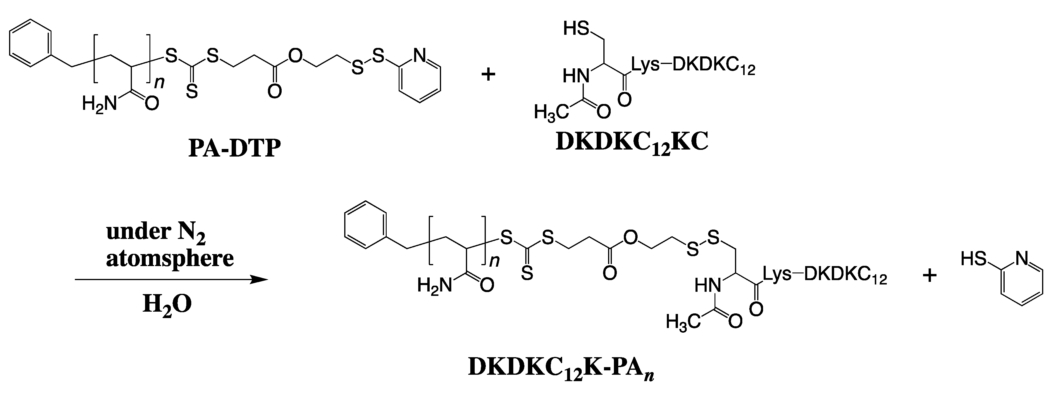
3.4. Synthesis of the PA(5, 7, 18 kDa)-Appended DKDKC12KC, DKDKC12K-PAn (n = 5, 7, 18)

To a N2-substituted water (3 mL), PA with dithiopyridyl group (Mn = 5, 7, 18 kDa, 7 mmol) and DKDKC12KC (3.0 mg, 3.5 µmol) was added and it was stirred for 2 h. at ambient temperature. It was purified by RP-HPLC with a core-shell-type ODS column (Kinetex, Shimadzu, Japan). A linear-gradient of CH3CN and H2O, both including 0.1 vol% TFA, was utilized as eluent. The HPLC profiles of DKDKC12K-PAn (n = 5, 7, 18) were summarized in Figures S2–S4.

3.5. Dynamic Light Scattering Measurements of PG-Surfactant Assemblies

The concentrations of each DKDKC12K-PAn (n = 5, 7, 18) in 100 mM phosphate buffer (pH 7) were set at 0.1 wt% and the mean hydrodynamic diameters of PG-surfactant assemblies for each concentration at 25 °C were estimated using a Zetasizer Nano ZS (Malvern Instruments, Ltd., Malvern, UK).

3.6. Critical Aggregation Concentration (CAC) Determination for PG-Surfactants Using 8-Anilino-Naphtharene-1-Sulfonic Acid (ANS)

The CACs of DKDKC12K-PAn (n = 5, 7, 18) in 100 mM phosphate buffer (pH 7) were evaluated by the fluorescent method [30] similar to the previous studies [20,21,23].

3.7. Replacement of Solubilization Surfactant via PEG Precipitation

The PSI sample, solubilized with DKDKC12K-PAn (n = 5, 7, 18) in the buffer K2 [40 mM HEPES–NaOH (pH 7.8), 100 mM NaCl, 15 mM CaCl2, and 15 mM MgCl2], containing 0.1 wt% of DKDKC12K-PAn (n = 5, 7, 18) was prepared similar to the previous studies [20,21].

3.8. Isolation of the Conjugates of PSI and DKDKC12K-PAn (n = 5, 7, 18)

The unbound-DKDKC12K-PAns (n = 5, 7, 18) were first removed by ultrafiltration (Amicon Ultra 0.5 mL Centrifugal Filters, Devise NMWL 100 kDa) for the PSI sample, solubilized with DKDKC12K-PAn (n = 5, 7, 18) in the buffer K2 (40 mM HEPES–NaOH (pH 7.8), 100 mM NaCl, 15 mM CaCl2, and 15 mM MgCl2). Then these were subjected to gel permeation chromatography (1 cm (i.d.) × 30 cm (h), Superose 6, GE Healthcare, Milwaukee, WI, USA) was applied using the buffer K2 as an eluent.

3.9. Evaluation of Photo-Induced Electron-Transfer Rate of PSI Based on Decreases in O2 Concentration

Measurements of the O2 uptake rate to evaluate photo-induced initial electron transfer rate of PSI were conducted at 25 °C using a Clark-type O2 electrode (Hansatech Instruments, DW1, Oxygen Electrode Unit; Norfolk, VA, USA), similar to the previous study [20,21].

3.10. Fluorescence Spectrum of PSI at 77 K

The fluorescence spectrum of the conjugates of PSI and DKDKC12K-PAn (n = 5, 7, 18) in the buffer K2 was observed similar to the previous study [20,21].

3.11. TEM Measurements of the Conjugates of PSI and DKDKC12K-PAn (n = 5, 7, 18)

TEM images were obtained with JEM-z2500 (JEOL). All the samples were prepared by dry-cast of protein solutions supported on a poval-coated Cu grid (400 mesh, Okenshoji Co., Ltd., Tokyo, Japan). Membrane protein concentration was 5 µg Chla/mL of the isolated PSI conjugates with DKDKC12K-PAn (n = 5, 7, 18). This solution was dropped on the Cu grid and left to stand for approximately 5 min at room temperature. Droplets were removed with filter paper and stained by sodium phosphotungstate solution (2 wt%) in pure water. This sample was rinse three times with pure water.

4. Conclusions

In this study, we constructed PA-modified PG-surfactants, DKDKC12K-PAn, and examined its ability to solubilize membrane proteins. All surfactants could solubilize PSI in a buffer containing 0.1 wt% of each surfactant without any denaturation. However, under surfactant-free conditions (i.e., after removal of the unbound surfactants from the sample solution), the ability to solubilize PSI in a buffer was different; DKDKC12K-PA5 and DKDKC12K-PA7 could solubilize PSI in a surfactant-free buffer without protein denaturation, but several PSI aggregations were observed. In contrast, DKDKC12K-PA18 could solubilize PSI without inducing protein denaturation and aggregation. This should be reasoned by the difference in affinity of PA chains, concentrated onto PSI surfaces; PA chains with 18 kDa of Mn had enough affinity without inducing elimination of DKDKC12K-PA18 molecules from the PSI surface.

Supplementary Materials

Supplementary Materials can be found at https://www.mdpi.com/1422-0067/22/4/1524/s1. Figure S1: Purification of PG-surfactant, DKDKC12KC, Figure S2: Purification of PA-modified PG-surfactant, DKDKC12K-PA5, Figure S3: Purification of PA-modified PG-surfactant, DKDKC12K-PA7, Figure S4: Purification of PA-modified PG-surfactant, DKDKC12K-PA18, Figure S5: Change in F478 of 8-anilino-1-naphthalene sulfonate (ANS) in accordance with increased concentration of DKDKC12K-PA5.

Author Contributions

Conceptualization, T.M.; investigation, T.S., T.N. (Tatsuki Nakakubo), T.N. (Tomoyasu Noji), S.K., K.K., N.K.; writing—original draft preparation, T.S.; writing—review and editing, T.M.; supervision, T.M.; project administration, T.M.; funding acquisition, T.M. All authors have read and agreed to the published version of the manuscript.

Funding

This work was supported by JSPS KAKENHI (Grant Numbers 20K05705), the Takahashi Industrial and Economic Research Foundation, and Ichihara International scholarship foundation.

Acknowledgments

We thank Makoto Miyata and Yu-hei Tahara, affiliated with the Department of Biology, Graduate School of Science, Osaka City University, for technical assistance with TEM measurements of the PSI/DKDKC12K-PAn conjugates (n = 5, 7, 18).

Conflicts of Interest

The authors declare no conflict of interest.

References

  1. Dekkera, J.P.; Boekema, E.J. Supramolecular organization of thylakoid membrane proteins in green plants. Biochim. Biophys. Acta 2005, 1706, 12–39. [Google Scholar] [CrossRef] [Green Version]
  2. Marco, P.; Elman, T.; Yacoby, I. Binding of ferredoxin NADP+ oxidoreductase (FNR) to plant photosystem I. Biochim. Biophys. Acta Bioenergy 2019, 1860, 689–698. [Google Scholar] [CrossRef]
  3. Utschig, L.M.; Soltau, S.R.; Mulfort, K.L.; Niklas, J.; Poluektov, O.G. Z-Scheme solar water splitting via self-assembly of photosystem I-catalyst hybrids in thylakoid membranes. Chem. Sci. 2018, 9, 8504–8512. [Google Scholar] [CrossRef] [Green Version]
  4. Ciesielski, P.N.; Scott, A.M.; Faulkner, C.J.; Berron, B.J.; Cliffel, D.E.; Jennings, G.K. Functionalized nanoporous gold leaf electrode films for the immobilization of photosystem I. ACS Nano 2008, 23, 2465–2472. [Google Scholar] [CrossRef]
  5. Utschig, L.M.; Silver, S.C.; Mulfort, K.L.; Tiede, D.M. Nature-Driven photochemistry for catalytic solar hydrogen production: A photosystem I-Transition metal catalyst hybrid. J. Am. Chem. Soc. 2011, 133, 16334–16337. [Google Scholar] [CrossRef]
  6. Zhao, F.; Conzuelo, F.; Hartmann, V.; Li, H.; Nowaczyk, M.M.; Plumere, N.; Rögner, M.; Schuhmann, W. Light induced H2 evolution from a biophotocathode based on photosystem 1-Pt nanoparticles complexes integrated in solvated redox polymers films. J. Phys. Chem. B 2015, 119, 13726–13731. [Google Scholar] [CrossRef]
  7. Ihara, M.; Nishihara, H.; Yoon, K.; Lenz, O.; Friedrich, B.; Nakamoto, H.; Kojima, K.; Honma, D.; Kamachi, T.; Okura, I. Light-Driven hydrogen production by a hybrid complex of a [NiFe]-Hydrogenase and the cyanobacterial photosystem I. Photochem. Photobiol. 2006, 82, 676–682. [Google Scholar] [CrossRef]
  8. Grimme, R.A.; Lubner, C.E.; Bryant, D.A.; Golbeck, J.H. Photosystem I/Molecular wire/metal nanoparticle bioconjugates for the photocatalytic production of H2. J. Am. Chem. Soc. 2008, 130, 6308–6309. [Google Scholar] [CrossRef]
  9. Nguyen, K.; Bruce, B.D. Growing green electricity: Progress and strategies for use of photosystem I for sustainable photovoltaic energy conversion. Biochim. Biophys. Acta 2014, 1837, 1553–1566. [Google Scholar] [CrossRef] [Green Version]
  10. Ciornii, D.; Riedel, M.; Stieger, K.R.; Feifel, S.C.; Hejazi, M.; Lokstein, H.; Zouni, A.; Lisdat, F. Bioelectronic circuit on a 3D electrode architecture: Enzymatic catalysis interconnected with photosystem I. J. Am. Chem. Soc. 2017, 139, 16478–16481. [Google Scholar] [CrossRef]
  11. Misawa, N.; Fujii, S.; Kamiya, K.; Osaki, T.; Takaku, T.; Takahashi, Y.; Takeuchi, S. Construction of a biohybrid odorant sensor using biological olfactory receptors embedded into bilayer lipid membrane on a chip. ACS Sens. 2019, 4, 711–716. [Google Scholar] [CrossRef] [PubMed]
  12. Nield, J.; Morris, E.P.; Bibby, T.S.; Barber, J. Structural analysis of the photosystem I supercomplex of cyanobacteria induced by iron deficiency. Biochemistry 2003, 42, 3180–3188. [Google Scholar] [CrossRef] [PubMed]
  13. Elter, S.; Raschle, T.; Arens, S.; Viegas, A.; Gelev, V.; Etzkorn, M.; Wagner, G. The use of amphipols for NMR structural characterization of 7-TM proteins. J. Membr. Biol. 2014, 247, 957–964. [Google Scholar] [CrossRef] [PubMed] [Green Version]
  14. Chae, P.S.; Rasmussen, S.G.F.; Rana, R.; Gotfryd, K.; Chandra, R.; Goren, M.A.; Kruse, A.C.; Nurva, S.; Loland, C.J.; Pierre, Y.; et al. Maltose-Neopentyl glycol (MNG) amphiphiles for solubilization, stabilization and crystallization of membrane proteins. Nat. Methods 2010, 7, 1003–1008. [Google Scholar] [CrossRef] [PubMed] [Green Version]
  15. Tribet, C.; Audebert, R.; Popot, J.-L. Amphipols: Polymers that keep membrane proteins soluble in aqueous solutions. Proc. Natl. Acad. Sci. USA 1996, 93, 15047–15050. [Google Scholar] [CrossRef] [PubMed] [Green Version]
  16. Dörr, J.M.; Koorengevel, M.C.; Schäfer, M.; Prokofyev, A.V.; Scheidelaar, S.; Cruijsen, E.A.W.; Dafforn, T.R.; Baldus, M.; Killian, J.A. Detergent-Free isolation, characterization, and functional reconstitution of a tetrameric K+ channel: The power of native nanodiscs. Proc. Natl. Acad. Sci. USA 2014, 111, 18607–18612. [Google Scholar] [CrossRef] [PubMed] [Green Version]
  17. Barniol-Xicota, M.; Verhelst, S.H.L. Stable and functional rhomboid proteases in lipid nanodiscs by using diisobutylene/maleic acid copolymers. J. Am. Chem. Soc. 2018, 140, 14557–14561. [Google Scholar] [CrossRef] [PubMed] [Green Version]
  18. Zhang, M.; Huang, R.; Ackermann, R.; Im, S.-C.; Waskell, L.; Schwendeman, A.; Ramamoorthy, A. Reconstitution of the Cytb5–CytP450 complex in nanodiscs for structural studies using NMR spectroscopy. Angew. Chem. Int. Ed. 2016, 55, 4497–4499. [Google Scholar] [CrossRef] [PubMed]
  19. Umezaki, K.; Sakai, S.; Koeda, S.; Yamamoto, Y.; Kondo, M.; Ikeda, A.; Dewa, T.; Taga, K.; Tanaka, T.; Mizuno, T. Formation of planar bilayer membranes on solid supports using peptide gemini surfactants. Chem. Lett. 2012, 41, 1430–1432. [Google Scholar] [CrossRef]
  20. Koeda, S.; Umezaki, K.; Noji, T.; Ikeda, A.; Kawakami, K.; Kondo, M.; Yamamoto, Y.; Shen, J.R.; Taga, K.; Dewa, T.; et al. Application of peptide gemini surfactants as novel solubilization surfactants for photosystems I and II of cyanobacteria. Langmuir 2013, 29, 11667–11680. [Google Scholar] [CrossRef]
  21. Koeda, S.; Suzuki, T.; Noji, T.; Kawakami, K.; Itoh, S.; Dewa, T.; Kamiya, N.; Mizuno, T. Rational design of novel high molecular weight solubilization surfactants for membrane proteins from the peptide gemini surfactants (PG-Surfactants). Tetrahedron 2016, 72, 6898–6908. [Google Scholar] [CrossRef]
  22. Shibata, M.; Koeda, S.; Noji, T.; Kawakami, K.; Ido, Y.; Amano, Y.; Umezawa, N.; Higuchi, T.; Dewa, T.; Itoh, S.; et al. Design of new extraction surfactants for membrane proteins from peptide gemini surfactants. Bioconjug. Chem. 2016, 27, 2469–2479. [Google Scholar] [CrossRef]
  23. Taniguchi, A.; Koeda, S.; Noji, T.; Kawakami, K.; Sumito, N.; Dewa, T.; Itoh, S.; Kamiya, N.; Mizuno, T. Synthesis and characterization of chemically-reactive solubilization surfactants for membrane proteins and preparation of membrane protein hydrogel microfibers. Colloid Interface Sci. Commun. 2019, 32, 100199. [Google Scholar] [CrossRef]
  24. Reisinger, V.; Eichacker, L.A. Analysis of membrane protein complexes by blue native PAGE. Proteomics 2006, 6 (Suppl. 2), 6–15. [Google Scholar] [CrossRef]
  25. Sassolas, A.; Hayat, A.; Marty, J.-L. Enzyme immobilization by entrapment within a gel network. Methods Mol. Biol. 2013, 1051, 229–239. [Google Scholar] [PubMed]
  26. McCormick, C.L.; Lowe, A.B. Aqueous RAFT polymerization: Recent developments in synthesis of functional water-soluble (co)polymers with controlled structures. Acc. Chem. Res. 2004, 37, 312–325. [Google Scholar] [CrossRef] [PubMed]
  27. Liu, J.; Bulmus, V.; Herlambang, D.L.; Barner-Kowollik, C.; Stenzel, M.H.; Davis, T.P. In situ formation of protein–polymer conjugates through reversible addition fragmentation chain transfer polymerization. Angew. Chem. Int. Ed. 2007, 46, 3099–3103. [Google Scholar] [CrossRef]
  28. Jordan, P.; Fromme, P.; Witt, H.T.; Klukas, O.; Saenger, W.; Krauû, N. Three-Dimensional structure of cyanobacterial photosystem I at 2.5 A resolution. Nature 2001, 411, 909–917. [Google Scholar] [CrossRef] [PubMed]
  29. Shen, J.R. Denshidentatsu kassei. Low Temp. Sci. 2009, 67, 551–560. [Google Scholar]
  30. De Vendittis, E.; Palumbo, G.; Parlato, G.; Bocchini, V. A fluorimetric method for the estimation of the critical micelle concentration of surfactants. Anal. Biochem. 1981, 115, 278–286. [Google Scholar] [CrossRef]
Figure 1. Chemical structure of PG-surfactants and schematic illustration of their conjugates with polyacrylamide (PA) (a) Chemical structure of PG-surfactants with C12 alkyl chains (X: linker peptide, Y, Z: peripheral peptide). (b) Schematic illustration of the PA-appended solubilization surfactants, DKDKC12K-PAn (n = 5, 7, and 18). X is -Asp-Lys-Asp-Lys and Z is -NH2. Y is Ac-Cys-Lys-, in which PA unit was covalently connected to side chain of Cys via S-S bond.
Ijms 22 01524 g001
Scheme 1. Synthesis of polyacrylamide (PA) with dithiopyridyl unit (PAn-DTP, n = 5, 7, 18) via RAFT polymerization.
Ijms 22 01524 sch001
Scheme 2. Synthesis of DKDKC12K-PAn (n = 5, 7, 18).
Ijms 22 01524 sch002
Figure 2. Absorption and fluorescence spectra of PSI, solubilized with 0.1 wt% β-DDM, or conjugated with DKDKC12K-PAn (n = 5, 7, 18). (a) Absorption spectra of PSI, solubilized with 0.1 wt% β-DDM (black), DKDKC12K-PA5 (red), DKDKC12K-PA7 (blue), and DKDKC12K-PA18 (green). (b) Florescence spectra of PSI, solubilized with 0.1 wt% β-DDM (black), DKDKC12K-PA5 (red), DKDKC12K-PA7 (blue), and DKDKC12K-PA18 (green).
Ijms 22 01524 g002
Figure 3. Comparison of the solution states of the PSI/DKDKC12K-PAn conjugates and the reference supernatant solution of PSI. The PSI/DKDKC12K-PAn conjugates were the isolated ones by removement of the unbound DKDKC12K-PAn by gel permeation chromatography. The reference supernatant solution of PSI was prepared by decrease in β-DDM concentration less than 0.00001 wt% by ultrafiltration. (a) PSI/DKDKC12K-PA5 conjugate, (b) PSI/DKDKC12K-PA7 conjugate, (c) PSI/DKDKC12K-PA18 conjugate, (d) the reference supernatant solution of PSI sample.
Ijms 22 01524 g003
Figure 4. Absorption and fluorescence spectra of PSI, solubilized with 0.1 wt% β-DDM, or conjugated with DKDKC12K-PAn (n = 5, 7, 18). (a) Absorption spectra of PSI, solubilized with 0.1 wt% β-DDM (black), DKDKC12K-PA5 (red), DKDKC12K-PA7 (blue), and DKDKC12K-PA18 (green). (b) Florescence spectra of PSI, solubilized with 0.1 wt% β-DDM (black), DKDKC12K-PA5 (red), DKDKC12K-PA7 (blue), and DKDKC12K-PA18 (green).
Ijms 22 01524 g004
Figure 5. TEM images of the isolated PSI/DKDKC12K-PAn conjugates (PSI/DKDKC12K-PA5 conjugates (left low), PSI/DKDKC12K-PA7 conjugates (center low), and PSI/DKDKC12K-PA18 conjugates (right low)) and the schematic illustrations of the expected molecular structures of aggregates, observed in TEM images (upper). 2D aggregates of PSI particles solubilized with DKDKC12K-PA5 or DKDKC12K-PA7 are indicated by red or blue enclosing regions and arrows. While single PSI particles solubilized with DKDKC12K-PA18 are indicated by purple enclosing regions and arrows. Accelerate voltage, 200 kV.
Ijms 22 01524 g005
Table 1. Summary of Mn and PDI off PAn-DTP (n = 5, 7, and 18).
PAn-DTPMn (PDI) a
PA5-DTP4500 (1.19)
PA7-DTP6900 (1.29)
PA18-DTP17,800 (1.14)
aMn and PDI of the polyacrylamide part determined using polyethylene glycol standard.
Table 2. CAC and hydrodynamic diameter (d, nm) of DKDKC12K-PAn in 100 mM phosphate buffer (pH 7).
CAC (wt%)CAC (µM)d (nm) a
DKDKC12K---8.3 b6 b
DKDKC12K-PA50.03051 c13
DKDKC12K-PA70.03846 c25
DKDKC12K-PA180.09047 c29
a The concentration of each was set to 0.1 wt%. b Referred from ref. 21. c The CACs with mol/L as a unit were calculated from CAC with wt% as a unit by the assumption of MW of DKDKC12K-PA5, DKDKC12K-PA7, and DKDKC12K-PA18, as 5900, 8300, and 19,200, respectively.
Table 3. Light-induced initial electron transfer rates in PSI solubilized with 0.1 wt% β-DDM in a buffer or conjugated with DKDKC12K-PA5, DKDKC12K-PA7, or DKDKC12K-PA18.
Solubilization
Surfactant
Electron Transfer Rate
in PSI (PSI−1s−1) a
β-DDM32.2 ± 5.9
DKDKC12K-PA535.8 ± 1.3
DKDKC12K-PA741.2 ± 0.6
DKDKC12K-PA1838.0 ± 0.6
a Concentration of PSI solubilized with 0.001 wt% surfactant was 24 nM. A buffer containing 40 mM HEPES−NaOH (pH 7.5), 100 mM NaCl, 15 mM CaCl2, 15 mM MgCl2, and 0.4 M sucrose supplemented with 0.5 mM dichloroindophenol (DCIP), 2 mM sodium ascorbate, and 0.5 mM MV2+ was used for oxygen uptake measurements.
Publisher’s Note: MDPI stays neutral with regard to jurisdictional claims in published maps and institutional affiliations.

