Article

eIF5A-Independent Role of DHPS in p21CIP1 and Cell Fate Regulation

Department of Biochemistry, Medical College of Wisconsin, Milwaukee, WI 53226, USA
*
Author to whom correspondence should be addressed.
Int. J. Mol. Sci. 2021, 22(24), 13187; https://doi.org/10.3390/ijms222413187
Submission received: 3 November 2021 / Revised: 1 December 2021 / Accepted: 2 December 2021 / Published: 7 December 2021
(This article belongs to the Special Issue 23rd Anniversary of IJMS: Advances in Biochemistry)

Abstract

:
Deoxyhypusine synthase (DHPS) catalyzes the first step of hypusination of the elongation translation factor 5A (eIF5A), and these two proteins have an exclusive enzyme–substrate relationship. Here we demonstrate that DHPS has a role independent of eIF5A hypusination in A375 and SK-MEL-28 human melanoma cells, in which the extracellular signal regulated kinase 1/2 (ERK1/2) pathway is deregulated. We found that RNA interference of DHPS induces G0/G1 cell cycle arrest in association with increased p21CIP1 expression in these cells whereas eIF5A knockdown induces cell death without increasing p21CIP1 expression. Interestingly, p21CIP1 knockdown switched DHPS knockdown-induced growth arrest to cell death in these cells, suggesting a specific relation between DHPS and p21CIP1 in determining cell fate. Surprisingly, ectopic expression of DHPS-K329R mutant that cannot hypusinate eIF5A abrogated DHPS knockdown-induced p21CIP1 expression in these cells, suggesting a non-canonical role of DHPS underlying the contrasting effects of DHPS and eIF5A knockdowns. We also show that DHPS knockdown induces p21CIP1 expression in these cells by increasing CDKN1A transcription through TP53 and SP1 in an ERK1/2-dependent manner. These data suggest that DHPS has a role independent of its ability to hypusinate eIF5A in cells, which appears to be important for regulating p21CIP1 expression and cell fate.

1. Introduction

Deoxyhypusine synthase (DHPS) is the only known enzyme that catalyzes the first step of hypusination of the elongation translation factor 5A (eIF5A), which is the only DHPS substrate known to date [1,2]. Given this exclusive enzyme–substrate relationship, it is generally expected that DHPS and eIF5A mediate overlapping contexts of cellular processes, and although DHPS mRNA has been shown to be a natural antisense transcript for the scaffold protein MORG1 [3], no mutually independent function of DHPS and eIF5A has been reported at protein levels. Hypusination is a highly specific post-translational modification that has been detected with only eIF5A [4,5,6]. eIF5A hypusination is mediated by two step enzyme reactions that sequentially occur. First, DHPS conjugates the aminobutyl moiety of spermidine to a specific lysine residue of eIF5A to produce deoxyhypusine-eIF5A [7,8,9]. Second, deoxyhypusine hydroxylase (DOHH) reduces deoxyhypusine-eIF5A to irreversibly produce hypusine-eIF5A [10,11]. Cells express two functionally redundant highly homologous eIF5A isoforms, eIF5A1 and eIF5A2 [12]. eIF5A1 is the major isoform ubiquitously expressed in almost any tissue type, whereas eIF5A2 expression is limited to specific tissue or tumor types [13,14]. Hypusination enables eIF5A to promote translational elongation of the polypeptide chains stalling due to disruptive sequences such as polyproline-motifs and sequentially charged residues [15,16,17] as well as translational termination on a global scale [15,18,19]. As determined in yeast Saccharomyces cerevisiae and cells of higher organisms, eIF5A hypusination is a fundamental process for eukaryotic cell viability [20]. Expression of DHPS, DOHH, and eIF5A is often upregulated in different tumors [21], and better understanding the role of the hypusination pathway is important.
p21CIP1/WAF is a cyclin-dependent kinase inhibitor (CDKI) that can mediate G0/G1 cell cycle arrest by inhibiting CDK2, CDK4, and CDK6, and subsequently preventing retinoblastoma protein (Rb) phosphorylation, E2F1 release from Rb, and entry into the S-phase of the cell cycle [22]. p21CIP1 has a role as a tumor suppressor along with other CDKIs such as p16INK4A and p27KIP1 in various tumor biological contexts [23]. Particularly, p21CIP1 has been identified as a key mediator of growth arrest induced upon sustained activation of the Raf/MEK/extracellular-signal regulated kinase (ERK) pathway in different cell types [24]. Interestingly, although mainly known for its role to mediate cell cycle arrest in the G0/G1 phase [25], p21CIP1 is also known to suppress cell death [23]. The Raf/MEK/ERK pathway is a mitogenic pathway and its deregulated signaling is detected in a wide variety of cancers [26]. Therefore, it is important to elucidate the mechanisms by which the pathway regulates p21CIP1 expression and how p21CIP1 expression is suppressed in tumor cells that exhibit aberrant MEK/ERK activity.
A previous study proposed a role for DHPS in p21CIP1 regulation using the specific DHPS inhibitor GC7 [27]. However, it was never reported whether the DHPS-eIF5A cascade has any role in p21CIP1 regulation in the context of Raf/MEK/ERK signaling. We therefore sought to determine whether the DHPS-eIF5A cascade has any role in p21CIP1 regulation and cell fate decision in Raf/MEK/ERK-activated tumor cells. In this study, we demonstrate in the BRAFV600E melanoma cells A375 and SK-MEL-28 that DHPS knockdown induces growth arrest associated with p21CIP1 upregulation whereas eIF5A knockdown induces cell death, and that p21CIP1 knockdown can switch the growth arrest induced by DHPS knockdown to cell death. Subsequently, we show that DHPS can negatively regulate p21CIP1 expression independently of its eIF5A hypusinating activity, identifying a previously unknown non-canonical function of DHPS.

2. Results

2.1. DHPS Depletion Increases p21CIP1 Levels and Induces G0/G1 Phase Cell Cycle Arrest in A375 and SK-MEL-28 Cells

Using RNA interference, we determined the effects of DHPS depletion in A375 and SK-MEL-28 that exhibit high MEK/ERK activity due to BRAFV600E mutation. We found that knockdown of DHPS for 5 days by lentiviruses expressing two independent shRNA, which was sufficient to curtail eIF5A hypusination in cells, consistently increased p21CIP1 expression in association with decreased phosphorylation of Rb in these cells (Figure 1A). Rb is a key cell cycle regulator that sequesters the S-phase transcription factor E2F1 [28]. However, DHPS knockdown did not affect the levels of E2F1 and another CDKI p27KIP1, while not inducing p16INK4a expression or notably increasing the cleavage of Poly ADP-ribose polymerase (PARP) and lamin A, the apoptotic cell death makers [29,30], in these cells (Figure 1A). Consistent with these changes, DHPS knockdown substantially decreased cell proliferation rates without increasing death rates in these cells (Figure 1B). Moreover, DHPS knockdown increased the G0/G1 phase cell population (Figure 1C). These data demonstrate that DHPS depletion can induce cell cycle arrest which is associated with p21CIP1 expression.

2.2. eIF5A Depletion Induces Cell Death without Increasing p21CIP1 Levels in A375 and SK-MEL-28 Cells

As determined by Western blotting using an antibody that reacts with both eIF5A1 and eIF5A2 isoforms, RNA interference of eIF5A1 substantially decreased eIF5A levels in SK-MEL-28 and A375 cells (Figure 2A). In these cells, the effects of eIF5A depletion were in stark contrast to the effects of DHPS depletion. eIF5A knockdown in these cells, mediated by two independent lentiviral shRNA constructs, robustly increased the cleavage of lamin A and PARP but did not increase p21CIP1 expression (Figure 2A). Consistent with the death markers, eIF5A knockdown substantially increased cell death in the cultures of these cells, as determined by the trypan blue exclusion analysis (Figure 2B). Importantly, DHPS knockdown and eIF5A knockdown decreased eIF5A hypusination at similar degrees (Figure 2C), and did not cause any correlative changes in ERK1/2 phosphorylation in these cells (Figure 1A and Figure 2A). Therefore, the different effects of these treatments are not due to different eIF5A hypusination levels or altered ERK1/2 activity. These data suggested that DHPS and eIF5A may have distinct effects on cell fate and p21CIP1 regulation in cells.
The contrasting effects of DHPS and eIF5A knockdowns were not limited to A375 and SK-MEL-28 cells. Similar as these cells, DHPS knockdown suppressed cell proliferation without inducing cell death whereas eIF5A knockdown induced cell death responses, albeit at varied degrees, in the KRAS-mutant human pancreatic cell lines, MIA-PaCa-2 and PANC-1 (Supplemental Figure S1A–C). However, unlike in A375 and SK-MEL-28 cells, DHPS depletion did not increase p21CIP1 levels in these cells (Supplemental Figure S1A), suggesting that cellular responses to DHPS knockdown are not entirely identical across different cell types.

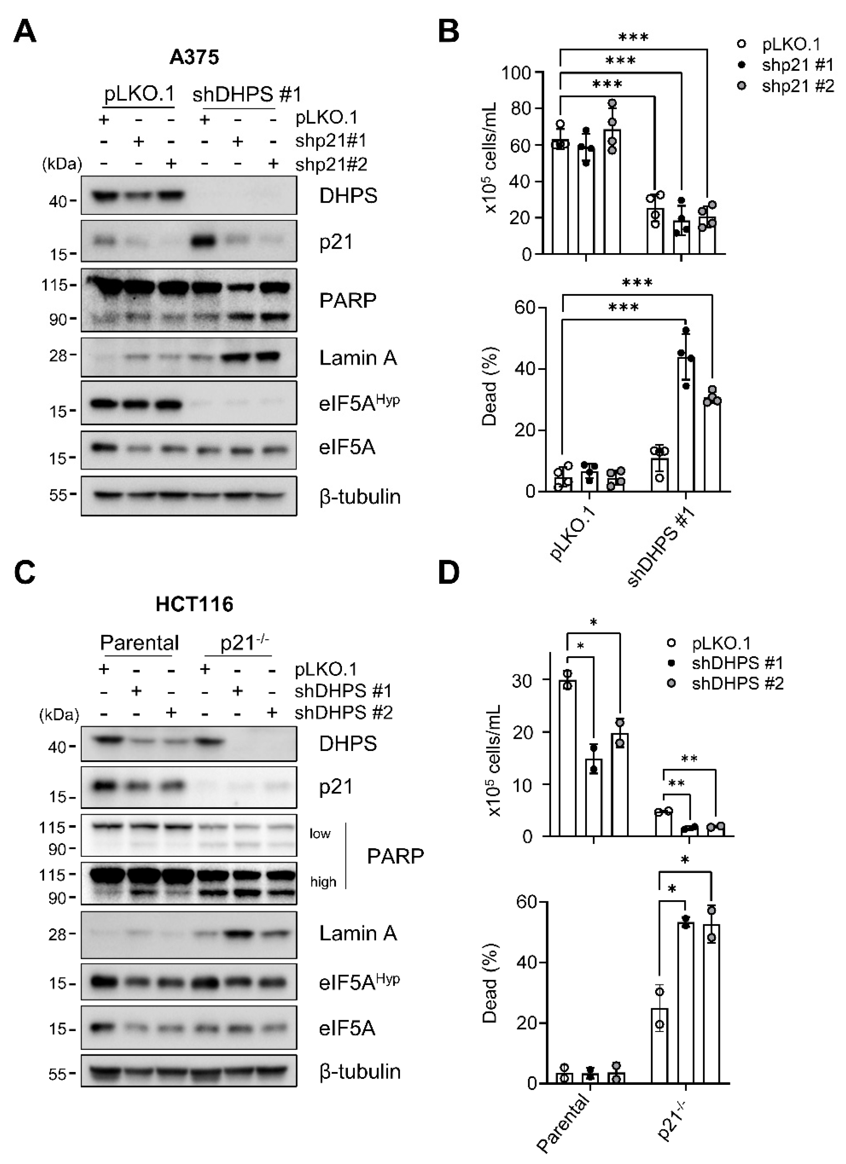
2.3. p21CIP1 Depletion Switches DHPS Depletion-Induced Growth Arrest to Cell Death

We previously showed that p21CIP1-mediated cell cycle arrest can suppress death responses in MEK/ERK-dependent tumor cells [31,32]. Given this and the data above, we asked whether p21CIP1 has a role in determining cell fate in the face of DHPS depletion. Indeed, we found that blocking p21CIP1 expression can switch DHPS depletion-induced cell cycle arrest to cell death responses. Knockdown of p21CIP1 using lentiviruses expressing two independent shRNA consistently increased the cleavage of PARP and lamin A, and dead cell populations in A375 cultures only when combined with DHPS knockdown (Figure 3A,B). Consistent with these effects, DHPS knockdown increased the cleavage of PARP and lamin A, and induced cell death in CDKN1A (encoding p21CIP1)-deleted HCT116 (p21−/−) cells, but not in the parental cells (Figure 3C,D). Nevertheless, DHPS knockdown did not increase p21CIP1 levels in HCT116 cells, further supporting the notion that DHPS-mediated p21CIP1 regulation is cell type specific.
p21CIP1 depletion did not alter the effects of DHPS knockdown on eIF5A hypusination in these cells when compared with their parental cells (Figure 3A,C), excluding potential interference by uneven eIF5A hypusination. These data demonstrate that p21CIP1 can regulate cellular responses to DHPS depletion.

2.4. Hypusinating Activity Is Not Necessary for DHPS to Regulate p21CIP1

Because the contrasting knockdown effects of DHPS and eIF5A may suggest an eIF5A-independent role for DHPS, we examined the effects of the spermidine analog GC7 in A375 and SK-MEL-28 cells. GC7 is a competitive inhibitor of DHPS catalytic activity [33], and we expected that GC7 would help us determine whether DHPS knockdown-induced p21CIP was due to depleted enzyme activity or protein. Our time-course analysis revealed that GC7, used at levels sufficient to inhibit eIF5A hypusination, did not increase but decreased the basal levels of p21CIP1 in A375 and SK-MEL-28 cells (Figure 4A). As an orthogonal approach, we also examined the effects of GC7 on p21CIP1 expression induced upon activation of ΔRaf-1:ER in HEK293 cells. ΔRaf-1:ER is the CR3 catalytic domain of Raf-1 fused to the ligand binding domain of the estrogen receptor for tamoxifen-controlled activity [34]. In these cells, GC7 treatment substantially inhibited ΔRaf-1:ER-induced p21CIP1 expression without affecting ERK1/2 phosphorylation (Figure 4B). These data indicated that inhibition of DHPS catalytic activity cannot mimic DHPS depletion in cells, leading us to hypothesize that DHPS has a role independent of its catalytic activity to regulate p21CIP1 in cells.
Lys329 of DHPS is necessary for the formation of an enzyme–imine intermediate required for eIF5A hypusination, and replacing this residue with Arg hinders the intermediate formation [35]. To test the aforementioned hypothesis, we generated a DHPS mutant in which Lys329 is replaced with Arg (DHPS-K329R) and determined whether this mutant can regulate p21CIP1 levels in cells similarly as wild type DHPS. First, we confirmed that DHPS-K329R is catalytically deficient in HEK293 cells in which DOHH and eIF5A were co-expressed with either DHPS or DHPS-K329R to establish a proper stoichiometry between these proteins. Our data showed that, unlike wild type DHPS, DHPS-K329R cannot increase eIF5A hypusination in this assay (Figure 4C). Expression of this mutant and wild type DHPS did not affect ERK1/2 phosphorylation induced by ΔRaf-1:ER activation (Figure 4C). Next, we determined whether DHPS-K329R, which was engineered to avoid RNA interference, can restore p21CIP1 suppression in DHPS-depleted A375 cells. Indeed, when ectopically expressed at similar levels, DHPS-K329R was similarly effective as wild type DHPS for abrogating the effect of DHPS knockdown to increase p21CIP1 levels in cells (Figure 4D). These data demonstrate that DHPS can negatively regulate cellular p21CIP1 levels independently of its hypusinating activity, thus its substrate eIF5A. We also attempted to determine the ability of DHPS-K329R to rescue cells from DHPS knockdown, but prolonged overexpression of this mutant as well as wild type DHPS suppressed cell proliferation, hampering our efforts. It may be possible that unbalanced stoichiometry between DHPS, DOHH and eIF5A is anti-proliferative.

2.5. MEK/ERK Activity Is Necessary for DHPS Knockdown to Increase p21CIP1 Levels in Cells

We previously demonstrated that MEK/ERK activity is sufficient to induce p21CIP1 [36] and that the MEK/ERK activity in BRAF-mutant tumor cells can be routed to induce p21CIP1 expression [37]. We therefore determined whether the MEK/ERK activity is necessary for DHPS knockdown to increase p21CIP1 levels in cells. First, we found that AZD6244, a highly selective MEK1/2 inhibitor [38], substantially attenuated p21CIP1 expression in DHPS depleted SK-MEL-28 cells (Figure 5A). Second, the combination of DHPS knockdown and ΔRaf-1:ER activation robustly increased p21CIP1 levels in HEK293 cells without affecting other cell cycle or death effectors (Figure 5B). Conversely, DHPS overexpression in LNCaP cells, a cell line that rapidly expresses p21CIP1 in response to ΔRaf-1:ER activation [32,36], substantially abrogated the p21CIP1 expression induced by ΔRaf-1:ER activation (Figure 5C). Moreover, consistent with its effect to augment p21CIP1 expression, the combination of DHPS knockdown and ΔRaf-1:ER activation more potently suppressed HEK293 cell proliferation than DHPS knockdown or ΔRaf-1:ER activation alone (Figure 5D). However, this combination did not notably induce cell death (Figure 5D), which is consistent with its lack of effects on PARP and lamin A cleavage (Figure 5B). Together, these data suggest that DHPS negatively regulates MEK/ERK-induced p21CIP1 expression.

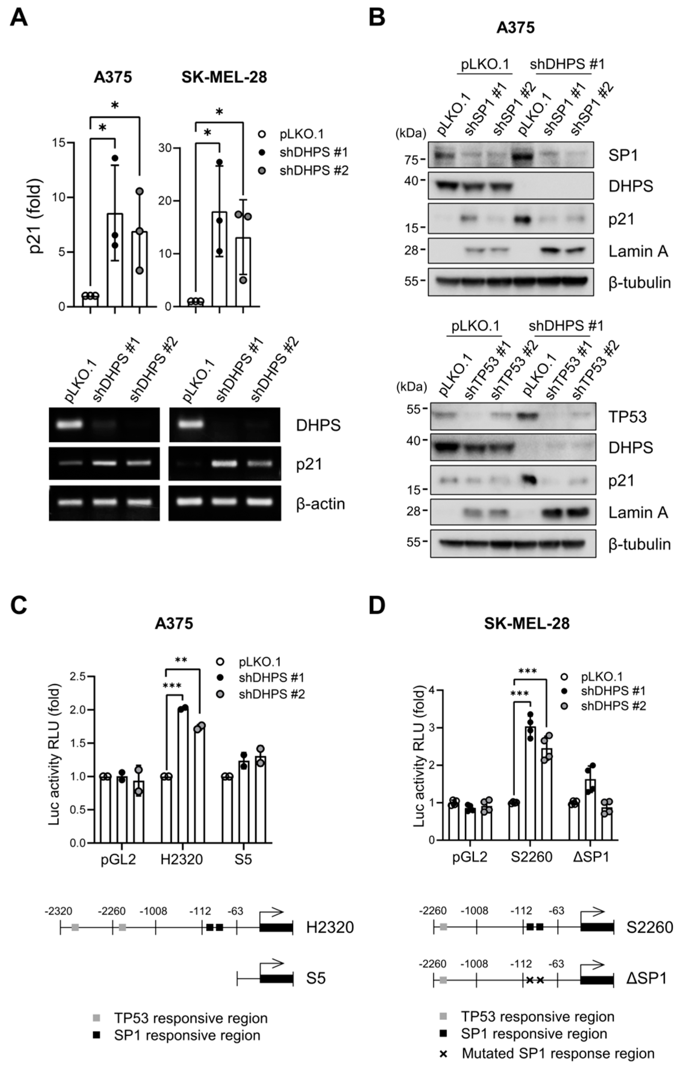
2.6. DHPS Knockdown Increases CDKN1A Transcription via TP53 and SP1 in A375 and SK-MEL-28 Cells

p21CIP1 is regulated at multiple levels including transcription, translation, and post-translation [39]. We found that DHPS knockdown substantially increased CDKN1A (encoding p21CIP1) mRNA levels in A375 and SK-MEL-28 cells (Figure 6A), and thus sought to determine the molecular mechanism underlying this increase. Although TP53 is the major regulator of CDKN1A transcription [40,41], we previously reported that SP1 can also mediate MEK/ERK-induced CDKN1A transcription in tumor cells deficient of a functional TP53 [42]. Accordingly, we examined the effects of TP53 knockdown and SP1 knockdown on DHPS knockdown-induced p21CIP1 expression in A375 (TP53 wild type) and SK-MEL-28 (TP53L145R) cells. As determined by Western blotting in A375 cells, TP53 knockdown and SP1 knockdown substantially attenuated p21CIP1 expression induced by DHPS knockdown while increasing lamin-A cleavage (Figure 6B). This effect is consistent with p21CIP1 depletion effects in the background of DHPS knockdown. Moreover, DHPS knockdown increased the activity of the luciferase reporter, H2320, that contains 2320 base pairs of CDKN1A promoter in A375 cells, but this increase was abrogated upon truncation of the region that contain TP53 and SP1 responsive elements (Figure 6C). Similarly, DHPS knockdown upregulated the activity of the luciferase reporter, S2260, that contains 2260 base pairs of CDKN1A promoter in SK-MEL-28 cells but this induction was abrogated upon disabling the SP1 binding sites by site-directed mutagenesis (Figure 6D). These data suggest that DHPS knockdown increases p21CIP1 expression by promoting CDKN1A transcription through TP53 or SP1 in these cells.

3. Discussion

In this report, we demonstrate that, although known for their exclusive enzyme–substrate relationship and thus expected to mediate largely overlapping cellular processes [5], DHPS and eIF5A depletion can result in distinct effects on cell fate. Mechanistically, our data suggest that DHPS has a novel eIF5A hypusination-independent function that can negatively regulate p21CIP1 expression in cells by suppressing TP53- and SP1-mediated MEK/ERK activity-sensitive CDKN1A transcription (a model depicted in Figure 7). Given that DHPS does not interact with TP53 and SP1 (data not shown), we speculate that DHPS indirectly regulates CDKN1A transcription in this model, although the detailed molecular mechanism requires to be elucidated.
Our data provide compelling evidence supporting that DHPS can regulate cell fate independently of its activity to catalyze hypusination, thus of its only known substrate eIF5A. First, knockdown of DHPS and eIF5A produced different physiological effects in A375, SK-MEL-28, MIA-PaCa-2, PANC-1, and HCT116 cell lines. Whereas DHPS knockdown induced growth arrest, direct knockdown of eIF5A induced cell death accompanied by increased cleavage of lamin A and PARP, albeit at varied levels. In these cells, DHPS knockdown and eIF5A knockdown reduced eIF5A hypusination to similar degrees, and knockdown of one did not affect the expression of the other. Therefore, we suggest that the differential outcomes in cell fate were not a result of varying levels of eIF5A hypusination but probably a specific effect due to distinct roles of DHPS and eIF5A. Second, our data suggest a clear functional difference between DHPS and eIF5A in p21CIP1 regulation. Whereas DHPS knockdown increased p21CIP1 expression in A375 and SK-MEL-28 cells, eIF5A knockdown decreased it. Third, similar as eIF5A knockdown, GC7 decreased basal p21CIP1 expression in these cells while exhibiting higher potency than eIF5A knockdown, which suggested that protein depletion but not hypusinating activity underlies the p21CIP1-inducing effect of DHPS knockdown. This notion is strongly supported by the data obtained with the catalytically disabled DHPS-K329R mutant. Given these contrasting effects on p21CIP1, we suspect that while DHPS maintains basal p21CIP1 expression via an eIF5A-dependent mechanism, which fits into the context of the global effect of hypusinated eIF5A to promote protein translation [16], DHPS also regulates an eIF5A-independent mechanism to prevent p21CIP1 overexpression caused by an abnormal signal such as hyper ERK1/2 activity (Figure 7).
Aberrant MEK/ERK activity can cause cell cycle arrest for which p21CIP1 induction is a key mechanism [32,36,37]. This anti-proliferative response addresses in part oncogene-induced senescence, a hypothetical innate tumor defense mechanism in cells [43]. Our data suggest a role for DHPS in this context, given that the aberrant MEK/ERK activity was necessary for DHPS knockdown to induce p21CIP1 expression in A375 and SK-MEL-28 cells, and ΔRaf-1:ER-activated HEK293 cells. Because DHPS knockdown or overexpression did not affect ERK1/2 phosphorylation in these cells, DHPS might function at a level downstream of ERK1/2. Indeed, our data suggest that DHPS regulates MEK/ERK-induced p21CIP1 expression through the transcription factors TP53 and SP1. While TP53 is a bonafide regulator for CDKN1A transcription [40], we previously reported that SP1 also facilitates CDKN1A transcription in response to aberrant MEK/ERK activity [42]. Interestingly, despite the presence of intact TP53, SP1 knockdown substantially abrogated p21CIP1 expression induced by DHPS knockdown in A375 cells. Of note, it was reported that TP53 can recruit SP1 to p21CIP1 promoter and this enhances the binding of these transcription factors to the DNA promoter [44]. Given this information, it may be possible that DHPS has a role in promoting the cooperative effects of SP1 and TP53. How DHPS functionally interacts with TP53 and SP1 remains to be elucidated in future work.
In summary, we report a previously unknown non-canonical DHPS function, which is independent of its activity to hypusinate eIF5A and regulates p21CIP1 expression induced by MEK/ERK via TP53 and SP1. We propose that this function may be important for BRAF-mutated tumor cells to suppress p21CIP1 overexpression, which can be triggered by aberrant MEK/ERK activity in these tumor cells if not prevented.

4. Materials and Methods

4.1. Cell Culture and Reagents

The human embryonic kidney cell line HEK293 (ATCC, Manassas, VA, USA) was maintained in minimal essential medium (Invitrogen, Waltham, MA, USA, 11095-080) supplemented with 10% fetal bovine serum (Gibco, Waltham, MA, USA, SH30541.03). The human prostate cancer cell line LNCaP (ATCC, Manassas, VA, USA) was maintained in phenol-red free RPMI (Invitrogen, Waltham, MA, USA, 11835-030) supplemented with 10% fetal bovine serum. The human melanoma cell line SK-MEL-28 (ATCC, Manassas, VA) were maintained in minimal essential medium supplemented with 10% fetal bovine serum, 100U/mL penicillin, and 100 μg/mL streptomycin (Gibco, Waltham, MA, USA, 15140-122), 1% sodium pyruvate (Gibco, Waltham, MA, USA, 11360-070), and 1% non-essential amino acids (Gibco, Waltham, MA, USA, 11140-050). The human melanoma cell line A375 (ATCC, Manassas, VA, USA), and human pancreatic cancer cell lines MIA-PaCa-2 and PANC-1 were maintained in Dulbecco’s modified eagle medium (Invitrogen, Waltham, MA, USA, 11965-092) with 10% fetal bovine serum, 100U/mL penicillin, and 100 μg/mL streptomycin. The human colorectal carcinoma cell line HCT116 was maintained in McCoy’s 5a medium (Gibco, Waltham, MA, USA, 16600) with 10% fetal bovine serum, 100U/mL penicillin and 100 μg/mL streptomycin. Generation of LNCaP-ΔRaf:ER and HEK293-ΔRaf:ER cells that are stably transduced with the lentiviral pHAGE-ΔRaf:ER was previously described [32,36]. ΔRaf:ER was activated by treating cells with 1 µM 4-Hydroxytamoxifen (Sigma, St. Louis, MO, USA, 68392-35-8) as previously described [34]. AZD6244 (606143-52-6) and PLX4032 (S1267) were obtained from Selleck Chemicals.

4.2. Plasmids and Viral Constructs for Gene Expression and RNA Interference

C-terminally HA-tagged DHPS in pHAGE-GFP was generated by ligating the full length DHPS cDNA from pCMV3-ORF-HA (Sino Biologicals, Bejing China, HG14407-CY) into the NheI/XhoI sites of pHAGE-GFP. pcDNA3.1-eIF5A and pCEFL-DOHH were obtained from Dr. Myung Hee Park [11,13]. DHPS was depleted using two independent pLKO.1 lentiviral shRNA systems (Sigma-Aldrich, St. Louis, MO, USA, TRCN0000045644 labeled as shDHPS-#1 and TRCN00000330717 labeled as shDHPS-#2). pLKO.1-shDHPS-#1 and pLKO.1-shDHPS-#2 target GCGACATGATCTTCTTCCATTC and AGTGCACTGGGATGATCATTC, respectively. Lentiviral shRNA expression systems pLL3.7-sheIF5A-#1 and pLL3.7-sheIF5A-#2 target GCATTACGTAAGAATGGCTTT and CTGGGAAGAAATATGAAGATA of EIF5A mRNA, respectively. pLL3.7-sheIF5A-#1 was generated by ligating the annealing product of TGCATTACGTAAGAATGGCTTTCTCGAGAAAGCCATTCTTACGTAATGCT TTTTC and TCGAGAAAAAGCATTACGTAAGAATGGCTTTCTCGAGAAAGCCATTCTT ACGTAATGCA into the XhoI/HpaII sites of pLL3.7 (ATCC). pLL3.7-sheIF5A-#2 was generated by ligating the annealing product of TCTGGGAAGAAATATGAAGATACTCGA GTATCTTCATATTTCTTCCCA GTTTTTC and TCGAGAAAAACTGGGAAGAAATATGA AGATACTCGAGTATCTTCATATTTCTTCCCAGA into the XhoI/HpaII sites of pLL3.7. To generate DHPS-K329R, the wild type DHPS in pHAGE was mutagenized using the QuickChange Lightning Site-Directed Mutagenesis Kit (Agilent Technologies, Santa Clara, CA, USA, 210518-5) and the primers GGCTGTCTCCTGGGGCAGGATCCGGGTGGAT and GCATCCACCCG GATCCTGCCCCAGGAGACAG. To generate shRNA non-targetable constructs, pHAGE-DHPS and pHAGE-DHPS-K329R were mutagenized using the primers GCTCGCTGGGC GACATGATTTTTTTTCATTCCTACAAGAACC and GGTTCTTGTAGGAATGAAAAAAAA TCATGTCGCCCAGCGAGC p21CIP1 was depleted using two independent pLKO.1 lentiviral shRNA systems (Sigma-Aldrich, St. Louis, MO, USA, TRCN0000040123 labeled as shp21 #1 and TRCN0000040125 labeled as shp21 #2). pLKO.1-shp21 #1 and pLKO.1-shp21 #2 target CGCTCTACATCTTCTGCCTTA and GAGCGATGGAACTTCGACTTT, respectively.
For lentivirus production, 293T cells were co-transfected with the lentiviral backbone and packaging vectors, as previously described [36]. Viral supernatants were collected after 72 h. Viral titers were determined by infecting the recipient cell lines with serially diluted viral supernatants, and then scoring cells expressing GFP at 48 h post-infection.

4.3. Analysis of Cell Viability and Cell Cycle

Cell viability was measured by trypan blue exclusion assay. The cell cycle analysis was performed as previously described [36]. Briefly, cells were washed with ice-cold 0.2% bovine serum albumin in phosphate-buffer saline, and then resuspended in 250 mM sucrose/40mM citrate buffer (pH 7.6) containing 0.5% dimethylsulfoxide. Cell nuclei were stained with propidium iodide and analyzed using the Guava EasyCyte flowcytometry system (Millipore Sigma, St. Louis, MO, USA). Gating targeted single cell nuclei within a normal size range. The cell-cycle parameters were determined from 5000 gated nuclei, and then analyzed with FCS EXPRESS 6 FLOW software (De Novo Software, Boulder, CO, USA).

4.4. Quantitative RT-PCR (qPCR) and Luciferase Reporter Assays

TRIzol® reagent (Invitrogen, Waltham, MA, USA, 15596026) was used to isolate total RNA from cells. Reverse transcription was performed using Superscript III (Invitrogen, Waltham, MA, USA, 18080-044) and oligo-dT according to the manufacturer’s instructions. Quantitative RT-PCR was performed by mixing the resulting cDNA with the SYBR GreenER qPCR Supermix Universal (Invitrogen, Waltham, MA, USA, 11762100) and the following primers: CTGGAGACTCTCAGGGTCGAA and CCAGCACTCTT AGGAACCTCTCA for p21CIP1; AAGTTTGAGGACTGGCTGATG and CAGGGATGTGGT TCTTCTGG for DHPS; and GTCCTCTCCCAAGTCCACAC and GGGAGA CCAAAAGCCTTC AT for β-actin. qPCR signal was obtained using a Stratagene MX3005P instrument.
Construction of the p21CIP1 promoter luciferase reporters H2320, S2260, and S5 that contains p21CIP1 promoter DNA fragments spanning −2320 to +16, −2260 to +16, and −63 to +16 base pairs respectively, were previously described [42]. To generate ΔSP1, two SP1 sites in S2260 were mutated using the QuikChange Lightning site-directed mutagenesis kit (Agilent Technologies, Santa Clara, CA, USA 210518-5) and the primers GTGGGCCGAGCGCGGGTCTTACCTCCTTGAGGTAAGCC and GGCTTACCTCAAGG AGGTAAGACCCGCGCTCGGCCCAC. To measure the reporter activity, A375 and SK-MEL-28 cells were transfected in 6-well plates using Lipofectamine 3000 (Invitrogen, Waltham MA, USA, L3000-008) or Lipofectamine LTX (Invitrogen, Waltham, MA, USA, 15338-100) respectively, and total cell lysates were extracted and analyzed using the Luciferase® Assay System (Promega, Madison, WI, USA, E1960) according to the manufacturer’s instructions. Data were normalized to total protein concentrations.

4.5. Immunoblotting

Cells were harvested in lysis buffer containing 62.5 mM Tris-HCl (pH 6.8)/2% SDS, and protease and phosphatase inhibitor cocktails 2 and 3 (Sigma-Aldrich, St. Louis, MO, USA, P8340, P5726, P0044). Protein concentrations were measured using the bicinchoninic acid reagent (Pierce, Waltham, MA, USA, 23228, 1859078). Proteins were resolved by sodium dodecyl sulfate polyacrylamide gel electrophoresis and transferred to polyvinylidene difluoride membrane filter (Bio-Rad, Hercules, CA, USA, 1620177). After transfer, membranes were blocked at 25 °C for 1h in buffer containing 0.1M Tris (pH 7.4), 0.9% NaCl, 0.05% Tween 20, and 5% nonfat dry milk.
Membranes were then incubated with the appropriate antibodies overnight at 4 °C at the dilutions indicated as follows: phospho-ERK1/2 1:2000 (Thr202/Tyr204, sc-16982), DHPS 1:2000 (sc-365077), eIF5A 1:2000 (sc-390202), p21CIP1 1:1000 (sc-756), p27 1:1000 (sc-528), E2F1 1:1000 (sc-193), p16 1:1000 (sc-1661) (Santa Cruz Biotechnology, Santa Cruz, CA, USA). PARP (9542S), Lamin A (2035S), ERK1/2 1:2000 (9102S), pRb 1:2000 (S780, 9307S), Rb 1:2000 (9309P), (Cell Signaling Technology, Danvers, MA, USA). β-actin 1:5000 (Sigma, St. Louis MO, USA, A1978), β-tubulin 1:5000 (Invitrogen, Waltham MA, USA, PA5-16863), eIF5AHyp 1:5000 (gift from Dr. Mirmira [45]). SuperSignal West Pico and Femto chemiluminescence kit (Pierce, Waltham, MA, USA, 34094) were used for visualization of the signal. For densitometry, immunoblots were analyzed using Image Lab software (Bio-Rad, Hercules, CA, USA).

4.6. Statistical Analysis

Statistical significance was determined using the two-tailed unpaired Student’s t-test using PRISM (Graph-Pad Software, La Jolla, CA, USA) of two data sets. p values of <0.05 were considered statistically significant.

Supplementary Materials

The following are available online at https://www.mdpi.com/article/10.3390/ijms222413187/s1.

Author Contributions

Conceptualization, A.E.B. and J.-I.P.; methodology, A.E.B., P.-K.W., and J.-I.P.; formal analysis, A.E.B.; investigation, A.E.B.; validation, P.-K.W.; writing—original draft preparation, A.E.B. and J.-I.P.; writing-review and editing, A.E.B., P.-K.W., and J.-I.P.; visualization, A.E.B., P.-K.W., and J.-I.P.; supervision, J.-I.P.; project administration, J.-I.P.; funding acquisition, J.-I.P. All authors have read and agreed to the published version of the manuscript.

Funding

This study was supported by the NIH/National Cancer Institute grant R01CA138441 to J.-I.P.

Institutional Review Board Statement

Not applicable.

Informed Consent Statement

Not applicable.

Data Availability Statement

Not applicable.

Acknowledgments

The eIF5A and DOHH expression vectors were a gift from Myung Hee Park (Molecular and Cellular Biochemistry, NIDCR). Anti-eIF5AHyp antibody was a gift from Raghu Mirmira (Indiana University School of Medicine). We thank Wenjing Chen for technical supports for qPCR.

Conflicts of Interest

The authors declare no conflict of interest.

References

  1. Joe, Y.A.; Wolff, E.C.; Park, M.H. Cloning and expression of human deoxyhypusine synthase cDNA. Structure-function studies with the recombinant enzyme and mutant proteins. J. Biol. Chem. 1995, 270, 22386–22392. [Google Scholar] [CrossRef] [Green Version]
  2. Wolff, E.C.; Lee, Y.B.; Chung, S.I.; Folk, J.E.; Park, M.H. Deoxyhypusine synthase from rat testis: Purification and characterization. J. Biol. Chem. 1995, 270, 8660–8666. [Google Scholar] [CrossRef] [PubMed] [Green Version]
  3. Su, W.Y.; Li, J.T.; Cui, Y.; Hong, J.; Du, W.; Wang, Y.C.; Lin, Y.W.; Xiong, H.; Wang, J.L.; Kong, X.; et al. Bidirectional regulation between WDR83 and its natural antisense transcript DHPS in gastric cancer. Cell Res. 2012, 22, 1374–1389. [Google Scholar] [CrossRef] [Green Version]
  4. Park, M.H. The post-translational synthesis of a polyamine-derived amino acid, hypusine, in the eukaryotic translation initiation factor 5A (eIF5A). J. Biochem. 2006, 139, 161–169. [Google Scholar] [CrossRef] [Green Version]
  5. Park, M.H.; Wolff, E.C. Hypusine, a polyamine-derived amino acid critical for eukaryotic translation. J. Biol. Chem. 2018, 293, 18710–18718. [Google Scholar] [CrossRef] [PubMed] [Green Version]
  6. Cooper, H.L.; Park, M.H.; Folk, J.E.; Safer, B.; Braverman, R. Identification of the hypusine-containing protein hy+ as translation initiation factor eIF-4D. Proc. Natl. Acad. Sci. USA 1983, 80, 1854–1857. [Google Scholar] [CrossRef] [PubMed] [Green Version]
  7. Chen, M.; Gai, Z.; Okada, C.; Ye, Y.; Yu, J.; Yao, M. Flexible NAD(+) Binding in Deoxyhypusine Synthase Reflects the Dynamic Hypusine Modification of Translation Factor IF5A. Int. J. Mol. Sci. 2020, 21, 5509. [Google Scholar] [CrossRef] [PubMed]
  8. Lee, C.H.; Um, P.Y.; Park, M.H. Structure-function studies of human deoxyhypusine synthase: Identification of amino acid residues critical for the binding of spermidine and NAD. Biochem. J. 2001, 355, 841–849. [Google Scholar] [CrossRef] [Green Version]
  9. Umland, T.C.; Wolff, E.C.; Park, M.H.; Davies, D.R. A new crystal structure of deoxyhypusine synthase reveals the configuration of the active enzyme and of an enzyme.NAD.inhibitor ternary complex. J. Biol. Chem. 2004, 279, 28697–28705. [Google Scholar] [CrossRef] [Green Version]
  10. Kim, Y.S.; Kang, K.R.; Wolff, E.C.; Bell, J.K.; McPhie, P.; Park, M.H. Deoxyhypusine hydroxylase is a Fe(II)-dependent, HEAT-repeat enzyme. Identification of amino acid residues critical for Fe(II) binding and catalysis [corrected]. J. Biol. Chem. 2006, 281, 13217–13225. [Google Scholar] [CrossRef] [Green Version]
  11. Park, J.H.; Aravind, L.; Wolff, E.C.; Kaevel, J.; Kim, Y.S.; Park, M.H. Molecular cloning, expression, and structural prediction of deoxyhypusine hydroxylase: A HEAT-repeat-containing metalloenzyme. Proc. Natl. Acad. Sci. USA 2006, 103, 51–56. [Google Scholar] [CrossRef] [PubMed] [Green Version]
  12. Wu, G.Q.; Xu, Y.M.; Lau, A.T.Y. Recent insights into eukaryotic translation initiation factors 5A1 and 5A2 and their roles in human health and disease. Cancer Cell Int. 2020, 20, 142. [Google Scholar] [CrossRef]
  13. Clement, P.M.; Johansson, H.E.; Wolff, E.C.; Park, M.H. Differential expression of eIF5A-1 and eIF5A-2 in human cancer cells. FEBS J. 2006, 273, 1102–1114. [Google Scholar] [CrossRef] [Green Version]
  14. Jenkins, Z.A.; Haag, P.G.; Johansson, H.E. Human eIF5A2 on chromosome 3q25-q27 is a phylogenetically conserved vertebrate variant of eukaryotic translation initiation factor 5A with tissue-specific expression. Genomics 2001, 71, 101–109. [Google Scholar] [CrossRef] [PubMed]
  15. Pelechano, V.; Alepuz, P. eIF5A facilitates translation termination globally and promotes the elongation of many non polyproline-specific tripeptide sequences. Nucleic Acids Res. 2017, 45, 7326–7338. [Google Scholar] [CrossRef] [Green Version]
  16. Schuller, A.P.; Wu, C.C.; Dever, T.E.; Buskirk, A.R.; Green, R. eIF5A Functions Globally in Translation Elongation and Termination. Mol. Cell 2017, 66, 194–205.e5. [Google Scholar] [CrossRef] [PubMed] [Green Version]
  17. Gutierrez, E.; Shin, B.S.; Woolstenhulme, C.J.; Kim, J.R.; Saini, P.; Buskirk, A.R.; Dever, T.E. eIF5A promotes translation of polyproline motifs. Mol. Cell 2013, 51, 35–45. [Google Scholar] [CrossRef] [PubMed] [Green Version]
  18. Park, M.H.; Wolff, E.C.; Smit-McBride, Z.; Hershey, J.W.; Folk, J.E. Comparison of the activities of variant forms of eIF-4D. The requirement for hypusine or deoxyhypusine. J. Biol. Chem. 1991, 266, 7988–7994. [Google Scholar] [CrossRef]
  19. Park, M.H. The essential role of hypusine in eukaryotic translation initiation factor 4D (eIF-4D). Purification of eIF-4D and its precursors and comparison of their activities. J. Biol. Chem. 1989, 264, 18531–18535. [Google Scholar] [CrossRef]
  20. Schnier, J.; Schwelberger, H.G.; Smit-McBride, Z.; Kang, H.A.; Hershey, J.W. Translation initiation factor 5A and its hypusine modification are essential for cell viability in the yeast Saccharomyces cerevisiae. Mol. Cell. Biol. 1991, 11, 3105–3114. [Google Scholar] [CrossRef]
  21. Mathews, M.B.; Hershey, J.W. The translation factor eIF5A and human cancer. Biochim. Biophys. Acta 2015, 1849, 836–844. [Google Scholar] [CrossRef] [PubMed] [Green Version]
  22. El-Deiry, W.S. p21(WAF1) Mediates Cell-Cycle Inhibition, Relevant to Cancer Suppression and Therapy. Cancer Res. 2016, 76, 5189–5191. [Google Scholar] [CrossRef] [PubMed] [Green Version]
  23. Abbas, T.; Dutta, A. p21 in cancer: Intricate networks and multiple activities. Nat. Rev. Cancer 2009, 9, 400–414. [Google Scholar] [CrossRef]
  24. Park, J.I. Growth arrest signaling of the Raf/MEK/ERK pathway in cancer. Front. Biol. 2014, 9, 95–103. [Google Scholar] [CrossRef] [PubMed] [Green Version]
  25. Warfel, N.A.; El-Deiry, W.S. p21WAF1 and tumourigenesis: 20 years after. Curr. Opin. Oncol. 2013, 25, 52–58. [Google Scholar] [CrossRef] [PubMed]
  26. Imperial, R.; Toor, O.M.; Hussain, A.; Subramanian, J.; Masood, A. Comprehensive pancancer genomic analysis reveals (RTK)-RAS-RAF-MEK as a key dysregulated pathway in cancer: Its clinical implications. Semin. Cancer Biol. 2019, 54, 14–28. [Google Scholar] [CrossRef] [PubMed]
  27. Bandino, A.; Geerts, D.; Koster, J.; Bachmann, A.S. Deoxyhypusine synthase (DHPS) inhibitor GC7 induces p21/Rb-mediated inhibition of tumor cell growth and DHPS expression correlates with poor prognosis in neuroblastoma patients. Cell Oncol. 2014, 37, 387–398. [Google Scholar] [CrossRef]
  28. Weinberg, R.A. The retinoblastoma protein and cell cycle control. Cell 1995, 81, 323–330. [Google Scholar] [CrossRef] [Green Version]
  29. Kaufmann, S.H.; Desnoyers, S.; Ottaviano, Y.; Davidson, N.E.; Poirier, G.G. Specific proteolytic cleavage of poly(ADP-ribose) polymerase: An early marker of chemotherapy-induced apoptosis. Cancer Res. 1993, 53, 3976–3985. [Google Scholar]
  30. Rao, L.; Perez, D.; White, E. Lamin proteolysis facilitates nuclear events during apoptosis. J. Cell Biol. 1996, 135, 1441–1455. [Google Scholar] [CrossRef] [PubMed]
  31. Park, J.I.; Strock, C.J.; Ball, D.W.; Nelkin, B.D. The Ras/Raf/MEK/extracellular signal-regulated kinase pathway induces autocrine-paracrine growth inhibition via the leukemia inhibitory factor/JAK/STAT pathway. Mol. Cell. Biol. 2003, 23, 543–554. [Google Scholar] [CrossRef] [Green Version]
  32. Hong, S.K.; Wu, P.K.; Park, J.I. A cellular threshold for active ERK1/2 levels determines Raf/MEK/ERK-mediated growth arrest versus death responses. Cell. Signal. 2018, 42, 11–20. [Google Scholar] [CrossRef]
  33. Lee, Y.; Kim, H.K.; Park, H.E.; Park, M.H.; Joe, Y.A. Effect of N1-guanyl-1,7-diaminoheptane, an inhibitor of deoxyhypusine synthase, on endothelial cell growth, differentiation and apoptosis. Mol. Cell. Biochem. 2002, 237, 69–76. [Google Scholar] [CrossRef] [PubMed]
  34. Samuels, M.L.; Weber, M.J.; Bishop, J.M.; McMahon, M. Conditional transformation of cells and rapid activation of the mitogen-activated protein kinase cascade by an estradiol-dependent human raf-1 protein kinase. Mol. Cell. Biol. 1993, 13, 6241–6252. [Google Scholar]
  35. Joe, Y.A.; Wolff, E.C.; Lee, Y.B.; Park, M.H. Enzyme-substrate intermediate at a specific lysine residue is required for deoxyhypusine synthesis. The role of Lys329 in human deoxyhypusine synthase. J. Biol. Chem. 1997, 272, 32679–32685. [Google Scholar] [CrossRef] [Green Version]
  36. Hong, S.K.; Yoon, S.; Moelling, C.; Arthan, D.; Park, J.I. Noncatalytic function of ERK1/2 can promote Raf/MEK/ERK-mediated growth arrest signaling. J. Biol. Chem. 2009, 284, 33006–33018. [Google Scholar] [CrossRef] [Green Version]
  37. Wu, P.K.; Hong, S.K.; Veeranki, S.; Karkhanis, M.; Starenki, D.; Plaza, J.A.; Park, J.I. A mortalin/HSPA9-mediated switch in tumor-suppressive signaling of Raf/MEK/extracellular signal-regulated kinase. Mol. Cell. Biol. 2013, 33, 4051–4067. [Google Scholar] [CrossRef] [PubMed] [Green Version]
  38. Wu, P.K.; Park, J.I. MEK1/2 Inhibitors: Molecular Activity and Resistance Mechanisms. Semin. Oncol. 2015, 42, 849–862. [Google Scholar] [CrossRef] [Green Version]
  39. Jung, Y.S.; Qian, Y.; Chen, X. Examination of the expanding pathways for the regulation of p21 expression and activity. Cell. Signal. 2010, 22, 1003–1012. [Google Scholar] [CrossRef] [Green Version]
  40. el-Deiry, W.S.; Tokino, T.; Velculescu, V.E.; Levy, D.B.; Parsons, R.; Trent, J.M.; Lin, D.; Mercer, W.E.; Kinzler, K.W.; Vogelstein, B. WAF1, a potential mediator of p53 tumor suppression. Cell 1993, 75, 817–825. [Google Scholar] [CrossRef]
  41. Waldman, T.; Kinzler, K.W.; Vogelstein, B. p21 is necessary for the p53-mediated G1 arrest in human cancer cells. Cancer Res. 1995, 55, 5187–5190. [Google Scholar]
  42. Karkhanis, M.; Park, J.I. Sp1 regulates Raf/MEK/ERK-induced p21(CIP1) transcription in TP53-mutated cancer cells. Cell. Signal. 2015, 27, 479–486. [Google Scholar] [CrossRef] [Green Version]
  43. Zou, J.; Lei, T.; Guo, P.; Yu, J.; Xu, Q.; Luo, Y.; Ke, R.; Huang, D. Mechanisms shaping the role of ERK1/2 in cellular senescence (Review). Mol. Med. Rep. 2019, 19, 759–770. [Google Scholar] [CrossRef] [PubMed] [Green Version]
  44. Oppenheim, A.; Lahav, G. The puzzling interplay between p53 and Sp1. Aging 2017, 9, 1355–1356. [Google Scholar] [CrossRef] [PubMed] [Green Version]
  45. Nishiki, Y.; Farb, T.B.; Friedrich, J.; Bokvist, K.; Mirmira, R.G.; Maier, B. Characterization of a novel polyclonal anti-hypusine antibody. Springerplus 2013, 2, 421. [Google Scholar] [CrossRef] [PubMed] [Green Version]
Figure 1. DHPS knockdown increases p21CIP1 levels and induces growth arrest without causing cell death in A375 andSK-MEL-28 cells. (A) Western blots of total lysates of cells infected for 5 days with pLKO.1 viruses expressing shRNA targeting different regions of DHPS mRNA (shDHPS #1 and shDHPS #2). β-tubulin is the control for equal protein loading. Empty virus was used as a control. (bottom) Densitometry of p21CIP1. (B) Time-course trypan blue exclusion assays to determine viability of cells infected as described in (A). (C) Propidium iodide staining and flowcytometry to determine cell cycle phases of cells treated as described in (A). Data are mean ± SD of biological triplicates. * p < 0.05, ** p < 0.005 and *** p < 0.001 by two-tailed Student t-test.
Ijms 22 13187 g001
Figure 2. Knockdown of eIF5A induces cell death in A375 andSK-MEL-28 cells. (A) Western blots of total lysates of cells infected for 4 days with pLL3.7 viruses expressing shRNA targeting different regions of EIF5A mRNA (sheIF5A #1 and sheIF5A #2). β-tubulin is the control for equal protein loading. Empty virus was used as a control. (right) Densitometry of cleaved lamin A normalized to β-tubulin. (B) Trypan blue exclusion assays to determine viability of cells infected as described in (A). (C) Densitometry of hypusine from (Figure 1A) and (Figure 2A) normalized to β-tubulin. Data are mean ± SD of biological triplicates. * p < 0.05, ** p < 0.005, and *** p < 0.001 by two-tailed Student t-test.
Ijms 22 13187 g002
Figure 3. p21CIP1 depletion switches DHPS knockdown-induced growth arrest to cell death. (A) Western blots of total lysates of cells co-infected for 5 days with pLKO.1-shDHPS virus and pLKO.1 viruses expressing shRNA targeting different regions of CDKN1A mRNA (shp21 #1 and shp21 #2). β-tubulin is the control for equal protein loading. Empty virus was used as a control. (B) Trypan blue exclusion assays to determine viability of A375 cells infected as described in (A). (C) Western blots of total lysates of parental and p21−/− HCT116 cells infected with pLKO.1-shDHPS virus for 5 days. (D) Trypan blue exclusion assays to determine viability of HCT116 cells infected as described in (C). Data are mean ± SD of biological triplicate and duplicate. * p < 0.05, ** p < 0.005, and *** p < 0.001 by two-tailed Student t-test.
Ijms 22 13187 g003
Figure 4. Hypusinating activity is not necessary for DHPS to regulate p21CIP1 levels. (A) Western blots of total lysates of cells treated with 10 µM GC7 for indicated time periods. Equal volume of acetic acid (A.A.) was used as the vehicle control. (right) Densitometry of p21CIP1 normalized to β-tubulin. (B) Western blots of total lysates of HEK293-ΔRaf:ER treated with 1 µM tamoxifen (Raf act.) with or without 10 µM GC7 for 24 h. (right) Densitometry of p21CIP1 normalized to β-tubulin. (C) Western blots of total lysates of HEK293-ΔRaf:ER cells co-transfected for 48 h with pcDNA3.1 plasmids expressing wild type DHPS (WT), DHPS-K329R (K329R), and C-terminal FLAG-tagged eIF5A (eIF5A-FLAG), and pCEFL plasmid expressing DOHH, and then treated with 1 µM tamoxifen (Raf act.) for an additional 24 h. Empty vectors were used as the control. β-actin is the control for equal protein loading. Exo and endo indicate exogenous eIF5A and endogenous eIF5A, respectively. (D) Western blots of total lysates of cells co-infected for 5 days with pLKO.1-shDHPS virus and pHAGE viruses expressing DHPS constructs engineered to avoid shDHPS. (bottom) Densitometry of p21CIP1 normalized to β-tubulin. Data are mean ± SD of biological duplicate and triplicate. * p < 0.05 and *** p < 0.001 by two-tailed Student t-test.
Ijms 22 13187 g004
Figure 5. Raf/MEK/ERK activity is necessary for DHPS depletion to increase p21CIP1 levels. (A) Western blots of total lysates of cells infected with pLKO.1-shDHPS viruses for 4 days, and then treated with 100 nM AZD6244 for an additional 24 h. Equal volume DMSO was used as a vehicle control. (bottom) Densitometry of p21CIP1 normalized to β-tubulin. (B) Western blots of total lysates of HEK293-ΔRaf:ER cells infected with pLKO.1-shDHPS for 5 days and then treated with 1 µM tamoxifen for an additional 24 h (Raf act.). (right) Densitometry of p21CIP1 normalized to β-actin. (C) Western blots of total lysates of LNCaP-ΔRaf:ER cells infected with pHAGE-DHPS virus for 48 h and treated with 1 µM tamoxifen for an additional 24 h (Raf act.). (bottom) Densitometry of p21CIP1 normalized to β-tubulin. (D) Trypan blue exclusion assays to determine viability of HEK293-ΔRaf:ER cells treated as described in (B). Cell death was determined at day 3. Data are mean ± SD of biological triplicate and duplicate. * p < 0.05 by two-tailed Student t-test.
Ijms 22 13187 g005
Figure 6. DHPS depletion increases CDKN1A transcription via TP53 and SP1 in BRAFV600E tumor cells. (A) (top) qPCR analysis of total RNA of cells infected with pLKO.1-shDHPS for 5 days. (bottom) RT-PCR analysis of the same total RNA samples. β-actin was the control for RNA amounts. (B) Western blots of total lysates of cells co-infected for 5 days with pLKO.1-shDHPS virus and pLKO.1-shSP1 viruses expressing shRNA targeting different regions of SP1 mRNA (shSP1 #1 and shSP1 #2, top panel) or with pLKO.1-shDHPS virus and pLKO.1-TP53 viruses expressing shRNA targeting different regions of TP53 mRNA (shTP53 #1 and shTP53 #2, bottom panel). (C,D) Luciferase reporter assays of cells infected with pLKO.1-shDHPS viruses for 5 days. At 2 days post-infection, cells were transfected with the luciferase reporters of p21CIP1-promoter, H2320 and S2260, and maintained for 3 days prior to luciferase activity measurement. Data are mean ± SD of biological triplicate and duplicate. * p < 0.05, ** p < 0.005, and *** p < 0.001 by two-tailed Student t-test.
Ijms 22 13187 g006
Figure 7. A proposed role for DHPS in p21CIP1 regulation in cells. (I) DHPS suppresses p21CIP1 overexpression, which can be induced by aberrant ERK1/2 activity and mediated by TP53 and SP1 in certain tumor cells. This DHPS function is independent of its ability to hypusinate eIF5A. (II) DHPS can also regulate basal p21CIP1 levels in cells through eIF5A hypusination. These mechnanisms may be involved in p21CIP1-mediated regulation of cell proliferation and survival.
Ijms 22 13187 g007
Publisher’s Note: MDPI stays neutral with regard to jurisdictional claims in published maps and institutional affiliations.

