Article

The Extracellular Vesicles from the Commensal Staphylococcus Epidermidis ATCC12228 Strain Regulate Skin Inflammation in the Imiquimod-Induced Psoriasis Murine Model

by
Fernando Gómez-Chávez
1,
Carlos Cedillo-Peláez
2,
Luis A. Zapi-Colín
3,
Guadalupe Gutiérrez-González
3,
Isaí Martínez-Torres
4,
Humberto Peralta
5,
Leslie Chavez-Galan
6,
Erick D. Avila-Calderón
7,
Araceli Contreras-Rodríguez
7,
Yaneth Bartolo-Aguilar
8,
Sandra Rodríguez-Martínez
3,
Mario E. Cancino-Diaz
3,* and
Juan C. Cancino-Diaz
7,*
1
Laboratorio de Enfermedades Osteoarticulares e Inmunológicas, Sección de Estudios de Posgrado e Investigación, Escuela Nacional de Medicina y Homeopatía, Instituto Politécnico Nacional, Mexico City 07320, Mexico
2
Laboratorio de Inmunología Experimental, Instituto Nacional de Pediatría, Secretaría de Salud, Mexico City 04530, Mexico
3
Laboratorio de Inmunidad Innata, Departamento de Inmunología, Escuela Nacional de Ciencias Biológicas-Instituto Politécnico Nacional, Mexico City 07340, Mexico
4
Departamento de Inmunología, Unidad de Investigación, Instituto de Oftalmología Conde de Valenciana, Mexico City 06800, Mexico
5
Programa de Genómica Funcional de Procariotes, Centro de Ciencias Genómicas, Universidad Nacional Autónoma de México, Cuernavaca 04510, Mexico
6
Laboratory of Integrative Immunology, Instituto Nacional de Enfermedades Respiratorias Ismael Cosió Villegas, Mexico City 14080, Mexico
7
Laboratorio de Inmunomicrobiología, Departamento Microbiología, Escuela Nacional de Ciencias Biológicas-Instituto Politécnico Nacional, Mexico City 11340, Mexico
8
Unidad Profesional Interdisciplinaria de Biotecnología del Instituto Politécnico Nacional (UPIBI-IPN), Mexico City 07340, Mexico
*
Authors to whom correspondence should be addressed.
Int. J. Mol. Sci. 2021, 22(23), 13029; https://doi.org/10.3390/ijms222313029
Submission received: 26 October 2021 / Revised: 20 November 2021 / Accepted: 24 November 2021 / Published: 2 December 2021

Abstract

:
Extracellular vesicles (EVs) are evaginations of the cytoplasmic membrane, containing nucleic acids, proteins, lipids, enzymes, and toxins. EVs participate in various bacterial physiological processes. Staphylococcus epidermidis interacts and communicates with the host skin. S. epidermidis’ EVs may have an essential role in this communication mechanism, modulating the immunological environment. This work aimed to evaluate if S. epidermidis’ EVs can modulate cytokine production by keratinocytes in vitro and in vivo using the imiquimod-induced psoriasis murine model. S. epidermidis’ EVs were obtained from a commensal strain (ATC12228EVs) and a clinical isolated strain (983EVs). EVs from both origins induced IL-6 expression in HaCaT keratinocyte cultures; nevertheless, 983EVs promoted a higher expression of the pro-inflammatory cytokines VEGF-A, LL37, IL-8, and IL-17F than ATCC12228EVs. Moreover, in vivo imiquimod-induced psoriatic skin treated with ATCC12228EVs reduced the characteristic psoriatic skin features, such as acanthosis and cellular infiltrate, as well as VEGF-A, IL-6, KC, IL-23, IL-17F, IL-36γ, and IL-36R expression in a more efficient manner than 983EVs; however, in contrast, Foxp3 expression did not significantly change, and IL-36 receptor antagonist (IL-36Ra) was found to be increased. Our findings showed a distinctive immunological profile induction that is dependent on the clinical or commensal EV origin in a mice model of skin-like psoriasis. Characteristically, proteomics analysis showed differences in the EVs protein content, dependent on origin of the isolated EVs. Specifically, in ATCC12228EVs, we found the proteins glutamate dehydrogenase, ornithine carbamoyltransferase, arginine deiminase, carbamate kinase, catalase, superoxide dismutase, phenol-soluble β1/β2 modulin, and polyglycerol phosphate α-glucosyltransferase, which could be involved in the reduction of lesions in the murine imiquimod-induced psoriasis skin. Our results show that the commensal ATCC12228EVs have a greater protective/attenuating effect on the murine imiquimod-induced psoriasis by inducing IL-36Ra expression in comparison with EVs from a clinical isolate of S. epidermidis.

1. Introduction

The delivery mechanisms of bacterial molecules to the host cells are a subject of extensive interest. The bacterial secretion systems release several molecules related to infection, competition, and communication [1]. These classical bacterial secretion systems release specific compounds; meanwhile, the massive release of bacterial molecules depends on the formation of extracellular vesicles (EVs) [2]. The EVs are formed from the evagination of the cytoplasmic membrane in Gram-positive or from the outer membrane in Gram-negative bacteria that can contain metabolites, proteins, lipids, nucleic acids, phages, enzymes, and toxins [3,4,5,6]. EVs may have a crucial role in bacterial physiology and ecology since they are involved in exchanging different molecules involved in the bacterial–host interaction, promoting survival, infection, invasion, immune evasion, and immune modulation [6,7]. The environmental conditions can regulate EV formation and release from Gram-positive bacteria [8,9]. Gram-positive bacteria EVs have been reported to be involved in cellular defense [10], cell communication [11], DNA transfer [12], pathogenesis by delivering virulence factors [13], and the inactivation of antimicrobials by enzymatic degradation [14].
In particular, S. aureus can use EVs for self-protection, releasing EVs containing β-lactamase (BlaZ), a protein that confers penicillin resistance [4,15]. Furthermore, S. aureus’ EVs can induce neutrophil cytotoxicity, increasing its survival when evaluate in animal models [16]. The application of S. aureus’ EVs to mouse skin exacerbates atopic dermatitis [17]. In contrast, mice immunized with S. aureus’ EVs induce protection against subcutaneous and systemic S. aureus infection [16]. S. aureus EV components can induce apoptosis or cytotoxicity, such as protein A and α-toxin [18,19]. Moreover, the EVs produced in a mouse pneumonia model by S. aureus [18] were found to inhibit the development of S. epidermidis biofilm but not the S. aureus biofilm, indicating a selective action [20]. In humans, the presence of S. aureus’ EVs has been detected in the serum of patients with osteomyelitis infected with this bacterium, but their role was not explored [21].
In contrast to the pathogenic S. aureus, S. epidermidis is an active inhabitant of the skin microbiome [22] with critical roles as a regulator of the skin immune response [23], as it can inhibit the establishment of pathogenic bacteria in the skin [24]. On the injured skin of subjects with psoriasis, S. epidermidis is under-represented, indicating that its absence may have a role in the development of psoriasis [25]; however, studies on the role of S. epidermidis in psoriasis are scarce. Psoriasis is the most common chronic inflammatory skin disease, characterized by epidermal hyperplasia (acanthosis) due to hyperproliferation and impaired differentiation of keratinocytes, scaling, and erythematous plaque formation, eventually resulting in loss of the protective skin barrier [26,27]. Psoriasis pathogeny depends on the inflammatory environment produced by the innate/adaptative cells and the skin inflammatory resident cells. In psoriasis, activated resident immune cells and keratinocytes can produce cytokines that initiate the inflammatory process such as IL-36, IL-23, and IL-22. Later, the environment induces the production of IL-17, TNF-α, IL-6, IL-8, and VEGF-A. These cytokines can exacerbate inflammation by the recruiting of T cells and neutrophils into dermis and epidermis [28,29].
Reports about the effects of S. epidermidis’ EVs are scarce. In S. epidermidis-infected rats, as a model of osteomyelitis development, the presence of EVs was detected in their serum, but the way in which EVs influence the evolution of the disease was not evaluated [21]. As S. epidermidis participates in the regulation of the immune response of healthy skin, we hypothesized that S. epidermidis’ EVs may regulate the immunological environment of psoriasis. Therefore, in this work, the immunoregulatory effect of EVs from commensal and clinical S. epidermidis strains on HaCat keratinocytes and psoriatic lesions using the imiquimod-induced psoriasis murine model was evaluated.

2. Results

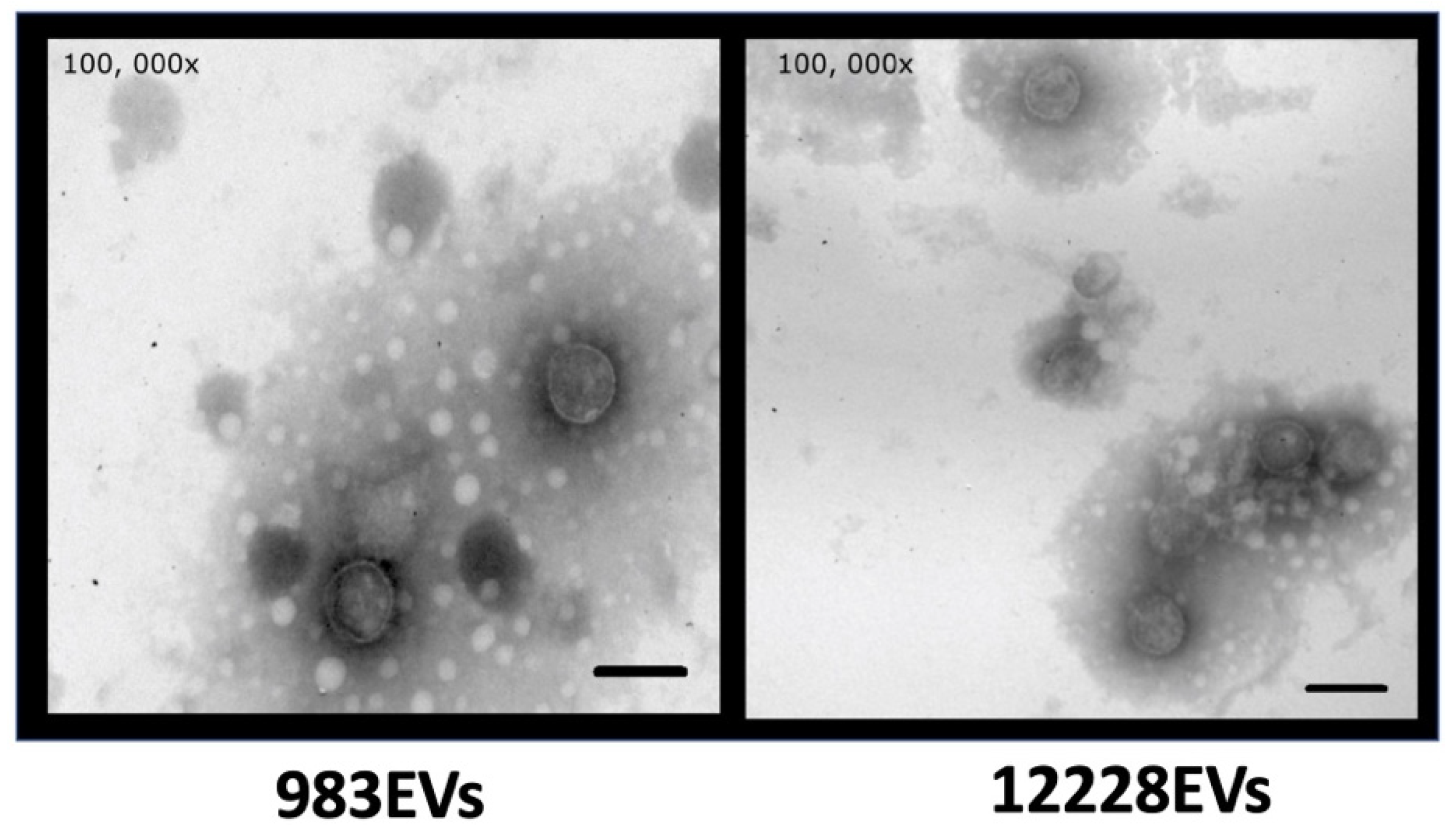
2.1. Characterization of S. epidermidis EVs

EVs isolated from the S. epidermidis commensal and clinical strains are shown in Figure 1. The EVs from the 983 clinical strain (983EVs) sized on average 56.27 nm (27.7 to 135 nm), while EVs from S. epidermidis ATCC12228 (ATCC12228EVs) sized on average 66.9 nm (from 27.9 to 135 nm) (Figure 1). In both strains, EVs showed a spherical shape and a bilayer-lipidic membrane. The amount of protein was similar in both strains, 1.421 mg/mL for ATCC12228EVs and 1.377 mg/mL for 983EVs.

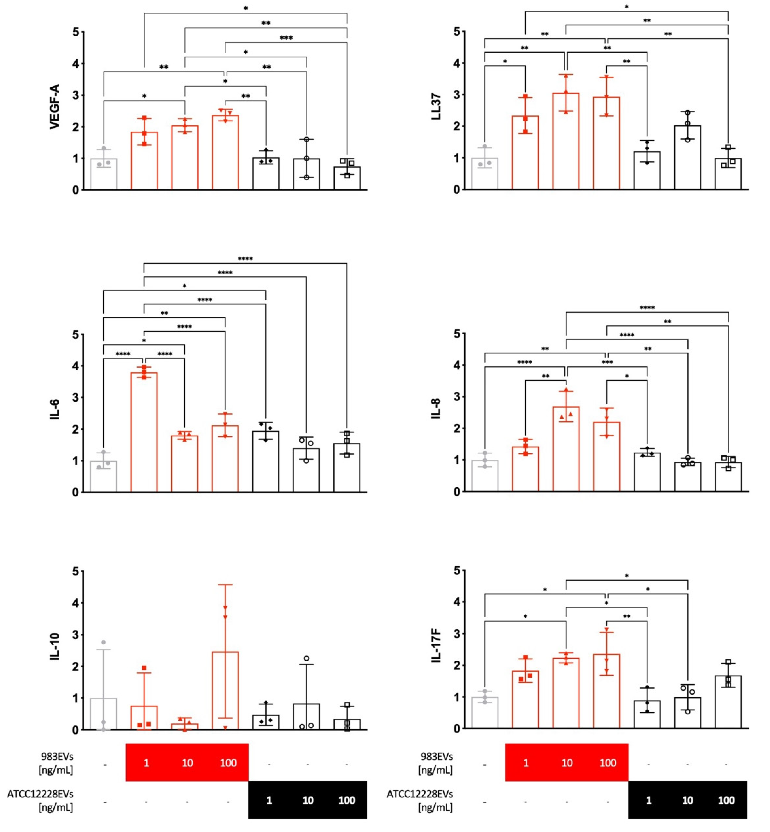
2.2. Effect of EVs on the Cytokine’s Expression in Human HaCaT Keratinocytes

The 983EVs, isolated from a clinical strain, significantly increased VEGF-A, LL37, IL-17F, and IL-6 mRNA expression at the different EVs concentrations evaluated compared to the non-stimulated HaCat keratinocytes (Figure 2). IL-8 mRNA expression was significantly induced only from 983EVs 10 and 100 ng/mL (Figure 2). Interestingly, the ATCC12228EVs did not induce significant changes in the expression of the cytokines mentioned above at any EVs concentrations tested (Figure 2), except for IL-6, which was increased at one ng/mL EVs concentration. The expression of IL-10 mRNA was not modified at any EV origin or concentration (Figure 2).

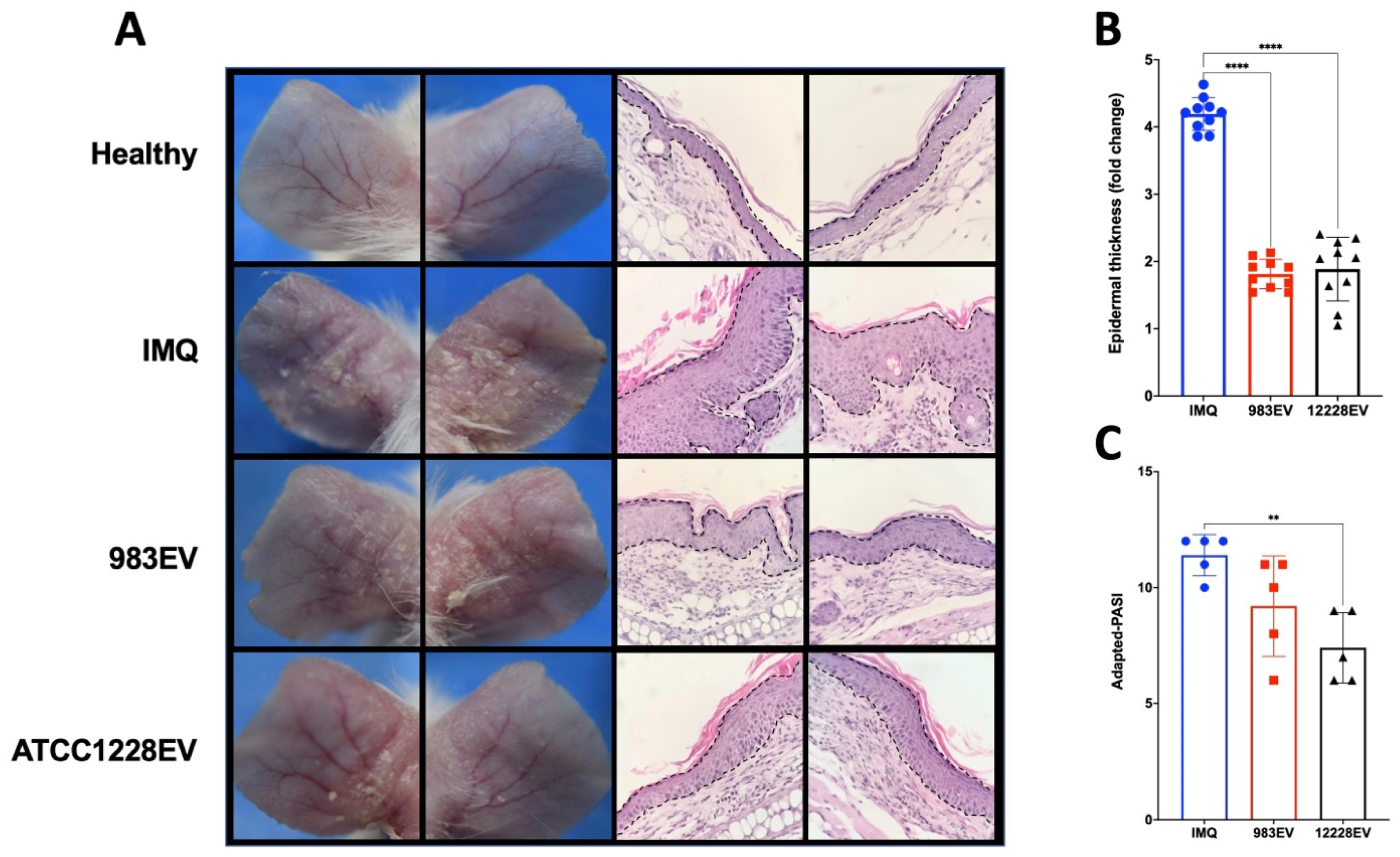
2.3. Effect of EVs on the Murine Imiquimod (IMQ)-Induced Psoriasis Model

In this murine psoriasis model, the topical IMQ treatment in the skin of the ears induce redness, skin thickness, and scaling with similar signs as occurring in the human psoriasis phenotype. Figure 3 shows the effect of EVs treatment in the ears of IMQ-induced psoriasis mice. EVs from both origins decreased the characteristic psoriatic phenotype observed in mice treated only with IMQ, with reduced cellular infiltrate and epidermal thickness (Figure 3A,B). However, the treatment with ATCC12228EVs showed a higher amelioration, reducing the degree of redness, epidermal thickness, and scaling than in the skin-like-psoriasis treated with 983EVs, as it is shown in the adapted-PASI score (Figure 3C).

2.4. ATCC12228EVs Decreased Cell Infiltration and Inflammatory Cytokine Expression in the IMQ-Induced Psoriasis Model

The recruitment of neutrophils as Gr1+ cells was evaluated in the ears of mice as an indication of the inflammation progress, as typically seen in psoriatic lesions. The IMQ-induced psoriasis ears of mice treated with ATCC12228EVs showed a significant decrease in the percentage of neutrophils, such as that found in healthy skin, an observation not found in those mice treated with 983EVs (Figure 4A).
On the other hand, the ATCC12228EVs treatment decreased the mRNAs skin expression of VEGF-A, IL-6, KC, IL-17F, IL-23 (Figure 4B), and the IL-36 family members IL-36γ and IL-36R (Figure 4C), compared to the IMQ-induced psoriasis skin mice with no EV treatment. In addition, IL-36α mRNA was expressed as in the IMQ control group, and IL-36β was induced (Figure 4B). In contrast, 983EVs treatment only significantly decreased IL-6, IL-17F, IL-36γ, and IL-36R, while the cytokines IL-36α and IL-36β were overexpressed compared to the only IMQ-induced psoriasis skin (Figure 4B). Furthermore, ATCC12228EVs did not significantly reduce the Foxp3 regulator expression but increased the IL-36 antagonist, the IL-36Ra, compared to the IMQ control group. In opposition, the 983EVs significantly inhibited Foxp3. In general, ATCC12228EVs reduced the IL-23, VEGF-A, KC, IL-17F, IL-6, and non-significantly stimulated IL-36-α, -γ, and -R expression compared to 983EV-treated mice.

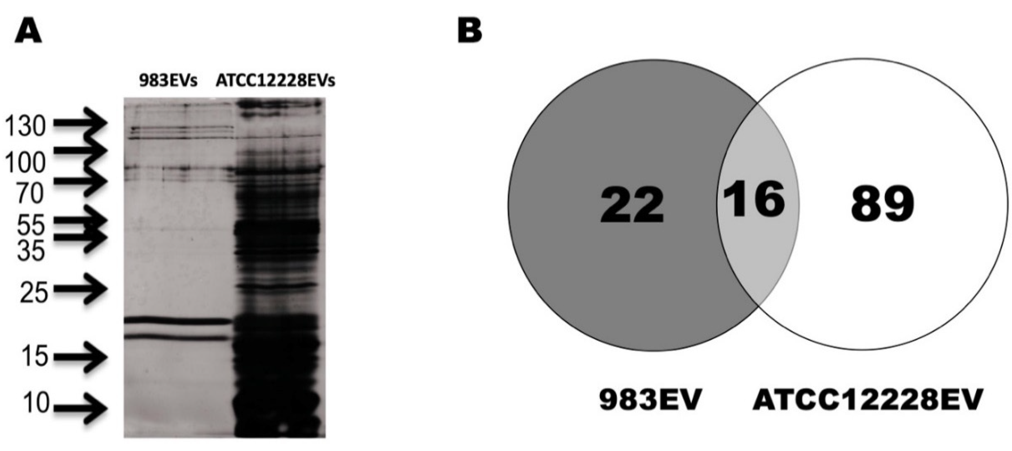
2.5. Protein Identification in EVs

The electrophoretic protein profile of EVs obtained from 983 strain showed proteins from 10 to 130 kDa. On the other hand, three major band groups were observed in ATCC12228EVs: 1 (18–23 kDa), 2 (68–80 kDa), and 3 (115–130 kDa). A differential protein pattern between the ATCC12228EVs and the 983EVs is shown in Figure 5A. After electrophoretic separation, we performed a proteomic analysis to identify the protein composition of S. epidermidis’ EVs.
The LC–MS/MS analysis identified 105 proteins in the 983EVs and ATCC12228EVs. From these proteins, 22 were exclusive for 983EVs, 89 for the ATCC12228EVs, and 16 proteins were shared between them (Figure 5B).
The identified proteins were assigned to their function key using the EggNOG program (Table 1 and Table 2). When the Clusters of Orthologous Groups of proteins (COG) functions of the proteins were compared between both EVs origins, we found that the 983EVs had more abundant proteins with unknown function (COG S) than in the ATCC12228EVs (Figure 6). Proteins with different functions were more abundant in the ATCC1228EVs, such as translation (COG J), transcription (COG K), replication and recombination (COG L), energy obtainment (COG C), carbohydrate metabolism (COG G), amino acid biosynthesis (COG E), coenzyme biosynthesis (COG H), inorganic ion transport/metabolism (COG P), and general activities (COG R). In the 983EVs, the proteins found were related to nucleotide synthesis (COG F) and intracellular trafficking and secretion (COG U). Importantly, we did not find any of the following functional classes in any of the EVs analyzed: cell division (COG D), defense mechanisms (COG V), secondary structure (COG Q), cell motility (COG N), extracellular structures (COG W), and lipid transport/metabolism (COG I).

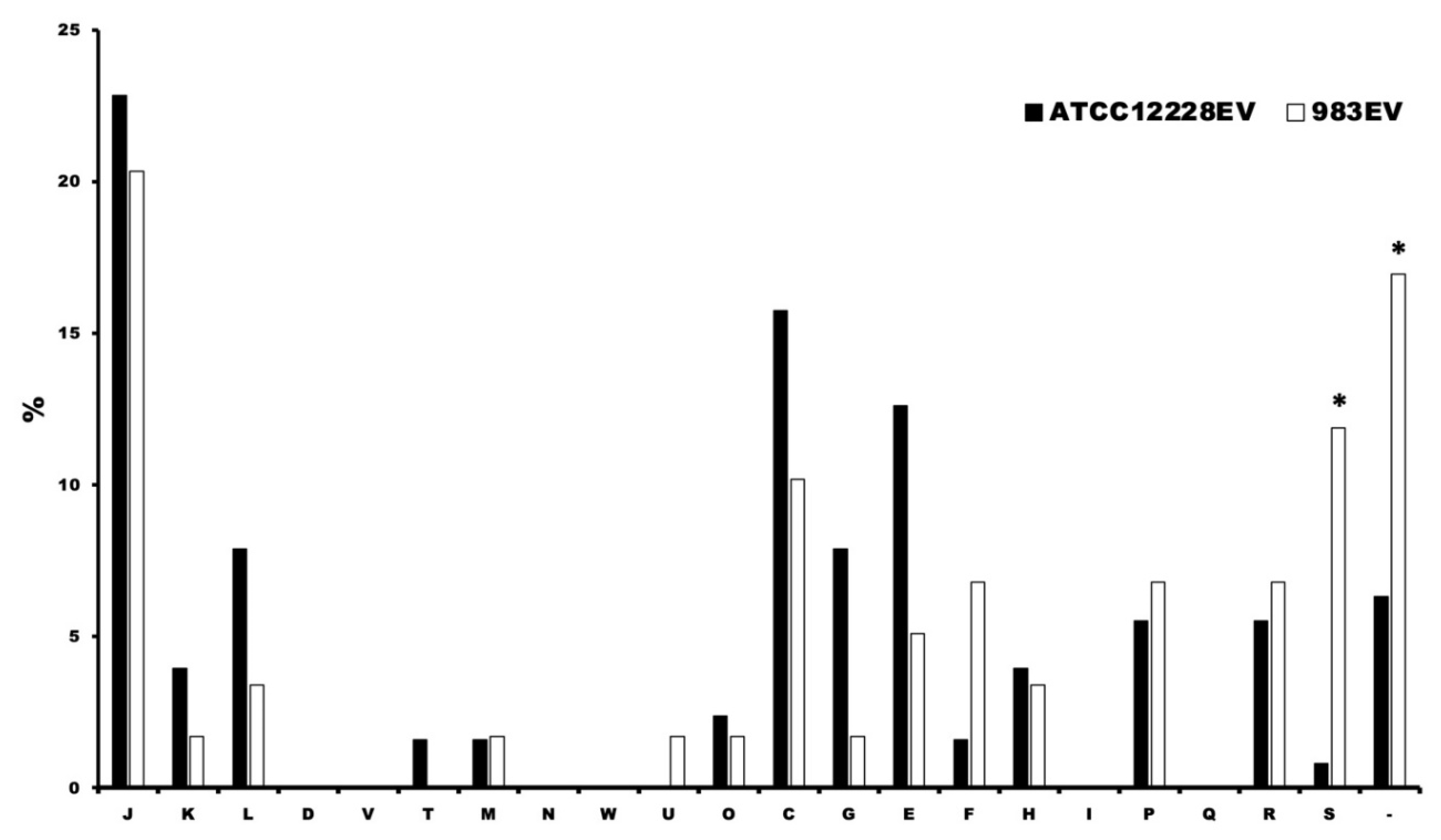
2.6. Detection of Functional Protein Enrichment Using Gene Ontology Software

According to the Gene Ontology software, the identified proteins were subjected to an analysis to determine the functional enrichment of EVs. Analysis showed that 13 biological processes are shared between both vesicles (Table 3). The ATCC12228EVs had proteins with exclusive biological processes, including metabolic process, catabolic process, tricarboxylic acid cycle, acetyl-CoA catabolic process, macromolecule biosynthetic process, and coenzyme catabolic process. The 983EVs had exclusive biological processes such as pathogenesis, cellular process, gene expression, multi-organism process, primary metabolic process, and nucleobase metabolic process (Table 3).
For molecular function (Table S1), we found that the proteins of both vesicles have functions like a structural constituent of ribosome, structural molecule activity, rRNA binding, and RNA binding. ATCC12228EVs have proteins involved in tRNA binding, oxidoreductase activity, activity on the amino acid, nucleic acid binding, oxidoreductase activity, and activity on CH–OH. Other molecular functions were found to not have significant representation.
From all these data and the analysis of the metabolic processes, we can conclude that the proteins in each vesicle could participate in essential functions of the cell, with ATCC12228EVs having a higher number of proteins. Some proteins present in ATCC12228EVs could suggest an interesting role such as HTH-type transcriptional regulator rot, universal stress protein UspA, penicillin-binding protein 2, glycosyltransferase, 60 kDa chaperonin, ornithine carbamoyltransferase, arginine deiminase, glutamate dehydrogenase, carbamate kinase, catalase, superoxide dismutase, phenol soluble modulin β1/β2, and poly (glycerol-phosphate) α-glucosyltransferase. In the case of 983EVs, the proteins found were antiholin-like protein LrgA, N-acetylmuramoyl-L-alanine amidase, antibacterial protein 2, and siphovirus Gp157.

3. Discussion

Bacterial EV research has concentrated on Gram-negative bacteria, and less is known about Gram-positive bacteria EV production and function. Concerning S. epidermidis, progress on EV research is scarce. It is well known that S. epidermidis contributes to human skin homeostasis [30], and its interaction with the skin is evolutionarily beneficial for both organisms [31]. The mechanism of communication between this bacterium and the skin is not yet known in detail. This work suggests that communication between S. epidermidis and the host skin may also occur by releasing bacterial products transported in EVs.
In this work, we obtained spherical-double membrane S. epidermidis’ EVs, with a diameter around 66.9 nm, similar to those reported in vitro and in vivo by Zaborowska et al. (2020) and Deng et al. (2020) [21,32]. It was also demonstrated that EVs from a commensal isolate (ATCC12228) and a clinical isolate (983) have the same morphology but a different protein content, even if they are obtained under the same culture conditions, and they can differentially modulate cytokine production on human keratinocytes and mice psoriatic-like skin.
The ATCC12228EVs significantly reduced the expression of LL37, IL-6, IL-23, IL-17, KC, IL- 36R, and IL-36Ra. All these molecules are essential in the initiation and development of the mice imiquimod-induced psoriasis and human psoriasis. In the context of skin inflammation, LL37 is overexpressed in the skin of psoriatic patients compared to healthy skin [33]. The LL37 overproduction is associated with the onset of psoriasis by the damaged keratinocytes releasing LL37 and genomic DNA or RNA. Then, DNA-LL37/RNA-LL37 complexes are recognized by plasmacytoid dendritic cells (pDC) and myeloid DC (mDC) inducing type I IFN expression, as well as TNFα, IL-6, and IL-36γ [34,35,36]. Furthermore, LL37 induces the chemokines CXCL8 and CXCL1 through the IL-36R signaling in psoriatic keratinocytes, leading to neutrophils’ recruitment [36]. Other cytokines such as IL-6 and IL-23 can promote a Th17 environment, favoring skin inflammation along the IL-23/IL-17 axis [37]. Furthermore, IL-6 inhibits the TGF-β-dependent differentiation of Treg cells [38] and induces mononuclear cells/macrophages IL-8 and MCP-1 production [39], resulting in more neutrophils recruiting. On the other hand, IL-6 is also capable of enhancing keratinocyte growth and proliferation [40]. A wide variety of cell types can produce IL-6 in response to stimulation by IL-36 [40]. IL-6 and IL-23 can stimulate IL-17F production in lesioned psoriatic skin compared with non-lesioned skin in humans, and it has been reported as one of the central cytokines in psoriasis development [41]. The axis formed by IL-23/IL-17/IL-22 was thought to be the trigger of psoriasis. However, the loss of IL-36R signaling successfully counteracts and protects against the development of imiquimod-induced psoriasis compared to deletions of the IL-23/IL-17/IL-22 axis, indicating that IL-36 activity should be early in the cascade of psoriasis initiation [42,43,44,45]. Importantly, IL-36R signaling in keratinocytes is crucial for the early production of IL-23, IL-17, and IL 22 and neutrophil infiltration [44,45]. The IL36 family members, the IL-36α, β, and γ, are highly expressed in psoriatic lesions and influence the function of DCs [46]. In humans, IL-36 has been shown to activate mDCs, stimulating the secretion of IL-1β and IL-6, promoting the differentiation of the Th17 cells [47]. Imiquimod via TLR-7 induces the expression of IL-36α in the keratinocyte, and this stimulation drives IL-23 and Th17-related cytokine/chemokine production [48]. TNF-α induces IL-36γ in psoriatic lesions, which promotes the expression of antimicrobial peptides and chemokines and Th17 cells and interferes with terminal differentiation and the cornification process of the psoriatic epidermis [49]. Furthermore, pretreatment with IL-36Ra has a protective effect on imiquimod-induced psoriasis and saves mice from the severe disease phenotype [50]; in addition, mice deficient in IL-36Ra show exacerbated psoriasiform disease [42]. Finally, VEGF-A is an angiogenic cytokine increased in psoriatic tissues that promote the hyperproliferation of keratinocytes [51]. Accordingly, keratin 14-VEGF-A transgenic mice develop psoriasis [51,52], indicating its critical role in the disease.
ATCC12228EVs decreased the expression of IL-36R and IL-36α but increased the IL-36Ra, suggesting that the mechanism of action for reducing imiquimod-induced psoriasis depends on the downregulation of the IL-36R signaling, in general attenuating the development of the psoriatic lesion. On the other hand, 983EVs showed a lower effect over the cytokine expression, and its most evident effect was found in the reduction of the master regulator of Treg cells (Foxp3), but it was not as effective in reducing the development of psoriatic lesion as it did the ATCC12228EVs. What is evident is that S. epidermidis vesicles have an anti-inflammatory effect on the skin compared to the cutaneous inflammatory effect of S. aureus vesicles reported elsewhere [17]. These results suggest that EVs from different origins or strains (commensal or clinical), grown under the same conditions, can differentially regulate cytokine production in the skin.
To our knowledge, this is the first report about proteomics in S. epidermidis’ EVs. Surprisingly, EVs composition analysis revealed that some proteins detected in S. epidermidis’ EVs have been reported in S. aureus’ EVs, highlighting the idea that some proteins are shared in the EVs of these two staphylococcal species. In 983EVs, we detected proteins such as the N-acetylmuramoyl-L-alanine amidase protein [4,53], ATP synthase subunit β [4,53], pyruvate dehydrogenase E1 component subunit α [4,18], dihydrolipoyllysine-residue acetyltransferase component of pyruvate dehydrogenase complex, 30S ribosomal protein S3, and 30S ribosomal protein S4 [4]. In ATCCC12228EVs, we found proteins such as the penicillin-binding protein 2 [4,18,53], ATP synthase subunit β [4,53], glutamine synthetase [53], pyruvate kinase, DNA-directed RNA polymerase subunit β [53], 30S ribosomal protein S7, 50S ribosomal protein L5, 50S ribosomal protein L1 [4], catalase (560), enolase [53], phenol-soluble modulin β 1, superoxide dismutase [53], HTH-type transcriptional regulator rot [16], glycosyltransferase, 60 kDa chaperonin, and glutamate dehydrogenase [53]. Previously, Silva Ribeiro et al. reported that dramatic differences in protein EVs from two distinct Trypanosoma cruzi strains may correlate with their infectivity/virulence during the host–parasite interaction. Thus, differences between specific proteins in 983EVs and ATCC12228EVs could determine the behavior as a clinical isolate and a commensal strain [54].
Regarding the regulation of psoriasis by S. epidermidis EVs, it is difficult to precisely denote which proteins are functioning as regulators of the inflammation in the psoriasis animal model since the proteome of the vesicles mainly identified proteins with participation in the essential metabolism, which was expected since S. epidermidis does not have many virulence factors as is the case for S. aureus [55].
We hypothesized that some proteins present in the commensal strain ATCC12228EVs that are not contained in 983EVs could regulate inflammation, avoiding the development of psoriatic lesions. However, there are no precedents that support this suggestion. Nevertheless, the presence of enzymes involved in the glutamate and glutamine metabolism are present in ATCC12228EVs, and these could be relevant participants that interfere in the production of characteristic plaques in this disease. It is known that amino acids, essential constituents of the skin’s natural moisturizing factors, are decreased in psoriasis [56]. A metabolic study of psoriasis patients showed that their serum has high α-ketoglutarate levels, glucuronic acid, and a low level of asparagine and glutamine [57]. Glutamate dehydrogenase could convert α-ketoglutarate to glutamate, and glutamate is a source to produce glutamine by the enzyme glutamine synthetase, with both enzymes being present in ATCC12228EVs. The enzyme glutamate-1-semialdehyde 2,1-aminomutase, also present in the ATCC12228EVs, converts L-glutamate 1-semialdehyde to the d-aminolevulinic acid, which is used for the treatment of psoriasis by photodynamic therapy with favorable results in humans [58]. Furthermore, S. epidermidis can convert aromatic amino acids into trace amines (TA), which accelerate skin wound healing [59].
The enzymes catalase and superoxide dismutase (presents ATCC12228EVs) may participate in the attenuation of reactive oxygen species (ROS) compounds. Liu et al. reported decreased levels of reduced glutathione (GSH), superoxide dismutase (SOD), and catalase (CAT), as well as the accumulation of malondialdehyde (MDA), in the skin of IMQ-induced psoriasis [60]. In this way, catalase and SOD from ATCC12228EVs could contribute to the reduction of ROS in psoriasis.
Other proteins present in ATCC12228EVs could ameliorate IMQ-induced psoriasis; for example, glycine dehydrogenase converts glyoxylate to glycine, and glycine has antioxidative and anti-inflammatory effects regulating apoptosis in various models [61]. The soluble phenol modulin (PSM) β1/β2 are related to biofilm maturation and removal [62], and it is also reported that these PSM regulate the release of vesicles in S. aureus [53]. Poly (glycerol-phosphate) α-glucosyltransferase participates in the synthesis of lipoteichoic acid (LTA). LTA can be released into the environment, modulating the keratinocyte immune response [63] and regulating the pathogenesis of Cutibacterium acnes [24].
The use of beneficial bacteria as probiotics for the skin represents another alternative method for treating skin diseases, such as activated Lactobacillus acidophilus [64], L. paracasei, L. brevis, or L. fermentum, which are ingredients in skincare products [65], as well as Streptococcus salivarius K12, hich stimulates an anti-inflammatory response in the skin [66]. A bacterial extract of the Gram-negative bacteria Vitreoscilla filiformis showed a beneficial effect when applied to lesions of patients with atopic dermatitis [67]. A note of caution is due here since bacterial extracts (cytoplasmic proteins) in some cases can have adverse effects, and more research in this area is needed.

4. Materials and Methods

4.1. EV Isolation

S. epidermidis clinical strain 983 was isolated from a corneal infection; it is an ST2 genotype and biofilm producer [68]. EVs from 983 and ATCC12228 strains were obtained according to Avila-Calderón et al. (2012) and Ruiz-Palma et al. (2021) under the same culture conditions [69,70]. Briefly, both strains were cultured on supplemented LB agar plates and incubated for 24 h at 37 °C. A bacterial suspension was grown up and cultured in bulk in 25 LB agar plates from the pure plate colonies, and then these plates were incubated for another 24 h at 37 °C. Bacteria culture was harvested with a sterile cell scraper and suspended in 200 mL of sterile phosphate saline buffer (PBS). The bacterial suspension was centrifuged at 10,000× g for 30 min to remove intact cells. The supernatant was filtered through a 0.22 µm pore filter (Millipore Corp., Billerica, MA, USA) to remove the remaining bacteria. Then, the supernatant was cultured on LB agar plates for 48 h at 37 °C to evaluate sterility. Then, the sterile supernatant was ultracentrifuged at 100,000× g for 2 h at 4 °C. The pellet containing the EVs was washed twice with 25 mL of sterile PBS. In the last washing, the EVs were suspended in 1 mL of sterile PBS. The EVs were finally purified by density gradient using OptiPrep (Sigma-Aldrich, Inc., Burlington, MA, USA). OptiPrep was diluted to final concentrations of 10, 15, 20, 25, and 30% in sterile PBS. Later, 2.6 mL of each OptiPrep solution was layered from the higher to the lower density in an ultracentrifuge tube, and the EVs were loaded at the top of the tube. The samples were centrifuged at 100,000× g for 16 h at 4 °C. EVs appeared as an opalescent band in the density gradient. EVs were collected, washed twice with sterile PBS, and centrifuged at 100,000× g for 2 h at 4 °C, and finally suspended in 300 μL of sterile PBS. This procedure was performed in three independent experiments for each strain. The total protein concentration was determined using PIERCE-BCA (Thermo-Fisher Scientific Inc., Waltham, MA, USA). The EV samples were divided into 0.5 mL aliquots and stored at −80 °C until used.

4.2. Observation of EVs by Electron Microscopy

A total of 20 µL of purified EVs equivalent to 25 µg from both strains were placed onto copper grids coated with formvar and dried using filter paper. One percent of phosphotungstic acid was added, and the grids were allowed to dry for 10 h at room temperature. All preparations were stained and observed in a transmission electron microscope (JEOL model JEM 10–10).

4.3. Stimulation of HaCaT Keratinocytes with S. epidermidis EVs

We cultured 1 × 10⁶ HaCaT keratinocyte cells in 6-well plates until 80% confluence. Before the stimulus, keratinocytes were washed with sterile 0.1 M PBS (pH 7.4) and with 1X DMEM (Life Technologies, CA, USA). Subsequently, we added to the cultures 800 µL of DMEM/F12-K (1:1) supplemented with 10% FBS and 1X antifungal antibiotic (Life Technologies, CA, USA). Keratinocyte cultures were stimulated with S. epidermidis EVs at the final concentrations of 1, 10, and 100 ng/mL and incubated for 6 h at 20% O2, 5% CO2, and 37 °C. Three independent experiments were carried out, and unstimulated cells were used as the control group.

4.4. HaCaT Keratinocyte RNA Extraction, cDNA Synthesis, and PCR

Total RNA was obtained from stimulated and unstimulated keratinocytes using the TRIzol reagent (Invitrogen, CA, USA). RNA purification and RT-qPCR were performed as previously described [71]. Briefly, cells were washed with 1× PBS, and total RNA was extracted with TRIzol (Invitrogen), treated with DNase I (Invitrogen), and then re-extracted. For the reverse transcriptase (RT) reaction, 3 µg of total RNA was denatured at 70 °C for 10 min in the presence of 0.5 μg of oligo-hexamers (Invitrogen). Then, we added 1× single strand buffer, 0.5 mM DTT, 10 mM of each dNTPs, and 200 U of MMLV reverse transcriptase (Invitrogen). Reverse transcriptase reactions were performed at 42 °C for 1 h. The PCR conditions for LL37, VEGF-A, IL-6, IL-17F, and IL-8 were performed as follows: 40 cycles of denaturation at 94 °C 30 s, hybridization at 60 °C 30 s, elongation at 72 °C 30 s. Endogenous gene (GADPH): 28 cycles of denaturation at 94 °C 30 s, hybridization at 60 °C 30 s, and elongation at 72 °C 30 s. The oligonucleotide sequences used for amplification are shown in Table S2. Relative expression was determined by the 2−ΔΔCt method. The results are expressed as means ± standard deviation of triplicate assays.

4.5. Imiquimod (IMQ)-Induced Murine Psoriasis Model Treated with Staphylococcus Epidermidis EVs

All the mice used in this work were supplied by the Animal Care Facility of the Escuela Superior de Medicina-IPN. We used female Balb/c mice (eight weeks old; 22–25 g), housed at 23 °C under 12 h light/dark cycles, 40–60% humidity, and ad libitum access to food/water. Two independent experiments were conducted with 20 mice, grouped as follows: control untreated healthy (n = 5), IMQ-treated (IMQ; n = 5), ATCC12228EV/IMQ-treated (n = 5), and 983EV/IMQ-treated (n = 5). To evaluate the EV effect on psoriasis-like skin, we depilated mice ears, and 30 min before the treatment with AldaraTM (6.25 µg, IMQ 5%, Graceway Laboratory), the EVs (10 ng of total protein) were topically administered using the same vehicle as used in the AldaraTM cream. This procedure was performed once a day for six days, in accordance with the work of Van der Fits et al. (2009) [72]. After the third day of IMQ administration in the only IMQ-treated group, the psoriasis-like lesions were visible and clearly defined after the sixth day of treatment. On the sixth day of treatment, the redness, scaling, and thickness of mice were evaluated to score the “adapted-PASI”, which assesses the severity of the induced murine psoriasis erythema, desquamation, and induration; these severity parameters are measured on a 0–4 scale (from none to the maximum damage). The sum of these four values is the value of the adapted-PASI. An adapted-PASI value of 12 indicates a maximum severe injury. On day 7, mice were euthanized with an overdose of pentobarbital sodium (150 mg/kg) by intravenous injection, and the ears were collected for histochemistry, flow cytometry, and RT-qPCR analysis. The handling of the ear samples was as follows: both ears of a mouse were divided into two equal parts. The one-half ear was used for histochemistry, and the other half for the determination of GR1 + cells by flow cytometry. The second ear of the same mouse was used for histochemistry and the other half ear for RT-qPCR analysis. Statistics were calculated using 10 data per group for histochemistry and 5 data per group for GR1 + cell determination and RT-qPCR.

4.6. Histochemistry

The ears sections used for histochemistry analysis were maintained in a fixative buffered formalin solution (10% in PBS) until used. The skin was placed into cassettes and dehydrated using an ethanol series (70, 80, 90, 96, 100, and 100% for 1 h each). Tissues were cleared twice in 100% xylene for 1 h and soaked in paraffin twice at 60 °C for 1 h. Subsequently, tissues were sectioned in 5 µm slices using a Leica RM 2132 microtome (Leica Microsystems GmbH). The slices were stained in hematoxylin and eosin (H&E) and Lillie’s trichrome solutions for 5 min at room temperature. Stained sections were observed in 10 randomly selected fields in a transmitted light microscope (magnification ×40). Finally, we performed a blind histological examination and interpretation, and photographs were taken to determine the size of the epidermal thickness using ImageJ [73].

4.7. Skin RT-PCR

The mice’s ears skin was cut and homogenized individually with a mortar and pestle in Trizol. Total RNA yield was around 10–30 μg per a half mice ear of ≈15–30 mg. Then, cDNA synthesis was performed following the same procedure mentioned above to detect GADPH, VEGF-A, IL-6, MIP2, KC, IL-17F, IL-23, Foxp3, IL-36α, IL-36β, IL-36γ, IL-36R, and IL-36 receptor antagonist (IL-36Ra) (Table S2). Relative expression was determined by the 2−ΔΔCt method. The results were expressed as means ± standard deviation of triplicate assays.

4.8. GR1+ Cell Detection in the Skin of Ears from Treated Mice

Ear skin sections were placed on a plate with 1 mL of RPMI. The inner and outer faces of the ears were separated and incubated in RPMI supplemented with collagenase type I (60 U/mL; Life Technologies, NY, USA), liberase TL (153.5 μg/mL; Roche Diagnostics, Mannheim, Germany), and DNAse I (6.5 ng/mL; Roche Diagnostics) for 2 h at 37 °C. After incubation, both faces were fragmented, transferred to 1 mL of fresh medium supplemented with enzymes, and incubated for 30 min more at 37 °C. The tissues were homogenized and filtered at 70 µm; the cell suspension was rinsed with 1 mL of FACS buffer, centrifuged for 5 min at 450× g, and finally resuspended in 500 μL of FACS buffer. A total of 1 × 10⁶ cells were stained with 100 μL of anti-CD45 PE-Cy5 antibody (1:100) (monoclonal antibody rat anti-mouse; Biolegend, CA, USA) and anti-GR1 Alexa Flour 488 antibody (1:100) (monoclonal antibody rat anti-mouse; Biolegend), and then incubated on ice for 30 min. The cells were then washed with FACs buffer and resuspended in 200 μL of FACs buffer. All samples were acquired in a FACSAria II cell sorter (BD Bioscience; Figure S1).

4.9. Protein Identification by Liquid Chromatography Coupled with Tandem Mass Spectrometry (LC–MS/MS)

EVs were mixed with the SDS-PAGE Leammli loading buffer, boiled for 90 s, and centrifuged at 10,000× g for 5 min to eliminate insoluble fraction. A total of 100 µg of soluble protein fractions were loaded to 12% SDS-PAGE. Proteins were visualized by staining with Coomassie blue. Then, gels were destained in a 50:50 (v/v) solution of 100 mM, pH 7.8 ammonium bicarbonate (Sigma-Aldrich, Mexico Edo, Mexico) and acetonitrile treated with 50 mM dithiothreitol (DTT, Sigma-Aldrich), alkylated with 50 mM iodoacetamide (Sigma-Aldrich). An “in-gel” digestion was performed using 10 ng/μL trypsin (Promega Sequencing Grade Modified Trypsin, WI, USA). The peptides were desalted using ZipTip® C18 (Merck KGaA, Darmstadt, Germany) solution and then concentrated in a Speed-Vac SPD 1010 Thermo-Electron. This procedure was performed in three independent experiments for each EVs.
The proteomic analysis was performed in the Proteomics Facility of the Instituto de Biotecnología-UNAM, Cuernavaca, Mexico. The peptide mixtures were eluted in 50% acetonitrile containing 1% acetic acid and loaded into a liquid chromatography system coupled to mass spectrometry (LC–MS/MS), consisting of an EASY-nLC II nanoflow pump, coupled with an LTQ-Orbitrap Velos (Thermo-Fisher; MA, USA) mass spectrometer with a nano-electrospray (ESI) ionization source. For nanoflow chromatography, a gradient system of 5–80% solvent B (water/acetonitrile with 0.1% formic acid) and solvent A (water with 0.1% formic acid) using a capillary column (ID 0.75 µm × 10 cm, RP-C18) with a flow rate of 300 nanoliters/min, in a time of 120 min, was used.
We performed an average mass scan of the total ions on the Orbitrap analyzer with a 60,000 (RP = m/FWHM) mass resolving power. The CID (collision-induced dissociation) and HCD (high-energy collision dissociation) methods were applied with a 15,000 (RP = m/FWHM) mass resolving power.
The protein analysis was performed using the Proteome Discoverer 1.4 program (Thermo Fisher) through the Sequest H and X! Tandem search engines. We configured the Sequest HT search engine to identify the S. epidermidis protein database (UniProt). The precursor ion’s mass tolerance and the ion fragments obtained by dissociation of the precursor ion were 20 ppm and 0.6 Da. MW using an FDR (false discovery rate) of 0-01 and 0.05 (maximum). We worked with the inverted database (Decoy database), the Scaffold program (version 4.8.9) as a tool of the “Percolator” validation program for the validation of the peptides, and protein identification based on MS/MS, establishing for the identification of the peptides a probability higher than 96% to achieve an FDR less than 5%, and for the identification of the proteins a probability higher than 93% to reach an FDR less than 1%. The Protein Prophet algorithm assigned the probabilities.

4.10. In Silico Analysis

The proteins identified by LC–MS/MS were related to their closest homolog of S. epidermidis strains RP62A and ATCC12228 by BLAST analysis (http://blast.ncbi.nlm.nih.gov, accessed on 8 March 2021) with its default parameter settings. The protein accession numbers were obtained in strains RP62A and ATCC12228 from the Uniprot database (http://www.uniprot.org/uniprot, accessed on 8 March 2021). The clusters of orthologous groups’ (COG) functional group were assigned using Eggnog (http://eggnog.embl.de, accessed on 8 March 2021), version_3.0. The participation in metabolic pathways were obtained using the Kyoto Encyclopedia of Genes and Genomes (KEGG; http://www.genome.jp/kegg, accessed on 8 March 2021).

4.11. Statistics

To determine the proportion significant differences, we used the accurate Fisher test. To analyze differences in the expression levels of the cytokines measured, we conducted a two-way ANOVA and a Tuckey post hoc test. These analyses were carried out with the software GraphPad Prism version 7.0 (GraphPad, San Diego, CA, USA).

5. Conclusions

Commensal S. epidermidis’ EVs can modulate inflammation and psoriasis development in part by the axis IL-36R and IL-36Ra. We suggest that bacterial extracellular vesicles could be a cleaner and safer new alternative for dermatosis treatment.

Supplementary Materials

The following are available online at https://www.mdpi.com/article/10.3390/ijms222313029/s1.

Author Contributions

Conceptualization, J.C.C.-D. and M.E.C.-D.; methodology, software, validation, formal analysis, investigation, data curation, J.C.C.-D., M.E.C.-D., F.G.-C., S.R.-M., C.C.-P., L.A.Z.-C., I.M.-T., Y.B.-A., H.P., L.C.-G., G.G.-G., E.D.A.-C. and A.C.-R.; writing—original draft preparation, writing—review and editing, supervision, project administration, F.G.-C., J.C.C.-D., S.R.-M. and M.E.C.-D.; funding acquisition, J.C.C.-D. and M.E.C.-D. All authors have read and agreed to the published version of the manuscript.

Funding

This research was funded by The Instituto Politécnico Nacional, grant number 20201026SIP and in part by the grant number 20210140SIP.

Institutional Review Board Statement

All experimental animal procedures, including the treatment with psoriasis inductors, administration of vesicles, and animal handling, were approved and performed according to the standards of the Ethics Committee for Animal Use of the Instituto Politécnico Nacional/Escuela Nacional de Ciencias Biológicas (IPN/ENCB), which follows the EU Directive 2010/63/EU animal care for animals. The protocol’s registration and approval are under the file number ENCB/CEI/036/2018, CONBIOETICA09CEI03720130520.

Informed Consent Statement

Not applicable.

Data Availability Statement

Not applicable.

Acknowledgments

M.E. Cancino-Diaz, S. Rodríguez-Martínez, and J.C. Cancino-Diaz appreciate the COFAA and EDI-IPN fellowships. All authors are SNI-CONACyT fellows. Finally, we thank Alfredo A. Díaz-Estrada (FMVZ, UNAM) and J. Eduardo Farfán-Morales (INP, SSA) for their assistance with the histological processing.

Conflicts of Interest

The authors declare no conflict of interest.

References

  1. Ulhuq, F.R.; Gomes, M.C.; Duggan, G.M.; Guo, M.; Mendonca, C.; Buchanan, G.; Chalmers, J.D.; Cao, Z.; Kneuper, H.; Murdoch, S.; et al. A membrane-depolarizing toxin substrate of the Staphylococcus aureus type VII secretion system mediates intraspecies competition. Proc. Natl. Acad. Sci. USA 2020, 117, 20836–20847. [Google Scholar] [CrossRef] [PubMed]
  2. Briaud, P.; Carroll, R.K. Extracellular vesicle biogenesis and functions in gram-positive bacteria. Infect. Immun. 2020, 88, 12. [Google Scholar] [CrossRef] [PubMed]
  3. Mashburn-Warren, L.M.; Whiteley, M. Special delivery: Vesicle trafficking in prokaryotes. Mol. Microbiol. 2006, 61, 839–846. [Google Scholar] [CrossRef]
  4. Lee, E.Y.; Choi, D.Y.; Kim, D.K.; Kim, J.W.; Park, J.O.; Kim, S.; Kim, S.H.; Desiderio, D.M.; Kim, Y.K.; Kim, K.P.; et al. Gram-positive bacteria produce membrane vesicles: Proteomics-based characterization of Staphylococcus aureus-derived membrane vesicles. Proteomics 2009, 9, 5425–5436. [Google Scholar] [CrossRef]
  5. Hong, S.W.; Kim, M.R.; Lee, E.Y.; Kim, J.H.; Kim, Y.S.; Jeon, S.G.; Yang, J.M.; Lee, B.J.; Pyun, B.Y.; Gho, Y.S.; et al. Extracellular vesicles derived from Staphylococcus aureus induce atopic dermatitis-like skin inflammation. Allergy 2011, 66, 351–359. [Google Scholar] [CrossRef] [PubMed] [Green Version]
  6. Liu, Y.; Defourny, K.A.Y.; Smid, E.J.; Abee, T. Gram-positive bacterial extracellular vesicles and their impact on health and disease. Front. Microbiol. 2018, 9, 1502. [Google Scholar] [CrossRef] [PubMed] [Green Version]
  7. Schooling, S.R.; Hubley, A.; Beveridge, T.J. Interactions of DNA with biofilm-derived membrane vesicles. J. Bacteriol. 2009, 191, 4097–4102. [Google Scholar] [CrossRef] [Green Version]
  8. Maredia, R.; Devineni, N.; Lentz, P.; Dallo, S.F.; Yu, J.; Guentzel, N.; Chambers, J.; Arulanandam, B.; Haskins, W.E.; Weitao, T. Vesiculation from Pseudomonas aeruginosa under SOS. Sci. World J. 2012, 2012, 402919. [Google Scholar] [CrossRef] [Green Version]
  9. Gamalier, J.P.; Silva, T.P.; Zarantonello, V.; Dias, F.F.; Melo, R.C. Increased production of outer membrane vesicles by cultured freshwater bacteria in response to ultraviolet radiation. Microbiol. Res. 2017, 194, 38–46. [Google Scholar] [CrossRef]
  10. Manning, A.J.; Kuehn, M.J. Contribution of bacterial outer membrane vesicles to innate bacterial defense. BMC Microbiol. 2011, 11, 258. [Google Scholar] [CrossRef] [Green Version]
  11. Mashburn, L.M.; Whiteley, M. Membrane vesicles traffic signals and facilitate group activities in a prokaryote. Nat. Cell Biol. 2005, 437, 422–425. [Google Scholar] [CrossRef]
  12. Perez-Cruz, C.; Carrion, O.; Delgado, L.; Martinez, G.; Lopez-Iglesias, C.; Mercade, E. New type of outer membrane vesicle produced by the Gram-negative bacterium Shewanella vesiculosa M7T: Implications for DNA content. Appl. Environ. Microbiol. 2013, 79, 1874–1881. [Google Scholar] [CrossRef] [Green Version]
  13. Rivera, J.; Cordero, R.J.; Nakouzi, A.S.; Frases, S.; Nicola, A.; Casadevall, A. Bacillus anthracis produces membrane-derived vesicles containing biologically active toxins. Proc. Natl. Acad. Sci. USA 2010, 107, 19002–19007. [Google Scholar] [CrossRef] [PubMed] [Green Version]
  14. Ciofu, O.; Beveridge, T.J.; Kadurugamuwa, J.; Walther-Rasmussen, J.; Hoiby, N. Chromosomal beta-lactamase is packaged into membrane vesicles and secreted from Pseudomonas aeruginosa. J. Antimicrob. Chemother. 2000, 45, 9–13. [Google Scholar] [CrossRef]
  15. Lee, J.; Lee, E.Y.; Kim, S.H.; Kim, D.K.; Park, K.S.; Kim, K.P.; Kim, Y.K.; Roh, T.Y.; Gho, Y.S. Staphylococcus aureus extracellular vesicles carry biologically active beta-lactamase. Antimicrob. Agents Chemother. 2013, 57, 2589–2595. [Google Scholar] [CrossRef] [Green Version]
  16. Askarian, F.; Lapek, J.D., Jr.; Dongre, M.; Tsai, C.-M.; Kumaraswamy, M.; Kousha, A.; Valderrama, J.A.; Ludviksen, J.A.; Cavanagh, P.; Uchiyama, S.; et al. Staphylococcus aureus membrane-derived vesicles promote bacterial virulence and confer protective immunity in murine infection models. Front. Microbiol. 2018, 9, 262. [Google Scholar] [CrossRef] [PubMed]
  17. Jun, S.H.; Lee, J.H.; Kim, S.I.; Choi, C.W.; Park, T.I.; Jung, H.R.; Cho, J.W.; Kim, S.H.; Lee, J.C. Staphylococcus aureus-derived membrane vesicles exacerbate skin inflammation in atopic dermatitis. Clin. Exp. Allergy 2017, 47, 85–96. [Google Scholar] [CrossRef] [PubMed]
  18. Gurung, M.; Moon, D.C.; Choi, C.W.; Lee, J.H.; Bae, Y.C.; Kim, J.; Lee, Y.C.; Seol, S.Y.; Cho, D.T.; Kim, S.I.; et al. Staphylococcus aureus produces membrane-derived vesicles that induce host cell death. PLoS ONE 2011, 6, e27958. [Google Scholar] [CrossRef] [Green Version]
  19. Thay, B.; Wai, S.N.; Oscarsson, J. Staphylococcus aureus alpha-toxin-dependent induction of host cell death by membrane-derived vesicles. PLoS ONE 2013, 8, e54661. [Google Scholar] [CrossRef]
  20. Chebotar, I.; Konchakova, E.D.; Maianskii, A.N. Vesicle formation as a result of interaction between polymorphonuclear neutrophils and Staphylococcus aureus biofilm. J. Med. Microbiol. 2013, 62 Pt 8, 1153–1159. [Google Scholar] [CrossRef]
  21. Deng, S.; Wang, Y.; Liu, S.; Chen, T.; Hu, Y.; Zhang, G.; Zhang, X.; Yu, B. Extracellular vesicles: A potential biomarker for quick identification of infectious osteomyelitis. Front. Cell. Infect. Microbiol. 2020, 10, 323. [Google Scholar] [CrossRef]
  22. Wang, Y.; Kuo, S.; Shu, M.; Yu, J.; Huang, S.; Dai, A.; Two, A.; Gallo, R.L.; Huang, C.-M. Staphylococcus epidermidis in the human skin microbiome mediates fermentation to inhibit the growth of Propionibacterium acnes: Implications of probiotics in acne vulgaris. Appl. Microbiol. Biotechnol. 2013, 98, 411–424. [Google Scholar] [CrossRef] [PubMed] [Green Version]
  23. Leech, J.M.; Dhariwala, M.O.; Lowe, M.M.; Chu, K.; Merana, G.R.; Cornuot, C.; Weckel, A.; Ma, J.M.; Leitner, E.G.; Gonzalez, J.R.; et al. Toxin-triggered interleukin-1 receptor signaling enables early-life discrimination of pathogenic versus commensal skin bacteria. Cell Host Microbe 2019, 26, 795–809.e5. [Google Scholar] [CrossRef] [PubMed]
  24. Xia, X.; Li, Z.; Liu, K.; Wu, Y.; Jiang, D.; Lai, Y. Staphylococcal LTA-induced miR-143 inhibits Propionibacterium acnes-Mediated inflammatory response in skin. J. Investig. Dermatol. 2016, 136, 621–630. [Google Scholar] [CrossRef] [Green Version]
  25. Chang, H.-W.; Yan, D.; Singh, R.; Liu, J.; Lu, X.; Ucmak, D.; Lee, K.; Afifi, L.; Fadrosh, D.; Leech, J.M.; et al. Alteration of the cutaneous microbiome in psoriasis and potential role in Th17 polarization. Microbiome 2018, 6, 1–27. [Google Scholar] [CrossRef]
  26. Lowes, M.A.; Bowcock, A.M.; Krueger, J.G. Pathogenesis and therapy of psoriasis. Nat. Cell Biol. 2007, 445, 866–873. [Google Scholar] [CrossRef] [PubMed]
  27. Nestle, F.O.; Kaplan, D.H.; Barker, J. Psoriasis. N. Engl. J. Med. 2009, 361, 496–509. [Google Scholar] [CrossRef]
  28. Al-Harbi, N.O.; Nadeem, A.; Ahmad, S.F.; Bakheet, S.A.; El-Sherbeeny, A.M.; Ibrahim, K.E.; Alzahrani, K.S.; Al-Harbi, M.M.; Mahmood, H.M.; Alqahtani, F.; et al. Therapeutic treatment with Ibrutinib attenuates imiquimod-induced psoriasis-like inflammation in mice through downregulation of oxidative and inflammatory mediators in neutrophils and dendritic cells. Eur. J. Pharmacol. 2020, 877, 173088. [Google Scholar] [CrossRef]
  29. Nadeem, A.; Ahmad, S.F.; Al-Harbi, N.O.; El-Sherbeeny, A.M.; Alasmari, A.F.; Alanazi, W.A.; Alasmari, F.; Ibrahim, K.E.; Al-Harbi, M.M.; Bakheet, S.A.; et al. Bruton’s tyrosine kinase inhibitor suppresses imiquimod-induced psoriasis-like inflammation in mice through regulation of IL-23/IL-17A in innate immune cells. Int. Immunopharmacol. 2020, 80, 106215. [Google Scholar] [CrossRef] [PubMed]
  30. Stacy, A.; Belkaid, Y. Microbial guardians of skin health. Science 2019, 363, 227–228. [Google Scholar] [CrossRef]
  31. Zhou, W.; Spoto, M.; Hardy, R.; Guan, C.; Fleming, E.; Larson, P.; Brown, J.S.; Oh, J. Host-specific evolutionary and transmission dynamics shape the functional diversification of staphylococcus epidermidis in human skin. Cell 2020, 180, 454–470.e18. [Google Scholar] [CrossRef] [PubMed]
  32. Zaborowska, M.; Flores, C.T.; Vazirisani, F.; Shah, F.A.; Thomsen, P.; Trobos, M. Extracellular vesicles influence the growth and adhesion of staphylococcus epidermidis under antimicrobial selective pressure. Front. Microbiol. 2020, 11, 1132. [Google Scholar] [CrossRef] [PubMed]
  33. Tovar-Castillo, L.E.; Cancino-Díaz, J.C.; García-Vázquez, F.; Cancino-Gómez, F.G.; León-Dorantes, G.; Blancas-González, F.; Jiménez-Zamudio, L.; García-Latorre, E.; Cancino-Díaz, M.E. Under-expression of VHL and over-expression of HDAC-1, HIF-1alpha, LL-37 and IAP-2 in affected skin biopsies of patients with psoriasis. Int. J. Dermatol. 2007, 46, 239–246. [Google Scholar] [CrossRef] [PubMed]
  34. Lande, R.; Gregorio, J.; Facchinetti, V.; Chatterjee, B.; Wang, Y.H.; Homey, B.; Cao, W.; Wang, Y.H.; Su, B.; Nestle, F.O.; et al. Plasmacytoid dendritic cells sense self-DNA coupled with antimicrobial peptide. Nature 2007, 449, 564–569. [Google Scholar] [CrossRef] [PubMed]
  35. Ganguly, D.; Chamilos, G.; Lande, R.; Gregorio, J.; Meller, S.; Facchinetti, V.; Homey, B.; Barrat, F.J.; Zal, T.; Gilliet, M. Self-RNA–antimicrobial peptide complexes activate human dendritic cells through TLR7 and TLR8. J. Exp. Med. 2009, 206, 1983–1994. [Google Scholar] [CrossRef]
  36. Li, N.; Yamasaki, K.; Saito, R.; Fukushi-Takahashi, S.; Shimada-Omori, R.; Asano, M.; Aiba, S. Alarmin function of cathelicidin antimicrobial peptide LL37 through IL-36gamma induction in human epidermal keratinocytes. J. Immunol. 2014, 193, 5140–5148. [Google Scholar] [CrossRef] [Green Version]
  37. Hansel, A.; Gunther, C.; Ingwersen, J.; Starke, J.; Schmitz, M.; Bachmann, M.; Meurer, M.; Rieber, E.P.; Schakel, K. Human slan (6-sulfo LacNAc) dendritic cells are inflammatory dermal dendritic cells in psoriasis and drive strong TH17/TH1 T-cell responses. J. Allergy Clin. Immunol. 2011, 127, 787–794.e1–9. [Google Scholar] [CrossRef]
  38. Goodman, W.A.; Levine, A.D.; Massari, J.V.; Sugiyama, H.; McCormick, T.S.; Cooper, K.D. IL-6 Signaling in psoriasis prevents immune suppression by regulatory T Cells. J. Immunol. 2009, 183, 3170–3176. [Google Scholar] [CrossRef] [Green Version]
  39. Bartoccioni, E.; Scuderi, F.; Marino, M.; Provenzano, C. IL-6, monocyte infiltration and parenchymal cells. Trends Immunol. 2003, 24, 298–299. [Google Scholar] [CrossRef]
  40. Lindroos, J.; Svensson, L.; Norsgaard, H.; Lovato, P.; Moller, K.; Hagedorn, P.H.; Olsen, G.M.; Labuda, T. IL-23-mediated epidermal hyperplasia is dependent on IL-6. J. Investig. Dermatol. 2011, 131, 1110–1118. [Google Scholar] [CrossRef] [Green Version]
  41. Watanabe, H.; Kawaguchi, M.; Fujishima, S.; Ogura, M.; Matsukura, S.; Takeuchi, H.; Ohba, M.; Sueki, H.; Kokubu, F.; Hizawa, N.; et al. Functional characterization of IL-17F as a selective neutrophil attractant in psoriasis. J. Investig. Dermatol. 2009, 129, 650–656. [Google Scholar] [CrossRef] [Green Version]
  42. Tortola, L.; Rosenwald, E.; Abel, B.; Blumberg, H.; Schäfer, M.; Coyle, A.J.; Renauld, J.-C.; Werner, S.; Kisielow, J.; Kopf, M. Psoriasiform dermatitis is driven by IL-36–mediated DC-keratinocyte crosstalk. J. Clin. Investig. 2012, 122, 3965–3976. [Google Scholar] [CrossRef]
  43. Uribe-Herranz, M.; Lian, L.-H.; Hooper, K.M.; Milora, K.A.; Jensen, L.E. IL-1R1 Signaling Facilitates Munro’s Microabscess Formation in Psoriasiform Imiquimod-Induced Skin Inflammation. J. Investig. Dermatol. 2013, 133, 1541–1549. [Google Scholar] [CrossRef] [PubMed] [Green Version]
  44. Goldstein, J.D.; Bassoy, E.Y.; Caruso, A.; Palomo, J.; Rodriguez, E.; Lemeille, S.; Gabay, C. IL-36 signaling in keratinocytes controls early IL-23 production in psoriasis-like dermatitis. Life Sci. Alliance 2020, 3, e202000688. [Google Scholar] [CrossRef] [PubMed]
  45. Hernández-Santana, Y.E.; Leon, G.; Leger, D.S.; Fallon, P.; Walsh, P.T. Keratinocyte interleukin-36 receptor expression orchestrates psoriasiform inflammation in mice. Life Sci. Alliance 2020, 3, e201900586. [Google Scholar] [CrossRef] [PubMed] [Green Version]
  46. Furue, K.; Yamamura, K.; Tsuji, G.; Mitoma, C.; Uchi, H.; Nakahara, T.; Kido-Nakahara, M.; Kadono, T.; Furue, M. Highlighting Interleukin-36 signalling in plaque psoriasis and pustular psoriasis. Acta Derm. Venereol. 2018, 98, 5–13. [Google Scholar] [CrossRef] [Green Version]
  47. Foster, A.M.; Baliwag, J.; Chen, C.S.; Guzman, A.M.; Stoll, S.W.; Gudjonsson, J.E.; Ward, N.L.; Johnston, A. IL-36 promotes myeloid cell infiltration, activation, and inflammatory activity in skin. J. Immunol. 2014, 192, 6053–6061. [Google Scholar] [CrossRef] [Green Version]
  48. Hashiguchi, Y.; Yabe, R.; Chung, S.-H.; Murayama, M.A.; Yoshida, K.; Matsuo, K.; Kubo, S.; Saijo, S.; Nakamura, Y.; Matsue, H.; et al. IL-36α from skin-resident cells plays an important role in the pathogenesis of imiquimod-induced psoriasiform dermatitis by forming a local autoamplification loop. J. Immunol. 2018, 201, 167–182. [Google Scholar] [CrossRef]
  49. Bassoy, E.Y.; Towne, J.E.; Gabay, C. Regulation and function of interleukin-36 cytokines. Immunol. Rev. 2018, 281, 169–178. [Google Scholar] [CrossRef]
  50. Mercurio, L.; Morelli, M.; Scarponi, C.; Eisenmesser, E.Z.; Doti, N.; Pagnanelli, G.; Gubinelli, E.; Mazzanti, C.; Cavani, A.; Ruvo, M.; et al. IL-38 has an anti-inflammatory action in psoriasis and its expression correlates with disease severity and therapeutic response to anti-IL-17A treatment. Cell Death Dis. 2018, 9, 1–13. [Google Scholar] [CrossRef]
  51. Canavese, M.; Altruda, F.; Silengo, L.; Castiglioni, V.; Scanziani, E.; Radaelli, E. Clinical, pathological and immunological features of psoriatic-like lesions affecting keratin 14-vascular endothelial growth factor transgenic mice. Histol. Histopathol. 2011, 26, 285–296. [Google Scholar]
  52. Hvid, H.; Teige, I.; Kvist, P.H.; Svensson, L.; Kemp, K. TPA induction leads to a Th17-like response in transgenic K14/VEGF mice: A novel in vivo screening model of psoriasis. Int. Immunol. 2008, 20, 1097–1106. [Google Scholar] [CrossRef] [Green Version]
  53. Schlatterer, K.; Beck, C.; Hanzelmann, D.; Lebtig, M.; Fehrenbacher, B.; Schaller, M.; Ebner, P.; Nega, M.; Otto, M.; Kretschmer, D.; et al. The mechanism behind bacterial lipoprotein release: Phenol-soluble modulins mediate toll-like receptor 2 activation via extracellular vesicle release from Staphylococcus aureus. mBio 2018, 9, e01851-18. [Google Scholar] [CrossRef] [Green Version]
  54. Ribeiro, K.; Vasconcellos, C.I.; Soares, R.P.; Mendes, M.T.; Ellis, C.C.; Aguilera-Flores, M.; Almeida, I.; Schenkman, S.; Iwai, L.K.; Torrecilhas, A.C. Proteomic analysis reveals different composition of extracellular vesicles released by two Trypanosoma cruzi strains associated with their distinct interaction with host cells. J. Extracell. Vesicles 2018, 7, 1463779. [Google Scholar] [CrossRef] [PubMed] [Green Version]
  55. Zhang, Y.-Q.; Ren, S.-X.; Li, H.-L.; Wang, Y.-X.; Fu, G.; Yang, J.; Qin, Z.-Q.; Miao, Y.-G.; Wang, W.-Y.; Chen, R.-S.; et al. Genome-based analysis of virulence genes in a non-biofilm-forming Staphylococcus epidermidis strain (ATCC 12228). Mol. Microbiol. 2003, 49, 1577–1593. [Google Scholar] [CrossRef] [Green Version]
  56. Hussain, H.; Ziegler, J.; Mrestani, Y.; Neubert, R.H.H. Studies of the corneocytary pathway across the stratum corneum. Part I: Diffusion of amino acids into the isolated corneocytes. Die Pharm. Int. J. Pharm. Sci. 2019, 74, 340–344. [Google Scholar]
  57. Armstrong, A.W.; Wu, J.; Johnson, M.A.; Grapov, D.; Azizi, B.; Dhillon, J.; Fiehn, O. Metabolomics in psoriatic disease: Pilot study reveals metabolite differences in psoriasis and psoriatic arthritis. F1000Research 2014, 3, 248. [Google Scholar] [CrossRef] [Green Version]
  58. Gupta, A.K.; Ryder, J.E. Photodynamic therapy and topical aminolevulinic acid: An overview. Am. J. Clin. Dermatol. 2003, 4, 699–708. [Google Scholar] [CrossRef]
  59. Luqman, A.; Muttaqin, M.Z.; Yulaipi, S.; Ebner, P.; Matsuo, M.; Zabel, S.; Tribelli, P.M.; Nieselt, K.; Hidayati, D.; Götz, F. Trace amines produced by skin bacteria accelerate wound healing in mice. Commun. Biol. 2020, 3, 1–10. [Google Scholar] [CrossRef] [PubMed]
  60. Liu, A.; Zhao, W.; Zhang, B.; Tu, Y.; Wang, Q.; Li, J. Cimifugin ameliorates imiquimod-induced psoriasis by inhibiting oxida-tive stress and inflammation via NF-kappaB/MAPK pathway. Biosci. Rep. 2020, 40. [Google Scholar] [CrossRef]
  61. Zhang, Y.; Jia, H.; Jin, Y.; Liu, N.; Chen, J.; Yang, Y.; Dai, Z.; Wang, C.; Wu, G.; Wu, Z. Glycine attenuates LPS-induced apoptosis and inflammatory cell infiltration in mouse liver. J. Nutr. 2020, 150, 1116–1125. [Google Scholar] [CrossRef]
  62. Wang, W.-M.; Jin, H.-Z. Role of neutrophils in psoriasis. J. Immunol. Res. 2020, 2020, 3709749. [Google Scholar] [CrossRef] [PubMed]
  63. Lai, Y.; Di Nardo, A.; Nakatsuji, T.; Leichtle, A.; Yang, Y.; Cogen, A.L.; Wu, Z.R.; Hooper, L.V.; Schmidt, R.R.; von Au-lock, S.; et al. Commensal bacteria regulate Toll-like receptor 3-dependent inflammation after skin injury. Nat. Med. 2009, 15, 1377–1382. [Google Scholar] [CrossRef]
  64. Iovieno, A.; Lambiase, A.; Sacchetti, M.; Stampachiacchiere, B.; Micera, A.; Bonini, S. Preliminary evidence of the efficacy of probiotic eye-drop treatment in patients with vernal keratoconjunctivitis. Graefes Arch. Clin. Exp. Ophthalmol. 2008, 246, 435–441. [Google Scholar] [CrossRef] [PubMed]
  65. Byrd, A.L.; Belkaid, Y.; Segre, J.A. The human skin microbiome. Nat. Rev. Microbiol. 2018, 16, 143–155. [Google Scholar] [CrossRef]
  66. Cosseau, C.; Devine, D.A.; Dullaghan, E.; Gardy, J.L.; Chikatamarla, A.; Gellatly, S.; Yu, L.L.; Pistolic, J.; Falsafi, R.; Tagg, J.; et al. The commensal Streptococcus salivarius K12 downregulates the innate immune responses of human epithelial cells and promotes host-microbe homeostasis. Infect. Immun. 2008, 76, 4163–4175. [Google Scholar] [CrossRef] [PubMed] [Green Version]
  67. Volz, T.; Skabytska, Y.; Guenova, E.; Chen, K.M.; Frick, J.S.; Kirschning, C.J.; Kaesler, S.; Rocken, M.; Biedermann, T. Nonpathogenic bacteria alleviating atopic dermatitis inflammation induce IL-10-producing dendritic cells and regulatory Tr1 cells. J. Investig. Dermatol. 2014, 134, 96–104. [Google Scholar] [CrossRef] [Green Version]
  68. Flores-Páez, L.A.; Zenteno, J.C.; Alcántar-Curiel, M.D.; Vargas-Mendoza, C.F.; Rodriguez, S.; Cancino-Diaz, M.E.; Jan-Roblero, J.; Cancino-Diaz, J.C. Molecular and phenotypic characterization of staphylococcus epidermidis isolates from healthy conjunctiva and a comparative analysis with isolates from ocular infection. PLoS ONE 2015, 10, e0135964. [Google Scholar] [CrossRef]
  69. Avila-Calderon, E.D.; Lopez-Merino, A.; Jain, N.; Peralta, H.; Lopez-Villegas, E.O.; Sriranganathan, N.; Boyle, S.M.; Witonsky, S.; Contreras-Rodriguez, A. Characterization of outer membrane vesicles from Brucella melitensis and protection induced in mice. Clin. Dev. Immunol. 2012, 2012, 352493. [Google Scholar] [CrossRef] [Green Version]
  70. Ruiz-Palma, M.D.S.; Avila-Calderón, E.D.; Aguilera-Arreola, M.G.; López-Merino, A.; Ruiz, E.A.; Morales-García, M.D.R.; López-Villegas, E.O.; Gomez-Lunar, Z.; Arellano-Reynoso, B.; Contreras-Rodríguez, A. Comparative proteomic analysis of outer membrane vesicles from Brucella suis, Brucella ovis, Brucella canis and Brucella neotomae. Arch. Microbiol. 2021, 203, 1611–1626. [Google Scholar] [CrossRef] [PubMed]
  71. Martinez-Garcia, S.; Ortega-Pena, S.; De Haro-Cruz, M.J.; Aguilera-Arreola, M.G.; Alcantar-Curiel, M.D.; Betan-zos-Cabrera, G.; Jan-Roblero, J.; Perez-Tapia, S.M.; Rodriguez-Martinez, S.; Cancino-Diaz, M.E.; et al. Non-biofilm-forming commensal Staphylococcus epidermidis isolates produce biofilm in the presence of trypsin. Microbiologyopen 2019, 8, e906. [Google Scholar] [CrossRef] [PubMed] [Green Version]
  72. van der Fits, L.; Mourits, S.; Voerman, J.S.; Kant, M.; Boon, L.; Laman, J.D.; Cornelissen, F.; Mus, A.M.; Florencia, E.; Prens, E.P.; et al. Imiquimod-induced psoriasis-like skin inflammation in mice is mediated via the IL-23/IL-17 axis. J. Immunol. 2009, 182, 5836–5845. [Google Scholar] [CrossRef] [PubMed]
  73. Schneider, C.A.; Rasband, W.S.; Eliceiri, K.W. NIH image to imagej: 25 years of image analysis. Nat. Methods 2012, 9, 671–675. [Google Scholar] [CrossRef] [PubMed]
Figure 1. Extracellular vesicles (EVs) microphotographs from 983 and ATCC12228 Staphylococcus epidermidis strains. The bar corresponds to 50 μm.
Ijms 22 13029 g001
Figure 2. HaCaT keratinocytes stimulated with Staphylococcus epidermidis EVs. HaCaT keratinocytes were stimulated with EVs at different concentrations for 6 h. Subsequently, RT-qPCR was performed to measure the mRNA cytokine expression. Results show mean ± SD. ANOVA and Tukey’s post hoc analysis were performed for the expression of VEGF-A (F (6, 14) = 10.67; p = 0.0002), LL37 (F (6, 14) = 10.83; p = 0.0001), IL-17F (F (6, 14) = 7.135; p = 0.0012), IL-6 (F (6, 14) = 30.99; p < 0.0001), IL-8 (F (6, 14) = 17.36; p < 0.0001), and IL-10 (F (6, 14) = 1.262; p = 0.3349). The data were compared with the non-EV-stimulated control cells. * p ≤ 0.05, ** p ≤ 0.01, *** p ≤ 0.001, and **** p ≤0.0001.
Ijms 22 13029 g002
Figure 3. Imiquimod-induced psoriasis mice skin-treated with Staphylococcus epidermidis EVs. The ears of mice were treated topically with EVs (10 ng) 30 min before psoriasis was induced in the ears. Then, psoriasis was induced by imiquimod (IMQ); this procedure was performed for 6 days. Representative images of the ears and its histology (magnification, ×40) are shown (A), along with epidermal thickness ((B) F (2, 27) = 165.1; p < 0.0001) and adapted-PASI ((C) F (2, 12) = 7.718; p = 0.0070) at the end of treatment. Data are presented as mean ± SD, n = 10. ANOVA and Tukey’s post hoc analysis were performed. ** p ≤ 0.01, and **** p ≤ 0.0001.
Ijms 22 13029 g003
Figure 4. Neutrophil infiltrate and pro-inflammatory cytokine expression in the ears skin of EV-treated, IMQ-induced psoriasis mice. The mice ears from different treatments were processed to determine neutrophil infiltrate of Gr1+ cells ((A) F (3, 16) = 35.18; p < 0.0001) and pro-inflammatory cytokine expression levels ((B,C) F (6, 16) = 213.6; p < 0.0001). Data are presented as mean ± SD, n = 5. ANOVA and Tukey’s post hoc analysis were performed in comparison with the IMQ-treated mice; * p ≤ 0.05, ** p ≤ 0.01, *** p ≤ 0.001, and **** p ≤ 0.0001.
Ijms 22 13029 g004
Figure 5. Protein profile and distribution of Staphylococcus epidermidis EVs. The EV total protein was separated on a 12% SDS-PAGE gel; arrows indicate the molecular weight marker (A). Venn diagram shows the distribution of the identified proteins by proteomics analysis in both EVs (B).
Ijms 22 13029 g005
Figure 6. Functional comparison between Staphylococcus epidermidis EVs. COG J translation, COG K transcription; COG L replication and repair; COG D cell cycle control and mitosis; COG V defense; COG T signal transduction; COG M cell wall/membrane/envelop biogenesis; COG N cell motility; COG W extracellular structures; COG U intracellular trafficking and secretion; COG O post-translational modification, protein turnover, chaperone functions; COG C energy production and conversion; COG G carbohydrate metabolism and transport; COG E amino acid metabolism and transport; COG F nucleotide metabolism and transport; COG H coenzyme metabolism; COG I lipid metabolism; COG P inorganic ion transport and metabolism; COG Q secondary structure; COG R general functional prediction only; COG S function unknown. * Proportion analysis by the accurate Fisher test (p = 0.0015 for COG S, p = 0.0318 for not COG).
Ijms 22 13029 g006
Table 1. Proteins found in ATCC12228EV.
ClassAccessionCOG
Number
Gene NameFunction
JQ8CQV5COG1190lysSLysine–tRNA ligase
JQ8CSD5COG0423glyQSAnticodon-binding domain protein
JQ8CSY9COG0016pheSPhenylalanine–tRNA ligase alpha subunit
JQ8CRH9COG0200rplO50S ribosomal protein L5
JQ8CS87COG0261rplU50S ribosomal protein L21
JQ8CRI3COG0099rpsM30S ribosomal protein S13
JP66618COG0049rpsG30S ribosomal protein S7
JQ8CRH2COG0094rplE50S ribosomal protein L5
JQ8CRH1COG0198rplX50S ribosomal protein L24
JQ8CTT4COG0081rplA50S ribosomal protein L1
JQ8CRH7COG0098rpsE30S ribosomal protein S5
JQ8CRG2COG0089rplW50S ribosomal protein L23
JQ8CS77COG0292rplT50S ribosomal protein L20
JQ8CRI5COG0203rplQ50S ribosomal protein L17
JQ8CTT5COG0080rplK50S ribosomal protein L11
JQ8CRJ0COG0103rpsI30S ribosomal protein S9
JQ8CRG4COG0185rpsS30S ribosomal protein S19
JP66336COG0051rpsJ30S ribosomal protein S10
JQ8CRH6COG0256rplR50S ribosomal protein L18
JQ8CRH4COG0096rpsH30S ribosomal protein S8
KQ8CQ84COG0085rpoBDNA-directed RNA polymerase subunit beta
KQ8CNU6COG1846RotHTH-type transcriptional regulator rot
KQ8CRX8COG1476CroCro/Cl family transcriptional regulator
KQ8CRJ1COG1349lacRLactose phosphotransferase system repressor
LQ8CNN0COG0582tnpATransposase A
LQ8CP25COG0648NfoProbable endonuclease 4
LQ8CPC6COG0420sbcDExonuclease SbcCD, C subunit
LQ8CP04COG0507-Helicase, RecD/TraA family
LQ8CRP6COG0513cshADEAD/DEAH box helicase domain protein
LQ8CNX7COG0258polADNA polymerase I
LQ8CPT9COG1074addAHelicase-exonuclease AddAB, AddB subunit
LQ8CNX7COG0258polADNA polymerase
LQ8CPZ0COG0556uvrBUvrABC system protein B
TQ8CS61COG0589uspAUniversal stress protein UspA
TQ8CQK0COG0745walRTranscriptional regulatory protein WalR
MQ8CP74COG0744-Penicillin-binding protein 2
MQ8CMV0COG0438gtf1Glycosyltransferase
OP0C0N7COG0459groEL60 kDa chaperonin
OQ8CTA6COG0396-ABC transporter (ATP-binding protein)
CQ8CN04COG1012rocA1-Pyrroline-5-carboxylate dehydrogenase
CQ8CSL9COG0508odhBDihydrolipoyllysine-residue succinyltransferase
CQ8CPH5COG0045sucCSuccinyl-CoA ligase (ADP-forming) subunit beta
CQ8CNX4COG0538IcdIsocitrate dehydrogenase (NADP)
CQ8CP83COG0567odhA2-Oxoglutarate dehydrogenase E1 component
CQ8CPL2COG1053sdhASuccinate dehydrogenase flavoprotein subunit
CQ8CQA3COG1249lpdADihydrolipoyl dehydrogenase
CQ8CT13COG0508odp2Dihydrolipoyllysine-residue acetyltransferase component of pyruvatedehydrogenase complex
CQ8CNJ7COG0055atpDATP synthase subunit beta
CQ8CS25COG1866pckAPhosphoenolpyruvate carboxykinase (ATP)
CQ8PCP2COG1048acnAAconitate hydratase1
CQ8CNI5COG1012-Aldehyde dehydrogenase (NAD) family protein
CQ8CNJ1COG0356atpAATP synthase subunit alpha
CQ8CN24COG1012aldAAldehyde dehydrogenase (NAD) family protein
CQ8CQB2COG0371gldAGlycerol dehydrogenase
CQ8CQA1COG0022-Branched-chain alpha-keto acid dehydrogenase E1
CQ8CMZ0COG0039idh2L-Lactate dehydrogenase
GQ8CPY5COG0057GapGlyceraldehyde 3-phosphate dehydrogenase, C-terminal domain protein
GQ8CPY3COG0148EnoEnolase
GQ8CTD6COG0126PgkPhosphoglycerate kinase
GQ8CS69COG0469PykPyruvate kinase
GQ8CN17COG1869rbsDD-ribose pyranase
GQ8CRJ4COG1105lacCTagatose-6-phosphate kinase
GQ8CN27COG3855FbpFructose-1,6-bisphosphatase class 3 O
GQ8CPC7COG0021TktTransketolase
EP0C0N1COG0078argFOrnithine carbamoyltransferase
EQ8CU41COG0078arcBOrnithine carbamoyltransferase
EQ8CSR8COG0174glnAGlutamine synthetase
EQ8CQG5COG2235arcAArginine deiminase
EQ8CPU5COG0334gdhAGlutamate dehydrogenase
EQ8CSR8COG0174glnAGlutamine synthetase
EQ8CU41COG0078otcC1Ornithine carbamoyltransferase
EQ8CMM1COG1003gcvPBGlycine dehydrogenase subunit 2
EQ8CMM0COG0403gcvPAProbable glycine dehydrogenase (decarboxylating) subunit 1
EQ8CP09COG0169aroEAroE
EQ8CPN0COG3842potA2ABC transporter, ATP-binding protein
EQ8CU42COG0549arcC2Carbamate kinase
EQ8CTA4COG0520CsdCysteine desulfurase
EQ8CNQ9COG0834-Glutamine ABC transporter, permease protein
FQ8CPJ6COG0044pyrCDihydroorotase
FQ8CMQ7COG0517guaBInosine-5’-monophosphate dehydrogenase
HQ8CNU2COG0108ribBARiboflavin biosynthesis protein
HQ8CQV7COG0214pdxSPyridoxal biosynthesis lyase
HQ8CNB8COG1052pdxBPutative 2-hydroxyacid dehydrogenase
HQ8CNZ1COG0001hemLGlutamate-1-semialdehyde 2,1-aminomutase
HQ8CPQ6COG1169-Isochorismate synthase
PQ8CR71COG0855PpkPolyphosphate kinase
PQ8CPN8COG1122-ABC transporter, ATP-binding protein
PQ8CPD0COG0753catACatalase
PP0C0Q6COG0605sodaSuperoxide dismutase
PQ8CPD0COG0753katACatalase
PQ8CN76COG0223narTNitrate ABC transporter substrate-binding protein
RQ8CNT0COG1106-Abortive phage resistance protein
RQ8CQ56COG1064adhPAcetaldehyde reductase
RQ8CMY4COG0579mqo4Probable malate:quinone oxidoreductase 4
RQ8CTE4COG0457-Uncharacterized protein
RQ8CPB7COG1942-4-Oxalocrotonate tautomerase
SQ8CT08COG4493-UPF0637 protein HMPREF9956_0818
-Q8CMZ9nog70990isaAPutative transglycosylase IsaA
-Q8CMZ9NOG70990gseFTransglycosylase-like domain protein
-Q7CCK7--Phenol soluble modulin beta 1/beta 2
-Q8CTA5-sufDFeS assembly protein SufD
-Q8CQK8NOG27742-Poly (glycerol-phosphate) alpha-glucosyltransferase
-Q8CS14NOG28792-Uncharacterized protein
-Q8CTN6--Uncharacterized protein
-Q8CU14--Putative uncharacterized protein
Table 2. Proteins found in 983EV.
ClassAccessionCOGGene NameFunction
JQ8CS54COG0522rpsD30S ribosomal protein S4
JQ8CRG6COG0092rpsC30S ribosomal protein S3
JQ5HQE9COG1514-2’,5’ RNA ligase family protein
LQ8CSH8COG0776-Transcriptional regulator
MQ8CP74COG0744-Transglycosylase
UQ8CPZ2COG0653secA1Protein translocase subunit
CQ8CNJ3COG0711atpFATP synthase subunit b
CQ8CT13COG0508pdhCDihydrolipoyllysine-residue acetyltransferase component of pyruvate dehydrogenase complex
CQ8CPN3COG1071pdhAPyruvate dehydrogenase E1 component, alpha subunit
CQ8CSL9COG0508odhBDihydrolipoyllysine-residue succinyltransferase, E2 component
GQ8CPY5COG0057gapA1Glyceraldehyde-3-phosphate dehydrogenase, type I
EQ8CSR8COG0174glnAPutative uncharacterized protein
FQ7CCJ0COG0503purRPurine operon repressor
FQ8CRN4COG0035UppUracil phosphoribosyltransferase
FQ8CPC9COG0516guaCGMP reductase
FQ5HL04COG1328nrdDAnaerobic ribonucleoside-triphosphate reductase
HQ8CRN9COG2145thiMHydroxyethylthiazole kinase
HQ8CRM3COG5146coaWType II pantothenate kinase
PQ5HM52COG4594fecBPeriplasmic-binding protein
PQ8CTM7COG0025-Putative Na+/H+ antiporter
RQ8CN54COG1380lrgAAntiholin-like protein LrgA
RQ8CMN2COG3942sle1N-acetylmuramoyl-L-alanine amidase
SQ8CRV3NOG42366ytxHUncharacterized protein
SQ8CS40NOG41643-Uncharacterized protein
SQ5HMW0NOG08342-Uncharacterized protein
SQ5HM45NOG40448-Putative uncharacterized protein
SQ5HMK5NOG80633-Uncharacterized protein
SQ8CN89NOG131938-Putative uncharacterized protein
SQ5HMK1NOG249085-Uncharacterized protein
-Q5HMJ2--Uncharacterized protein
-Q5HMJ9--Uncharacterized protein
-Q5HMK0--Uncharacterized protein
-Q7CCK7--Antibacterial protein 2
-J0ZSL7--Siphovirus Gp157
-Q5HK77--Methylase S
-Q8CT20NOG44554-Putative uncharacterized protein
-Q8CRB3--Membrane protein
-Q8CTN6--Uncharacterized protein
Table 3. Gene Ontology (GO) biological process.
GO IDGO TermATCC12228EV983EV
Number of MoleculesNumber of Molecules
GO:0006412Translation2810
GO:0044267cellular protein metabolic process3111
GO:0006091generation of precursor metabolites136
GO:0019538protein metabolic process3111
GO:0046365monosaccharide catabolic process104
GO:0006006glucose metabolic process114
GO:0006096Glycolysis84
GO:0005996monosaccharide metabolic process124
GO:0016052carbohydrate catabolic process124
GO:0006007glucose catabolic process94
GO:0019318hexose metabolic process114
GO:0019320hexose catabolic process94
GO:0034645cellular macromolecule biosynthetic process3512
GO:0008152metabolic process1020
GO:0009056catabolic process280
GO:0006099tricarboxylic acid cycle60
GO:0046356acetyl-CoA catabolic process60
GO:0009059macromolecule biosynthetic process350
GO:0009109coenzyme catabolic process60
GO:0009405Pathogenesis03
GO:0009987cellular process033
GO:0010467gene expression012
GO:0051704multi-organism process03
GO:0044238primary metabolic process028
GO:0009112nucleobase metabolic process02
Publisher’s Note: MDPI stays neutral with regard to jurisdictional claims in published maps and institutional affiliations.

