Review

Rapidly Growing Mycobacterium Species: The Long and Winding Road from Tuberculosis Vaccines to Potent Stress-Resilience Agents

by
Mattia Amoroso
1,
Dominik Langgartner
1,
Christopher A. Lowry
2,3,4,5,6 and
Stefan O. Reber
1,*
1
Laboratory for Molecular Psychosomatics, Department of Psychosomatic Medicine and Psychotherapy, University of Ulm, 89081 Ulm, Germany
2
Department of Integrative Physiology, Center for Neuroscience and Center for Microbial Exploration, University of Colorado Boulder, Boulder, CO 80309, USA
3
Department of Physical Medicine and Rehabilitation and Center for Neuroscience, University of Colorado Anschutz Medical Campus, Aurora, CO 80045, USA
4
Veterans Health Administration, Rocky Mountain Mental Illness Research Education and Clinical Center (MIRECC), The Rocky Mountain Regional Veterans Affairs Medical Center (RMRVAMC), Aurora, CO 80045, USA
5
Military and Veteran Microbiome: Consortium for Research and Education (MVM-CoRE), Aurora, CO 80045, USA
6
Senior Fellow, inVIVO Planetary Health, of the Worldwide Universities Network (WUN), West New York, NJ 07093, USA
*
Author to whom correspondence should be addressed.
Int. J. Mol. Sci. 2021, 22(23), 12938; https://doi.org/10.3390/ijms222312938
Submission received: 2 November 2021 / Revised: 22 November 2021 / Accepted: 26 November 2021 / Published: 29 November 2021
(This article belongs to the Section Molecular Pathology, Diagnostics, and Therapeutics)

Abstract

:
Inflammatory diseases and stressor-related psychiatric disorders, for which inflammation is a risk factor, are increasing in modern Western societies. Recent studies suggest that immunoregulatory approaches are a promising tool in reducing the risk of suffering from such disorders. Specifically, the environmental saprophyte Mycobacterium vaccae National Collection of Type Cultures (NCTC) 11659 has recently gained attention for the prevention and treatment of stress-related psychiatric disorders. However, effective use requires a sophisticated understanding of the effects of M. vaccae NCTC 11659 and related rapidly growing mycobacteria (RGMs) on microbiome–gut–immune–brain interactions. This historical narrative review is intended as a first step in exploring these mechanisms and provides an overview of preclinical and clinical studies on M. vaccae NCTC 11659 and related RGMs. The overall objective of this review article is to increase the comprehension of, and interest in, the mechanisms through which M. vaccae NCTC 11659 and related RGMs promote stress resilience, with the intention of fostering novel clinical strategies for the prevention and treatment of stressor-related disorders.

1. Introduction

1.1. The “Old Friends” Hypothesis: A Biological Concept to Explain the Increasing Prevalence Rates of Stress-Associated Inflammatory Disorders in Modern Urban Societies

The prevalence of many stress-associated somatic disorders including allergies [1] and autoimmune diseases [2,3,4,5] as well as mental pathologies such as depression and posttraumatic stress disorder (PTSD) [6] has increased over the past decades in Westernized countries, overall representing a serious health and economic burden for our modern society. Although the mechanisms underlying both development and progression of these stress-associated disorders are not fully understood, and, consequently, prevention and treatment options for many of these disorders are still limited, a common feature of these disorders is a dysregulated immune system and increased inflammation [7]. As many of these stress-associated disorders are further characterized by a compromised regulatory T cell (Treg) compartment [8,9,10], a failure of immunoregulation has been hypothesized to be involved in promoting an over-reacting immune system, thus, predisposing an individual to disease development. Thus, anti-inflammatory and immunoregulatory approaches might be a useful tool in prevention and treatment of stress-related disorders. According to the “old friends” hypothesis, deficits in immunoregulation are due to reduced contact with harmless microorganisms that accompanied mammalian evolution in high abundance and had to be tolerated by an individual’s immune system to avoid damage caused by chronic inflammatory processes [11]. Interestingly, these “old friend” organisms promote their own survival and, as a beneficial side effect, the health of their host by facilitating immunoregulation. “Old friends” fall into three main categories: (1) microorganisms associated with “old infections” that were common in human evolutionary past (helminths, Salmonella, Helicobacter pylori [12,13]); (2) microorganisms that were part of the human microbiota (gut, airway, skin, genitourinary, oropharyngeal; [14,15,16,17]); and (3) harmless microorganisms from the natural environment in water, air, and soil with which humans inevitably had regular contact (reviewed in [18,19]). Two such microorganisms attracting attention for their immunoregulatory effects are Mycobacterium vaccae National Collection of Type Cultures 11659 (M. vaccae NCTC 11659) and Mycobacterium vaccae American Type Culture Collection 15483 Typestrain (M. vaccae ATCC 15483T) [20,21,22,23,24,25,26,27,28,29,30,31,32,33]. In the current article, we aim to provide a narrative review of the research history of these two immunoregulatory mycobacteria in a chronological way, starting with the first observational studies on their promising effects as tuberculosis (TB) vaccines up to the most recent studies indicating that these “old friends” have stress-protective effects and promote stress resilience. Special emphasis is given to the cellular and molecular mechanisms known so far to mediate the effects of these rapidly growing mycobacteria (RGMs) including the recently discovered 10(Z)-hexadecenoic acid (10(Z)-HDA), a free fatty acid synthetized by M. vaccae NCTC 11659 that mediates its anti-inflammatory effects via enhancement of peroxisome proliferator-activated receptor alpha (PPARα) signaling. Finally, the effectiveness of different routes of M. vaccae NCTC 11659 and M. vaccae ATCC 15483T administration and possible strategies to increase it are discussed. Important to note is that M. vaccae NCTC 11659 (Colección Española de Cultivos Tipo (CECT) 9646T; DSMZ-Deutsche Sammlung von Mikroorganismen und Zellkulturen GmbH (DSM) 107316T) was reclassified in 2018 as Mycobacterium kyogaense sp. nov. NCTC 11659T [34,35]. However, to avoid any confusion as this Mycobacterium strain has been referenced in many previously published articles including our own [6,20,21,32,33] as M. vaccae NCTC 11659, we keep this nomenclature consistent and refer to this Mycobacterium strain in the current review article as M. vaccae NCTC 11659 (please see Table 1 for alternative designations and different preparations and production processes of M. vaccae NCTC 11659). When discussing studies investigating the effects of the M. vaccae type strain, we refer to it as M. vaccae (ATCC 15483T; DSM 43292T; NCTC 10916T), and to M. vaccae when the exact M. vaccae strain was not further specified in the original articles. An overview of the exact nomenclature, preparation, and production details and dose of the Mycobacterium species/strain used in each study discussed in the current review article is provided in Table 1, Table 2, Table 3 and Table 4.

1.2. Mycobacterium vaccae NCTC 11659: General Information

M. vaccae NCTC 11659 is a rapidly growing, aerobic, Gram-positive, acid-alcohol-fast, rod-shaped soil saprophyte, which forms rough yellow pigmented colonies in culture [34]. Mycobacterium strains can be ubiquitously found in water and soil as well as in manufactured water distribution systems [77,78] and are the dominant taxa in municipal showerheads [79]. Although mycobacteria are not normally found in the human gut microbiome, they are abundant in the human oral cavity (buccal mucosa and dental plaque) and upper respiratory tract (nostrils and oropharynx) [80]. Studies comparing the airway microbiome in urban versus rural children in Denmark have found greater abundance of mycobacteria in rural children versus urban children at three months of age [81]. Although no evidence of pathogenicity of M. vaccae NCTC 11659 has ever been shown, in 1996, a non-identified strain of M. vaccae was for the first time reported to cause non-severe infections in immunocompromised individuals [82]. More recently, another non-identified strain of M. vaccae was reported to cause catheter-related sepsis in a patient with follicular non-Hodgkin lymphoma [83]. Its name is derived from the Latin word for cow, “vacca”, as the first discovered strain was isolated from cow dung in Austria [50] and known in the literature under the designations ATCC 15483T, DSM 43292T, and NCTC 10916T [34,84]. On the other hand, M. vaccae NCTC 11659 was first isolated from the mud of Lake Kyoga in Uganda by Stanford and Paul [85]. Although incorrectly classified as M. vaccae NCTC 11659, data based on 16S rRNA gene and genome sequencing provided a rationale for its reclassification as M. kyogaense sp. nov [34]. This strain was originally grown on Sauton’s medium solidified with 1.5% agar at 32 °C, but showed optimal growth at 37 °C on glucose, yeast, and malt (GYM) agar, Middlebrook (MB) 7H10 agar, and proteose peptone-meat extract-glycerol (PMG) agar [34,86]. At the end of the period of logarithmic growth (about seven days), the bacterial growth is usually scraped from the surface of the medium, weighed, and suspended in borate-buffered saline (BBS) at a concentration of 10 mg of wet weight/mL [34]. Heat-killed preparations of M. vaccae NCTC 11659 can be prepared by autoclaving in BBS at 121 °C for 15 min [87].
Several lines of evidence have shown that a heat-killed preparation of M. vaccae NCTC 11659 had remarkable immunomodulatory and, thus, health promoting properties in both preclinical and clinical studies [88]. This is indicated by its beneficial effects against infectious diseases such as leprosy and TB [85,87,89], chronic inflammatory disorders such as asthma [40,69,90], colitis [32,33] as well as various forms of cancer in humans [66,68,71,91,92,93,94,95]. Strikingly, M. vaccae NCTC 11659 was also protective in animal models of inescapable stress, fear conditioning, post-operative cognitive impairment in aged rats, models of “two hit” stressors involving sleep deprivation or chronic disruption of rhythms and social defeat, and chronic psychosocial stress [20,21,22,23,24,25,26,27,28,29,30,31,32,33]. The effects of M. vaccae NCTC 11659 in all of the above-referenced studies are discussed in a more detailed manner in the following sections of this review article. Both M. vaccae NCTC 11659 [88] and M. vaccae ATCC 15483T [77] have peculiar immune modulating properties that are hypothesized to depend on the extraordinary complexity of the cell envelope, a feature characteristic for the whole Mycobacterium genus [96,97,98,99]. The envelope of mycobacteria adopts a unique dual membrane structure with a waxy outer membrane rich in mycolic acids and free lipids [100,101], a polysaccharide cell wall, and an inner cytoplasmic membrane (reviewed in [96,97,98]). Molecules in the outer membrane can be recognized by macrophages and dendritic cells (DCs) through pattern recognition receptors (PRRs) such as Toll-like receptors (TLRs), nucleotide-binding oligomerization domain (NOD)-like receptors (NLRs), and C-type lectin receptors (CLRs) (reviewed in [98]). These interactions influence DC maturation and, as a consequence, modulate subsequent immune responses, for example, by promoting naïve T cells to develop either into classic T helper (Th) 1 and Th2 cells and, in the case of M. vaccae NCTC 11659, into Th1 cells [71,102,103] and Tregs [33,40,88].
Considering M. vaccae ATCC 15483T, it can form both rough and smooth colonies in solid culture, and the shift from smooth to rough colony type occurs at temperatures above 30 °C [53]. Interestingly, while the rough colonies induced a Th1 response upon subcutaneous (s.c.) injection in mice, as shown by increased interferon (IFN)γ and interleukin (IL)-12 (p40) production, the smooth variant induced a significantly weaker production of the above-mentioned cytokines, but a higher IL-10 production instead [53]. Analyzing the lipid profiles of the two colony variants, the authors identified a long-chain saturated fatty acid polyester of estolide-like structure that is produced by the smooth, but not rough, M. vaccae ATCC 15483T colony variant. This substance, named RC by the authors (i.e., “red color”, as it stained red with anthrone), seems to be the putative substance that explains the differential immune-polarizing properties of the two kinds of M. vaccae ATCC 15483T colonies [53].
The immunomodulatory properties of M. vaccae NCTC 11659 seem to be best retained when the microorganism is heat-killed by autoclaving in BBS. In turn, M. vaccae NCTC 11659 autoclaved in phosphate-buffered saline (PBS) is much less effective, potentially because autoclaving in a borate solution breaks down proteins into short amino acid chains, which are stably preserved [88,104]. Autoclaving M. vaccae NCTC 11659 in PBS is also considered to reduce the amount of the so called group I antigens [88], which are common among the whole Mycobacterium genus and can suppress inflammation given their high homology with some heat shock proteins located in the mitochondria of eukaryotic cells, namely the heat-shock protein (hsp) 60 [105,106] and hsp70 [107,108]. Autoclaving M. vaccae NCTC 11659 in BBS ensures appropriate presentation of the amino acid chains of those antigens to naïve T cells, thereby resulting in a more stable product as opposed to autoclaving in PBS [88,103] or killing by exposing the bacterium to 60Co [109].

2. History of Mycobacterium vaccae NCTC 11659 Research

The history of M. vaccae NCTC 11659 research is strongly linked to the efforts to find an effective TB and leprosy vaccine. To date, Bacillus Calmette–Guèrin (BCG) remains the only effective vaccine against TB available for human use [110]. BCG was developed at the beginning of the 20th century as a suspension of live, attenuated Mycobacterium bovis bacilli isolated from a calf believed to be infected with the bovine form of TB, and was found to protect children from active TB [111]. With respect to the underlying mechanisms, it has been shown in BCG-vaccinated mice that the immune response against Mycobacterium tuberculosis is characterized by an increased accumulation of effector T cells at the site of active infection as well as increased production of Th1 cytokines, leading to restricted growth of the bacilli [112,113]. Of note, the immunotherapeutic efficacy of BCG seems to rely on both cluster of differentiation (CD) 4+ and CD8+ T cell subsets, as depletion of either cell type results in the failure of BCG therapy [114]. Both CD4+ and CD8+ T cell subsets in turn are dependent on the enhanced survival and prolonged lifespan of DCs following BCG injection, which are achieved through reduced rates of apoptosis [115]. However, BCG vaccination results in variable degrees of protection against TB and leprosy [116,117], being very effective in certain areas such as Uganda [85] and poorly effective in others such as India [118] and Myanmar [119]. The reason behind the geography-dependent effects of BCG vaccination was believed to be environmental in nature. Interestingly, in the search for an environmental factor that could explain the high success rate of BCG vaccination in Uganda, Dr. John Stanford noticed that the mud in and around Lake Kyoga in Uganda was particularly rich in M. vaccae NCTC 11659 [85,120,121], while Mycobacterium scrofulaceum was abundantly present in Myanmar [119]. Some years later, Stanford and colleagues could indeed show that M. vaccae NCTC 11659 enhances the protective post-BCG immune responses in Uganda, while M. scrofulaceum blocks them in Myanmar [119].
In more detail, the geography-dependent effectiveness of BCG vaccination against TB seems to be dependent, among other factors, on the environmental mycobacterial species present in the areas where studies on BCG were conducted. Rook and colleagues postulated that the latter was due to the two types of cell-mediated immune responses generally driven by M. tuberculosis infection [122]. The first, the “Koch-type”, initially described by Robert Koch in guinea pigs at the end of the 19th century [123,124] develops 4–6 weeks after M. tuberculosis infection, as indicated by a positive tuberculin skin test, and is characterized by a mixed Th1 and Th2 immune response, promoting the necrotizing effects of tumor necrosis factor (TNF) in the presence of the Th2 cytokines IL-4 and IL-5 and immunoglobulin E (IgE) [125,126]. The second cell-mediated immune response, the “Listeria-type” [127], occurs within days following M. tuberculosis infection and is characterized by the appearance of macrophage-activating Th1 lymphocytes [128]. The term “Listeria-type” immune response has been coined by George Mackaness [129] investigating the immune response against Listeria monocytogenes in mice. In contrast to the “Koch-type” response, this response was not accompanied by necrosis and strongly correlated with protection against M. tuberculosis and Mycobacterium leprae [130]. Interestingly, different species of mycobacteria have been demonstrated in animal models to induce these two types of immune responses, characterized by either an activation of Th1/Th2 (“Koch-type”) or solely Th1 (“Listeria-type”) immune response, to varying degrees [122,131]. For instance, while some mycobacterial strains induce only the “Listeria-type” of response, others promote only the “Koch-type”. Therefore, given the influence of different mycobacterial species on cellular-mediated immune responses, the predominant immune response to BCG vaccination found in a particular geographic region and, thus, the success rate of the BCG vaccine to protect individuals from M. tuberculosis-induced TB, was hypothesized to strongly depend on the environmental abundance of particular mycobacterial species as well as the relative abundances of different mycobacterial strains in the environment [119,122]. From this point of view, Uganda turned out to be an ideal place for testing this hypothesis, as environmental mycobacteria vary in their individual abundance and general composition from place to place, depending on the humidity and pH of the soil from where they are retrieved. In fact, Uganda is a country with a great variety of environmental conditions including forests, grasslands, and both acid and alkaline swamplands that guarantees the optimal habitat for a variety of mycobacteria species [132]. Among the many species isolated, M. avium, M. nonchromogenicum, M. engbaekii, M. gordonae, M. fortuitum, M. vaccae, M. neoaurum, and M. kansasii were the most abundant [132].
In these studies, it turned out that M. vaccae NCTC 11659 can only induce a “Listeria-type” response [133,134], which, if pre-existing, markedly boosts the immune response toward the BCG vaccine and thereby enhances the capacity of an organism to recognize and control further environmental mycobacterial species [119]. Thus, these data support the hypothesis that the high success rates of BCG vaccination against TB in particular areas of Uganda are due to the high environmental abundance of M. vaccae NCTC 11659 and related strains [85]. The “Listeria-type” response induced by M. vaccae NCTC 11659 thereby promotes the ability of the organism to induce a Th1 response; the Th1-polarizing effects of M. vaccae NCTC 11659 were then demonstrated in studies in mice [70,90,135], humans [67,136,137], and in in vitro studies employing human DCs [46]. This is thought to counteract the pathological shift toward the detrimental M. tuberculosis-induced “Koch-type” immune response, which prevents clearance of pathogen-infected cells [74]. Interestingly, the ability of M. vaccae NCTC 11659 to shift the immune response from a Th2 toward a Th1 response depends on the dose of M. vaccae NCTC 11659 administered. While a low-dose of M. vaccae NCTC 11659 (107 bacterial cells given subcutaneously in mice) induces a protective Th1 response, a high-dose of M. vaccae NCTC 11659 (109 bacterial cells) promotes a mixed Th1/Th2 response with detrimental effects for infection with M. tuberculosis [138]. These data are in agreement with previous findings of increased pathogenicity of TB when a mixed Th1/Th2, rather than a pure Th1 immune response, ensues after M. tuberculosis infection [139,140]. In contrast, M. scrofulaceum can induce responses of either the “Koch-“ or the “Listeria-type” depending on the frequency with which it and other environmental mycobacterial species are encountered [119]. Thus, the high amount of “Koch-type” reactions found in children in Myanmar, together with the high abundance of M. scrofulaceum present in the environment, may explain the low rate of success of BCG vaccination in Myanmar [141]. Following these early studies, many more observational, and later also mechanistic, studies were conducted to investigate the protective effects of M. vaccae NCTC 11659 in a variety of contexts. These studies are discussed in a chronological way in the following sections.

2.1. Observational Studies on the Protective Effects of M. vaccae NCTC 11659 and M. vaccae ATCC 15483T: Chronological Evidence

The following section summarizes the relevant literature on M. vaccae NCTC 11659 immunotherapy in the context of several conditions including TB, leprosy, psoriasis, dermatitis, asthma, and cancer. Specifically, the focus will be on observational studies (i.e., clinical trials in humans, and on the final outcomes of different formulations of M. vaccae NCTC 11659 in the progression of the above-mentioned pathologies. We subsequently focus on the cellular and molecular mechanisms of action of M. vaccae NCTC 11659. In a similar way, studies employing M. vaccae ATCC 15483T will also be mentioned and discussed.

2.1.1. M. vaccae NCTC 11659 and TB

Single Intradermal M. vaccae NCTC 11659 Administration as an Adjunct Therapy for First-Line Drug Therapy for Treatment of TB

TB represents a global health problem that is further aggravated by malnutrition and poor hygiene in developing countries, and is one of the most common co-infections and causes of death among human immunodeficiency virus (HIV)-infected individuals [142]. Moreover, certain M. tuberculosis strains are known to cause difficult-to-treat infections such as multi-drug-resistant [143], extremely drug resistant, and totally drug resistant TB [96,144], overall generating high socioeconomic burden [43]. Therefore, there is a clear unmet need for developing novel and effective drugs for the prevention and treatment of TB. Noteworthy, immune-based interventions employing M. vaccae NCTC 11659 as an adjunct therapy to standard anti-TB treatment have shown promising results in this context. M. tuberculosis is an intracellular pathogen, and it can express hsps that are highly cross-reactive with the hsps of the host [145]. The inflammatory response directed against the hsps of M. tuberculosis can result in the production of a spectrum of autoantibodies similar to what is seen in rheumatoid arthritis patients [146]. Although the main target of M. tuberculosis are phagocytic cells, in vitro studies have indicated that it can also infect other cell types [147,148]. Interestingly, infected endothelial cells and fibroblasts can only rarely be detected in vivo in histological sections of tissues. One possible explanation might be that in vivo these cells are killed very rapidly, which is supported by the observation that cells containing M. tuberculosis are exquisitely sensitive to killing by TNF [147,148]. Therefore, macrophages infected in vitro may be killed by their own production of TNF, while non-macrophage cells survive in vitro in the absence of TNF, but are rapidly killed in vivo since TNF is abundant in TB lesions [149]. As above-mentioned, the immune response against M. tuberculosis can further promote necrosis of the infected tissue through the combined action of TNF [150], type 2 cytokines IL-4 and IL-5, and IgE [125,126,151]. Studies in mice [138] revealed that M. vaccae NCTC 11659 (107 bacilli) induces a strong Th1 immune response, activating infected macrophages to kill bacteria surviving in their phagosomes as well as promoting clearance of these infected macrophages by CD8+ cytotoxic T lymphocyte (CTL), together ameliorating TB pathogenesis [152,153]. On the other hand, a higher dose (109 bacilli) induces a mixed Th1/Th2 response with detrimental effects against tuberculosis [138]. Based on these animal studies, several clinical trials were initiated in areas where TB is still endemic.
In a clinical study conducted in 1999 in Argentina by Dlugovitzky and colleagues [55], individuals with TB received a single intradermal injection of heat-killed M. vaccae NCTC 11659 (SRL 172; batch A4, containing 10 mg wet-weight of bacilli per mL of M/l5 BBS at pH 8.0, equivalent to 109 bacilli per dose; injected volume: 0.1 mL) together with standard immunotherapy for TB (isoniazid, also known as isonicotinic acid hydrazide (INH), rifampicin, and streptomycin for two months, followed by four months of INH and rifampicin alone). After one month, serum levels of IL-4, IL-10, and TNF decreased (p < 0.00l, p < 0.0l, and p < 0.01, respectively) while levels of IFNγ (p = 0.005) increased more in M. vaccae NCTC 11659-treated individuals than in those receiving drug therapy alone. Another randomized, placebo-controlled clinical trial conducted in Uganda included 120 HIV-negative adults with newly diagnosed pulmonary TB, recruited from August 1995 to February 1997 [56]. After screening, standard immunotherapy for TB began (two months of self-administered daily INH, rifampicin, pyrazinamide, and ethambutol, followed by four months of daily INH and rifampicin with doses adjusted for body weight). In addition to drug therapy, individuals received either a single intradermal injection of 0.1 mL heat-killed M. vaccae NCTC 11659 (containing 109 organisms) in sterile BBS, or 0.1 mL sterile BBS placebo-excipient on the eighth day of anti-TB drug therapy. Heat-killed M. vaccae NCTC 11659 was generally safe and well tolerated. The major finding of the study was that the number of individuals receiving M. vaccae NCTC 11659 and having negative sputum cultures after one month of anti-TB treatment was significantly higher than the number of those with sputum culture conversion in the placebo group (35% in the M. vaccae NCTC 11659 group vs. 14% in the placebo group; p = 0.01). The results from the above-mentioned studies suggest that co-administration of M. vaccae NCTC 11659 favors a switch from Th2 to Thl immune response during M. tuberculosis infection, and this is associated with faster recovery and clinical benefits such as reduced recovery time from fever, improved smear conversion, and greater reduction in erythrocyte sedimentation rate (ESR). These results are in accordance with other studies conducted in the 1990s employing M. vaccae NCTC 11659 as an immunomodulatory agent in the treatment of TB in Nigeria [57] and Romania [54,154].

Repeated Intradermal M. vaccae NCTC 11659 Administrations as an Adjunct Therapy for First-Line Drug Therapy for Treatment of TB

In a follow-up study, Dlugovitzky and colleagues [41] administered Argentinian individuals with newly diagnosed pulmonary TB between 18–70 years of age with a triple-dose immunotherapy with heat-killed M. vaccae NCTC 11659 (SRL 172) combined with drug therapy for TB, consisting of daily INH, rifampicin, ethambutol, and pyrazinamide for two months followed by daily INH and rifampicin for a continuation phase of four months. M. vaccae NCTC 11659 was administered at days 1, 30, and 60 of drug therapy (10 mg of heat-killed M. vaccae NCTC 11659 suspended in 1.0 mL of BBS (pH 8); placebo contained BBS alone; a volume of 0.1 mL of M. vaccae NCTC 11659 (equivalent to 109 bacilli per dose) or placebo was given by intradermal injection over alternating deltoid muscles). In confirmation of their previous study applying a single injection of M. vaccae NCTC 11659 [55], individuals receiving M. vaccae NCTC 11659 repeatedly showed faster clearance of tuberculous bacilli from sputum (p < 0.03), better radiological clearance of pulmonary cavities, and a faster fall in erythrocyte sedimentation rate (ESR; 63% vs. 35%; p < 0.001) compared to placebo-treated individuals. Serum TNF (p < 0.001) and IL-4 (p < 0.001) were lower in the group receiving M. vaccae NCTC 11659 vs. placebo.

Repeated Oral M. vaccae NCTC 11659 Administrations Promote Treatment of TB

A few years later, the same group conducted another clinical study to investigate whether M. vaccae NCTC 11659 (SLR 172) has beneficial effects in 10 individuals aged 16–52 with moderate to advanced pulmonary TB at Carrasco Hospital, Argentina, when administered via the non-invasive oral route [58]. All ten participants received two months of daily rifampicin, INH, ethambutol, and pyrazinamide followed by four months of daily rifampicin and INH. M. vaccae NCTC 11659 was absorbed into a gelatin made from potato starch/lactose powder (46 g starch to 184 g lactose) and encapsulated so that each gelatin capsule contained 1 mg of bacilli (109). Each patient swallowed a single capsule on the first day of drug therapy, then on days 7, 14, 21, and 28. Thereafter, the capsules were taken at two fortnightly intervals, followed by monthly doses to the end of six months, for a total of ten doses. The results of this study indicated that M. vaccae NCTC 11659 is as successful when administered via the oral route as when given via intradermal injection in the treatment of TB, as shown by the negative sputum conversion, normalization of the ESR, recovery of body weight, increased IFNγ and IL-10 levels as well as decreased TNF levels from in vitro-cultured peripheral blood mononuclear cells (PBMCs), respectively) in M. vaccae NCTC 11659-treated TB patients, suggesting that M. vaccae NCTC 11659 can also induce its immunomodulatory effects via the mucosal immune system, where microfold cells (M cells) [155] phagocytize mycobacteria and mycobacterial antigens and transport them to macrophages in the epithelium [156].
More recently, a phase III clinical trial was conducted between 2014 and 2018, comprised of an ethnically diverse population of Ukrainian and Mongolian TB patients [43]. In this study, M. vaccae NCTC 11659 (V7, a hydrolyzed form of M. vaccae NCTC 11659) was administered in the form of an oral tablet containing 10 µg of hydrolyzed and heat-killed bacteria, administered once-daily for one month, in combination with standard TB drug therapy consisting of daily doses of INH (300 mg), rifampicin (600 mg), ethambutol (1200 mg), pyrazinamide (2000 mg), and streptomycin (1000 mg). The results of this phase III study indicate that when daily oral administrations of M. vaccae NCTC 11659 are combined with TB drug therapy, the M. tuberculosis clearance rate in the sputum is significantly improved compared to the placebo group receiving TB drug therapy only, as is body weight gain (eight fold higher than placebo) and reduction in ESR (72% in the M. vaccae NCTC 11659-treated group vs. 53.8% in the placebo-treated group). These results support the findings of two prior phase II trials comprising individuals with diverse forms of TB and using two different mycobacteria (i.e., M. vaccae NCTC 11659 [44] and M. vaccae ATCC 15483T (Longcom batch; No M20111124)) [51] administered with the same dose and formulation. Of note, in the here referenced clinical trials [43,44,51], M. vaccae NCTC 11659 and M. vaccae ATCC 15483T (Longcom batch) were administered daily for one month at a dose of 10 µg (equivalent to 107 bacilli per dose), which is 100-fold lower than the oral dose administered earlier by Dlugovitzky and colleagues in a weekly, two-weekly, or monthly fashion (ten doses of 1 mg each) [58].

Repeated Intradermal M. vaccae NCTC 11659 Administration Prevents TB in Persons with HIV Infection

HIV infection is a major contributor to the TB epidemic, and neither INH preventive therapy (IPT) nor antiretroviral therapy (ART) is completely effective in reducing the infection risk [157,158,159,160]. Consequently, TB remains the major cause of death in most regions where TB and HIV coexist [157,158,161] and represents the most important opportunistic infection affecting HIV-positive people in the developing world [42,142]. Therefore, von Reyn and colleagues [42] conducted a randomized, placebo-controlled, double-blind clinical trial (DarDar trial) in Tanzania investigating the hypothesis that mycobacterial immunity primed by childhood BCG immunization has to be boosted by mycobacterial re-exposure to provide protection against TB in patients with HIV infection. The authors further hypothesized that a successful prime-boost strategy against TB in HIV infection would need to meet the following criteria: (1) given early in HIV infection for an optimal immune response; (2) present multiple antigens because of the reduced T cell diversity in HIV infection; and (3) be well tolerated and, therefore, excluding the possibility of administering live mycobacteria. As timely administration of an inactivated whole-cell mycobacterial reagent would fulfill all these criteria, the authors employed the same M. vaccae NCTC 11659 formulation developed by Stanford and Rook (Strain R877R NCTC 11659, 109 bacilli in 0.1 mL) [49]. Although single-dose studies [56,162] turned out to be unsuccessful, a phase II study [163] in Zambia indicated that five doses of heat-killed M. vaccae NCTC 11659 administered intradermally promoted mycobacteria-directed T cell responses in HIV-infected participants and that responses were maximal in recipients primed with BCG during childhood. Of note, studies conducted beforehand have demonstrated the safety of a multiple-dose series of intradermal M. vaccae NCTC 11659 in healthy adults and in HIV-infected adults and children [47,164,165]. In line with these findings, a subsequent, randomized, controlled, phase II trial [48] in Finland demonstrated that five doses of intradermal M. vaccae NCTC 11659 (MV 007) were well-tolerated in HIV-infected participants and boosted mycobacteria-directed T cell responses in recipients primed with BCG. The above-mentioned DarDar trial [42] aimed at determining whether repeated administrations of inactivated whole cell M. vaccae NCTC 11659 could boost childhood BCG vaccination to increase protection against TB and whether it could also prove successful in preventing HIV-associated TB among BCG-primed recipients in a TB endemic country such as Tanzania [45]. To be eligible for the study [42], participants had to be HIV-positive, at least 18 years of age, with a CD4 T cell count of at least 200 cells/mL, a visible BCG scar from childhood immunization (sensitivity > 90%, as reported in a study conducted in the Malawi region [166]), a negative pregnancy test, and no evidence of active TB. A total of 2013 individuals entered the study, were randomized (1006 to M. vaccae NCTC 11659 and 1007 to placebo), and followed [42]. Individuals in the M. vaccae NCTC 11659 group received a five-dose series of 0.1 mL intradermal M. vaccae NCTC 11659 (SRL 172, 1 mg, 109 colony-forming units in BBS; Immodulon, London, UK), while those in the placebo group received BBS (same appearance, identical vial) at respective time points over the deltoid at 0, 2, 4, 6, and 12 months. Tuberculin skin tests were performed every three months for a median of 3.3 years, and individuals with reactions of at least 5 mm were administered INH for six months. Blood sampling was performed at baseline (prior to administering either BBS or M. vaccae NCTC 11659) and two months after the final (fifth) dose of treatment, in order to assess PBMC proliferation and IFNγ production and serum IgG against lipoarabinomannan, a widely expressed mycobacterial lipopeptide [167]. Other outcome measures were “disseminated (primary endpoint)”, “definite”, and “probable TB (secondary endpoints)”. In confirmation with the phase II study results, this phase III study demonstrated that a multiple-dose series of inactivated M. vaccae NCTC 11659 given to BCG-primed recipients with HIV infection in Tanzania significantly (39%) reduced the risk of developing HIV-associated definite TB [42]. Overall, repeated immunizations were well-tolerated, with no adverse effect on CD4+ T cell counts or HIV viral load, and no increase in the rate of serious adverse events was recorded. Noteworthy, another study showed that PBMCs isolated from HIV-infected and BCG-vaccinated adults with a CD4+ count ≥ 200 cells/mL administered with five intradermal doses of whole cell heat-inactivated M. vaccae NCTC 11659 further showed a boosted IFNγ production and proliferation when exposed in vitro to M. vaccae NCTC 11659 sonicated at a concentration of 2 µg/mL over five days [45]; in addition, an increased serum antibody response to lipoarabinomannan, indicative of protective immunity against TB, was detected following treatment with M. vaccae NCTC 11659 in HIV-infected adults [45]. More recently, the same authors showed similar BCG-boosting effects of M. vaccae NCTC 11659 in mice [168]. Briefly, mice were vaccinated with BCG (TICE strain, 1 × 10⁵ colony forming units (CFU) in saline, intradermal (i.d.), week 0), administered with two doses of M. vaccae NCTC 11659 (DAR-901; SRL 172; i.d., 1 mg/50 µL citrate buffer/dose on weeks 12 and 14) and infected with aerosolized M. tuberculosis (H37Rv strain, 100 CFU/lung/mouse, week 20). The results show that two doses of DAR-901 (equivalent to 109 bacilli per dose) can boost the efficacy of BGC vaccine, as shown by a significant reduction in the number of M. tuberculosis cells from lungs and spleen of infected mice in the group receiving BCG + two doses of M. vaccae NCTC 11659 compared to the group receiving BCG alone. In addition, this effect was paralleled by increased IFNγ secretion in splenocytes of M. vaccae NCTC 11659-treated mice [168].
The above-mentioned clinical studies are consistent with the first observational studies in mice [119,169,170] showing that M. vaccae NCTC 11659 suppresses Th2 while boosting Th1 immune response in the host, resulting in significantly increased protection against, and clearance of, M. tuberculosis bacilli.

2.1.2. M. vaccae NCTC 11659 and Leprosy

Leprosy is a chronic granulomatous infection caused by the obligate intracellular organism M. leprae, which primarily affects the skin and peripheral nerves [171,172] with a peculiar affinity for Schwann cells, resulting in demyelination and loss of axonal conductance of peripheral nerves [173]. There are two major types of clinical leprosy. Tuberculoid leprosy is characterized by a vigorous cellular Th1 immune response to the bacterium, which limits the disease to a few well-defined skin patches or nerve trunks [174]. These are infiltrated by IFNγ, TNF, IL-12, IL-15, and IL-18-secreting CD4+ T lymphocytes [175,176,177,178] forming granulomas containing multinucleated giant cells that prevent the bacterium from spreading [175]. In contrast, lepromatous leprosy lacks a specific cellular immune response and lesions are rich in cells secreting the Th2 cytokines IL-4 and IL-10 [175], allowing uncontrolled proliferation of leprosy bacilli with many lesions and extensive infiltration of the skin and nerves [173]. Most individuals have intermediate forms, which are clinically unstable and can shift toward either the tuberculoid or the lepromatous pole. Thus, these findings from persons infected with M. leprae suggest, similarly to what is the case for M. tuberculosis, that a Th1 rather than Th2 immune response is beneficial in containing the disease. As previous studies have shown that M. vaccae NCTC 11659 modulates the immune response via Th1 polarization [51,103,107], and as this immunomodulatory approach has been shown to be effective in the prevention and treatment of TB [55,58,152,179], studies were conducted in humans investigating whether the immunomodulatory properties of M. vaccae NCTC 11659 would also be beneficial for people with leprosy. The first clinical trials in humans started in Spain between 1983–1985 amongst volunteers with long-treated lepromatous leprosy to determine the dose of M. vaccae NCTC 11659 required to induce a positive skin test response to leprosin A [62]. The latter was originally isolated from M. leprae bacilli extracted from the tissues of experimentally infected armadillos. The rationale behind the use of the leprosin A skin test in this study was that individuals with lepromatous leprosy do not respond to leprosin A, whereas individuals with tuberculoid leprosy show a positive response [180]. Thus, a positive skin response to leprosin A in individuals with lepromatous leprosy would indicate a favorable shift toward a less dangerous tuberculoid type of leprosy. Moreover, studies conducted in India [181] and Malawi [182] have shown that skin test positivity to leprosin A correlates with protection from subsequent development of lepromatous leprosy. In the study from Stanford and colleagues [62], leprosin A negative individuals with lepromatous leprosy were treated i.d. at yearly intervals with ascending doses of M. vaccae NCTC 11659 (107, 108, 109, equivalent to 0.01, 0.1 and 1 mg wet weight of M. vaccae NCTC 11659, respectively) or BBS. Interestingly, one year after the 109 dose, about one third of participants developed positive responses to leprosin A for the first time, suggesting that a shift from Th2 to Th1 cellular response against M. leprae requires at least a 1 mg dose of M. vaccae NCTC 11659 and several months to develop. Another study showed that i.d. administration of M. vaccae NCTC 11659 (one single injection of 108 heat-killed bacilli in 0.1 mL BBS) to healthy individuals with regular contact with people with leprosy increased immune responses against leprosin A, suggesting the use of M. vaccae NCTC 11659 as a potential vaccine against lepromatous leprosy [59]. Of note, so far and similarly to what has been shown for TB, only vaccination using BCG is considered to be effective in reducing the risk for developing leprosy [183,184]. Thus, studies were conducted to establish whether M. vaccae NCTC 11659 could also induce protective immunity against leprosy, or boost the efficacy of BCG vaccination. In a study by Truoc and colleagues [60] performed in Vietnam, children living in close contact with persons with leprosy were vaccinated with a single i.d. injection of BCG alone, BCG + 108 heat-killed M. vaccae NCTC 11659 (R877R), or 109 heat-killed M. vaccae NCTC 11659 (R877R) alone. The results showed that although all three vaccines significantly increased the number of recipients being skin-test positive to leprosin A, the best protection was seen in children receiving BCG + M. vaccae NCTC 11659. Similar protective effects of M. vaccae NCTC 11659 against leprosy were also found in other studies carried out in India [185], Iran [186,187,188], and Lebanon [189]. Another study showed that immunotherapy with M. vaccae NCTC 11659 (i.d., 1 mg wet weight in 0.1 mL BBS) increased skin capillary blood flow, important for a proper transport of oxygen and nutrients [190,191] and increased skin temperature, a marker for leprosy severity (cooler tissues are more severely affected [192]), in individuals with chronic leprosy [61].

2.1.3. M. vaccae NCTC 11659 and Psoriasis

Psoriasis is a chronic autoimmune skin disease characterized by the production of erythematous squamous lesions with abnormal keratinocyte proliferation, vascular alterations, and dermal–epidermal inflammatory infiltrates [193], with plaque psoriasis being the most common variant [194]. Although the pathogenesis of this disease remains poorly understood, lesions are likely to be mediated by activated Th cells releasing growth promoting and pro-inflammatory cytokines [195,196]. Interest in the use of M. vaccae NCTC 11659 for treatment of psoriasis started with the observation that M. vaccae NCTC 11659 ameliorated psoriasis in persons with and without comorbid leprosy [197]. In a clinical study conducted later by Lehrer and colleagues [63], individuals with chronic plaque psoriasis were recruited. To clinically assess psoriasis, an index taking into account the extent of the affected skin and the intensity of erythema, desquamation, and infiltration was used (psoriasis area severity index (PASI) [198]). Participants received a single dose of M. vaccae NCTC 11659 (Batch A4, 1 mg, i.d.) or placebo (tetanus toxoid, Tetavax: Merieux, Institut Pasteur). As a result, the recipients of M. vaccae NCTC 11659 showed a reduced PASI, indicating improved skin lesions, and reduced blood lymphoproliferative response to concanavalin A when compared with placebo recipients six months after treatment. Of note, tetanus toxoid was chosen as a placebo because it produces a small local response, helping to maintain study blindness and had some benefit for the participants. Although the underlying mechanisms are not fully understood, the authors of the study suggested that a reduction in the toxic effects of TNF, known to be high in psoriasis skin lesions [199,200], plays an important role. This hypothesis is also supported by other studies suggesting TNF to have higher toxicity when Th2 cytokines prevail over Th1 [140] and that the Th1-polarizing effect of M. vaccae NCTC 11659 overall contributes to the clinical improvement in TB patients [58].

2.1.4. M. vaccae NCTC 11659 and Atopic Dermatitis

Atopic dermatitis is an inflammatory skin disorder characterized by intense itching and recurrent eczematous lesions with usual age of onset in early childhood [201,202]. The prevalence of atopic dermatitis has doubled during the last half century in Western society [203] and this has been attributed to a reduced microbial exposure including to infectious diseases such as TB [204,205,206]. Supporting the role of reduced exposure to TB in the increasing incidence rates of atopic dermatitis, it was shown that reduced exposure to mycobacteria is associated with an increased prevalence of atopic dermatitis and asthma [207]. Based on these observations, Arkwright and David hypothesized in their clinical trial that immunizing atopic individuals with M. vaccae NCTC 11659 ameliorates their disease [64]. This hypothesis is further supported by animal data showing that M. vaccae NCTC 11659 induces anti-inflammatory responses in animal models of atopy [70]. In their study, Arkwright and David immunized children aged 5–18 years with moderate-to-severe atopic dermatitis with an i.d. injection of 0.3 mL of a heat-killed preparation of M. vaccae NCTC 11659 (SRL 172, 1010 organisms per mL). The severity of the children’s dermatitis was assessed just before treatment and then at one and three months after treatment with a score accounting for erythema, excoriation, exudation, and lichenification [208]. Serum total IgE concentration as well as absolute blood eosinophil counts were measured at the same time points. The results of this study showed that children with atopic dermatitis receiving M. vaccae NCTC 11659, relative to children receiving BBS, showed reduced surface area of dermatitis lesions at one month as well as three months after treatment, although no child showed a complete resolution of the disease. On the other hand, no significant reduction in the amount of serum IgE or in the absolute eosinophil count were measured. Although the cellular mechanisms by which M. vaccae NCTC 11659 exerted these effects were not the focus of the study, the authors hypothesize a prominent role of Langerhans cells. These cells are the major antigen-presenting cells in the skin and, compared to other types of DCs, express large amounts of the non-classical major histocompatibility complex (MHC) receptor CD1a on their cell surface, which is known to present lipid antigens, especially those derived from mycobacteria [209]. Moreover, there is evidence that IL-10 and transforming growth factor beta 1 (TGFβ1) are important mediators in inducing tolerance and preventing atopy [210], and it was shown that polymorphisms of the TGFB1 gene associated with low production of this cytokine predisposes individuals to atopic dermatitis [67]. Thus, it is likely that M. vaccae NCTC 11659 may restore adequate levels of those cytokines, a hypothesis that is also supported by studies in mice showing increased numbers of Tregs secreting IL-10 and TGFβ1 in a model of allergy [40]. Noteworthy, younger children aged 2–6 years with atopic dermatitis did not benefit from a subdermal single dose of M. vaccae NCTC 11659 (SRP299; 1 mg in 0.1 mL) with respect to the surface area of dermatitis lesions [38]. This was also confirmed in a large cohort study [37] involving male and female atopic children (aged five to 16 years) of 19 different centers between the United Kingdom and Croatia with dermatitis severity required to be moderate to severe according to the six area, six sign, atopic dermatitis score [208]. Participants in this study [37] received either M. vaccae NCTC 11659 (SRP 299; 1 or 0.1 mg) or placebo (PBS) as a single 0.1 mL i.d. injection.

2.1.5. M. vaccae NCTC 11659 and Asthma

Bronchial asthma is a chronic airway inflammatory disease characterized by a predominant Th2 over Th1 immune response [211] with large production of IL-4 and IL-5, which in turn promote airway eosinophilia and IgE synthesis [212]. Interestingly, the prevalence of asthma is higher in developed, Westernized countries and relatively low in developing countries [213]. Moreover, migration studies indicate that immigrants from countries with a lower asthma incidence than the natives of the host country show rising incidence rates with increasing length of residence, further suggesting that environmental factors play an important role in the etiopathogenesis of the disease [214,215]. Epidemiological studies explain environment-dependent differences in asthma incidence with the “hygiene hypothesis”, according to which the increased prevalence of atopic diseases has been, at least partly, due to reduced early childhood exposure to environmental microbes, resulting in inadequate development of immunity against infectious agents and inappropriate inflammation in response to harmless antigens [216,217,218,219]. M. vaccae NCTC 11659 is known to shift the immune response from Th2 to Th1 [4,103] and studies involving animal models of allergic asthma revealed both a suppressive effect of M. vaccae NCTC 11659 on IL-5 and IgE synthesis in ovalbumin (OVA)-sensitized mice [70] as well as an inductive effect on Tregs, which in turn downregulated Th2 responses [40]. Therefore, a placebo-controlled phase I clinical trial was designed to test the hypothesis that M. vaccae NCTC 11659 is protective in asthmatic humans [65]. In this clinical trial, a total of 24 asthmatic male volunteers with mild-to-moderate disease received a single i.d. injection of M. vaccae NCTC 11659 (SRL 172, 1 mg in 0.1 mL BBS, equivalent to 109 bacilli per dose) or BBS alone on day 0 of the experimental protocol. A bronchial allergen challenge [220] was performed on days −14 and 21. PBMCs were isolated right before M. vaccae NCTC 11659 injection on day 0 and subsequently on days 21 and 42, and cultured for 48 h with or without dust mite allergen extract from Dermatophagoides pteronyssinus. In support of an asthma-protective effect of M. vaccae NCTC 11659, participants receiving the latter showed a by trend (p = 0.06) decreased area under the curve (AUC) for IL-5 concentration in the culture supernatants considering the three time points investigated when compared with participants receiving the placebo. As a similar trend was also seen in serum IgE (p = 0.07), these data suggest that M. vaccae NCTC 11659 potentially has beneficial effects in asthmatic patients.

2.1.6. M. vaccae NCTC 11659 and Cancer

Interest in the use of mycobacteria in the treatment of cancer started about a hundred years ago, when the first studies indicated lower cancer risk in TB patients [221,222]. As a consequence, anti-tumor effects of BCG vaccine were tested [223], and proved to be successful against bladder carcinoma [224,225]. Moreover, BCG reduces the susceptibility to develop malignant melanoma and increases successful treatment outcome, both by about 50% [226]. Of note, contrary to M. vaccae NCTC 11659, BCG does not modulate from Th2 toward Th1 maturation of T cells, which is an essential step in effective immunotherapy against cancer [103,223]. Therefore, the mechanisms through which BCG influences the development of certain cancers remains largely unknown. Studies were performed in which persons with a variety of different malignancies received repeated i.d. injections of M. vaccae NCTC 11659 (SRL 172, 1 mg in 0.1 mL BBS), resulting in a significant improvement in quality of life scores [68], better tolerance of drug therapy side effects [94], and increased survival [95]. Other studies confirmed cancer protective effects of M. vaccae NCTC 11659 (SRL 172), indicated, for instance, by an improved survival of participants with melanoma [66,67] as well as advanced prostate cancer—the latter was paralleled by a switch from Th2 to Th1 polarization [137]. O’Brien and colleagues performed a randomized phase II trial in which M. vaccae NCTC 11659 (SRL 172) was administered once a week for three weeks and then once a month for three to six months via i.d. injections (109 bacilli; 1 mg in 0.1 mL BBS) together with intravenous injection of drug therapy in individuals with inoperable non-small-cell lung adenocarcinoma and mesothelioma. The combination of drug therapy with M. vaccae NCTC 11659 immunotherapy improved the participants’ median and one year survival, sleep, and appetite but did not affect the serum Th1 cytokines IFNγ and TNF [93]. Although a beneficial interaction between drug therapy and M. vaccae NCTC 11659 administration was confirmed in small cell lung cancer patients two years later [227], a follow-up phase III study from O’Brien and colleagues in 2004 revealed that five i.d. injections of M. vaccae NCTC 11659 (SRL 172, 1 mg in 0.1 mL BBS) once a month following standard drug therapy only by trend prolonged the survival of participants with advanced non-small-cell lung adenocarcinoma. Interestingly, the latter trial found that M. vaccae NCTC 11659 co-administration significantly improved the participants’ cognitive functioning and vitality while reducing treatment-related adverse effects such as nausea, vomiting, peripheral neuropathy, body pain, and dyspnea [68]. Of note, re-analysis of the data revealed that participants with better compliance also showed a strongly increased survival rate [95], suggesting that the lack of a significant effect for cancer protection in the initial study [68] could be explained by the poor compliance of study participants. In line with cancer-protective effects of M. vaccae NCTC 11659, a phase II clinical trial further showed promising effects of a related species, M. obuense NCTC 13365 (IMM-101, six i.d. injections of 1 mg in 0.1 mL BBS; 3 doses administered every two weeks, followed by four weeks rest; the remaining three doses were administered every four weeks) in advanced pancreatic ductal adenocarcinoma [228].

2.2. Mechanistic Studies on the Protective Effects of M. vaccae NCTC 11659 and M. vaccae ATCC 15483T

As extensively reported above, one of the most acknowledged mechanisms through which M. vaccae NCTC 11659 exerts its immunomodulatory effects is by facilitating the development of naïve T cells into Th1 instead of Th2 cells, and this proved to be beneficial in the above-discussed pathologies characterized by an imbalanced Th2 over Th1 immune response. However, the mechanisms of action of M. vaccae NCTC 11659 seem to be much more complex than that. Therefore, in the following sections, studies unraveling further mechanistic details about the immunomodulatory effects of M. vaccae NCTC 11659 are discussed and summarized in Figure 1. Studies investigating the mechanisms of action of the closely related strain, M. vaccae ATCC 15483T, are also presented. In detail, besides the impact of both bacterial species on the Th1/Th2 immune profile, their effects on DCs, CD11b+ myeloid cells, γδ T cells, CD8+ CTL, and Tregs are also outlined. As measures of absolute and relative immune cell numbers, i.e., neutrophils and monocytes, are particularly emerging as important predictors of anxiety-disorders, affective disorders, trauma and stressor-related disorders, and suicide, it is hoped that understanding the impact of M. vaccae NCTC 11659 on peripheral immune function may inform potential mechanisms through which M. vaccae NCTC 11659 promotes stress resilience. For example, inflammation in general is thought to increase the risk of anxiety disorders [229], affective disorders [230,231], trauma and stressor-related disorders including PTSD [232], and suicide [233]. In support, increases in granulocyte:lymphocyte ratios, neutrophil cell counts, or neutrophil:lymphocyte ratios, thought to be a reliable marker of chronic low-grade inflammation [234,235], have been associated with increases in blood inflammatory markers, major depressive disorder (MDD) [236,237], impulsivity [238], and suicidal behavior [239,240,241,242]. In addition, an increase in the number of circulating monocytes is also thought to reflect chronic inflammation, and has been identified, among the white blood cell subtype counts, to be an independent predictor of cardiovascular disease risk [243]. Recently, in a study of polygenic, epigenetic, metabolomic, endocrine, inflammatory, and routine clinical lab markers, computerized neurocognitive testing, and symptom self-reports, machine learning models revealed that absolute numbers of monocytes measured prior to deployment of soldiers to Afghanistan were among the highest ranking predictors of provisional PTSD diagnosis 90–180 days post-deployment [232]. Finally, monocyte:lymphocyte ratios have also been shown to be predictive of a chronic inflammatory state [244], and the inflammatory state of monocytes has been linked to depression severity, childhood adversity, and suicide risk [230,245,246,247]. Given preclinical studies suggesting that stress-mobilized IL-6-secreting inflammatory monocytes from the bone marrow traffic to the brain and mediate stress-induced anxiety- and depressive-like behavioral responses [248,249,250,251,252,253], understanding the effects of M. vaccae NCTC 11659 on peripheral immune signaling may inform potential mechanisms through which M. vaccae NCTC 11659 promotes stress resilience.

2.2.1. M. vaccae NCTC 11659 Effects on DCs and Th1/Th2 Immune Profile

In their in vitro study, Le Bert and colleagues [46] investigated the effects of M. vaccae NCTC 11659 on human DC maturation. PBMCs from healthy participants were used to isolate CD14+ monocytes, which were subsequently differentiated into DCs during a 4–day incubation with granulocyte-macrophage colony-stimulating factor (GM-CSF) and IL-4. DCs were then cultured for 24 h in the presence of different doses of heat-killed M. vaccae NCTC 11659 (batch MV07, 1 µg/mL, 10 µg/mL, 100 µg/mL), before they were co-cultured with naïve CD4+ T cells. While M. vaccae NCTC 11659 in a dose-dependent manner promoted maturation of DCs, indicated by upregulation of the co-stimulatory molecule CD86 and the maturation marker CD83 [254], co-culturing of these M. vaccae NCTC 11659-primed DCs with naïve T cells also reduced the number of IL-4+ T cells in a dose dependent manner, with the greatest reduction assessed at the 100 µg/mL dose. Together with the above referenced studies, these findings suggest that M. vaccae NCTC 11659 is mediating the shift from Th2 to Th1 immunity and, thus, its allergy protective effects [255,256], at least in part, via DCs. Of note, although this M. vaccae NCTC 11659-mediated inhibition of Th2 responses was critically dependent on TLR2 activation, and as other mycobacterial strains also activate TLR2 signaling [257,258,259], using a specific TLR2 ligand alone (i.e., Pam3CSK4; (Pam3CysSerLys4), a synthetic triacylated lipopeptide and a TLR2/TLR1 ligand) failed to induce a Th2 polarization of T cells, suggesting that TLR2 signaling alone is not sufficient to mediate this effect. Although further studies are certainly required to fully understand the underlying mechanisms, the inhibiting effect of M. vaccae NCTC 11659 on Th2 polarization of T cells was dependent on selective activation of the transcription factor cAMP-response element binding protein (CREB) in DCs, which can antagonize the pro-inflammatory transcription factor nuclear factor-kappa B (NF-κB) and upregulate IL-10 [260]. Interestingly, mycobacteria have also been shown to induce the CREB signaling pathway in macrophages [261,262] and PBMCs [263]. Evidence for M. vaccae NCTC 11659 to promote a Th1 over Th2 polarization comes from mouse models of OVA-induced asthma. In an early work from Wang and Rook [70], mice were sensitized with OVA (on day 0 and 21) and then a single s.c. dose of M. vaccae NCTC 11659 (107, 108, or 109 bacilli in saline solution) was administered 42 days after the first OVA challenge. M. vaccae NCTC 11659, irrespective of the concentration administered, significantly lowered the levels of serum IgE compared to vehicle (Veh)-treated mice. Furthermore, splenocytes cultured in vitro from M. vaccae NCTC 11659-treated mice showed decreased levels of the Th2 cytokine IL-4 and increased levels of the Th1 cytokine IL-2 in the supernatants. Similar results were also obtained by Smit and colleagues [69], who administered M. vaccae NCTC 11659 (SRL 172; 106, 107, or 108 CFU in 0.1 mL BBS injected s.c.) immediately before challenging mice with OVA, although only the 107 dose of M. vaccae NCTC 11659 effectively prevented OVA-induced increases in eosinophil count and increases in IL-4 and IL-5 in the BAL fluid. In addition, M. vaccae NCTC 11659 significantly reduced the levels of IgE and IgG1 antibodies in the serum of mice after OVA challenge.
In support also of M. vaccae NCTC 11659-related strains affecting DC function, a study by Strygin and colleagues [264] found effects of both heat-killed and sonicated M. vaccae ATCC 15483T (DSM 43292T) on DC function in co-cultures of DCs and CD4+ T cells from both human and murine sources. In their study, M. vaccae ATCC 15483T was cultured in Middelbrook 7H9 medium, 1010 cells/mL were diluted in PBS, and either sonicated on ice or autoclaved for 15 min at 120 °C. Mouse DCs were differentiated from bone marrow cells following erythrocyte lysis (eight days of culture in the presence of 100 ng/mL GM-CSF). DCs were stimulated with 10 µg/mL of heat-killed or lysate M. vaccae ATCC 15483T (3.2 x 10⁵ DCs/mL, 24 h) and 104 DCs were co-incubated with 105 spleen-derived naïve allogeneic CD4+CD62L+ T lymphocytes for five days. Human DCs and CD4+ T lymphocytes were isolated from PBMCs of healthy donors using magnetic separation (CD4, CD14 microbeads). Isolated cells were cultured in the presence of 100 ng/mL GM-CSF and 50 ng/mL IL-4 with and without heat-killed/sonicated M. vaccae ATCC 15483T for four days. Considering both human and murine DCs, heat-killed and sonicated M. vaccae ATCC 15483T both induced the upregulation of the co-stimulatory marker CD86 and of the maturation marker CD83, although sonicated M. vaccae ATCC 15483T increased the expression of CD86 to a greater extent compared to heat-killed M. vaccae ATCC 15483T. The secretion of TNF and IL-10 was also significantly increased in the supernatants of lysate vs. heat-killed M. vaccae ATCC 15483T co-cultures, while no differences were detected between the two bacterial preparations in terms of the supernatant cytokines IL-6, IL-12p70, IL-1β, IL-4, IL-5, IL-13, IFNγ, and IL-17.

2.2.2. M. vaccae NCTC 11659 Effects on γδ T Cells

The γδ T cells are characterized by the expression of the γ and δ chain in their TCR (T cell receptor) [265] and NKG2D (natural killer group 2D) [266]. They have cytotoxic activity and show protective immunosurveillance against cancerous cells [267,268], being able to recognize antigens independent of MHC I, which is often downregulated in a variety of cancers [269]. Interestingly, γδ T cells are highly reactive against mycobacterial antigens [270] and display vigorous cross-reactivity against tumor cells [271]. In order to investigate the mechanisms underlying the cancer protective effects of M. vaccae NCTC 11659 immunotherapy as reported above, Fowler and colleagues [71] focused on γδ T cells. In detail, following overnight stimulation of PBMCs isolated from healthy donors with heat-killed M. vaccae NCTC 11659 (100 µg/mL in BBS), the percentage of IFNγ and TNF secreting γδ T cells was increased. These Th1 cytokines have documented anti-tumor effects including upregulation of MHC class I molecules on the surface of tumor cells promoting recognition by cytotoxic CD8+ T cells [272,273], induction of cell cycle arrest and apoptosis in tumor cells [274,275], and facilitation of anti-tumor Th1 cell differentiation [276,277]. M. vaccae NCTC 11659 further upregulated the expression of granzyme B in γδ T cells, which is an important effector molecule in γδ T cell-induced cytotoxicity [278]. Interestingly, these effects are mediated by the direct activation of type 1 myeloid DCs, which in turn activate γδ T cells via production of IL-12, IL-1β, and TNF.

2.2.3. M. vaccae NCTC 11659 Effects on CD11b+ Myeloid Cells

In order to gain more detailed insights into the modulatory effects of M. vaccae NCTC 11659 on the immune system, human PBMCs were isolated from healthy volunteers and incubated with M. vaccae NCTC 11659 (300 µg/mL) for 3 h [72]. Interestingly, the data revealed that the phagocytic cells, namely neutrophils and monocytes, are the main direct targets of the bacterium. Specifically, with respect to surface markers involved in adhesion/trafficking, co-culturing of PBMCs with M. vaccae NCTC 11659 downregulates CD62L and upregulates CD18, CD11a, CD44, CD54, and CD58k on monocytes. Among the co-stimulatory receptors and antigen presentation molecules, CD80, CD86, CD45, and CD137L were upregulated on monocytes following stimulation with M. vaccae NCTC 11659. Interestingly, a previous study suggests that increasing the expression of costimulatory receptors on monocytes facilitate anti-tumor immunity [91]. Finally, in vitro co-incubation of human PBMCs with M. vaccae NCTC 11659 further upregulates the PRRs TLR2, TLR4, CD14, CD36, and CD206. Therefore, although further functional studies are clearly required to fully understand the meaning of these changes, M. vaccae NCTC 11659 seems to have direct effects on CD11b+ neutrophil and macrophage function and, consequently, to affect first-line defense provided by the innate immune system as well as antigen presentation required for an adequate adaptive immunity [279]. Support for M. vaccae NCTC 11659 to affect the function of CD11b+ innate immune cells comes from the finding that stimulation of human whole blood cultures with M. vaccae NCTC 11659 for 24 h also resulted in increased production of the typical myeloid cell line-derived cytokines IL-6, IL-10, and TNF [72]. Interestingly, the secretion of signature T cell cytokines IFNγ, IL-2, and IL-4 by in vitro cultured human whole blood was not affected by M. vaccae NCTC 11659 co-incubation, at least not when stimulation was performed over 24 h, suggesting that M. vaccae NCTC 11659 specifically modulates the function and activity of CD11b+ myeloid cells of the innate immune system and, in an indirect process involving innate immune cells with antigen presenting function as well as the adaptive immune system and here specifically T cells [72].
Although the mechanisms through which M. vaccae NCTC 11659 specifically modulates the function and activity of CD11b+ myeloid cells of the innate immune system are not fully understood, recent studies have identified a mycobacteria-specific lipid, 1,2,3-tri [Z-10-hexadecenoyl] glycerol, isolated from M. vaccae NCTC 11659, that may play an important role [76]. Studies using RNA-Seq analysis of mRNA expression in freshly isolated murine peritoneal macrophages have shown that the free fatty acid form of the lipid, 10(Z)-HDA, suppresses lipopolysaccharide (LPS)-induced inflammation. Genes that were downregulated by 10(Z)-HDA included those encoding the transcription factor NF-κB (i.e., Nfkb1, Nfkb2), Irf8, pro-inflammatory cytokines (i.e., Il1a, Il1b, Il6, Il11, Il12a, Il12b, and Tnf), chemokine ligands, (i.e., Ccl2, Ccl3, Ccl4, Ccl7, Ccl6, Ccl17, Cxcl2 (a functional homologue of human Il8), Ccl22, and Cxcl3), and chemokine receptors (i.e., Cmklr1). Further studies revealed that the anti-inflammatory effects of 10(Z)-HDA are mediated by PPARα. Thus, following phagocytosis of heat-killed M. vaccae NCTC 11659, the mycobacterial lipid 10(Z)-HDA may mimic endogenous ligands of PPARα such as palmitoleic acid [280,281], and the endocannabinoid palmitoylethanolamide (PEA) [282,283], or increase/restore adequate PPARα expression to limit host inflammatory responses. Of note, recent findings suggest that the activation of PPARα may affect resilience, neuronal plasticity, and cognitive functioning in the aftermath of traumatic exposure, as is the case in PTSD [284].

2.2.4. M. vaccae ATCC 15483T Effects on CD8+ CTL

A study from Skinner and colleagues [74] aimed at analyzing the effects of M. vaccae ATCC 15483T on CD8+ CTL function against macrophages infected with M. tuberculosis. Therefore, M. vaccae ATCC 15483T was suspended in PBS at a concentration of 10 mg/mL (equivalent to 1010 bacilli per ml), autoclaved (15 min, 120 °C), and a single dose of 1 mg was injected intraperitoneally (i.p.) in specific pathogen-free (SPF) BALB/c mice. CD8+ CTL were isolated from the spleen two, three, or four weeks after injection. Another set of syngeneic and non-treated mice were used to isolate peritoneal macrophages for in vitro infection with live M. tuberculosis (105 macrophages + 2 × 10⁵ bacilli overnight). CD8+ CTL were then co-cultured with infected macrophages to assess their cytotoxic activity. Interestingly, co-culturing of CD8+ CTL isolated from spleens of M. vaccae ATCC 15483T-treated mice with M. tuberculosis-infected macrophages revealed that CD8+ CTL isolated two weeks, but not three or four weeks following M. vaccae ATCC 15483T immunization, were specifically cytotoxic against M. tuberculosis–infected, but not uninfected, macrophages. In line with these findings, M. vaccae ATCC 15483T-primed CD8+ T cells produce more IFNγ and, consequently, enhance the production of IL-12 by M. tuberculosis-infected macrophages during in vitro co-incubation. This finding is of particular note considering that the interplay between IFNγ and IL-12 is crucial in the M. tuberculosis clearance of infected macrophages [285]. Noteworthy, in vitro M. vaccae ATCC 15483T pre-incubation of CD8+ cytotoxic T cells isolated later than two weeks following the initial in vivo immunization prior to co-incubation with M. tuberculosis-infected macrophages re-activated their specific cytotoxic activity, suggesting that M. vaccae ATCC 15483T can induce memory CD8+ T cells capable of recognizing and killing M. tuberculosis-infected macrophages upon re-stimulation.

2.2.5. M. vaccae NCTC 11659 Effects on Tregs

Similarly to TB, a shift from a Th2 to a Th1 immune response has proven to also be beneficial against asthma, which is characterized by an increased Th2 over Th1 immune response [211]. However, although s.c. administration of a single dose of M. vaccae NCTC 11659 (SRP299, 0.1 mg in 200 µL saline) had protective effects in a mouse model of OVA-induced allergic asthma [40,286], it did not induce a shift from Th2 to Th1 immunity, indicated by the fact that the levels of the Th1 cytokines IFNγ, IL-2, and IL-12 were not different between the groups. Interestingly, and in line with increased levels of IL-5 and IL-10 in cultured splenocytes of M. vaccae NCTC 11659T-treated mice [40], which correlated with decreased lung eosinophilia, numbers of IL-10 producing Tregs were also elevated following M. vaccae NCTC 11659 treatment [40]. As transfer of CD4+CD45RBLo Tregs from M. vaccae NCTC 11659-treated mice into asthmatic mice strongly suppressed allergen-induced eosinophilic lung inflammation in recipient mice [40], these studies were the first describing a novel Treg-based mechanism by which M. vaccae NCTC 11659 modulates immune function.
Support for a role of Tregs in the allergy protective effects of M. vaccae NCTC 11659 comes from Hunt and colleagues [75], who first showed higher IL-10, but not IFNγ and IL-12, concentrations in supernatants of mesenteric lymph node cells (mesLNCs) isolated from naïve mice and cultured in vitro for 72 h in the presence of different doses of M. vaccae NCTC 11659 (100, 200, and 400 µg/mL) compared with respective PBS conditions. In follow-up experiments, M. vaccae NCTC 11659 (0.1 mg/100 µL of sterile water) was administered intragastric (i.g.) and mesLNCs and splenocytes were isolated one, four, or seven days after administration. After 72 h of culturing in the presence of M. vaccae NCTC 11659 (300 µg/mL), splenocyte supernatants showed increased IL-10 and IFNγ, but not IL-12, while mesLNC supernatants did not show variations in those cytokines. Moreover, mice treated with M. vaccae NCTC 11659 i.g. (0.1 mg/100 µL) and then immunized with two i.p. injections of OVA after three weeks and challenged with intratracheal OVA after six weeks from treatment showed decreased numbers of cell infiltrates and increased levels of IL-10, but not IL-5, in the broncheoalveolar lavage (BAL) fluid. Interestingly, and opposed to what was observed in the BAL, cultured splenocytes showed increased IL-5 but not IL-10 following in vitro stimulation for 72 h with OVA.
In line with and extending the so far reported studies, Adams and colleagues showed that a single administration of M. vaccae NCTC 11659 (s.c.; 100 µL; 1 mg/mL) vs. saline to mice prior (day-21) to OVA immunization (day 8) and OVA challenge (day 19; i.t.) reduced pulmonary inflammation, indexed by a decreased BAL total cell number and increased IL-10 levels [39]. Interestingly, CD11c+ cells isolated from the lungs of M. vaccae NCTC 11659-treated mice were characterized by increased levels of IL-10, TGFβ1, and IFNα mRNA expression, supporting the above-mentioned data indicating that M. vaccae NCTC 11659 is able to facilitate the effect of antigen-presenting DCs to promote Treg differentiation [287,288].
Evidence that the protective effects of M. vaccae NCTC 11659 at least in part are mediated by the induction of Tregs also comes from our own studies, in which we administered M. vaccae NCTC 11659 repeatedly via the s.c. route prior (days −21, −14, −7) to the start (day 1) of a mouse model of PTSD [33,289]. To induce this PTSD-like phenotype, the chronic subordinate colony housing (CSC) paradigm, which is based on the repeated psychosocial traumatization (i.e., social defeat) in combination with chronic subordination of four male CSC mice toward a dominant resident male conspecific, was used [289]. Briefly, compared with single-housed controls (SHC), CSC mice avoid trauma-related external reminders, indicated by a lack of social preference toward unfamiliar male mice, and develop a long-lasting increase in general anxiety-related behavior and alcohol consumption/preference, hyperactivity, spontaneous colitis, and an aggravated dextran sulfate sodium (DSS)-induced colitis. CSC exposure is further associated with basal hypocorticism, increased dexamethasone suppression of adrenocorticotropic hormone (ACTH), and increased hypothalamic–pituitary–adrenal (HPA) axis reactivity toward novel stressors. Importantly, CSC mice further showed functional glucocorticoid resistance of isolated and LPS-stimulated splenocytes and reduced mesLNC Treg counts, together contributing to an overall increased systemic inflammatory state [289]. Importantly, repeated s.c administration of a heat-killed preparation of M. vaccae NCTC 11659 (0.1 mg in 100 µL) induced a shift toward proactive stress coping, prevented/ameliorated CSC-induced anxiety, social anxiety, spontaneous colitis, and aggravation of DSS-induced colitis [33]. As reported at the beginning of this section in a mouse model of airway inflammation [40], M. vaccae NCTC 11659 propagated its immunoregulatory and, thus, PTSD-protective effects via induction of Tregs and IL-10 secretion [33]. The latter was indicated by the fact that pretreatment with an anti-CD25 antibody, which depletes Tregs, but not pretreatment with a control-antibody, prevented the stress-protective effects of prior M. vaccae NCTC 11659 immunization.

2.2.6. M. vaccae NCTC 11659 Effects on Brain Microglia

In addition to the well-documented immunomodulatory effects of M. vaccae NCTC 11659 on peripheral immune functioning, data suggest that immunization with M. vaccae NCTC 11659 also has the potential to attenuate stress-induced neuroinflammation. For example, immunization of adult male Sprague Dawley rats with M. vaccae NCTC 11659 (s.c., 0.1 mg in 0.1 mL sterile BBS) on days –21, –14, and –7, prevents inescapable stress-induced increases in anxiety-like defensive behavioral responses assessed 24 h following stress exposure in a model of learned helplessness [24]. This effect is associated with M. vaccae NCTC 11659-induced increases in the expression of hippocampal IL-4 mRNA and protein, which has anti-inflammatory effects in the central nervous system and has been shown to induce anxiolytic and antidepressant-like behavioral responses following central administration [290,291]. Supporting effects of M. vaccae NCTC 11659 on anti-inflammatory signaling in the central nervous system, immunization of rats with M. vaccae NCTC 11659 increases expression of IL-4-responsive genes including Cd200r1 and mannose receptor C-type 1 (Mrc1; Cd206). Cd200r1 is the cognate receptor for Cd200, which inhibits microglial function. In line with a direct effect of M. vaccae NCTC 11659 on microglia, immunization of rats with this “old friend” prevented stress-induced microglial priming [24]. Specifically, immunization with M. vaccae NCTC 11659 prevented stress-induced exaggeration of LPS-induced secretion of IL-1β from freshly isolated and cultured hippocampal microglia [24]. Immunization with either M. vaccae NCTC 11659 or M. vaccae ATCC 15483T (both given s.c., 0.1 mg in 0.1 mL sterile BBS on days –21, –14, and –7) has been shown to prevent stress-induced increases in hippocampal Il6 mRNA expression in adult male Sprague Dawley rats, suggesting that both strains can induce anti-inflammatory signaling in the central nervous system [29]. Consistent with these findings, immunization with M. vaccae NCTC 11659 (s.c., 0.1 mg in 0.1 mL sterile BBS on days –19, –12, and –5 prior to laparotomy) increases hippocampal IL-4 and arginase 1 mRNA expression (a biological signature of alternatively activated, M2-like macrophages) as well as forkhead-box-protein (Foxp)3 mRNA expression (a marker of Tregs) while preventing surgery-induced increases in IL-1β mRNA and protein and stress-induced cognitive impairment in a model of post-operative cognitive function, in aged (24 months) male F344XBN F1 rats [22].
An increasing body of evidence suggests that exaggerated neuroinflammation mediates stress-induced exaggeration of fear learning and stress-induced impairment of fear extinction [229,292,293,294,295,296]. Consistent with these findings, and consistent with the potential for M. vaccae NCTC 11659 in prevention of stress-induced impairment of fear extinction, repeated immunization of male Sprague Dawley rats with M. vaccae NCTC 11659, either before (s.c., 0.1 mg in 0.1 mL sterile BBS on days –35, –28, and –21 before baseline acoustic startle testing) or after (s.c., 0.1 mg in 0.1 mL sterile BBS on day 1, 8, and 15 following fear conditioning; –35, –28, and –21 before fear extinction training), enhances within-session and between-session fear extinction in the fear-potentiated startle model [25,27,28]. Finally, while sleep deprivation has been identified as an important risk factor for development of PTSD, repeated immunization of male C57BL/6N mice with M. vaccae NCTC 11659 (s.c., 0.1 mg in 0.1 mL sterile BBS on day –18, –11, and –4 days before the onset of sleep deprivation) has been shown to prevent a stress-induced sleep and behavioral phenotype that shares features with human PTSD [23].

2.2.7. M. vaccae ATCC 15483T Effects on Gene Expression in the Context of TB Infection

In order to understand the protective effects of M. vaccae ATCC 15483T in the context of TB reviewed in [297], Gong and colleagues aimed at analyzing the expression of an array of genes involved in inflammatory responses in a mouse model of tuberculosis [52]. In their study, mice were infected with live M. tuberculosis (strain H37Rv, 5 × 10⁵ CFU via the caudal vein) on day 0, and received intramuscular (i.m.) injections of M. vaccae ATCC 15483T (M. vaccaeTM, Longcom batch) or vehicle on day 7, 21, and 35 (22.5 μg in 100 μL distilled water). Fifty-two days following the last injection, (day 87), lung and spleen were homogenized and plated for four weeks to check for M. tuberculosis colony formation. To check for gene expression, total RNA from PBMCs was isolated. Interestingly, mice that received M. vaccae ATCC 15483T developed significantly less M. tuberculosis CFU in the spleen and by trend less M. tuberculosis CFU in the lungs, suggesting that M. vaccae ATCC 15483T hinders M. tuberculosis from establishing infections in those organs. In parallel, M. vaccae ATCC 15483T induced the upregulation of genes associated with the TNF signaling pathway, NOD-like receptor signaling pathway, TLR signaling pathway, and mitogen-activated protein kinase (MAPK) signaling pathway. Specifically, the expression of TLR2 was enhanced, accompanied by activation of the NF-κB and MAPK signaling pathways and upregulation of Th1 cytokines (TNF, IL-1, IL-6, IL-12, IL-18) and chemokines (C-X-C motif chemokine ligand 2 (CXCL2), monocyte chemoattractant protein (MCP)-1 (also referred to as CC-chemokine ligand (CCL)2)), together promoting clearance of M. tuberculosis [298,299,300]. Furthermore, myeloid differentiation primary response 88 (MYD88) innate immune signal transduction adaptor (MyD88), an adaptor protein in the TLR2 and TLR4 signaling pathways [301] that plays a critical role in immune responses against M. tuberculosis infections [302,303], was also upregulated in response to M. vaccae ATCC 15483T administration.

3. The Route of M. vaccae NCTC 11659 Administration Affects Its Immunoregulatory Effects

During the past years, M. vaccae NCTC 11659 has been administered to humans and animals via different routes. Although the i.d. route was preferably used in humans, and the s.c. route was preferably used in rodents, some studies employed non-invasive mucosal administration of M. vaccae NCTC 11659, e.g., oral (per os; p.o.) in humans as well as i.g. and intranasal (i.n.) in rodents. The reason for dedicating a whole section of this review article to this topic is that different routes of administration are likely to result in: (1) presentation of the antigen to different immune cell types; (2) different antigen concentrations available to be presented to the respective immune cells; and (3) different effects on or at least kinetics of the subsequent immune response.

3.1. Invasive Route: s.c. Administration of M. vaccae NCTC 11659 and M. vaccae ATCC 15483T

The invasive s.c. route provides a stable and long-lasting presence of the injected antigen, serving as a reservoir of bacterial material that is available for antigen presentation to tissue-resident Langerhans DCs for a longer period of time, thereby increasing its immunomodulatory function [32]. The immunoregulatory and anti-inflammatory effects of s.c. administered M. vaccae NCTC 11659 are at least in part mediated via the induction of CD4+CD45RBlow Type 1 regulatory (Tr1) Tregs or CD4+CD25+ forkhead box P3 (FoxP3)+ Tregs. This was shown in a mouse model of asthma [40,286] as well as in a mouse model of chronic psychosocial stress [33]. Although it is not yet clear for the in vivo condition whether M. vaccae NCTC 11659 can increase Treg counts directly via affecting naïve T cells, in vitro studies support the hypothesis that this represents an indirect process involving M. vaccae NCTC 11659-primed immunoregulatory CD11c+ DCs, which in turn promotes the differentiation of naïve T cells into Tregs [39]. In addition, data on the use of s.c. M. vaccae NCTC 11659 in individuals with TB revealed a DC- and TLR2-dependent general switch from Th2 to Th1 immunity, resulting in improved clearance of TB bacilli [126,127,131,142,150]. Studies in mice confirmed the Th2 to Th1 shift induced by s.c. administration of M. vaccae NCTC 11659 or M. vaccae ATCC 15483T and extended these findings by showing that both mycobacterial strains administered via the s.c. route enhanced the ability of M. tuberculosis-infected macrophages to eliminate these intracellular bacilli and enhance the ability of CD8+ CTLs to kill infected macrophages [74,138].
Invasive administration of M. vaccae NCTC 11659 also has beneficial effects on mood and cognition. For instance, participants diagnosed with non-small cell lung cancer [68,93] and treated with standard drug therapy in combination with i.d. administered M. vaccae NCTC 11659 reported improved quality of life, indexed by improved cognitive function and mood, reduced body pain, nausea, and peripheral neuropathy [68], besides better tolerance of drug therapy side effects [94] and increased survival [95]. Follow-up studies in rats revealed that s.c. administration of M. vaccae NCTC 11659 in M. vaccae NCTC 11659-preimmunized mice is able to induce anti-depressive-like behavior, paralleled by an activation of serotonergic neurons specifically in the interfascicular part of the dorsal raphe nucleus 6 h after administration [21,30]. In line with these stress-protective effects, repeated administration of M. vaccae NCTC 11659 via the s.c. route prior to chronic psychosocial stress exposure promoted an active stress-coping style, and in a Treg-dependent manner prevented stress-induced anxiety [20,33] and colitis [33]. Furthermore, repeated administration of M. vaccae NCTC 11659 via the s.c. route prevents inescapable stress-induced exaggeration of anxiety-like defensive behavioral responses in a model of learned helplessness [24,29], and enhances fear extinction in the fear-potentiated startle model [25,27]. Finally, repeated administration of M. vaccae NCTC 11659 via the s.c. route prevents development of a PTSD-like syndrome following a two-hit stressor of sleep deprivation followed by social defeat in C57BL/6N mice [23].

3.2. Non-Invasive Routes of Administration of M. vaccae NCTC 11659

3.2.1. i.n. Administration of M. vaccae NCTC 11659

In contrast, via both the i.g. and i.n. routes, the mycobacterial antigens are presented to M cells and DCs in the mucosa [304,305]. The i.n. route, for instance, is well known for its tolerance promoting effects, mediated in the nose-draining lymph node microenvironment (i.e., cervical lymph nodes and pulmonary lymph nodes) via immunoregulatory DCs promoting Treg development [306,307]. In addition to Tregs, intraepithelial CD8+ γδ T cells from the respiratory mucosa and from the small intestine are also involved in mucosal tolerance [308,309]. Noteworthy in this context is that antigens administered via the mucosal route are not available for local immune cells for a long time, but are readily washed away or degraded by mucosal enzymes, thereby potentially compromising the efficacy of the treatment.
Interestingly, while it could be shown that the non-invasive i.g. administration of M. vaccae NCTC 11659 promotes immunoregulation via an increased secretion of the typical Treg cytokine IL-10 from mesLNC and splenocytes [75], repeated administration of M. vaccae NCTC 11659 (100 μg in BBS) via the i.n. route does not affect spleen and mesLN Treg counts in a model of chronic psychosocial stress (Reber et al., unpublished data). Although this was independent of whether M. vaccae NCTC 11659 was administered prior to (days –21, –14, –7) or during (days 2, 8, 15) chronic psychosocial stress exposure, it cannot be excluded at the moment that Treg function or other immune cell types with regulatory properties are involved. In fact, several cell types, besides the CD4+CD25+Foxp3+ Treg subset, have been recognized in mice for their regulatory function. Immune cells with immunoregulatory potential include tissue-resident memory cells [310], IL-10-producing DCs [311], CD4+ Th2-like cells that produce IL-4 and IL-10, and antagonize the activity of Th1 effector cells [312], CD4+CD45RBlow Tr1 cells that function through the production of IL-10 [40,313], and CD4+ or CD8+ T cells producing TGFβ (Th3 cells) [314]. Furthermore, as above-mentioned, intraepithelial CD8+ γδ T cells from the respiratory mucosa and from the small intestine have also been suggested to be involved in mucosal tolerance [308,309]. In summary, although i.n.-administered M. vaccae NCTC 11659 prevents the aggravating effects of stress on DSS-induced colitis when administered during chronic psychosocial stress exposure and shows at least mild stress protective effects when administered prior to stressor exposure [32], future studies are required to elucidate the exact underlying mechanisms.

3.2.2. i.g./p.o. Administration of M. vaccae NCTC 11659

Studies employing i.g./p.o.-administered M. vaccae NCTC 11659 assessed its effects on TB in humans [43,58], pulmonary allergic inflammation in mice [75], and (using M. vaccae ATCC 15483T) anxiety-related behavior in mice [315]. TB participants receiving standard drug therapy in addition to daily p.o. administration with M. vaccae NCTC 11659 for one month showed an ameliorated TB-associated weight loss and inflammation, reduced hepatotoxicity of TB drugs, and an improved clearance of sputum from M. tuberculosis [43]. Although the underlying mechanisms were not investigated in this phase III trial, an improved sputum clearance in TB participants suggests that p.o. M. vaccae NCTC 11659 administration promoted Th1-dependent intracellular killing of TB bacilli inside macrophages [138]. In line with this, TB protective effects of p.o.-administered M. vaccae NCTC 11659, either a total of 10 capsules [58] or 3 i.d. injections [41] of M. vaccae NCTC 11659 together with TB drug therapy improved body weight, TB bacilli clearance, and normalization of ESR to a comparable extent compared with TB drug therapy alone. Cultured monocytes from both p.o. and i.d. treatment groups revealed an increase in Th1 and a decrease in Th2 cytokines, once again confirming the effects of M. vaccae NCTC 11659 on Th1/Th2 polarization. The only difference between the groups was that TB participants administered M. vaccae NCTC 11659 via the p.o. route, as opposed to the i.d. route, showed no downregulation of TNF secretion from cultured monocytes [41,58]. Although not investigated in detail, the authors suggest that the immunomodulatory effects of p.o. administered M. vaccae NCTC 11659 could be mediated by intestinal M cells, which phagocytize mycobacteria and mycobacterial antigens and transport them to macrophages in the epithelium [155,156]. In analogy, Hunt and colleagues [75] aimed at assessing whether the protective effects of i.g. and s.c. administered M. vaccae NCTC 11659 differed in a mouse model of allergy. One single dose (0.1 mg/100 µL) of M. vaccae NCTC 11659 was administered either i.g. (prevention protocol, day –21) before both OVA sensitization (days 0, 12; i.p.) and OVA challenge (days 19, 21; i.t.), or s.c. (treatment protocol, day 21) during OVA sensitizations (days 0, 12, 42, 54; i.p.) but before OVA challenge (days 61, 63; i.t.). Interestingly and in line with the human data reported above, both the prevention (i.g.) and treatment (s.c.) protocol were comparably effective in reducing pulmonary inflammation by restraining eosinophil infiltration and increasing IL-10 in the BAL. The cytokine environment in the BAL showed a bias toward increased IL-10 production, suggesting for the first time an involvement of Tregs following i.g.-administered M. vaccae NCTC 11659, with potentially beneficial consequences for the treatment of allergy. Noteworthy, mice treated p.o. with M. vaccae ATCC 15483T via food pellets (4.5 × 10⁶ CFU/mL per food pellet) on days –21 and –7 before behavioral testing in the Hebb-Williams-style complex maze or the elevated zero-maze (EZM) [315] showed a faster maze run time and reduced expression of anxiety-related behavior. Although the neurobiological mechanisms were not elucidated, the authors speculate that the effects of M. vaccae ATCC 15483T might be due to its influence on the serotonergic system in the midbrain and pontine raphe nuclei, as shown previously [21].

4. Summary and Conclusions

  • Preparations of M. vaccae NCTC 11659 have been shown, regardless of their administration route, to have immunomodulatory properties (for summary see Figure 1).
  • Preparations of M. vaccae NCTC 11659 have been shown to be beneficial in a plethora of conditions such as TB, leprosy, psoriasis, dermatitis, allergy, asthma, and several cancers as well as inescapable and chronic psychosocial stress.
  • While invasive s.c. and non-invasive i.g. administration of M. vaccae NCTC 11659 mediate their protective effects at least in part via induction of Tregs, the non-invasive i.n. administration of M. vaccae NCTC 11659 protects against the negative pro-inflammatory consequences of chronic psychosocial stress without affecting splenic and mesLN Treg counts.

5. Future Perspectives

Together, data covered in this historical narrative review suggest that combining invasive or non-invasive administration of M. vaccae NCTC 11659 with other immunomodulatory substances known to additionally facilitate Treg counts or function in the future might be able to boost the immunoregulatory and, thus, stress-protective effects of M. vaccae NCTC 11659. Promising candidates would be Lactobacillus reuteri (e.g., WU and 100-23 strains) and retinoic acid (RA). While both substances are well-known for their facilitating effect on the number of Tregs [316,317], RA can, similarly to M. vaccae NCTC 11659, induce tolerogenic DCs that facilitate the de novo conversion of Foxp3 CD4+ cells into Foxp3+ Treg cells [317,318]. In addition, in the presence of RA, TGFβ1 inhibits IL-6-dependent Th17 cell formation and promotes Treg development [319,320]. Another Mycobacterium, M. obuense IMM-101 (NCTC 13365; [321]) has shown, similarly to M. vaccae NCTC 11659, remarkable immunomodulatory properties. An interesting concept expressed by Kleen and colleagues [102] states that M. obuense IMM-101 as well as other mycobacteria could have pleiotropic effects on the immune system. This concept, also known as “trained immunity”, can be defined as any contact with microbial stimuli that can induce long-lasting epigenetic changes in innate immune cells, which can not only result in enhanced response to a second challenge by the same microbe (immunological memory), but also to unrelated microbial insults [322,323,324]. Interestingly, M. obuense IMM-101 has been demonstrated to induce Th1 responses while counter-regulating Th2 responses and showed promising results in clinical trials of melanoma and pancreatic cancer [228,325]. Mechanistically, M. obuense IMM-101 enhanced antigen presentation in DCs [72,326], IFNγ production by multiple cell types like natural killer (NK) cells and γδ T cells [71,321] and induced the activation of CD4+ Th1 and CD8+ CTL [72,321,326,327]. The fact that M. obuense IMM-101 has such pleiotropic activity on the innate and adaptive type 1 immune response makes its use an attractive candidate for a therapeutic agent against cancers [228,325] as well as viral infections such as severe acute respiratory syndrome coronavirus type 2 (SARS-CoV-2) [102]. This last statement is supported by data showing that M. obuense IMM-101 induces the DC-dependent generation of T cells that secrete the anti-viral molecules IFNγ, perforin, and granzyme B [327,328] and has led to the approval of a Phase 3 trial of immunization with M. obuense IMM-101 for the prevention of severe respiratory and coronavirus disease (COVID)-19 related infections in cancer patients [329]. Of note, just like M. vaccae NCTC 11659, M. obuense IMM-101 is also a rapidly dividing environmental saprophyte [330] and its type strain (DSM 44075T, NCTC 10778T, ATCC 27023T; [330,331]) is highly related to M. vaccae NCTC 11659 (99.9% sequence similarity between 16S rRNA genes; [34]). Given the above-mentioned report and the highly similar effects on the immune system between M. obuense IMM-101 and M. vaccae NCTC 11659, future studies should investigate the immune-training abilities of M. vaccae NCTC 11659 against viral infections such as SARS-CoV-2. Combining these two mycobacteria in the same formulation should be at least considered to possibly benefit from their additive/synergistic immunoregulatory effects. Given their anti-inflammatory and immunoregulatory properties, RGMs may be particularly useful for addressing emergent psychiatric conditions associated with SARS-CoV-2 infection [332].
Although more work is needed to fully define the effects of RGM including M. vaccae NCTC 11659 on microbiome–gut–immune–brain mechanisms relevant to stress-related psychiatric disorders including PTSD, studies to date support continued research to define the mechanisms involved. In addition, particularly given the extensive use of M. vaccae NCTC 11659 in clinical trials and overall safety record, data support phase I or phase I/phase II clinical trials evaluating M. vaccae NCTC 11659 for the prevention or treatment of stress-related psychiatric disorders including PTSD. M. vaccae NCTC 11659 may have particular promise in an inflammatory subset of individuals, or in individuals with comorbid inflammatory disease and psychiatric symptoms.

Author Contributions

Writing—original draft preparation, M.A.; Writing—review and editing, M.A., D.L., C.A.L. and S.O.R. All authors have read and agreed to the published version of the manuscript.

Funding

Parts of our own work presented in this review article were supported by the Office of Naval Research Global (ONRG; Project number: N00014-17-S-B001). M. vaccae NCTC 11659 research of SOR is currently supported by the Deutsche Forschungsgemeinschaft (DFG, German Research Foundation, Project numbers: RE2911/21-1; RE2911/23-1).

Acknowledgments

Special thanks goes to Servier for providing parts of the artwork adapted and presented in Figure 1 of the current manuscript (Servier Medical Art https://smart.servier.com, accessed on 28 July 2021). Servier Medical Art by Servier is licensed under a Creative Commons Attribution 3.0 Unreported License.

Conflicts of Interest

C.A.L. serves on the Scientific Advisory Board of Immodulon Therapeutics, Ltd., is Cofounder and Chief Scientific Officer of Mycobacteria Therapeutics Corporation, and is a member of the faculty of the Integrative Psychiatry Institute, Boulder, Colorado. All other authors declare no conflict of interest.

Abbreviations

10(Z)-HDA10(Z)-hexadecenoic acid
ACTHadrenocorticotropic hormone
ARTantiretroviral therapy
ATCCAmerican Type Culture Collection, Manassas, VA, USA
AUCarea under the curve
BALbronchoalveolar lavage
BBSborate-buffered saline
BCGBacillus Calmette Guérin
CCL2C–C motif chemokine ligand 2, also referred to as monocyte chemoattractant protein-1 (MCP-1)
CDcluster of differentiation
CECTColección Española de Cultivos Tipo
CFUcolony-forming units
CCUGCulture Collection
University of GoteborgSweden
CLRC-type lectin receptor
COVID-19coronavirus disease 19
CREBcAMP-response element binding protein
CSCchronic subordinate colony housing
CTLcytotoxic T lymphocyte
CXCL2C-X-C motif chemokine ligand 2
DCdendritic cell
DSMDSMZ-Deutsche Sammlung von Mikroorganismen und Zellkulturen GmbH
BraunschweigGermany
DSSdextran sulfate sodium
ESRerythrocyte sedimentation rate
EZMelevated zero-maze
FoxP3forkhead box protein P3
GM-CSFgranulocyte-macrophage colony-stimulating factor
GYMGlucose, yeast, and malt agar
HIVhuman immunodeficiency virus
HPAhypothalamic-pituitary-adrenal
hspheat-shock protein
i.d.intradermal
IFNγinterferon gamma
i.g.intragastric
Igimmunoglobulin
i.m.intramuscular
i.n.intranasal
KCTCKorean Collection of Type Cultures
ILinterleukin
INHisonicotinic acid hydrazide (isoniazid)
IPTINH preventative therapy
i.t.intratracheal
LPSlipopolysaccharide
MAPKmitogen-activated protein kinase
MB7H10middlebrook 7H10 agar
M cellsmicrofold cells
MCP-1monocyte chemoattractant protein-1, also referred to as C-C motif chemokine ligand 2 (CCL2)
MDDmajor depressive disorder
mesLNmesenteric lymph nodes
mesLNCmesenteric lymph node cells
MHCmajor histocompatibility complex
M. kyogaenseMycobacterium kyogaense
M. vaccaeMycobacterium vaccae
MyD88MYD88 innate immune signal transduction adaptor
NCIBNational Collection of Industrial Bacteria
NCTCNational Collection of Type Cultures, Central Public Laboratory Service, London, UK
NF-κBnuclear factor-κB
NKnatural killer cell
NKG2Dnatural killer group 2D
NLRnucleotide-binding oligomerization domain (NOD)-like receptors
NODnucleotide-binding oligomerization domain
OVAovalbumin
PASIPsoriasis Area Severity Index
PBMCperipheral blood mononuclear cells
PBSphosphate-buffered saline
PEApalmitoylethanolamide
PMGproteose peptone-meat extract-glycerol agar
p.o.per os (i.e., orally)
PPARαperoxisome proliferator-activated receptor alpha
PRRpattern recognition receptor
PTSDposttraumatic stress disorder
RAretinoic acid
RGMsrapidly growing mycobacteria
SARS-CoV-2severe acute respiratory syndrome coronavirus type 2
s.c.subcutaneous
SHCsingle-housed control
SNAustralian Mycological Panel
SPFspecific pathogen-free
TBtuberculosis
TCRT cell receptor
TGFβ1transforming growth factor beta 1
ThT helper cell
TLRToll-like receptor
TMCTrudeau Mycobacterial Culture Collection
TNFtumor necrosis factor, also referred to as tumor necrosis factor alpha
Tregregulatory T cells
Vehvehicle

References

  1. Eder, W.; Ege, M.J.; von Mutius, E. The asthma epidemic. N. Engl. J. Med. 2006, 355, 2226–2235. [Google Scholar] [CrossRef] [Green Version]
  2. Bach, J.F. The effect of infections on susceptibility to autoimmune and allergic diseases. N. Engl. J. Med. 2002, 347, 911–920. [Google Scholar] [CrossRef] [PubMed]
  3. Rook, G.A. Review series on helminths, immune modulation and the hygiene hypothesis: The broader implications of the hygiene hypothesis. Immunology 2009, 126, 3–11. [Google Scholar] [CrossRef]
  4. Rook, G.A.; Stanford, J.L. Give us this day our daily germs. Immunol. Today 1998, 19, 113–116. [Google Scholar] [CrossRef]
  5. Stene, L.C.; Nafstad, P. Relation between occurrence of type 1 diabetes and asthma. Lancet 2001, 357, 607–608. [Google Scholar] [CrossRef]
  6. Langgartner, D.; Lowry, C.A.; Reber, S.O. Old Friends, immunoregulation, and stress resilience. Pflug. Arch. 2019, 471, 237–269. [Google Scholar] [CrossRef] [Green Version]
  7. Rook, G.A.; Lowry, C.A. The hygiene hypothesis and psychiatric disorders. Trends Immunol. 2008, 29, 150–158. [Google Scholar] [CrossRef]
  8. Li, Y.; Xiao, B.; Qiu, W.; Yang, L.; Hu, B.; Tian, X.; Yang, H. Altered expression of CD4+ CD25+ regulatory T cells and its 5-HT(1a) receptor in patients with major depression disorder. J. Affect. Disord. 2010, 124, 68–75. [Google Scholar] [CrossRef]
  9. Sommershof, A.; Aichinger, H.; Engler, H.; Adenauer, H.; Catani, C.; Boneberg, E.M.; Elbert, T.; Groettrup, M.; Kolassa, I.T. Substantial reduction of naive and regulatory T cells following traumatic stress. Brain Behav. Immun. 2009, 23, 1117–1124. [Google Scholar] [CrossRef] [Green Version]
  10. Jergović, M.; Bendelja, K.; Vidović, A.; Savić, A.; Vojvoda, V.; Aberle, N.; Rabatić, S.; Jovanovic, T.; Sabioncello, A. Patients with posttraumatic stress disorder exhibit an altered phenotype of regulatory T cells. Allergy Asthma Clin. Immunol. 2014, 10, 43. [Google Scholar] [CrossRef] [Green Version]
  11. Rook, G.A. 99th Dahlem conference on infection, inflammation and chronic inflammatory disorders: Darwinian medicine and the ‘hygiene’ or ‘old friends’ hypothesis. Clin. Exp. Immunol. 2010, 160, 70–79. [Google Scholar] [CrossRef] [Green Version]
  12. Comas, I.; Coscolla, M.; Luo, T.; Borrell, S.; Holt, K.E.; Kato-Maeda, M.; Parkhill, J.; Malla, B.; Berg, S.; Thwaites, G.; et al. Out-of-Africa migration and Neolithic coexpansion of Mycobacterium tuberculosis with modern humans. Nat. Genet. 2013, 45, 1176–1182. [Google Scholar] [CrossRef]
  13. Wolfe, N.D.; Dunavan, C.P.; Diamond, J. Origins of major human infectious diseases. Nature 2007, 447, 279–283. [Google Scholar] [CrossRef]
  14. Hanski, I.; von Hertzen, L.; Fyhrquist, N.; Koskinen, K.; Torppa, K.; Laatikainen, T.; Karisola, P.; Auvinen, P.; Paulin, L.; Makela, M.J.; et al. Environmental biodiversity, human microbiota, and allergy are interrelated. Proc. Natl. Acad. Sci. USA 2012, 109, 8334–8339. [Google Scholar] [CrossRef] [Green Version]
  15. Hoffmann, C.; Dollive, S.; Grunberg, S.; Chen, J.; Li, H.; Wu, G.D.; Lewis, J.D.; Bushman, F.D. Archaea and fungi of the human gut microbiome: Correlations with diet and bacterial residents. PLoS ONE 2013, 8, e66019. [Google Scholar] [CrossRef] [Green Version]
  16. Naik, S.; Bouladoux, N.; Wilhelm, C.; Molloy, M.J.; Salcedo, R.; Kastenmuller, W.; Deming, C.; Quinones, M.; Koo, L.; Conlan, S.; et al. Compartmentalized control of skin immunity by resident commensals. Science 2012, 337, 1115–1119. [Google Scholar] [CrossRef] [Green Version]
  17. Round, J.L.; Lee, S.M.; Li, J.; Tran, G.; Jabri, B.; Chatila, T.A.; Mazmanian, S.K. The Toll-like receptor 2 pathway establishes colonization by a commensal of the human microbiota. Science 2011, 332, 974–977. [Google Scholar] [CrossRef] [PubMed] [Green Version]
  18. Rook, G.A.; Lowry, C.A.; Raison, C.L. Microbial ‘Old Friends’, immunoregulation and stress resilience. Evol. Med. Public Health 2013, 2013, 46–64. [Google Scholar] [CrossRef] [Green Version]
  19. Rook, G.A.; Raison, C.L.; Lowry, C.A. Microbial ‘old friends’, immunoregulation and socioeconomic status. Clin. Exp. Immunol. 2014, 177, 1–12. [Google Scholar] [CrossRef]
  20. Amoroso, M.; Bottcher, A.; Lowry, C.A.; Langgartner, D.; Reber, S.O. Subcutaneous Mycobacterium vaccae promotes resilience in a mouse model of chronic psychosocial stress when administered prior to or during psychosocial stress. Brain Behav. Immun. 2020, 87, 309–317. [Google Scholar] [CrossRef] [PubMed]
  21. Lowry, C.A.; Hollis, J.H.; de Vries, A.; Pan, B.; Brunet, L.R.; Hunt, J.R.; Paton, J.F.; van Kampen, E.; Knight, D.M.; Evans, A.K.; et al. Identification of an immune-responsive mesolimbocortical serotonergic system: Potential role in regulation of emotional behavior. Neuroscience 2007, 146, 756–772. [Google Scholar] [CrossRef] [Green Version]
  22. Fonken, L.K.; Frank, M.G.; D’Angelo, H.M.; Heinze, J.D.; Watkins, L.R.; Lowry, C.A.; Maier, S.F. Mycobacterium vaccae immunization protects aged rats from surgery-elicited neuroinflammation and cognitive dysfunction. Neurobiol. Aging 2018, 71, 105–114. [Google Scholar] [CrossRef] [PubMed]
  23. Bowers, S.J.; Lambert, S.; He, S.; Lowry, C.A.; Fleshner, M.; Wright, K.P.; Turek, F.W.; Vitaterna, M.H. Immunization with a heat-killed bacterium, Mycobacterium vaccae NCTC 11659, prevents the development of cortical hyperarousal and a PTSD-like sleep phenotype after sleep disruption and acute stress in mice. Sleep 2021, 44, zsaa271. [Google Scholar] [CrossRef]
  24. Frank, M.G.; Fonken, L.K.; Dolzani, S.D.; Annis, J.L.; Siebler, P.H.; Schmidt, D.; Watkins, L.R.; Maier, S.F.; Lowry, C.A. Immunization with Mycobacterium vaccae induces an anti-inflammatory milieu in the CNS: Attenuation of stress-induced microglial priming, alarmins and anxiety-like behavior. Brain Behav. Immun. 2018, 73, 352–363. [Google Scholar] [CrossRef]
  25. Fox, J.H.; Hassell, J.E., Jr.; Siebler, P.H.; Arnold, M.R.; Lamb, A.K.; Smith, D.G.; Day, H.E.W.; Smith, T.M.; Simmerman, E.M.; Outzen, A.A.; et al. Preimmunization with a heat-killed preparation of Mycobacterium vaccae enhances fear extinction in the fear-potentiated startle paradigm. Brain Behav. Immun. 2017, 66, 70–84. [Google Scholar] [CrossRef]
  26. Foxx, C.L.; Heinze, J.D.; González, A.; Vargas, F.D.; Baratta, M.V.; Elsayed, A.I.; Stewart, J.R.; Loupy, K.M.; Arnold, M.R.; Flux, M.C.; et al. Effects of immunization with the soil-derived bacterium Mycobacterium vaccae on stress coping behaviors and cognitive performance in a” two hit” stressor model. Front. Physiol. 2020, 11, 1602. [Google Scholar] [CrossRef]
  27. Hassell, J.E., Jr.; Fox, J.H.; Arnold, M.R.; Siebler, P.H.; Lieb, M.W.; Schmidt, D.; Spratt, E.J.; Smith, T.M.; Nguyen, K.T.; Gates, C.A.; et al. Treatment with a heat-killed preparation of Mycobacterium vaccae after fear conditioning enhances fear extinction in the fear-potentiated startle paradigm. Brain Behav. Immun. 2019, 81, 151–160. [Google Scholar] [CrossRef]
  28. Loupy, K.M.; Arnold, M.R.; Hassell, J.E., Jr.; Lieb, M.W.; Milton, L.N.; Cler, K.E.; Fox, J.H.; Siebler, P.H.; Schmidt, D.; Noronha, S.; et al. Evidence that preimmunization with a heat-killed preparation of Mycobacterium vaccae reduces corticotropin-releasing hormone mRNA expression in the extended amygdala in a fear-potentiated startle paradigm. Brain Behav. Immun. 2019, 77, 127–140. [Google Scholar] [CrossRef]
  29. Loupy, K.M.; Cler, K.E.; Marquart, B.M.; Yifru, T.W.; D’Angelo, H.M.; Arnold, M.R.; Elsayed, A.I.; Gebert, M.J.; Fierer, N.; Fonken, L.K.; et al. Comparing the effects of two different strains of mycobacteria, Mycobacterium vaccae NCTC 11659 and M. vaccae ATCC 15483, on stress-resilient behaviors and lipid-immune signaling in rats. Brain Behav. Immun. 2021, 91, 212–229. [Google Scholar] [CrossRef]
  30. Siebler, P.H.; Heinze, J.D.; Kienzle, D.M.; Hale, M.W.; Lukkes, J.L.; Donner, N.C.; Kopelman, J.M.; Rodriguez, O.A.; Lowry, C.A. Acute administration of the nonpathogenic, saprophytic bacterium, Mycobacterium vaccae, induces activation of serotonergic neurons in the dorsal raphe nucleus and antidepressant-like behavior in association with mild hypothermia. Cell. Mol. Neurobiol. 2018, 38, 289–304. [Google Scholar] [CrossRef]
  31. Smith, Z.Z.; Kubiak, R.A.; Arnold, M.R.; Loupy, K.M.; Taylor, J.A.; Crist, T.G.; Bernier, A.E.; D’Angelo, H.M.; Heinze, J.D.; Lowry, C.A.; et al. Effects of immunization with heat-killed Mycobacterium vaccae on autism spectrum disorder-like behavior and epileptogenesis in a rat model of comorbid autism and epilepsy. Brain Behav. Immun. 2020, 88, 763–780. [Google Scholar] [CrossRef]
  32. Amoroso, M.; Kempter, E.; Eleslambouly, T.; Lowry, C.A.; Langgartner, D.; Reber, S.O. Intranasal Mycobacterium vaccae administration prevents stress-induced aggravation of dextran sulfate sodium (DSS) colitis. Brain Behav. Immun. 2019, 80, 595–604. [Google Scholar] [CrossRef]
  33. Reber, S.O.; Siebler, P.H.; Donner, N.C.; Morton, J.T.; Smith, D.G.; Kopelman, J.M.; Lowe, K.R.; Wheeler, K.J.; Fox, J.H.; Hassell, J.E., Jr.; et al. Immunization with a heat-killed preparation of the environmental bacterium Mycobacterium vaccae promotes stress resilience in mice. Proc. Natl. Acad. Sci. USA 2016, 113, E3130–E3139. [Google Scholar] [CrossRef] [Green Version]
  34. Nouioui, I.; Brunet, L.R.; Simpson, D.; Klenk, H.P.; Goodfellow, M. Description of a novel species of fast growing mycobacterium: Mycobacterium kyogaense sp. nov. a scotochromogenic strain received as Mycobacterium vaccae. Int. J. Syst. Evol. Microbiol. 2018, 68, 3726–3734. [Google Scholar] [CrossRef]
  35. Gupta, R.S.; Lo, B.; Son, J. Phylogenomics and comparative genomic studies robustly support division of the genus Mycobacterium into an emended genus Mycobacterium and four novel genera. Front. Microbiol. 2018, 9, 67. [Google Scholar] [CrossRef] [Green Version]
  36. Breivik, T.; Rook, G.A. Oral treatment with SRP299 (killed Mycobacterium vaccae) inhibits experimental periodontal disease in Wistar rats. J. Clin. Periodontol. 2003, 30, 931–936. [Google Scholar] [CrossRef]
  37. Berth-Jones, J.; Arkwright, P.D.; Marasovic, D.; Savani, N.; Aldridge, C.R.; Leech, S.N.; Morgan, C.; Clark, S.M.; Ogilvie, S.; Chopra, S.; et al. Killed Mycobacterium vaccae suspension in children with moderate-to-severe atopic dermatitis: A randomized, double-blind, placebo-controlled trial. Clin. Exp. Allergy 2006, 36, 1115–1121. [Google Scholar] [CrossRef]
  38. Arkwright, P.D.; David, T.J. Effect of Mycobacterium vaccae on atopic dermatitis in children of different ages. Br. J. Derm. 2003, 149, 1029–1034. [Google Scholar] [CrossRef]
  39. Adams, V.C.; Hunt, J.R.F.; Martinelli, R.; Palmer, R.; Rook, G.A.W.; Brunet, L.R. Mycobacterium vaccae induces a population of pulmonary CD11c+ cells with regulatory potential in allergic mice. Eur. J. Immunol. 2004, 34, 631–638. [Google Scholar] [CrossRef]
  40. Zuany-Amorim, C.; Sawicka, E.; Manlius, C.; Le Moine, A.; Brunet, L.R.; Kemeny, D.M.; Bowen, G.; Rook, G.; Walker, C. Suppression of airway eosinophilia by killed Mycobacterium vaccae-induced allergen-specific regulatory T-cells. Nat. Med. 2002, 8, 625–629. [Google Scholar] [CrossRef]
  41. Dlugovitzky, D.; Fiorenza, G.; Farroni, M.; Bogue, C.; Stanford, C.; Stanford, J. Immunological consequences of three doses of heat-killed Mycobacterium vaccae in the immunotherapy of tuberculosis. Respir. Med. 2006, 100, 1079–1087. [Google Scholar] [CrossRef] [Green Version]
  42. von Reyn, C.F.; Mtei, L.; Arbeit, R.D.; Waddell, R.; Cole, B.; Mackenzie, T.; Matee, M.; Bakari, M.; Tvaroha, S.; Adams, L.V.; et al. Prevention of tuberculosis in Bacille Calmette-Guerin-primed, HIV-infected adults boosted with an inactivated whole-cell mycobacterial vaccine. Aids 2010, 24, 675–685. [Google Scholar] [CrossRef]
  43. Bourinbaiar, A.S.; Batbold, U.; Efremenko, Y.; Sanjagdorj, M.; Butov, D.; Damdinpurev, N.; Grinishina, E.; Mijiddorj, O.; Kovolev, M.; Baasanjav, K.; et al. Phase III, placebo-controlled, randomized, double-blind trial of tableted, therapeutic TB vaccine (V7) containing heat-killed M. vaccae administered daily for one month. J. Clin. Tuberc. Other Mycobact. Dis. 2020, 18, 100141. [Google Scholar] [CrossRef]
  44. Butov, D.A.; Efremenko, Y.V.; Prihoda, N.D.; Zaitzeva, S.I.; Yurchenko, L.V.; Sokolenko, N.I.; Butova, T.S.; Stepanenko, A.L.; Kutsyna, G.A.; Jirathitikal, V.; et al. Randomized, placebo-controlled Phase II trial of heat-killed Mycobacterium vaccae (Immodulon batch) formulated as an oral pill (V7). Immunotherapy 2013, 5, 1047–1054. [Google Scholar] [CrossRef]
  45. Lahey, T.; Arbeit, R.D.; Bakari, M.; Horsburgh, C.R.; Matee, M.; Waddell, R.; Mtei, L.; Vuola, J.M.; Pallangyo, K.; von Reyn, C.F. Immunogenicity of a protective whole cell mycobacterial vaccine in HIV-infected adults: A phase III study in Tanzania. Vaccine 2010, 28, 7652–7658. [Google Scholar] [CrossRef] [Green Version]
  46. Le Bert, N.; Chain, B.M.; Rook, G.; Noursadeghi, M. DC priming by M. vaccae inhibits Th2 responses in contrast to specific TLR2 priming and is associated with selective activation of the CREB pathway. PLoS ONE 2011, 6, e18346. [Google Scholar] [CrossRef] [Green Version]
  47. Johnson, D.; Waddell, R.D.; Pelton, S.I.; Jaeger, A.S.; Modlin, J.F.; Yogev, R.; Morin, P.; Arbeit, R.D.; von Reyn, C.F. Randomised trial of intradermal Mycobacterium vaccae or intradermal hepatitis B immunisation in children with HIV infection. Vaccine 1999, 17, 2583–2587. [Google Scholar] [CrossRef]
  48. Vuola, J.M.; Ristola, M.A.; Cole, B.; Jarviluoma, A.; Tvaroha, S.; Ronkko, T.; Rautio, O.; Arbeit, R.D.; von Reyn, C.F. Immunogenicity of an inactivated mycobacterial vaccine for the prevention of HIV-associated tuberculosis: A randomized, controlled trial. Aids 2003, 17, 2351–2355. [Google Scholar] [CrossRef]
  49. Stanford, J.L.; Bahr, G.M.; Rook, G.A.; Shaaban, M.A.; Chugh, T.D.; Gabriel, M.; al-Shimali, B.; Siddiqui, Z.; Ghardani, F.; Shahin, A.; et al. Immunotherapy with Mycobacterium vaccae as an adjunct to chemotherapy in the treatment of pulmonary tuberculosis. Tubercle 1990, 71, 87–93. [Google Scholar] [CrossRef]
  50. Boenickse, R.; Juhasz, E. [Description of the new species Mycobacterium vaccae N. Sp]. Zentralblatt fur Bakteriologie, Parasitenkunde, Infektionskrankheiten und Hygiene. 1. Abt. Medizinisch-hygienische Bakteriologie, Virusforschung und Parasitologie. Originale 1964, 192, 133–135. [Google Scholar]
  51. Efremenko, Y.V.; Butov, D.A.; Prihoda, N.D.; Zaitzeva, S.I.; Yurchenko, L.V.; Sokolenko, N.I.; Butova, T.S.; Stepanenko, A.L.; Kutsyna, G.A.; Jirathitikal, V.; et al. Randomized, placebo-controlled phase II trial of heat-killed Mycobacterium vaccae (Longcom batch) formulated as an oral pill (V7). Hum. Vaccines Immunother. 2013, 9, 1852–1856. [Google Scholar] [CrossRef] [Green Version]
  52. Gong, W.P.; Liang, Y.; Ling, Y.B.; Zhang, J.X.; Yang, Y.R.; Wang, L.; Wang, J.; Shi, Y.C.; Wu, X.Q. Effects of Mycobacterium vaccae vaccine in a mouse model of tuberculosis: Protective action and differentially expressed genes. Mil. Med. Res. 2020, 7, 25. [Google Scholar] [CrossRef]
  53. Rodriguez-Guell, E.; Agusti, G.; Corominas, M.; Cardona, P.J.; Casals, I.; Parella, T.; Sempere, M.A.; Luquin, M.; Julian, E. The production of a new extracellular putative long-chain saturated polyester by smooth variants of Mycobacterium vaccae interferes with Th1-cytokine production. Antonie Van Leeuwenhoek 2006, 90, 93–108. [Google Scholar] [CrossRef] [PubMed]
  54. Corlan, E.; Marica, C.; Macavei, C.; Stanford, J.L.; Stanford, C.A. Immunotherapy with Mycobacterium vaccae in the treatment of tuberculosis in Romania. 2. Chronic or relapsed disease. Respir. Med. 1997, 91, 21–29. [Google Scholar] [CrossRef] [Green Version]
  55. Dlugovitzky, D.; Bottasso, O.; Dominino, J.C.; Valentini, E.; Hartopp, R.; Singh, M.; Stanford, C.; Stanford, J. Clinical and serological studies of tuberculosis patients in Argentina receiving immunotherapy with Mycobacterium vaccae (SRL 172). Respir. Med. 1999, 93, 557–562. [Google Scholar] [CrossRef] [Green Version]
  56. Johnson, J.L.; Kamya, R.M.; Okwera, A.; Loughlin, A.M.; Nyole, S.; Hom, D.L.; Wallis, R.S.; Hirsch, C.S.; Wolski, K.; Foulds, J.; et al. Randomized controlled trial of Mycobacterium vaccae immunotherapy in non-human immunodeficiency virus-infected ugandan adults with newly diagnosed pulmonary tuberculosis. The Uganda-Case Western Reserve University Research Collaboration. J. Infect. Dis. 2000, 181, 1304–1312. [Google Scholar] [CrossRef] [PubMed] [Green Version]
  57. Onyebujoh, P.C.; Abdulmumini, T.; Robinson, S.; Rook, G.A.; Stanford, J.L. Immunotherapy with Mycobacterium vaccae as an addition to chemotherapy for the treatment of pulmonary tuberculosis under difficult conditions in Africa. Respir. Med. 1995, 89, 199–207. [Google Scholar] [CrossRef] [Green Version]
  58. Dlugovitzky, D.; Notario, R.; Martinel-Lamas, D.; Fiorenza, G.; Farroni, M.; Bogue, C.; Stanford, C.; Stanford, J. Immunotherapy with oral, heat-killed, Mycobacterium vaccae in patients with moderate to advanced pulmonary tuberculosis. Immunotherapy 2010, 2, 159–169. [Google Scholar] [CrossRef]
  59. Bottasso, O.; Merlin, V.; Cannon, L.; Cannon, H.; Ingledew, N.; Keni, M.; Hartopp, R.; Stanford, C.; Stanford, J. Studies of vaccination of persons in close contact with leprosy patients in Argentina. Vaccine 1998, 16, 1166–1171. [Google Scholar] [CrossRef]
  60. Truoc, L.V.; Ly, H.M.; Thuy, N.K.; Trach, D.D.; Stanford, C.A.; Stanford, J.L. Vaccination against leprosy at Ben San Leprosy Centre, Ho Chi Minh City, Vietnam. Vaccine 2001, 19, 3451–3458. [Google Scholar] [CrossRef]
  61. Abbot, N.C.; Beck, J.S.; Feval, F.; Weiss, F.; Mobayen, M.H.; Ghazi-Saidi, K.; Dowlati, Y.; Velayati, A.A.; Stanford, J.L. Immunotherapy with Mycobacterium vaccae and peripheral blood flow in long-treated leprosy patients, a randomised, placebo-controlled trial. Eur. J. Vasc. Endovasc. Surg. 2002, 24, 202–208. [Google Scholar] [CrossRef] [Green Version]
  62. Stanford, J.; de las Aguas, J.T.; Torres, P.; Gervasioni, B.; Ravioli, R.J.H.C.P. Studies on the effects of a potential immunotherapeutic agent in leprosy patients. Health Coop. Pap. 1987, 7, 201–206. [Google Scholar]
  63. Lehrer, A.; Bressanelli, A.; Wachsmann, V.; Bottasso, O.; Bay, M.L.; Singh, M.; Stanford, C.; Stanford, J. Immunotherapy with Mycobacterium vaccae in the treatment of psoriasis. Fems Immunol. Med. Microbiol. 1998, 21, 71–77. [Google Scholar] [CrossRef] [PubMed] [Green Version]
  64. Arkwright, P.D.; David, T.J. Intradermal administration of a killed Mycobacterium vaccae suspension (SRL 172) is associated with improvement in atopic dermatitis in children with moderate-to-severe disease. J. Allergy Clin. Immunol. 2001, 107, 531–534. [Google Scholar] [CrossRef]
  65. Camporota, L.; Corkhill, A.; Long, H.; Lordan, J.; Stanciu, L.; Tuckwell, N.; Cross, A.; Stanford, J.L.; Rook, G.A.; Holgate, S.T.; et al. The effects of Mycobacterium vaccae on allergen-induced airway responses in atopic asthma. Eur. Respir. J. 2003, 21, 287–293. [Google Scholar] [CrossRef] [Green Version]
  66. Cananzi, F.C.; Mudan, S.; Dunne, M.; Belonwu, N.; Dalgleish, A.G. Long-term survival and outcome of patients originally given Mycobacterium vaccae for metastatic malignant melanoma. Hum. Vaccines Immunother. 2013, 9, 2427–2433. [Google Scholar] [CrossRef]
  67. Maraveyas, A.; Baban, B.; Kennard, D.; Rook, G.A.; Westby, M.; Grange, J.M.; Lydyard, P.; Stanford, J.L.; Jones, M.; Selby, P.; et al. Possible improved survival of patients with stage IV AJCC melanoma receiving SRL 172 immunotherapy: Correlation with induction of increased levels of intracellular interleukin-2 in peripheral blood lymphocytes. Ann. Oncol. 1999, 10, 817–824. [Google Scholar] [CrossRef]
  68. O’Brien, M.E.; Anderson, H.; Kaukel, E.; O’Byrne, K.; Pawlicki, M.; Von Pawel, J.; Reck, M.; Group, S.-O.-S. SRL172 (killed Mycobacterium vaccae) in addition to standard chemotherapy improves quality of life without affecting survival, in patients with advanced non-small-cell lung cancer: Phase III results. Ann. Oncol. 2004, 15, 906–914. [Google Scholar] [CrossRef]
  69. Smit, J.J.; Van Loveren, H.; Hoekstra, M.O.; Schijf, M.A.; Folkerts, G.; Nijkamp, F.P. Mycobacterium vaccae administration during allergen sensitization or challenge suppresses asthmatic features. Clin. Exp. Allergy 2003, 33, 1083–1089. [Google Scholar] [CrossRef] [PubMed]
  70. Wang, C.C.; Rook, G.A. Inhibition of an established allergic response to ovalbumin in BALB/c mice by killed Mycobacterium vaccae. Immunology 1998, 93, 307–313. [Google Scholar] [CrossRef]
  71. Fowler, D.W.; Copier, J.; Wilson, N.; Dalgleish, A.G.; Bodman-Smith, M.D. Mycobacteria activate gammadelta T-cell anti-tumour responses via cytokines from type 1 myeloid dendritic cells: A mechanism of action for cancer immunotherapy. Cancer Immunol. Immunother. 2012, 61, 535–547. [Google Scholar] [CrossRef] [Green Version]
  72. Bazzi, S.; Modjtahedi, H.; Mudan, S.; Akle, C.; Bahr, G.M. Analysis of the immunomodulatory properties of two heat-killed mycobacterial preparations in a human whole blood model. Immunobiology 2015, 220, 1293–1304. [Google Scholar] [CrossRef] [PubMed] [Green Version]
  73. Baran, J.; Baj-Krzyworzeka, M.; Weglarczyk, K.; Ruggiero, I.; Zembala, M. Modulation of monocyte-tumour cell interactions by Mycobacterium vaccae. Cancer Immunol. Immunother. 2004, 53, 1127–1134. [Google Scholar] [CrossRef]
  74. Skinner, M.A.; Yuan, S.; Prestidge, R.; Chuk, D.; Watson, J.D.; Tan, P.L. Immunization with heat-killed Mycobacterium vaccae stimulates CD8+ cytotoxic T cells specific for macrophages infected with Mycobacterium tuberculosis. Infect. Immun. 1997, 65, 4525–4530. [Google Scholar] [CrossRef] [Green Version]
  75. Hunt, J.R.; Martinelli, R.; Adams, V.C.; Rook, G.A.; Brunet, L.R. Intragastric administration of Mycobacterium vaccae inhibits severe pulmonary allergic inflammation in a mouse model. Clin. Exp. Allergy 2005, 35, 685–690. [Google Scholar] [CrossRef]
  76. Smith, D.G.; Martinelli, R.; Besra, G.S.; Illarionov, P.A.; Szatmari, I.; Brazda, P.; Allen, M.A.; Xu, W.; Wang, X.; Nagy, L.; et al. Identification and characterization of a novel anti-inflammatory lipid isolated from Mycobacterium vaccae, a soil-derived bacterium with immunoregulatory and stress resilience properties. Psychopharmacology 2019, 236, 1653–1670. [Google Scholar] [CrossRef]
  77. de Carvalho, C.; Teixeira, R.; Fernandes, P. Mycobacterium vaccae adaptation to disinfectants and hand sanitisers, and evaluation of cross-tolerance with antimicrobials. Antibiotics 2020, 9, 544. [Google Scholar] [CrossRef] [PubMed]
  78. Hoisington, A.J.; Brenner, L.A.; Kinney, K.A.; Postolache, T.T.; Lowry, C.A. The microbiome of the built environment and mental health. Microbiome 2015, 3, 60. [Google Scholar] [CrossRef] [Green Version]
  79. Gebert, M.J.; Delgado-Baquerizo, M.; Oliverio, A.M.; Webster, T.M.; Nichols, L.M.; Honda, J.R.; Chan, E.D.; Adjemian, J.; Dunn, R.R.; Fierer, N. Ecological analyses of mycobacteria in showerhead biofilms and their relevance to human health. mBio 2018, 9, e01614-18. [Google Scholar] [CrossRef] [PubMed] [Green Version]
  80. Macovei, L.; McCafferty, J.; Chen, T.; Teles, F.; Hasturk, H.; Paster, B.J.; Campos-Neto, A. The hidden ‘mycobacteriome’ of the human healthy oral cavity and upper respiratory tract. J. Oral Microbiol. 2015, 7, 26094. [Google Scholar] [CrossRef]
  81. Lehtimaki, J.; Thorsen, J.; Rasmussen, M.A.; Hjelmso, M.; Shah, S.; Mortensen, M.S.; Trivedi, U.; Vestergaard, G.; Bonnelykke, K.; Chawes, B.L.; et al. Urbanized microbiota in infants, immune constitution, and later risk of atopic diseases. J. Allergy Clin. Immunol. 2021, 148, 234–243. [Google Scholar] [CrossRef]
  82. Hachem, R.; Raad, I.; Rolston, K.V.; Whimbey, E.; Katz, R.; Tarrand, J.; Libshitz, H. Cutaneous and pulmonary infections caused by Mycobacterium vaccae. Clin. Infect. Dis. 1996, 23, 173–175. [Google Scholar] [CrossRef] [Green Version]
  83. Congedo, P.; Gardellini, A.; Corich, L.; Papa, A.; Turrini, M. The first case of Mycobacterium vaccae sepsis in a non-Hodgkin lymphoma patient: Biological understandings and clinical consequencies. Access Microbiol. 2020, 2, acmi000161. [Google Scholar] [CrossRef]
  84. Publich Health England Culture Collections Bacteria Collection: Mycobacterium vaccae. Available online: https://www.phe-culturecollections.org.uk/products/bacteria/detail.jsp?refId=NCTC+10916&collection=nctc (accessed on 28 July 2021).
  85. Stanford, J.L.; Paul, R.C. A preliminary report on some studies of environmental mycobacteria. Ann. Soc. Belg. Med. Trop. 1973, 53, 389–393. [Google Scholar]
  86. BacDive Mycobacterium kyogaense CECT 9646 Is an Aerobe, Mesophilic, Gram-Positive Bacterium That Was Isolated from Mud. Available online: https://bacdive.dsmz.de/search?search=NCTC+11659&submit (accessed on 28 July 2021).
  87. Stanford, J.L.; Rook, G.A.; Bahr, G.M.; Dowlati, Y.; Ganapati, R.; Ghazi Saidi, K.; Lucas, S.; Ramu, G.; Torres, P.; Minh Ly, H.; et al. Mycobacterium vaccae in immunoprophylaxis and immunotherapy of leprosy and tuberculosis. Vaccine 1990, 8, 525–530. [Google Scholar] [CrossRef]
  88. Stanford, J.; Stanford, C.; Stansby, G.; Bottasso, O.; Bahr, G.; Grange, J. The common mycobacterial antigens and their importance in the treatment of disease. Curr. Pharm. Des. 2009, 15, 1248–1260. [Google Scholar] [CrossRef] [PubMed]
  89. Stanford, J.L.; Stanford, C.A. Immunotherapy of tuberculosis with Mycobacterium vaccae NCTC 11659. Immunobiology 1994, 191, 555–563. [Google Scholar] [CrossRef]
  90. Smit, J.J.; Van Loveren, H.; Hoekstra, M.O.; Van der Kant, P.A.; Folkerts, G.; Nijkamp, F.P. Therapeutic treatment with heat-killed Mycobacterium vaccae (SRL172) in a mild and severe mouse model for allergic asthma. Eur. J. Pharmacol. 2003, 470, 193–199. [Google Scholar] [CrossRef]
  91. Capece, D.; Verzella, D.; Fischietti, M.; Zazzeroni, F.; Alesse, E. Targeting costimulatory molecules to improve antitumor immunity. J. Biomed. Biotechnol. 2012, 2012, 926321. [Google Scholar] [CrossRef] [PubMed]
  92. Eaton, J.D.; Perry, M.J.; Nicholson, S.; Guckian, M.; Russell, N.; Whelan, M.; Kirby, R.S. Allogeneic whole-cell vaccine: A phase I/II study in men with hormone-refractory prostate cancer. Bju Int. 2002, 89, 19–26. [Google Scholar] [CrossRef]
  93. O’Brien, M.E.; Saini, A.; Smith, I.E.; Webb, A.; Gregory, K.; Mendes, R.; Ryan, C.; Priest, K.; Bromelow, K.V.; Palmer, R.D.; et al. A randomized phase II study of SRL172 (Mycobacterium vaccae) combined with chemotherapy in patients with advanced inoperable non-small-cell lung cancer and mesothelioma. Br. J. Cancer 2000, 83, 853–857. [Google Scholar] [CrossRef] [PubMed] [Green Version]
  94. Patel, P.M.; Sim, S.; O’Donnell, D.O.; Protheroe, A.; Beirne, D.; Stanley, A.; Tourani, J.M.; Khayat, D.; Hancock, B.; Vasey, P.; et al. An evaluation of a preparation of Mycobacterium vaccae (SRL172) as an immunotherapeutic agent in renal cancer. Eur. J. Cancer 2008, 44, 216–223. [Google Scholar] [CrossRef]
  95. Stanford, J.L.; Stanford, C.A.; O’Brien, M.E.; Grange, J.M. Successful immunotherapy with Mycobacterium vaccae in the treatment of adenocarcinoma of the lung. Eur. J. Cancer 2008, 44, 224–227. [Google Scholar] [CrossRef] [PubMed]
  96. Batt, S.M.; Minnikin, D.E.; Besra, G.S. The thick waxy coat of mycobacteria, a protective layer against antibiotics and the host’s immune system. Biochem J. 2020, 477, 1983–2006. [Google Scholar] [CrossRef]
  97. Dulberger, C.L.; Rubin, E.J.; Boutte, C.C. The mycobacterial cell envelope—A moving target. Nat. Rev. Microbiol. 2020, 18, 47–59. [Google Scholar] [CrossRef] [PubMed]
  98. Ishikawa, E.; Mori, D.; Yamasaki, S. Recognition of Mycobacterial Lipids by Immune Receptors. Trends Immunol. 2017, 38, 66–76. [Google Scholar] [CrossRef]
  99. Brennan, P.J.; Nikaido, H. The envelope of mycobacteria. Annu. Rev. Biochem. 1995, 64, 29–63. [Google Scholar] [CrossRef]
  100. Zuber, B.; Chami, M.; Houssin, C.; Dubochet, J.; Griffiths, G.; Daffé, M. Direct visualization of the outer membrane of mycobacteria and corynebacteria in their native state. J. Bacterial. 2008, 190, 5672–5680. [Google Scholar] [CrossRef] [Green Version]
  101. Christensen, H.; Garton, N.J.; Horobin, R.W.; Minnikin, D.E.; Barer, M.R. Lipid domains of mycobacteria studied with fluorescent molecular probes. Mol. Microbiol. 1999, 31, 1561–1572. [Google Scholar] [CrossRef]
  102. Kleen, T.O.; Galdon, A.A.; MacDonald, A.S.; Dalgleish, A.G. Mitigating coronavirus induced dysfunctional immunity for at-risk populations in COVID-19: Trained immunity, BCG and “New Old Friends”. Front. Immunol. 2020, 11, 2059. [Google Scholar] [CrossRef]
  103. Stanford, J.; Stanford, C. Mycobacteria and their world. Int. J. Mycobacteriol. 2012, 1, 3–12. [Google Scholar] [CrossRef] [Green Version]
  104. Stanford, J.L.; Grange, J.M. The meaning and structure of species as applied to mycobacteria. Tubercle 1974, 55, 143–152. [Google Scholar] [CrossRef]
  105. Burdon, R.H. Heat shock and the heat shock proteins. Biochem. J. 1986, 240, 313–324. [Google Scholar] [CrossRef] [Green Version]
  106. Dudani, A.K.; Gupta, R.S. Immunological characterization of a human homolog of the 65-kilodalton mycobacterial antigen. Infect. Immun. 1989, 57, 2786–2793. [Google Scholar] [CrossRef] [Green Version]
  107. Deocaris, C.C.; Kaul, S.C.; Wadhwa, R. On the brotherhood of the mitochondrial chaperones mortalin and heat shock protein 60. Cell Stress Chaperones 2006, 11, 116–128. [Google Scholar] [CrossRef]
  108. Garsia, R.J.; Hellqvist, L.; Booth, R.J.; Radford, A.J.; Britton, W.J.; Astbury, L.; Trent, R.J.; Basten, A. Homology of the 70-kilodalton antigens from Mycobacterium leprae and Mycobacterium bovis with the Mycobacterium tuberculosis 71-kilodalton antigen and with the conserved heat shock protein 70 of eucaryotes. Infect. Immun. 1989, 57, 204–212. [Google Scholar] [CrossRef] [PubMed] [Green Version]
  109. Bahr, G.M.; Shaaban, M.A.; Gabriel, M.; al-Shimali, B.; Siddiqui, Z.; Chugh, T.D.; Denath, F.M.; Shahin, A.; Behbehani, K.; Chedid, L.; et al. Improved immunotherapy for pulmonary tuberculosis with Mycobacterium vaccae. Tubercle 1990, 71, 259–266. [Google Scholar] [CrossRef]
  110. Andersen, P.; Doherty, T.M. The success and failure of BCG—Implications for a novel tuberculosis vaccine. Nat. Rev. Microbiol. 2005, 3, 656–662. [Google Scholar] [CrossRef]
  111. Grange, J.M.; Gibson, J.; Osborn, T.W.; Collins, C.H.; Yates, M.D. What is BCG? Tubercle 1983, 64, 129–139. [Google Scholar] [CrossRef]
  112. Goter-Robinson, C.; Derrick, S.C.; Yang, A.L.; Jeon, B.Y.; Morris, S.L. Protection against an aerogenic Mycobacterium tuberculosis infection in BCG-immunized and DNA-vaccinated mice is associated with early type I cytokine responses. Vaccine 2006, 24, 3522–3529. [Google Scholar] [CrossRef]
  113. Irwin, S.M.; Izzo, A.A.; Dow, S.W.; Skeiky, Y.A.; Reed, S.G.; Alderson, M.R.; Orme, I.M. Tracking antigen-specific CD8 T lymphocytes in the lungs of mice vaccinated with the Mtb72F polyprotein. Infect. Immun. 2005, 73, 5809–5816. [Google Scholar] [CrossRef] [Green Version]
  114. Kawai, K.; Miyazaki, J.; Joraku, A.; Nishiyama, H.; Akaza, H. Bacillus Calmette-Guerin (BCG) immunotherapy for bladder cancer: Current understanding and perspectives on engineered BCG vaccine. Cancer Sci. 2013, 104, 22–27. [Google Scholar] [CrossRef]
  115. Kumar, P.; John, V.; Gupta, A.; Bhaskar, S. Enhanced survival of BCG-stimulated dendritic cells: Involvement of anti-apoptotic proteins and NF-kappaB. Biol. Open 2018, 7, bio032045. [Google Scholar] [CrossRef] [PubMed] [Green Version]
  116. Fine, P.E. BCG vaccination against tuberculosis and leprosy. Br. Med. Bull. 1988, 44, 691–703. [Google Scholar] [CrossRef] [PubMed]
  117. Setia, M.S.; Steinmaus, C.; Ho, C.S.; Rutherford, G.W. The role of BCG in prevention of leprosy: A meta-analysis. Lancet. Infect. Dis. 2006, 6, 162–170. [Google Scholar] [CrossRef]
  118. Trial, T.P. Trial of BCG vaccines in south India for tuberculosis prevention: First report. J. Bull. World Health Organ. 1979, 57, 819–827. [Google Scholar]
  119. Stanford, J.L.; Shield, M.J.; Rook, G.A. How environmental mycobacteria may predetermine the protective efficacy of BCG. Tubercle 1981, 62, 55–62. [Google Scholar] [CrossRef]
  120. Lord, R.; Naish, C.; Taylor, C.; Stanford, C.A.; Stanford, C.J.; Chacko, J.G.; Debanbu, V.; Samson, P.D.; Berchmans, J.; Surendran, D.; et al. Skin test studies on close contacts of leprosy patients in India. Int. J. Lepr. Other Mycobact. Dis. 1989, 57, 801–809. [Google Scholar]
  121. Paul, R.C.; Stanford, J.L.; Misljenovic, O.; Lefering, J. Multiple skin testing of Kenyan schoolchildren with a series of new tuberculins. J. Hyg. 1975, 75, 303–313. [Google Scholar] [CrossRef] [Green Version]
  122. Rook, G.A.; Bahr, G.M.; Stanford, J.L. The effect of two distinct forms of cell-mediated response to mycobacteria on the protective efficacy of BCG. Tubercle 1981, 62, 63–68. [Google Scholar] [CrossRef]
  123. Koch, R. An address on bacteriological research. Br. Med. J. 1890, 2, 380–383. [Google Scholar] [CrossRef] [PubMed]
  124. Koch, R. A further communication on a remedy for tuberculosis. Br. Med. J. 1891, 1, 125. [Google Scholar] [CrossRef] [Green Version]
  125. Rook, G.A.; Hernandez-Pando, R. Immunological and endocrinological characteristics of tuberculosis that provide opportunities for immunotherapeutic intervention. Novartis Found. Symp. 1998, 217, 73–87; discussion 87–98. [Google Scholar] [PubMed]
  126. Yong, A.J.; Grange, J.M.; Tee, R.D.; Beck, J.S.; Bothamley, G.H.; Kemeny, D.M.; Kardjito, T. Total and anti-mycobacterial IgE levels in serum from patients with tuberculosis and leprosy. Tubercle 1989, 70, 273–279. [Google Scholar] [CrossRef]
  127. Debré, R.; Bonnet, H. Surinfection du cobaye tuberculoux avant et après l’établissement de l’état allergique. C. R. Séances Société Biol. 1922, 87, 449. [Google Scholar]
  128. Huebner, R.E. BCG vaccination in the control of tuberculosis. Curr. Top. Microbiol. Immunol. 1996, 215, 263–282. [Google Scholar]
  129. Mackaness, G.B. The Immunology of Antituberculous Immunity; American Lung Association: Chicago, IL, USA, 1968. [Google Scholar]
  130. Patel, P.J.; Lefford, M.J. Induction of cell-mediated immunity to Mycobacterium leprae in mice. Infect. Immun. 1978, 19, 87–93. [Google Scholar] [CrossRef] [Green Version]
  131. Stanford, J.L.; Shield, M.J.; Rook, G.A.W. Mycobacterium leprae, other mycobacteria and a possible vaccine. Int. J. Lepr. 1979, 47, 357. [Google Scholar]
  132. Stanford, J.L.; Paul, R.C. A preliminary study of the effect of contact with environmental mycobacteria on the pattern of sensitivity to a range of new tuberculins amongst Ugandan adults. Epidemiol. Infect. 1976, 76, 205–214. [Google Scholar] [CrossRef] [PubMed] [Green Version]
  133. Shepard, C.C.; Draper, P.; Rees, R.J.; Lowe, C. Effect of purification steps on the immunogenicity of Mycobacterium leprae. Br. J. Exp. Pathol. 1980, 61, 376–379. [Google Scholar]
  134. Stanford, J.L.; Rook, G.A.; Samuel, N.; Madlener, F.; Khamenei, A.A.; Nemati, T.; Modabber, F.; Rees, R.J. Preliminary immunological studies in search of correlates of protective immunity carried out on some Iranian leprosy patients and their families. Lepr. Rev. 1980, 51, 303–314. [Google Scholar] [CrossRef] [PubMed]
  135. Abou-Zeid, C.; Gares, M.P.; Inwald, J.; Janssen, R.; Zhang, Y.; Young, D.B.; Hetzel, C.; Lamb, J.R.; Baldwin, S.L.; Orme, I.M.; et al. Induction of a type 1 immune response to a recombinant antigen from Mycobacterium tuberculosis expressed in Mycobacterium vaccae. Infect. Immun. 1997, 65, 1856–1862. [Google Scholar] [CrossRef] [PubMed] [Green Version]
  136. Grange, J.M.; Stanford, J.L.; Rook, G.A. Tuberculosis and cancer: Parallels in host responses and therapeutic approaches? Lancet 1995, 345, 1350–1352. [Google Scholar] [CrossRef]
  137. Hrouda, D.; Baban, B.; Dunsmuir, W.; Kirby, R.; Dalgleish, A.G. Immunotherapy of advanced prostate cancer: A phase I/II trial using Mycobacterium vaccae (SRL172). Brit. J. Urol. 1998, 82, 568–573. [Google Scholar] [CrossRef]
  138. Hernandez-Pando, R.; Pavon, L.; Arriaga, K.; Orozco, H.; Madrid-Marina, V.; Rook, G. Pathogenesis of tuberculosis in mice exposed to low and high doses of an environmental mycobacterial saprophyte before infection. Infect. Immun. 1997, 65, 3317–3327. [Google Scholar] [CrossRef] [Green Version]
  139. Hernandez-Pando, R.; Orozcoe, H.; Sampieri, A.; Pavon, L.; Velasquillo, C.; Larriva-Sahd, J.; Alcocer, J.M.; Madrid, M.V. Correlation between the kinetics of Th1, Th2 cells and pathology in a murine model of experimental pulmonary tuberculosis. Immunology 1996, 89, 26–33. [Google Scholar]
  140. Hernandez-Pando, R.; Rook, G.A. The role of TNF-alpha in T-cell-mediated inflammation depends on the Th1/Th2 cytokine balance. Immunology 1994, 82, 591–595. [Google Scholar]
  141. Bechelli, L.M.; Lwin, K.; Gallego Garbajosa, P.; Mg Mg, G.; Uemura, K.; Sundaresan, T.; Tamondong, C.; Matejka, M.; Sansarricq, H.; Walter, J. BCG vaccination of children against leprosy: Nine-year findings of the controlled WHO trial in Burma. Bull. World Health Organ. 1974, 51, 93–99. [Google Scholar] [PubMed]
  142. Bloom, B.R.; Atun, R.; Cohen, T.; Dye, C.; Fraser, H.; Gomez, G.B.; Knight, G.; Murray, M.; Nardell, E.; Rubin, E.; et al. Tuberculosis. In Major Infectious Diseases; Holmes, K.K., Bertozzi, S., Bloom, B.R., Jha, P., Eds.; The International Bank for Reconstruction and Development/The World Bank © 2021; The International Bank for Reconstruction and Development/The World Bank: Washington, DC, USA, 2017. [Google Scholar]
  143. Nunn, A.J.; Phillips, P.P.J.; Meredith, S.K.; Chiang, C.Y.; Conradie, F.; Dalai, D.; van Deun, A.; Dat, P.T.; Lan, N.; Master, I.; et al. A trial of a shorter regimen for rifampin-resistant tuberculosis. N. Engl. J. Med. 2019, 380, 1201–1213. [Google Scholar] [CrossRef]
  144. Almeida Da Silva, P.E.; Palomino, J.C. Molecular basis and mechanisms of drug resistance in Mycobacterium tuberculosis: Classical and new drugs. J. Antimicrob. Chemother. 2011, 66, 1417–1430. [Google Scholar] [CrossRef] [PubMed]
  145. Roca, F.J.; Whitworth, L.J.; Redmond, S.; Jones, A.A.; Ramakrishnan, L. TNF Induces pathogenic programmed macrophage necrosis in tuberculosis through a mitochondrial-lysosomal-endoplasmic reticulum circuit. Cell 2019, 178, 1344–1361. [Google Scholar] [CrossRef] [Green Version]
  146. Shoenfeld, Y.; Isenberg, D.A. Mycobacteria and autoimmunity. Immunol. Today 1988, 9, 178–182. [Google Scholar] [CrossRef]
  147. Filley, E.A.; Bull, H.A.; Dowd, P.M.; Rook, G.A. The effect of Mycobacterium tuberculosis on the susceptibility of human cells to the stimulatory and toxic effects of tumour necrosis factor. Immunology 1992, 77, 505–509. [Google Scholar]
  148. Filley, E.A.; Rook, G.A. Effect of mycobacteria on sensitivity to the cytotoxic effects of tumor necrosis factor. Infect. Immun. 1991, 59, 2567–2572. [Google Scholar] [CrossRef] [PubMed] [Green Version]
  149. Rook, G.; Stanford, J. The Koch phenomenon and the immunopathology of tuberculosis. In Tuberculosis; Springer: Berlin/Heidelberg, Germany, 1996; pp. 239–262. [Google Scholar]
  150. Karopoulos, C.; Rowley, M.J.; Handley, C.J.; Strugnell, R.A. Antibody reactivity to mycobacterial 65 kDa heat shock protein: Relevance to autoimmunity. J. Autoimmun. 1995, 8, 235–248. [Google Scholar] [CrossRef] [PubMed]
  151. Stanford, J.; Stanford, C.; Grange, J. Immunotherapy with Mycobacterium vaccae in the treatment of tuberculosis. Front. Biosci. 2004, 9, 1701–1719. [Google Scholar] [CrossRef] [Green Version]
  152. Hernandez-Pando, R.; Pavon, L.; Orozco, E.; Rangel, J.; Rook, G. Interactions between hormone-mediated and vaccine-mediated immunotherapy for pulmonary tuberculosis in BALB/c mice. Immunology 2000, 100, 391–398. [Google Scholar] [CrossRef] [PubMed]
  153. Marshall, B.G.; Chambers, M.A.; Wangoo, A.; Shaw, R.J.; Young, D.B. Production of tumor necrosis factor and nitric oxide by macrophages infected with live and dead mycobacteria and their suppression by an interleukin-10-secreting recombinant. Infect. Immun. 1997, 65, 1931–1935. [Google Scholar] [CrossRef] [Green Version]
  154. Corlan, E.; Marica, C.; Macavei, C.; Stanford, J.L.; Stanford, C.A. Immunotherapy with Mycobacterium vaccae in the treatment of tuberculosis in Romania. 1. Newly-diagnosed pulmonary disease. Respir. Med. 1997, 91, 13–19. [Google Scholar] [CrossRef] [Green Version]
  155. Gebert, A.; Rothkotter, H.J.; Pabst, R. M cells in Peyer’s patches of the intestine. Int. Rev. Cytol. 1996, 167, 91–159. [Google Scholar]
  156. Fujimura, Y. Functional morphology of microfold cells (M cells) in Peyer’s patches--phagocytosis and transport of BCG by M cells into rabbit Peyer’s patches. Gastroenterol. Jpn. 1986, 21, 325–335. [Google Scholar] [CrossRef]
  157. Golub, J.E.; Saraceni, V.; Cavalcante, S.C.; Pacheco, A.G.; Moulton, L.H.; King, B.S.; Efron, A.; Moore, R.D.; Chaisson, R.E.; Durovni, B. The impact of antiretroviral therapy and isoniazid preventive therapy on tuberculosis incidence in HIV-infected patients in Rio de Janeiro, Brazil. Aids 2007, 21, 1441–1448. [Google Scholar] [CrossRef] [PubMed] [Green Version]
  158. Havlir, D.V.; Getahun, H.; Sanne, I.; Nunn, P. Opportunities and challenges for HIV care in overlapping HIV and TB epidemics. JAMA 2008, 300, 423–430. [Google Scholar] [PubMed]
  159. Lawn, S.D.; Myer, L.; Bekker, L.G.; Wood, R. Burden of tuberculosis in an antiretroviral treatment programme in sub-Saharan Africa: Impact on treatment outcomes and implications for tuberculosis control. Aids 2006, 20, 1605–1612. [Google Scholar] [CrossRef]
  160. Whalen, C.C.; Johnson, J.L.; Okwera, A.; Hom, D.L.; Huebner, R.; Mugyenyi, P.; Mugerwa, R.D.; Ellner, J.J. A trial of three regimens to prevent tuberculosis in Ugandan adults infected with the human immunodeficiency virus. Uganda-Case Western Reserve University Research Collaboration. N. Engl. J. Med. 1997, 337, 801–808. [Google Scholar] [CrossRef] [PubMed]
  161. Ansari, N.A.; Kombe, A.H.; Kenyon, T.A.; Hone, N.M.; Tappero, J.W.; Nyirenda, S.T.; Binkin, N.J.; Lucas, S.B. Pathology and causes of death in a group of 128 predominantly HIV-positive patients in Botswana, 1997–1998. Int. J. Tuberc. Lung Dis. 2002, 6, 55–63. [Google Scholar]
  162. Immunotherapy with Mycobacterium vaccae in patients with newly diagnosed pulmonary tuberculosis: A randomised controlled trial. Durban Immunotherapy Trial Group. Lancet 1999, 354, 116–119. [CrossRef]
  163. Waddell, R.D.; Chintu, C.; Lein, A.D.; Zumla, A.; Karagas, M.R.; Baboo, K.S.; Habbema, J.D.; Tosteson, A.N.; Morin, P.; Tvaroha, S.; et al. Safety and immunogenicity of a five-dose series of inactivated Mycobacterium vaccae vaccination for the prevention of HIV-associated tuberculosis. Clin. Infect. Dis. 2000, 30 (Suppl. 3), S309–S315. [Google Scholar] [CrossRef] [Green Version]
  164. von Reyn, C.F.; Arbeit, R.D.; Yeaman, G.; Waddell, R.D.; Marsh, B.J.; Morin, P.; Modlin, J.F.; Remold, H.G. Immunization of healthy adult subjects in the United States with inactivated Mycobacterium vaccae administered in a three-dose series. Clin. Infect. Dis. 1997, 24, 843–848. [Google Scholar] [CrossRef] [Green Version]
  165. von Reyn, C.F.; Marsh, B.J.; Waddell, R.; Lein, A.D.; Tvaroha, S.; Morin, P.; Modlin, J.F. Cellular immune responses to mycobacteria in healthy and human immunodeficiency virus-positive subjects in the United States after a five-dose schedule of Mycobacterium vaccae vaccine. Clin. Infect. Dis. 1998, 27, 1517–1520. [Google Scholar] [CrossRef] [Green Version]
  166. Floyd, S.; Ponnighaus, J.M.; Bliss, L.; Warndorff, D.K.; Kasunga, A.; Mogha, P.; Fine, P.E. BCG scars in northern Malawi: Sensitivity and repeatability of scar reading, and factors affecting scar size. Int. J. Tuberc. Lung Dis. 2000, 4, 1133–1142. [Google Scholar]
  167. Nigou, J.; Gilleron, M.; Puzo, G. Lipoarabinomannans: From structure to biosynthesis. Biochimie 2003, 85, 153–166. [Google Scholar] [CrossRef]
  168. Lahey, T.; Laddy, D.; Hill, K.; Schaeffer, J.; Hogg, A.; Keeble, J.; Dagg, B.; Ho, M.M.; Arbeit, R.D.; von Reyn, C.F. Immunogenicity and Protective Efficacy of the DAR-901 Booster Vaccine in a Murine Model of Tuberculosis. PLoS ONE 2016, 11, e0168521. [Google Scholar] [CrossRef] [Green Version]
  169. Rook, G.A.; Stanford, J.L. The relevance to protection of three forms of delayed skin-test response evoked by m. leprae and other mycobacteria in mice. Correlation with the classical work in the guinea-pig. Parasite Immunol. 1979, 1, 111–123. [Google Scholar] [CrossRef]
  170. Stanford, J.; Cordess, G.; Rook, G.; Barnass, S.; Lucas, S. Immunotherapy of tuberculosis in mice and guinea pigs. Bull. Int. Union Tuberc. Lung Dis. 1988, 62, 10–11. [Google Scholar]
  171. Bhandari, J.; Awais, M.; Robbins, B.A.; Gupta, V. Leprosy. In StatPearls; StatPearls Publishing Copyright © 2021; StatPearls Publishing LLC.: Treasure Island, FL, USA, 2021. [Google Scholar]
  172. Mungroo, M.R.; Khan, N.A.; Siddiqui, R. Mycobacterium leprae: Pathogenesis, diagnosis, and treatment options. Microb. Pathog. 2020, 149, 104475. [Google Scholar] [CrossRef] [PubMed]
  173. Britton, W.J.; Lockwood, D.N. Leprosy. Lancet 2004, 363, 1209–1219. [Google Scholar] [CrossRef]
  174. Britton, W.J. Immunology of leprosy. Trans. R Soc. Trop Med. Hyg. 1993, 87, 508–514. [Google Scholar] [CrossRef]
  175. Yamamura, M.; Wang, X.H.; Ohmen, J.D.; Uyemura, K.; Rea, T.H.; Bloom, B.R.; Modlin, R.L. Cytokine patterns of immunologically mediated tissue damage. J. Immunol. 1992, 149, 1470–1475. [Google Scholar] [PubMed]
  176. Jullien, D.; Sieling, P.A.; Uyemura, K.; Mar, N.D.; Rea, T.H.; Modlin, R.L. IL-15, an immunomodulator of T cell responses in intracellular infection. J. Immunol. 1997, 158, 800–806. [Google Scholar] [PubMed]
  177. Little, D.; Khanolkar-Young, S.; Coulthart, A.; Suneetha, S.; Lockwood, D.N. Immunohistochemical analysis of cellular infiltrate and gamma interferon, interleukin-12, and inducible nitric oxide synthase expression in leprosy type 1 (reversal) reactions before and during prednisolone treatment. Infect. Immun. 2001, 69, 3413–3417. [Google Scholar] [CrossRef] [Green Version]
  178. García, V.E.; Uyemura, K.; Sieling, P.A.; Ochoa, M.T.; Morita, C.T.; Okamura, H.; Kurimoto, M.; Rea, T.H.; Modlin, R.L. IL-18 promotes type 1 cytokine production from NK cells and T cells in human intracellular infection. J. Immunol. 1999, 162, 6114–6121. [Google Scholar]
  179. Hernandez-Pando, R.; Aguilar, D.; Orozco, H.; Cortez, Y.; Brunet, L.R.; Rook, G.A. Orally administered Mycobacterium vaccae modulates expression of immunoregulatory molecules in BALB/c mice with pulmonary tuberculosis. Clin. Vaccine Immunol. 2008, 15, 1730–1736. [Google Scholar] [CrossRef] [PubMed] [Green Version]
  180. Brennan, P.J. Skin test development in leprosy: Progress with first-generation skin test antigens, and an approach to the second generation. Lepr. Rev. 2000, 71, S50–S54. [Google Scholar] [CrossRef]
  181. Stanford, J.L. The history and future of vaccination and immunotherapy for leprosy. Trop. Geogr. Med. 1994, 46, 93–107. [Google Scholar] [PubMed]
  182. Fine, P.E.; Sterne, J.A.; Ponnighaus, J.M.; Rees, R.J. Delayed-type hypersensitivity, mycobacterial vaccines and protective immunity. Lancet 1994, 344, 1245–1249. [Google Scholar] [CrossRef]
  183. Brown, J.A.; Stone, M.M.B.C.G. vaccination of children against leprosy: First results of a trial in Uganda. Br. Med. J. 1966, 1, 7–14. [Google Scholar] [CrossRef] [PubMed] [Green Version]
  184. Fine, P.E. Variation in protection by BCG: Implications of and for heterologous immunity. Lancet 1995, 346, 1339–1345. [Google Scholar] [CrossRef]
  185. Ganapati, R.; Revankar, C.R.; Lockwood, D.N.; Wilson, R.C.; Price, J.E.; Ashton, P.; Ashton, L.A.; Holmes, R.M.; Bennett, C.; Stanford, J.L.; et al. A pilot study of three potential vaccines for leprosy in Bombay. Int. J. Lepr. Other Mycobact. Dis. 1989, 57, 33–37. [Google Scholar] [PubMed]
  186. Ghazi Saidi, K.; Stanford, J.L.; Stanford, C.A.; Dowlati, Y. containing Mycobacterium vaccae and their use in the children of leprosy patients in Iran. Med. J. Islam. Repub. Iran 1994, 8, 87–91. [Google Scholar]
  187. Ghazi Saidi, K.; Stanford, J.L.; Stanford, C.A.; Dowlati, Y.; Farshchi, Y.; Rook, G.A.; Rees, R.J. Vaccination and skin test studies on children living in villages with differing endemicity for leprosy and tuberculosis. Int. J. Lepr. Mycobact. Dis. Off. Organ. Int. Lepr. Assoc. 1989, 57, 45–53. [Google Scholar]
  188. Stanford, J.L.; Stanford, C.A.; Ghazi Saidi, K.; Dowlati, Y.; Weiss, S.F.; Farshchi, Y.; Madlener, F.; Rees, R.J. Vaccination and skin test studies on the children of leprosy patients. Int. J. Lepr. Mycobact. Dis. Off. Organ. Int. Lepr. Assoc. 1989, 57, 38–44. [Google Scholar]
  189. Bahr, G.M.; Stanford, J.L.; Rook, G.A.; Rees, R.J.; Abdelnoor, A.M.; Frayha, G.J. Two potential improvements to BCG and their effect on skin test reactivity in the Lebanon. Tubercle 1986, 67, 205–218. [Google Scholar] [CrossRef]
  190. Jonk, A.M.; Houben, A.J.; Schaper, N.C.; de Leeuw, P.W.; Serne, E.H.; Smulders, Y.M.; Stehouwer, C.D. Meal-related increases in microvascular vasomotion are impaired in obese individuals: A potential mechanism in the pathogenesis of obesity-related insulin resistance. Diabetes Care 2011, 34, S342–S348. [Google Scholar] [CrossRef] [Green Version]
  191. Kraemer-Aguiar, L.G.; Laflor, C.M.; Bouskela, E. Skin microcirculatory dysfunction is already present in normoglycemic subjects with metabolic syndrome. Metab. Clin. Exp. 2008, 57, 1740–1746. [Google Scholar] [CrossRef]
  192. Shepard, C.C. Temperature optimum of Mycobacterium leprae in mice. J. Bacteriol. 1965, 90, 1271–1275. [Google Scholar] [CrossRef] [Green Version]
  193. Gottlieb, A.B. Immunologic mechanisms in psoriasis. J. Investig. Dermatol. 1990, 95, 18S–19S. [Google Scholar] [CrossRef] [Green Version]
  194. Armstrong, A.W.; Read, C. Pathophysiology, clinical presentation, and Treatment of psoriasis: A review. JAMA 2020, 323, 1945–1960. [Google Scholar] [CrossRef] [PubMed]
  195. Bata-Csorgo, Z.; Hammerberg, C.; Voorhees, J.J.; Cooper, K.D. Intralesional T-lymphocyte activation as a mediator of psoriatic epidermal hyperplasia. J. Investig. Dermatol. 1995, 105, 89S–94S. [Google Scholar] [CrossRef] [Green Version]
  196. Wong, R.L.; Winslow, C.M.; Cooper, K.D. The mechanisms of action of cyclosporin A in the treatment of psoriasis. Immunol. Today 1993, 14, 69–74. [Google Scholar] [CrossRef] [Green Version]
  197. Ramu, G.; Prema, G.; Balakrishnan, S.; Shanker Narayan, N.; Stanford, J.J.I.M.G. A preliminary report on the immunotherapy of psoriasis. Indian Med. Gaz. 1990, 124, 381–382. [Google Scholar]
  198. Fredriksson, T.; Pettersson, U.J.D. Severe psoriasis–oral therapy with a new retinoid. Dermatology 1978, 157, 238–244. [Google Scholar] [CrossRef] [PubMed]
  199. Ettehadi, P.; Greaves, M.W.; Wallach, D.; Aderka, D.; Camp, R.D. Elevated tumour necrosis factor-alpha (TNF-alpha) biological activity in psoriatic skin lesions. Clin. Exp. Immunol. 1994, 96, 146–151. [Google Scholar] [CrossRef] [PubMed]
  200. Nickoloff, B.J.; Karabin, G.D.; Barker, J.N.; Griffiths, C.E.; Sarma, V.; Mitra, R.S.; Elder, J.T.; Kunkel, S.L.; Dixit, V.M. Cellular localization of interleukin-8 and its inducer, tumor necrosis factor-alpha in psoriasis. Am. J. Pathol. 1991, 138, 129–140. [Google Scholar]
  201. Langan, S.M.; Irvine, A.D.; Weidinger, S. Atopic dermatitis. Lancet 2020, 396, 345–360. [Google Scholar] [CrossRef]
  202. Williams, H.C. Atopic Dermatitis: The Epidemiology, Causes and Prevention of Atopic Eczema; Cambridge University Press: Cambridge, UK, 2000. [Google Scholar]
  203. Von Mutius, E. The environmental predictors of allergic disease. J. Allergy Clin. Immunol. 2000, 105, 9–19. [Google Scholar] [CrossRef]
  204. Romagnani, S. The role of lymphocytes in allergic disease. J. Allergy Clin. Immunol. 2000, 105, 399–408. [Google Scholar] [CrossRef]
  205. Stead, W.W.J.C.i.c.m. The origin and erratic global spread of tuberculosis: How the past explains the present and is the key to the future. Clin. Chest Med. 1997, 18, 65–77. [Google Scholar] [CrossRef]
  206. Tocque, K.; Bellis, M.A.; Tam, C.M.; Chan, S.L.; Syed, Q.; Remmington, T.; Davies, P.D. Long-term trends in tuberculosis: Comparison of age-cohort data between Hong Kong and England and Wales. Am. J. Respir. Crit. Care Med. 1998, 158, 484–488. [Google Scholar] [CrossRef]
  207. Shirakawa, T.; Enomoto, T.; Shimazu, S.-I.; Hopkin, J.M. The inverse association between tuberculin responses and atopic disorder. Science 1997, 275, 77–79. [Google Scholar] [CrossRef]
  208. Berth-Jones, J. Six area, six sign atopic dermatitis (SASSAD) severity score: A simple system for monitoring disease activity in atopic dermatitis. Br. J. Derm. 1996, 135, 25–30. [Google Scholar] [CrossRef]
  209. Schaible, U.E.; Hagens, K.; Fischer, K.; Collins, H.L.; Kaufmann, S.H. Intersection of group I CD1 molecules and mycobacteria in different intracellular compartments of dendritic cells. J. Immunol. 2000, 164, 4843–4852. [Google Scholar] [CrossRef] [Green Version]
  210. Strannegard, I.L.; Larsson, L.O.; Wennergren, G.; Strannegard, O. Prevalence of allergy in children in relation to prior BCG vaccination and infection with atypical mycobacteria. Allergy 1998, 53, 249–254. [Google Scholar] [CrossRef] [PubMed]
  211. Robinson, D.S.; Hamid, Q.; Ying, S.; Tsicopoulos, A.; Barkans, J.; Bentley, A.M.; Corrigan, C.; Durham, S.R.; Kay, A.B. Predominant TH2-like bronchoalveolar T-lymphocyte population in atopic asthma. N. Engl. J. Med. 1992, 326, 298–304. [Google Scholar] [CrossRef]
  212. Romagnani, S. Regulation of the development of type 2 T-helper cells in allergy. Curr. Opin. Immunol. 1994, 6, 838–846. [Google Scholar] [CrossRef]
  213. To, T.; Stanojevic, S.; Moores, G.; Gershon, A.S.; Bateman, E.D.; Cruz, A.A.; Boulet, L.P. Global asthma prevalence in adults: Findings from the cross-sectional world health survey. Bmc Public Health 2012, 12, 204. [Google Scholar] [CrossRef] [PubMed] [Green Version]
  214. Cabieses, B.; Uphoff, E.; Pinart, M.; Anto, J.M.; Wright, J. A systematic review on the development of asthma and allergic diseases in relation to international immigration: The leading role of the environment confirmed. PLoS ONE 2014, 9, e105347. [Google Scholar] [CrossRef] [PubMed]
  215. Papi, A.; Brightling, C.; Pedersen, S.E.; Reddel, H.K. Asthma. Lancet 2018, 391, 783–800. [Google Scholar] [CrossRef]
  216. Björkstén, B. Risk factors in early childhood for the development of atopic diseases. Allergy 1994, 49, 400–407. [Google Scholar] [CrossRef] [PubMed]
  217. Holt, P.G. Environmental factors and primary T-cell sensitisation to inhalant allergens in infancy: Reappraisal of the role of infections and air pollution. Pediatric Allergy Immunol. 1995, 6, 1–10. [Google Scholar] [CrossRef]
  218. Holt, P.G. Infections and the development of allergy. Toxicol. Lett. 1996, 86, 205–210. [Google Scholar] [CrossRef]
  219. Matricardi, P.M.; Rosmini, F.; Riondino, S.; Fortini, M.; Ferrigno, L.; Rapicetta, M.; Bonini, S. Exposure to foodborne and orofecal microbes versus airborne viruses in relation to atopy and allergic asthma: Epidemiological study. BMJ 2000, 320, 412–417. [Google Scholar] [CrossRef] [PubMed] [Green Version]
  220. Twentyman, O.P.; Finnerty, J.P.; Harris, A.; Palmer, J.; Holgate, S.T. Protection against allergen-induced asthma by salmeterol. Lancet 1990, 336, 1338–1342. [Google Scholar] [CrossRef]
  221. Carlson, H.; Bell, E.T. A statistical study of the occurrence of cancer and tuberculosis in 11,195 postmortem examinations. J. Cancer Res. 1929, 13, 126–135. [Google Scholar]
  222. Pearl, R. Cancer and tuberculosis. Am. J. Hyg. 1929, 9, 97–159. [Google Scholar] [CrossRef]
  223. Grange, J.M.; Stanford, J.L. BCG vaccination and cancer. Tubercle 1990, 71, 61–64. [Google Scholar] [CrossRef]
  224. Alexandroff, A.B.; Jackson, A.M.; O’Donnell, M.A.; James, K. BCG immunotherapy of bladder cancer: 20 years on. Lancet 1999, 353, 1689–1694. [Google Scholar] [CrossRef]
  225. Krone, B.; Kölmel, K.F.; Henz, B.M.; Grange, J.M. Protection against melanoma by vaccination with Bacille Calmette-Guerin (BCG) and/or vaccinia: An epidemiology-based hypothesis on the nature of a melanoma risk factor and its immunological control. Eur. J. Cancer 2005, 41, 104–117. [Google Scholar] [CrossRef]
  226. Kölmel, K.; Grange, J.; Krone, B.; Mastrangelo, G.; Rossi, C.; Henz, B.; Seebacher, C.; Botev, I.; Niin, M.; Lambert, D. Prior immunisation of patients with malignant melanoma with vaccinia or BCG is associated with better survival. An European Organization for Research and Treatment of Cancer cohort study on 542 patients. Eur. J. Cancer 2005, 41, 118–125. [Google Scholar] [CrossRef]
  227. Assersohn, L.; Souberbielle, B.E.; O’Brien, M.E.; Archer, C.D.; Mendes, R.; Bass, R.; Bromelow, K.V.; Palmer, R.D.; Bouilloux, E.; Kennard, D.A.; et al. A randomized pilot study of SRL172 (Mycobacterium vaccae) in patients with small cell lung cancer (SCLC) treated with chemotherapy. Clin. Oncol. 2002, 14, 23–27. [Google Scholar] [CrossRef]
  228. Dalgleish, A.G.; Stebbing, J.; Adamson, D.J.; Arif, S.S.; Bidoli, P.; Chang, D.; Cheeseman, S.; Diaz-Beveridge, R.; Fernandez-Martos, C.; Glynne-Jones, R.; et al. Randomised, open-label, phase II study of gemcitabine with and without IMM-101 for advanced pancreatic cancer. Br. J. Cancer 2016, 115, 789–796. [Google Scholar] [CrossRef]
  229. Michopoulos, V.; Powers, A.; Gillespie, C.F.; Ressler, K.J.; Jovanovic, T. Inflammation in fear- and anxiety-based disorders: PTSD, GAD, and beyond. Neuropsychopharmacology 2017, 42, 254–270. [Google Scholar] [CrossRef] [Green Version]
  230. Miller, A.H.; Raison, C.L. The role of inflammation in depression: From evolutionary imperative to modern treatment target. Nat. Rev. Immunol. 2016, 16, 22–34. [Google Scholar] [CrossRef] [Green Version]
  231. Raison, C.L.; Rook, G.W.; Miller, A.H.; Begay, T.K. Role of inflammation in psychiatric disease. In Neurobiology of Brain Disorders; Zigmond, M.J., Rowland, L.P., Coyle, J.T., Eds.; Academic Press: San Diego, CA, USA, 2015; pp. 396–421. [Google Scholar]
  232. Schultebraucks, K.; Qian, M.; Abu-Amara, D.; Dean, K.; Laska, E.; Siegel, C.; Gautam, A.; Guffanti, G.; Hammamieh, R.; Misganaw, B.; et al. Pre-deployment risk factors for PTSD in active-duty personnel deployed to Afghanistan: A machine-learning approach for analyzing multivariate predictors. Mol. Psychiatry 2021, 26, 5011–5022. [Google Scholar] [CrossRef] [PubMed]
  233. Gould, T.D.; Georgiou, P.; Brenner, L.A.; Brundin, L.; Can, A.; Courtet, P.; Donaldson, Z.R.; Dwivedi, Y.; Guillaume, S.; Gottesman, I.I.; et al. Animal models to improve our understanding and treatment of suicidal behavior. Transl. Psychiatry 2017, 7, e1092. [Google Scholar] [CrossRef] [PubMed] [Green Version]
  234. Bhat, T.; Teli, S.; Rijal, J.; Bhat, H.; Raza, M.; Khoueiry, G.; Meghani, M.; Akhtar, M.; Costantino, T. Neutrophil to lymphocyte ratio and cardiovascular diseases: A review. Expert Rev. Cardiovasc. Ther. 2013, 11, 55–59. [Google Scholar] [CrossRef]
  235. Everds, N.E.; Snyder, P.W.; Bailey, K.L.; Bolon, B.; Creasy, D.M.; Foley, G.L.; Rosol, T.J.; Sellers, T. Interpreting stress responses during routine toxicity studies: A review of the biology, impact, and assessment. Toxicol. Pathol. 2013, 41, 560–614. [Google Scholar] [CrossRef]
  236. Liang, M.; Du, B.; Zhang, H.; Lu, X.; Chen, C.; Fan, C.; Bi, X. NLR Is associated with geriatric depression in chinese women: A community-based cross-sectional study in eastern China. Front. Psychol. 2019, 10, 2941. [Google Scholar] [CrossRef]
  237. Lynall, M.E.; Turner, L.; Bhatti, J.; Cavanagh, J.; de Boer, P.; Mondelli, V.; Jones, D.; Drevets, W.C.; Cowen, P.; Harrison, N.A.; et al. Peripheral blood cell-stratified subgroups of inflamed depression. Biol. Psychiatry 2020, 88, 185–196. [Google Scholar] [CrossRef] [PubMed] [Green Version]
  238. Sutin, A.R.; Milaneschi, Y.; Cannas, A.; Ferrucci, L.; Uda, M.; Schlessinger, D.; Zonderman, A.B.; Terracciano, A. Impulsivity-related traits are associated with higher white blood cell counts. J. Behav. Med. 2012, 35, 616–623. [Google Scholar] [CrossRef] [Green Version]
  239. Ekinci, O.; Ekinci, A. The connections among suicidal behavior, lipid profile and low-grade inflammation in patients with major depressive disorder: A specific relationship with the neutrophil-to-lymphocyte ratio. Nord. J. Psychiatry 2017, 71, 574–580. [Google Scholar] [CrossRef]
  240. Ivkovic, M.; Pantovic-Stefanovic, M.; Dunjic-Kostic, B.; Jurisic, V.; Lackovic, M.; Totic-Poznanovic, S.; Jovanovic, A.A.; Damjanovic, A. Neutrophil-to-lymphocyte ratio predicting suicide risk in euthymic patients with bipolar disorder: Moderatory effect of family history. Compr. Psychiatry 2016, 66, 87–95. [Google Scholar] [CrossRef]
  241. Orum, M.H.; Kara, M.Z.; Egilmez, O.B. Relationship between immune cells and alcohol dependents and controls: What about the lymphocyte-related ratios? J. Immunoass. Immunochem. 2018, 39, 348–350. [Google Scholar] [CrossRef] [PubMed]
  242. Velasco, A.; Rodriguez-Revuelta, J.; Olie, E.; Abad, I.; Fernandez-Pelaez, A.; Cazals, A.; Guillaume, S.; de la Fuente-Tomas, L.; Jimenez-Trevino, L.; Gutierrez, L.; et al. Neutrophil-to-lymphocyte ratio: A potential new peripheral biomarker of suicidal behavior. Eur. Psychiatry 2020, 63, e14. [Google Scholar] [CrossRef]
  243. Kim, J.H.; Lee, Y.J.; Park, B. Higher monocyte count with normal white blood cell count is positively associated with 10-year cardiovascular disease risk determined by Framingham risk score among community-dwelling Korean individuals. Medicine 2019, 98, e15340. [Google Scholar] [CrossRef] [PubMed]
  244. Cherfane, C.E.; Gessel, L.; Cirillo, D.; Zimmerman, M.B.; Polyak, S. Monocytosis and a low lymphocyte to monocyte ratio are effective biomarkers of ulcerative colitis disease activity. Inflamm. Bowel Dis. 2015, 21, 1769–1775. [Google Scholar] [CrossRef] [PubMed] [Green Version]
  245. Nowak, W.; Grendas, L.N.; Sanmarco, L.M.; Estecho, I.G.; Arena, A.R.; Eberhardt, N.; Rodante, D.E.; Aoki, M.P.; Daray, F.M.; Carrera Silva, E.A.; et al. Pro-inflammatory monocyte profile in patients with major depressive disorder and suicide behaviour and how ketamine induces anti-inflammatory M2 macrophages by NMDAR and mTOR. EBioMedicine 2019, 50, 290–305. [Google Scholar] [CrossRef] [Green Version]
  246. Schiweck, C.; Claes, S.; Van Oudenhove, L.; Lafit, G.; Vaessen, T.; de Beeck, G.O.; Berghmans, R.; Wijkhuijs, A.; Muller, N.; Arolt, V.; et al. Childhood trauma, suicide risk and inflammatory phenotypes of depression: Insights from monocyte gene expression. Transl. Psychiatry 2020, 10, 296. [Google Scholar] [CrossRef]
  247. Serafini, G.; Parisi, V.M.; Aguglia, A.; Amerio, A.; Sampogna, G.; Fiorillo, A.; Pompili, M.; Amore, M. A specific inflammatory profile underlying suicide risk? Systematic review of the main literature findings. Int. J. Environ. Res. Public Health 2020, 17, 2393. [Google Scholar] [CrossRef] [Green Version]
  248. Foertsch, S.; Reber, S.O. The role of physical trauma in social stress-induced immune activation. Neurosci. Biobehav. Rev. 2020, 113, 169–178. [Google Scholar] [CrossRef]
  249. Hodes, G.E.; Pfau, M.L.; Leboeuf, M.; Golden, S.A.; Christoffel, D.J.; Bregman, D.; Rebusi, N.; Heshmati, M.; Aleyasin, H.; Warren, B.L.; et al. Individual differences in the peripheral immune system promote resilience versus susceptibility to social stress. Proc. Natl. Acad. Sci. USA 2014, 111, 16136–16141. [Google Scholar] [CrossRef] [Green Version]
  250. McKim, D.B.; Patterson, J.M.; Wohleb, E.S.; Jarrett, B.L.; Reader, B.F.; Godbout, J.P.; Sheridan, J.F. Sympathetic release of splenic monocytes promotes recurring anxiety following repeated social defeat. Biol. Psychiatry 2016, 79, 803–813. [Google Scholar] [CrossRef]
  251. Niraula, A.; Witcher, K.G.; Sheridan, J.F.; Godbout, J.P. Interleukin-6 induced by social stress promotes a unique transcriptional signature in the monocytes that facilitate anxiety. Biol. Psychiatry 2019, 85, 679–689. [Google Scholar] [CrossRef]
  252. Wohleb, E.S.; McKim, D.B.; Shea, D.T.; Powell, N.D.; Tarr, A.J.; Sheridan, J.F.; Godbout, J.P. Re-establishment of anxiety in stress-sensitized mice is caused by monocyte trafficking from the spleen to the brain. Biol. Psychiatry 2014, 75, 970–981. [Google Scholar] [CrossRef] [PubMed] [Green Version]
  253. Wohleb, E.S.; Patterson, J.M.; Sharma, V.; Quan, N.; Godbout, J.P.; Sheridan, J.F. Knockdown of interleukin-1 receptor type-1 on endothelial cells attenuated stress-induced neuroinflammation and prevented anxiety-like behavior. J. Neurosci. 2014, 34, 2583–2591. [Google Scholar] [CrossRef]
  254. Banchereau, J.; Briere, F.; Caux, C.; Davoust, J.; Lebecque, S.; Liu, Y.J.; Pulendran, B.; Palucka, K. Immunobiology of dendritic cells. Annu. Rev. Immunol. 2000, 18, 767–811. [Google Scholar] [CrossRef] [PubMed]
  255. Bhatt, K.; Salgame, P. Host innate immune response to Mycobacterium tuberculosis. J. Clin. Immunol. 2007, 27, 347–362. [Google Scholar] [CrossRef] [PubMed]
  256. Flynn, J.L.; Chan, J. Immunology of tuberculosis. Annu. Rev. Immunol. 2001, 19, 93–129. [Google Scholar] [CrossRef]
  257. Drage, M.G.; Pecora, N.D.; Hise, A.G.; Febbraio, M.; Silverstein, R.L.; Golenbock, D.T.; Boom, W.H.; Harding, C.V. TLR2 and its co-receptors determine responses of macrophages and dendritic cells to lipoproteins of Mycobacterium tuberculosis. Cell. Immunol. 2009, 258, 29–37. [Google Scholar] [CrossRef] [Green Version]
  258. Krutzik, S.R.; Ochoa, M.T.; Sieling, P.A.; Uematsu, S.; Ng, Y.W.; Legaspi, A.; Liu, P.T.; Cole, S.T.; Godowski, P.J.; Maeda, Y.; et al. Activation and regulation of Toll-like receptors 2 and 1 in human leprosy. Nat. Med. 2003, 9, 525–532. [Google Scholar] [CrossRef]
  259. Sweet, L.; Schorey, J.S. Glycopeptidolipids from Mycobacterium avium promote macrophage activation in a TLR2- and MyD88-dependent manner. J. Leukoc. Biol. 2006, 80, 415–423. [Google Scholar] [CrossRef] [PubMed]
  260. Alvarez, Y.; Municio, C.; Alonso, S.; Sanchez Crespo, M.; Fernandez, N. The induction of IL-10 by zymosan in dendritic cells depends on CREB activation by the coactivators CREB-binding protein and TORC2 and autocrine PGE2. J. Immunol. 2009, 183, 1471–1479. [Google Scholar] [CrossRef] [PubMed] [Green Version]
  261. Agarwal, N.; Lamichhane, G.; Gupta, R.; Nolan, S.; Bishai, W.R. Cyclic AMP intoxication of macrophages by a Mycobacterium tuberculosis adenylate cyclase. Nature 2009, 460, 98–102. [Google Scholar] [CrossRef]
  262. Roach, S.K.; Lee, S.B.; Schorey, J.S. Differential activation of the transcription factor cyclic AMP response element binding protein (CREB) in macrophages following infection with pathogenic and nonpathogenic mycobacteria and role for CREB in tumor necrosis factor alpha production. Infect. Immun. 2005, 73, 514–522. [Google Scholar] [CrossRef] [Green Version]
  263. Samten, B.; Howard, S.T.; Weis, S.E.; Wu, S.; Shams, H.; Townsend, J.C.; Safi, H.; Barnes, P.F. Cyclic AMP response element-binding protein positively regulates production of IFN-gamma by T cells in response to a microbial pathogen. J. Immunol. 2005, 174, 6357–6363. [Google Scholar] [CrossRef] [Green Version]
  264. Strygin, A.V.; Nesmiyanov, P.P.; Petrov, V.I.; Tolkachev, B.E.; Morkovin, E.I.; Gutov, M.V.; Strygina, A.O. Mycobacterium vaccae lysate induces anti-allergic immune response in vitro. Bull. Exp. Biol. Med. 2020, 170, 226–229. [Google Scholar] [CrossRef]
  265. Li, H.; Lebedeva, M.I.; Llera, A.S.; Fields, B.A.; Brenner, M.B.; Mariuzza, R.A. Structure of the Vdelta domain of a human gammadelta T-cell antigen receptor. Nature 1998, 391, 502–506. [Google Scholar] [CrossRef]
  266. Lawand, M.; Dechanet-Merville, J.; Dieu-Nosjean, M.C. Key features of gamma-delta T-cell subsets in human diseases and their immunotherapeutic implications. Front. Immunol. 2017, 8, 761. [Google Scholar] [CrossRef] [PubMed] [Green Version]
  267. Girardi, M.; Oppenheim, D.E.; Steele, C.R.; Lewis, J.M.; Glusac, E.; Filler, R.; Hobby, P.; Sutton, B.; Tigelaar, R.E.; Hayday, A.C. Regulation of cutaneous malignancy by gammadelta T cells. Science 2001, 294, 605–609. [Google Scholar] [CrossRef]
  268. Liu, Z.; Eltoum, I.E.; Guo, B.; Beck, B.H.; Cloud, G.A.; Lopez, R.D. Protective immunosurveillance and therapeutic antitumor activity of gammadelta T cells demonstrated in a mouse model of prostate cancer. J. Immunol. 2008, 180, 6044–6053. [Google Scholar] [CrossRef]
  269. Garrido, F.; Algarra, I.; García-Lora, A.M. The escape of cancer from T lymphocytes: Immunoselection of MHC class I loss variants harboring structural-irreversible “hard” lesions. Cancer Immunol. Immunother. 2010, 59, 1601–1606. [Google Scholar] [CrossRef]
  270. Morita, C.T.; Jin, C.; Sarikonda, G.; Wang, H. Nonpeptide antigens, presentation mechanisms, and immunological memory of human Vgamma2Vdelta2 T cells: Discriminating friend from foe through the recognition of prenyl pyrophosphate antigens. Immunol. Rev. 2007, 215, 59–76. [Google Scholar] [CrossRef] [PubMed]
  271. Wang, M.H.; Chen, Y.Q.; Gercken, J.; Ernst, M.; Bohle, A.; Flad, H.D.; Ulmer, A.J. Specific activation of human peripheral blood gamma/delta + lymphocytes by sonicated antigens of Mycobacterium tuberculosis: Role in vitro in killing human bladder carcinoma cell lines. Scand. J. Immunol. 1993, 38, 239–246. [Google Scholar] [CrossRef]
  272. Hallermalm, K.; Seki, K.; Wei, C.; Castelli, C.; Rivoltini, L.; Kiessling, R.; Levitskaya, J. Tumor necrosis factor-alpha induces coordinated changes in major histocompatibility class I presentation pathway, resulting in increased stability of class I complexes at the cell surface. Blood 2001, 98, 1108–1115. [Google Scholar] [CrossRef] [PubMed]
  273. Wroblewski, J.M.; Bixby, D.L.; Borowski, C.; Yannelli, J.R. Characterization of human non-small cell lung cancer (NSCLC) cell lines for expression of MHC, co-stimulatory molecules and tumor-associated antigens. Lung Cancer 2001, 33, 181–194. [Google Scholar] [CrossRef]
  274. Dormond, O.; Lejeune, F.J.; Ruegg, C. Modulation of cdk2, cyclin D1, p16INK4a, p21WAF and p27Kip1 expression in endothelial cells by TNF/IFN gamma. Anticancer Res. 2002, 22, 3159–3163. [Google Scholar]
  275. Egwuagu, C.E.; Li, W.; Yu, C.R.; Che Mei Lin, M.; Chan, C.C.; Nakamura, T.; Chepelinsky, A.B. Interferon-gamma induces regression of epithelial cell carcinoma: Critical roles of IRF-1 and ICSBP transcription factors. Oncogene 2006, 25, 3670–3679. [Google Scholar] [CrossRef] [Green Version]
  276. Bradley, L.M.; Dalton, D.K.; Croft, M. A direct role for IFN-gamma in regulation of Th1 cell development. J. Immunol. 1996, 157, 1350–1358. [Google Scholar] [PubMed]
  277. Prevost-Blondel, A.; Roth, E.; Rosenthal, F.M.; Pircher, H. Crucial role of TNF-alpha in CD8 T cell-mediated elimination of 3LL-A9 Lewis lung carcinoma cells in vivo. J. Immunol. 2000, 164, 3645–3651. [Google Scholar] [CrossRef] [Green Version]
  278. Alexander, A.A.; Maniar, A.; Cummings, J.S.; Hebbeler, A.M.; Schulze, D.H.; Gastman, B.R.; Pauza, C.D.; Strome, S.E.; Chapoval, A.I. Isopentenyl pyrophosphate-activated CD56+ {gamma}{delta} T lymphocytes display potent antitumor activity toward human squamous cell carcinoma. Clin. Cancer Res. 2008, 14, 4232–4240. [Google Scholar] [CrossRef] [Green Version]
  279. Gramaglia, I.; Cooper, D.; Miner, K.T.; Kwon, B.S.; Croft, M. Co-stimulation of antigen-specific CD4 T cells by 4-1BB ligand. Eur. J. Immunol. 2000, 30, 392–402. [Google Scholar] [CrossRef]
  280. Kliewer, S.A.; Sundseth, S.S.; Jones, S.A.; Brown, P.J.; Wisely, G.B.; Koble, C.S.; Devchand, P.; Wahli, W.; Willson, T.M.; Lenhard, J.M.; et al. Fatty acids and eicosanoids regulate gene expression through direct interactions with peroxisome proliferator-activated receptors α and γ. Proc. Natl. Acad. Sci. USA 1997, 94, 4318–4323. [Google Scholar] [CrossRef] [Green Version]
  281. Kota, B.P.; Huang, T.H.; Roufogalis, B.D. An overview on biological mechanisms of PPARs. Pharm. Res. 2005, 51, 85–94. [Google Scholar] [CrossRef] [PubMed]
  282. Guida, F.; Luongo, L.; Boccella, S.; Giordano, M.E.; Romano, R.; Bellini, G.; Manzo, I.; Furiano, A.; Rizzo, A.; Imperatore, R.; et al. Palmitoylethanolamide induces microglia changes associated with increased migration and phagocytic activity: Involvement of the CB2 receptor. Sci Rep. 2017, 7, 375. [Google Scholar] [CrossRef]
  283. Lo Verme, J.; Fu, J.; Astarita, G.; La Rana, G.; Russo, R.; Calignano, A.; Piomelli, D. The nuclear receptor peroxisome proliferator-activated receptor-alpha mediates the anti-inflammatory actions of palmitoylethanolamide. Mol. Pharm. 2005, 67, 15–19. [Google Scholar] [CrossRef] [PubMed]
  284. Wuchty, S.; Myers, A.J.; Ramirez-Restrepo, M.; Huentelman, M.; Richolt, R.; Gould, F.; Harvey, P.D.; Michopolous, V.; Steven, J.S.; Wingo, A.P.; et al. Integration of peripheral transcriptomics, genomics, and interactomics following trauma identifies causal genes for symptoms of post-traumatic stress and major depression. Mol. Psychiatry 2021, 26, 3077–3092. [Google Scholar] [CrossRef]
  285. Flesch, I.E.; Hess, J.H.; Huang, S.; Aguet, M.; Rothe, J.; Bluethmann, H.; Kaufmann, S.H. Early interleukin 12 production by macrophages in response to mycobacterial infection depends on interferon gamma and tumor necrosis factor alpha. J. Exp. Med. 1995, 181, 1615–1621. [Google Scholar] [CrossRef] [Green Version]
  286. Zuany-Amorim, C.; Manlius, C.; Trifilieff, A.; Brunet, L.R.; Rook, G.; Bowen, G.; Pay, G.; Walker, C. Long-term protective and antigen-specific effect of heat-killed Mycobacterium vaccae in a murine model of allergic pulmonary inflammation. J. Immunol. 2002, 169, 1492–1499. [Google Scholar] [CrossRef] [Green Version]
  287. Akbari, O.; DeKruyff, R.H.; Umetsu, D.T. Pulmonary dendritic cells producing IL-10 mediate tolerance induced by respiratory exposure to antigen. Nat. Immunol. 2001, 2, 725–731. [Google Scholar] [CrossRef]
  288. Akbari, O.; Freeman, G.J.; Meyer, E.H.; Greenfield, E.A.; Chang, T.T.; Sharpe, A.H.; Berry, G.; DeKruyff, R.H.; Umetsu, D.T. Antigen-specific regulatory T cells develop via the ICOS-ICOS-ligand pathway and inhibit allergen-induced airway hyperreactivity. Nat. Med. 2002, 8, 1024–1032. [Google Scholar] [CrossRef]
  289. Reber, S.O.; Langgartner, D.; Foertsch, S.; Postolache, T.T.; Brenner, L.A.; Guendel, H.; Lowry, C.A. Chronic subordinate colony housing paradigm: A mouse model for mechanisms of PTSD vulnerability, targeted prevention, and treatment-2016 Curt Richter Award Paper. Psychoneuroendocrinology 2016, 74, 221–230. [Google Scholar] [CrossRef]
  290. Park, H.J.; Shim, H.S.; An, K.; Starkweather, A.; Kim, K.S.; Shim, I. IL-4 Inhibits IL-1beta-induced depressive-like behavior and central neurotransmitter alterations. Mediat. Inflamm. 2015, 2015, 941413. [Google Scholar] [CrossRef] [PubMed] [Green Version]
  291. Wachholz, S.; Knorr, A.; Mengert, L.; Plumper, J.; Sommer, R.; Juckel, G.; Friebe, A. Interleukin-4 is a participant in the regulation of depressive-like behavior. Behav. Brain Res. 2017, 326, 165–172. [Google Scholar] [CrossRef]
  292. Doenni, V.M.; Song, C.M.; Hill, M.N.; Pittman, Q.J. Early-life inflammation with LPS delays fear extinction in adult rodents. Brain Behav. Immun. 2017, 63, 176–185. [Google Scholar] [CrossRef]
  293. Jones, M.E.; Lebonville, C.L.; Barrus, D.; Lysle, D.T. The role of brain interleukin-1 in stress-enhanced fear learning. Neuropsychopharmacology 2015, 40, 1289–1296. [Google Scholar] [CrossRef]
  294. Parekh, S.V.; Paniccia, J.E.; Lebonville, C.L.; Lysle, D.T. Dorsal hippocampal interleukin-1 signaling mediates heroin withdrawal-enhanced fear learning. Psychopharmacology 2020, 237, 3653–3664. [Google Scholar] [CrossRef] [PubMed]
  295. Quiñones, M.M.; Maldonado, L.; Velazquez, B.; Porter, J.T.J.B. Candesartan ameliorates impaired fear extinction induced by innate immune activation. Brain Behav. Immun. 2016, 52, 169–177. [Google Scholar] [CrossRef] [PubMed] [Green Version]
  296. Young, M.B.; Howell, L.L.; Hopkins, L.; Moshfegh, C.; Yu, Z.; Clubb, L.; Seidenberg, J.; Park, J.; Swiercz, A.P.; Marvar, P.J. A peripheral immune response to remembering trauma contributes to the maintenance of fear memory in mice. Psychoneuroendocrinology 2018, 94, 143–151. [Google Scholar] [CrossRef]
  297. Huang, C.Y.; Hsieh, W.Y. Efficacy of Mycobacterium vaccae immunotherapy for patients with tuberculosis: A systematic review and meta-analysis. Hum. Vaccines Immunother. 2017, 13, 1960–1971. [Google Scholar] [CrossRef] [PubMed] [Green Version]
  298. Giacomini, E.; Iona, E.; Ferroni, L.; Miettinen, M.; Fattorini, L.; Orefici, G.; Julkunen, I.; Coccia, E.M. Infection of human macrophages and dendritic cells with Mycobacterium tuberculosis induces a differential cytokine gene expression that modulates T cell response. J. Immunol. 2001, 166, 7033–7041. [Google Scholar] [CrossRef] [Green Version]
  299. Sugawara, I.; Yamada, H.; Mizuno, S.; Takeda, K.; Akira, S. Mycobacterial infection in MyD88-deficient mice. Microbiol. Immunol. 2003, 47, 841–847. [Google Scholar] [CrossRef] [PubMed] [Green Version]
  300. Wright, K.M.; Friedland, J.S. Regulation of monocyte chemokine and MMP-9 secretion by proinflammatory cytokines in tuberculous osteomyelitis. J. Leukoc. Biol. 2004, 75, 1086–1092. [Google Scholar] [CrossRef] [PubMed] [Green Version]
  301. Deguine, J.; Barton, G.M. MyD88: A central player in innate immune signaling. F1000prime Rep. 2014, 6, 97. [Google Scholar] [CrossRef]
  302. Cervantes, J.L. MyD88 in Mycobacterium tuberculosis infection. Med. Microbiol. Immunol. 2017, 206, 187–193. [Google Scholar] [CrossRef] [Green Version]
  303. Gu, X.; Gao, Y.; Mu, D.G.; Fu, E.Q. MiR-23a-5p modulates mycobacterial survival and autophagy during mycobacterium tuberculosis infection through TLR2/MyD88/NF-kappaB pathway by targeting TLR2. Exp. Cell Res. 2017, 354, 71–77. [Google Scholar] [CrossRef]
  304. Coombes, J.L.; Powrie, F. Dendritic cells in intestinal immune regulation. Nat. Rev. Immunol. 2008, 8, 435–446. [Google Scholar] [CrossRef] [PubMed]
  305. Kim, D.-Y.; Sato, A.; Fukuyama, S.; Sagara, H.; Nagatake, T.; Kong, I.G.; Goda, K.; Nochi, T.; Kunisawa, J.; Sato, S.; et al. The airway antigen sampling system: Respiratory M cells as an alternative gateway for inhaled antigens. J. Immunol. 2011, 186, 4253–4262. [Google Scholar] [CrossRef] [PubMed] [Green Version]
  306. Ruane, D.; Brane, L.; Reis, B.S.; Cheong, C.; Poles, J.; Do, Y.; Zhu, H.; Velinzon, K.; Choi, J.H.; Studt, N.; et al. Lung dendritic cells induce migration of protective T cells to the gastrointestinal tract. J. Exp. Med. 2013, 210, 1871–1888. [Google Scholar] [CrossRef] [Green Version]
  307. Unger, W.W.; Hauet-Broere, F.; Jansen, W.; van Berkel, L.A.; Kraal, G.; Samsom, J.N. Early events in peripheral regulatory T cell induction via the nasal mucosa. J. Immunol. 2003, 171, 4592–4603. [Google Scholar] [CrossRef] [Green Version]
  308. Huang, C.T.; Workman, C.J.; Flies, D.; Pan, X.; Marson, A.L.; Zhou, G.; Hipkiss, E.L.; Ravi, S.; Kowalski, J.; Levitsky, H.I.; et al. Role of LAG-3 in regulatory T cells. Immunity 2004, 21, 503–513. [Google Scholar] [CrossRef] [Green Version]
  309. McMenamin, C.; Schon-Hegrad, M.; Oliver, J.; Girn, B.; Holt, P.G. Regulation of IgE responses to inhaled antigens: Cellular mechanisms underlying allergic sensitization versus tolerance induction. Int. Arch. Allergy Appl. Immunol. 1991, 94, 78–82. [Google Scholar] [CrossRef]
  310. Perdomo, C.; Zedler, U.; Kuhl, A.A.; Lozza, L.; Saikali, P.; Sander, L.E.; Vogelzang, A.; Kaufmann, S.H.; Kupz, A. Mucosal BCG vaccination induces protective lung-resident memory T cell populations against tuberculosis. MBio 2016, 7, e01686-16. [Google Scholar] [CrossRef] [PubMed] [Green Version]
  311. Iwasaki, A.; Kelsall, B.L. Freshly isolated Peyer’s patch, but not spleen, dendritic cells produce interleukin 10 and induce the differentiation of T helper type 2 cells. J. Exp. Med. 1999, 190, 229–239. [Google Scholar] [CrossRef] [PubMed] [Green Version]
  312. Polanski, M.; Melican, N.S.; Zhang, J.; Weiner, H.L. Oral administration of the immunodominant B-chain of insulin reduces diabetes in a co-transfer model of diabetes in the NOD mouse and is associated with a switch from Th1 to Th2 cytokines. J. Autoimmun. 1997, 10, 339–346. [Google Scholar] [CrossRef] [PubMed]
  313. Groux, H.; O’Garra, A.; Bigler, M.; Rouleau, M.; Antonenko, S.; de Vries, J.E.; Roncarolo, M.G. A CD4+ T-cell subset inhibits antigen-specific T-cell responses and prevents colitis. Nature 1997, 389, 737–742. [Google Scholar] [CrossRef]
  314. Chen, Y.; Kuchroo, V.K.; Inobe, J.; Hafler, D.A.; Weiner, H.L. Regulatory T cell clones induced by oral tolerance: Suppression of autoimmune encephalomyelitis. Science 1994, 265, 1237–1240. [Google Scholar] [CrossRef] [PubMed]
  315. Matthews, D.M.; Jenks, S.M. Ingestion of Mycobacterium vaccae decreases anxiety-related behavior and improves learning in mice. Behav. Process. 2013, 96, 27–35. [Google Scholar] [CrossRef]
  316. Cervantes-Barragan, L.; Chai, J.N.; Tianero, M.D.; Di Luccia, B.; Ahern, P.P.; Merriman, J.; Cortez, V.S.; Caparon, M.G.; Donia, M.S.; Gilfillan, S.; et al. Lactobacillus reuteri induces gut intraepithelial CD4+ CD8alphaalpha+ T cells. Science 2017, 357, 806–810. [Google Scholar] [CrossRef] [Green Version]
  317. Sun, C.M.; Hall, J.A.; Blank, R.B.; Bouladoux, N.; Oukka, M.; Mora, J.R.; Belkaid, Y. Small intestine lamina propria dendritic cells promote de novo generation of Foxp3 T reg cells via retinoic acid. J. Exp. Med. 2007, 204, 1775–1785. [Google Scholar] [CrossRef] [PubMed] [Green Version]
  318. Coombes, J.L.; Siddiqui, K.R.; Arancibia-Carcamo, C.V.; Hall, J.; Sun, C.M.; Belkaid, Y.; Powrie, F. A functionally specialized population of mucosal CD103+ DCs induces Foxp3+ regulatory T cells via a TGF-beta and retinoic acid-dependent mechanism. J. Exp. Med. 2007, 204, 1757–1764. [Google Scholar] [CrossRef]
  319. Mucida, D.; Cheroutre, H. TGFbeta and retinoic acid intersect in immune-regulation. Cell Adhes. Migr. 2007, 1, 142–144. [Google Scholar] [CrossRef] [Green Version]
  320. Mucida, D.; Park, Y.; Kim, G.; Turovskaya, O.; Scott, I.; Kronenberg, M.; Cheroutre, H. Reciprocal TH17 and regulatory T cell differentiation mediated by retinoic acid. Science 2007, 317, 256–260. [Google Scholar] [CrossRef] [Green Version]
  321. Crooks, J.; Brown, S.; Gauthier, A. The effects of combination treatment of IMM-101, a heat-killed whole cell preparation of Mycobacterium obuense (NCTC 13365) with checkpoint inhibitors in pre-clinical models. In Proceedings of the Annual Meeting of the SITC 2016, National Harbor, MD, USA, 11–13 November 2016; pp. 9–13. [Google Scholar]
  322. Dominguez-Andres, J.; Joosten, L.A.; Netea, M.G. Induction of innate immune memory: The role of cellular metabolism. Curr. Opin. Immunol. 2019, 56, 10–16. [Google Scholar] [CrossRef]
  323. Netea, M.G. Training innate immunity: The changing concept of immunological memory in innate host defence. Eur. J. Clin. Investig. 2013, 43, 881–884. [Google Scholar] [CrossRef]
  324. Netea, M.G.; Quintin, J.; van der Meer, J.W. Trained immunity: A memory for innate host defense. Cell Host Microbe 2011, 9, 355–361. [Google Scholar] [CrossRef] [PubMed] [Green Version]
  325. Dalgleish, A.G.; Mudan, S.; Fusi, A. Enhanced effect of checkpoint inhibitors when given after or together with IMM-101: Significant responses in four advanced melanoma patients with no additional major toxicity. J. Transl. Med. 2018, 16, 227. [Google Scholar] [CrossRef]
  326. Bazzi, S.; Modjtahedi, H.; Mudan, S.; Achkar, M.; Akle, C.; Bahr, G.M. Immunomodulatory effects of heat-killed Mycobacterium obuense on human blood dendritic cells. Innate Immun. 2017, 23, 592–605. [Google Scholar] [CrossRef] [PubMed]
  327. Crooks, J.; Brown, S.; Forss, C.; Phythian-Adams, A.; Cook, P.; Brunet, L.R.; MacDonald, A. The impact of Mycobacterium obuense on innate and adaptive immunity. Eur. J. Cancer 2016, 1, S72. [Google Scholar] [CrossRef]
  328. Galdon, A.; Crooks, J.; Brown, S.L.; Kampinga, J.; Brunet, L.R.; MacDonald, A.S. Defining the immunomodulatory effects of IMM-101: A promising, novel co-therapy for cancer. In Proceedings of the CRI-CIMT-EATI-AACR International Cancer Immunotherapy Conference, Paris, France, 25–28 September 2019. [Google Scholar]
  329. ClinicalTrials.gov Immunization with IMM-101 vs. Observation for Prevention of Respiratory and Severe COVID-19 Related Infections in Cancer Patients at Increased Risk of Exposure (COV-IMMUNO): ClinicalTrials.gov Identifier: NCT04442048. Available online: https://clinicaltrials.gov (accessed on 28 July 2021).
  330. Tsukamura, M.; Mizuno, S. Mycobacterium obuense, a rapidly growing scotochromogenic mycobacterium capable of forming a black product from p-aminosalicylate and salicylate. J. Gen. Microbiol. 1971, 68, 129–134. [Google Scholar] [CrossRef] [PubMed] [Green Version]
  331. Stone, B.B.; Nietupski, R.M.; Breton, G.L.; Weisburg, W.G. Comparison of Mycobacterium 23S rRNA sequences by high-temperature reverse transcription and PCR. Int. J. Syst. Bacteriol. 1995, 45, 811–819. [Google Scholar] [CrossRef] [Green Version]
  332. Postolache, T.T.; Benros, M.E.; Brenner, L.A. Targetable Biological Mechanisms Implicated in Emergent Psychiatric Conditions Associated with SARS-CoV-2 Infection. JAMA Psychiatry 2020. Available online: https://www.amedeolucente.it/public/Targetable%20Biological%20Mechanisms%20Implicated%20in%20Emergent%20Psychiatric%20Conditions%20Associated%20With%20SARS-CoV-2%20Infection.pdf (accessed on 28 July 2021).
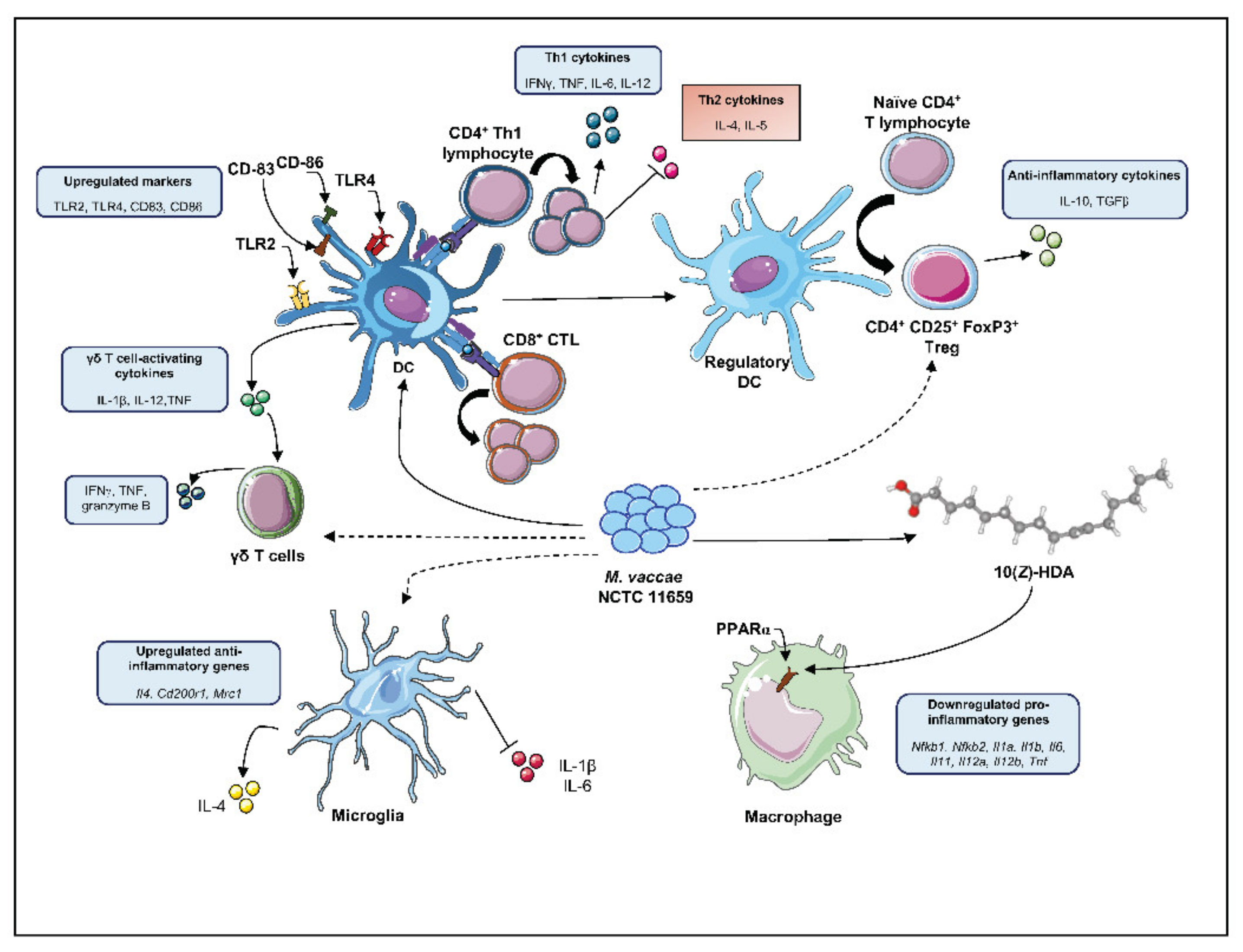
Figure 1. Pleiotropic effects of M. vaccae National Collection of Type Cultures (NCTC) 11659 on the immune system, promoting immunoregulation. Recognition of M. vaccae NCTC 11659 by dendritic cells (DCs) results in the upregulation of Toll-like receptor (TLR)2, TLR4, and of the maturation markers cluster of differentiation (CD)83 and CD86. It also results in the polarization and proliferation of CD4+ T lymphocytes toward a T helper (Th)1 phenotype with production of Th1 cytokines like interferon gamma (IFNγ), tumor necrosis factor (TNF), interleukin (IL)6, and IL-12. M. vaccae NCTC 11659-stimulated DCs also promote the differentiation of CD8+ cytotoxic T lymphocytes (CTL) and gammadelta (γδ) T cells with antitumor activity as well as of CD4+CD25+ forkhead box P3 (FoxP3)+ regulatory T cells (Treg). M. vaccae NCTC 11659 upregulates anti-inflammatory genes (i.e., Il4, Cd220r1, mannose receptor C-type 1 (Mrc1)) in hippocampal microglia, indicated by increased secretion of IL-4, while in contrast reducing secretion of proinflammatory markers such as IL-1β and IL-6. Finally, the M. vaccae NCTC 11659-produced lipid 10(Z)-hexadecenoic acid (10(Z)-HDA) induces anti-inflammatory responses in isolated peritoneal macrophages via activation of peroxisome proliferator-activated receptor alpha (PPARα) and downregulation of proinflammatory genes (i.e., transcription factor nuclear factor-kappa B (Nfkb1, Nfkb2), Il1a, Il1b, Il6, Il11, Il12a, Il12b, Tnf). Solid-line arrows represent direct effects of M. vaccae NCTC 11659 while dashed-line arrows represent indirect effects. 3D image of 10(Z)-HDA retrieved from pubchem.ncbi.nlm.nih.gov.
Ijms 22 12938 g001
Table 1. List of alternative names of the mycobacterium strains used in the present review article and related details 1.
Reference StrainOriginal Source and Year of IsolationAlternative Collection NumbersCulture MediumBatch NameInactivation MethodAdministration VehicleReference
Mycobacterium vaccae NCTC 11659 [34]Mud (lake Kyoga, Uganda), 1973- DSM 107316
- CECT 9646
- Peptone meat extract glycerol agar
- Middlebrook medium
- Löwenstein-Jensen medium
- Tryptic Soy Agar
- MB7H10 agar
- PMG agar
- GYM agar
SRP 299Heat-killed 2PBS[36,37,38]
Saline[39,40]
SRL172Heat-killed 2BBS[41]
Irradiation-killed 3,*BBS[42]
V7Hydrolyzed and heat-killed 2Oral pill excipients[43,44]
Dar-901 (SRL 172)Heat-killed 2Citrate buffer[45]
MV07Heat-killed 2Culture media[46]
MV001Heat-killed 2BBS *[47]
MV007Heat-killed 2BBS[48]
ENG1Heat-killed 2BBS[20,22,24,29,32]
R877RIrradiation-killed 3BBS[49]
Mycobacterium vaccae ATCC 15483T [50]Bovine milk, 1964- ATCC 23004
- CCUG 21003T
- DSM 43292
- KCTC 19087
- NCIB 9937
- NCTC 10916
- SN 920 (Bonicke SN920)
- TMC 1526
- Middlebrook medium
- Medium 55 for Mycobacterium
V7Hydrolyzed and heat-killed 2Oral pill excipients[51]
M. vaccaeTMHeat-killed 2Distilled water[52]
ATCC® 15483Heat-killed 2PBS[53]
BBS[29]
1 Reference strain, original source and year of isolation, alternative collection numbers, culture medium, batch name, administration vehicle and relevant references. ATCC, American Type Culture Collection, Manassas, VA, USA; BBS, borate-buffered saline; CCUG, Culture Collection, University of Göteborg, Department of Clinical Bacteriology, Guldhedsg, Göteborg, Sweden; CETC, Colección Española de Cultivos Tipo; DSM, DSMZ-Deutsche Sammlung von Mikroorganismen und Zellkulturen GmbH, Braunschweig, Germany; KCTC, Korean Collection for Type Cultures, Genetic Resources Center, Korea Research Institute of Bioscience and Biotechnology, Taejon, Republic of Korea; NCIB, National Collection of Industrial Bacteria, Torry Research Station, Aberdeen, Scotland, UK (incorporated with NCIMB); NCTC, National Collection of Type Cultures, Central Public Laboratory Service, London, UK; PBS, phosphate-buffered saline; PMG, Pombe Minimal Glutamate; GYM, glucose, yeast and malt; MB7H10, Middlebrook 7H10; SN, Australian Mycological Panel (AMP; formerly Aust or SN); TMC, Trudeau Mycobacterial Culture Collection, Trudeau Institute, Denver, CO, USA. 2 Heat-killed through autoclaving at 121 °C for 15 min (Stanford et al., 1990b). 3 Irradiation-killed refers to the sterilization procedure, usually performed through exposure to 2.5 Mrad from a 60Co source (Stanford et al., 1990a). * indicates that we did not explicitly find this information in the publication, but deduced it based on other facts given in the publication.
Table 2. List of observational studies employing rapidly growing mycobacteria strains (RGMs) in clinical studies 1.
Disease InvestigatedM. vaccae StrainDosageVehicleEffectsUnderlying MechanismsReference
TuberculosisNCTC 116591 dose (i.d.; 1 mg in 0.1 mL)BBS- improved clearance of TB bacilli
- normalized ESR
- improved sputum conversion
- increased survival
- reduced serum IL-4, IL-10, TNF
- increased serum IFNγ
[54,55,56,57]
TuberculosisNCTC 11659 (SRL172)3 doses (i.d.; 1 mg in 0.1 mL)BBS- improved clearance of TB bacilli
- normalized ESR
- improved sputum conversion
increased serum IL-4 & TNF[41]
TuberculosisNCTC 11659 (SRL172)10 doses (p.o; day 0,7,14,21,28 and then monthly; 1 mg/dose)Gelatine tablet - normalized ESR
- improved sputum conversion and body weight gain
- increased Th1 parameters
- decreased Th2 parameters
[58]
TuberculosisNCTC 11659 (SRL172)5 doses (i.d.; 1 mg in 0.1 mL)BBSproliferation of PBMCsincreased IFNγ in PBMCs[42]
TuberculosisNCTC 11659 (V7)30 doses (p.o.; 10 μg/tablet)V7 tablet (Immunitor®)improved clearance of TB bacilli-reduced blood leukocyte number[43,44]
TuberculosisLongcom batch (ATCC 15483T)30 doses (p.o.; 10 μg/tablet)V7 tablet (Immunitor®)improved clearance of TB bacillinot investigated[51]
LeprosyNCTC 116591 dose (i.d.; 0.1 mg in 0.1 mL)BBSpositive Leprosin A responsenot investigated[59,60]
LeprosyNCTC 116591 dose (i.d.; 1 mg in 0.1 mL)BBSimproved skin capillary flownot investigated[61]
LeprosyNCTC 116593 doses (i.d.; 0.01, 0.1, 1 mg in 0.1 mL)BBSpositive Leprosin A responsenot investigated[62]
PsoriasisNCTC 116591 dose (i.d.; 1 mg in 0.1 mL)BBSreduced PASIreduced lymphocyte proliferation[63]
DermatitisNCTC 11659 (SRL172)1 dose (i.d.; 3 mg in 0.3 mL)BBSreduced dermatitis lesion areanot investigated[64]
AsthmaNCTC 11659 (SRL172)1 dose (i.d.; 1 mg in 0.1 mL)BBStrend towards improved airway response to allergenreduced IL-5 and IgE in PBMCs[65]
Cancer 2NCTC 11659 (SRL172)- 1 dose (i.d.; 0.5 mg in 0.1 mL);
- 3 doses (i.d.; 1 mg in 0.1 mL)
BBSincreased survivalnot investigated[66]
Cancer 2NCTC 11659 (SRL172)- 1 dose (i.d.; 50 µL (5 × 10⁸ heat-killed bacilli));
- 4 doses (i.d.; 100 µL (109 heat-killed bacilli))
BBSimproved survivalintracellular IL-2 induction[67]
Cancer 3NCTC 11659 (SRL172)5 doses (i.d.; 1 mg in 0.1 mL)BBS- improved quality of life (cognitive function and vitality);
- reduced body pain, nausea and dyspnea
not investigated[68]
1 Disease investigated, strain of M. vaccae, dosage and vehicle used, effects, cellular/molecular mechanisms involved, and respective sources are listed. 2 Metastatic malignant melanoma. 3 Non-small-cell lung adenocarcinoma. Abbreviations: ATCC 15483T, M. vaccae American Type Culture Collection 15483T; BBS, borate-buffered saline; ESR, erythrocyte sedimentation rate; i.d., intradermal; IFN, interferon; IgE, immunoglobulin E; IL, interleukin; NCTC 11659, M. vaccae National Collection of Type Cultures 11659; PASI, psoriasis area severity index; PBMC, peripheral blood mononuclear cells; p.o, per os (i.e., orally); TB, tuberculosis; Th, T helper cell; TNF, tumor necrosis factor.
Table 3. List of observational studies employing rapidly growing mycobacteria strains (RGMs) in preclinical studies 1.
Disease InvestigatedM. vaccae StrainDosageVehicleEffectsUnderlying MechanismsReference
Negative consequences of stressNCTC 116593 doses (s.c.; 0.1 mg in 0.1 mL)BBSreduced stress-induced anxiety and colitis- increased number of Treg (CD4+ CD25+ FoxP3+)
- increased IL-10
[20,33]
Negative consequences of stressNCTC 116593 doses (i.n.; 0.1 mg in 0.012 mL)BBSreduced stress-induced colitisnot investigated[32]
Negative consequences of stressNCTC 116593 doses (s.c.; 0.1 mg in 0.1 mL)BBSenhanced between- and within-session extinction in fear-potentiated startle paradigmalteration in serotonergic gene expression[25,27,28]
Negative consequences of stressNCTC 116593 doses (s.c.; 0.1 mg in 0.1 mL)BBSprevention of stress-induced exaggeration of anxiety and microglial priming- upregulated hippocampal IL4, Cd200r1 and Mrc1
- downregulated hippocampal Nlrp3 and Nfkbia
[24]
Negative consequences of stressNCTC 116593 doses (s.c.; 0.1 mg in 0.1 mL)BBSprevention of post-operative cognitive dysfunction- upregulated hippocampal IL4, Arg1 and Foxp3
- downregulated hippocampal NfκBbia and IL1β
[22]
Negative consequences of stressNCTC 116593 doses (s.c.; 0.1 mg in 0.1 mL)BBSprevention of negative outcomes of a two-hit stress models- prevention of stress-induced decreased Tph2 and Slc6a4 expression
- prevention of stress-induced REM sleep disturbances
[23,26]
1 Disease investigated, strain of M. vaccae, dosage and vehicle used, effects, cellular/molecular mechanisms involved, and respective sources are listed. Abbreviations: Arg1, arginase 1 gene; BBS, borate-buffered saline; CD, cluster of differentiation; FoxP3, forkhead box P3; IL, interleukin; i.n., intranasal; NCTC 11659, M. vaccae National Collection of Type Cultures 11659; NfκBbia: gene encoding nuclear factor of kappa light polypeptide gene enhancer in B-cells inhibitor, alpha; Nlrp3, NLR family pyrin domain containing 3; s.c., subcutaneous; REM, rapid eye movement; Slc6a4, solute carrier family 6 member 4; Tph2, tryptophan hydroxylase 2; Treg, regulatory T cells.
Table 4. List of preclinical studies investigating the underlying mechanisms induced by rapidly growing mycobacteria strains (RGMs) 1.
Cellular/ Molecular Target InvestigatedM. vaccae StrainDosageVehicleSpeciesUnderlying MechanismsReference
Th1/Th2 balanceNCTC 11659 (MV07)1, 10, 100 µg/mL (in vitro)BBShuman (DCs)- reduced IL-4
- upregulated CD83 and CD86
[46]
Th1/Th2 balanceNCTC 11659 (SRL172)3 doses (s.c.; 106,107,108 bacteria in 0.1 mL)BBSmouse- reduced serum IgE, IL-4 and IL-5
- reduced eosinophil count in BAL
[69]
Th1/Th2 balanceNCTC 116591 dose (s.c.; 107, 108 or 109 bacteria in 0.1 mL)BBSmouse- reduced serum IgE and IL-4
- increased IL-2 in splenocytes
[70]
γδ T cellsNCTC 11659 (SRL172)100 μg/mL (in vitro)BBShuman (PBMCs)upregulated IFNγ, TNF and granzyme B[71]
CD11b+ myeloid cellsNCTC 11659300 μg/mL (in vitro)BBShuman (PBMCs)- downregulated CD62L
- upregulated TLR2, TLR4, CD18, CD11a, CD14, CD36, CD44, CD45, CD54 CD58k, CD80, CD86, CD137L, CD206
[72]
CD11c+ APCNCTC 11659 (SRP299)1 dose (s.c.; 0.1 mg in 0.1 mL)NaClmouse- decreased cell number in BAL;
- increased IL-10+ and TGFβ in lung DCs
[39]
CD14+ monocytesATCC 15483T (SN920)in vitro incubation with 1:10 ratio cells:mycobacteriaMediumhuman (PBMCs)increased secretion of TNF and IL-12[73]
CD8+ CTLATCC 15483T1 dose (i.p.; 1 mg in 0.1 mL)PBSmouse- increased expression of IFNγ in TB-infected macrophages
- increased cytotoxic activity of CTL against TB-infected macrophages
[74]
TregsNCTC 11659 (SRP299)1 dose (s.c.; 0.1 mg in 0.2 mL)NaClmouse- increased number of Treg (CD4+ CD45RBLo)
- suppressed airway inflammation upon Treg transfer
[40]
TregsNCTC 11659 (SRP299)- 1 dose (i.g.; 0.1 mg in 0.1 mL)
- 100, 200 or 400 μg/mL (in vitro)
-H2O-NaClmouse- reduced cellular infiltrate in lungs
- increased IL-10 in mesLNC
[75]
TregsNCTC 116593 doses (s.c.; 0.1 mg in 0.1 mL)BBSmouse- increased number of Treg (CD4+ CD25+ FoxP3+)
- increased IL-10
- reduced stress-induced anxiety and colitis
[33]
PPARα10(Z)-HDA from NCTC 11659200 µM (in vitro)DMEM/F-12mousePPARα-dependent downregulation of pro-inflammatory transcription factors, cytokines and chemokines[76]
Serotonergic neuronsNCTC 116593 doses (s.c.; 0.1 mg in 0.1 mL)BBSmouseactivation of serotonergic neurons in interfascicular part of dorsal raphe nucleus[21]
MicrogliaNCTC 116593 doses (s.c.; 0.1 mg in 0.1 mL)BBSrat- increased expression of Il4 mRNA and IL-4-responsive genes (Cd200r1, Mrc1)
- reduced IL-1β secretion from freshly isolated and LPS-stimulated hippocampal microglia
[24]
MicrogliaNCTC 11659, ATCC 15483T3 doses (s.c.; 0.1 mg in 0.1 mL)BBSratprevention of stress-induced upregulation of hippocampal Il6 mRNA expression[29]
MicrogliaNCTC 116593 doses (s.c.; 0.1 mg in 0.1 mL)BBSratincreased hippocampal Il4, Foxp3, Arg1, decreased Il1β, Il6 and Nfκbia mRNA expression[22]
1 Cellular/molecular target investigated, strain of M. vaccae, dosage and vehicle used, species, cellular/molecular mechanisms involved, and respective sources are listed. Abbreviations: Arg1, arginase 1 gene; ATCC 15483T, M. vaccae American Type Culture Collection 15483T; BAL, bronchoalveolar lavage; BBS, borate-buffered saline; CD, cluster of differentiation; DMEM, Dulbecco’s modified Eagle’s medium; FoxP3, forkhead box P3; HPC, human pancreatic carcinoma cell line, IFN, interferon; i.g., intragastric; IL, interleukin; i.p., intraperitoneal; Mrc1, gene encoding mannose receptor C-type 1; NCTC 11659, M. vaccae National Collection of Type Cultures 11659; NfκBbia: gene encoding nuclear factor of kappa light polypeptide gene enhancer in B-cells inhibitor, alpha; PBMC, peripheral blood mononuclear cells; PBS, phosphate-buffered saline; p.o, per os (i.e., orally); PPARα, peroxisome proliferator-activated receptor α; s.c., subcutaneous; TB, tuberculosis; TNF, tumor necrosis factor.
Publisher’s Note: MDPI stays neutral with regard to jurisdictional claims in published maps and institutional affiliations.

