Article

Photoactivation of Cell-Free Expressed Archaerhodopsin-3 in a Model Cell Membrane
Experimental Physics and Center for Biophysics, Saarland University, 66123 Saarbrücken, Germany
*
Authors to whom correspondence should be addressed.
Navid Khangholi and Marc Finkler contributed equally to this paper.
Int. J. Mol. Sci. 2021, 22(21), 11981; https://doi.org/10.3390/ijms222111981
Submission received: 14 October 2021 / Revised: 2 November 2021 / Accepted: 3 November 2021 / Published: 5 November 2021
(This article belongs to the Special Issue Membrane Proteins: Structure, Function and Motion)

Abstract

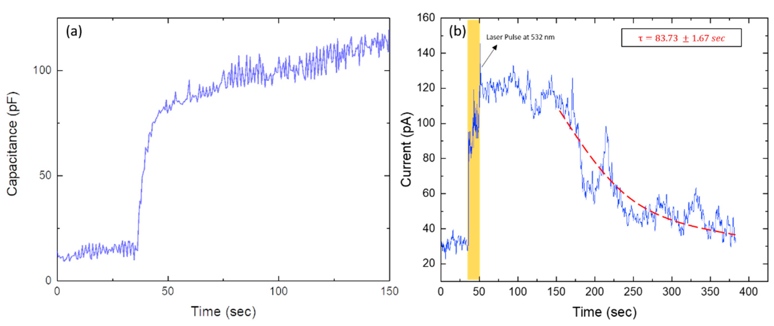
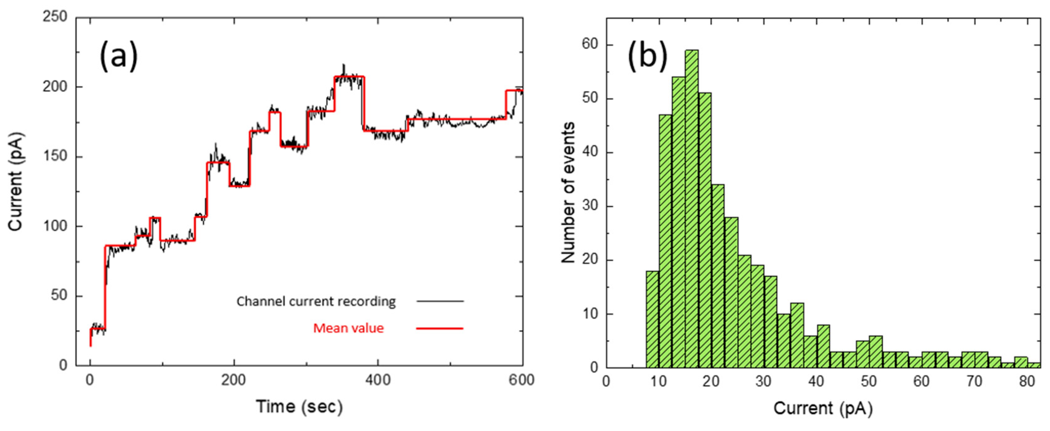
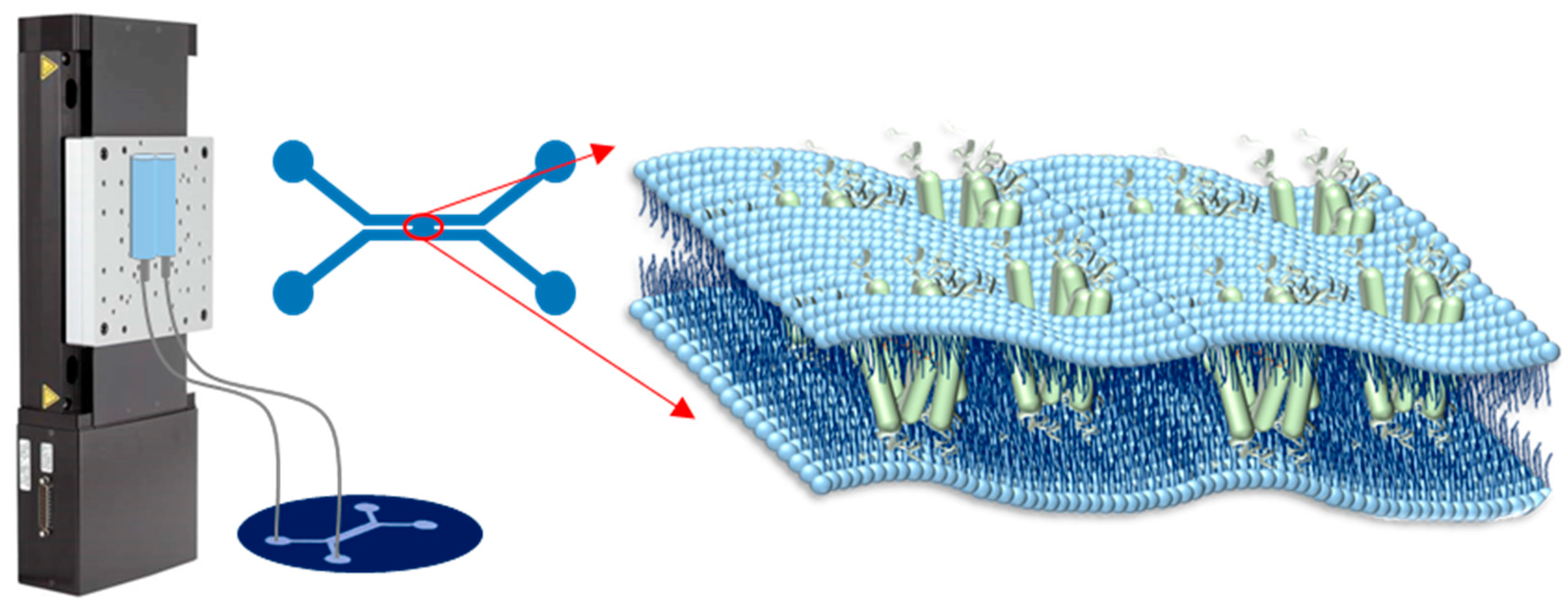
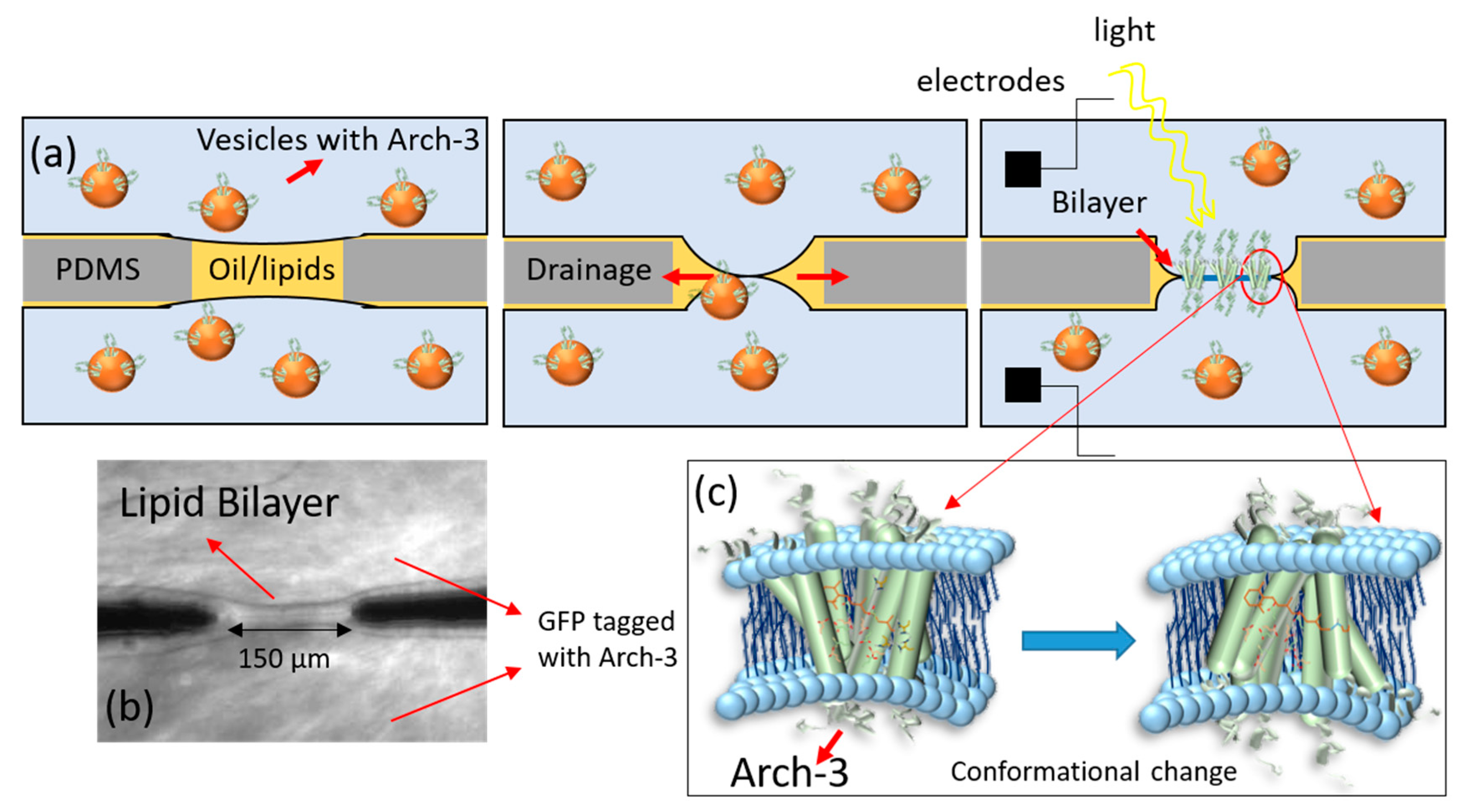
Transmembrane receptor proteins are located in the plasma membranes of biological cells where they exert important functions. Archaerhodopsin (Arch) proteins belong to a class of transmembrane receptor proteins called photoreceptors that react to light. Although the light sensitivity of proteins has been intensely investigated in recent decades, the electrophysiological properties of pore-forming Archaerhodopsin (Arch), as studied in vitro, have remained largely unknown. Here, we formed unsupported bilayers between two channels of a microfluidic chip which enabled the simultaneous optical and electrical assessment of the bilayer in real time. Using a cell-free expression system, we recombinantly produced a GFP (green fluorescent protein) labelled as a variant of Arch-3. The label enabled us to follow the synthesis of Arch-3 and its incorporation into the bilayer by fluorescence microscopy when excited by blue light. Applying a green laser for excitation, we studied the electrophysiological properties of Arch-3 in the bilayer. The current signal obtained during excitation revealed distinct steps upwards and downwards, which we interpreted as the opening or closing of Arch-3 pores. From these steps, we estimated the pore radius to be 0.3 nm. In the cell-free extract, proteins can be modified simply by changing the DNA. In the future, this will enable us to study the photoelectrical properties of modified transmembrane protein constructs with ease. Our work, thus, represents a first step in studying signaling cascades in conjunction with coupled receptor proteins.
Keywords: Archaerhodopsin-3; lipid bilayer; microfluidics; cell-free gene expression Archaerhodopsin-3; lipid bilayer; microfluidics; cell-free gene expression