Review

The Role of Long Non-Coding RNAs in Endometriosis

Department of Obstetrics and Gynecology, Medical University of Vienna, Waehringer Guertel 18-20, A-1090 Vienna, Austria
*
Author to whom correspondence should be addressed.
These authors contributed equally to this work.
Int. J. Mol. Sci. 2021, 22(21), 11425; https://doi.org/10.3390/ijms222111425
Submission received: 22 August 2021 / Revised: 14 October 2021 / Accepted: 19 October 2021 / Published: 22 October 2021
(This article belongs to the Special Issue Molecular and Cellular Advances in Endometriosis Research)

Abstract

:
Endometriosis is a chronic gynecological disorder affecting the quality of life and fertility of many women around the world. Heterogeneous and non-specific symptoms may lead to a delay in diagnosis, with treatment options limited to surgery and hormonal therapy. Hence, there is a need to better understand the pathogenesis of the disease to improve diagnosis and treatment. Long non-coding RNAs (lncRNAs) have been increasingly shown to be involved in gene regulation but remain relatively under investigated in endometriosis. Mutational and transcriptomic studies have implicated lncRNAs in the pathogenesis of endometriosis. Single-nucleotide polymorphisms (SNPs) in lncRNAs or their regulatory regions have been associated with endometriosis. Genome-wide transcriptomic studies have identified lncRNAs that show deregulated expression in endometriosis, some of which have been subjected to further experiments, which support a role in endometriosis. Mechanistic studies indicate that lncRNAs may regulate genes involved in endometriosis by acting as a molecular sponge for miRNAs, by directly targeting regulatory elements via interactions with chromatin or transcription factors or by affecting signaling pathways. Future studies should concentrate on determining the role of uncharacterized lncRNAs revealed by endometriosis transcriptome studies and the relevance of lncRNAs implicated in the disease by in vitro and animal model studies.

1. Introduction

Endometriosis is a chronic inflammatory disorder defined by endometrial-like lesions growing ectopically outside of the uterus that affects many reproductive-age women worldwide. The disease has heterogeneous symptoms that may not be specific to the disease, such as different types of chronic gynecological pain and reduced fertility. The cause of the disease remains unclear, but lesions are thought to originate from endometrial cells that escape the uterus [1], although the presence of rare endometriosis-like lesions in males supports a trans-differentiation origin [2]. Endometriosis is thought to be an immunological disease, first because lesions must evade the immune system in order to establish and develop, and second because the inflammatory nature of the disease accounts for many of the symptoms [3]. Additionally, a meta-analysis has found that there is some association between endometriosis and a risk for developing an autoimmune disease [4]. Treatment options for the disease remain limited to laparoscopic surgery to remove lesions and hormone-based treatments that are not compatible with pregnancy [3]. Given that diagnosis is often delayed due to the non-specific symptoms and that there are limited treatment options, there is a need to better understand the molecular pathogenesis of the disease in order to identify key players that may be used as biomarkers for diagnosis or as targets for new treatments. One class of molecules that may play a role in the disease are long non-coding RNAs (lncRNAs).
LncRNAs are a class of genes that since the advent of high-throughput sequencing technology have been revealed to be much more numerous than was previously realized. Currently over 16,000 human lncRNAs are annotated by the GENCODE project, while some other studies have indicated that the total number may be over 100,000 [5]. Although most of these lncRNAs remain uncharacterized, an increasing number have been shown to have a biological function, often involved in gene regulation [6]. LncRNAs remain relatively under investigated in endometriosis, but given their roles in development and disease in other contexts [7], it is reasonable to assume that there may be lncRNAs that also play a role in endometriosis.
LncRNAs may exert their function in either the nucleus or the cytoplasm via a variety of mechanisms. In the nucleus, lncRNAs may act as epigenetic gene regulators by recruiting chromatin remodeling or modification complexes to target gene promoters either in cis (Figure 1a) [8] and/or in trans to activate or suppress their transcription (Figure 1b) [9]. LncRNAs can act as decoys of specific chromatin modifiers by sequestering them from the promoters of target genes (Figure 1c) [10]. In other contexts, lncRNAs may act as transcriptional regulators by competing with a transcription factor for binding DNA and/or by binding its DNA binding domain (Figure 1d) [11]. LncRNAs have also been associated with the regulation of pre-mRNA alternative splicing in the nucleus, thereby affecting which isoforms predominate (Figure 1e) [12]. In the cytoplasm, lncRNAs are involved in post-transcriptional regulation processes that can affect the stability of transcripts (Figure 1f) [13] or whether a transcript is translated into a protein or not (Figure 1g) [14]. LncRNAs can also be involved in the modulation of cell signaling pathways by binding to signaling proteins and affecting their activation state (Figure 1h) [15]. Finally, a common mechanism whereby lncRNAs are thought to affect mRNA abundance is by acting as so-called “sponges” of miRNAs to reduce miRNA-mediated degradation. Here, lncRNAs may share miRNA target sequences with mRNAs, thereby reducing miRNA availability to target mRNAs (Figure 1i) [16,17].
These studies show that lncRNAs can act at multiple subcellular sites to affect various aspects of cell biology, although in most cases they affect either transcription or processing of mRNAs and their steady state levels in the cell. That is, in a broad sense, they regulate other genes. An increasing body of literature implicates abnormal expression of specific lncRNAs in disease, particularly in different types of cancer [18], but also in a variety of other diseases including neural disorders [19], cardiovascular disease [20], and diabetes [21]. Evidence for the involvement of individual lncRNAs in the pathogenesis of these diseases ranges from a correlation with dysregulated lncRNA expression to detailed mechanistic studies that indicate a functional role in the disease. The implication from these studies and others is that lncRNAs are important players in the pathogenesis of many diseases, suggesting that there may be lncRNAs that play a role in endometriosis. Furthermore, a number of studies in have shown that lncRNAs can either be regulated by estrogen-dependent enhancers [22] or that lncRNAs can regulate estrogen receptor expression [23]. As endometriosis is an estrogen-dependent disease [3], these studies give further impetus to investigate the role of lncRNAs in the disease. Given this evidence for the role of lncRNAs in other contexts, in this review, we aim to provide a comprehensive picture of the current state of knowledge over the role of lncRNAs in endometriosis.

2. Aims and Methodology of This Review

In this review, we aim to provide a comprehensive picture of the current knowledge of the role of lncRNAs in the pathogenesis of endometriosis and the mechanisms by which they affect the disease. We included patient studies to detect lncRNAs that have been associated with endometriosis, along with in vitro studies and in vivo animal studies to analyze the mechanism of action and function of lncRNAs in the disease. To gather studies for this review, we searched both the PubMed and Google Scholar databases for endometriosis and lncRNA combined with the following keywords: mechanism of action, sponging, ceRNA networks, chromatin, biomarkers, genome-wide, microenvironment, and infertility. We concentrated on original research papers, excluding literature reviews, pre-prints, meeting abstracts, and articles that were not in English and then assessed the remaining publications for this review.

3. Evidence for the Role of lncRNAs in Endometriosis

The evidence for the role of lncRNAs in the pathogenesis of endometriosis has been largely uncovered by using high-throughput screening technologies to detect differences between control and disease cohorts from either patient samples or animal models. Selected lncRNAs identified using these approaches were then subjected to further validation experiments to confirm their role. Below, we discuss the current evidence, most of which is derived from genome-wide genomic or transcriptomic studies.

3.1. Genetic Evidence for lncRNA Involvement in Endometriosis

Genome-wide association studies (GWAS) have been conducted for many diseases including endometriosis [24,25]. This powerful approach surveys single-nucleotide polymorphisms (SNPs) throughout the genome in a large cohort of individuals to identify variants and genomic regions associated with an increased chance of developing the disease. SNPs identified in GWAS studies often occur in intergenic regions, and therefore, it has been speculated that they may affect the regulation of nearby genes [26]. One mechanism by which this could work is via changing the sequence of an lncRNA and, thereby, affecting its regulatory function [27].
Genome-wide DNA sequencing technology has identified genetic variations in lncRNA loci that may affect the pathogenesis of endometriosis. The rs10965235 SNP located on chromosome 9p21.3 is associated with severe endometriosis in a Korean patient cohort and lies within the CDKN2B-AS gene locus [28]. This indicates that disruption of lncRNA function by the SNP is a possible mechanism through which this variant may predispose patients to endometriosis. The rs3820282 SNP located within a WNT4 intron on chromosome 1p36.12 is associated with endometriosis and appears to affect an enhancer–promoter interaction resulting in the downregulation of LINC00339 and upregulation of CDC42 [29]. This indicates another possible mechanism for lncRNA regulation, whereby dysregulation of CDC42 due to the risk of an SNP affecting enhancer competition with the LINC00339 promoter leads to predisposition to endometriosis. Recently, genetic variations at the rs1838169 and rs17720428 SNPs sites on chromosome 12q13.3 in HOTAIR have been shown to be frequently detected in patients with endometriosis [30]. These variants appear to increase the stability of the lncRNA, resulting in reduced levels of HOXD10 and HOXA5 transcripts regulated by HOTAIR. A variant of the rs591291 SNP located on chromosome 11q13.1 in the promoter region of MALAT1 was associated with an increase in endometriosis risk in a Chinese population, indicating that a change in the MALAT1 expression level may affect endometriosis risk, although this was not examined in this study [31]. Finally, the rs710886 SNP in PCAT1 has been associated with increased risk of developing endometriosis [32]. This SNP appears to disrupt sponging by PCAT1 of miR-145, affecting the expression of FASCIN1, SOX2, MSI2, SERPINE1, and JAM-A, and the proliferative and invasive ability of endometriosis stem cells. Together, these studies indicate that genetic variants associated with endometriosis may predispose patients to the disease by disrupting the lncRNA gene regulatory function via diverse mechanisms.

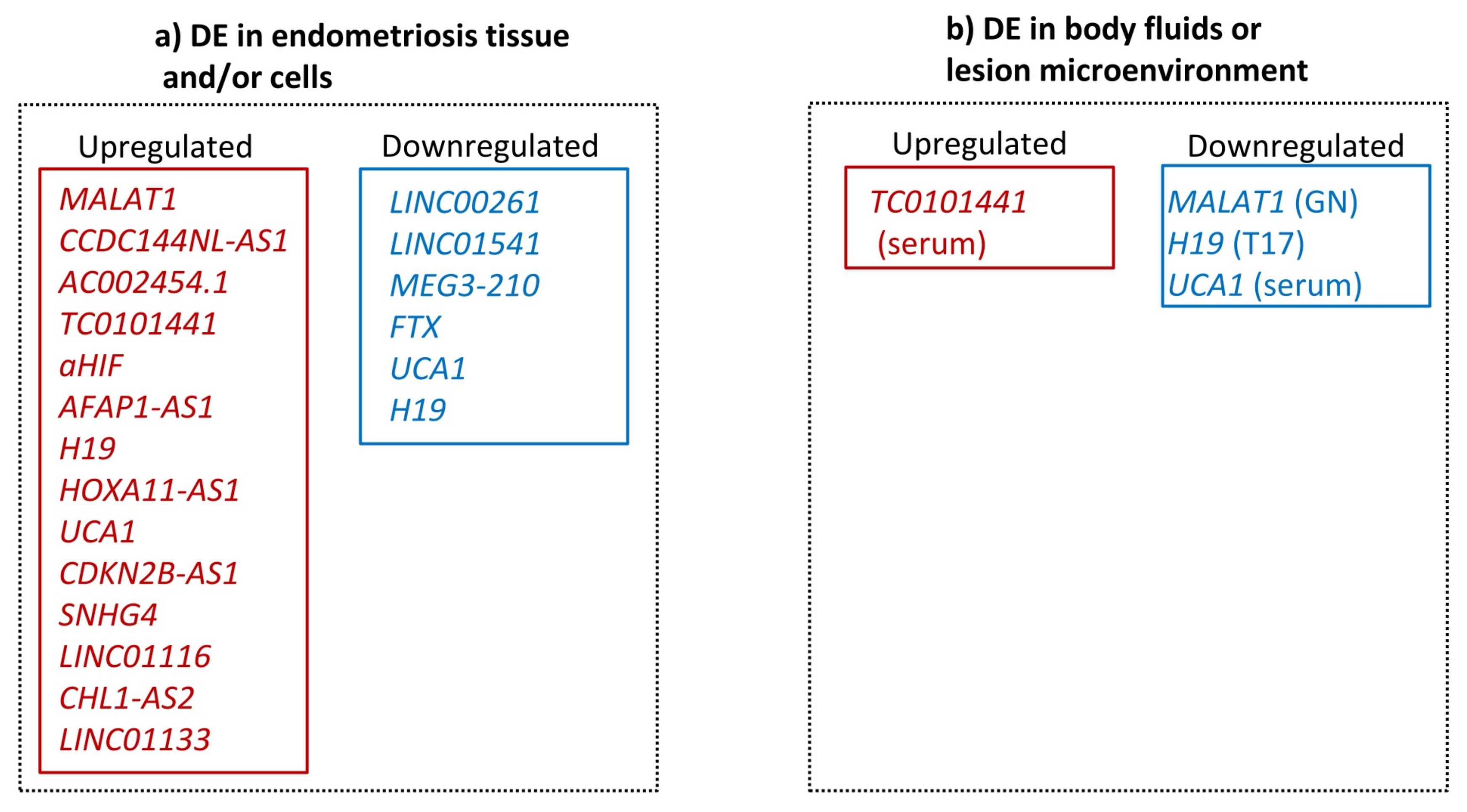
3.2. Transcriptional Evidence for lncRNA Involvement in Endometriosis

To date, there have been around 14 genome-wide profiling transcriptome studies of human patient samples and animal models for endometriosis (Table 1). These studies are mainly descriptive in nature, with experimental validation limited to a few selected candidates. These studies often include in silico analysis examining the relationship between differentially expressed protein coding and lncRNA transcripts. Further bioinformatic approaches, such as gene ontology analysis, are then often used to predict the relevance of the identified targets to biological pathways involved in the pathogenesis of endometriosis. Initial studies were mostly conducted using microarray platforms to assess the expression of lncRNAs and mRNAs. Using a human lncRNA expression microarray, Sun et al. [33] were the first to assess genome-wide the relationship between lncRNA and mRNA expression in ovarian endometriosis lesions compared to paired autologous eutopic endometrial samples. They identified 948 lncRNAs and 4088 mRNAs as differentially expressed in the study cohort and validated the differential expression of the top 10 lncRNA candidates using qRT-PCR. Based on co-expression with mRNAs, lncRNAs in this study were predicted to take part in tissue adhesion, angiogenesis, estrogen production, and immune response, all processes known to be associated with the pathogenesis of endometriosis. Co-expression and genomic proximity were then used to predict 49 cis-regulating lncRNAs and their protein coding targets, while trans-regulating lncRNAs were predicted by co-expression with transcription factors and network analysis. This indicated that the top candidates were in a network with MYC, CTCF, and E2F4. Another study from Cai et al. (2019) used microarrays to profile lncRNA and mRNA expression in an endometriosis rat model, resulting in the identification of 115 upregulated and 51 downregulated lncRNAs together with 182 differentially expressed protein coding mRNA transcripts [34]. Co-expression analysis revealed five lncRNAs (LOC102551276, NONRTT006252, LOC103691820, LOC102546604, and NONRATT003997) that show a similar expression pattern to four protein coding mRNAs (Adamts7, P2ry6, Dlx3, and TP53), indicating a possible functional relationship in endometriosis.
Using a transcriptome array, Wang et al. [35] investigated the expression profile of lncRNAs in serum and tissue samples from patients with and without endometriosis. Following qRT-PCR validation of differentially expressed lncRNAs, they identified a combination of five circulating lncRNAs (NR_038395, NR_038452, ENST00000482343, ENST00000544649, and ENST00000393610) that they proposed could act as non-invasive biomarkers for the disease.
In the last few years, high-throughput RNA sequencing technology has surpassed microarrays as the preferred genome-wide technology for the identification of differentially expressed lncRNAs in endometriosis. Unlike microarrays, RNA sequencing is not biased by prior knowledge, enabling the discovery and characterization of lncRNAs that may play a role in the disease. A number of RNA sequencing studies have now been conducted comparing tissues from either patients or endometriosis animal models (Table 1). For example, using RNA sequencing, Cui et al. identified 86 differentially expressed lncRNAs and 1228 differentially expressed mRNAs in patients with ovarian endometriosis lesions compared to eutopic endometrial controls [36]. Pathway and gene ontology analysis showed that differentially expressed lncRNAs were involved in the regulation of cell proliferation, adhesion, migration, steroidogenesis, and angiogenesis, all processes implicated in endometriosis lesion formation and survival at ectopic sites of implantation.
Table 1. Genome-wide studies that identified differentially expressed lncRNAs in endometriosis.
Method (Reference)Cohort
(Tissue Type)
Validation
(Type; Cohort; lncRNAs)
Predicted Function in EndometriosisLimitations
Expression
Microarray [33]
n = 8
(paired ovarian:
4 eutopic and
4 ectopic tissues)
RT-qPCR
n = 42
(paired ovarian: 21 eutopic and
21 ectopic tissues)
CHL1-AS2, MGC24103, XLOC_007433, HOXA11-AS, KLP1, LOC100505776, XLOC_012981, LIMS3-LOC4408, LOC389906
Cis and trans regulation of protein
coding genes
1, 6
Expression
Microarray [37]
n = 6
(3 eutopic tissues of women with EM of
undefined entity and
3 eutopic tissues of women without EM)
RT-qPCR
n = 68
(40 eutopic tissues of women with EM of undefined entity and 28 eutopic
tissues of women without EM)
RP11-369C8.1, RP11-432J24.5, AC068282.3, GBP1P1, SNHG1, AC002454.1, AC007246.3, FTX
Cell cycle regulation and immune
response
1, 2, 4, 6
Expression
Microarray [35]
Serum: n = 20
(10 women with peritoneal and/or ovarian EM and 10 women without EM)
Tissue: n = 15
(paired peritoneal and/or ovarian: 5 eutopic and
5 ectopic EM patients and 5 eutopic tissues
of women without EM
RT-qPCR
Serum: n = 110
(59 women with peritoneal and/or ovarian EM and 51 women without EM)
Tissue: qPCR n = 24
(paired peritoneal and/or ovarian: 9 eutopic and
9 ectopic tissues of EM patients and 6 eutopic tissues of women without EM)
 
DE lncRNAs
16
Combination of NR_038395, NR_038452, ENST00000482343, ENST00000544649, and ENST00000393610 suggested as a non-invasive
diagnosis marker
1,
2 (because of pooling)
Expression
microarray [38]
n = 8
(paired ovarian: 4 eutopic and 4 ectopic tissues of EM patients)
RT-qPCR
n = 87
(paired ovarian: 30 eutopic and 30 ectopic tissues of EM patients and 27 eutopic tissues of women without EM))
 
CHL1-AS2
CCDC144NL-AS1 expression was upregulated in ectopic tissues compared to eutopic and control endometrial tissues1
Re-analysis of existing microarray data [39]n = 18
GSE120103
(eutopic tissue from 9 fertile and 9 infertile women with ovarian EM)
Validation of 14 hub mRNAs using
GSE26787
(5 fertile and 5 infertile women with unknown EM status)
Identification of putative infertility-associated lncRNAs LOC390705 and LOC1005058541, 3, 6
Re-analysis of existing microarray data [40]GSE7305
(paired ovarian:
10 eutopic and 10 ectopic tissues of EM patients and 10 eutopic tissues of women without EM)
 
GSE7846
(HEECs of eutopic tissues of 5 EM patients with ovarian EM and HEECs of eutopic tissues of 5 women without EM),
 
GSE29981
(LMD epithelial cells of 20 women without EM)
 
E-MTAB-694
(paired peritoneal: 18 eutopic and
18 ectopic tissues of EM patients and 17 eutopic tissues of women without EM)
No validationProposed cell cycle regulatory functions for LINC012792, 3, 4, 5
RNA-Seq [41]n = 16
(8 eutopic tissues of women with EM of undefined entity and 8 eutopic tissues of women without EM)
No validationPredicted oxidative stress and endometrial receptivity regulatory functions1, 2, 3, 4, 6
RNA-Seq [36] n = 10
(5 eutopic tissues of patients with ovarian EM and 5 eutopic tissues of women without EM)
RT-qPCR
n = 24
(12 eutopic tissues of patients with ovarian EM and 12 eutopic tissues of women without EM)
USP46-AS1, RP11-1143G9.4, RP11-217B1.2, AC004951.6, RP11-182J1.12
Predicted proliferation, adhesion, migration, invasion, and angiogenesis regulatory functions1, 4, 6
RNA-Seq [42] n = 9
(paired ovarian: 3 eutopic and
3 ectopic tissues of EM patients and 3 eutopic tissues of women without EM)
RT-qPCR
n = 45
(paired:15 eutopic and
15 ectopic tissues of EM patients with undefined entity and 15 eutopic tissues of women without EM)
PRKAR2B, CLEC2D
Predicted angiogenesis, cell adhesion, cell migration, immune response, inflammatory response, NF-κB signaling, regulatory functions1, 2, 4
Re-analysis of existing RNA-Seq and expression array
datasets [43]
GSE105764
(paired ovarian:
8 eutopic and
8 ectopic tissues of EM patients)
GSE121406
(paired ovarian: FACS sorted stromal cells of 4 eutopic and 4 ectopic tissues of EM patients)
GSE105765
(same as GSE105764)
in silico validation by microarray
GSE124010
(3 ectopic tissues of patients with EM of undefined entity and 3 eutopic tissues of women without EM)
GSE86534
(paired ovarian: 4 eutopic and 4 ectopic tissues of patients with EM)
Predicted function in regulation of inflammation and
prediction of sponging LINC01018 and SMIM25 functions for hsa-miR-182-5p in the regulation of CHL1 protein coding gene to promote endometriosis
1, 4 (validation study), 5, 6
Re-analysis of existing RNA-Seq datasets [44]n = 28
(paired ovarian: 14 eutopic and 14 ectopic tissues of infertile EM patients)
GSE105764
(paired ovarian: 8 eutopic and 8 ectopic tissues of EM patients)
GSE105765
(same patients as in GSE105764)
 
GSE25628
(unpaired DIE: 8 eutopic and 8 ectopic tissues of EM patients and 6 eutopic tissues of women without EM)
No validationConstruction of a competitive
endogenous (ce) RNA network
promoting growth and death of endometrial stroma cells. CDK1 and PCNA proposed as treatment targets for endometriosis-
associated
infertility
1, 3, 5
RNA-Seq [45]n = 12
(paired ovarian: 6 eutopic and 6 ectopic tissues of EM patients)
RT-qPCR
 
n = 60
(paired ovarian: 30 eutopic and 30 ectopic tissues of EM patients)
MIR202HG, LINC00261, UCA1, GAGA2-AS1
Immunity,
inflammation
1, 6
Re-analysis of existing RNA-Seq
data [46]
GSE105764 and GSE105765
include same patients
 
n = 16
(paired ovarian: 8 eutopic and 8 ectopic tissues of EM patients)
No validationLncRNAs:H19, GS1-358P8.4, and RP11-96D1.10 strongly associated with
ovarian
endometriosis
1, 3, 6
Animal Studies
Expression
microarray [34]
Rat
n = 35
(EM uterine tissue
EM = 13, adipose tissue control = 8, blank = 14)
RT-qPCR;
NONRATT003997;
gi|672033904|ref|, XR_589853.1|; NONRATT006252; gi|672027621|ref|; XR_592747.1|; gi|672045999|ref|; XR_591544.1|
Regulation of
endometrial
receptivity
EM, endometriosis; HEECs, human endometrial endothelial cells; LMD, laser microdissection; DIE, deep infiltrating endometriosis. 1, small sample size (less than 30 per group); 2, no comprehensive clinical information (i.e., American Fertility Society (rAFS) disease stage, lesion entities, menstrual cycle phase); 3, no validation in an adequate independent cohort; 4, EM-free controls are not appropriate (i.e., CIN patients, no laparoscopic proof); 5, combining heterogeneous datasets (i.e., different lesion entities, cycle phases, stages, cell types); 6, not all relevant tissues analyzed (i.e., eutopic tissues of EM-free controls, eutopic and ectopic tissues of EM patients).
In spite of the advantages listed above, RNA sequencing studies have some limitations. Due to their high cost, they are often limited to a small number of samples and lack extensive validation. However, they can form the basis for further studies that perform functional validation of candidate genes and investigate their value as diagnostic and prognostic markers in endometriosis. These validation studies usually have cohorts with a larger sample size and may include functional experiments that attempt to uncover the molecular mechanism of lncRNA action (Table S1). For example, AFAP1-AS1 was identified as one of the most differentially expressed lncRNAs in the microarray study of Sun et al. [33]. In a new study, Lin et al. [47] could confirm and extend these findings in a cohort of n = 36 patients. They show that AFAP1-AS1 is overexpressed in ectopic endometrium of women with endometriosis (n = 18), compared to paired eutopic endometriosis tissue (n = 18) and normal endometrium of women without the disease (n = 10). In vitro gene targeting assays in primary human endometriotic stroma cells (ESCs) indicated that this lncRNA regulates epithelial–mesenchymal transition (EMT) in endometriosis by regulating transcription of the EMT-related transcription factor ZEB1. Furthermore, experiments in a xenograft mouse model indicated that AFAP1-AS1 was required for the growth of ectopic tissue. In this case, a shRNA knockdown of AFAP1-AS1 in the Ishikawa endometrial cancer cell line led to reduction of the subcutaneous tumor size, compared to animals injected with a non-targeting shRNA-transfected cell line [47].
Liu et al. analyzed the value of lncRNA H19 expression as an endometriosis biomarker [48]. They found that H19 expression in the both the ectopic and eutopic endometrium of endometriosis patients was significantly higher than in the normal endometrium. Overexpression of H19 lncRNA in endometriosis lesions was associated with infertility, recurrence of disease, bilateral ovarian lesions, an increased CA125 level and with progression in the revised American Fertility Society (rAFS) disease stage. Further multivariate logistic regression analysis showed that H19 overexpression in endometriosis lesions is a prognostic factor for endometriosis recurrence.
In summary, recently there has been an effort by a number of studies using genome-wide high-throughput technologies to identify differentially expressed lncRNAs in endometriosis not only for their potential clinical application as diagnostic or prognostic biomarkers of the disease, but also in order to gain insights into the pathogenesis of the disease (Table 1). The lncRNAs where further work has been done to validate a role in endometriosis are listed in Table S1 and summarized in Figure 2.

3.3. Critical Assessment of the Evidence for the Role of lncRNAs in Endometriosis

In the previous sections, and in Table 1 and Table S1, we summarized the evidence for a role for lncRNAs candidates in endometriosis derived from genomic and transcriptomic studies. However, the strengths and weaknesses of these studies should be taken into account when assessing the role of individual lncRNAs in the pathology of the disease.
We have assessed the studies listed in Table 1 and Table S1 and noted common limitations, namely small sample size, incomplete clinical information, no validation in an independent cohort, inappropriate controls, or failure to examine all relevant tissues. A limitation that can apply in particular to re-analysis of published datasets is that differences in the type of tissue collected, such as lesion type, menstrual cycle stage, or the type of controls, used may limit the validity of comparisons between studies. Following assessment of all the studies in Table 1 and Table S1, we found that 38.5% (5/13) of the transcriptome studies had no validation in an adequate independent cohort and that 23.1% (3/13) compared heterogeneous datasets that containing either different lesion types, cell types, or disease stages, and/or samples from different menstrual cycle phases. In total, 81.1% (30/37) of all studies analyzing lncRNA expression in human tissues had a small sample size (defined as less than 30 per group), and 45.9% (17/37) did not provide comprehensive clinical information regarding the lesion type, rAFS stage, or menstrual cycle phase. Around half of the studies (19/37) had inappropriate endometriosis-free controls, in that they included cervical intraepithelial neoplasia (CIN) patients or had no laparoscopic proof that the controls were endometriosis free. Finally, 45.9% (17/37) of the studies did not evaluate all the relevant tissue types, which are eutopic tissues of endometriosis-free controls and eutopic and ectopic tissues from endometriosis patients. Therefore, while these genome-wide studies can be valuable in detecting lncRNAs that may be novel players in endometriosis pathogenesis, careful validation studies are required, as well as mechanistic studies to understand how they may function.

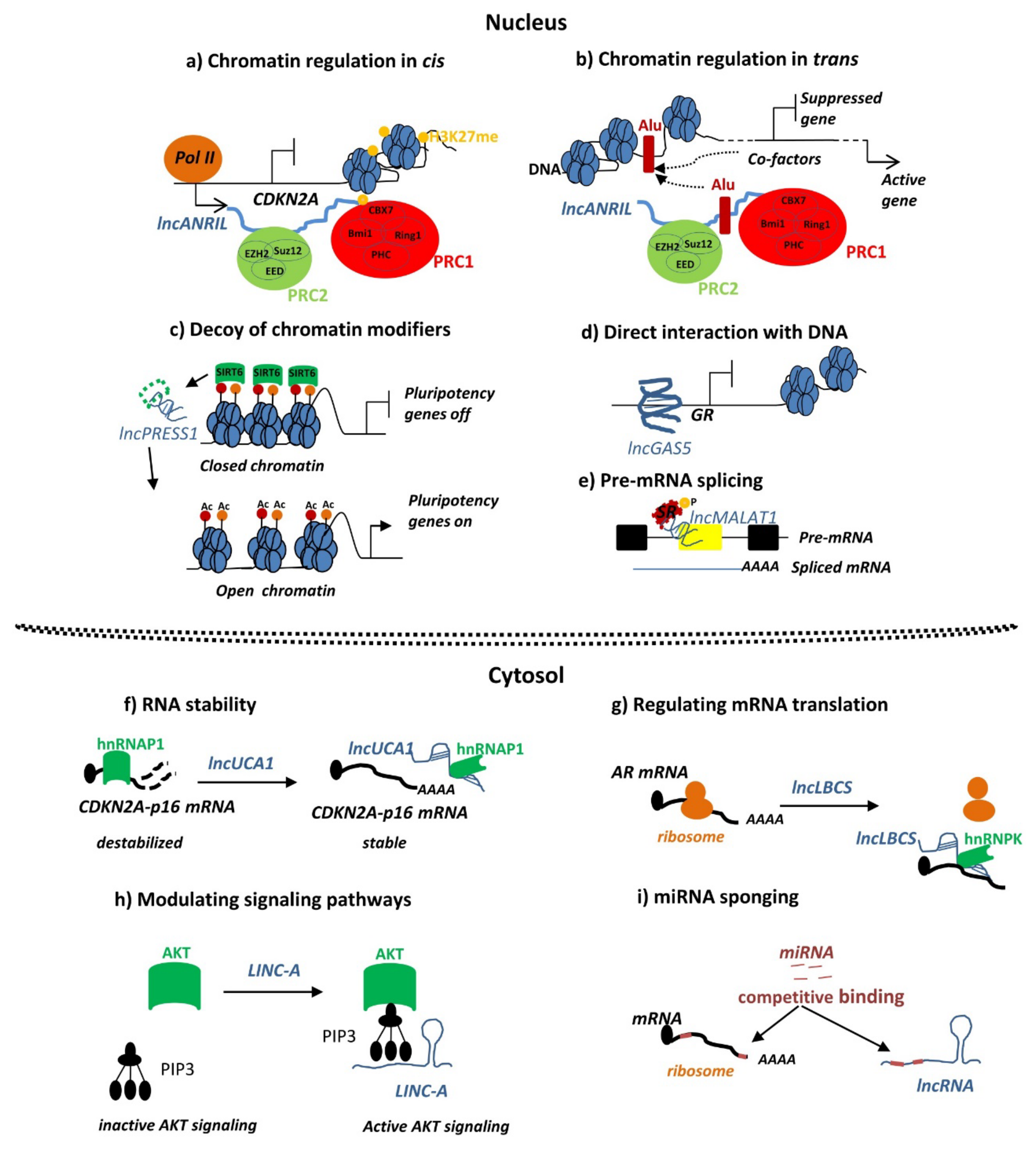
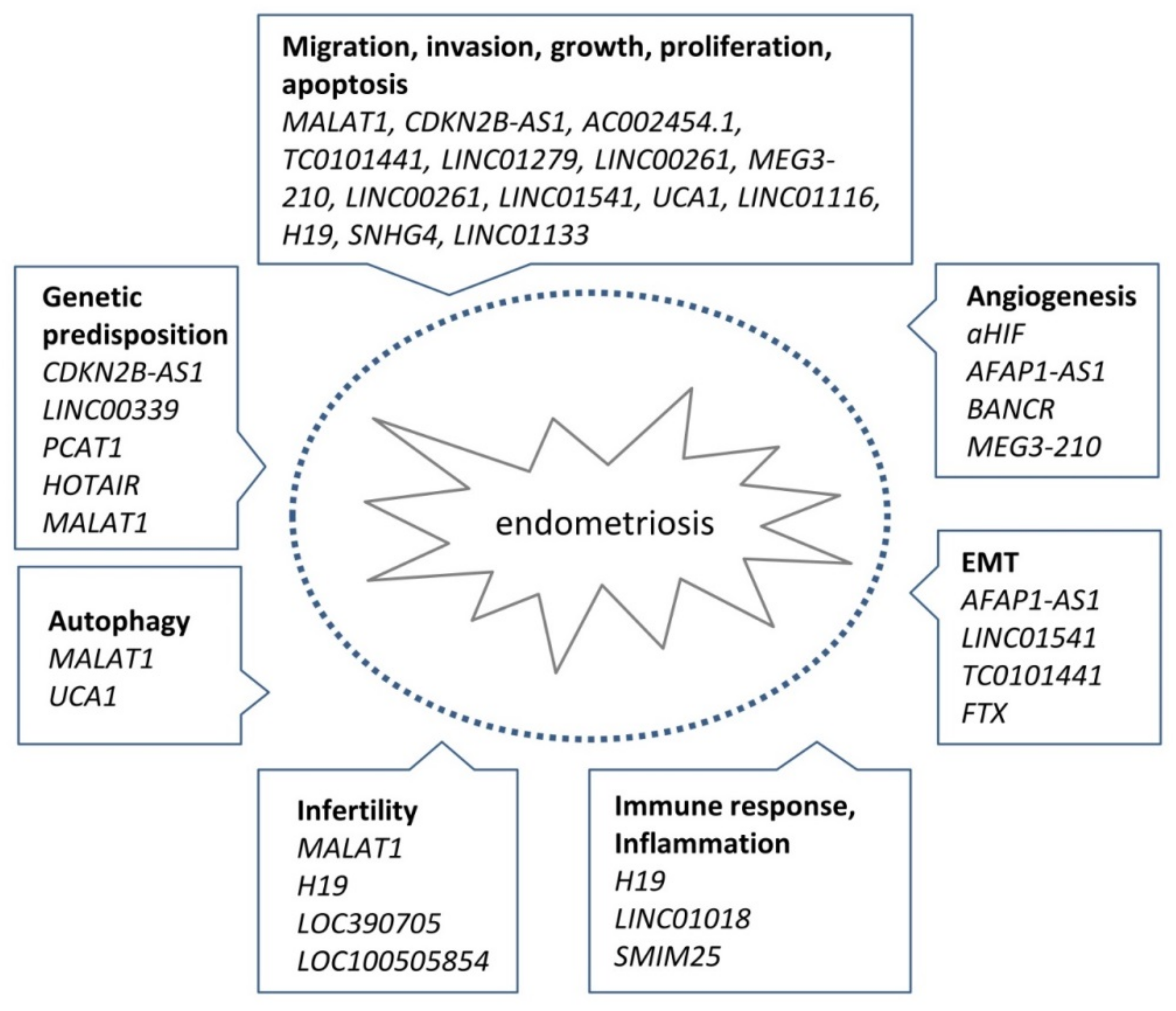
4. Functional Evidence for the Mechanism of lncRNA Action in Endometriosis

The diverse range of mechanisms for lncRNA action described above show what is possible, but so far, only a subset of these mechanisms have been described in the context of endometriosis. Based on the current knowledge, the mechanism of lncRNA action in endometriosis can be divided into the following groups: (i) lncRNAs that recruit and target chromatin remodeling or transcriptional regulatory factors, (ii) lncRNAs with miRNA sponging functions, and (iii) lncRNAs that modulate cellular signaling pathways.

4.1. Chromatin Remodeling and Transcriptional Control by lncRNAs in Endometriosis

The HOTAIR lncRNA is known as a critical regulator of HOX gene expression. Maintaining appropriate expression of genes within the HOX-gene network has been shown to be critical for endometrial homeostasis during embryonic implantation, and alterations in HOXA10 and HOXA11 expression have been associated with endometriosis-associated infertility [49,50]. Mechanistically, HOTAIR interacts with the PRC2 or REST/CoREST chromatin remodeling complex to guide the recruitment of H3K27 tri-methylation or H3K4-demethylation at target gene loci resulting in target gene silencing [51]. As described above, it has been shown that genetic variations at SNPs sites in HOTAIR are relatively frequently detected in patients with endometriosis [30]. These genetic changes alter the thermostability of mature HOTAIR leading to epigenetic silencing of HOXD10 and HOXA5 genes and are associated with more advanced endometriosis [30].
Overexpression of the lncRNA AFAP1-AS1 has been shown to be associated with ovarian endometriosis [33]. In another example of direct transcriptional regulation by an lncRNA in endometriosis, AFAP1-AS1 directly activates expression of EMT-related transcription factor ZEB1 in vitro [47]. ZEB1 has been previously shown to be involved in 17β-estradiol-induced EMT in endometriosis [52], indicating a possible mechanism by which AFAP1-AS1 could affect endometriosis.

4.2. MiRNA Sponging by LncRNAs in Endometriosis

A growing body of evidence indicates that lncRNAs can act as miRNA sponges in endometriosis (summarized in Table 2). In these cases, a binding site for the miRNA is present in both the lncRNA and the targeted protein-coding gene, and there is a correlation between expression of the lncRNA and the protein-coding gene. In the first reported example of sponging in endometriosis, decreased levels of the H19 lncRNA was shown to be associated with an increase in let-7 miRNA activity, which inhibits IGF1R expression resulting in the reduced proliferation of endometrial stroma cells [53]. These results suggested that the H19/let7/IGF1R pathway may contribute to impaired endometrial receptivity in women suffering from the disease. H19 was also shown to regulate cell proliferation and invasion of ectopic endometrial cells by increasing ITGB3 expression via sponging of miR-124-3p [54]. H19 has also been implicated in the impaired immune responses of women with the disease, by acting as a sponge for miR-342-3p, which regulates the IER3 pathway. This pathway has been implicated in Th-17 cell differentiation and endometrial stroma cell proliferation in ectopic sites in women with the disease [55]. Another lncRNA that has been shown to act as a sponge in endometriosis is CDKN2B-AS1, which acts as a regulator of AKT3 expression by sponging miR-424-5p in an in vitro model of ovarian endometriosis [56]. In another example, LINC01116 promoted the proliferation and migration of endometrial stroma cells by targeting FOXP1 via sponging of miR-9-5p, thereby promoting endometriosis lesion formation and growth [57]. MALAT1 was identified as a sponge of miR-200c involved in the regulation of endometriosis stoma cell proliferation and migration by promoting ZEB1 and ZEB2 expression in women with the disease [58]. This regulation is not restricted to miR-200c and may include the entire miR-200 family, consisting of miR-200a, miR-200b, miR-200c, miR-141, and miR-429 [59]. In addition to these examples, other lncRNAs have also been implicated in endometriosis via their role as sponges for miRNAs, as summarized in Table 2.

4.3. LncRNAs Modulating Cellular Signaling Pathways in Endometriosis

Cell signaling pathways have a pivotal role in the regulation of a variety of cellular processes in response to intracellular or extracellular stimuli. The regulation of components of a signaling pathway by lncRNAs can be direct or indirect and result in functional changes in the signaling cascades. Direct regulation can be achieved by direct binding of the lncRNA to signaling proteins leading to changes in either their free cellular levels or their activity. We define indirect regulation as cases where no direct lncRNA binding to signaling molecules has been shown and where the lncRNA is thought to alter the transcription of genes associated with the signaling pathway resulting in an altered cellular response. In Table 3, we summarize currently known cases where a lncRNA directly or indirectly affects a signaling pathway in endometriosis.
In an example of direct regulation of a signaling pathway in endometriosis, the lncRNA MEG3-210 has been shown to regulate endometriosis stromal cell migration, invasion, and apoptosis through the p38 MAPK and PKA/SERCA2 signaling pathways. In this case, MEG3-210 directly interacts with Galectin-1 in vitro and affects the growth of endometriotic lesions in vivo in a murine model of the disease [64]. Mechanistically, MEG3-210 titrates away the cellular levels of Galectin-1, preventing its action on the p38 MAPK and PKA/SERCA2 signaling cascades in endometrial stromal cells. In endometriosis, the levels of MEG3-210 are downregulated and the levels of free Galectin-1 are upregulated, which is associated with the subsequent activation of p38 MAPK signaling–mediated phosphorylation of ATF2. Activated ATF2 increases the expression of BCL-2 and MMP contributing to the anti-apoptotic, pro-migratory, and invasive phenotype of endometriosis cells. Simultaneously, MEG3-210 downregulation leads to suppression of the PKA/SERCA2 signaling cascade [64].
In an example of indirect regulation of signaling pathway, knockdown of MALAT1 lncRNA in endometriosis cells leads to enhanced cell death, reduced migration and invasion associated with activation of Caspase-3, and downregulation of MMP-9 and the NFkB/iNOS signaling pathway (Figure 3a) [65]. In contrast to endometriosis tissue where MALAT1 is overexpressed, in the granulosa cells of women with endometriosis MALAT1 expression is reduced [66]. This is associated with a reduced follicle count, due to impaired cell proliferation resulting from ERK/MAPK-dependent p21/p53 cell cycle arrest. This implicates altered expression of MALAT1 in endometriosis-related infertility. In cultured primary endometrial stromal cells, depletion of MALAT1 by siRNA knockdown results in the suppression of hypoxia-induced autophagy, as indicated by a reduction in the expression of autophagy markers Beclin-1 and LC3-II [67]. In this signaling cascade, expression of MALAT1 is regulated by the HIF1α transcription factor, known to be overexpressed in endometriosis lesions and to regulate multiple gene targets in response to hypoxia (Figure 3a) (reviewed in [76]).
In another example, the most downregulated lncRNA in ectopic tissue of women with ovarian endometriosis was LINC01541 [33], which has been shown respond to levels of estradiol [68]. A gene-targeting in vitro study in human endometrial stromal cells showed that the cellular levels of LINC01541 affect the activity of WNT/β-catenin, pro- and anti-apoptotic signaling regulators Caspase-3 and BCL2 and the levels of VEGFA production [68].
The presence of extracellular lncRNA in exosomes or microvesicles raises the possibility that these lncRNAs may be able to serve as extracellular signals for cell signaling regulation in endometriosis. This was supported by reports that exosomal lncRNAs promote angiogenesis in endometriosis [77]. Based on an in vitro co-culture model and patient serum analysis, the authors developed a novel mechanistic model explaining how endometriosis stromal cells of the lesion induce angiogenesis. They suggested that these cells in an ectopic environment can produce exosomes that are enriched in aHIF lncRNA, a pro-angiogenic lncRNA, highly expressed in ectopic endometrial stromal tissue. These aHIF-rich exosomes are then taken up by recipient macrovascular cells, where they cause upregulation of the angiogenesis-related genes VEGF-A, VEGF-D, and bFGF.
Endometriosis is an estrogen-dependent disease, and therefore, lncRNAs involved in the estrogen pathway or those targeted by estrogen signaling could play a role in the disease. The lncRNA H19 is positively regulated by estrogen, and its expression in the endometrium increases during the proliferative stage of the menstrual cycle [78,79]. H19 has been shown regulate several pathways that are relevant in endometriosis including IGF1R, ITGB3, IER3, and ACTA2 [53,54,55,80]. Another lncRNA, steroid receptor RNA activator 1 (SRA1) lncRNA, has been reported to act in concert with SRA1 to regulate the expression of estrogen receptors by affecting alternative splicing, and thereby the growth of stromal cells in ovarian endometriosis [81]. These examples illustrate how lncRNAs could play a role in endometriosis via the estrogen pathway or as targets of estrogen regulation.
Conceptually, the mechanism of lncRNAs’ action in endometriosis may involve influencing cellular pathways, where one lncRNA may regulate several targets using different molecular strategies (Figure 3a) or one targeted signaling pathway may be affected by multiple lncRNAs (Figure 3b). To better understand this complexity, integrative in silico and experimental analyses interrogating the molecular networks in endometriosis cells need to be applied. This approach should help to dissect the relationship between lncRNA, mRNA, miRNA expression, genetic- and epigenetic-driven chromatin remodeling, and signal transduction activity and to enable lncRNA target identification. The use of such in silico bioinformatics algorithms for cellular network construction in endometriosis has already been attempted by several studies [41,43,46]. For example, Wang et al. [41] constructed an lncRNA–miRNA–mRNA network, revealing lncRNAs that act as competing endogenous RNAs (ceRNA), the miRNAs they sponge, and the target genes that were involved in regulating endometrial receptivity in endometriosis. Moreover, Jiang et al. [43] identified the lncRNAs (SMIM25, LINC01018), miRNA (miR-182-5p), and mRNA (CHLI) as part of a ceRNA network implicated in the regulation of immune responses in endometriosis. These in silico studies may be valuable to predict how lncRNAs may function in endometriosis, but experimental validation is required in order for these predictions to be confirmed.

5. Summary and Perspectives

In recent years, a growing number of studies have implicated the abnormal expression of specific lncRNAs with various aspects of endometriosis pathogenesis. This altered expression may be due to genetic predisposition or an unknown environmental trigger and affect pathogenetic processes including EMT, endometriosis cell stemness, angiogenesis, lesion establishment and growth, endometriosis cell survival, proliferation and invasion, oxidative stress, autophagy, and endometrial receptivity (summarized in Figure 4). Evidence for the role of lncRNAs has mainly come from large-scale transcriptome studies comparing normal and diseased tissue, followed by validation studies of a small number of candidate lncRNAs. These studies can be valuable for uncovering potential novel players in endometriosis but often have weaknesses that should be considered including small sample size, incomplete clinical information on patient samples, and inappropriate controls (Table 1, Table S1). A general weakness of these studies is that they do not assess the complexity of the disease to take into account different lesion types, the stage or severity of the disease, or the effect of hormone status or age of the patient. The vast majority of lncRNAs revealed in these studies remain unvalidated by an independent method or by functional studies. One challenge for future work is to prioritize lncRNAs identified in these studies for validation and then conduct further functional studies to determine their role in endometriosis and their possible use as biomarkers or targets for treatment.
One avenue of endometriosis research that should be pursued in the future is the connection between genetic variants and abnormal lncRNA expression that affects aspects of endometriosis pathogenesis. The majority of disease-associated SNPs in GWAS studies, including those investigating endometriosis, are in non-coding regions. The hypothesis that these SNPs may affect lncRNA function is supported by a number of studies in endometriosis, with SNPs in or near lncRNAs including HOTAIR, MALAT1, CDKN2B-AS, PCAT1, and LINC00339 affecting their expression and regulation of genes and the endometriosis phenotype [28,29,30,31,32]. The possibility that other non-coding SNPs associated with endometriosis may also affect the disease via lncRNAs should be investigated.
In vitro cell culture models and in vivo animal models are valuable systems that enable the mechanism of action and function of individual lncRNAs to be investigated experimentally. However, a weakness of some studies investigating endometriosis, including those investigating the role of lncRNAs, is that the available in vitro and in vivo models may not always represent all aspects of the disease accurately. Therefore, it should be a priority for the field to continue to strive for improved models to investigate the disease.
To conclude, genome-wide genomic and transcriptomic studies have implicated numerous lncRNAs in a wide range of diseases, including in endometriosis. The challenge remains to distinguish the lncRNAs that play a relevant role in the disease from those that are merely associated with the transcriptional changes in the disease and to functionally show their role.

Supplementary Materials

The following are available online at https://www.mdpi.com/article/10.3390/ijms222111425/s1.

Author Contributions

Conceptualization, Q.J.H., K.P. and I.Y.; methodology, Q.J.H., K.P. and I.Y.; data curation, Q.J.H., K.P. and I.Y.; writing—original draft preparation, Q.J.H., K.P. and I.Y.; writing—review and editing, A.P., L.K., H.H. and R.W.; visualization, Q.J.H., K.P. and I.Y.; supervision, I.Y.; funding acquisition, H.H. All authors have read and agreed to the published version of the manuscript.

Funding

This work was co-funded by the Ingrid Flick Foundation (grant no. FA751C0801), which played no role in the review design and analysis, decision to publish, or preparation of the manuscript.

Data Availability Statement

Not applicable.

Conflicts of Interest

The authors declare no conflict of interest.

References

  1. Sampson, J.A. Peritoneal Endometriosis Due to the Menstrual Dissemination of Endometrial Tissue into the Peritoneal Cavity. Am. J. Obstet. Gynecol. 1927, 14, 442–469. [Google Scholar] [CrossRef]
  2. Rei, C.; Williams, T.; Feloney, M. Endometriosis in a Man as a Rare Source of Abdominal Pain: A Case Report and Review of the Literature. Case Rep. Obstet. Gynecol. 2018, 2018, 2083121. [Google Scholar] [CrossRef] [PubMed] [Green Version]
  3. Zondervan, K.T.; Becker, C.M.; Missmer, S.A. Endometriosis. N. Engl. J. Med. 2020, 382, 1244–1256. [Google Scholar] [CrossRef] [PubMed]
  4. Shigesi, N.; Kvaskoff, M.; Kirtley, S.; Feng, Q.; Fang, H.; Knight, J.C.; Missmer, S.A.; Rahmioglu, N.; Zondervan, K.T.; Becker, C.M. The association between endometriosis and autoimmune diseases: A systematic review and meta-analysis. Hum. Reprod. Update 2019, 25, 486–503. [Google Scholar] [CrossRef] [PubMed]
  5. Statello, L.; Guo, C.-J.; Chen, L.-L.; Huarte, M. Gene regulation by long non-coding RNAs and its biological functions. Nat. Rev. Mol. Cell Biol. 2021, 22, 96–118. [Google Scholar] [CrossRef] [PubMed]
  6. Gil, N.; Ulitsky, I. Regulation of gene expression by cis-acting long non-coding RNAs. Nat. Rev. Genet. 2020, 21, 102–117. [Google Scholar] [CrossRef] [PubMed]
  7. Delás, M.J.; Hannon, G.J. lncRNAs in development and disease: From functions to mechanisms. Open Biol. 2017, 7, 170121. [Google Scholar] [CrossRef] [PubMed] [Green Version]
  8. Yap, K.L.; Li, S.; Munoz-Cabello, A.M.; Raguz, S.; Zeng, L.; Mujtaba, S.; Gil, J.; Walsh, M.J.; Zhou, M.M. Molecular interplay of the noncoding RNA ANRIL and methylated histone H3 lysine 27 by polycomb CBX7 in transcriptional silencing of INK4a. Mol. Cell 2010, 38, 662–674. [Google Scholar] [CrossRef] [PubMed] [Green Version]
  9. Holdt, L.M.; Hoffmann, S.; Sass, K.; Langenberger, D.; Scholz, M.; Krohn, K.; Finstermeier, K.; Stahringer, A.; Wilfert, W.; Beutner, F.; et al. Alu elements in ANRIL non-coding RNA at chromosome 9p21 modulate atherogenic cell functions through trans-regulation of gene networks. PLoS Genet. 2013, 9, e1003588. [Google Scholar] [CrossRef] [PubMed]
  10. Jain, A.K.; Xi, Y.; McCarthy, R.; Allton, K.; Akdemir, K.C.; Patel, L.R.; Aronow, B.; Lin, C.; Li, W.; Yang, L.; et al. LncPRESS1 Is a p53-Regulated LncRNA that Safeguards Pluripotency by Disrupting SIRT6-Mediated De-acetylation of Histone H3K56. Mol. Cell 2016, 64, 967–981. [Google Scholar] [CrossRef] [PubMed] [Green Version]
  11. Kino, T.; Hurt, D.E.; Ichijo, T.; Nader, N.; Chrousos, G.P. Noncoding RNA Gas5 Is a Growth Arrest– and Starvation-Associated Repressor of the Glucocorticoid Receptor. Sci. Signal. 2010, 3, ra8. [Google Scholar] [CrossRef] [PubMed] [Green Version]
  12. Tripathi, V.; Ellis, J.D.; Shen, Z.; Song, D.Y.; Pan, Q.; Watt, A.T.; Freier, S.M.; Bennett, C.F.; Sharma, A.; Bubulya, P.A.; et al. The nuclear-retained noncoding RNA MALAT1 regulates alternative splicing by modulating SR splicing factor phosphorylation. Mol. Cell 2010, 39, 925–938. [Google Scholar] [CrossRef] [PubMed] [Green Version]
  13. Kumar, P.P.; Emechebe, U.; Smith, R.; Franklin, S.; Moore, B.; Yandell, M.; Lessnick, S.L.; Moon, A.M. Coordinated control of senescence by lncRNA and a novel T-box3 co-repressor complex. Elife 2014, 3, e02805. [Google Scholar] [CrossRef] [PubMed]
  14. Gu, P.; Chen, X.; Xie, R.; Xie, W.; Huang, L.; Dong, W.; Han, J.; Liu, X.; Shen, J.; Huang, J.; et al. A novel AR translational regulator lncRNA LBCS inhibits castration resistance of prostate cancer. Mol. Cancer 2019, 18, 109. [Google Scholar] [CrossRef] [PubMed]
  15. Lin, A.; Hu, Q.; Li, C.; Xing, Z.; Ma, G.; Wang, C.; Li, J.; Ye, Y.; Yao, J.; Liang, K.; et al. The LINK-A lncRNA interacts with PtdIns(3,4,5)P3 to hyperactivate AKT and confer resistance to AKT inhibitors. Nat. Cell Biol. 2017, 19, 238–251. [Google Scholar] [CrossRef] [PubMed]
  16. Gong, C.; Maquat, L.E. lncRNAs transactivate STAU1-mediated mRNA decay by duplexing with 3′ UTRs via Alu elements. Nature 2011, 470, 284–288. [Google Scholar] [CrossRef] [PubMed] [Green Version]
  17. Liu, W.; Yao, D.; Huang, B. LncRNA PVT1 promotes cervical cancer progression by sponging miR-503 to upregulate ARL2 expression. Open Life Sci. 2021, 16, 1–13. [Google Scholar] [CrossRef] [PubMed]
  18. Jiang, M.C.; Ni, J.J.; Cui, W.Y.; Wang, B.Y.; Zhuo, W. Emerging roles of lncRNA in cancer and therapeutic opportunities. Am. J. Cancer Res. 2019, 9, 1354–1366. [Google Scholar] [PubMed]
  19. Policarpo, R.; Sierksma, A.; De Strooper, B.; d’Ydewalle, C. From Junk to Function: LncRNAs in CNS Health and Disease. Front. Mol. Neurosci. 2021, 14, 151. [Google Scholar] [CrossRef] [PubMed]
  20. Yu, B.; Wang, S. Angio-LncRs: LncRNAs that regulate angiogenesis and vascular disease. Theranostics 2018, 8, 3654–3675. [Google Scholar] [CrossRef] [PubMed]
  21. He, X.; Ou, C.; Xiao, Y.; Han, Q.; Li, H.; Zhou, S. LncRNAs: Key players and novel insights into diabetes mellitus. Oncotarget 2017, 8, 71325–71341. [Google Scholar] [CrossRef] [PubMed] [Green Version]
  22. Sedano, M.J.; Harrison, A.L.; Zilaie, M.; Das, C.; Choudhari, R.; Ramos, E.; Gadad, S.S. Emerging Roles of Estrogen-Regulated Enhancer and Long Non-Coding RNAs. Int. J. Mol. Sci. 2020, 21, 3711. [Google Scholar] [CrossRef] [PubMed]
  23. Basak, P.; Chatterjee, S.; Bhat, V.; Su, A.; Jin, H.; Lee-Wing, V.; Liu, Q.; Hu, P.; Murphy, L.C.; Raouf, A. Long Non-Coding RNA H19 Acts as an Estrogen Receptor Modulator that is Required for Endocrine Therapy Resistance in ER+ Breast Cancer Cells. Cell. Physiol. Biochem. 2018, 51, 1518–1532. [Google Scholar] [CrossRef] [PubMed]
  24. Sapkota, Y.; Steinthorsdottir, V.; Morris, A.P.; Fassbender, A.; Rahmioglu, N.; De Vivo, I.; Buring, J.E.; Zhang, F.; Edwards, T.L.; Jones, S.; et al. Meta-analysis identifies five novel loci associated with endometriosis highlighting key genes involved in hormone metabolism. Nat. Commun. 2017, 8, 15539. [Google Scholar] [CrossRef] [PubMed] [Green Version]
  25. Gallagher, C.S.; Mäkinen, N.; Harris, H.R.; Rahmioglu, N.; Uimari, O.; Cook, J.P.; Shigesi, N.; Ferreira, T.; Velez-Edwards, D.R.; Edwards, T.L.; et al. Genome-wide association and epidemiological analyses reveal common genetic origins between uterine leiomyomata and endometriosis. Nat. Commun. 2019, 10, 4857. [Google Scholar] [CrossRef] [PubMed] [Green Version]
  26. Giral, H.; Landmesser, U.; Kratzer, A. Into the Wild: GWAS Exploration of Non-coding RNAs. Front. Cardiovasc. Med. 2018, 5, 181. [Google Scholar] [CrossRef] [PubMed]
  27. Mirza, A.H.; Kaur, S.; Brorsson, C.A.; Pociot, F. Effects of GWAS-associated genetic variants on lncRNAs within IBD and T1D candidate loci. PLoS ONE 2014, 9, e105723. [Google Scholar] [CrossRef] [PubMed] [Green Version]
  28. Lee, G.H.; Choi, Y.M.; Hong, M.A.; Yoon, S.H.; Kim, J.J.; Hwang, K.; Chae, S.J. Association of CDKN2B-AS and WNT4 genetic polymorphisms in Korean patients with endometriosis. Fertil. Steril. 2014, 102, 1393–1397. [Google Scholar] [CrossRef] [PubMed]
  29. Powell, J.E.; Fung, J.N.; Shakhbazov, K.; Sapkota, Y.; Cloonan, N.; Hemani, G.; Hillman, K.M.; Kaufmann, S.; Luong, H.T.; Bowdler, L.; et al. Endometriosis risk alleles at 1p36.12 act through inverse regulation of CDC42 and LINC00339. Hum. Mol. Genet. 2016, 25, 5046–5058. [Google Scholar] [PubMed] [Green Version]
  30. Chang, C.Y.; Tseng, C.C.; Lai, M.T.; Chiang, A.J.; Lo, L.C.; Chen, C.M.; Yen, M.J.; Sun, L.; Yang, L.; Hwang, T.; et al. Genetic impacts on thermostability of onco-lncRNA HOTAIR during the development and progression of endometriosis. PLoS ONE 2021, 16, e0248168. [Google Scholar] [CrossRef] [PubMed]
  31. Chen, G.; Zhang, M.; Liang, Z.; Chen, S.; Chen, F.; Zhu, J.; Zhao, M.; Xu, C.; He, J.; Hua, W.; et al. Association of polymorphisms in MALAT1 with the risk of endometriosis in Southern Chinese women. Biol. Reprod. 2019, 102, 943–949. [Google Scholar] [CrossRef] [PubMed]
  32. Wang, L.; Xing, Q.; Feng, T.; He, M.; Yu, W.; Chen, H. SNP rs710886 A>G in long noncoding RNA PCAT1 is associated with the risk of endometriosis by modulating expression of multiple stemness-related genes via microRNA-145 signaling pathway. J. Cell Biochem. 2020, 121, 1703–1715. [Google Scholar] [CrossRef] [PubMed]
  33. Sun, P.R.; Jia, S.Z.; Lin, H.; Leng, J.H.; Lang, J.H. Genome-wide profiling of long noncoding ribonucleic acid expression patterns in ovarian endometriosis by microarray. Fertil. Steril. 2014, 101, 1038–1346. [Google Scholar] [CrossRef] [PubMed]
  34. Cai, H.; Zhu, X.; Li, Z.; Zhu, Y.; Lang, J. lncRNA/mRNA profiling of endometriosis rat uterine tissues during the implantation window. Int. J. Mol. Med. 2019, 44, 2145–2160. [Google Scholar] [CrossRef] [PubMed] [Green Version]
  35. Wang, W.T.; Sun, Y.M.; Huang, W.; He, B.; Zhao, Y.N.; Chen, Y.Q. Genome-wide Long Non-coding RNA Analysis Identified Circulating LncRNAs as Novel Non-invasive Diagnostic Biomarkers for Gynecological Disease. Sci. Rep. 2016, 6, 23343. [Google Scholar] [CrossRef] [PubMed]
  36. Cui, D.; Ma, J.; Liu, Y.; Lin, K.; Jiang, X.; Qu, Y.; Lin, J.; Xu, K. Analysis of long non-coding RNA expression profiles using RNA sequencing in ovarian endometriosis. Gene 2018, 673, 140–148. [Google Scholar] [CrossRef] [PubMed]
  37. Wang, Y.; Li, Y.; Yang, Z.; Liu, K.; Wang, D. Genome-Wide Microarray Analysis of Long Non-Coding RNAs in Eutopic Secretory Endometrium with Endometriosis. Cell Physiol. Biochem. 2015, 37, 2231–2245. [Google Scholar] [CrossRef] [PubMed]
  38. Zhang, C.; Wu, W.; Ye, X.; Ma, R.; Luo, J.; Zhu, H.; Chang, X. Aberrant expression of CHL1 gene and long non-coding RNA CHL1-AS1, CHL1-AS2 in ovarian endometriosis. Eur. J. Obstet. Gynecol. Reprod. Biol. 2019, 236, 177–182. [Google Scholar] [CrossRef] [PubMed]
  39. Wu, J.; Xia, X.; Hu, Y.; Fang, X.; Orsulic, S. Identification of Infertility-Associated Topologically Important Genes Using Weighted Co-expression Network Analysis. Front. Genet. 2021, 12, 580190. [Google Scholar] [CrossRef] [PubMed]
  40. Liu, J.; Wang, Q.; Zhang, R.; Zhang, C.; Lin, J.; Huang, X. Identification of LINC01279 as a cell cycle-associated long non-coding RNA in endometriosis with GBA analysis. Mol. Med. Rep. 2018, 18, 3850–3858. [Google Scholar] [CrossRef] [PubMed] [Green Version]
  41. Wang, X.; Yu, Q. Endometriosis-related ceRNA network to identify predictive biomarkers of endometrial receptivity. Epigenomics 2019, 11, 147–167. [Google Scholar] [CrossRef] [PubMed]
  42. Liu, S.-P.; Tian, X.; Cui, H.-Y.; Zhang, Q.; Hua, K.-Q. The messenger RNA and long non-coding RNA expression profiles in ectopic and eutopic endometrium provide novel insights into endometriosis. Reprod. Dev. Med. 2019, 3, 11. [Google Scholar]
  43. Jiang, L.; Zhang, M.; Wang, S.; Xiao, Y.; Wu, J.; Zhou, Y.; Fang, X. LINC01018 and SMIM25 sponged miR-182-5p in endometriosis revealed by the ceRNA network construction. Int. J. Immunopathol. Pharmacol. 2020, 34, 2058738420976309. [Google Scholar] [CrossRef] [PubMed]
  44. Zhang, M.; Li, J.; Duan, S.; Fang, Z.; Tian, J.; Yin, H.; Zhai, Q.; Wang, X.; Zhang, L. Comprehensive characterization of endometrial competing endogenous RNA network in infertile women of childbearing age. Aging 2020, 12, 4204–4221. [Google Scholar] [CrossRef] [PubMed]
  45. Bi, J.; Wang, D.; Cui, L.; Yang, Q. RNA sequencing-based long non-coding RNA analysis and immunoassay in ovarian endometriosis. Am. J. Reprod. Immunol. 2021, 85, e13359. [Google Scholar] [CrossRef] [PubMed]
  46. Bai, J.; Wang, B.; Wang, T.; Ren, W. Identification of Functional lncRNAs Associated With Ovarian Endometriosis Based on a ceRNA Network. Front. Genet. 2021, 12, 534054. [Google Scholar] [CrossRef] [PubMed]
  47. Lin, D.; Huang, Q.; Wu, R.; Dai, S.; Huang, Z.; Ren, L.; Huang, S.; Chen, Q. Long non-coding RNA AFAP1-AS1 promoting epithelial-mesenchymal transition of endometriosis is correlated with transcription factor ZEB1. Am. J. Reprod. Immunol. 2019, 81, e13074. [Google Scholar] [CrossRef] [PubMed]
  48. Liu, S.; Xin, W.; Tang, X.; Qiu, J.; Zhang, Y.; Hua, K. LncRNA H19 Overexpression in Endometriosis and its Utility as a Novel Biomarker for Predicting Recurrence. Reprod. Sci. 2020, 27, 1687–1697. [Google Scholar] [CrossRef] [PubMed]
  49. Du, H.; Taylor, H.S. The Role of Hox Genes in Female Reproductive Tract Development, Adult Function, and Fertility. Cold Spring Harb. Perspect. Med. 2015, 6, a023002. [Google Scholar] [CrossRef] [PubMed] [Green Version]
  50. Liu, X.H.; Liu, Z.L.; Sun, M.; Liu, J.; Wang, Z.X.; De, W. The long non-coding RNA HOTAIR indicates a poor prognosis and promotes metastasis in non-small cell lung cancer. BMC Cancer 2013, 13, 464. [Google Scholar] [CrossRef] [PubMed] [Green Version]
  51. Tsai, M.C.; Manor, O.; Wan, Y.; Mosammaparast, N.; Wang, J.K.; Lan, F.; Shi, Y.; Segal, E.; Chang, H.Y. Long noncoding RNA as modular scaffold of histone modification complexes. Science 2010, 329, 689–693. [Google Scholar] [CrossRef] [PubMed] [Green Version]
  52. Wu, R.F.; Chen, Z.X.; Zhou, W.D.; Li, Y.Z.; Huang, Z.X.; Lin, D.C.; Ren, L.L.; Chen, Q.X.; Chen, Q.H. High expression of ZEB1 in endometriosis and its role in 17beta-estradiol-induced epithelial-mesenchymal transition. Int. J. Clin. Exp. Pathol. 2018, 11, 4744–4758. [Google Scholar] [PubMed]
  53. Ghazal, S.; McKinnon, B.; Zhou, J.; Mueller, M.; Men, Y.; Yang, L.; Mueller, M.; Flannery, C.; Huang, Y.; Taylor, H.S. H19 lncRNA alters stromal cell growth via IGF signaling in the endometrium of women with endometriosis. EMBO Mol. Med. 2015, 7, 996–1003. [Google Scholar] [CrossRef] [PubMed]
  54. Liu, S.; Qiu, J.; Tang, X.; Cui, H.; Zhang, Q.; Yang, Q. LncRNA-H19 regulates cell proliferation and invasion of ectopic endometrium by targeting ITGB3 via modulating miR-124-3p. Exp. Cell Res. 2019, 381, 215–222. [Google Scholar] [CrossRef] [PubMed]
  55. Liu, Z.; Liu, L.; Zhong, Y.; Cai, M.; Gao, J.; Tan, C.; Han, X.; Guo, R.; Han, L. LncRNA H19 over-expression inhibited Th17 cell differentiation to relieve endometriosis through miR-342-3p/IER3 pathway. Cell Biosci. 2019, 9, 84. [Google Scholar] [CrossRef] [PubMed] [Green Version]
  56. Wang, S.; Yi, M.; Zhang, X.; Zhang, T.; Jiang, L.; Cao, L.; Zhou, Y.; Fang, X. Effects of CDKN2B-AS1 on cellular proliferation, invasion and AKT3 expression are attenuated by miR-424-5p in a model of ovarian endometriosis. Reprod. Biomed. Online 2021, 42, 1057–1066. [Google Scholar] [CrossRef] [PubMed]
  57. Cui, L.; Chen, S.; Wang, D.; Yang, Q. LINC01116 promotes proliferation and migration of endometrial stromal cells by targeting FOXP1 via sponging miR-9-5p in endometriosis. J. Cell Mol. Med. 2021, 25, 2000–2012. [Google Scholar] [CrossRef] [PubMed]
  58. Liang, Z.; Chen, Y.; Zhao, Y.; Xu, C.; Zhang, A.; Zhang, Q.; Wang, D.; He, J.; Hua, W.; Duan, P. miR-200c suppresses endometriosis by targeting MALAT1 in vitro and in vivo. Stem Cell Res. Ther. 2017, 8, 251. [Google Scholar] [CrossRef] [PubMed] [Green Version]
  59. Du, Y.; Zhang, Z.; Xiong, W.; Li, N.; Liu, H.; He, H.; Li, Q.; Liu, Y.; Zhang, L. Estradiol promotes EMT in endometriosis via MALAT1/miR200s sponge function. Reproduction 2019, 157, 179–188. [Google Scholar] [CrossRef] [PubMed] [Green Version]
  60. Xu, Z.; Zhang, L.; Yu, Q.; Zhang, Y.; Yan, L.; Chen, Z.J. The estrogen-regulated lncRNA H19/miR-216a-5p axis alters stromal cell invasion and migration via ACTA2 in endometriosis. Mol. Hum. Reprod. 2019, 25, 550–561. [Google Scholar] [CrossRef] [PubMed]
  61. Mai, H.; Xu, H.; Lin, H.; Wei, Y.; Yin, Y.; Huang, Y.; Huang, S.; Liao, Y. LINC01541 Functions as a ceRNA to Modulate the Wnt/beta-Catenin Pathway by Decoying miR-506-5p in Endometriosis. Reprod. Sci. 2021, 28, 665–674. [Google Scholar] [CrossRef] [PubMed]
  62. Liu, Y.; Huang, X.; Lu, D.; Feng, Y.; Xu, R.; Li, X.; Yin, C.; Xue, B.; Zhao, H.; Wang, S.; et al. LncRNA SNHG4 promotes the increased growth of endometrial tissue outside the uterine cavity via regulating c-Met mediated by miR-148a-3p. Mol. Cell Endocrinol. 2020, 514, 110887. [Google Scholar] [CrossRef] [PubMed]
  63. Feng, Y.; Tan, B.Z. LncRNA MALAT1 inhibits apoptosis of endometrial stromal cells through miR-126-5p-CREB1 axis by activating PI3K-AKT pathway. Mol. Cell Biochem. 2020, 475, 185–194. [Google Scholar] [CrossRef] [PubMed]
  64. Wang, H.; Sha, L.; Huang, L.; Yang, S.; Zhou, Q.; Luo, X.; Shi, B. LINC00261 functions as a competing endogenous RNA to regulate BCL2L11 expression by sponging miR-132-3p in endometriosis. Am. J. Transl. Res. 2019, 11, 2269–2279. [Google Scholar] [PubMed]
  65. Liu, Y.; Ma, J.; Cui, D.; Fei, X.; Lv, Y.; Lin, J. LncRNA MEG3-210 regulates endometrial stromal cells migration, invasion and apoptosis through p38 MAPK and PKA/SERCA2 signalling via interaction with Galectin-1 in endometriosis. Mol. Cell Endocrinol. 2020, 513, 110870. [Google Scholar] [CrossRef] [PubMed]
  66. Yu, J.; Chen, L.H.; Zhang, B.; Zheng, Q.M. The modulation of endometriosis by lncRNA MALAT1 via NF-kappaB/iNOS. Eur Rev. Med. Pharmacol. Sci. 2019, 23, 4073–4080. [Google Scholar] [PubMed]
  67. Li, Y.; Liu, Y.D.; Chen, S.L.; Chen, X.; Ye, D.S.; Zhou, X.Y.; Zhe, J.; Zhang, J. Down-regulation of long non-coding RNA MALAT1 inhibits granulosa cell proliferation in endometriosis by up-regulating P21 via activation of the ERK/MAPK pathway. Mol. Hum. Reprod. 2019, 25, 17–29. [Google Scholar] [CrossRef] [PubMed]
  68. Liu, H.; Zhang, Z.; Xiong, W.; Zhang, L.; Du, Y.; Liu, Y.; Xiong, X. Long non-coding RNA MALAT1 mediates hypoxia-induced pro-survival autophagy of endometrial stromal cells in endometriosis. J. Cell Mol. Med. 2019, 23, 439–452. [Google Scholar] [CrossRef] [PubMed] [Green Version]
  69. Zhan, L.; Wang, W.; Zhang, Y.; Song, E.; Fan, Y.; Wei, B. Hypoxia-inducible factor-1alpha: A promising therapeutic target in endometriosis. Biochimie 2016, 123, 130–137. [Google Scholar] [CrossRef] [PubMed]
  70. Mai, H.; Wei, Y.; Yin, Y.; Huang, S.; Lin, H.; Liao, Y.; Liu, X.; Chen, X.; Shi, H.; Liu, C.; et al. LINC01541 overexpression attenuates the 17beta-Estradiol-induced migration and invasion capabilities of endometrial stromal cells. Syst. Biol. Reprod. Med. 2019, 65, 214–222. [Google Scholar] [CrossRef] [PubMed]
  71. Qiu, J.J.; Lin, X.J.; Zheng, T.T.; Tang, X.Y.; Zhang, Y.; Hua, K.Q. The Exosomal Long Noncoding RNA aHIF is Upregulated in Serum From Patients With Endometriosis and Promotes Angiogenesis in Endometriosis. Reprod. Sci. 2019, 26, 1590–1602. [Google Scholar] [CrossRef] [PubMed]
  72. Yotova, I.; Hudson, Q.J.; Pauler, F.M.; Proestling, K.; Haslinger, I.; Kuessel, L.; Perricos, A.; Husslein, H.; Wenzl, R. LINC01133 Inhibits Invasion and Promotes Proliferation in an Endometriosis Epithelial Cell Line. Int. J. Mol. Sci. 2021, 22, 8385. [Google Scholar] [CrossRef] [PubMed]
  73. Wang, H.; Ni, C.; Xiao, W.; Wang, S. Role of lncRNA FTX in invasion, metastasis, and epithelial-mesenchymal transition of endometrial stromal cells caused by endometriosis by regulating the PI3K/Akt signaling pathway. Ann. Transl. Med. 2020, 8, 1504. [Google Scholar] [CrossRef] [PubMed]
  74. Zhu, M.B.; Chen, L.P.; Hu, M.; Shi, Z.; Liu, Y.N. Effects of lncRNA BANCR on endometriosis through ERK/MAPK pathway. Eur. Rev. Med. Pharmacol. Sci. 2019, 23, 6806–6812. [Google Scholar] [PubMed]
  75. Jiang, L.; Wan, Y.; Feng, Z.; Liu, D.; Ouyang, L.; Li, Y.; Liu, K. Long Noncoding RNA UCA1 Is Related to Autophagy and Apoptosis in Endometrial Stromal Cells. Front. Oncol. 2020, 10, 618472. [Google Scholar] [CrossRef] [PubMed]
  76. Liu, J.; Wang, Y.; Chen, P.; Ma, Y.; Wang, S.; Tian, Y.; Wang, A.; Wang, D. AC002454.1 and CDK6 synergistically promote endometrial cell migration and invasion in endometriosis. Reproduction 2019, 157, 535–543. [Google Scholar] [CrossRef] [PubMed]
  77. Zhang, C.; Wu, W.; Zhu, H.; Yu, X.; Zhang, Y.; Ye, X.; Cheng, H.; Ma, R.; Cui, H.; Luo, J.; et al. Knockdown of long noncoding RNA CCDC144NL-AS1 attenuates migration and invasion phenotypes in endometrial stromal cells from endometriosis†. Biol. Reprod. 2019, 100, 939–949. [Google Scholar] [CrossRef] [PubMed]
  78. Qiu, J.J.; Lin, Y.Y.; Tang, X.Y.; Ding, Y.; Yi, X.F.; Hua, K.Q. Extracellular vesicle-mediated transfer of the lncRNA-TC0101441 promotes endometriosis migration/invasion. Exp. Cell Res. 2020, 388, 111815. [Google Scholar] [CrossRef] [PubMed]
  79. Korucuoglu, U.; Biri, A.A.; Konac, E.; Alp, E.; Onen, I.H.; Ilhan, M.N.; Turkyilmaz, E.; Erdem, A.; Erdem, M.; Menevse, S. Expression of the imprinted IGF2 and H19 genes in the endometrium of cases with unexplained infertility. Eur. J. Obstet. Gynecol. Reprod. Biol. 2010, 149, 77–81. [Google Scholar] [CrossRef] [PubMed]
  80. Adriaenssens, E.; Lottin, S.; Dugimont, T.; Fauquette, W.; Coll, J.; Dupouy, J.P.; Boilly, B.; Curgy, J.J. Steroid hormones modulate H19 gene expression in both mammary gland and uterus. Oncogene 1999, 18, 4460–4473. [Google Scholar] [CrossRef] [PubMed] [Green Version]
  81. Lin, K.; Zhan, H.; Ma, J.; Xu, K.; Wu, R.; Zhou, C.; Lin, J. Silencing of SRA1 Regulates ER Expression and Attenuates the Growth of Stromal Cells in Ovarian Endometriosis. Reprod. Sci. 2017, 24, 836–843. [Google Scholar] [CrossRef] [PubMed]
Figure 1. Description of the general mechanisms of the lncRNA actions in the nucleus (ae) or in the cytosol (fi). (a) Regulation in cis by lncRNA ANRIL, which mediates polycomb repressive complex (PRC) 1 and 2 recruitment to the promoter of the neighboring CDKN2A and CDKN2B genes, thereby controlling their expression [8]. (b) Regulation in trans by ANRIL, which acts through Alu sequences to recruit PRC1 and PRC2 complexes to distant targets [9]. (c) The embryonic stem cell–specific lncRNA lncPRESS1 sequesters the histone deacetylase Sirtuin 6 (SIRT6) from the promoters of numerous pluripotency genes. LncPRESS1 keeps histone H3 acetylated, thereby activating the transcription of pluripotency genes. During differentiation or following depletion of lncPRESS1, SIRT6 localizes to the chromatin, blocking the transcription of pluripotency genes [10]. (d) The lncRNA GAS5 folds into a DNA-like structure and binds to the glucocorticoid receptor (GR), thereby inhibiting its transcriptional activity [11]. (e) The nuclear-retained lncRNA MALAT1 can regulate alternative splicing by modulating the phosphorylation of the SR splicing factor [12]. (f) In the cytosol, the lncRNA urothelial carcinoma associated 1 (UCA1) stabilizes CDKN2A-p16 mRNA by sequestering the heterogeneous nuclear ribonucleoprotein A1 (hnRNPA1) [13]. (g) LncRNA LBCS suppresses the androgen receptor (AR) translation efficiency by forming a complex with hnRNPK and AR mRNA [14]. (h) The lncRNA for kinase activation (LINK-A) directly interacts with the AKT pleckstrin homology domain and PIP3 facilitating AKT–PIP3 interaction and consequent enzymatic activation [15]. (i) The lncRNA PVT1 sponges miR-503 to upregulate ARL2 expression in cervical cancer [17].
Ijms 22 11425 g001
Figure 2. Differentially expressed (DE) lncRNAs in endometriosis based on validation studies: (a) Up- and downregulated lncRNAs in endometriosis tissues and/or cells (b) in body fluids or lesion microenvironment.
Ijms 22 11425 g002
Figure 3. (a) MALAT1 was identified as a sponge of miR-200c. This regulation is not restricted to miR-200c and might include the entire miR-200 family (miR-200s), consisting of miR-200a, miR-200b, miR-200c, miR-141, and miR-429. Upregulation of MALAT1 in women with endometriosis leads to enhanced sponging of miR200s and promotes zinc finger E-box binding homeobox transcription factor 1 (ZEB1) and ZEB2 expression leading to higher EMT. In HESCs, the lncRNA MALAT1 directly interacts with miR-126-5p, which regulates cAMP responsive element-binding protein (CREB1) expression. Upregulation of MALAT1 inhibits apoptosis probably via activation of the PI3K–AKT pathway through the miR-126-5p–CREB1 axis. MALAT1 lncRNA can also lead to reduced apoptosis in HESCs through the upregulation of the NFkB/iNOS signaling pathway activity, which also enhances migration and invasion of cells. In cultured primary endometrial stromal cells, MALAT1 leads to upregulation of hypoxia-induced autophagy. In this signaling cascade, regulation of MALAT1 expression is under the control of the HIF1α transcription factor. (b) In granulosa cells (GCs) of women with endometriosis, significant downregulation of MALAT1 expression was reported. MALAT1 knockdown induced an increase in phosphorylated ERK1/2 (p-ERK1/2) that was associated with altered follicle count, due to impaired cell proliferation resulting from ERK/MAPK-dependent activation of p21/p53 cell cycle arrest. In an autograft transplantation rat model of endometriosis, inhibition of the lncRNA BANCR led to a decrease in ectopic tissue volume associated with a significant reduction in serum levels of VEGF, MMP-2, and MMP-9, ERK, and MAPK mRNA and in phosphorylated ERK and MAPK protein levels in tissues. HESCs: human endometrial stromal cells.
Ijms 22 11425 g003
Figure 4. Phenotypical changes caused by altered expression of lncRNAs in endometriosis. Altered expression of lncRNAs in endometriosis is involved in the regulation of numerous processes known to be associated with the pathogenesis of the disease. These processes include EMT, endometriosis cell stemness, angiogenesis, lesion establishment and growth, endometriosis cell survival, proliferation and invasion, oxidative stress, autophagy, and endometrial receptivity.
Ijms 22 11425 g004
Table 2. LncRNAs involved in endometriosis as molecular sponges of miRNAs.
lncRNA (Reference)Sponged miRNATarget mRNA (Pathway) in Endometriosis
H19 [53]Let-7; miR-125-3p; miR-342-3p; miR-216a-5pIGF1R; ITGB3; IER3; ACTA2
CDKN2B-AS1 [56]miR-424-5pAKT3
LINC01541 [60]miR-506-3pWIF1 (Wnt/β-catenin)
LINC01116 [57]miR-9-5pFOXP1
SNHG4 [61]miR-148a-3pc-Met
LINC01018 [43] miR-182-5pCHLI (inflammatory)
SMIM25 [43]miR-182-5pCHLI (inflammatory)
MALAT1 [62]miR-126-5p; miR200s; miR200cCREB1 (PI3K/AKT);
ZEB1, ZEB2, VIM (EMT); ZEB1, ZEB2; CDH2 (EMT)
LINC00261 [63]miR-132-3pBCL2L11
PCAT1 [32]miR-145FASCIN1, SOX2, MSI2, SERPIN
Table 3. Mechanisms of cell signaling regulation via lncRNAs in endometriosis.
lncRNA (Reference)Model
System
Signaling
Molecules
Signaling
Pathways
Type of RegulationFunction in Endometriosis
MEG3-210 [64]Primary HESC, HEEC
EM mouse model
Galectin-1P38 MAPK,
PKA/SERCA2
directRegulation of migration, invasion and apoptosis, lesion growth and vascularization
MALAT1 [65]EMs cellsCaspase-3,
MMP-9
NFkB/iNOSIndirectRegulation of apoptosis, migration, invasion
MALAT1 [66]Granulosa cells
(KGN cell line)
p21, p53, CDK1ERK/MAPKIndirectRegulation of cell proliferation, ovarian follicle count, infertility
MALAT1 [67]HESCHIF-1α, LC3-II, beclin1-IndirectRegulation of hypoxia-induced pro-survival and autophagy
LINC01541 [68]HESCβ-Catenin,
VEGFA, BCL2, caspase-3
WNT/β-CateninIndirectRegulates EMT, migration, invasion, survival, and angiogenesis
LINC01541 [69]12Z epithelial endometriosis cell linep21, cyclin ATESK1/CofilinIndirectRegulation of cell proliferation and invasion
FTX [70]EESC (ectopic endometrial stromal cells), HESC (normal)E-Cadherin, N-cadherin, ZEB1,
vimentin
PI3K/AKTIndirectRegulates EMT and cell cycle
BANCR [71]Rat model of EM, ectopic tissue, and serumVEGF, MMP-9,
MMP-2
MAPK/ERKIndirectRegulation of angiogenesis
UCA1 [72]HESCIC3, VMP1-IndirectRegulation of autophagy and apoptosis
AC002454.1 [73]EESCCDK6-IndirectRegulation of cell migration and invasion
CCDC144NL-AS1 [74]HESCVimentin, MMP-9-IndirectRegulation of cell migration and invasion
TC0101441 [75]ECSCN-Cadherin, SNAIL, SLUG, TCF8/ZEB1-IndirectPromotes endometriosis cyst stromal cell (ECSC) migration and invasion
Publisher’s Note: MDPI stays neutral with regard to jurisdictional claims in published maps and institutional affiliations.

