Review

Mechanisms of DNA Methylation in Virus-Host Interaction in Hepatitis B Infection: Pathogenesis and Oncogenetic Properties
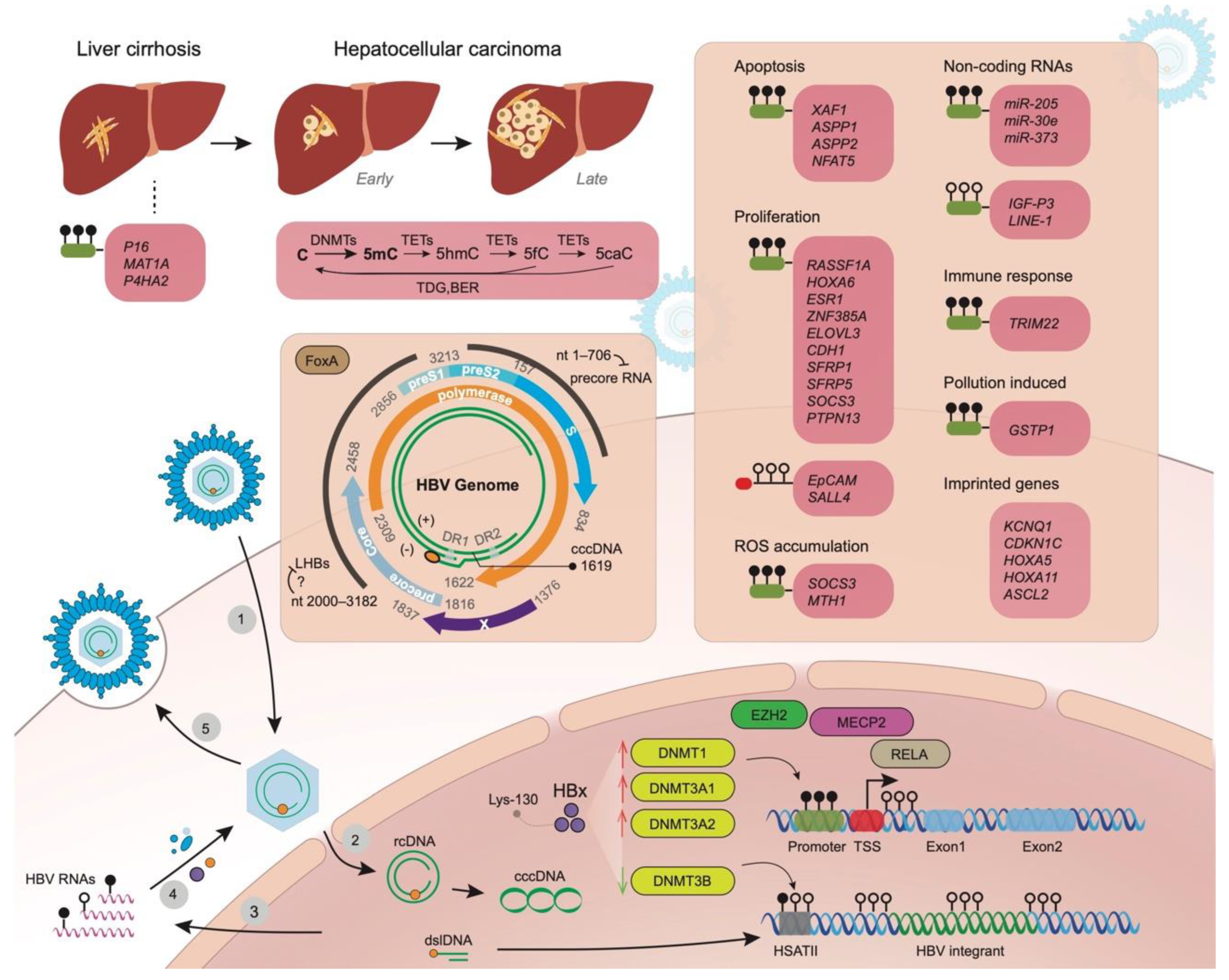
1
Key Laboratory of Biomechanics and Mechanobiology, Ministry of Education, Beijing Advanced Innovation Center for Biomedical Engineering, School of Biological Science and Medical Engineering, Beihang University, Beijing 100083, China
2
Department of Medical Genetics, University of Wisconsin-Madison, Madison, WI 53706, USA
3
Computation and Informatics in Biology and Medicine, University of Wisconsin-Madison, Madison, WI 53706, USA
*
Authors to whom correspondence should be addressed.
Int. J. Mol. Sci. 2021, 22(18), 9858; https://doi.org/10.3390/ijms22189858
Submission received: 22 August 2021 / Revised: 10 September 2021 / Accepted: 10 September 2021 / Published: 12 September 2021
(This article belongs to the Special Issue Role of Signaling Pathways in the Viral Life Cycle 2.0)

Abstract

:
Hepatitis B virus (HBV), the well-studied oncovirus that contributes to the majority of hepatocellular carcinomas (HCC) worldwide, can cause a severe inflammatory microenvironment leading to genetic and epigenetic changes in hepatocyte clones. HBV replication contributes to the regulation of DNA methyltransferase gene expression, particularly by X protein (HBx), and subsequent methylation changes may lead to abnormal transcription activation of adjacent genes and genomic instability. Undoubtedly, the altered expression of these genes has been known to cause diverse aspects of infected hepatocytes, including apoptosis, proliferation, reactive oxygen species (ROS) accumulation, and immune responses. Additionally, pollutant-induced DNA methylation changes and aberrant methylation of imprinted genes in hepatocytes also complicate the process of tumorigenesis. Meanwhile, hepatocytes also contribute to epigenetic modification of the viral genome to affect HBV replication or viral protein production. Meanwhile, methylation levels of HBV integrants and surrounding host regions also play crucial roles in their ability to produce viral proteins in affected hepatocytes. Both host and viral changes can provide novel insights into tumorigenesis, individualized responses to therapeutic intervention, disease progress, and early diagnosis. As such, DNA methylation-mediated epigenetic silencing of cancer-related genes and viral replication is a compelling therapeutic goal to reduce morbidity and mortality from liver cancer caused by chronic HBV infection. In this review, we summarize the most recent research on aberrant DNA methylation associated with HBV infection, which is involved in HCC development, and provide an outlook on the future direction of the research.

1. Background

Hepatocellular carcinoma (HCC) is a common human malignancy disease with a particularly high prevalence in Asia and Africa [1]. The risk factors for HCC are well documented, including hepatitis B/C virus infections, aflatoxin B exposure, and heavy consumption of tobacco and alcohol. However, its molecular pathogenesis remains poorly understood. In the past two decades, there has been great progress in the identification of candidate HCC-related genetic and epigenetic risk or disease causal factors such as HLA-DRB, HLA-DQB1, and perhaps other HLA regions, PBX4, PNPLA3, GLUL, and STAT4 from genome-wide association studies (GWAS) [2,3,4,5,6]; genome-wide methylation differential analysis (GWMA) also have identified such examples as Ras association domain family member 1A (RASSF1A), P16, SLC22A20, PNPLA7 as epigenetic factors as well as their interaction [7,8,9,10]. However, the mechanism of how the virus, as the most important risk factor, interacts with genetic and epigenetics, is still not clear.
DNA methyltransferases (DNMTs) turn cytosine (C) into 5-methylcytosine (5mC) and ten-eleven translocation (TET) enzymes oxidize 5mC into 5-hydroxymethlcytosine (5hmC), 5-formylcytosine (5fC), and 5-carboxylcytosine (5caC) sequentially, the last two of which can be restored to an unmodified C (Figure 1). By far, 5mC is the most well studied and almost all methylation changes here in the review are referred to 5mC. Different viral hepatitis may cause distinct epigenetic changes within the host genome. As observed by Shih et al. (2016), increased methylation of Paired-box 6 (PAX6) was more frequently seen in HCV-positive HCC (61.3%) in comparison with HBV-positive HCC (22.1%) in a cohort of 160 patients [11]. The promoter methylation is thought to account for the lack of RB1 expression in both HBV and HCV-associated HCC [12]. Meanwhile, certain methylation changes may be common in HCC development and are not specific to viral infection. Li et al. (2018) reported the promotor of TMEM176A, an activator of ERK signaling, was methylated in 75.4% of HCC, which was not associated with HBV infection status [13]. The differentially-methylated CpG sites were enriched in enhancers, promoters, CpG islands, and surrounding regions [14]. To provide a better understanding of the interaction role of HBV/HCV with genetic and epigenetic factors, we performed a comprehensive review of the most recent findings between virus and DNA methylation at different molecule regulation levels.

2. Aberrant Methylation of Promoter Regions of Genes in Liver Disease Progression Related to Tumorigenesis

DNA methylation changes are a feature of tumorigenesis, and one important mechanism by which hepatocytes respond to HBV infection is through upregulation of DNA methyltransferase genes, which may contribute to the increased tumor risk during chronic infection [15]. Liu et al. (2019) measured DNA methylation in HBV-related HCC and paired normal tissues, and found HBV infection corresponded to (i) 5mC, 5hmC, or 5fC decreases at the early stage of HCC (Figure 1) [16], (ii) was associated with decreased TET enzyme activity, and (iii) partial or complete reduction in DNA methylation-related enzymes. Although global hypomethylation is a feature of tumor genomes, hypermethylated sites can differentiate particular tumors from normal tissues.
Numerous studies have shown that the HBV-encoded X protein (HBx) is heavily involved in interactions with host gene networks and impacts HBV-mediated HCC progression. Park et al. (2007) comprehensively evaluated the HBx expression on host epigenetic changes, and found increased total DNMT activity, up-regulation of DNMT1, DNMT3A1, and DNMT3A2, as well as selectively promoted regional hypermethylation of specific tumor suppressor genes [17]. HBx specifically repressed insulin-like growth factor-3 (IGF3) expression through de novo methylation via DNMT3A1 and DNMT3A2 and by inhibiting SP1 binding via recruiting methyl CpG binding protein 2 (MECP2) to the newly methylated SP1 binding element (Figure 1) [17]. HBx also induced global hypomethylation of human satellite 2 repeat sequences (HSATII) by down-regulating DNMT3B (Figure 1). The prevalence of these specific methylation abnormalities by HBx was significantly correlated with HBx expression in HBV-infected HCC patients [17]. Additionally, Huang et al. (2010) reported that HBx downregulated miR-152, and its target DNMT1 was subsequently upregulated, leading to aberrant DNA methylation [18]. Vivekanandan et al. (2010) transfected HeG2 with full-length HBV DNA according to the procedure designed by Gunther et al. [19,20]. They observed that DNMT2 activity had the greatest increase of DNMTs [19]. Benhenda et al. (2013) reported that the upregulation of DNMTs was seen within 2–4 days after transfection, indicating a quick response of infected cells [21]. Intriguingly, they also found that fresh medium alone may also induce the upregulation of DNMT activity of the cell lines, strongly suggesting that care should be taken in interpreting results from functional experiments evaluating DNMT induction [21]. To observe the changes in host CpG sites, they collected DNA samples after 15 days of infection and applied a microarray to screen for these changes. Interestingly, they also reported that the HBV DNA fragments without replication activity could induce methylation changes in the host genome.
Promoter methylation changes of diverse genes are associated with liver disease progression. Generally, HBV infection causes promoter hypermethylation to suppress gene activity. Su et al. (2007) compared the promoter methylation in p14 (ARF), p16 (INK4a), O(6)-methylguanine-DNA methyltransferase (MGMT), glutathione S-transferase pi (GSTP1), and E-cadherin (E-Cad, also CDH1) in noncirrhotic, cirrhotic, and HCC tissues; p16(INK4a) promoter methylation increased along with liver disease progression; GSTP1 promoter hypermethylation occurred more frequently in HBV related HCC; while p16(INK4a), MGMT, and p14(ARF) promoter hypermethylation did not show this tendency [22]. P16 hypermethylation may occur early before HCC development, and Shim et al. (2003) found that P16 hypermethylation increased from cirrhotic nodules through dysplastic nodules [23]. Kuramoto et al. (2017) investigated the methylation alterations in non-cancerous liver tissue, showing NASH had featured DNA methylation changes with good confidence to discern them from lesions of cirrhosis or viral hepatitis; further, aberrant methylation identified in NASH-related HCC were inherited from the non-cancerous liver tissue showing NASH—a finding which was aggravated with disease progression [24]. Methionine metabolism is known to occur in human liver cirrhosis, and Avila et al. (2000) showed that promoter hypermethylation of methionine adenosyltransferase (MAT1A) could contribute to its decline in cirrhosis [25]. Bing et al. (2014) further showed HBx could recruit DNMT1 causing the promoter hypermethylation and inhibiting the glucocorticoid receptor (GR) to the glucocorticoid-response element (GRE) of the MAT1A promoter [26]. ZNF382 was frequently downregulated by promoter methylation in HBV-related HCCs relative to HBV-infected liver cirrhosis tissues and decreased expression of ZNF382 was strongly correlated with poor survival in early-stage HCC patients [27]. Nevertheless, not all abnormal gene expression is due to aberrant promoter methylation. Qiu et al. (2013) reported the expression of DNMTs induced by HBx failed to change the methylation status of the PDCD4 promoter, and the deregulation of PDCD4 transcription in hepatocarcinogenesis was due to HBx-mediated miR-21 upregulation [28].
In some cases, HBx-introduced promoter hypermethylation happens to miRNAs, and it removes the suppression of miRNAs on their target genes. Zhang et al. (2013) reported that although miR-205 targets HBx mRNA, HBx was able to induce hypermethylation of the miR-205 promoter and abrogate its effect on tumor suppression in carcinogenesis [29]. Feng et al. (2017) illustrated that HBx suppressed miR-30e expression by inducing the methylation of its promoter, possibly mediated by EZH2-formed complexes [30]. The subsequent upregulation of prolyl 4-hydroxylase subunit α2 (P4HA2), as a crucial enzyme catalyzing collagen formation, increased the collagen deposition in the liver in vivo and in vitro, promoting liver fibrosis and tumorigenesis [30].
The mechanism of promoter methylation of E-Cad has been thoroughly studied. As a tumor suppressor, E-Cad under-expression was one feature of a subgroup of HCC (G6) identified by Boyault et al. (2007) in their transcriptome classification of HCC [31]. For HBsAg-positive patients, E-Cad promoter hypermethylation remained high in both nontumorous tissues and HCCs, while it was lower in HCCs than in nontumorous tissues of HBsAg-negative patients [22]. Wei et al. (2002) suggested the HBV infection may lead to hypermethylation of the promoter region of E-Cad, whose expression is frequently lost in HCC [32]. Lee et al. (2005) demonstrated that this methylation change accounting for the HBx and the Lys-130 in its transactivation domain is essential. They also showed HBx induces DNMT1 expression by stimulating its transcription [33]. Liu et al. (2006) also showed the HBx is responsible for the hypermethylation of the E-Cad promoter and further pointed out that decreased expression of E-Cad was also associated with beta-catenin accumulation in the cytoplasm and/or nuclei, resulting in increased cell migration contributing to hepatocarcinogenesis [34]. In addition to promoter hypermethylation of E-Cad, Arzumanyan et al. (2012) further proposed that HBx may recruit the mSin3A/histone deacetylase complex to the Snail-binding sites in human CDH1 and cause its histone deacetylation [35]. They also pointed out that miR-373, upregulating E-Cad expression, was also downregulated by HBx, which may also explain the loss of E-cadherin in HBV-related carcinogenesis [35].

3. Apoptosis, Proliferation, and Reactive Oxygen Species (ROS) Accumulation

Efforts have been made to reveal the contribution of DNA methylation changes underlying distinct biological processes. It is known that HBV infection inhibits apoptosis by interfering with multiple signaling pathways. Zhang et al. (2008) showed that in hepatoma cell lines, promoter methylation was responsible for the low expression of X-linked inhibitor of apoptosis protein-associated factor 1 (XAF1) [36]. The XAF1 promoter methylation occurred in 66% of the HCC samples they collected, and low expression of XAF1 resulted in a reduction in recurrence-free survival of patients by over one year [36]. The promoter hypermethylation s contributed to lower expression of ASPP1 and ASPP2 in tumor tissues, suppressing their regulation of apoptosis through interaction with p53 and its family members. However, only the methylation of ASPP2 may be induced by HBx expression [37]. Qin et al. (2017) illustrated that HBV induced hypermethylation of the AP1-binding site in the NFAT5 promoter in hepatoma cells, and suppression of NFAT5 may lead to reduced apoptosis of hepatoma cells [38].
During chronic infection, methylation changes can be observed for genes influencing cell proliferation, e.g., RASSF1A, Homeobox A6 (HOXA6), estrogen receptor 1 (ESR1), zinc finger protein 385A (ZNF385A), and ELOVL fatty acid elongase 3 (ELOVL3) [39]. According to proliferation ability, HCCs have been broadly grouped into two classes: proliferation and nonproliferation, which reflects the fundamental differences in cell division in these two HCC classes. Tumors of the proliferation class are likely to be recurrent or lead to a shorter survival time, and they demonstrate heterogeneous changes of signaling pathways related to proliferation [40]. LAMA2 mutations are found in 5%–15% of diverse tumors, and Jhunjhunwala et al. (2014) reported that promoter hypermethylation induced low expression of LAMA2, which represents a highly recurrent and proliferative HCC [41]. Xie et al. (2014) reported that HBx can induce the hypermethylation of Secreted frizzled-related protein 1 and 5 (SFRP1 and SFRP5), and epigenetic silencing of these antagonists of the Wnt signaling pathway leads to the pathway activation which subsequently promotes cell growth and HBx induced epithelial–mesenchymal transition [42]. Zhang et al. (2015) demonstrated the hypermethylation of the CpG island close to cytokine signaling 3 (SOCS3) TSS accounted for its suppression in HCC losing its inhibition role on proliferation [43]. HCC had the promoter region of TMEM176A frequently methylated independent of HBV infection (Li et al., 2018), whose protein product suppresses HCC growth by inhibiting the ERK signaling pathway [13]. Yang et al. (2019) discovered that HBx may induce the methylation of CpG islands of miR-18 and subsequently inhibit its expression, and the suppression of miR-18 induced the nucleolar spindle-associated protein 1 (NUSAP1) upregulation which promotes the proliferation of hepatoma cells [44]. In hepatocytes. polycomb repressive complex 2 (PRC2) silences the EpCAM expression, which upregulates c-myc and induces cell proliferation [45]. Fan H et al. (2016) showed that this suppression can be removed by HBx induced demethylation of the CpG dinucleotide located adjacent to NF-κB/RelA half-site downstream from EpCAM transcriptional start site [46]. By chromatin immunoprecipitation assays, they further showed that RelA forms a complex with EZH2, TET2, and DNMT3L in the presence of HBx, and proposed the binding of RelA to the half-site may direct TET2 to demethylate the nearby CpG sites [46]. HBV infection may contribute to the re-expression of Sal-like protein 4 (SALL4), an embryonic stem cell transcriptional regulator, in HCC. Fan et al. (2017) pointed out that HBV-related HCC and HBV replicating cells had demethylation of specific CpG sites downstream of SALL4 TSS, which influences SALL4 transcriptional elongation [47]. They further showed that these demethylated CpGs are within binding sites of octamer-binding transcription factor 4 (OCT4) and signal transducer and activator of transcription3 (STAT3), which interact with embryonic stem cell BAF (esBAF) complex and recruit BRG1, a subunit of the Brg1/BAF chromatin-remodeling complex [47]. In particular, OCT4 is repressed by PRC2 [48], and HBx may account for the loss of PRC2 function in HBV-related HCC [46,49,50,51,52]. Yan et al. (2020) report that protein phosphatase nonreceptor type 13 (PTPN13) expression was significantly lower in HBV-positive hepatocellular carcinoma tissues. Patients with low PTPN13 expression and an HBV-positive history had the poorest overall survival. PTPN13 downregulation induced by HBx-induced promoter methylation promoted HCC proliferation due to increased c-Myc mRNA [53].
Tumors are known to have increased ROS production, which can activate pro-tumorigenic signaling and enhance cell survival and proliferation [54]. Roles of ROS in HBV-related tumorigenesis and DNA methylation changes have also been explored recently. HBV-induced mitochondrial ROS accumulation upregulated Snail expression, which epigenetically suppressed SOCS3 expression together with DNMT1 and HDAC1 [55]. The low expression of SOCS3 led to the sustained activation of the IL-6/STAT3 pathway ultimately contributing to hepatocarcinogenesis [55]. High levels of ROS can induce oxidative DNA damage, which may produce oxidized nucleotides, such as 8-hydroxy-2-deoxyguanosine (8-OHdG). Nucleotide pool sanitization enzymes, including MTH1 (NUDT1), MTH2 (NUDT15), MTH3 (NUDT18), and NUDT5, prevent oxidized nucleotides from incorporation into DNA. Lin et al. (2018) observed that in vitro and in vivo HBV caused an increase of 8-OHdG within infected hepatocytes due to reduced expression of MTH1, whose promoter activity was inhibited by HBx induced hypermethylation [56].

4. Hypomethylation, Methylation Changes to Non-Coding Genes Other Than miRNAs and Imprinted Genes

In the aforementioned promoter methylation changes of miRNAs, HBx commonly introduces promoter hypermethylation and inhibits miRNA expression. However, it is not always the case for all non-coding RNAs. Tang et al. (2015) speculated that the overexpression of transcripts from P3 and P4 promoters of the IGF2 gene may be driven by HBx and showed in cell lines that HBx induced hypomethylation of P3 promoter and improved IGF2-P3 transcript expression [57]. Gao et al. (2014) claimed nearly 90% HCC showed hypomethylation of LINE-1 promoter compared with HBV-related cirrhosis and normal controls, and LINE-1 promoter hypomethylation accounted for the significantly reduced survival time of patients (e.g., overall survival post-resection of a median of 22 months in comparison with ~60 months of patients without this change) [58].
Imprinted genes are mono-allelically expressed in a parent-of-origin-dependent manner and methylation is the crucial modification to sustain their status. It is suggested that their methylation status is susceptible to deregulation in cancer considering genome-wide epigenetic changes as well-known tumor genomic features. Lambert et al. (2015) demonstrated the aberrant methylation process during liver disease progression was more likely to affect the imprinted genes and found HCC was likely to have hypomethylation of 15q11–13, which is the imprinting control region harboring the cluster of imprinted genes [59]. They further pointed out that methylation changes of paternally expressed genes may be more susceptible to epigenetic disruption in carcinogenesis, such as KCNQ1, CDKN1C, HOXA5, HOXA11, and ASCL2 [59].

5. Changes of Host Immune Status, Infected Hepatocytes, and Exposure to Environmental Pollutant

The CpG sites in PBMC (peripheral blood mononuclear cell) DNA may have their quantitative state of methylation correlated with the liver disease progression in patients with HBV infection. Recently, Li et al. (2020) identified 7888 sites of this type in a cohort of 48 HBV-related liver disease patients and claimed their methylation level reflected the epigenetic reprogramming of the immune and inflammatory responses during chronic infection [60]. Peripheral blood DNA methylation level is used to calculate the epigenetic age (Horvath clock), and Gindin et al. (2020) reported the inconsistency between epigenetic age and chronological age in patients with HBV and HCV infection. They proposed that accelerated epigenetic age is a feature of viral hepatitis, which can be slowed down by antiviral treatment [61]. Okamoto et al. (2014) also presented the time-dependent manner of methylation changes during HBV infection in hepatocytes [39]. Nevertheless, unlike HBV, they found HCV infection did not induce aberrant methylation in HCC cell lines, which inspired them to identify the involved immune factors. They proposed the natural killer cell activity via IFN (interferon)-gamma as the major cause of these aforementioned DNA methylations [39]. The superfamily of tripartite motif-containing (TRIM) proteins induces a response to IFNs that involves innate immunity and antiviral defense against a broad range of viral infections [62]. Lim et al. (2018) found the HBx-mediated suppression of TRIM22 may evade the antiviral effect of IFNs, and further showed that among the multiple CpG sites within its promotor that they examined, only the CpG (+71) had its methylation status regulated by HBx, which reduces the IFN regulatory factor-1(IRF1) binding affinity and thereby suppresses the IFN-stimulated induction of TRIM22h which impedes IFN regulatory factor-1 [63]. This may help HBV evade the host’s innate immune system. Jin et al. (2019) attributed the reduced activity of pathways in PBMCs to hypermethylation of their member genes, which can be a feature of particular phases during infection, e.g., RASSSF1 and cyclin-dependent kinase inhibitor 2A (CDKN2A) for chronic infection [64].
Exposure to environmental pollutants can lead to methylation level changes in gene promoter regions. Glutathione S-transferases are a type of enzyme known to defend cells against diverse damage caused by oxidant and electrophilic carcinogens, and GSTP1 is the pi-class glutathione S-transferase. GSTP promoter hypermethylation and B[a]P diol epoxide-albumin (BPDE-Alb) were positively correlated with HCC incidence [65]. In HBV-associated HCC, Zhong et al. reported the common hypermethylation of GSTP1 gene and low protein expression, and the DNA methyltransferase inhibitor 5-aza-deoxycytidine reversed the hypermethylation of GSTP1 in Hep3B and HepG2 cell lines [66]. GSTP1 expression was completely depleted in HepG2.2.15 cells due to its promoter hypermethylation, and Niu et al. (2009) suggested the HBx accounted for the methylation change [67]. Lambert et al. (2011) reported the hypermethylation of RASSF1A, GSTP1, CHRNA3, and DOK1 in HCC tumors, and they also claimed that alcohol intake was related to hypomethylation of MGMT and hypermethylation of GSTP1 associated with HBV infection [68]. Recently, Tian et al. (2020) found that the concentration of environmental pollutant organochlorine pesticides (OCPs) in blood correlated with GSTP1 methylation and increased the risk of hepatocellular carcinoma [69].

6. Viral Genome Methylation in HBV Replication, Modifications of cccDNA and HBV Integrants

Pioneer transcription factors are critical determinants of tissue-specific gene expression and functions of the HBV genome within hepatocytes should be under their regulation [70]. Vivekanandan et al. (2008) illustrated that methylation patterns of the HBV genome differ between patients with occult and noncult infections, and the former cases had HBV CpG island 2 more densely methylated and the latter had island 1 more densely methylated [71]. Later in 2009, they also illustrated that the methylation level of viral DNA affected the protein production ability [72]. Uchiad et al. (2017) applied combined treatment of Peginterferon and Entecavir and successfully achieved a long-term loss of HBsAg in several human hepatocyte chimeric mice, and according to HBV genome analysis, they attributed the suppression of HBV activity to the G-to-A hypermutation and GpG III island methylation and speculated their antiviral effects without the requirement of cellular immune response [73]. FoxA pioneer transcription factor is known to have seven binding sites in the HBV genome and cell culture experiments show FoxA is involved in the regulation of all four HBV promoters [74]. Oropeza et al. (2020) illustrated the dynamic changes in HBV DNA methylation during neonatal liver development, balanced by FoxA-recruited Tet-mediated DNA demethylation and Dnmt-mediated DNA methylation activities [75]. McFadden et al. (2017) and colleagues showed that in FoxA-deficiency in the HBV transgenic mice producing a limited amount of FoxA3, significantly increased methylation levels were observed in nt 1–706 and nt 2000–3182 (Figure 1) [76]. The former was almost 100% methylated, leading to significantly decreased HBV 3.5 kb precore RNA synthesis; and the latter harbors the large and major surface antigen promoter and three FoxA binding sites [74,77]. This observation is consistent with the study of Nakamura et al. (2020), comparing the effects of in vitro methylation on viral protein productions in distinct genotypes. They found for genotypes B and C, the reduction of HBeAg was more than that of HBsAg [78]. Using nanopore sequencing, Goldsmith et al. also reported the relatively high methylation level in preS1/preS2 region for genotypes A and D [79].
DNA methylation is essential for the activity of covalently closed circular DNA (cccDNA), and diverse protocols have been developed to determine its patterns [80]. The cotransfection of HBV with DNMT3a showed that the methylation of cccDNA led to decreased production of both pregenomic and precore RNA, but the precore RNA decline seemed to be more significant, possibly complicated by RNA methylation [21]. Lee et al. (2019) verified that HBx could induce the methylation of C-1619 in cccDNA, which is a highly conserved nucleotide in the binding site of HBV negative regulatory element-binding protein [81]. Its methylation interrupted that binding process and led to the activation of the core promoter responsible for a four-fold increase in HBV particles compared to the situation when HBx was absent [81]. In addition, the methylation status of cccDNA may also influence the therapeutic strategies aiming at eliminating cccDNA. Kostyushev and colleagues (2019) first reported that methylated HBV cccDNA may be resistant to the degradation effect of CRISPR/Cas9-based anti-HBV therapeutics [82]. van Breugel et al. (2012) argued that the gene expression activation of HBx functions may only work for extrachromosomal DNA, rather than integrants [83]. They demonstrated its consistent activation effect on transiently transfected constructs independent of regulatory elements within them, and this effect disappeared once these constructs were incorporated into the host genome. They showed that the activity required HBx binding to the cellular UV-damaged DDB1 E3 ubiquitin ligase [83].
It has long been noticed (Miller et al., 1983) that integrated HBV DNA in PLC/PRF/5 cell line is methylated, but the region coding for viral surface protein always remains unmethylated unlike the extensively methylated region coding for core protein [84]. Bowyer et al. (1987) observed the methylation status of HBV integrants was not consistent among diverse HCC samples, among which some were hypomethylated [85]. Miyoshi et al. (1992) claimed that 5-azacytidine (5-AZA) treatment may induce hypomethylation of HBV integrants and thereby increase the HBsAg production of a hepatoblastoma-derived cell line HB611 [86]. Watanabe et al. (2015) showed in cell lines and tissues that the methylation level of HBV integrants was associated with that of the neighboring human genome, and HBx regions within integrants stayed unmethylated even when other regions all became methylated. HBV integrations seemed not to change but “follow” the methylation level of interrupted regions of the host genome [87]. Hama et al. (2018) attributed frequent detection of HBV integration sites within inactive chromatin regions in cancer cells to negative selection for integrations in active chromatin regions [88]. Nevertheless, we observed that the hypomethylation of previously reported integration sites may be a good indicator of HCC development [89]. HBV integrations are known to predispose the liver to HCC and have attracted increasing research interest, and recently their biological impacts, particularly how viral integrants complicate the damaging of hepatocytes due to their diverse ability of viral protein production, are extensively reviewed elsewhere [90,91].

7. Technical Challenges in Future Research

Frommer et al. combined bisulfite conversion with sequencing in 1992 [92], and it offered a viable method to measure DNA methylation through sequencing. The treatment converts cytosine residues to uracil but leaves 5-methylcytosine residues unaffected. By comparing the sequence composition of the resulting products with the reference genome, methylated sites can be identified. Since then, diverse solutions have been developed, such as methylation-specific PCR [93], methylation-sensitive single-nucleotide primer extension [94], and the reduced representation bisulfite-sequencing method [95]. In the early stage, the PCR-based detection method typically interrogates limited CpG sites, perhaps one or two CpG sites for each promoter. In recent years, detection methods for CpG sites or specific regions have gradually become the alternatives for the validation of interesting candidates. Whole-genome bisulfite sequencing (WGBS) theoretically captures all the DNA methylation changes [96], but its high cost limited the sample size in one study. Hence, microarray techniques help to profile CpG sites across the genome in relatively large sample sizes [97]. Nevertheless, the detection sensitivity of this technique may be lower than the PCR-based or sequencing solutions. Recently, nanopore sequencing has been used to directly detect the 5mC in the naïve DNA/RNA sequence without bisulfite treatment, avoiding sample loss and introduced biases during bisulfite conversion [98].
By far, the majority of genome-wide methylation analyses have been performed on bulk tissues, which are composed of diverse types of cells with an inconsistent percentage in tissue aliquot due to random sampling or individual differences. This ignores the fact that DNA methylation is cell-type specific, and the averaging of methylation levels of a specific site may obscure the biological significance contributed by a subgroup of the cell population. Overall, cell-type based highly accurate deconvolution [99,100,101] and single-cell-based DNA methylation (sc-5mC) [102] detection assays will be more powerful approaches for future DNA methylation research. Finally, causal inference based on genetic, epigenetic, and interaction will identify more disease-causing genes and therapeutic targets through the modeling of interactions between HCC and host pathways.

Author Contributions

Substantial contributions to conception, D.Z., S.J.S.; Manuscript writing and organizing, D.Z., S.G., S.J.S.; Collection of data and data interpretation, D.Z., S.G., S.J.S. All authors have read and agreed to the published version of the manuscript.

Funding

This work was supported by the Higher Education Discipline Innovation Project (the 111 Project No.: B13003) and National Natural Science Foundation of China (81201700) to D.Z. This work was also supported by faculty start-up funds by the Laboratory of Genetics, UW-Madison, the School of Medicine and Public Health, UW-Madison, the Office of the Vice Chancellor for Research and Graduate Education, UW-Madison, and the Center for Human Genomics and Precision Medicine, UW-Madison to S.J.S.

Institutional Review Board Statement

Not applicable.

Informed Consent Statement

Not applicable.

Data Availability Statement

Not applicable.

Conflicts of Interest

All authors have no conflict of interest related to this publication.

References

  1. Brechot, C.; Pourcel, C.; Louise, A.; Rain, B.; Tiollais, P. Presence of integrated hepatitis B virus DNA sequences in cellular DNA of human hepatocellular carcinoma. Nature 1980, 286, 533–535. [Google Scholar] [CrossRef] [PubMed]
  2. Liu, Q.; Liu, G.; Lin, Z.; Lin, Z.; Tian, N.; Lin, X.; Tan, J.; Huang, B.; Ji, X.; Pi, L.; et al. The association of lncRNA SNPs and SNPs-environment interactions based on GWAS with HBV-related HCC risk and progression. Mol. Genet. Genomic Med. 2021, 9, e1585. [Google Scholar] [CrossRef] [PubMed]
  3. Li, C.; Bi, X.; Huang, Y.; Zhao, J.; Li, Z.; Zhou, J.; Zhang, M.; Huang, Z.; Zhao, H.; Cai, J. Variants identified by hepatocellular carcinoma and chronic hepatitis B virus infection susceptibility GWAS associated with survival in HBV-related hepatocellular carcinoma. PLoS ONE 2014, 9, e101586. [Google Scholar] [CrossRef] [PubMed]
  4. Li, S.; Qian, J.; Yang, Y.; Zhao, W.; Dai, J.; Bei, J.X.; Foo, J.N.; McLaren, P.J.; Li, Z.; Yang, J.; et al. GWAS identifies novel susceptibility loci on 6p21.32 and 21q21.3 for hepatocellular carcinoma in chronic hepatitis B virus carriers. PLoS Genet. 2012, 8, e1002791. [Google Scholar] [CrossRef]
  5. Jiang, D.K.; Sun, J.; Cao, G.; Liu, Y.; Lin, D.; Gao, Y.Z.; Ren, W.H.; Long, X.D.; Zhang, H.; Ma, X.P.; et al. Genetic variants in STAT4 and HLA-DQ genes confer risk of hepatitis B virus-related hepatocellular carcinoma. Nat. Genet. 2013, 45, 72–75. [Google Scholar] [CrossRef] [Green Version]
  6. Lin, Y.Y.; Yu, M.W.; Lin, S.M.; Lee, S.D.; Chen, C.L.; Chen, D.S.; Chen, P.J. Genome-wide association analysis identifies a GLUL haplotype for familial hepatitis B virus-related hepatocellular carcinoma. Cancer 2017, 123, 3966–3976. [Google Scholar] [CrossRef]
  7. Zhao, Y.; Xue, F.; Sun, J.; Guo, S.; Zhang, H.; Qiu, B.; Geng, J.; Gu, J.; Zhou, X.; Wang, W.; et al. Genome-wide methylation profiling of the different stages of hepatitis B virus-related hepatocellular carcinoma development in plasma cell-free DNA reveals potential biomarkers for early detection and high-risk monitoring of hepatocellular carcinoma. Clin. Epigenet. 2014, 6, 30. [Google Scholar] [CrossRef] [PubMed] [Green Version]
  8. Zhang, X.; Zhang, J.; Wang, R.; Guo, S.; Zhang, H.; Ma, Y.; Liu, Q.; Chu, H.; Xu, X.; Zhang, Y.; et al. Hypermethylation reduces the expression of PNPLA7 in hepatocellular carcinoma. Oncol. Lett. 2016, 12, 670–674. [Google Scholar] [CrossRef] [Green Version]
  9. He, Y.; Cui, Y.; Wang, W.; Gu, J.; Guo, S.; Ma, K.; Luo, X. Hypomethylation of the hsa-miR-191 locus causes high expression of hsa-mir-191 and promotes the epithelial-to-mesenchymal transition in hepatocellular carcinoma. Neoplasia 2011, 13, 841–853. [Google Scholar] [CrossRef] [Green Version]
  10. Wang, R.; Zhang, J.; Ma, Y.; Chen, L.; Guo, S.; Zhang, X.; Ma, Y.; Wu, L.; Pei, X.; Liu, S.; et al. Association study of miR149 rs2292832 and miR608 rs4919510 and the risk of hepatocellular carcinoma in a largescale population. Mol. Med. Rep. 2014, 10, 2736–2744. [Google Scholar] [CrossRef]
  11. Shih, Y.L.; Kuo, C.C.; Yan, M.D.; Lin, Y.W.; Hsieh, C.B.; Hsieh, T.Y. Quantitative methylation analysis reveals distinct association between PAX6 methylation and clinical characteristics with different viral infections in hepatocellular carcinoma. Clin. Epigenet. 2016, 8, 41. [Google Scholar] [CrossRef] [PubMed] [Green Version]
  12. Edamoto, Y.; Hara, A.; Biernat, W.; Terracciano, L.; Cathomas, G.; Riehle, H.M.; Matsuda, M.; Fujii, H.; Scoazec, J.Y.; Ohgaki, H. Alterations of RB1, p53 and Wnt pathways in hepatocellular carcinomas associated with hepatitis C, hepatitis B and alcoholic liver cirrhosis. Int. J. Cancer 2003, 106, 334–341. [Google Scholar] [CrossRef]
  13. Li, H.; Zhang, M.; Linghu, E.; Zhou, F.; Herman, J.G.; Hu, L.; Guo, M. Epigenetic silencing of TMEM176A activates ERK signaling in human hepatocellular carcinoma. Clin. Epigenet. 2018, 10, 137. [Google Scholar] [CrossRef] [PubMed]
  14. Hensel, K.O.; Rendon, J.C.; Navas, M.C.; Rots, M.G.; Postberg, J. Virus-host interplay in hepatitis B virus infection and epigenetic treatment strategies. FEBS J. 2017, 284, 3550–3572. [Google Scholar] [CrossRef] [Green Version]
  15. Ozen, C.; Yildiz, G.; Dagcan, A.T.; Cevik, D.; Ors, A.; Keles, U.; Topel, H.; Ozturk, M. Genetics and epigenetics of liver cancer. New Biotechnol. 2013, 30, 381–384. [Google Scholar] [CrossRef]
  16. Liu, J.; Jiang, J.; Mo, J.; Liu, D.; Cao, D.; Wang, H.; He, Y.; Wang, H. Global DNA 5-Hydroxymethylcytosine and 5-Formylcytosine Contents Are Decreased in the Early Stage of Hepatocellular Carcinoma. Hepatology 2019, 69, 196–208. [Google Scholar] [CrossRef]
  17. Park, I.Y.; Sohn, B.H.; Yu, E.; Suh, D.J.; Chung, Y.H.; Lee, J.H.; Surzycki, S.J.; Lee, Y.I. Aberrant epigenetic modifications in hepatocarcinogenesis induced by hepatitis B virus X protein. Gastroenterology 2007, 132, 1476–1494. [Google Scholar] [CrossRef]
  18. Huang, J.; Wang, Y.; Guo, Y.; Sun, S. Down-regulated microRNA-152 induces aberrant DNA methylation in hepatitis B virus-related hepatocellular carcinoma by targeting DNA methyltransferase 1. Hepatology 2010, 52, 60–70. [Google Scholar] [CrossRef] [PubMed]
  19. Vivekanandan, P.; Daniel, H.D.; Kannangai, R.; Martinez-Murillo, F.; Torbenson, M. Hepatitis B virus replication induces methylation of both host and viral DNA. J. Virol. 2010, 84, 4321–4329. [Google Scholar] [CrossRef] [Green Version]
  20. Gunther, S.; Li, B.C.; Miska, S.; Kruger, D.H.; Meisel, H.; Will, H. A novel method for efficient amplification of whole hepatitis B virus genomes permits rapid functional analysis and reveals deletion mutants in immunosuppressed patients. J. Virol. 1995, 69, 5437–5444. [Google Scholar] [CrossRef] [PubMed] [Green Version]
  21. Benhenda, S.; Ducroux, A.; Rivière, L.; Sobhian, B.; Ward, M.D.; Dion, S.; Hantz, O.; Protzer, U.; Michel, M.L.; Benkirane, M.; et al. Methyltransferase PRMT1 is a binding partner of HBx and a negative regulator of hepatitis B virus transcription. J. Virol. 2013, 87, 4360–4371. [Google Scholar] [CrossRef] [PubMed] [Green Version]
  22. Su, P.F.; Lee, T.C.; Lin, P.J.; Lee, P.H.; Jeng, Y.M.; Chen, C.H.; Liang, J.D.; Chiou, L.L.; Huang, G.T.; Lee, H.S. Differential DNA methylation associated with hepatitis B virus infection in hepatocellular carcinoma. Int. J. Cancer 2007, 121, 1257–1264. [Google Scholar] [CrossRef]
  23. Shim, Y.H.; Yoon, G.S.; Choi, H.J.; Chung, Y.H.; Yu, E. p16 Hypermethylation in the early stage of hepatitis B virus-associated hepatocarcinogenesis. Cancer Lett. 2003, 190, 213–219. [Google Scholar] [CrossRef]
  24. Kuramoto, J.; Arai, E.; Tian, Y.; Funahashi, N.; Hiramoto, M.; Nammo, T.; Nozaki, Y.; Takahashi, Y.; Ito, N.; Shibuya, A.; et al. Genome-wide DNA methylation analysis during non-alcoholic steatohepatitis-related multistage hepatocarcinogenesis: Comparison with hepatitis virus-related carcinogenesis. Carcinogenesis 2017, 38, 261–270. [Google Scholar] [CrossRef]
  25. Avila, M.A.; Berasain, C.; Torres, L.; Martin-Duce, A.; Corrales, F.J.; Yang, H.; Prieto, J.; Lu, S.C.; Caballeria, J.; Rodes, J.; et al. Reduced mRNA abundance of the main enzymes involved in methionine metabolism in human liver cirrhosis and hepatocellular carcinoma. J. Hepatol. 2000, 33, 907–914. [Google Scholar] [CrossRef] [Green Version]
  26. Bing, Y.; Zhu, S.; Yu, G.; Li, T.; Liu, W.; Li, C.; Wang, Y.; Qi, H.; Guo, T.; Yuan, Y.; et al. Glucocorticoid-induced S-adenosylmethionine enhances the interferon signaling pathway by restoring STAT1 protein methylation in hepatitis B virus-infected cells. J. Biol. Chem. 2014, 289, 32639–32655. [Google Scholar] [CrossRef] [Green Version]
  27. Dang, S.; Zhou, J.; Chen, Y.; Chen, P.; Ji, M.; Shi, B.; Yang, Q.; Hou, P. Dynamic expression of ZNF382 and its tumor-suppressor role in hepatitis B virus-related hepatocellular carcinogenesis. Oncogene 2019, 38, 4804–4819. [Google Scholar] [CrossRef] [PubMed]
  28. Qiu, X.; Dong, S.; Qiao, F.; Lu, S.; Song, Y.; Lao, Y.; Li, Y.; Zeng, T.; Hu, J.; Zhang, L.; et al. HBx-mediated miR-21 upregulation represses tumor-suppressor function of PDCD4 in hepatocellular carcinoma. Oncogene 2013, 32, 3296–3305. [Google Scholar] [CrossRef] [Green Version]
  29. Zhang, T.; Zhang, J.; Cui, M.; Liu, F.; You, X.; Du, Y.; Gao, Y.; Zhang, S.; Lu, Z.; Ye, L.; et al. Hepatitis B virus X protein inhibits tumor suppressor miR-205 through inducing hypermethylation of miR-205 promoter to enhance carcinogenesis. Neoplasia 2013, 15, 1282–1291. [Google Scholar] [CrossRef] [Green Version]
  30. Feng, G.X.; Li, J.; Yang, Z.; Zhang, S.Q.; Liu, Y.X.; Zhang, W.Y.; Ye, L.H.; Zhang, X.D. Hepatitis B virus X protein promotes the development of liver fibrosis and hepatoma through downregulation of miR-30e targeting P4HA2 mRNA. Oncogene 2017, 36, 6895–6905. [Google Scholar] [CrossRef] [Green Version]
  31. Boyault, S.; Rickman, D.S.; de Reyniès, A.; Balabaud, C.; Rebouissou, S.; Jeannot, E.; Hérault, A.; Saric, J.; Belghiti, J.; Franco, D.; et al. Transcriptome classification of HCC is related to gene alterations and to new therapeutic targets. Hepatology 2007, 45, 42–52. [Google Scholar] [CrossRef] [PubMed] [Green Version]
  32. Wei, Y.; Van Nhieu, J.T.; Prigent, S.; Srivatanakul, P.; Tiollais, P.; Buendia, M.A. Altered expression of E-cadherin in hepatocellular carcinoma: Correlations with genetic alterations, beta-catenin expression, and clinical features. Hepatology 2002, 36, 692–701. [Google Scholar] [CrossRef]
  33. Lee, J.O.; Kwun, H.J.; Jung, J.K.; Choi, K.H.; Min, D.S.; Jang, K.L. Hepatitis B virus X protein represses E-cadherin expression via activation of DNA methyltransferase 1. Oncogene 2005, 24, 6617–6625. [Google Scholar] [CrossRef] [Green Version]
  34. Liu, J.; Lian, Z.; Han, S.; Waye, M.M.; Wang, H.; Wu, M.C.; Wu, K.; Ding, J.; Arbuthnot, P.; Kew, M.; et al. Downregulation of E-cadherin by hepatitis B virus X antigen in hepatocellullar carcinoma. Oncogene 2006, 25, 1008–1017. [Google Scholar] [CrossRef] [Green Version]
  35. Arzumanyan, A.; Friedman, T.; Kotei, E.; Ng, I.O.; Lian, Z.; Feitelson, M.A. Epigenetic repression of E-cadherin expression by hepatitis B virus x antigen in liver cancer. Oncogene 2012, 31, 563–572. [Google Scholar] [CrossRef] [Green Version]
  36. Zhang, F.; Wu, L.M.; Zhou, L.; Chen, Q.X.; Xie, H.Y.; Feng, X.W.; Zheng, S.S. Predictive value of expression and promoter hypermethylation of XAF1 in hepatitis B virus-associated hepatocellular carcinoma treated with transplantation. Ann. Surg. Oncol. 2008, 15, 3494–3502. [Google Scholar] [CrossRef]
  37. Zhao, J.; Wu, G.; Bu, F.; Lu, B.; Liang, A.; Cao, L.; Tong, X.; Lu, X.; Wu, M.; Guo, Y. Epigenetic silence of ankyrin-repeat-containing, SH3-domain-containing, and proline-rich-region- containing protein 1 (ASPP1) and ASPP2 genes promotes tumor growth in hepatitis B virus-positive hepatocellular carcinoma. Hepatology 2010, 51, 142–153. [Google Scholar] [CrossRef]
  38. Qin, X.; Li, C.; Guo, T.; Chen, J.; Wang, H.T.; Wang, Y.T.; Xiao, Y.S.; Li, J.; Liu, P.; Liu, Z.S.; et al. Upregulation of DARS2 by HBV promotes hepatocarcinogenesis through the miR-30e-5p/MAPK/NFAT5 pathway. J. Exp. Clin. Cancer Res. 2017, 36, 148. [Google Scholar] [CrossRef] [Green Version]
  39. Okamoto, Y.; Shinjo, K.; Shimizu, Y.; Sano, T.; Yamao, K.; Gao, W.; Fujii, M.; Osada, H.; Sekido, Y.; Murakami, S.; et al. Hepatitis virus infection affects DNA methylation in mice with humanized livers. Gastroenterology 2014, 146, 562–572. [Google Scholar] [CrossRef] [PubMed]
  40. Sia, D.; Villanueva, A.; Friedman, S.L.; Llovet, J.M. Liver Cancer Cell of Origin, Molecular Class, and Effects on Patient Prognosis. Gastroenterology 2017, 152, 745–761. [Google Scholar] [CrossRef] [PubMed] [Green Version]
  41. Jhunjhunwala, S.; Jiang, Z.; Stawiski, E.W.; Gnad, F.; Liu, J.; Mayba, O.; Du, P.; Diao, J.; Johnson, S.; Wong, K.F.; et al. Diverse modes of genomic alteration in hepatocellular carcinoma. Genome Biol. 2014, 15, 436. [Google Scholar] [CrossRef] [Green Version]
  42. Xie, Q.; Chen, L.; Shan, X.; Shan, X.; Tang, J.; Zhou, F.; Chen, Q.; Quan, H.; Nie, D.; Zhang, W.; et al. Epigenetic silencing of SFRP1 and SFRP5 by hepatitis B virus X protein enhances hepatoma cell tumorigenicity through Wnt signaling pathway. Int. J. Cancer 2014, 135, 635–646. [Google Scholar] [CrossRef]
  43. Zhang, X.; You, Q.; Zhang, X.; Chen, X. SOCS3 Methylation Predicts a Poor Prognosis in HBV Infection-Related Hepatocellular Carcinoma. Int. J. Mol. Sci. 2015, 16, 22662–22675. [Google Scholar] [CrossRef] [Green Version]
  44. Yang, Z.; Li, J.; Feng, G.; Wang, Y.; Yang, G.; Liu, Y.; Zhang, S.; Feng, J.; Zhang, X. Hepatitis B virus X protein enhances hepatocarcinogenesis by depressing the targeting of NUSAP1 mRNA by miR-18b. Cancer Biol. Med. 2019, 16, 276–287. [Google Scholar] [CrossRef] [PubMed] [Green Version]
  45. Munz, M.; Kieu, C.; Mack, B.; Schmitt, B.; Zeidler, R.; Gires, O. The carcinoma-associated antigen EpCAM upregulates c-myc and induces cell proliferation. Oncogene 2004, 23, 5748–5758. [Google Scholar] [CrossRef] [PubMed]
  46. Fan, H.; Zhang, H.; Pascuzzi, P.E.; Andrisani, O. Hepatitis B virus X protein induces EpCAM expression via active DNA demethylation directed by RelA in complex with EZH2 and TET2. Oncogene 2016, 35, 715–726. [Google Scholar] [CrossRef] [PubMed] [Green Version]
  47. Fan, H.; Cui, Z.; Zhang, H.; Mani, S.K.; Diab, A.; Lefrancois, L.; Fares, N.; Merle, P.; Andrisani, O. DNA demethylation induces SALL4 gene re-expression in subgroups of hepatocellular carcinoma associated with Hepatitis B or C virus infection. Oncogene 2017, 36, 2435–2445. [Google Scholar] [CrossRef] [Green Version]
  48. Pasini, D.; Bracken, A.P.; Hansen, J.B.; Capillo, M.; Helin, K. The polycomb group protein Suz12 is required for embryonic stem cell differentiation. Mol. Cell Biol. 2007, 27, 3769–3779. [Google Scholar] [CrossRef] [PubMed] [Green Version]
  49. Salerno, D.; Chiodo, L.; Alfano, V.; Floriot, O.; Cottone, G.; Paturel, A.; Pallocca, M.; Plissonnier, M.L.; Jeddari, S.; Belloni, L.; et al. Hepatitis B protein HBx binds the DLEU2 lncRNA to sustain cccDNA and host cancer-related gene transcription. Gut 2020, 69, 2016–2024. [Google Scholar] [CrossRef] [PubMed] [Green Version]
  50. Mani, S.K.; Zhang, H.; Diab, A.; Pascuzzi, P.E.; Lefrancois, L.; Fares, N.; Bancel, B.; Merle, P.; Andrisani, O. EpCAM-regulated intramembrane proteolysis induces a cancer stem cell-like gene signature in hepatitis B virus-infected hepatocytes. J. Hepatol. 2016, 65, 888–898. [Google Scholar] [CrossRef] [Green Version]
  51. Zhang, H.; Xing, Z.; Mani, S.K.; Bancel, B.; Durantel, D.; Zoulim, F.; Tran, E.J.; Merle, P.; Andrisani, O. RNA helicase DEAD box protein 5 regulates Polycomb repressive complex 2/Hox transcript antisense intergenic RNA function in hepatitis B virus infection and hepatocarcinogenesis. Hepatology 2016, 64, 1033–1048. [Google Scholar] [CrossRef] [PubMed]
  52. Zhang, H.; Diab, A.; Fan, H.; Mani, S.K.; Hullinger, R.; Merle, P.; Andrisani, O. PLK1 and HOTAIR Accelerate Proteasomal Degradation of SUZ12 and ZNF198 during Hepatitis B Virus-Induced Liver Carcinogenesis. Cancer Res. 2015, 75, 2363–2374. [Google Scholar] [CrossRef] [PubMed] [Green Version]
  53. Yan, Y.; Huang, P.; Mao, K.; He, C.; Xu, Q.; Zhang, M.; Liu, H.; Zhou, Z.; Zhou, Q.; Zhou, Q.; et al. Anti-oncogene PTPN13 inactivation by hepatitis B virus X protein counteracts IGF2BP1 to promote hepatocellular carcinoma progression. Oncogene 2020, 40, 28–45. [Google Scholar] [CrossRef] [PubMed]
  54. Moloney, J.N.; Cotter, T.G. ROS signalling in the biology of cancer. Semin. Cell Dev. Biol. 2018, 80, 50–64. [Google Scholar] [CrossRef]
  55. Yuan, K.; Lei, Y.; Chen, H.N.; Chen, Y.; Zhang, T.; Li, K.; Xie, N.; Wang, K.; Feng, X.; Pu, Q.; et al. HBV-induced ROS accumulation promotes hepatocarcinogenesis through Snail-mediated epigenetic silencing of SOCS3. Cell Death Differ. 2016, 23, 616–627. [Google Scholar] [CrossRef] [Green Version]
  56. Lin, Y.T.; Liu, W.; He, Y.; Wu, Y.L.; Chen, W.N.; Lin, X.J.; Lin, X. Hepatitis B Virus X Protein Increases 8-Oxo-7,8-Dihydro-2’-Deoxyguanosine (8-Oxodg) Level via Repressing MTH1/ MTH2 Expression in Hepatocytes. Cell Physiol. Biochem. 2018, 51, 80–96. [Google Scholar] [CrossRef]
  57. Tang, S.; Hu, W.; Hu, J.; Wu, S.; Li, J.; Luo, Y.; Cao, M.; Zhou, H.; Jiang, X. Hepatitis B virus X protein promotes P3 transcript expression of the insulin-like growth factor 2 gene via inducing hypomethylation of P3 promoter in hepatocellular carcinoma. Liver Int. 2015, 35, 608–619. [Google Scholar] [CrossRef]
  58. Gao, X.D.; Qu, J.H.; Chang, X.J.; Lu, Y.Y.; Bai, W.L.; Wang, H.; Xu, Z.X.; An, L.J.; Wang, C.P.; Zeng, Z.; et al. Hypomethylation of long interspersed nuclear element-1 promoter is associated with poor outcomes for curative resected hepatocellular carcinoma. Liver Int. 2014, 34, 136–146. [Google Scholar] [CrossRef] [Green Version]
  59. Lambert, M.P.; Ancey, P.B.; Esposti, D.D.; Cros, M.P.; Sklias, A.; Scoazec, J.Y.; Durantel, D.; Hernandez-Vargas, H.; Herceg, Z. Aberrant DNA methylation of imprinted loci in hepatocellular carcinoma and after in vitro exposure to common risk factors. Clin. Epigenet. 2015, 7, 15. [Google Scholar] [CrossRef] [Green Version]
  60. Li, K.; Qin, L.; Jiang, S.; Li, A.; Zhang, C.; Liu, G.; Sun, J.; Sun, H.; Zhao, Y.; Li, N.; et al. The signature of HBV-related liver disease in peripheral blood mononuclear cell DNA methylation. Clin. Epigenet. 2020, 12, 81. [Google Scholar] [CrossRef]
  61. Gindin, Y.; Gaggar, A.; Lok, A.S.; Janssen, H.L.A.; Ferrari, C.; Subramanian, G.M.; Jiang, Z.; Masur, H.; Emmanuel, B.; Poonia, B.; et al. DNA Methylation and Immune Cell Markers Demonstrate Evidence of Accelerated Aging in Patients with Chronic HBV or HCV, with or without HIV Co-Infection. Clin. Infect. Dis. 2020, 73, e184–e190. [Google Scholar] [CrossRef] [PubMed]
  62. Ozato, K.; Shin, D.M.; Chang, T.H.; Morse, H.C., III. TRIM family proteins and their emerging roles in innate immunity. Nat. Rev. Immunol. 2008, 8, 849–860. [Google Scholar] [CrossRef] [PubMed] [Green Version]
  63. Lim, K.H.; Park, E.S.; Kim, D.H.; Cho, K.C.; Kim, K.P.; Park, Y.K.; Ahn, S.H.; Park, S.H.; Kim, K.H.; Kim, C.W.; et al. Suppression of interferon-mediated anti-HBV response by single CpG methylation in the 5’-UTR of TRIM22. Gut 2018, 67, 166–178. [Google Scholar] [CrossRef] [PubMed] [Green Version]
  64. Jin, J.; Xu, H.; Wu, R.; Gao, N.; Wu, N.; Li, S.; Niu, J. Identification of key genes and pathways associated with different immune statuses of hepatitis B virus infection. J. Cell Mol. Med. 2019, 23, 7474–7489. [Google Scholar] [CrossRef] [PubMed]
  65. Tian, M.; Zhao, B.; Zhang, J.; Martin, F.L.; Huang, Q.; Liu, L.; Shen, H. Association of environmental benzo[a]pyrene exposure and DNA methylation alterations in hepatocellular carcinoma: A Chinese case-control study. Sci. Total Environ. 2016, 541, 1243–1252. [Google Scholar] [CrossRef] [Green Version]
  66. Zhong, S.; Tang, M.W.; Yeo, W.; Liu, C.; Lo, Y.M.; Johnson, P.J. Silencing of GSTP1 gene by CpG island DNA hypermethylation in HBV-associated hepatocellular carcinomas. Clin. Cancer Res. 2002, 8, 1087–1092. [Google Scholar]
  67. Niu, D.; Zhang, J.; Ren, Y.; Feng, H.; Chen, W.N. HBx genotype D represses GSTP1 expression and increases the oxidative level and apoptosis in HepG2 cells. Mol. Oncol. 2009, 3, 67–76. [Google Scholar] [CrossRef]
  68. Lambert, M.P.; Paliwal, A.; Vaissière, T.; Chemin, I.; Zoulim, F.; Tommasino, M.; Hainaut, P.; Sylla, B.; Scoazec, J.Y.; Tost, J.; et al. Aberrant DNA methylation distinguishes hepatocellular carcinoma associated with HBV and HCV infection and alcohol intake. J. Hepatol. 2011, 54, 705–715. [Google Scholar] [CrossRef]
  69. Tian, M.; Zhao, B.; Martin, F.L.; Morais, C.L.M.; Liu, L.; Huang, Q.; Zhang, J.; Shen, H. Gene-environment interactions between GSTs polymorphisms and targeted epigenetic alterations in hepatocellular carcinoma following organochlorine pesticides (OCPs) exposure. Environ. Int. 2020, 134, 105313. [Google Scholar] [CrossRef]
  70. Lee, C.S.; Friedman, J.R.; Fulmer, J.T.; Kaestner, K.H. The initiation of liver development is dependent on Foxa transcription factors. Nature 2005, 435, 944–947. [Google Scholar] [CrossRef]
  71. Vivekanandan, P.; Kannangai, R.; Ray, S.C.; Thomas, D.L.; Torbenson, M. Comprehensive genetic and epigenetic analysis of occult hepatitis B from liver tissue samples. Clin. Infect. Dis. 2008, 46, 1227–1236. [Google Scholar] [CrossRef] [Green Version]
  72. Vivekanandan, P.; Thomas, D.; Torbenson, M. Methylation regulates hepatitis B viral protein expression. J. Infect. Dis. 2009, 199, 1286–1291. [Google Scholar] [CrossRef] [Green Version]
  73. Uchida, T.; Imamura, M.; Hayes, C.N.; Hiraga, N.; Kan, H.; Tsuge, M.; Abe-Chayama, H.; Zhang, Y.; Makokha, G.N.; Aikata, H.; et al. Persistent Loss of Hepatitis B Virus Markers in Serum without Cellular Immunity by Combination of Peginterferon and Entecavir Therapy in Humanized Mice. Antimicrob. Agents Chemother. 2017, 61, e00725-17. [Google Scholar] [CrossRef] [Green Version]
  74. Raney, A.K.; Zhang, P.; McLachlan, A. Regulation of transcription from the hepatitis B virus large surface antigen promoter by hepatocyte nuclear factor 3. J. Virol. 1995, 69, 3265–3272. [Google Scholar] [CrossRef] [Green Version]
  75. Oropeza, C.E.; Tarnow, G.; Taha, T.Y.; Shalaby, R.E.; Hyde, M.V.; Maienschein-Cline, M.; Green, S.J.; McLachlan, A. Relative DNA methylation and demethylation efficiencies during postnatal liver development regulate hepatitis B virus biosynthesis. J. Virol. 2020, 95, e02148-20. [Google Scholar] [CrossRef]
  76. McFadden, V.C.; Shalaby, R.E.; Iram, S.; Oropeza, C.E.; Landolfi, J.A.; Lyubimov, A.V.; Maienschein-Cline, M.; Green, S.J.; Kaestner, K.H.; McLachlan, A. Hepatic deficiency of the pioneer transcription factor FoxA restricts hepatitis B virus biosynthesis by the developmental regulation of viral DNA methylation. PLoS Pathog. 2017, 13, e1006239. [Google Scholar] [CrossRef]
  77. Raney, A.K.; McLachlan, A. Characterization of the hepatitis B virus major surface antigen promoter hepatocyte nuclear factor 3 binding site. J. Gen. Virol. 1997, 78, 3029–3038. [Google Scholar] [CrossRef] [Green Version]
  78. Nakamura, T.; Inoue, J.; Ninomiya, M.; Kakazu, E.; Iwata, T.; Takai, S.; Sano, A.; Kogure, T.; Shimosegawa, T.; Masamune, A. Effect of viral DNA methylation on expression of hepatitis B virus proteins depends on the virus genotype. Virus Genes 2020, 56, 439–447. [Google Scholar] [CrossRef] [PubMed]
  79. Goldsmith, C.; Cohen, D.; Dubois, A.; Martinez, M.G.; Petitjean, K.; Corlu, A.; Testoni, B.; Hernandez-Vargas, H.; Chemin, I. Cas9-targeted nanopore sequencing reveals epigenetic heterogeneity after de novo assembly of native full-length hepatitis B virus genomes. Microb. Genom. 2021, 7, 000507. [Google Scholar] [CrossRef]
  80. Zhang, Y.; Mao, R.; Guo, H.; Zhang, J. Detection of HBV cccDNA Methylation from Clinical Samples by Bisulfite Sequencing and Methylation-Specific PCR. Methods Mol. Biol. 2017, 1540, 73–84. [Google Scholar] [CrossRef] [PubMed] [Green Version]
  81. Lee, H.; Jeong, H.; Lee, S.Y.; Kim, S.S.; Jang, K.L. Hepatitis B Virus X Protein Stimulates Virus Replication Via DNA Methylation of the C-1619 in Covalently Closed Circular DNA. Mol. Cells 2019, 42, 67–78. [Google Scholar] [CrossRef]
  82. Kostyushev, D.; Brezgin, S.; Kostyusheva, A.; Zarifyan, D.; Goptar, I.; Chulanov, V. Orthologous CRISPR/Cas9 systems for specific and efficient degradation of covalently closed circular DNA of hepatitis B virus. Cell Mol. Life Sci. 2019, 76, 1779–1794. [Google Scholar] [CrossRef]
  83. van Breugel, P.C.; Robert, E.I.; Mueller, H.; Decorsière, A.; Zoulim, F.; Hantz, O.; Strubin, M. Hepatitis B virus X protein stimulates gene expression selectively from extrachromosomal DNA templates. Hepatology 2012, 56, 2116–2124. [Google Scholar] [CrossRef] [PubMed]
  84. Miller, R.H.; Robinson, W.S. Integrated hepatitis B virus DNA sequences specifying the major viral core polypeptide are methylated in PLC/PRF/5 cells. Proc. Natl. Acad. Sci. USA 1983, 80, 2534–2538. [Google Scholar] [CrossRef] [PubMed] [Green Version]
  85. Bowyer, S.M.; Dusheiko, G.M.; Schoub, B.D.; Kew, M.C. Expression of the hepatitis B virus genome in chronic hepatitis B carriers and patients with hepatocellular carcinoma. Proc. Natl. Acad. Sci. USA 1987, 84, 847–850. [Google Scholar] [CrossRef] [PubMed] [Green Version]
  86. Miyoshi, E.; Fujii, J.; Hayashi, N.; Ueda, K.; Towata, T.; Fusamoto, H.; Kamada, T.; Taniguchi, N. Enhancement of hepatitis-B surface-antigen expression by 5-azacytidine in a hepatitis-B-virus-transfected cell line. Int. J. Cancer 1992, 52, 137–140. [Google Scholar] [CrossRef] [PubMed]
  87. Watanabe, Y.; Yamamoto, H.; Oikawa, R.; Toyota, M.; Yamamoto, M.; Kokudo, N.; Tanaka, S.; Arii, S.; Yotsuyanagi, H.; Koike, K.; et al. DNA methylation at hepatitis B viral integrants is associated with methylation at flanking human genomic sequences. Genome Res. 2015, 25, 328–337. [Google Scholar] [CrossRef] [Green Version]
  88. Hama, N.; Totoki, Y.; Miura, F.; Tatsuno, K.; Saito-Adachi, M.; Nakamura, H.; Arai, Y.; Hosoda, F.; Urushidate, T.; Ohashi, S.; et al. Epigenetic landscape influences the liver cancer genome architecture. Nat. Commun. 2018, 9, 1643. [Google Scholar] [CrossRef]
  89. Zhang, H.; Dong, P.; Guo, S.; Tao, C.; Chen, W.; Zhao, W.; Wang, J.; Cheung, R.; Villanueva, A.; Fan, J.; et al. Hypomethylation in HBV integration regions aids non-invasive surveillance to hepatocellular carcinoma by low-pass genome-wide bisulfite sequencing. BMC Med. 2020, 18, 200. [Google Scholar] [CrossRef]
  90. Zhang, D.; Zhang, K.; Protzer, U.; Zeng, C. HBV Integration Induces Complex Interactions between Host and Viral Genomic Functions at the Insertion Site. J. Clin. Transl. Hepatol. 2021, 9, 399–408. [Google Scholar] [CrossRef]
  91. Zhao, K.; Liu, A.; Xia, Y. Insights into Hepatitis B Virus DNA Integration-55 Years after Virus Discovery. Innovation 2020, 1, 100034. [Google Scholar] [CrossRef]
  92. Frommer, M.; McDonald, L.E.; Millar, D.S.; Collis, C.M.; Watt, F.; Grigg, G.W.; Molloy, P.L.; Paul, C.L. A genomic sequencing protocol that yields a positive display of 5-methylcytosine residues in individual DNA strands. Proc. Natl. Acad. Sci. USA 1992, 89, 1827–1831. [Google Scholar] [CrossRef] [Green Version]
  93. Herman, J.G.; Graff, J.R.; Myohanen, S.; Nelkin, B.D.; Baylin, S.B. Methylation-specific PCR: A novel PCR assay for methylation status of CpG islands. Proc. Natl. Acad. Sci. USA 1996, 93, 9821–9826. [Google Scholar] [CrossRef] [PubMed] [Green Version]
  94. Gonzalgo, M.L.; Jones, P.A. Rapid quantitation of methylation differences at specific sites using methylation-sensitive single nucleotide primer extension (Ms-SNuPE). Nucleic Acids Res. 1997, 25, 2529–2531. [Google Scholar] [CrossRef] [Green Version]
  95. Meissner, A.; Gnirke, A.; Bell, G.W.; Ramsahoye, B.; Lander, E.S.; Jaenisch, R. Reduced representation bisulfite sequencing for comparative high-resolution DNA methylation analysis. Nucleic Acids Res. 2005, 33, 5868–5877. [Google Scholar] [CrossRef] [Green Version]
  96. Lister, R.; Pelizzola, M.; Dowen, R.H.; Hawkins, R.D.; Hon, G.; Tonti-Filippini, J.; Nery, J.R.; Lee, L.; Ye, Z.; Ngo, Q.M.; et al. Human DNA methylomes at base resolution show widespread epigenomic differences. Nature 2009, 462, 315–322. [Google Scholar] [CrossRef] [Green Version]
  97. Schumacher, A.; Kapranov, P.; Kaminsky, Z.; Flanagan, J.; Assadzadeh, A.; Yau, P.; Virtanen, C.; Winegarden, N.; Cheng, J.; Gingeras, T.; et al. Microarray-based DNA methylation profiling: Technology and applications. Nucleic Acids Res. 2006, 34, 528–542. [Google Scholar] [CrossRef]
  98. Xie, S.; Leung, A.W.-S.; Zheng, Z.; Zhang, D.; Xiao, C.; Luo, R.; Luo, M.; Zhang, S. Applications and potentials of nanopore sequencing in the (epi)genome and (epi)transcriptome era. Innovation 2021, 2, 100153. [Google Scholar] [CrossRef]
  99. Zhang, H.; Cai, R.; Dai, J.; Sun, W. EMeth: An EM algorithm for cell type decomposition based on DNA methylation data. Sci. Rep. 2021, 11, 5717. [Google Scholar] [CrossRef]
  100. Scherer, M.; Nazarov, P.V.; Toth, R.; Sahay, S.; Kaoma, T.; Maurer, V.; Vedeneev, N.; Plass, C.; Lengauer, T.; Walter, J.; et al. Reference-free deconvolution, visualization and interpretation of complex DNA methylation data using DecompPipeline, MeDeCom and FactorViz. Nat. Protoc. 2020, 15, 3240–3263. [Google Scholar] [CrossRef]
  101. Gervin, K.; Salas, L.A.; Bakulski, K.M.; van Zelm, M.C.; Koestler, D.C.; Wiencke, J.K.; Duijts, L.; Moll, H.A.; Kelsey, K.T.; Kobor, M.S.; et al. Systematic evaluation and validation of reference and library selection methods for deconvolution of cord blood DNA methylation data. Clin. Epigenet. 2019, 11, 125. [Google Scholar] [CrossRef] [PubMed] [Green Version]
  102. Zhu, P.; Guo, H.; Ren, Y.; Hou, Y.; Dong, J.; Li, R.; Lian, Y.; Fan, X.; Hu, B.; Gao, Y.; et al. Single-cell DNA methylome sequencing of human preimplantation embryos. Nat. Genet. 2018, 50, 12–19. [Google Scholar] [CrossRef] [PubMed]
Figure 1. DNA methylation changes in host and viral genomes during chronic HBV infection. In the DNA methylation and demethylation cycle, DNA methyltransferases (DNMTs) catalyze the transfer of a methyl group from S-adenosyl-L-methionine to the 5th carbon atom position of cytosine (C) residues, forming 5-methylcytosine (5mC). 5mC is oxidized by ten-eleven translocation (TET) enzymes to 5-hydroxymethlcytosine (5hmC), then 5-formylcytosine (5fC), and finally 5-carboxylcytosine (5caC). Both 5fC and 5caC can be removed from DNA by thymine DNA glycosylase (TDG) in combination with base excision repair (BER) to restore an unmodified C. This process occurs along with liver disease progression, from liver cirrhosis to hepatocellular carcinoma. HBV infection can cause changes of DNMTs expression, particularly via viral X protein (HBx) whose Lys-130 may be crucial in its activity. The subsequent upregulation of DNMT1, DNMT3A1, and DNM3A2 is believed to contribute to the promoter methylation (green rounded rectangle with black lollipops) and suppressed expression of target genes in the majority of cases, in company of host factors such as EZH2, MECP2, and RelA. Meanwhile demethylation of CpG sites downstream of the transcription start site (TSS) (red rounded rectangle with hollow lollipops) has also been observed. Meanwhile, the downregulation of DNMT3B can cause the demethylation of some host genomic regions, such as human satellite 2 repeat sequences (HSATII). For non-coding RNAs, both hypermethylation and hypomethylation changes in promoters are also observed. Methylation changes of host genes may result in the phenotype changes of affected hepatocytes, e.g., apoptosis, proliferation, and reactive oxygen species (ROS) accumulation; and they are also able to influence the interactions of cells with immune cells and pollutants. In addition, aberrant methylation of imprinted genes occurs during tumorigenesis. After the HBV DNA imported into the hepatocyte nucleus, it forms covalently closed circular DNA (cccDNA), serving as the template for all viral RNAs. HBx-induced methylation of C-1619 in cccDNA increases its ability of viral particle production. The human FoxA pioneer transcription factor is known to bind to HBV promoters, and its deficiency is reported to introduce increased methylation of nt 1–706 and nt 2000–182, particularly leading to decreased precore RNA synthesis. HBV replication generates double strand linear DNA (dslDNA), which can be incorporated into the host genome and its subsequent methylation level seems to be consistent with the surrounding regions at the integration sites.
Ijms 22 09858 g001
Publisher’s Note: MDPI stays neutral with regard to jurisdictional claims in published maps and institutional affiliations.

