Article

Probiotic Administration Mitigates Bisphenol A Reproductive Toxicity in Zebrafish

by
Christian Giommi
1,
Hamid R. Habibi
2,
Michela Candelma
1,
Oliana Carnevali
1,3,* and
Francesca Maradonna
1,3,*
1
Dipartimento Scienze della Vita e dell’Ambiente, Università Politecnica delle Marche, Via Brecce Bianche, 60131 Ancona, Italy
2
Department of Biological Sciences, University of Calgary, Calgary, AB T2N 1N4, Canada
3
INBB—Consorzio Interuniversitario di Biosistemi e Biostrutture, 00136 Roma, Italy
*
Authors to whom correspondence should be addressed.
Int. J. Mol. Sci. 2021, 22(17), 9314; https://doi.org/10.3390/ijms22179314
Submission received: 22 July 2021 / Revised: 19 August 2021 / Accepted: 25 August 2021 / Published: 27 August 2021
(This article belongs to the Collection Feature Papers in Molecular Toxicology)

Abstract

:
Although the use of bisphenol A (BPA) has been banned in a number of countries, its presence in the environment still creates health issues both for humans and wildlife. So far, BPA toxicity has been largely investigated on different biological processes, from reproduction to development, immune system, and metabolism. In zebrafish, Danio rerio, previous studies revealed the ability of environmentally relevant concentrations of this contaminant to significantly impair fertility via epigenetic modification. In addition, several studies demonstrated the ability of different probiotic strains to improve organism health. This study provides information on the role of the probiotic mixture SLAb51 to counteract adverse BPA effects on reproduction. A 28-day trial was set up with different experimental groups: BPA, exposed to 10 µg/L BPA; P, receiving a dietary supplementation of SLAb51 at a final concentration of 109 CFU/g; BPA+P exposed to 10 µg/L BPA and receiving SLAb51 at a final concentration of 109 CFU/g and a C group. Since oocyte growth and maturation represent key aspects for fertility in females, studies were performed on isolated class III (vitellogenic) and IV (in maturation) follicles and liver, with emphasis on the modulation of the different vitellogenin isoforms. In males, key signals regulating spermatogenesis were investigated. Results demonstrated that in fish exposed to the combination of BPA and probiotic, most of the transcripts were closer to C or P levels, supporting the hypothesis of SLAb51 to antagonize BPA toxicity. This study represents the first evidence related to the use of SLAb51 to improve reproduction and open new fields of investigation regarding its use to reduce endocrine disrupting compound impacts on health.

Graphical Abstract

1. Introduction

The surrounding environment is contaminated by a broad range of organic pollutants with endocrine-disrupting properties able to alter the endocrine system and cause various health problems by interfering with the organism’s physiology [1,2,3]. It is well known that among them, bisphenol A (BPA), abundantly used in plastic food containers, water bottles, and personal care devices, affects reproduction, in part, by impairing gametogenesis in humans and wildlife [4,5,6,7]. In zebrafish, chronic exposure to 5 µg/L BPA blocked ovulation by deregulating epigenetic mechanism [8,9]. Feeding of BPA-contaminated diet in juvenile seabream led to increased feminization process [10], induced hepatotoxicity, altered lipid metabolism [11], and altered fillet macromolecular composition [12]. There is increasing awareness of the need to come up with strategies to minimize the impact of EDCs. At the present time, wastewater treatment plants cannot completely remove EDCs, which are found in wastewater effluents discharged into the aquatic environment. There is increasing effort to improve EDC management strategies and develop technologies to promote sustainable and environmentally responsible wastewater treatment plants [13]. One promising option for the remediation of EDCs is to select more appropriate microbial communities, microalgae, or fungi to enhance wastewater treatment plants [13]. An additional approach would be to improve the organism’s capacity to minimize adverse actions of contaminants by enhancing the host stress tolerance and immune response. Recent studies suggest that probiotics may improve tolerance to EDC toxicity, as demonstrated by a plethora of studies describing the beneficial effects of probiotic strain administration on different physiological processes [14,15,16,17,18,19,20]. In this context, a recent study demonstrated the ability of Lactiplantibacillus plantarum strain to lower BPA toxicity [21], in part, by reducing its biosorption and increasing its biodegradation [22]. Evidence suggests that probiotic strains can differently modulate biological processes and display different modes of action in a sex-specific manner [23]. The present study provides novel information on the ability of SLAb51 to counteract the adverse effects of BPA on reproduction in zebrafish.

2. Results

2.1. Fertility

Fertility is expressed as the mean ± standard deviation (SD) of fertilized eggs per female per day. Treatments administered did not show significant differences among experimental groups (Control -C-; Bisphenol A -BPA-; Bisphenol A+Probiotic -BPA+P; Probiotic -P-), although a decrease was observed in BPA treated groups compared to the control. The number of collected embryos includes BPA 86.08 ± 37.63, BPA+P 76.33 ± 42.89 and P 96.25 ± 13.73 eggs, compared to C fish (107.75 ± 24.97 eggs).

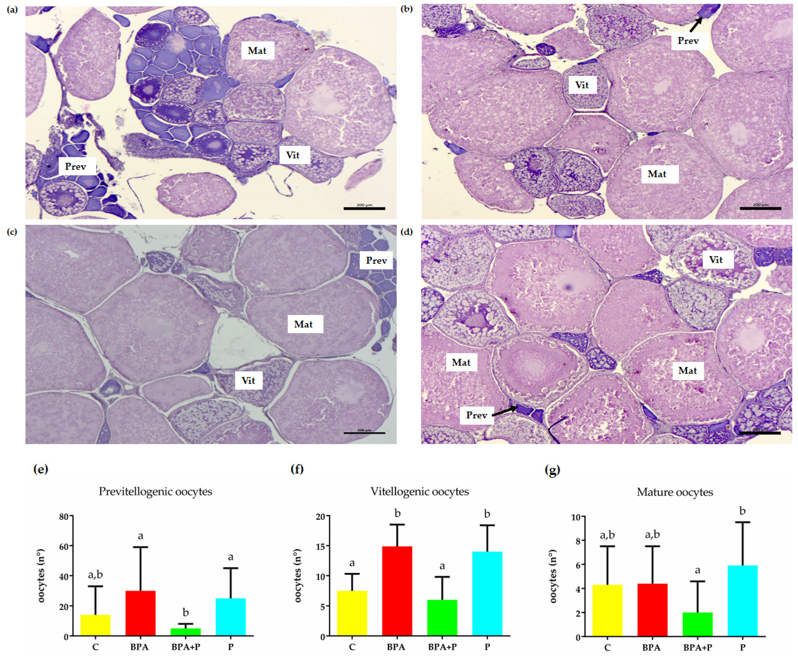
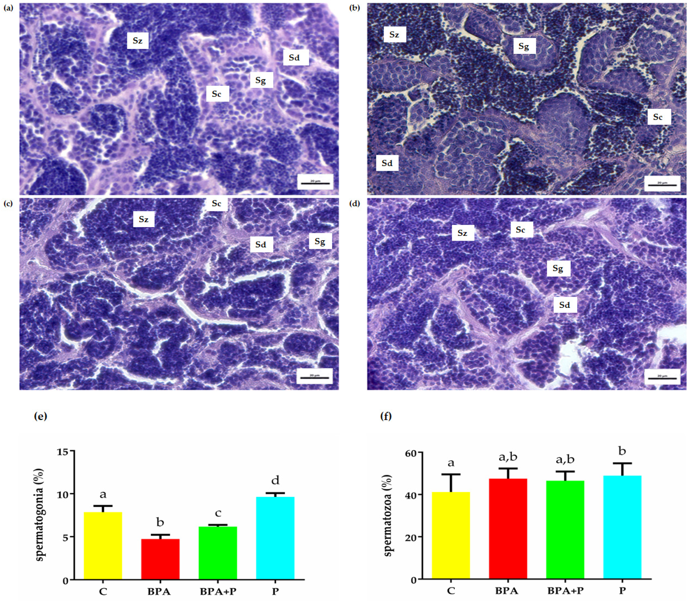
2.2. Gonadal Histological Analysis

The area covered by spermatogonia and spermatozoa was measured in the testis sections obtained from different groups (Figure 1). The spermatogonia number decreased in groups exposed to BPA and increased in testis from P fish. The results demonstrate that P mitigates BPA toxicity since, in the BPA+P group, a significant increase in spermatogonia number was observed compared to BPA. Moreover, P treatment significantly increased the number of spermatozoa compared to C (Figure 1e,f).
Histological analysis of the female ovary isolated from all experimental groups demonstrate the presence of all follicle stages, including previtellogenic- Prev-(Class I-II follicles), vitellogenic- Vit- (class III follicles), and in maturation -Mat- (class IV follicles) oocytes (Figure 2a–d). There were no significant differences in the number of Prev follicles between C and the different experimental groups. However, treatment with P in BPA+P reduced the number of BPA-induced Prev follicles to a level closer to C (Figure 2e). Similarly, treatment with BPA significantly increased the number of Vit follicles, compared to the control which was reduced following treatment with P in BPA+P (Figure 2f). Treatment with BPA did not alter the number of mature follicles compared to the control (Figure 2). The only difference observed was a significantly lower level of maturing follicles in the BPA+P compared to P treated groups (Figure 2g).

2.3. Real Time PCR Analisys

2.3.1. Hepatic Vitellogenin (vtg) Transcription

In this study, we measured the hepatic mRNA levels of different vtg isoforms using Real-time PCR (Table 1). BPA treatment caused a decrease of vtg1 and vtg6 mRNA levels and an upregulation of vtg7 form. Probiotic administration downregulated vtg1 mRNA levels and upregulated vtg 3, 4, 5, and 7 isoforms. Surprisingly, a negative synergic action of BPA and P was observed in case of vtg1 mRNA, which reaches lower levels in respect to those of BPA or P treatment alone, suggesting a clear antiestrogenic action. BPA and P coadministration upregulated vtg7 transcript, but to a lower extent in respect to BPA or P group alone, although still statistically higher than in C fish. Regarding vtg3, the downregulation, although not statistically significant of BPA, is mitigated by P co-administration and in BPA+P group levels result similar to those measured in C fish.
Concerning vtg4, when co-administered, BPA antagonizes the upregulation induced by P and levels result similar to those measure in BPA and C groups. In BPA+P group vtg5 form is significantly upregulated in respect to C and BPA fish, with levels similar to those observed in P group, suggesting an estrogenic effect of P and that this form is not BPA responsive. In BPA+P group, vtg 6 mRNA is downregulated and levels are similar to those in BPA fish, clearly suggesting that P does not modulate this vtg form.

2.3.2. Transcript of Genes Involved in Spermatogenesis

In this study, we measured transcript levels for a number of genes involved in the control of spermatogenesis in the zebrafish testis. Treatment with BPA significantly reduced the follicle stimulation hormone receptor, fshr, transcript level compared to the control. Treatment with P alone was without an effect but reversed the BPA-induced response (in BPA+P) to the control level (Table 2). Treatment with BPA significantly reduced the basal luteinizing hormone receptor, lhcgr, transcript level. Treatment with P alone did not change the basal lhcgr level and was without effect on the BPA-induced response (Table 2). Similarly, treatment with BPA significantly reduced the basal androgen receptor, ar, transcript level. Treatment with P was without a significant effect on the ar transcript level, compared to the control, but reversed the BPA-induced response to a level not different from the basal (Table 2). Treatment with BPA was without effect on the basal estrogen receptor 1, esr1, which was reduced following treatment with P alone and BPA+P, compared to the control (Table 2). Treatment with BPA significantly increased the basal esr2a transcript level, which was significantly reduced below the control and BPA level following treatments with either P alone or BPA+P (Table 2). Treatment with BPA was without effect on the basal esr2b transcript level, which was reduced following treatment with P alone and BPA+P, compared to the control (Table 2). While there were fluctuations, the basal membrane associated progesterone receptor 1, pgrmc1, transcript level was not altered significantly following treatments with BPA, P, and BPA+P (Table 2). Treatments with BPA or P alone significantly reduced the basal pgrmc2 transcript level. Paradoxically, combined treatment with BPA and P (BPA+P) increased the pgrmc2 transcript level to the control level (Table 2).

2.3.3. Transcript of Genes Involved in Follicle Growth and Maturation

In this study, we measured transcript levels for a number of genes involved in the control of follicular growth and maturation in the isolated class III and IV follicles. Treatment with BPA alone or in combination with P, significantly increased the fshr transcript level compared to control in the class III follicles. Treatment with P was without effect (Table 3). Similarly, in class IV follicles, BPA-treatment significantly increased the basal fshr transcript level, and treatment with P was without effect on the basal or the BPA-induced response (Table 3). In class III follicles, treatment with BPA significantly increased the basal lhcgr transcript level, while P alone significantly reduced the basal lhcgr level and the BPA-induced response to the control level (Table 3). In class IV follicles, BPA treatment also significantly increased the basal lhcgr transcript level, but unlike class III follicles, treatment with P alone significantly increased the basal lhcgr transcript level. In the latter follicles, treatment with P reduced the BPA-induced response to the basal level (Table 3). In the class III follicles, treatments with either BPA or P alone significantly elevated the basal pgrmc1 transcript level, and co-treatment with P did not alter the BPA-induced response. In the class IV follicles, treatments with either BPA or P alone did not alter the basal pgrmc1 transcript level, but co-treatments with BPA+P significantly reduced the pgrmc1 transcript level, compared to other groups (Table 3). In class III follicles, treatments with either BPA or P alone significantly reduced the basal pgrmc2 transcript level well below the control. Co-treatment with both BPA and P paradoxically increased and restored the pgrmc2 transcript level to the control level (Table 3). In class IV follicles, treatments with either BPA or P significantly reduced the basal pgrmc2 transcript level. In the latter class of follicles, co-treatment with BPA and P resulted in a pgrmc2 transcript level similar to P alone (Table 3).

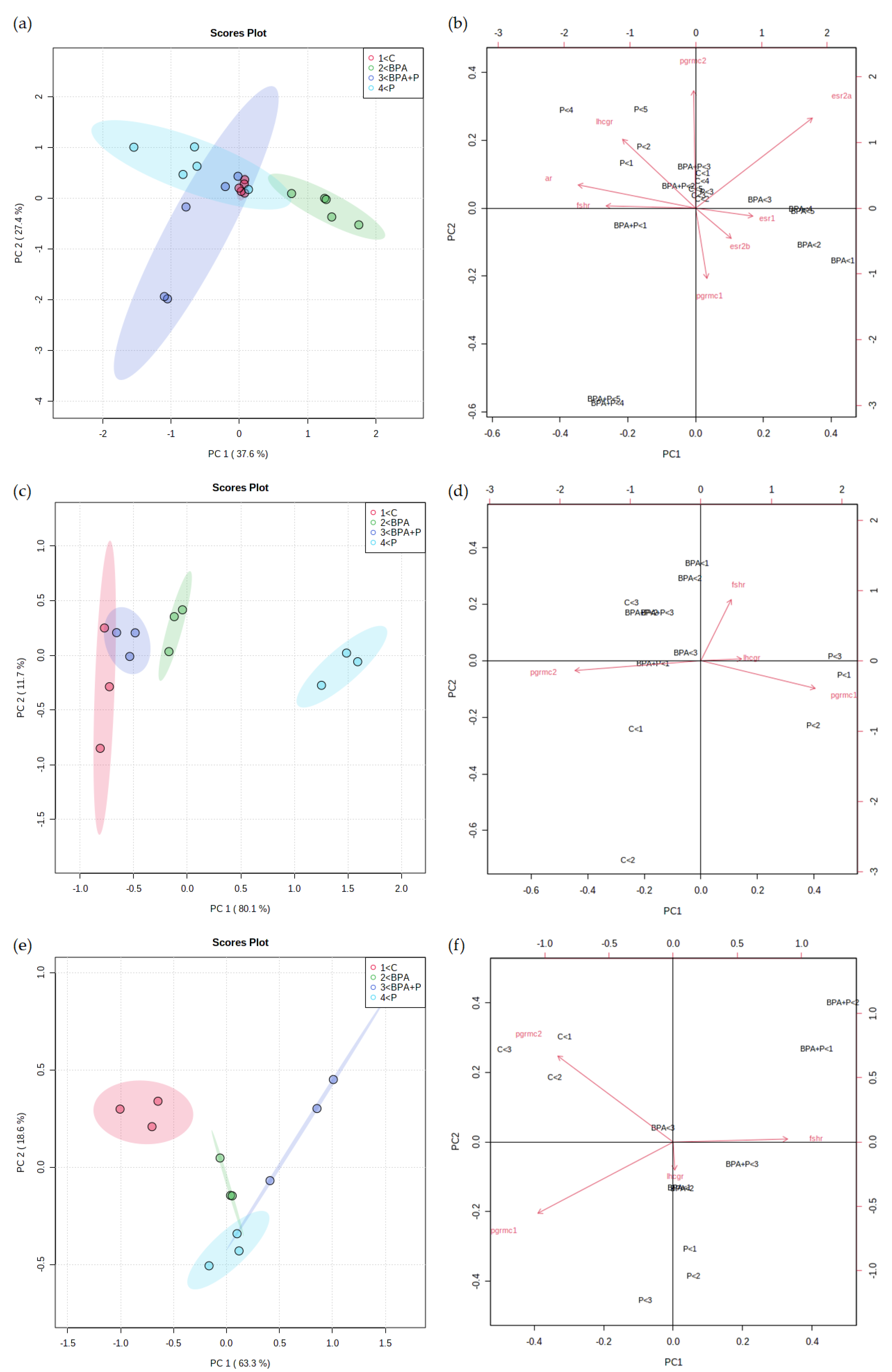
2.3.4. Multivariate Statistical Analysis

In this study, we performed unsupervised Principal Component Analysis (PCA) on three datasets obtained following the treatment of testis, class III, and class IV follicles in order to visualize the entire data set. Each point represents transcript data for each animal obtained from all treatment groups as single points corresponding to single samples and plotted into a reduced dimensional space, built with the calculated Principal Components (PCs). The position of samples into this space is given by the PC scores. The order of the PCs indicates their importance to the dataset (e.g., PC1 explains the highest amount of variation). PC loadings represent the relation between the input variables and the PCs and are used to assess how each variable contributes to a specific PC.
PCA results on testis analysis (Figure 3a), provides satisfactory cumulative explained variance (65%) with an overlapped region between P and BPA+P, containing the C group, provided by PC1, while PC2 shows separation between BPA and all other groups. Considering class III and IV follicles analysis, Figure 3c,e show that the combination of PC1 and PC2 value provide a result in terms of cumulative explained variance for class III (91.8%) and IV (81.9%) respectively, and of group segregation. In fact, samples appeared in the plot organized in compact clusters, demonstrating the presence of no outliers. The distribution of experimental groups was consistent with the working hypothesis, considering that in class III follicles PC1 (Figure 3c), accounting for the major variance, discriminates between C and BPA and P, but not BPA+P, while PC2 did not show a separation among groups. In class IV oocytes, PC1 discriminates between C and all treatments, while PC2 does not show separation between C and BPA+P (Figure 3e).
In general terms, data demonstrate the ability of P, when co-administered with BPA, to bring the BPA+P group closer to P, as observed in males or in class IV follicles or to C as seen in class III ones. These data suggest the ability of P to counteract BPA effect, showing the mitigating role of this probiotic formulation at a gonadal level.
The biplot shows the analysis of loadings together with the PCA, making it possible to understand the variable’s contribution to model building. Red arrows display the loading of each variable. Male PCA present fshr, ar, and esr1, contributing to PC1, pgrmc 1 and pgrmc2 have an effect on PC2, while lhcgr, esr2a, and esr2b have an impact on both PCs (Figure 3b).
PC1 big variance is explained by the fact that most of the variable presents the large effect of this component: lhcgr, pgrmc1, and pgrmc2 in class III oocytes (Figure 3d), and fshr and pgrmc1 in class IV oocytes (Figure 3f), while in this class pgrmc2 accounts for separation in both PC1 and PC2. Only fshr in class III follicles, and lhcgr in class IV follicles contribute to PC2, with lhcgr showing little impact.

3. Discussion

The beneficial role of SLAb51 administration was previously demonstrated in different animal models: and enhancement of specific immune functions associated to changes of intestinal microbiota was observed in healthy dogs [24], while in mice models affected by Alzheimer’s disease, a reduction of brain oxidative damages [25] was described.
This study presents the first results related to the role of SLAb51 in female and male zebrafish reproduction and evidenced its ability to mitigate BPA reproductive toxicity [9,26], in some cases antagonizing its well-known disruptive actions. Reproduction, indeed, is controlled by a delicate balance that is established between endocrine and paracrine factors [27,28] and a set of genes are involved in the intricate processes leading to the production of gonadal steroids that facilitate the production of viable gametes. Consistent with previous observations in rats [29,30], the results herein obtained in fish exposed to BPA clearly show an alteration of spermatogenesis and confirm a previous study in zebrafish [26]. These last authors demonstrated that the adverse actions of BPA are in part mediated by disrupting the endocannabinoid system (ECS) functioning by competing with its endogenous ligands. The correct ECS functioning is, in fact, essential for the normal progression of the male and female reproductive processes and the loss of anandamide binding is responsible for the observed decrease in spermatogonia numbers. Aside from interacting with the ECS, BPA can also decrease GnRH levels [31], in turn, disrupt the production of gonadotropins and testosterone, leading to a reduction of spermatogenesis [32]. In this study, we observed a BPA-mediated reduction of both gonadotropin receptor mRNA levels, which could potentially affect the FSH activity needed for conversion of spermatogonia A into B, and in case of LH, affect the final spermiogenic phase [33]. Regarding this last aspect, since the whole spermatogenesis in zebrafish lasts 28 days, we cannot exclude that longer exposure to BPA may also affect spermatozoa numbers. In addition, BPA could also exert antiandrogenic effects as indicated by the observed decrease in ar mRNA, with the possible consequence of reducing the activity of androgens such as T and 11-ketosterone [34,35]. In this context, the production of androgens may also be caused by the decrease of FSH [36]. Moving to SLAb51, the present results demonstrate its potential beneficial actions on spermatogenesis, either when administered alone or with BPA. In BPA+P group, P counteracts BPA-induced fshr mRNA downregulation, and this could be responsible for the observed increase in the number of spermatogonia. The increase of fshr transcript can be associated to an increase of FSH, as previously demonstrated in zebrafish [33] or in eels fed Lactobacillus rhamnosus [37].
In females, the number of vitellogenic oocytes observed following BPA exposure confirms previous data [26,38] indicating the estrogen-like activity of this contaminant thus upregulating vitellogenin transcript levels [39,40]. Similarly, as previously observed in fish treated with L. rhanmosus [14,16], also SLAb51 administration stimulated certain forms of vtg. Focusing on vitellogenesis, in oviparous vertebrates this process starts in the liver and is triggered by gonadal estrogens [41,42]. Zebrafish contain 8 different forms of vitellogenin genes [43] encoding for 3 proteins including type I (vtg1, vtg4, vtg5, vtg6, and vtg7), type II (vtg2 and vtg8) and type III (vtg3) [44], which differently contribute to the embryonic morphogenesis, hatching, larval kinetics and survival (vtg1, 3, 4, and 5) or provide homeostatic regulation of total vtg levels (vtg7) [45,46]. In the present study, the observed increase in vtg7 mRNA levels in all groups suggests the activation of a compensatory mechanism due to the alterations of the other gene forms. An increase of vtg7, in fact, has been observed both at the mRNA and protein levels in knock-out zebrafish phenotypes [46]. The role of vtg subtypes may be different among fish species. Differently from what is observed in this study, in seabream fed with BPA-contaminated feed, an increase of vtga and b, corresponding to zebrafish type 1 and 2 forms was induced. The observed increase in transcription of vtg forms following treatment with SLAb51 is consistent with the results obtained in fish receiving L. rhamnosus, possibly involving changes in neuropeptides and metabolic signals, thus suggesting its positive effect on reproduction as a food additive [47]. The present results also demonstrate the ability of SLAb51 to mitigate BPA effects on vtg 3 and 5 forms, which are crucial for fertility and embryo development. As previously described in catfish [48] or in zebrafish larvae [49] exposed to similar BPA concentrations, also in the present study, BPA exposure determined an increase of gonadotrophin receptor mRNA in both follicle classes, while data in the whole ovary reported that the same dose did not affect fshr levels, and significantly inhibits the transcription of lhcgr [8]. The present results demonstrate that SLAb51 can differently modulate fshr and lhcgr mRNA, and counteract BPA effects.
P differently modulates fshr and lhcgr mRNA, and in class III and IV follicles, its ability to contrast BPA effect was evidenced: mRNA levels were closer to those of C fish. However, thr fshr transcript trend does not reflect the number of vitellogenic oocytes, suggesting that BPA and P most likely may affect not only gene transcription but also protein synthesis.
In addition, BPA and SLAb51 do not affect maturation process which is featured by the localization of progesterone receptors on class III oocyte membrane, which are therefore defined as maturationally competent [50]. Previous studies have shown that pgrmc1 and 2 mRNA levels vary during follicle development, increasing in later stages. Zebrafish pgrmc1 −/− show a reduction of fecundity and fertility [51] and similarly, in female mice, conditional ablation of pgrmc1 results in reduced fertility, while elimination of pgrmc2 causes premature reproductive senescence [52]. In class III oocytes isolated from fish exposed to BPA or P, an increase in the expression of form 1 and a down-regulation of form 2 was observed and therefore since they are both involved in the maturation process [53], this could result in the lack of increase of maturing oocytes, despite the higher number of vitellogenic ones. In BPA+P group, mRNA transcripts suggest the ability of P to antagonize BPA as clearly demonstrated by levels similar to C ones. The scenario herein described is very similar to that observed in a study on zebrafish co-exposed to L. rhamnosus and perfluorobutanesulfonate (PFBS) [23], where co-exposure almost ceased the fecundity, which was accompanied by disturbances in sex hormones and oocyte maturation in females [54], in contrast, in males, probiotic additive efficiently antagonized the estrogenic activity of PFBS. Nevertheless, these authors demonstrated the antagonistic interaction between PFBS and L. rhamnosus regarding the metabolic activities along the microbe, gut and liver axis [55] with an efficient mitigation of lipid [56] and glucose [57] metabolic disorders associated with PFBS exposure, highlighting the potential values of probiotic bacteria used to protect the aquatic ecosystem. Different bacteria strains have been proven so far to promote the degradation of specific environmental pollutants that result in being harmful to organisms while increasing host resistance and resilience [58,59] This was clearly demonstrated in a trial where the toxicity of triclosan (TCS) was alleviated by feeding zebrafish Lactobacillus plantarum ST-III; dietary probiotic administration alleviated the intestinal metabolic syndromes and neurodegenerative diseases resulting from exposure to TCS, through modifying the gut flora [60].
In conclusion, these results present the first preliminary data supporting the hypothesis that SLAb51 can antagonize/mitigate some BPA toxic action in zebrafish and likely in other vertebrate species. The findings also suggest the ability of SLAb51 to positively interact with spermatogenesis, while regarding oocyte growth and maturation further investigation are needed. Changes of probiotic concentrations or trial duration could be useful to clarify the SLAb51 reproductive effects also in female zebrafish. This would help in building up a comprehensive scenario regarding Slab51 effects in zebrafish often used as aquatic species model, in the light of supporting aquaculture practices or within a bioremediation contest. In the last years, interest on this last aspect is in fact increasing and the use of bacteria is considered a good strategy to guarantee sustainability, as it has a relatively low cost and can be applied in different ecosystems, causing minimal impact to the environment.
On this regard, the results obtained in this study, together with those obtained by other authors showing the possible role of probiotics in counteracting the toxic effect of different EDCs on several physiological process should encourage further research in order to optimize the use of probiotics to mitigate the effect of the many toxicants ubiquitously present worldwide.

4. Materials and Methods

4.1. SLAb51® (SivoMixx®)

SLAb51® (SivoMixx®, Ormendes SA, Jouxtens-Mézery, CH, Switzerland) is a commercial multi-strain probiotic containing 200 billion lactic acid bacteria per 1.5 grams of product, comprised of the following strains: Streptococcus thermophilus DSM 32245, Bifidobacterium lactis DSM 32246, Bifidobacterium lactis DSM 32247, Lactobacillus acidophilus DSM 32241, Lactobacillus helveticus DSM 32242, Lactobacillus paracasei DSM 32243, Lactobacillus plantarum DSM 32244, and Lactobacillus brevis DSM 27961.

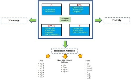
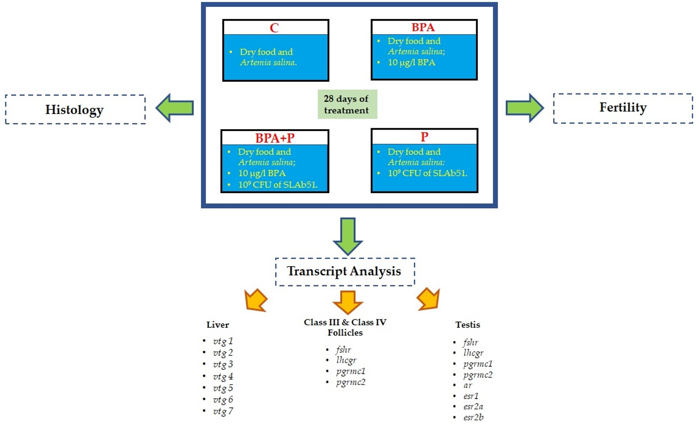
4.2. Animal Treatment

A total of 80 adult zebrafish (40 male and 40 female) (D. rerio, AB wild-type strain) were divided into 8 10-L aquaria (10 fish/tank) with oxygenated water under controlled conditions (28.0 ± 0.5 °C) and maintained on a 14/10 h light/dark cycle. They were fed twice a day commercial food (Vipagran; Sera, Loessnitz, Germany). The experiment was set up in duplicate as follows:
C: control fish fed twice a day with commercial food (Vipagran; Sera, Loessnitz, Germany)
BPA: fish were fed commercial food and were exposed to 10 μg/L BPA (98% analytical purity, Sigma-Aldrich, Milano, Italy)
BPA+P: fish were exposed to 10 μg/L BPA and fed commercial food supplemented with SLAb51 at a final concentration of 109 CFU/g
P: fish were fed commercial food fish and received a dietary supplementation of SLAb51 at a final concentration of 109 CFU/g
All groups were sampled after 4 weeks of treatment.
At the end of the trial, fish were lethally anesthetized with 500 mg/L MS-222 (3-aminobenzoic acid ethyl ester, Sigma Aldrich) buffered to pH 7.4. Livers were stored at −80 °C for molecular analysis. Testis and ovaries were dissected out and divided as follows: 5 samples were fixed in Bouin’s fixative for histology. The remaining 5 testis were stored at −80 °C, while ovaries were teased into separate follicles using transfer pipettes (Semco Scientific Corp., San Diego, CA, USA) without trypsinization; thereafter, follicles were separated into different maturation stages according to their diameters, as previously described [61,62] and class III and IV follicles were collected and stored at −80 °C for molecular analysis.

4.3. Fish Fertility

Reproductive performances of each experimental group were assayed in spawning tanks under standard conditions as previously described [8]. Starting on the 8th day of treatment, male and female zebrafish from the 4 groups were crossed and fertility was determined during the following 28 days. Fertilized eggs were counted, and the fertility rate was calculated as the mean ± standard deviation (SD) of fertilized egg number/female/day from the 8th to the 21st day of treatment.

4.4. Gonad Histology and Image Analysis

Hystological analyses were performed on 5 ovaries and testis for each experimental group according to Forner-Piquer et al., 2017 [63].

4.5. RNA Extraction and cDNA Synthesis

For each experimental group, total RNA was extracted from 5 female livers and 5 testis, and from 3, classes III and IV pools, containing 50 follicles each, using RNAeasy® Minikit (Qiagen, Milano, Italy). RNA quality assessment and cDNA synthesis were performed as previously described [61].

4.6. Real-Time PCR

qRT-PCRs were performed with SYBR green in an CFX thermal cycler, as previously described [26]. Ribosomal protein 13 (rpl13) and ribosomal protein 0 (rplp0) mRNAs were used as internal standards in each sample in order to standardize the results by eliminating variation in mRNA and cDNA quantity and quality. Primer sequences, GenBank accession numbers and primer efficiency of the examined genes are reported in Table 4.
mRNA levels of each target gene analyzed were calculated using the Pfaffl method [64], relative to the geometric mean of the two reference genes once demonstrated they were stably expressed by the geNorm algorithm, both implemented in the Bio-Rad CFX Manager 3.1. software. Modification of gene expression among the experimental groups is reported as relative mRNA abundance (arbitrary units). Primers were used at a final concentration of 10 pmol/mL

4.7. Statistical Analysis

All the data were analyzed by One-Way ANOVA followed by Dunnett’s multiple comparison test. When the collected data was expressed in percentage, arcsin transformation was conducted before ANOVA. All statistical analyses were performed using the statistical soft- ware package Prism5 (GraphPad Software, Inc., San Diego, CA, USA) with significance accepted at p < 0.05. Unsupervised Principal Component Analysis (PCA) were performed using Metaboanalyst 5.0 online platform (University of Aberta, Edmonton, AB, Canada). Data sets were created using genes with p-value < 0.05, and underwent normalization, using normalization by median, transformation, with log transformation, and scaling, using pareto scaling. A 2D score plot was generated, plotting the first two principal component, to show separation among groups based on gene expression profile.

Author Contributions

Formal analysis, C.G., M.C. and F.M.; conceptualization, O.C. and H.R.H.; validation, F.M., C.G.; methodology, O.C., F.M. and C.G.; writing—original draft preparation, F.M., C.G.; writing—review and editing, F.M., C.G., H.R.H. and O.C. All authors have read and agreed to the published version of the manuscript.

Funding

This research was funded by Ricerca Scientifica d’Ateneo 2019, Università Politecnica delle Marche to O.C.

Institutional Review Board Statement

The study was conducted according to the guidelines of the Declaration of Helsinki and was approved by the Ethics Committee of University of Calgary—Animal Care protocol (AC15-0183).

Informed Consent Statement

Not applicable.

Acknowledgments

The authors wish to thank Francesco Citarella for his help with histological analysis.

Conflicts of Interest

The authors declare no conflict of interest.

References

  1. Naidu, R.; Biswas, B.; Willett, I.R.; Cribb, J.; Kumar Singh, B.; Paul Nathanail, C.; Coulon, F.; Semple, K.T.; Jones, K.C.; Barclay, A.; et al. Chemical pollution: A growing peril and potential catastrophic risk to humanity. Environ. Int. 2021, 156, 106616. [Google Scholar] [CrossRef]
  2. Maradonna, F.; Carnevali, O. Lipid Metabolism Alteration by Endocrine Disruptors in Animal Models: An Overview. Front. Endocrinol. 2018, 9, 1–14. [Google Scholar] [CrossRef] [Green Version]
  3. Carnevali, O.; Santangeli, S.; Forner-Piquer, I.; Basili, D.; Maradonna, F. Endocrine-disrupting chemicals in aquatic environment: What are the risks for fish gametes? Fish Physiol. Biochem. 2018, 44, 1561–1576. [Google Scholar] [CrossRef]
  4. Zhu, L.; Wang, L.; Fan, X.; Dong, C.; Wang, G.; Wang, Z. Chronic exposure to Bisphenol A resulted in alterations of reproductive functions via immune defense, oxidative damage and disruption DNA/histone methylation in male rare minnow Gobiocypris rarus. Aquat. Toxicol. 2021, 236, 105849. [Google Scholar] [CrossRef]
  5. Kaimal, A.; Al Mansi, M.H.; Dagher, J.B.; Pope, C.; Varghese, M.G.; Rudi, T.B.; Almond, A.E.; Cagle, L.A.; Beyene, H.K.; Bradford, W.T.; et al. Prenatal exposure to bisphenols affects pregnancy outcomes and offspring development in rats. Chemosphere 2021, 276, 130118. [Google Scholar] [CrossRef]
  6. Lin, M.; Hua, R.; Ma, J.; Zhou, Y.; Li, P.; Xu, X.; Yu, Z.; Quan, S. Bisphenol A promotes autophagy in ovarian granulosa cells by inducing AMPK/mTOR/ULK1 signalling pathway. Environ. Int. 2021, 147, 106298. [Google Scholar] [CrossRef]
  7. Mathew, H.; Mahalingaiah, S. Do prenatal exposures pose a real threat to ovarian function? Bisphenol A as a case study. Reproduction 2019, 157, R143–R157. [Google Scholar] [CrossRef] [PubMed] [Green Version]
  8. Santangeli, S.; Maradonna, F.; Gioacchini, G.; Cobellis, G.; Piccinetti, C.C.; Dalla Valle, L.; Carnevali, O. BPA-Induced Deregulation Of Epigenetic Patterns: Effects On Female Zebrafish Reproduction. Sci. Rep. 2016, 6, 21982. [Google Scholar] [CrossRef]
  9. Santangeli, S.; Maradonna, F.; Olivotto, I.; Piccinetti, C.C.; Gioacchini, G.; Carnevali, O. Effects of BPA on female reproductive function: The involvement of epigenetic mechanism. Gen. Comp. Endocrinol. 2017, 245, 122–126. [Google Scholar] [CrossRef]
  10. Maradonna, F.; Nozzi, V.; Dalla Valle, L.; Traversi, I.; Gioacchini, G.; Benato, F.; Colletti, E.; Gallo, P.; Di Marco Pisciottano, I.; Mita, D.G.; et al. A developmental hepatotoxicity study of dietary bisphenol A in Sparus aurata juveniles. Comp. Biochem. Physiol. C Toxicol. Pharmacol. 2014, 166, 1–13. [Google Scholar] [CrossRef]
  11. Maradonna, F.; Nozzi, V.; Santangeli, S.; Traversi, I.; Gallo, P.; Fattore, E.; Mita, D.G.; Mandich, A.; Carnevali, O. Xenobiotic-contaminated diets affect hepatic lipid metabolism: Implications for liver steatosis in Sparus aurata juveniles. Aquat. Toxicol. 2015, 167, 257–264. [Google Scholar] [CrossRef]
  12. Carnevali, O.; Giorgini, E.; Canuti, D.; Mylonas, C.C.; Forner-Piquer, I.; Maradonna, F. Diets contaminated with Bisphenol A and Di-isononyl phtalate modify skeletal muscle composition: A new target for environmental pollutant action. Sci. Total Environ. 2019, 658, 250–259. [Google Scholar] [CrossRef]
  13. Gao, X.; Kang, S.; Xiong, R.; Chen, M. Environment-friendly removal methods for endocrine disrupting chemicals. Sustainability 2020, 12, 7615. [Google Scholar] [CrossRef]
  14. Gioacchini, G.; Maradonna, F.; Lombardo, F.; Bizzaro, D.; Olivotto, I.; Carnevali, O. Increase of fecundity by probiotic administration in zebrafish (Danio rerio). Reproduction 2010, 140, 953–959. [Google Scholar] [CrossRef] [PubMed] [Green Version]
  15. Carnevali, O.; Avella, M.A.; Gioacchini, G. Effects of probiotic administration on zebrafish development and reproduction. Gen. Comp. Endocrinol. 2012, 188, 297–302. [Google Scholar] [CrossRef]
  16. Giorgini, E.; Conti, C.; Ferraris, P.; Sabbatini, S.; Tosi, G.; Rubini, C.; Vaccari, L.; Gioacchini, G.; Carnevali, O. Effects of Lactobacillus rhamnosus on zebrafish oocyte maturation: An FTIR imaging and biochemical analysis. Anal. Bioanal. Chem. 2010, 398, 3063–3072. [Google Scholar] [CrossRef]
  17. Arani, M.M.; Salati, A.P.; Keyvanshokooh, S.; Safari, O. The effect of Pediococcus acidilactici on mucosal immune responses, growth, and reproductive performance in zebrafish (Danio rerio). Fish Physiol. Biochem. 2021, 47, 153–162. [Google Scholar] [CrossRef]
  18. Hu, J.; Kim, Y.H.; Kim, I.H. Effects of two bacillus strains probiotic supplement on reproduction performance, nutrient digestibility, blood profile, fecal score, excreta odor contents and fecal microflora in lactation sows, and growth performance in sucking piglets. Livest. Sci. 2021, 244, 104293. [Google Scholar] [CrossRef]
  19. Ceccarelli, G.; Borrazzo, C.; Pinacchio, C.; Santinelli, L.; Innocenti, G.P.; Cavallari, E.N.; Celani, L.; Marazzato, M.; Alessandri, F.; Ruberto, F.; et al. Oral Bacteriotherapy in Patients With COVID-19: A Retrospective Cohort Study. Front. Nutr. 2021, 7, 1–8. [Google Scholar] [CrossRef]
  20. d’Ettorre, G.; Ceccarelli, G.; Marazzato, M.; Campagna, G.; Pinacchio, C.; Alessandri, F.; Ruberto, F.; Rossi, G.; Celani, L.; Scagnolari, C.; et al. Challenges in the Management of SARS-CoV2 Infection: The Role of Oral Bacteriotherapy as Complementary Therapeutic Strategy to Avoid the Progression of COVID-19. Front. Med. 2020, 7, 1–7. [Google Scholar]
  21. Cai, S.-S.; Zhou, Y.; Ye, B.-C. Reducing the reproductive toxicity activity of Lactiplantibacillus plantarum: A review of mechanisms and prospects. Environ. Sci. Pollut. Res. 2021, 1–15. [Google Scholar] [CrossRef]
  22. Taghizadeh Moghaddam, S.; Javadi, A.; Matin, A.A. Reduction of bisphenol A by Lactobacillus acidophilus and Lactobacillus plantarum in yoghurt. Int. J. Dairy Technol. 2020, 73, 737–742. [Google Scholar] [CrossRef]
  23. Tang, L.; Song, S.; Hu, C.; Lam, J.C.W.; Liu, M.; Zhou, B.; Lam, P.K.S.; Chen, L. Unexpected Observations: Probiotic Administration Greatly Aggravates the Reproductive Toxicity of Perfluorobutanesulfonate in Zebrafish. Chem. Res. Toxicol. 2020, 33, 1605–1608. [Google Scholar] [CrossRef]
  24. Rossi, G.; Pengo, G.; Galosi, L.; Berardi, S.; Tambella, A.M.; Attili, A.R.; Gavazza, A.; Cerquetella, M.; Jergens, A.E.; Guard, B.C.; et al. Effects of the Probiotic Mixture Slab51® (SivoMixx®) as Food Supplement in Healthy Dogs: Evaluation of Fecal Microbiota, Clinical Parameters and Immune Function. Front. Vet. Sci. 2020, 7, 7. [Google Scholar] [CrossRef]
  25. Castelli, V.; Angelo, M.; Lombardi, F.; Alfonsetti, M.; Catanesi, M.; Benedetti, E.; Palumbo, P.; Grazia, M.; Giordano, A.; Desideri, G.; et al. Effects of the probiotic formulation SLAB51 in in vitro and in vivo Parkinson’s disease models. Aging 2020, 12, 4641–4659. [Google Scholar] [CrossRef]
  26. Forner-Piquer, I.; Beato, S.; Piscitelli, F.; Santangeli, S.; Di Marzo, V.; Habibi, H.R.; Maradonna, F.; Carnevali, O. Effects of BPA on zebrafish gonads: Focus on the endocannabinoid system. Environ. Pollut. 2020, 264, 114710. [Google Scholar] [CrossRef]
  27. Biran, J.; Levavi-Sivan, B. Endocrine Control of Reproduction, Fish. Encycl. Reprod. 2018, 63, 362–368. [Google Scholar]
  28. Yaron, Z.; Gur, G.; Melamed, P.; Rosenfeld, H.; Elizur, A.; Levavi-Sivan, B. Regulation of fish gonadotropins. Int. Rev. Cytol. 2003, 225, 131–185. [Google Scholar]
  29. Jin, P.; Wang, X.; Chang, F.; Bai, Y.; Li, Y.; Zhou, R.; Chen, L. Low dose bisphenol A impairs spermatogenesis by suppressing eproductive hormone production and promoting germ cell apoptosis in adult rats. J. Biomed. Res. 2013, 27, 135–144. [Google Scholar]
  30. Ullah, A.; Pirzada, M.; Jahan, S.; Ullah, H.; Turi, N.; Ullah, W.; Siddiqui, M.F.; Zakria, M.; Lodhi, K.Z.; Khan, M.M. Impact of low-dose chronic exposure to bisphenol A and its analogue bisphenol B, bisphenol F and bisphenol S on hypothalamo-pituitary-testicular activities in adult rats: A focus on the possible hormonal mode of action. Food Chem. Toxicol. 2018, 121, 24–36. [Google Scholar] [CrossRef]
  31. Klenke, U.; Constantin, S.; Wray, S. BPA directly decreases gnrh neuronal activity via noncanonical pathway. Endocrinology 2016, 157, 1980–1990. [Google Scholar] [CrossRef] [PubMed] [Green Version]
  32. Castellini, C.; Totaro, M.; Parisi, A.; D’Andrea, S.; Lucente, L.; Cordeschi, G.; Francavilla, S.; Francavilla, F.; Barbonetti, A. Bisphenol A and Male Fertility: Myths and Realities. Front. Endocrinol. 2020, 11, 1–10. [Google Scholar] [CrossRef]
  33. Fallah, H.P.; Rodrigues, M.S.; Zanardini, M.; Nóbrega, R.H.; Habibi, H.R. Effects of gonadotropin-inhibitory hormone on early and late stages of spermatogenesis in ex-vivo culture of zebrafish testis. Mol. Cell. Endocrinol. 2021, 520, 111087. [Google Scholar] [CrossRef] [PubMed]
  34. Miura, T.; Yamauchi, K.; Nagahama, Y.; Takahashi, H. Induction of spermatogenesis in male japanese eel, Anguilla japonica, by a single injection of human chorionic gonadotropin. Zoolog. Sci. 1991, 8, 63–73. [Google Scholar]
  35. Schulz, R.; de França, L.; Lareyre, J.; Le Gac, F.; Chiarini-Garcia, H.; Nobrega, R.; Miura, T. Spermatogenesis in fish. Gen. Comp. Endocrinol. 2010, 165, 390–411. [Google Scholar] [CrossRef]
  36. Ohta, T.; Miyake, H.; Miura, C.; Kamei, H.; Aida, K.; Miura, T. Follicle-stimulating hormone induces spermatogenesis mediated by androgen production in Japanese eel, Anguilla japonica. Biol. Reprod. 2007, 77, 970–977. [Google Scholar] [CrossRef] [PubMed] [Green Version]
  37. Vílchez, M.C.; Santangeli, S.; Maradonna, F.; Gioacchini, G.; Verdenelli, C.; Gallego, V.; Peñaranda, D.S.; Tveiten, H.; Pérez, L.; Carnevali, O.; et al. Effect of the probiotic Lactobacillus rhamnosus on the expression of genes involved in European eel spermatogenesis. Theriogenology 2015, 84, 1321–1331. [Google Scholar] [CrossRef]
  38. Migliaccio, M.; Chioccarelli, T.; Ambrosino, C.; Suglia, A.; Manfrevola, F.; Carnevali, O.; Fasano, S.; Pierantoni, R.; Cobellis, G. Characterization of follicular atresia responsive to BPA in zebrafish by morphometric analysis of follicular stage progression. Int. J. Endocrinol. 2018, 2018, 4298195. [Google Scholar] [CrossRef] [Green Version]
  39. Molina, A.M.; Lora, A.J.; Blanco, A.; Monterde, J.G.; Ayala, N.; Moyano, R. Endocrine-active compound evaluation: Qualitative and quantitative histomorphological assessment of zebrafish gonads after bisphenol-A exposure. Ecotoxicol. Environ. Saf. 2013, 88, 155–162. [Google Scholar] [CrossRef]
  40. Molina, A.M.; Abril, N.; Morales-Prieto, N.; Monterde, J.G.; Lora, A.J.; Ayala, N.; Moyano, R. Evaluation of toxicological endpoints in female zebrafish after bisphenol A exposure. Food Chem. Toxicol. 2018, 112, 19–25. [Google Scholar] [CrossRef]
  41. Gioacchini, G.; Marisaldi, L.; Basili, D.; Candelma, M.; Pignalosa, P.; Aiese Cigliano, R.; Sanseverino, W.; Hardiman, G.; Carnevali, O. A de novo transcriptome assembly approach elucidates the dynamics of ovarian maturation in the swordfish (Xiphias gladius). Sci. Rep. 2019, 9, 1–12. [Google Scholar] [CrossRef] [Green Version]
  42. Miccoli, A.; Maradonna, F.; De Felice, A.; Caputo Barucchi, V.; Estonba, A.; Genangeli, M.; Vittori, S.; Leonori, I.; Carnevali, O. Detection of endocrine disrupting chemicals and evidence of their effects on the HPG axis of the European anchovy Engraulis encrasicolus. Mar. Environ. Res. 2017, 127, 137–147. [Google Scholar] [CrossRef]
  43. Yilmaz, O.; Patinote, A.; Nguyen, T.V.; Com, E.; Lavigne, R.; Pineau, C.; Sullivan, C.V.; Bobe, J. Scrambled eggs: Proteomic portraits and novel biomarkers of egg quality in zebrafish (Danio rerio). PLoS ONE 2017, 12, e0188084. [Google Scholar] [CrossRef] [Green Version]
  44. Wang, H.; Yan, T.; Tan, J.T.T.; Gong, Z. A zebrafish vitellogenin gene (vg3) encodes a novel vitellogenin without a phosvitin domain and may represent a primitive vertebrate vitellogenin gene. Gene 2000, 256, 303–310. [Google Scholar] [CrossRef]
  45. Yilmaz, O.; Patinote, A.; Com, E.; Pineau, C.; Bobe, J. Knock out of specific maternal vitellogenins in zebrafish (Danio rerio) evokes vital changes in egg proteomic profiles that resemble the phenotype of poor quality eggs. BMC Genom. 2021, 22, 308. [Google Scholar] [CrossRef]
  46. Yilmaz, O.; Patinote, A.; Nguyen, T.; Com, E.; Pineau, C.; Bobe, J. Genome editing reveals reproductive and developmental dependencies on specific types of vitellogenin in zebrafish (Danio rerio). Mol. Reprod. Dev. 2019, 86, 1168–1188. [Google Scholar] [CrossRef] [Green Version]
  47. Carnevali, O.; Maradonna, F.; Gioacchini, G. Integrated control of fish metabolism, wellbeing and reproduction: The role of probiotic. Aquaculture 2017, 472, 144–155. [Google Scholar] [CrossRef]
  48. Faheem, M.; Lone, K.P. Oxidative stress and histopathologic biomarkers of exposure to bisphenol-A in the freshwater fish, Ctenopharyngodon idella. Braz. J. Pharm. Sci. 2018, 53, 1–9. [Google Scholar] [CrossRef] [Green Version]
  49. Qiu, W.; Chen, J.; Li, Y.; Chen, Z.; Jiang, L.; Yang, M.; Wu, M. Oxidative stress and immune disturbance after long-term exposure to bisphenol A in juvenile common carp (Cyprinus carpio). Ecotoxicol. Environ. Saf. 2016, 130, 93–102. [Google Scholar] [CrossRef]
  50. Clelland, E.; Peng, C. Endocrine/paracrine control of zebrafish ovarian development. Mol. Cell. Endocrinol. 2009, 312, 42–52. [Google Scholar] [CrossRef]
  51. Wu, X.J.; Thomas, P.; Zhu, Y. Pgrmc1 knockout impairs oocyte maturation in zebrafish. Front. Endocrinol. 2018, 9, 1–12. [Google Scholar] [CrossRef] [PubMed] [Green Version]
  52. Clark, N.C.; Pru, C.A.; Yee, S.P.; Lydon, J.P.; Peluso, J.J.; Pru, J.K. Conditional ablation of progesterone receptor membrane component 2 causes female premature reproductive senescence. Endocrinology 2017, 158, 640–651. [Google Scholar] [CrossRef]
  53. Wu, X.J.; Zhu, Y. Downregulation of nuclear progestin receptor (Pgr) and subfertility in double knockouts of progestin receptor membrane component 1 (pgrmc1) and pgrmc2 in zebrafish. Gen. Comp. Endocrinol. 2020, 285, 113275. [Google Scholar] [CrossRef]
  54. Hu, C.; Liu, M.; Tang, L.; Sun, B.; Huang, Z.; Chen, L. Probiotic Lactobacillus rhamnosus modulates the impacts of perfluorobutanesulfonate on oocyte developmental rhythm of zebrafish. Sci. Total Environ. 2021, 776, 1–9. [Google Scholar] [CrossRef]
  55. Hu, C.; Liu, M.; Tang, L.; Liu, H.; Sun, B.; Chen, L. Probiotic intervention mitigates the metabolic disturbances of perfluorobutanesulfonate along the gut-liver axis of zebrafish. Chemosphere 2021, 284, 131374. [Google Scholar] [CrossRef]
  56. Chen, L.; Lam, J.C.W.; Tang, L.; Tang, L.; Hu, C.; Liu, M.; Liu, M.; Lam, P.K.S.; Zhou, B. Probiotic Modulation of Lipid Metabolism Disorders Caused by Perfluorobutanesulfonate Pollution in Zebrafish. Environ. Sci. Technol. 2020, 54, 7494–7503. [Google Scholar] [CrossRef]
  57. Liu, M.; Tang, L.; Hu, C.; Sun, B.; Huang, Z.; Chen, L. Interaction between probiotic additive and perfluorobutanesulfonate pollutant on offspring growth and health after parental exposure using zebrafish. Ecotoxicol. Environ. Saf. 2021, 214, 112107. [Google Scholar] [CrossRef]
  58. Li, C.; Ma, Y.; Mi, Z.; Huo, R.; Zhou, T.; Hai, H.; Kwok, L.Y.; Sun, Z.; Chen, Y.; Zhang, H. Screening for Lactobacillus plantarum strains that possess organophosphorus pesticide-degrading activity and metabolomic analysis of phorate degradation. Front. Microbiol. 2018, 9, 1–13. [Google Scholar] [CrossRef] [PubMed] [Green Version]
  59. Feng, P.; Ye, Z.; Kakade, A.; Virk, A.K.; Li, X.; Liu, P. A review on gut remediation of selected environmental contaminants: Possible roles of probiotics and gut microbiota. Nutrients 2019, 11, 22. [Google Scholar] [CrossRef] [Green Version]
  60. Zang, L.; Ma, Y.; Huang, W.; Ling, Y.; Sun, L.; Wang, X.; Zeng, A.; Dahlgren, R.A.; Wang, C.; Wang, H. Dietary Lactobacillus plantarum ST-III alleviates the toxic effects of triclosan on zebrafish (Danio rerio) via gut microbiota modulation. Fish Shellfish Immunol. 2019, 84, 1157–1169. [Google Scholar] [CrossRef] [PubMed]
  61. Maradonna, F.; Gioacchini, G.; Notarstefano, V.; Fontana, C.M.; Citton, F.; Valle, L.D.; Giorgini, E.; Carnevali, O. Knockout of the glucocorticoid receptor impairs reproduction in female zebrafish. Int. J. Mol. Sci. 2020, 21, 9073. [Google Scholar] [CrossRef]
  62. Selman, K.; Wallace, R.A.; Sarka, A.; Qi, X. Stages of oocyte development in the zebrafish, Brachydanio rerio. J. Morphol. 1993, 218, 203–224. [Google Scholar] [CrossRef]
  63. Forner-Piquer, I.; Maradonna, F.; Gioacchini, G.; Santangeli, S.; Allarà, M.; Piscitelli, F.; Habibi, H.R.; Di Marzo, V.; Carnevali, O. Dose-Specific Effects of Di-Isononyl Phthalate on the Endocannabinoid System and on Liver of Female Zebrafish. Endocrinology 2017, 158, 3462–3476. [Google Scholar] [CrossRef] [Green Version]
  64. Pfaffl, M.W. Validities of mRNA quantification using recombinant RNA and recombinant. Biotechnol. Lett. 2001, 23, 275–282. [Google Scholar] [CrossRef]
Figure 1. Histological sections of testis: C (a), BPA (b), BPA+P (c), and P (d). Eosin-Mayer’s haematoxylin staining. Sg: spermatogonia; Sc: spermatocyte; Sd: spermatid; Sz: spermatozoa. Scale bar: 20 µm. Percentage of zebrafish testicular area occupied by spermatogonia (e) and spermatozoa (f). Data reported as means ± SEM. Different letters denote statistically significant differences among experimental groups (one-way ANOVA, p < 0.05, Dunnett’s multiple comparison test).
Ijms 22 09314 g001
Figure 2. Histological analysis of ovaries from C (a), BPA (b), BPA+P (c) and P (d). Ovarian sections show different follicular stages. Eosin and Mayer’s haematoxylin staining. Prev: previtellogenic oocytes; Vit: Vitellogenic oocytes; Mat: mature oocytes. Scale bar: 200 µm. Percentage of different follicle classes (eg). Data are reported as mean ± SEM. Different letters denote statistically significant differences among experimental groups (one-way ANOVA, p < 0.05, Dunnett’s multiple comparison test).
Ijms 22 09314 g002
Figure 3. Scores plot and biplot of testis (a,b), class III (c,d) and class IV (e,f) follicles of mRNA levels data obtained in C (red), BPA (green), BPA+P (blue) and P (light blue) groups. (a,c,e) Axes show scores on PC1 and PC2; (b,d,f) bottom and left axes show scores on PC1 and PC2, top and right axes show loadings; red arrows show variable loadings.
Ijms 22 09314 g003
Table 1. Hepatic vtg mRNA expression values in the different experimental groups. Data are reported as means ± SD. Different letters indicate statistically significant variations among groups. (one-way ANOVA followed by Dunnett’s multiple comparison test p < 0.05).
Female LiverCBPABPA+PP
vtg17.89 ± 1.05 (a)3.25 ± 1.20 (b)1.82 ± 0.73 (c)5.36 ± 0.87 (d)
vtg21.69 ± 0.20 (a)2.00 ± 0.82 (a)1.51 ± 0.49 (a)2.52 ± 0.35 (a)
vtg34.52 ± 0.37 (a,b)3.69 ± 0.91 (a)5.03 ± 0.59 (b)6.49± 0.49 (c)
vtg43.51 ± 1.20 (a)3.26 ± 0.52 (a)2.78 ± 0.87 (a)8.66 ± 0.50 (b)
vtg52.10 ± 0.63 (a)2.37 ± 0.35 (a)4.41 ± 0.65 (b)5.08 ± 0.59 (b)
vtg68.39 ± 0.32 (a)3.29 ± 0.96 (b)3.46 ± 0.68 (b)9.39 ± 0.89 (a)
vtg72.59 ± 0.44 (a)11.42 ± 0.84 (b)6.08 ± 0.60 (c)23.02 ± 0.79 (d)
Table 2. mRNA expression values of genes regulating spermatogenesis in the testis of the different experimental groups. Data are reported as means ± SD. Different letters indicate statistically significant variations among the groups (one-way ANOVA followed by Dunnett’s multiple comparison test, p < 0.05).
TestisCBPABPA+PP
fshr10.77 ± 2.22 (a)2.82 ± 0.27 (b)7.3 ± 1.82 (a)9.72 ± 4.67 (a)
lhcgr6.9 ± 0.69 (a)3.92 ± 0.94 (b)3.45 ± 1.45 (b)5.6 ± 2.14 (a,b)
ar5.15 ± 0.76 (a)1.88 ± 0.69 (b)3.57 ± 2.10 (a,b)2.81 ± 1.64 (a,b)
esr18.41 ± 0.61 (a)8.57 ± 1.20 (a)3.4 ± 1.12 (b)3.03 ± 1.10 (b)
esr2a5.31 ± 0.84 (a)8.11 ± 1.52 (b)2.05 ± 0.70 (c)2.12 ± 1.41 (c)
esr2b6.17± 1.23 (a)4.03 ± 1.2 (a,b)2.05 ± 0.41 (b,c)1.1 ± 0.32 (c)
pgrmc15.60 ± 1.37 (a,b)7.96 ± 2.06 (a)5.52 ± 1.89 (a,b)2.63 ± 0.93 (b)
pgrmc27.34 ± 2.06 (a)3.99 ± 1.50 (b)5.64 ± 1.14 (a,b)3.46 ± 1.17 (b)
Table 3. mRNA expression values of genes regulating follicle growth and maturation in class III (a) and IV (b) follicles. Data are reported as means ± SD. Different letters indicate statistically significant variations among groups (one-way ANOVA followed by Dunnett’s multiple comparison test, p < 0.05).
(a)
Class III FolliclesCBPABPA+PP
fshr1.85 ± 0.80 (a)5.15 ± 0.85 (b)4.03 ± 0.12 (b)1.18 ± 0.25 (a)
lhcgr3.39 ± 0.82 (a)5.63 ± 0.19 (b)3.32 ± 0.4 (a)1.41 ± 0.57 (c)
pgrmc14.95 ± 0.07 (a)6.91 ± 0.20 (b)6.33 ± 1.19 (a,b)11.70 ± 0.02 (c)
pgrmc240.04 ± 5.60 (a)19.85 ± 1.54 (b)38.75 ± 1.90 (a)1.25 ± 0.36 (c)
(b)
Class IV FolliclesCBPABPA+PP
fshr1.72 ± 0.68 (a)4.43 ± 0.77 (b)4.05 ± 0.24 (b,c)3.02 ± 0.49 (a,c)
lhcgr4.30 ± 0.27 (a)6.65 ± 0.36 (b)4.44 ± 0.29 (a)6.01 ± 0.47 (b)
pgrmc13.41 ± 0.77 (a)4.22 ± 0.69 (a)1.65 ± 0.92 (b)4.11 ± 0.84 (a)
pgrmc233.21 ± 1.82 (a)24.79 ± 4.68 (b)15.37 ± 0.66 (c)16.45 ± 0.35 (c)
Table 4. Primer list.
Gene NameSymbolForwardReverseSource
Vitellogenin 1vtg 1GATTAAGCGTACACTGAGACCAAGCCACTTCTTGTCCAAATACT[61]
Vitellogenin 2vtg 2TGCCGCATGAAACTTGAATCTGTTCTTACTGGTGCACAGCC[61]
Vitellogenin 3vtg 3GGGAAAGGATTCAAGATGTTCAGAATTTGCTGATTTCAACTGGGAGAC[61]
Vitellogenin 4vtg 4TCCAGACGGTACTTTCACCACTGACAGTTCTGCATCAACACA[61]
Vitellogenin 5vtg 5ATTGCCAAGAAAGAGCCCAATTCAGCCTCAAACAGCACAA[61]
Vitellogenin 6vtg 6TTTGGTGTGAGAACTGGAGGCCAGTTTGTGAGTGCTTTCAG[61]
Vitellogenin 7vtg 7TTGGTGTGAGAACTGGAGGATTGCAAGTGCCTTCAGTGTA[61]
Luteinizing hormone receptorlhcgrGGCGAAGGCTAGATGGCACATTCGCAATCTGGTTCATCAATA[8]
Follicle stimulating hormonefshrGGATTCTTCACCGTCTTCTCCTGTAGCTGCTCAACTCAAACA[8]
Estrogen recptor 1esr1GGTCCAGTGTGGTGTCCTCTAGAAAGCTTTGCATCCCTCA[8]
Estrogen receptor 2aesr2aTTGTGTTCTCCAGCATGAGCCCACATATGGGGAAGGAATG[8]
Estrogen receptor 2besr2bTAGTGGGACTTGGACCGAACTTCACACGACCACACTCCAT[8]
Membrane-associated progesterone receptor component 1pgrmc1CGGTTGTGATGGAGCAGATTAGTAGCGCCAGTTCTGGTCA[8]
Membrane-associated progesterone receptor component 2pgrmc2ACAACGAGCTGCTGAATGTGATGGGCCAGTTCAGAGTGAG[8]
Androgen receptorarACTGGGACCGAATAAAGCCCATGTAATCGCAGCCGGAGAC[8]
Ribosomal protein 13rpl13TCTGGAGACTGTAAGAGGTATGCAGACGCACAATCTTGAGAGCAG[8]
Ribosomal protein large, P0rplp0CTGAACATCTCGCCCTTCTCTAGCCGATCTGCAGACACAC[8]
Publisher’s Note: MDPI stays neutral with regard to jurisdictional claims in published maps and institutional affiliations.

