Article

Dexamethasone Inhibits the Pro-Angiogenic Potential of Primary Human Myoblasts
Department of Orthopedics and Traumatology, University Medical Center of the Johannes Gutenberg University Mainz, 55131 Mainz, Germany
*
Author to whom correspondence should be addressed.
Int. J. Mol. Sci. 2021, 22(15), 7986; https://doi.org/10.3390/ijms22157986
Submission received: 22 June 2021 / Revised: 19 July 2021 / Accepted: 23 July 2021 / Published: 26 July 2021
(This article belongs to the Special Issue Glucocorticoid Signaling Pathway: From Bench to Bedside)

Abstract

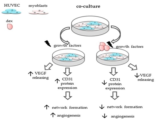
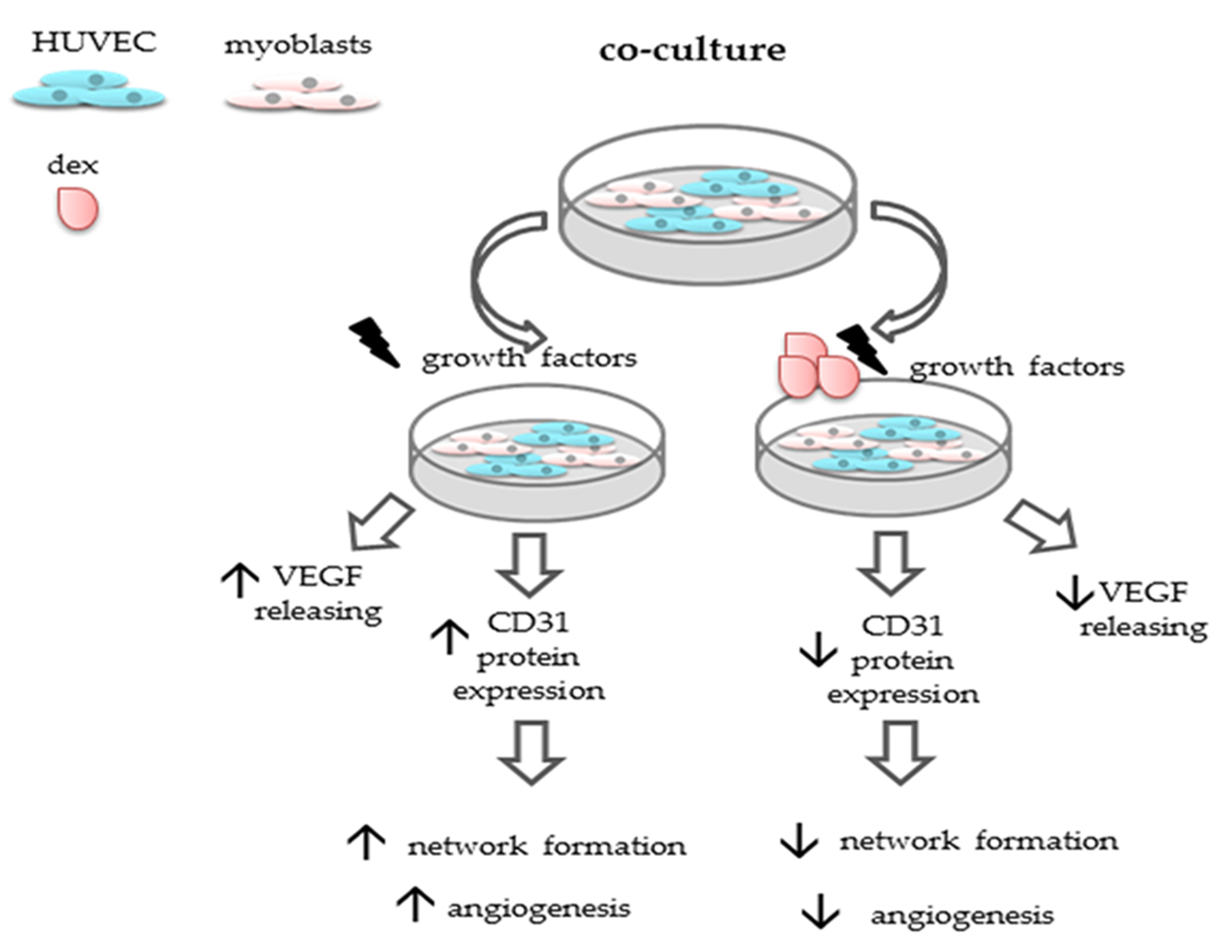
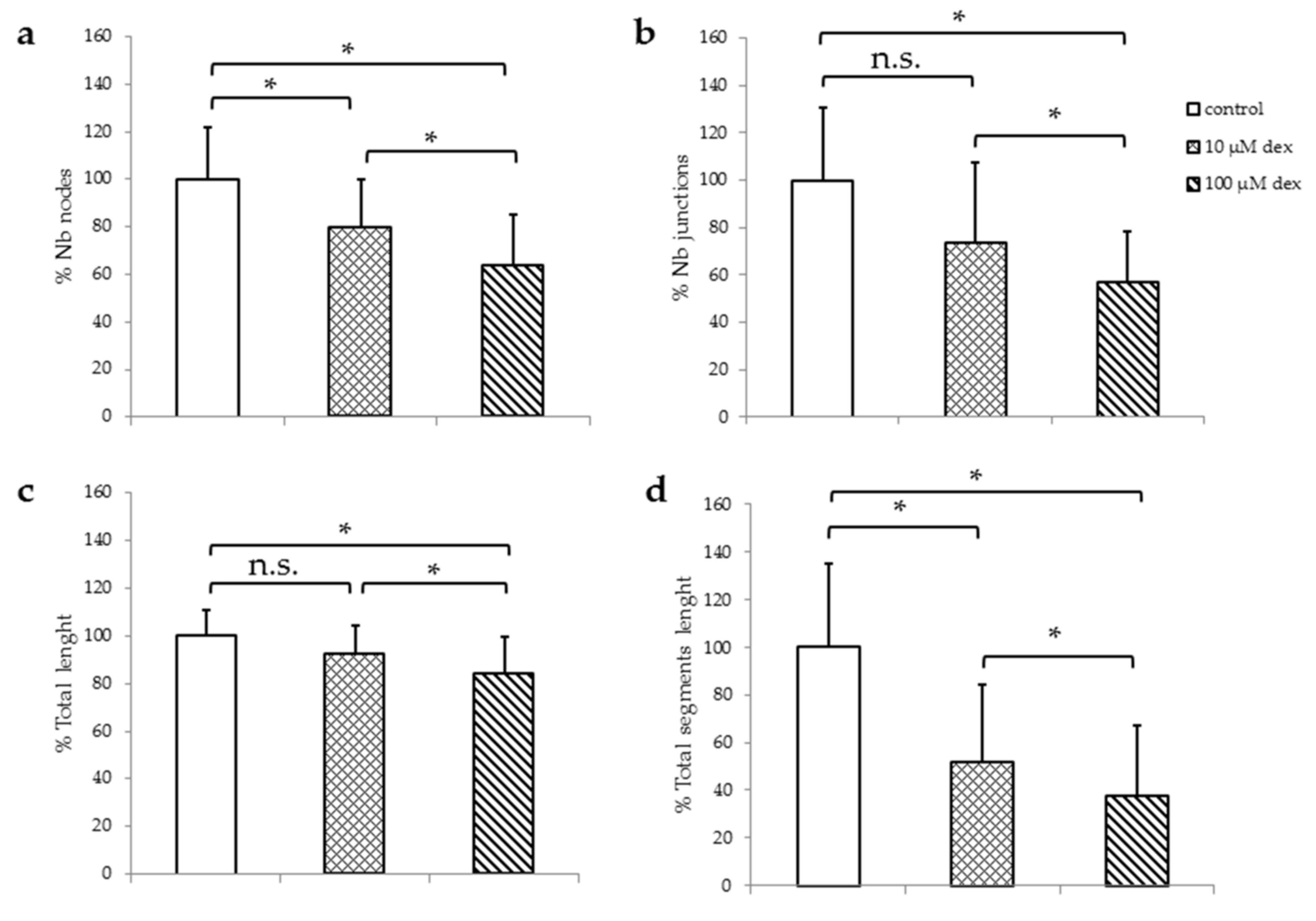
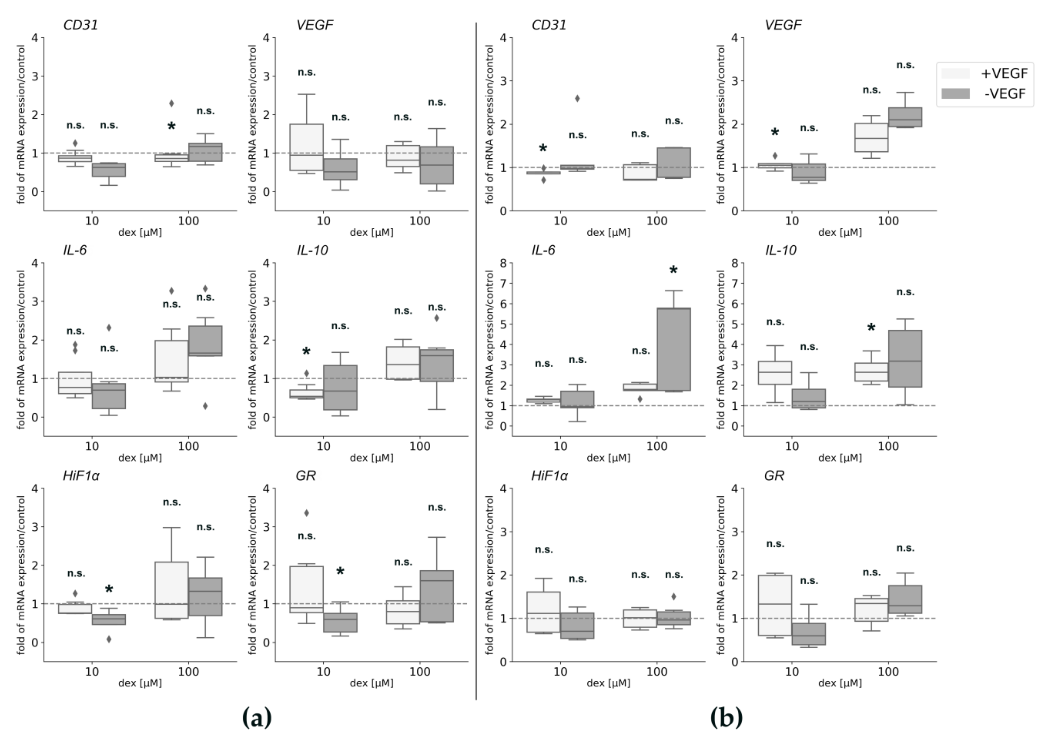
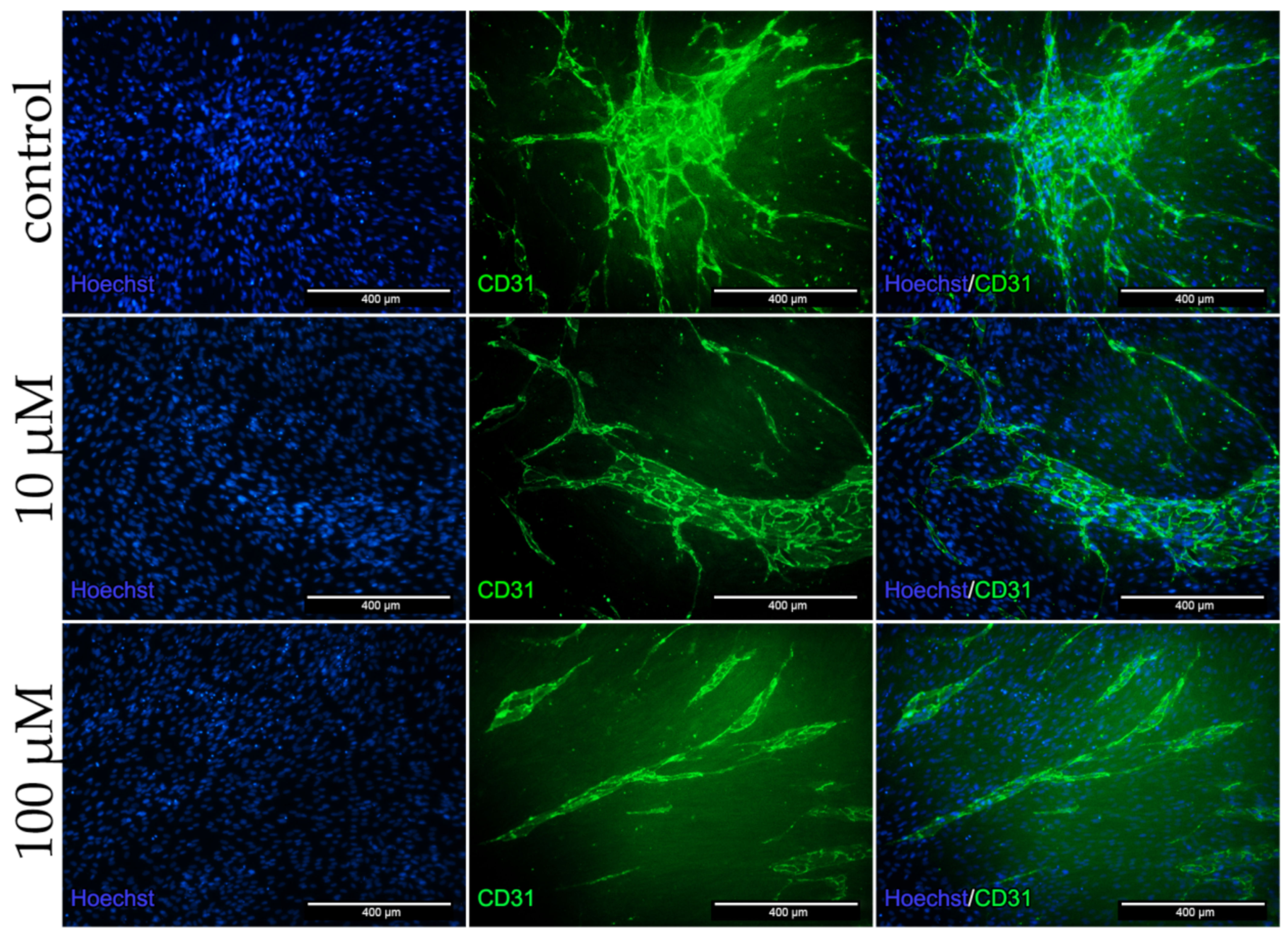
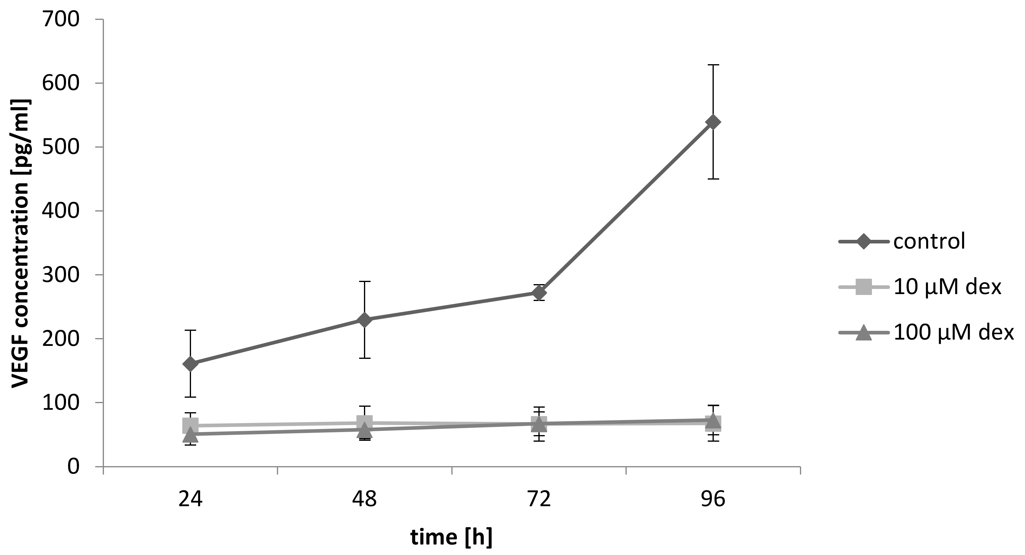
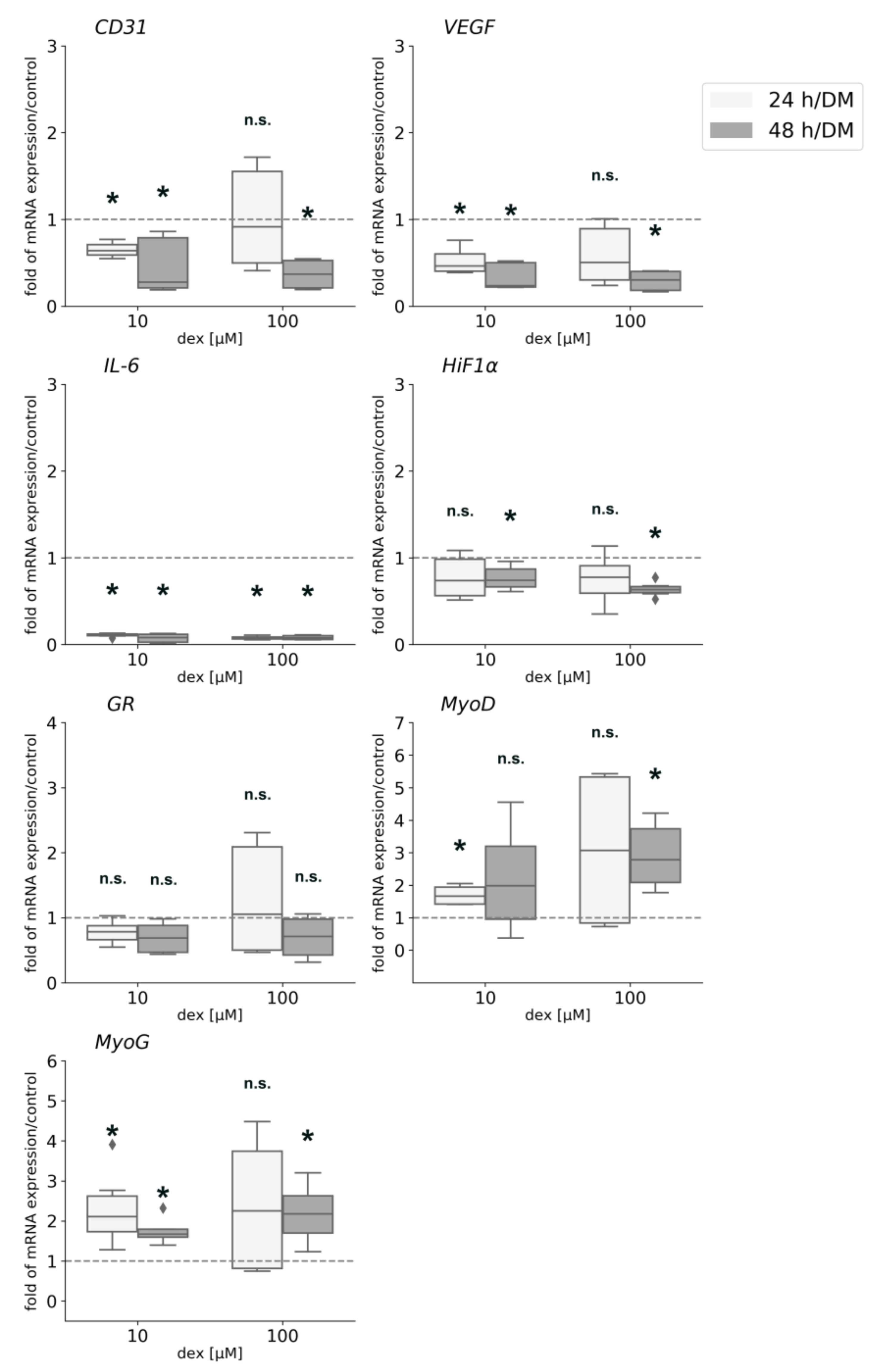
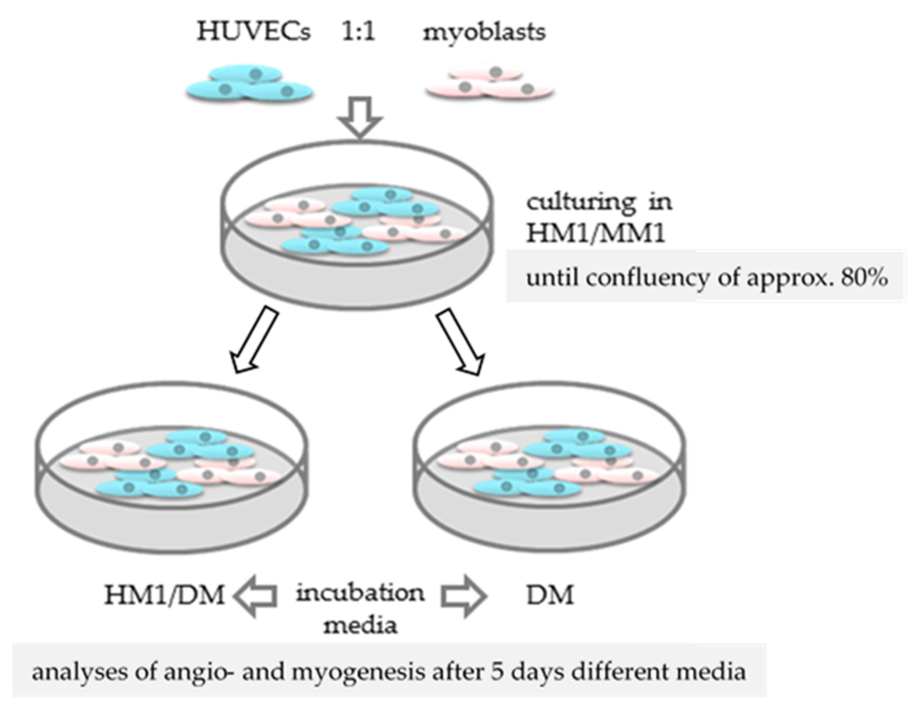
Tissue regeneration depends on the complex processes of angiogenesis, inflammation and wound healing. Regarding muscle tissue, glucocorticoids (GCs) inhibit pro-inflammatory signalling and angiogenesis and lead to muscle atrophy. Our hypothesis is that the synthetic GC dexamethasone (dex) impairs angiogenesis leading to muscle atrophy or inhibited muscle regeneration. Therefore, this study aims to elucidate the effect of dexamethasone on HUVECs under different conditions in mono- and co-culture with myoblasts to evaluate growth behavior and dex impact with regard to muscle atrophy and muscle regeneration. Viability assays, qPCR, immunofluorescence as well as ELISAs were performed on HUVECs, and human primary myoblasts seeded under different culture conditions. Our results show that dex had a higher impact on the tube formation when HUVECs were maintained with VEGF. Gene expression was not influenced by dex and was independent of cells growing in a 2D or 3D matrix. In co-culture CD31 expression was suppressed after incubation with dex and gene expression analysis revealed that dex enhanced expression of myogenic transcription factors, but repressed angiogenic factors. Moreover, dex inhibited the VEGF mediated pro angiogenic effect of myoblasts and inhibited expression of angiogenic inducers in the co-culture model. This is the first study describing a co-culture of human primary myoblast and HUVECs maintained under different conditions. Our results indicate that dex affects angiogenesis via inhibition of VEGF release at least in myoblasts, which could be responsible not only for the development of muscle atrophy after dex administration, but also for inhibition of muscle regeneration after vascular damage.
Keywords: dexamethasone; human primary myoblasts; HUVECs; co-culture; CD31; VEGF dexamethasone; human primary myoblasts; HUVECs; co-culture; CD31; VEGF
Graphical Abstract