Article

In Silico Structural Modeling and Analysis of Interactions of Tremellomycetes Cytochrome P450 Monooxygenases CYP51s with Substrates and Azoles

by
Olufunmilayo Olukemi Akapo
1,†,
Joanna M. Macnar
2,3,†,
Justyna D. Kryś
3,
Puleng Rosinah Syed
4,
Khajamohiddin Syed
1,* and
Dominik Gront
3,*
1
Department of Biochemistry and Microbiology, Faculty of Science and Agriculture, University of Zululand, KwaDlangezwa 3886, South Africa
2
College of Inter-Faculty Individual Studies in Mathematics and Natural Sciences, University of Warsaw, Stefana Banacha 2C, 02-097 Warsaw, Poland
3
Biological and Chemical Research Center, Faculty of Chemistry, University of Warsaw, Pasteura 1, 02-093 Warsaw, Poland
4
Department of Pharmaceutical Chemistry, College of Health Sciences, University of KwaZulu-Natal, Durban 4000, South Africa
*
Authors to whom correspondence should be addressed.
Authors contributed equally to the work.
Int. J. Mol. Sci. 2021, 22(15), 7811; https://doi.org/10.3390/ijms22157811
Submission received: 25 February 2021 / Revised: 21 May 2021 / Accepted: 25 May 2021 / Published: 22 July 2021
(This article belongs to the Special Issue Cytochromes P450: Drug Metabolism, Bioactivation and Biodiversity 3.0)

Abstract

:
Cytochrome P450 monooxygenase CYP51 (sterol 14α-demethylase) is a well-known target of the azole drug fluconazole for treating cryptococcosis, a life-threatening fungal infection in immune-compromised patients in poor countries. Studies indicate that mutations in CYP51 confer fluconazole resistance on cryptococcal species. Despite the importance of CYP51 in these species, few studies on the structural analysis of CYP51 and its interactions with different azole drugs have been reported. We therefore performed in silico structural analysis of 11 CYP51s from cryptococcal species and other Tremellomycetes. Interactions of 11 CYP51s with nine ligands (three substrates and six azoles) performed by Rosetta docking using 10,000 combinations for each of the CYP51-ligand complex (11 CYP51s × 9 ligands = 99 complexes) and hierarchical agglomerative clustering were used for selecting the complexes. A web application for visualization of CYP51s’ interactions with ligands was developed (http://bioshell.pl/azoledocking/). The study results indicated that Tremellomycetes CYP51s have a high preference for itraconazole, corroborating the in vitro effectiveness of itraconazole compared to fluconazole. Amino acids interacting with different ligands were found to be conserved across CYP51s, indicating that the procedure employed in this study is accurate and can be automated for studying P450-ligand interactions to cater for the growing number of P450s.

1. Introduction

Among fungal diseases, cryptococcosis remains a significant cause of morbidity and mortality in immunocompromised people especially in sub-Saharan Africa [1]. Cryptococcal species such as Cryptococcus neoformans and C. gatti are primarily responsible for this disease [2]. Across the world, an average of 6% of HIV-infected people have contract this disease, of which 73% of cases were reported in sub-Saharan Africa, with a death rate of 15% [1]. Treatment options for cryptococcosis are limited to only three drugs (amphotericin B, flucytosine, and fluconazole), with few compounds reaching clinical trials [3]. The treatment involves use of an azole drug, fluconazole, as primary or consolidation and maintenance therapy, followed by induction therapy with intravenous amphotericin B with or without flucytosine [4]. In developed countries, patients are initially subjected to induction therapy and then treatment with fluconazole. However, in resource-limited countries such as low- and middle-income countries, treatment is solely based on fluconazole [4,5].
Non-susceptibility of cryptococcal species to fluconazole has been observed around the world and this has become a growing problem [6,7,8,9,10,11]. Interestingly, quite a number of laboratory-based studies reported better performance of other azoles (itraconazole, voriconazole, posaconazole, and isavuconazole) including the new class of tetrazole compound, VT-1129, against cryptococcal species [10,12,13,14,15,16,17,18,19,20,21]. Azole compounds exert their anti-fungal activity by inhibiting the enzyme cytochrome P450 monooxygenase (CYP/P450) CYP51 involved in the synthesis of fungal membrane ergosterol [22,23]. CYP51, also known as sterol 14α-demethylase, is highly conserved across the phyla and stimulates a key enzymatic reaction that involves stereoselective three-step oxidative removal of the 14α-methyl group from the sterol [24]. Because of this important enzymatic reaction, this enzyme has become a drug target against fungal pathogens and protozoan parasites where azole compounds are in use as drugs in clinical practice targeting this enzyme [24,25,26]. A search using the word “CYP51” at Research Collaboratory for Structural Bioinformatics Protein Data Bank (RCSB PDB) [27] indicated the presence of 109 crystal structures (as of 22 January 2021), clearly indicating the enormous importance of this enzyme, especially its exploitation as a drug target [24,28].
Some progress has been made with unraveling the molecular mechanisms responsible for fluconazole non-susceptibility based on CYP51 in cryptococcal species [25,29,30,31,32]. Over-expression of this gene either by sterol regulatory element-binding protein or by virtue of the presence of more than one copy of this gene due to genomic aneuploidy or the mutations in this gene has been to be responsible for fluconazole resistance [25,29,30,31,32]. A point mutation in CYP51 at G484S resulted in conferring resistance on fluconazole [29]. Azole drug resistance patterns were changed by a single mutation Y145F that caused high fluconazole/voriconazole resistance but increased susceptibility to itraconazole and posaconazole [30]. G344S mutation caused multi-drug resistance to fluconazole, itraconazole, and voriconazole [31]. Detailed information on different mechanisms employed to deal with fluconazole resistance of cryptococcal species was recently reviewed [25].
The CYP51 gene mutational studies mentioned above are based on sequencing the CYP51 gene from the fluconazole non-susceptible strains and comparing its sequence with fluconazole-susceptible strains. To date, structure–activity relationship studies on CYP51 from cryptococcal species have not been reported, with the exception of one study published in 2009 reporting the first three-dimensional (3D) model of CYP51 from C. neoformans. Based on the in silico results, the authors suggested that G484S substitution changed the orientation of the heme-binding domain, leading to decreased catalytic activity and thus, azole binding conferring the drug resistance [33]. Recently, our laboratory performed genome-wide analysis and annotation of P450s in the fungal class Tremellomycetes, which included quite a number of cryptococcal species as well [34]. The study revealed the presence of the CYP51 gene in all species of Tremellomycetes. Considering that structure-activity studies are scarcely reported on CYP51 of cryptococcal species and the availability of a large number of CYP51 sequences [34], this study is aimed at addressing this research gap by performing comparative modeling of CYP51s, which includes assessing their binding patterns to substrates and azoles. CYP51s from other Tremellomycetes were included for comparative analysis with CYP51s of cryptococcal species.

2. Results and Discussion

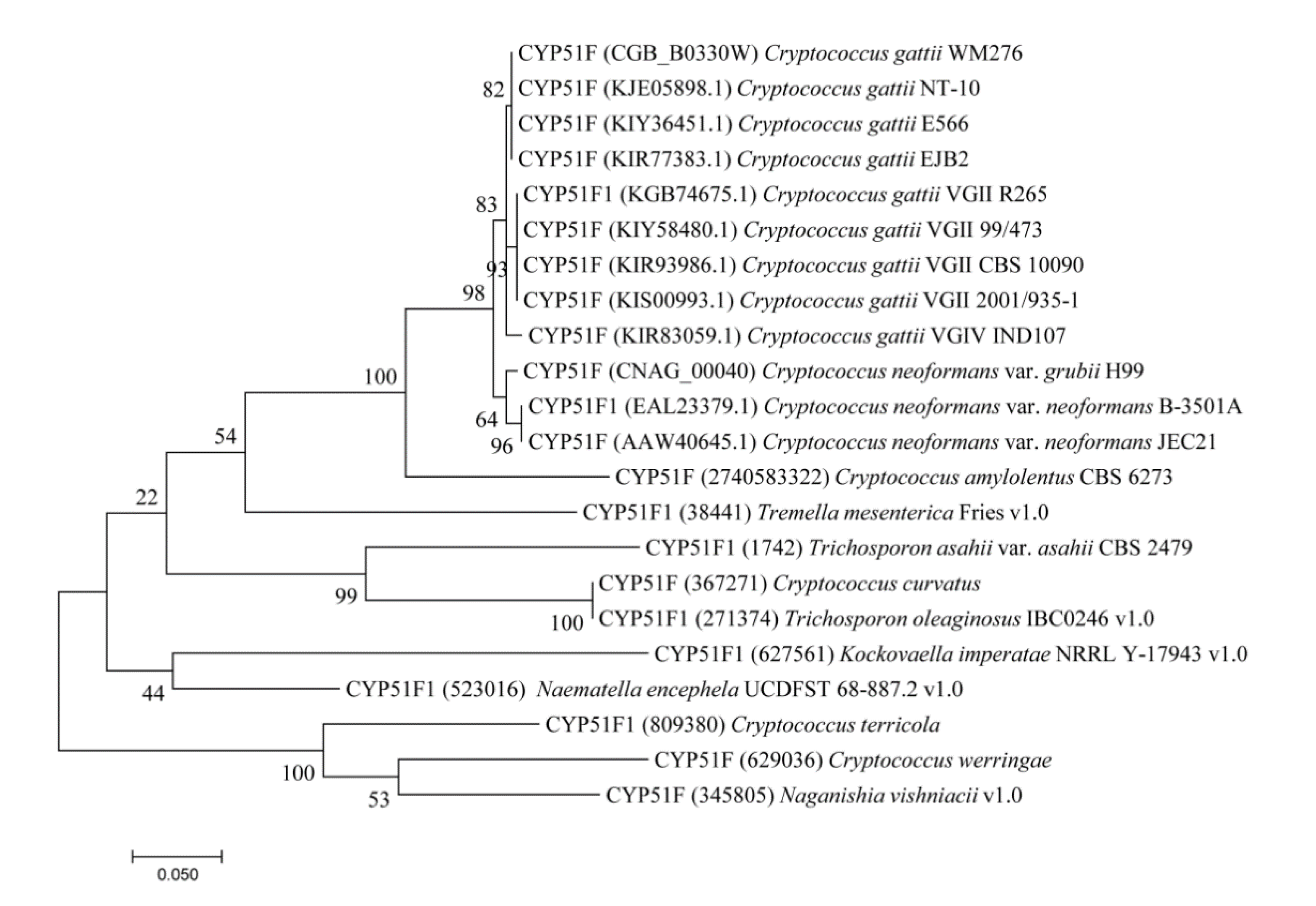
2.1. CYP51s Grouped as per Tremellomycetes Lifestyle

Phylogenetic analysis of CYP51s from different Tremellomycetes revealed an interesting pattern where CYP51s from species with a similar lifestyle or adaption to similar ecological niches grouped together (Figure 1). CYP51s from human pathogenic species such as C. neoformans and C. gatti grouped together whereas CYP51s from oleaginous yeasts C. curvatus and Trichosporon oleaginosus IBC0246 were grouped together with the exception of CYP51 from C. terricola (Figure 1). The CYP51s from the Tremellomycetes having diverse lifestyles standalone on the tree, indicating considerable changes in CYP51 amino acids after speciation. This phenomenon of grouping P450s from species with similar life style/adaption to similar ecological niches was observed in some bacterial [35,36,37] and fungal species [38,39,40]. This indicates that after speciation, lifestyle/ecological niches played a key role in changing/preserving the amino acid content, as observed for different P450s families in bacteria [35,36,37] and fungi [38,39,40].

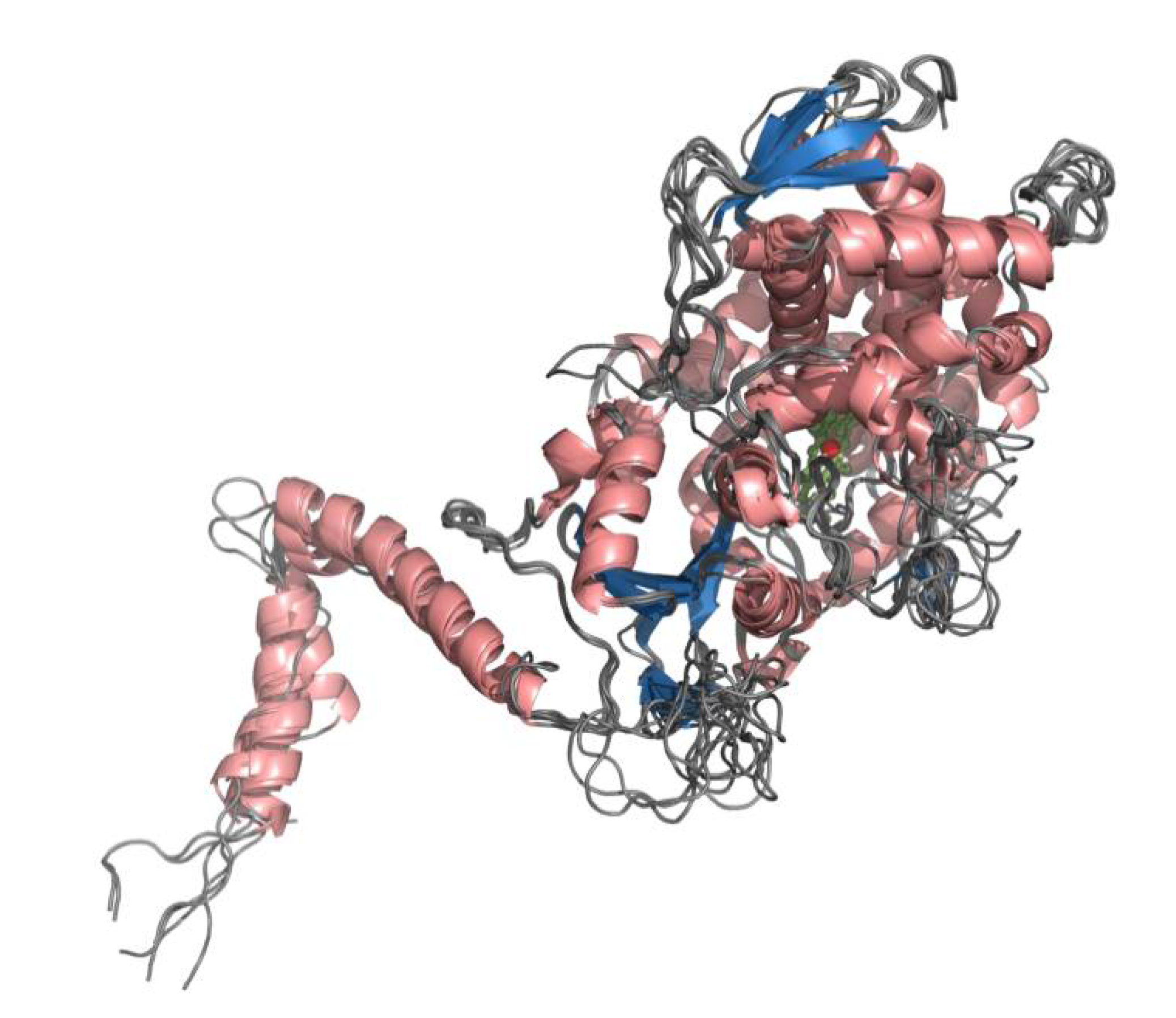
2.2. CYP51s of Tremellomycetes Have All the CYP Characteristic Motifs

Eleven CYP51s from Tremellomycetes as representative of diverse lifestyle/ecological niches were selected for structural analysis (Table 1). Three-dimensional structural models were built with template-based modeling using Modeller software version 9.21 [41].
The template 4LXJ (CYP51F from S. cerevisiae) [42] was selected as the best template for modeling as the sequence identity with CYP51s from Tremellomycetes ranged from 41% to 50% at very high alignment coverage above 90% and the sequence similarity was always well above 70% (Table S1). Multiple sequence alignment of Tremellomycetes CYP51s with the template showed very good alignment, with no major deletions when using the template (Supplementary Dataset S1). Three-dimensional modeling of Tremellomycetes CYP51s on such good alignment should produce high-quality models as described elsewhere [43]. Furthermore, a point to be noted is that all these CYPs belong to the same subfamily where they share >55% identity criteria to be part of the same subfamily [44]. Considering these facts, the 11 CYP51 models generated in the study, as expected, were found to have highly similar structures with characteristic CYP helices and beta sheets [45,46], with only a few minor differences (Figure 2 and Supplementary Dataset S1). An amphipathic helix and two transmembrane helices are missing from the N-terminal region of CYP51F Cter whereas an amphipathic helix, two transmembrane helices, αA helix and αL helix, and subsequent C-terminal end, are missing from CYP51F1 Nen UCDFST68-887 (protein ID: 523016) (Figure 2 and Supplementary Dataset S1). Lack of these helices may be due to genome editing or gene prediction error where prediction of a complete gene sequence is impossible at present. Compared to the rest of the 10 CYP51s, the αC helix of CYP51F1 TaaCBS2479 is found to be longer with 34 amino acids (Figure 2 and Supplementary Dataset S1). Tremellomycetes CYP51s have substrate recognition sites 1–6 (SRS 1-6) with the exception of CYP51F1 Nen UCDFST68-887, where the SRS6 region is missing because the C-terminal region is missing, as described earlier (Figure 2 and Supplementary Dataset 1). Amino acids, part of the SRSs, are highly conserved across the Tremellomycetes CYP51s (Supplementary Dataset S1).

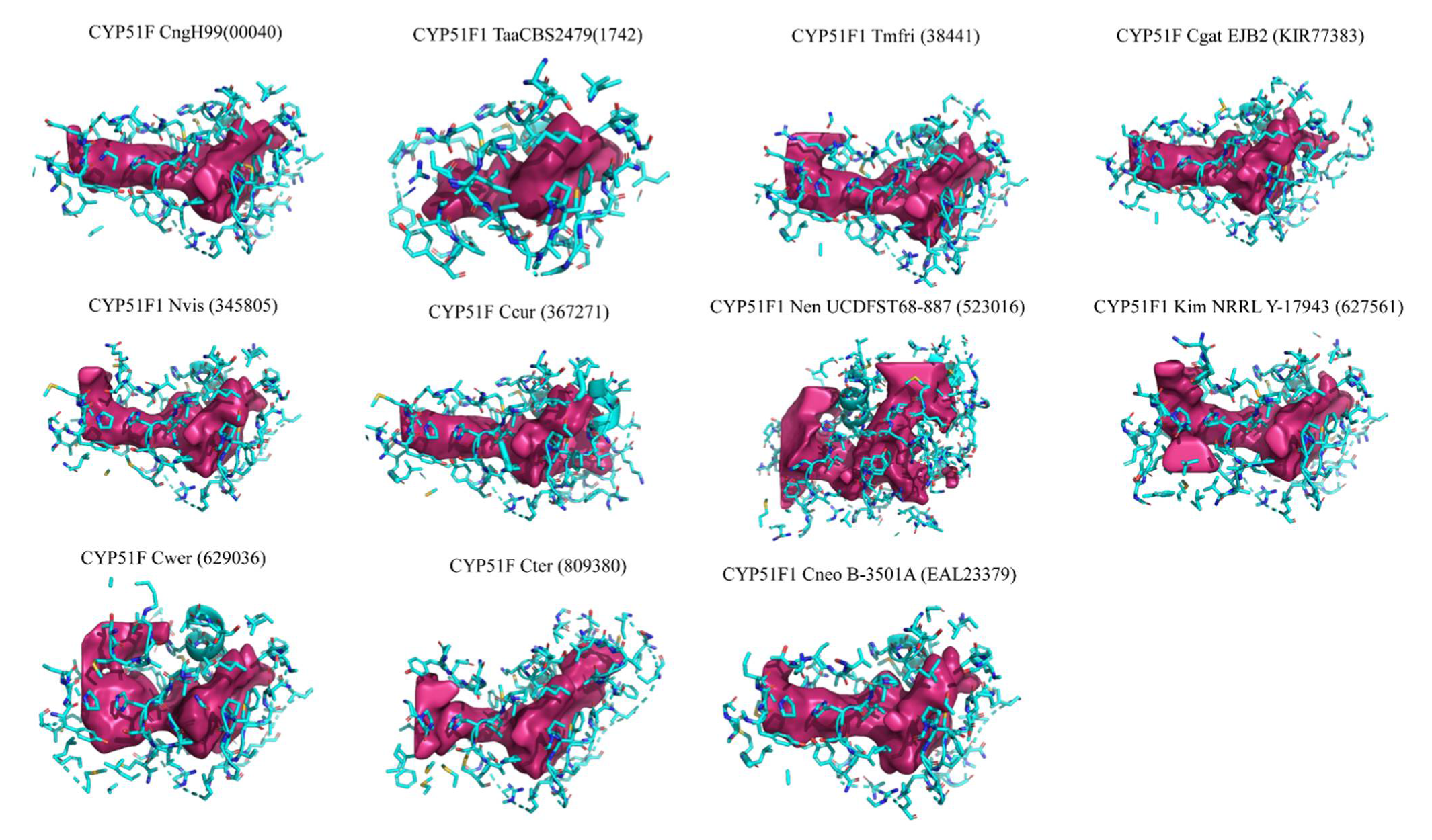
2.3. Tremellomycetes CYP51s Active Site Cavities Are Highly Hydrophobic

Analysis of active site cavities revealed that Tremellomycetes CYP51s active site cavities mostly comprise hydrophobic amino acids (Figure 3 and Table S2). The active site cavity volumes range from 685–4173 Å3, where CYP51F1 Nen UCDFST68-887 has the largest active site cavity and CYP51F1 TaaCBS2479 has the smallest active site cavity (Figure 3 and Table S2). One of the reasons why CYP51F1 Nen UCDFST68-887 has the largest active site cavity is the opening of a channel right next to heme because of the missing C-terminal region that encompasses SRS6, as described earlier. Thus, the active site cavity volume of this P450 is not an actual volume and a full-length CYP sequence is needed to deduce the correct volume of the active site cavity. CYP51s from prominent human pathogens such as C. neoformans var. grubii H99, C. neoformans var. neoformans B-3501A, and C. gattii EJB2 have similar sizes to active site cavities (Figure 3 and Table S2). CYP51s from other cryptococcal species have larger active site cavities compared to CYP51s from these three prominent human pathogens (Figure 3 and Table S2). Analysis of the composition of active site cavity amino acids revealed that 50–60% of cavity amino acids are hydrophobic (Table S2), suggesting that Tremellomycetes CYP51s’ active site cavities are highly hydrophobic. Among Tremellomycetes CYP51s, CYP51F from C. werringae has the highest percentage of hydrophobic amino acids (60%). CYP51s active site cavities’ acidic residues range from 1.64–5.73% and the basic residues range from 6.25–12.17%, where CYP51F1 Nen UCDFST68-887 has most acidic amino acids in its cavity and CYP51F from C. curvatus has most basic amino acids in its active site cavity (Table S2). The hydrophobic nature of these CYP51s’ active sites is expected considering that they accept highly hydrophobic substrates such as sterols.

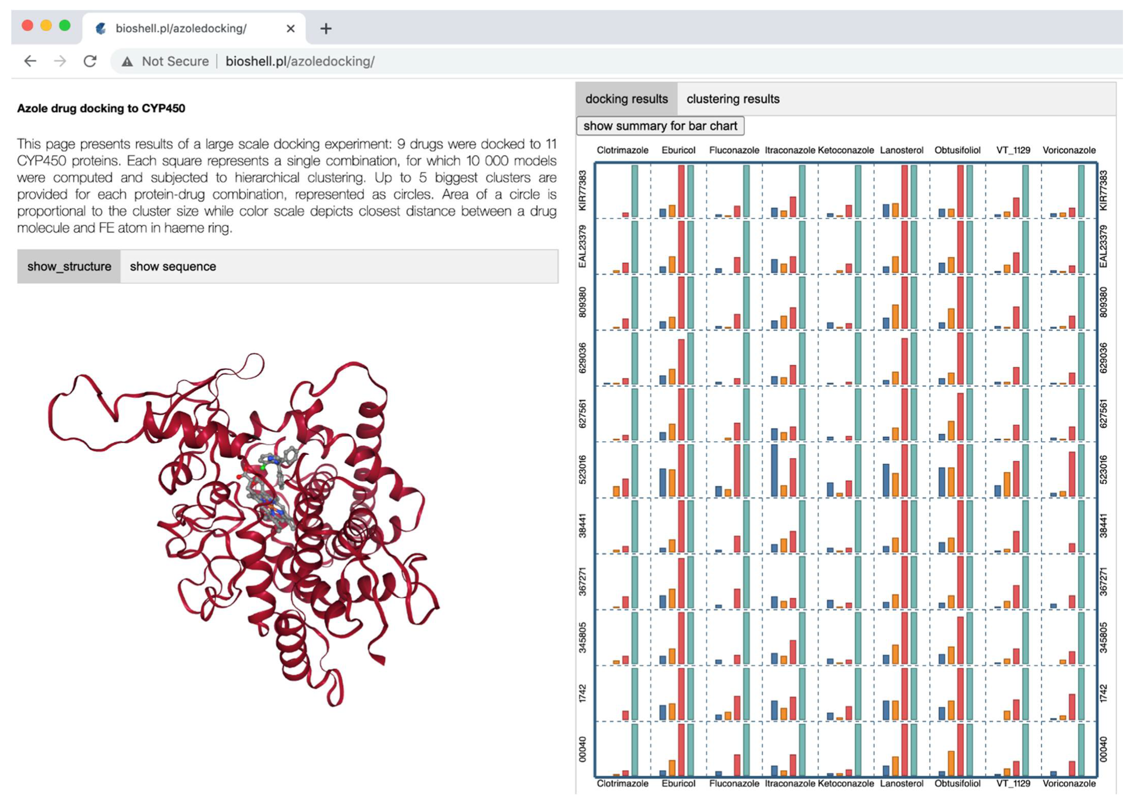
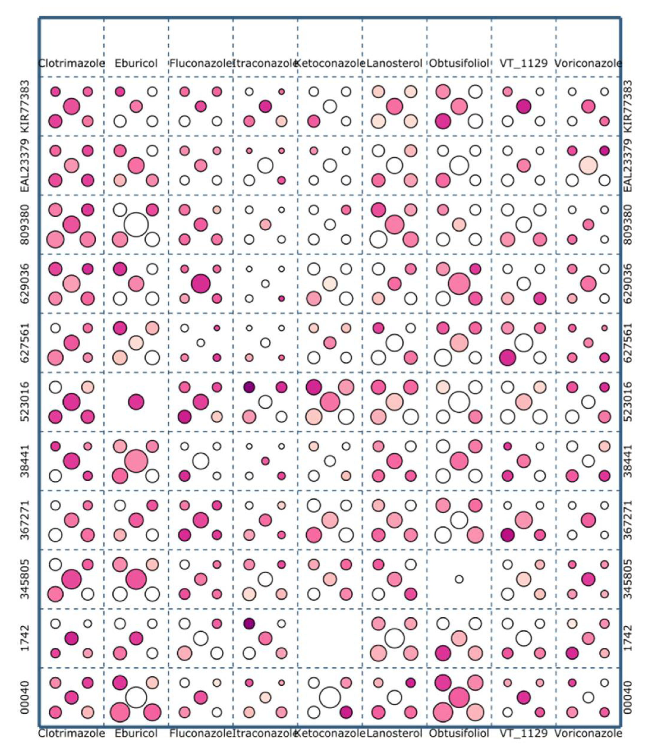
2.4. A Web Application for Visualization of Tremellomycetes CYP51s Interactions with Ligands

After successful construction of 11 CYP51s 3D models, we then proceeded to understand their interactions with ligands such as substrates and azoles. Among substrates, apart from the fungal CYP51 substrates lanosterol and eburicol [24], we also included the plant CYP51 substrate obtusifoliol [24] as a positive control to assess the accuracy of the study. Among the azoles, five are triazoles (clotrimazole, fluconazole, itraconazole, ketoconazole, and voriconazole) and one is tetrazole (VT_1129). The rationale for including these azoles, especially itraconazole, is to assess if any corroboration occurs between the in silico study and the laboratory studies that showed itraconazole performing better than the fluconazole [10,12,13,14,15,16,17].
Docking of each CYP51 vs. each ligand in 11 × 9 resulted in generating 99 combinations (Figure 4). For each of these combinations, we generated 10,000 models as described in the Methods section. Each of these combinations was analyzed independently. The huge amount of data generated during these docking calculations posed a formidable challenge to the analysis. Therefore, we developed a custom web application for this purpose, and made it publicly available at the address: http://bioshell.pl/azoledocking/. The application can display every model that was generated with its basic numerical parameters, such as Rosetta energy, number of hydrogen bonds, etc. The results can be browsed by cluster, sorted, etc.

2.5. Tremellomycetes CYP51s Have the Highest Preference for Itraconazole

Structures for the complexes (CYP51-ligand) considered in this study have not yet been established experimentally. Therefore, we cannot compare these study results to any reference structure. Nevertheless, the geometry of interaction between a heme moiety of P450s and azole drugs has been studied extensively in P450 enzyme systems where azole drugs are known to coordinate with the ferric core of heme group [47]. A lone pair of aromatic nitrogen of azole rings forms a semi-covalent bond with the Fe atom of length dNFe = 2.1 Å [47]. In our analysis, we report the closest distance between heme’s iron and an aromatic nitrogen atom of an azole drug as a measure of success. For the cases of eburicol, lanosterol, and obtusifoliol, dNFe denotes the closest distance between Fe and any atom of a ligand. Since we allowed Rosetta to sample a wide range of binding spots, we observe ligands located in virtually any region of a receptor. In order to assess the performance of Rosetta LigandDocking protocol critically, we included all models in our analysis.
The values of dNFe range from below 2 Å to 80 Å. For the analysis, we defined the following four groups, based on the dNFe distance: Bulleted lists look like this:
  • ‘clashes’ where a ligand was closer to the Fe than 1.5 Å;
  • ‘correct’ where dNFe was in the range 1.5–2.5 Å;
  • ‘near’ with dNFe in the range 2.5–3.5 Å;
  • ‘pocket’ with dNFe in the range 3.5–8.0 Å.
The number of models in each of the four categories is provided in Table 1, as well as shown in Figure 4 by blue, orange, red, and cyan bars, respectively. The definition of these categories was based on a visual analysis of the resulting structures using the online application. The distance ranges defining them were manually adjusted to keep the classification meaningful for as many models as possible. Information on each of these four were elaborated in the supplementary information under the title “CYP51-ligand groups”.
Ligand binding affinity under physiological conditions results from free energy gain upon binding. This free energy change includes entropic contributions as well as other effects such as solvent, ions, etc. Although some of these effects (most notably the interactions with solvent) are included in the Rosetta force field as mean field potentials, the energy value reported by Rosetta primarily accounts for conformational energy (i.e., enthalpy). In order to assess relative binding affinity, one has to take into account the number of observations for a particular conformation. The docking procedure that was applied allowed Rosetta to place a ligand virtually anywhere at the protein surface to avoid any conformational bias. The numbers of correctly docked models given in Table 1 therefore provide an estimation of binding affinity for different ligands. Based on these results, it is clear that Tremellomycetes CYP51s’ natural substrate, lanosterol, achieved the top score, followed by obtusifoliol and eburicol, indicating that our method of analysis is correct, as one can expect the highest binding affinity to natural substrates. It is also interesting to see the pattern of preference for azoles by Tremellomycetes CYP51s where itraconazole is preferred azole followed by VT_1129 and fluconazole, which is the least favored before ketoconazole (Table 1). This preference pattern perfectly matches wet laboratory data where itraconazole and VT_1129 were shown to perform better than fluconazole [10,12,13,14,15,16,17,18,19,20,21]. Overall, based on these results, we can safely say that our method of analysis is correct.
Given the drawbacks of the Rosetta scoring functions as elaborated on the supplementary section “CYP51-ligand”, the chance of selecting a correct complex without prior knowledge of the actual in vivo conformation is low. A commonly used remedy to solve this problem is structural clustering. Thus, in this study, we used this method to select the correct CYP-ligand complex.
Clustering analysis is a method commonly used in the field of biomacromolecular modeling. It helps to analyze large amounts of data generated by computer simulations and to overcome inaccuracies of molecular force fields. A group of structurally similar conformations corresponds to a single local energy minimum [48]. In general, it is expected that a conformation corresponding to a larger cluster will be more favored entropically. Entropic contributions, necessary to estimate the free energy of a given conformation, are typically not described properly by molecular modeling force fields. Giving preference to larger clusters during the process of selecting the final docking result, implicitly allows inclusion of these entropic contributions. Here, we employed a hierarchical agglomerative clustering algorithm, as described in the Methods section. Following the standard protocols in the field, we report the five largest clusters for every protein–ligand combination. The clusters are indicated graphically in Figure 5 as circles. A circular area is proportional to a cluster size while the color scale represents the dNFe values, in a range from 1 to 80 Å (see Figure 5). In three cases, no suitable clusters were found (Figure 5). All these clustering results can be browsed online at: http://bioshell.pl/azoledocking/.
All the following analyses require selection of a single representative model for each of the 99 modeling cases. The clustering procedure described above is certainly helpful, but on its own, is unable to select the final solution in every case. Therefore, we used a web application (http://bioshell.pl/azoledocking/) to browse the results and manually selected final conformations. In this process, we took into account the classification resulting from the clustering procedure, the chemical correctness of a conformation, and the number of interactions (stacking, H-bonding, and Van der Waals) between a ligand and a protein. The number of such interactions listed in a table of the web application facilitates the selection process.

2.6. High Conservation Observed in Tremellomycetes CYP51s Amino Acids Interacting with Ligands

After selection of each of the CYP51-ligand complex as described above, we further analyzed the amino acids interacting with different ligands and their location. This is a very important aspect to check the accuracy of our method employed to dock and select CYP51-ligand complexes. As all of these CYPs belong to the same subfamily and high conservation of amino acids is observed in SRS regions (Supplementary Dataset S1), if our analysis is correct, we should observe high conservation of amino acids interacting with analyzed ligands across the CYPs. As predicted, our analysis revealed conservation of a large number of amino acids interacting with docked ligands across the CYPs (Table 2 and Table S2). Mapping of the amino acids interacting with ≥5 ligands revealed that these amino acids are part of SRSs (Supplementary Dataset S1). We also identified amino acids across the CYPs that are able to form hydrogen bonds with ligands and interact with ligands via stacking and Van der Waals interactions (Table S2). This suggests that our method of docking and selecting the CYP-ligand complexes is correct and in future these aspects can be automated to understand the CYP-ligand interactions to cater for the growing number of CYPs. A point to be noted that CYP51F1 TaaCBS2479 due to its compact active site cavity, has more amino acids interacting with ligands compared CYP51F1 Nen UCDFST68-887 where due to its larger active site cavity ligands are not strongly binding and thus, less number of amino acids found to be interacting with the ligands (Table 2).
Analysis of amino acid substitutions in CYP51s that are found to be critical in response to different azole drugs [29,30,31] revealed that Y145 and G484 amino acids are absolutely conserved in the Tremellomycetes CYP51s (Supplementary Dataset 1 indicated with “X”). However, substitution of glycine at 410 position (formerly reported as G344) [31] with alanine (in three CYP51s), serine (in two CYP51s), lysine (in two CYP51s), and arginine (in a single CYP51) is found (Supplementary Dataset 1 indicated with “X”). This is quite an interesting observation as it has been reported that G344S substitution in six strains of C. neoformans var. grubii strain, NUBS14020, resulted in the development of multi-drug resistance to azoles such as fluconazole, itraconazole, and voriconazole [31]. Considering these findings, it would be interesting to see the response of the strains containing G344 substitutions in their CYP51s.

3. Materials and Methods

3.1. CYP51s Used in the Study

Twenty-one CYP51s from Tremellomycetes were used in this study. Among 21 CYP51s, 11 CYP51s from different Tremellomycetes representing diverse lifestyles/adaptation to different ecological niches were selected for structural analysis (Table 3). A detailed description of the well-known characteristics of Tremellomycetes can be found in a recently published article [34]. All CYP51 sequences from Tremellomycetes were retrieved from a recently published article from our laboratory [34] and used in the study. CYP51 sequences used in the study are presented in Supplementary Information.

3.2. Phylogenetic Analysis

Phylogenetic analysis of CYP51s from Tremellomycetes was carried out using the maximum likelihood method based on the JTT matrix-based model [49] and 500 bootstrap replications. Analysis and visualization of the phylogenetic tree were carried out using molecular evolutionary genetic analysis [50].

3.3. Homology Modeling of CYP51s

A 3D structural model for each CYP51s (Table 3) was constructed by employing a template-based modeling procedure. The template was selected from a non-redundant subset of the Protein Data Bank [51] by searching the database with the HHpred algorithm [52] available on the website. CYP51 from Saccharomyces cerevisiae (PDB code: 4lxj) [42] which has the highest E-value, highest percentage identity, and the highest alignment coverage with all 11 CYP51s of Tremellomycetes was selected as the best template for modeling of CYP51s. Although some CYP51s share more than 50% identity with other templates, we did not see much added value from using multiple templates. Unfortunately, they will not help in bridging gaps introduced by an alignment. On the other hand, we decided to include a ligand from the template structure in the homology modeling protocol to render the pocket in the active state. The use of multiple templates would considerably have complicated the modeling process. Thus, in this study, we used CYP51 from S. cerevisiae as a template. Query to template alignments were also obtained from the HHPred results. Manual editing was introduced when necessary. CYP51s structural models were built based on these alignments with Modeller software (version 9.21) [41]. Three-dimensional modeling of CYP51s was performed based on structural information from the template 4LXJ where heme cofactor and ligand molecules (lanosterol and heme molecule) were explicitly included during the modeling to ensure correct geometry of an active site. The models were checked with Molprobity [NN] which reported 7 to 21 residues (for CYP51F1 Kim NRRL Y-17943 and TaaCBS2479, respectively) outside the allowed Ramachandran region. Detailed statistics are provided in Table S3.

3.4. Preparation of CYP51s Models for Docking

Most calculations related to docking were performed with the Rosetta [53] software package, dedicated to biomolecular structure prediction, design, and docking. The software was collaboratively developed by over 60 laboratories from all over the world [54]. The clean-pdb.py script (distributed with the software package) [55] was used to remove all non-protein molecules from the model. Optimization of bonds and dihedral angles was also performed [55]. Rosetta’s molfile_to_param.py script was used to prepare input files: PDB and params (internal Rosetta format) for each ligand, based on the particular SDF files, downloaded from PubChem following the protocol described elsewhere [56], obtained from Rosetta Ligand Docking Tutorial online [55]. Ligand conformers generated with Openbabel [57], and BCL [58] were used, following the protocol used in the Rosetta Community developed at the Meiler’s laboratory. A Rosetta program named ligand_rpkmin was used to minimize protein side chains present near the heme molecule following the protocol used in the Rosetta Community developed by our laboratory. The protein conformation with the lowest energy was chosen for docking procedures.

3.5. Ligands and Their Docking Procedure

CYP51 substrates (lanosterol, obtusifoliol, and eburicol), azole drugs such as itraconazole, clotrimazole, voriconazole, fluconazole, and ketoconazole including the novel fungal CYP51 inhibitor VT-1129, were used in the study for assessing their binding pattern with 11 CYP51s of Tremellomycetes (Table 3). Chemical structures of the ligands used in the study are presented in Figure S1. In silico docking of these nine ligands to each of the CYP51 3D models was performed. Docking simulations were done with a PyRosetta script [59]. For this purpose, we adapted a standard script distributed with Rosetta Ligand Docking Tutorial [55] and PyRosetta notebook 08.01-Ligand-Docking-XMLObjects [60].
The ligand docking procedure was divided into two stages: low and high resolution. During the first, semi-global stage, a ligand was free to sample the protein surface within a sphere of 20 Å, which covers nearly the whole protein. A soft variant of the Rosetta score function called “ligand_soft_rep” was used at this stage to ease energy barrier crossing. Repulsive LJ term and Coulomb interactions were ramped down while hydrogen bonds were promoted. Any side chain within 6 Å from a ligand was also allowed to change its conformation. During the second, high-resolution stage, HighResDocker mover was used for local refinement of a conformation obtained from the previous stage. A final model was subjected to all-atom energy minimization and scoring. The procedure was repeated independently 10,000 for each ortholog-ligand combination and 10,000 resulting poses were subjected to further analysis.

3.6. Hierarchical Agglomerative Clustering

All clustering analyses in this work were performed with the ap_ligand_clustering program of the BioShell 3.0 package [61]. Here, we briefly summarize the procedure as the details of this approach have been reviewed and the software has already been published [62]. The procedure begins with placing every structure in a separate cluster of size 1. Then, at every step, the two closest clusters are found and merged into one. The newly merged cluster contains all structures from its ancestors and the total number of clusters decreases by one. The procedure is repeated until a convergence condition is met. Here, we stop clustering when the distance between the two closest clusters exceeds a certain distance cutoff, e.g., 5.0 Å. There are a few widely used approaches to compute a distance between two clusters. In this work, we assumed the complete link strategy: the distance between two clusters i.e., groups of structures, is defined as the smallest distance possible between any structure from the first group and a structure from the second group. Clustering with complete link strategy results in relatively many small but tight clusters. A single clustering calculation of 10,000 models takes around 5 min on a single CPU core.
It is also very convenient to be able to select a “representative” element of a cluster. Many methods have been devised for this purpose in the literature, e.g., taking an average of all the cluster elements. Averaged conformation, however, may not represent a chemically correct structure. Therefore, we defined a “middle” structure (often named ‘medoid’ in the literature) as the one for which the sum of distances to all other members of the cluster is the smallest.

3.7. Protocols

The detailed description of our modeling protocols, comparative modeling, docking, and analysis are given on the laboratory ‘Protocols’ website: https://labnotes.readthedocs.io. The website provides all the necessary files, scripts, and commands that would allow other researchers access to our calculations.

3.8. Data Visualization

In order to analyze the results of the calculations extensively and thoroughly, we built a custom visualization tool (http://bioshell.pl/azoledocking/). The application displays plots, statistics, and 3D structural models in a web browser. The server side is run by a Flask application server, also implemented in Python. The client side (i.e., the web browser part) utilizes VisuaLife Python library (visualife.readthedocs.io/) to render plots, charts, and tables. NGL biomolecular viewer [63] is employed to display structural models.

3.9. Active Site Cavity and Ligand Interacting Amino Acids Detection

Active site cavities in CYP51 models were detected using CavityPlus [64]. Interactions between a ligand and the protein were detected automatically with a BioShell package [61]. Van der Waals interactions were assigned when any atoms of two residues (including hydrogens) were closer to each other than the sum of their VdW radii + 0.5 Å. Hydrogen bonds were assigned when the distance between the donated hydrogen atom and its acceptor was shorter than 3 Å and the planar angle formed by the three atoms, pre-acceptor, acceptor, and the hydrogen was wider than 90 deg. Stacking interactions were recorded when the distance between the two aromatic ring centers was shorter than 7 Å.

4. Conclusions

Cytochrome P450 monooxygenases (CYPs/P450s) are some of the well-studied enzymes in the field of biology. Among CYPs, CYP51 serves as drug target against fungal pathogens. In the case of cryptococcosis, a life-threatening fungal disease in immunocompromised patients, studies indicated that cryptococcal species are developing resistance to the currently used azole drug fluconazole. Despite knowing that CYP51 is the prime target of this drug, mutations in this gene conferring resistance on fluconazole and a large number of in vitro studies indicating that other azoles perform better than fluconazole, to date very few studies on CYP51s structure from cryptococcal or other Tremellomycetes and their interactions with azoles have been reported. To address this research gap, in this study, we performed comparative modeling of CYP51s and their interactions with different ligands from Tremellomycetes. Phylogenetic analysis of CYP51s indicated some kind of amino acid conservation after speciation, as CYP51s grouped as per their species lifestyle/ecological niches. Structural models built for 11 CYP51s revealed the presence of P450 characteristic motifs and high conservation of amino acids in substrate recognition sites (SRSs) 1–6. The active site cavities of CYP51s were found to be highly hydrophobic to accommodation of hydrophobic sterol substrates. Since the structures for the complexes (CYP51-ligand) we considered have not yet been established experimentally, we performed global docking of ligands (three substrates and six azoles) with 10,000 combinations for each of the complex and the correct complexes were selected by hierarchical agglomerative clustering using the cluster program of the BioShell package. To analyze the huge data generated in this study, we developed an online visualization tool (http://bioshell.pl/azoledocking/). These study results indicated that Tremellomycetes CYP51s have the highest preference for its natural substrate lanosterol, followed by obtusifoliol and eburicol. Among azoles, Tremellomycetes CYP51s have the highest preference for itraconazole, followed by VT_1129, clotrimazole, voriconazole, fluconazole, and ketoconazole. Tremellomycetes CYP51s’ high preference for itraconazole is possibly the reason why this azole perform better than fluconazole, as observed in a large number of in vitro studies. The methodology followed in this study is correct, as the amino acids we identified as interacting with different ligands are highly conserved across the CYP51s, indicating no errors in the procedure. Furthermore, most of these amino acids were found to be part of SRSs. In this study, we focused on a single subfamily of CYPs simply to assess the methods employed in the study, but this method needs to be validated for the analysis of different CYP families and their interactions with different ligands. Studies are in progress to develop a website on which this program will be automated to study CYPs’ interactions with different compounds. This type of dedicated website for this family of enzymes is desperately needed, as the number of CYPs is growing exponentially every day. The results of this study further strengthen the previous laboratory results that itraconazole and VT_1129 will be potential compounds in the fight against cryptococcosis and their efficacy against cryptococcal species needs to be determined. One of the interesting results from this study is the identification of non-conservation of G344 amino acid in Tremellomycetes CYP51s, including in some cryptococcal species. It would be interesting to see if these Tremellomycetes are resistant to fluconazole, itraconazole, and voriconazole, as was reported for G344S substituted species.

Supplementary Materials

The following are available online at https://www.mdpi.com/article/10.3390/ijms22157811/s1, Reference [42] is cited in the Supplementary Materials.

Author Contributions

Conceptualization, K.S. and D.G.; data curation, O.O.A., J.M.M., J.D.K., P.R.S., K.S. and D.G.; formal analysis, O.O.A., J.M.M., J.D.K., P.R.S., K.S. and D.G.; funding acquisition, D.G. and K.S.; investigation, O.O.A., J.M.M., J.D.K., P.R.S., K.S. and D.G.; methodology, O.O.A., J.M.M., J.D.K., P.R.S., K.S. and D.G.; project administration, K.S.; resources, K.S. and D.G.; supervision, K.S. and D.G.; validation, O.O.A., J.M.M., J.D.K., P.R.S., K.S. and D.G.; visualization, O.O.A., J.M.M., J.D.K., P.R.S., K.S. and D.G.; writing—original draft, O.O.A., J.M.M., J.D.K., P.R.S., K.S. and D.G.; writing—review and editing, O.O.A., J.M.M., J.D.K., P.R.S., K.S. and D.G. All authors have read and agreed to the published version of the manuscript.

Funding

Olufunmilayo Olukemi Akapo thanks the University of Zululand for a visiting scholar fellowship to Dominik Gront’s laboratory at the University of Warsaw, Poland. Puleng Rosinah Syed thanks the Department of Science and Technology—National Research Foundation (DST-NRF), South Africa for a doctoral scholarship (Grant number MND190606443406). Khajamohiddin Syed expresses sincere gratitude to the NRF, South Africa for a research grant (Grant number 114159) and the University of Zululand (Grant number C686). Dominik Gront, Justyna D. Kryś and Joanna M. Macnar were supported by the Polish National Science Centre (NCN) 2018/29/B/ST6/01989.

Institutional Review Board Statement

Not applicable.

Informed Consent Statement

Not applicable.

Data Availability Statement

Data is contained within the article or supplementary material.

Acknowledgments

The authors want to thank Barbara Bradley, Pretoria, South Africa for English language editing.

Conflicts of Interest

The authors declare no conflict of interest. The funders had no role in the design of the study; in the collection, analyses, or interpretation of data; in the writing of the manuscript, or in the decision to publish the results.

References

  1. Rajasingham, R.; Smith, R.M.; Park, B.J.; Jarvis, J.N.; Govender, N.P.; Chiller, T.M.; Denning, D.W.; Loyse, A.; Boulware, D.R. Global burden of disease of HIV-associated cryptococcal meningitis: An updated analysis. Lancet Infect. Dis. 2017, 17, 873–881. [Google Scholar] [CrossRef] [Green Version]
  2. Kwon-Chung, K.J.; Fraser, J.A.; Doering, T.L.; Wang, Z.A.; Janbon, G.; Idnurm, A.; Bahn, Y.-S. Cryptococcus neoformans and Cryptococcus gattii, the etiologic agents of cryptococcosis. Cold Spring Harb. Perspect. Med. 2014, 4, a019760. [Google Scholar] [CrossRef]
  3. Coelho, C.; Casadevall, A. Cryptococcal therapies and drug targets: The old, the new and the promising. Cell. Microbiol. 2016, 18, 792–799. [Google Scholar] [CrossRef] [Green Version]
  4. WHO. Guidelines for the Diagnosis, Prevention, and Management of Cryptococcal Disease in HIV-Infected Adults, Adolescents and Children, March 2018: Supplement to the 2016 Consolidated Guidelines of the Use of Antiretroviral Drugs for Treating and Preventing HIV Infection; WHO: Geneva, Switzerland, 2018. [Google Scholar]
  5. Loyse, A.; Thangaraj, H.; Easterbrook, P.; Ford, N.; Roy, M.; Chiller, T.; Govender, N.; Harrison, T.S.; Bicanic, T. Cryptococcal meningitis: Improving access to essential antifungal medicines in resource-poor countries. Lancet Infect. Dis. 2013, 13, 629–637. [Google Scholar] [CrossRef] [Green Version]
  6. Chen, Y.-C.; Chang, T.-Y.; Liu, J.-W.; Chen, F.-J.; Chien, C.-C.; Lee, C.-H.; Lu, C.-H. Increasing trend of fluconazole-non-susceptible Cryptococcus neoformans in patients with invasive cryptococcosis: A 12-year longitudinal study. BMC Infect. Dis. 2015, 15, 277. [Google Scholar] [CrossRef] [PubMed] [Green Version]
  7. Wang, H.; Xiao, M.; Chen, S.C.-A.; Kong, F.; Sun, Z.-Y.; Liao, K.; Lu, J.; Shao, H.-F.; Yan, Y.; Fan, H. In vitro susceptibilities of yeast species to fluconazole and voriconazole as determined by the 2010 National China Hospital Invasive Fungal Surveillance Net (CHIF-NET) study. J. Clin. Microbiol. 2012, 50, 3952–3959. [Google Scholar] [CrossRef] [PubMed] [Green Version]
  8. Hsueh, P.-R.; Lau, Y.-J.; Chuang, Y.-C.; Wan, J.-H.; Huang, W.-K.; Shyr, J.-M.; Yan, J.-J.; Yu, K.-W.; Wu, J.-J.; Ko, W.-C. Antifungal susceptibilities of clinical isolates of Candida species, Cryptococcus neoformans, and Aspergillus species from Taiwan: Surveillance of multicenter antimicrobial resistance in Taiwan program data from 2003. Antimicrob. Agents Chemother. 2005, 49, 512–517. [Google Scholar] [CrossRef] [Green Version]
  9. Sar, B.; Monchy, D.; Vann, M.; Keo, C.; Sarthou, J.L.; Buisson, Y. Increasing in vitro resistance to fluconazole in Cryptococcus neoformans Cambodian isolates: April 2000 to March 2002. J. Antimicrob. Chemother. 2004, 54, 563–565. [Google Scholar] [CrossRef]
  10. Pan, W.; Khayhan, K.; Hagen, F.; Wahyuningsih, R.; Chakrabarti, A.; Chowdhary, A.; Ikeda, R.; Taj-Aldeen, S.J.; Khan, Z.; Imran, D. Resistance of Asian Cryptococcus neoformans serotype A is confined to few microsatellite genotypes. PLoS ONE 2012, 7, e32868. [Google Scholar] [CrossRef]
  11. Smith, K.D.; Achan, B.; Hullsiek, K.H.; McDonald, T.R.; Okagaki, L.H.; Alhadab, A.A.; Akampurira, A.; Rhein, J.R.; Meya, D.B.; Boulware, D.R. Increased antifungal drug resistance in clinical isolates of Cryptococcus neoformans in Uganda. Antimicrob. Agents Chemother. 2015, 59, 7197–7204. [Google Scholar] [CrossRef] [Green Version]
  12. Trilles, L.; Fernández-Torres, B.; dos Santos Lazéra, M.; Wanke, B.; Guarro, J. In vitro antifungal susceptibility of Cryptococcus gattii. J. Clin. Microbiol. 2004, 42, 4815–4817. [Google Scholar] [CrossRef] [PubMed] [Green Version]
  13. Thompson, G.R.; Wiederhold, N.P.; Fothergill, A.W.; Vallor, A.C.; Wickes, B.L.; Patterson, T.F. Antifungal susceptibilities among different serotypes of Cryptococcus gattii and Cryptococcus neoformans. Antimicrob. Agents Chemother. 2009, 53, 309–311. [Google Scholar] [CrossRef] [PubMed] [Green Version]
  14. Barchiesi, F.; Schimizzi, A.M.; Caselli, F.; Novelli, A.; Fallani, S.; Giannini, D.; Arzeni, D.; Di Cesare, S.; Di Francesco, L.F.; Fortuna, M. Interactions between triazoles and amphotericin B against Cryptococcus neoformans. Antimicrob. Agents Chemother. 2000, 44, 2435–2441. [Google Scholar] [CrossRef] [Green Version]
  15. Illnait-Zaragozi, M.-T.; Martínez, G.F.; Curfs-Breuker, I.; Fernández, C.M.; Boekhout, T.; Meis, J.F. In vitro activity of the new azole isavuconazole (BAL4815) compared with six other antifungal agents against 162 Cryptococcus neoformans isolates from Cuba. Antimicrob. Agents Chemother. 2008, 52, 1580–1582. [Google Scholar] [CrossRef] [Green Version]
  16. Hagen, F.; Illnait-Zaragozi, M.-T.; Bartlett, K.H.; Swinne, D.; Geertsen, E.; Klaassen, C.H.; Boekhout, T.; Meis, J.F. In vitro antifungal susceptibilities and amplified fragment length polymorphism genotyping of a worldwide collection of 350 clinical, veterinary, and environmental Cryptococcus gattii isolates. Antimicrob. Agents Chemother. 2010, 54, 5139–5145. [Google Scholar] [CrossRef] [PubMed] [Green Version]
  17. Armengou, A. Possible development of resistance to fluconazole during suppressive therapy for AIDS-associated cryptococcal meningitis. Clin. Infect. Dis. Off. Publ. Infect. Dis. Soc. Am. 1996, 23, 1337–1338. [Google Scholar] [CrossRef]
  18. Hoekstra, W.J.; Garvey, E.P.; Moore, W.R.; Rafferty, S.W.; Yates, C.M.; Schotzinger, R.J. Design and optimization of highly-selective fungal CYP51 inhibitors. Bioorganic Med. Chem. Lett. 2014, 24, 3455–3458. [Google Scholar] [CrossRef]
  19. Warrilow, A.; Hull, C.; Parker, J.; Garvey, E.; Hoekstra, W.; Moore, W.; Schotzinger, R.; Kelly, D.; Kelly, S. The clinical candidate VT-1161 is a highly potent inhibitor of Candida albicans CYP51 but fails to bind the human enzyme. Antimicrob. Agents Chemother. 2014, 58, 7121–7127. [Google Scholar] [CrossRef] [Green Version]
  20. Lockhart, S.R.; Fothergill, A.W.; Iqbal, N.; Bolden, C.B.; Grossman, N.T.; Garvey, E.P.; Brand, S.R.; Hoekstra, W.J.; Schotzinger, R.J.; Ottinger, E. The investigational fungal Cyp51 inhibitor VT-1129 demonstrates potent in vitro activity against Cryptococcus neoformans and Cryptococcus gattii. Antimicrob. Agents Chemother. 2016, 60, 2528–2531. [Google Scholar] [CrossRef] [Green Version]
  21. Nielsen, K.; Vedula, P.; Smith, K.D.; Meya, D.B.; Garvey, E.P.; Hoekstra, W.J.; Schotzinger, R.J.; Boulware, D.R. Activity of VT-1129 against Cryptococcus neoformans clinical isolates with high fluconazole MICs. Med. Mycol. 2017, 55, 453–456. [Google Scholar]
  22. Daum, G.; Lees, N.D.; Bard, M.; Dickson, R. Biochemistry, cell biology and molecular biology of lipids of Saccharomyces cerevisiae. Yeast 1998, 14, 1471–1510. [Google Scholar] [CrossRef]
  23. Kelly, S.L.; Lamb, D.C.; Kelly, D.E. Inhibitors of CYP51 as antifungal agents and resistance to azole antifungals. In Molecular and Applied Aspects of Oxidative Drug Metabolizing Enzymes; Springer: Boston, MA, USA, 1999; pp. 157–172. [Google Scholar]
  24. Lepesheva, G.I.; Friggeri, L.; Waterman, M.R. CYP51 as drug targets for fungi and protozoan parasites: Past, present and future. Parasitology 2018, 145, 1820–1836. [Google Scholar] [CrossRef] [PubMed]
  25. Zhang, J.; Li, L.; Lv, Q.; Yan, L.; Wang, Y.; Jiang, Y. The fungal CYP51s: Their functions, structures, related drug resistance, and inhibitors. Front. Microbiol. 2019, 10, 691. [Google Scholar] [CrossRef]
  26. Choi, J.Y.; Podust, L.M.; Roush, W.R. Drug strategies targeting CYP51 in neglected tropical diseases. Chem. Rev. 2014, 114, 11242–11271. [Google Scholar] [CrossRef] [PubMed] [Green Version]
  27. Burley, S.K.; Berman, H.M.; Bhikadiya, C.; Bi, C.; Chen, L.; Di Costanzo, L.; Christie, C.; Dalenberg, K.; Duarte, J.M.; Dutta, S. RCSB Protein Data Bank: Biological macromolecular structures enabling research and education in fundamental biology, biomedicine, biotechnology and energy. Nucleic Acids Res. 2019, 47, D464–D474. [Google Scholar] [CrossRef] [PubMed] [Green Version]
  28. Kelly, S.L.; Kelly, D.E. Microbial cytochromes P450: Biodiversity and biotechnology. Where do cytochromes P450 come from, what do they do and what can they do for us? Philos. Trans. R. Soc. B Biol. Sci. 2013, 368, 20120476. [Google Scholar] [CrossRef] [Green Version]
  29. Rodero, L.; Mellado, E.; Rodriguez, A.C.; Salve, A.; Guelfand, L.; Cahn, P.; Cuenca-Estrella, M.; Davel, G.; Rodriguez-Tudela, J.L. G484S amino acid substitution in lanosterol 14-alpha demethylase (ERG11) is related to fluconazole resistance in a recurrent Cryptococcus neoformans clinical isolate. Antimicrob. Agents Chemother. 2003, 47, 3653–3656. [Google Scholar] [CrossRef] [PubMed] [Green Version]
  30. Sionov, E.; Chang, Y.C.; Garraffo, H.M.; Dolan, M.A.; Ghannoum, M.A.; Kwon-Chung, K.J. Identification of a Cryptococcus neoformans cytochrome P450 lanosterol 14alpha-demethylase (Erg11) residue critical for differential susceptibility between fluconazole/voriconazole and itraconazole/posaconazole. Antimicrob. Agents Chemother. 2012, 56, 1162–1169. [Google Scholar] [CrossRef] [PubMed] [Green Version]
  31. Kano, R.; Okubo, M.; Hasegawa, A.; Kamata, H. Multi-azole-resistant strains of Cryptococcus neoformans var. grubii isolated from a FLZ-resistant strain by culturing in medium containing voriconazole. Med. Mycol. 2017, 55, 877–882. [Google Scholar]
  32. Lamb, D.; Corran, A.; Baldwin, B.C.; Kwon-Chung, J.; Kelly, S. Resistant P45051A1 activity in azole antifungal tolerant Cryptococcus neoformans from AIDS patients. FEBS Lett. 1995, 368, 326–330. [Google Scholar] [CrossRef] [Green Version]
  33. Sheng, C.; Miao, Z.; Ji, H.; Yao, J.; Wang, W.; Che, X.; Dong, G.; Lü, J.; Guo, W.; Zhang, W. Three-dimensional model of lanosterol 14α-demethylase from Cryptococcus neoformans: Active-site characterization and insights into azole binding. Antimicrob. Agents Chemother. 2009, 53, 3487–3495. [Google Scholar] [CrossRef] [Green Version]
  34. Akapo, O.O.; Padayachee, T.; Chen, W.; Kappo, A.P.; Yu, J.H.; Nelson, D.R.; Syed, K. Distribution and diversity of cytochrome P450 monooxygenases in the fungal class Tremellomycetes. Int. J. Mol. Sci. 2019, 20, 2889. [Google Scholar] [CrossRef] [Green Version]
  35. Parvez, M.; Qhanya, L.B.; Mthakathi, N.T.; Kgosiemang, I.K.; Bamal, H.D.; Pagadala, N.S.; Xie, T.; Yang, H.; Chen, H.; Theron, C.W.; et al. Molecular evolutionary dynamics of cytochrome P450 monooxygenases across kingdoms: Special focus on mycobacterial P450s. Sci. Rep. 2016, 6, 33099. [Google Scholar] [CrossRef] [PubMed] [Green Version]
  36. Ngcobo, N.S.; Chiliza, Z.E.; Chen, W.; Yu, J.-H.; Nelson, D.R.; Tuszynski, J.A.; Preto, J.; Syed, K. Comparative analysis, structural insights, and substrate/drug interaction of CYP128A1 in Mycobacterium tuberculosis. Int. J. Mol. Sci. 2020, 21, 4816. [Google Scholar] [CrossRef]
  37. Syed, P.R.; Chen, W.; Nelson, D.R.; Kappo, A.P.; Yu, J.H.; Karpoormath, R.; Syed, K. Cytochrome P450 monooxygenase CYP139 family involved in the synthesis of secondary metabolites in 824 mycobacterial species. Int. J. Mol. Sci. 2019, 20, 2690. [Google Scholar] [CrossRef] [PubMed] [Green Version]
  38. Jawallapersand, P.; Mashele, S.S.; Kovacic, L.; Stojan, J.; Komel, R.; Pakala, S.B.; Krasevec, N.; Syed, K. Cytochrome P450 monooxygenase CYP53 family in fungi: Comparative structural and evolutionary analysis and its role as a common alternative anti-fungal drug target. PLoS ONE 2014, 9, e107209. [Google Scholar] [CrossRef] [PubMed] [Green Version]
  39. Syed, K.; Shale, K.; Pagadala, N.S.; Tuszynski, J. Systematic identification and evolutionary analysis of catalytically versatile cytochrome P450 monooxygenase families enriched in model basidiomycete fungi. PLoS ONE 2014, 9, e86683. [Google Scholar] [CrossRef]
  40. Qhanya, L.B.; Matowane, G.; Chen, W.; Sun, Y.; Letsimo, E.M.; Parvez, M.; Yu, J.H.; Mashele, S.S.; Syed, K. Genome-wide annotation and comparative analysis of cytochrome P450 monooxygenases in Basidiomycete biotrophic plant pathogens. PLoS ONE 2015, 10, e0142100. [Google Scholar] [CrossRef] [Green Version]
  41. Webb, B.; Sali, A. Comparative Protein Structure Modeling Using MODELLER. Curr. Protoc. Bioinform. 2016, 54, 5.6.1–5.6.37. [Google Scholar] [CrossRef] [Green Version]
  42. Monk, B.C.; Tomasiak, T.M.; Keniya, M.V.; Huschmann, F.U.; Tyndall, J.D.; O’Connell, J.D.; Cannon, R.D.; McDonald, J.G.; Rodriguez, A.; Finer-Moore, J.S. Architecture of a single membrane spanning cytochrome P450 suggests constraints that orient the catalytic domain relative to a bilayer. Proc. Natl. Acad. Sci. USA 2014, 111, 3865–3870. [Google Scholar] [CrossRef] [Green Version]
  43. Baker, D.; Sali, A. Protein structure prediction and structural genomics. Science 2001, 294, 93–96. [Google Scholar] [CrossRef] [Green Version]
  44. Nelson, D.R. The cytochrome p450 homepage. Hum. Genom. 2009, 4, 59–65. [Google Scholar] [CrossRef] [Green Version]
  45. Podust, L.M.; Stojan, J.; Poulos, T.L.; Waterman, M.R. Substrate recognition sites in 14α-sterol demethylase from comparative analysis of amino acid sequences and X-ray structure of Mycobacterium tuberculosis CYP51. J. Inorg. Biochem. 2001, 87, 227–235. [Google Scholar] [CrossRef]
  46. Gotoh, O. Substrate recognition sites in cytochrome P450 family 2 (CYP2) proteins inferred from comparative analyses of amino acid and coding nucleotide sequences. J. Biol. Chem. 1992, 267, 83–90. [Google Scholar] [CrossRef]
  47. Balding, P.R.; Porro, C.S.; McLean, K.J.; Sutcliffe, M.J.; Maréchal, J.-D.; Munro, A.W.; Visser, S.P.d. How do azoles inhibit cytochrome P450 enzymes? A density functional study. J. Phys. Chem. A 2008, 112, 12911–12918. [Google Scholar] [CrossRef] [PubMed]
  48. Gront, D.; Hansmann, U.H.; Kolinski, A. Exploring protein energy landscapes with hierarchical clustering. Int. J. Quantum Chem. 2005, 105, 826–830. [Google Scholar] [CrossRef] [PubMed] [Green Version]
  49. Jones, D.T.; Taylor, W.R.; Thornton, J.M. The rapid generation of mutation data matrices from protein sequences. Bioinformatics 1992, 8, 275–282. [Google Scholar] [CrossRef]
  50. Kumar, S.; Stecher, G.; Tamura, K. MEGA7: Molecular evolutionary genetics analysis version 7.0 for bigger datasets. Mol. Biol. Evol. 2016, 33, 1870–1874. [Google Scholar] [CrossRef] [PubMed] [Green Version]
  51. Berman, H.M. The protein data bank: A historical perspective. Acta Crystallogr. Sect. A Found. Crystallogr. 2008, 64, 88–95. [Google Scholar] [CrossRef]
  52. Zimmermann, L.; Stephens, A.; Nam, S.-Z.; Rau, D.; Kübler, J.; Lozajic, M.; Gabler, F.; Söding, J.; Lupas, A.N.; Alva, V. A completely reimplemented MPI bioinformatics toolkit with a new HHpred server at its core. J. Mol. Biol. 2018, 430, 2237–2243. [Google Scholar] [CrossRef]
  53. Leman, J.K.; Weitzner, B.D.; Lewis, S.M.; Adolf-Bryfogle, J.; Alam, N.; Alford, R.F.; Aprahamian, M.; Baker, D.; Barlow, K.A.; Barth, P. Macromolecular modeling and design in Rosetta: Recent methods and frameworks. Nat. Methods 2020, 17, 665–680. [Google Scholar] [CrossRef] [PubMed]
  54. Koehler Leman, J.; Weitzner, B.D.; Renfrew, P.D.; Lewis, S.M.; Moretti, R.; Watkins, A.M.; Mulligan, V.K.; Lyskov, S.; Adolf-Bryfogle, J.; Labonte, J.W. Better together: Elements of successful scientific software development in a distributed collaborative community. PLoS Comput. Biol. 2020, 16, e1007507. [Google Scholar] [CrossRef] [PubMed]
  55. Meiler, J.; Baker, D. ROSETTALIGAND: Protein-small molecule docking with full side-chain flexibility. Proteins Struct. Funct. Bioinform. 2006, 65, 538–548. [Google Scholar] [CrossRef]
  56. Lemmon, G.; Meiler, J. Rosetta Ligand docking with flexible XML protocols. In Computational Drug Discovery and Design; Springer: New York, NY, USA, 2012; pp. 143–155. [Google Scholar]
  57. O’Boyle, N.M.; Banck, M.; James, C.A.; Morley, C.; Vandermeersch, T.; Hutchison, G.R. Open Babel: An open chemical toolbox. J. Cheminformatics 2011, 3, 33. [Google Scholar] [CrossRef] [Green Version]
  58. Kothiwale, S.; Mendenhall, J.L.; Meiler, J. BCL:: C onf: Small molecule conformational sampling using a knowledge based rotamer library. J. Cheminformatics 2015, 7, 1–15. [Google Scholar] [CrossRef] [Green Version]
  59. Chaudhury, S.; Lyskov, S.; Gray, J.J. PyRosetta: A script-based interface for implementing molecular modeling algorithms using Rosetta. Bioinformatics 2010, 26, 689–691. [Google Scholar] [CrossRef]
  60. Le, K.; Adolf-Bryfogle, J.; Klima, J.; Lyskov, S.; Labonte, J.; Bertolani, S.; Burman, S.R.; Leaver-Fay, A.; Weitzner, B.; Maguire, J. PyRosetta Jupyter Notebooks Teach Biomolecular Structure Prediction and Design. Biophysicist 2021, 2, 108–122. [Google Scholar] [CrossRef]
  61. Macnar, J.M.; Szulc, N.A.; Kryś, J.D.; Badaczewska-Dawid, A.E.; Gront, D. BioShell 3.0: Library for Processing Structural Biology Data. Biomolecules 2020, 10, 461. [Google Scholar] [CrossRef] [Green Version]
  62. Gront, D.; Kolinski, A. HCPM—Program for hierarchical clustering of protein models. Bioinformatics 2005, 21, 3179–3180. [Google Scholar] [CrossRef] [PubMed] [Green Version]
  63. Rose, A.S.; Hildebrand, P.W. NGL Viewer: A web application for molecular visualization. Nucleic Acids Res. 2015, 43, W576–W579. [Google Scholar] [CrossRef] [PubMed]
  64. Xu, Y.; Wang, S.; Hu, Q.; Gao, S.; Ma, X.; Zhang, W.; Shen, Y.; Chen, F.; Lai, L.; Pei, J. CavityPlus: A web server for protein cavity detection with pharmacophore modelling, allosteric site identification and covalent ligand binding ability prediction. Nucleic Acids Res. 2018, 46, W374–W379. [Google Scholar] [CrossRef] [PubMed]
Figure 1. Phylogenetic analysis of CYP51s from Tremellomycetes. The analysis involved 22 CYP51 protein sequences. Phylogenetic analysis was carried out as described in the Methods section and the percentage of trees in which the associated taxa clustered together is shown next to the branches.
Ijms 22 07811 g001
Figure 2. Superimposition of 3D structural models of 11 CYP51s from Tremellomycetes. Helices and beta sheets are shown in salmon and sky blue. Heme is shown in green along with the Fe atom in red.
Ijms 22 07811 g002
Figure 3. Active site cavities of 11 CYP51s from Tremellomycetes. The active site cavity surface is shown in pink and amino acids, part of the active site cavity, are represented using a ball and stick model. A detailed list of amino acids, part of the active site cavity, is presented in Table S2.
Ijms 22 07811 g003
Figure 4. This graphical overview of docking results at online web application. There are 99 squares representing the 11 × 9 = 99 CYP51-ligand combinations considered in this work. CYP51s were indicated with their protein IDs. Each square displays up to four bars, corresponding to models classified as ‘clashes’, ‘correct’, ‘near’, and ‘pocket’ (blue, orange, red, and cyan, respectively). Bars taller than 60 observations were reduced to 60 to fit into a plotting area; actual values are provided in Table 1. Docking results and corresponding information can be accessed at our online web application (Http://Bioshell.Pl/Azoledocking/).
Ijms 22 07811 g004
Figure 5. Graphical representation of results of hierarchical clustering. Each of the 99 squares displays up to five clusters, represented by circles. The circular area is proportional to the size of the respective cluster, while the color scale denotes the dNFe distance, with darker colors representing shorter values. All clusters of at least 10 structures found by hierarchical clustering are marked on this figure.
Ijms 22 07811 g005
Table 1. Analysis of Tremellomycetes CYP51s and ligand complexes. Number of models falling into ‘clashes’, ‘correct’, ‘near’, and ‘pocket’ group summarized by ligands. These numbers are summed up over all 11 proteins, so the total number of models for each inhibitor is 99,000.
LigandNo. ClashesNo. CorrectNo. NearNo. Pocket
Lanosterol13121280912,032
Obtusifoliol11620972011,670
Eburicol11620282812,511
Itraconazole1851082569041
average76983877555
VT_112924762504552
Clotrimazole125982825
Voriconazole21221955020
Fluconazole42192405040
Ketoconazole5514875307
Table 2. Analysis of Tremellomycetes CYP51s amino acids interacting with different ligands. Amino acids interacting with ≥5 ligands are presented in the table. Amino acids are colored based on their interactions with nine ligands (red), eight ligands (blue), seven ligands (brown), six ligands (green), and five ligands (pink). A complete list of amino acids interacting with different ligands is listed in Table S2.
CYP51FAmino Acids Interacting with Ligands (≥5 Ligands)
CYP51F CngH99F240, Y145, A317, F139, I386, M528, T321, V144, Y131
CYP51F1 TaaCBS2479S159, A158, A334, F127, F257, H337, I403, M333, M545, P402, S157, T123, T338, T544
CYP51F1 TmfriA307, F126, F228, I376, M521, T311, V131, Y118, Y132
CYP51F CcurA307, F128, F230, I376, M518, T124, T311, Y120, Y134
CYP51F1 NvisF228, Y120, A305, F128, I374, T309, Y134
CYP51F1 Nen UCDFST68-887A210, A214, F137, F35, H217,
I283, T218, T31, V40, Y27, Y41
CYP51F1 Kim NRRL Y-17943A313, F235, H316, I382, L129, M526, T130, T317, T525, Y126, Y140
CYP51F CwerA304, F128, F227, I373, S375,
T124, T308, Y120, Y134
CYP51F CterA237, F160, F60, H240, L306, L55, M236, M451, T241, T56, Y52, Y66
CYP51F1 Cneo B-3501AF234, I380, A311, F133, H314, I523, M310, T129, T315, V524, Y125, Y139
CYP51F Cgat EJB2F240, A317, F139, I386, M316, M528, T135, T321, Y131, Y145
Table 3. Information on CYP51s from Tremellomycetes used for structural analysis in this study. Amino acid sequences of CYP51s have been retrieved from the published data [34] and used in the study. The databases used for collecting CYP51 sequences were presented in parenthesis under the P450 ID column.
Species NameSpecies AbbreviationCYP IDCYP Abbreviation Used in the Study
Cryptococcus neoformans var. grubii H99CngH9900040 (NCBI)CYP51F CngH99
Trichosporon asahii var. asahii CBS 2479TaaCBS24791742 (JGI)CYP51F1 TaaCBS2479
Tremella mesenterica Fries v1.0Tmfri38441 (JGI)CYP51F1 Tmfri
Cryptococcus curvatusCcur367271 (JGI)CYP51F Ccur
Naganishia vishniacii v1.0Nvis345805 (JGI)CYP51F1 Nvis
Naematella encephela UCDFST 68-887Nen UCDFST68-887523016 (JGI)CYP51F1 Nen UCDFST68-887
Kockovaella imperatae NRRL Y-17943Kim NRRL Y-17943627561 (JGI)CYP51F1 Kim NRRL Y-17943
Cryptococcus werringaeCwer629036 (JGI)CYP51F Cwer
Cryptococcus terricolaCter809380 (JGI)CYP51F Cter
Cryptococcus neoformans var. neoformans B-3501ACneo B-3501AEAL23379 (NCBI)CYP51F1 Cneo B-3501A
Cryptococcus gattii EJB2Cgat EJB2KIR77383 (NCBI)CYP51F Cgat EJB2
Abbreviations: NCBI, National Center for Biotechnology Information; JGI, Joint-Genome Institute, USA.
Publisher’s Note: MDPI stays neutral with regard to jurisdictional claims in published maps and institutional affiliations.

