Article

The Sodium Channel B4-Subunits are Dysregulated in Temporal Lobe Epilepsy Drug-Resistant Patients

by
Mariam A. Sheilabi
1,
Louise Y. Takeshita
2,
Edward J. Sims
1,
Francesco Falciani
2 and
Alessandra P. Princivalle
1,*
1
Biomolecular Sciences Research Centre, Sheffield Hallam University, Howard Street, Sheffield S1 1WB, UK
2
Institute of Integrative Biology, University of Liverpool, Biosciences Building, Liverpool L69 7ZB, UK
*
Author to whom correspondence should be addressed.
Int. J. Mol. Sci. 2020, 21(8), 2955; https://doi.org/10.3390/ijms21082955
Submission received: 15 March 2020 / Revised: 14 April 2020 / Accepted: 21 April 2020 / Published: 22 April 2020
(This article belongs to the Special Issue Genomics of Brain Disorders 2.0)

Abstract

:
Temporal lobe epilepsy (TLE) is the most common type of partial epilepsy referred for surgery due to antiepileptic drug (AED) resistance. A common molecular target for many of these drugs is the voltage-gated sodium channel (VGSC). The VGSC consists of four domains of pore-forming α-subunits and two auxiliary β-subunits, several of which have been well studied in epileptic conditions. However, despite the β4-subunits’ role having been reported in some neurological conditions, there is little research investigating its potential significance in epilepsy. Therefore, the purpose of this work was to assess the role of SCN4β in epilepsy by using a combination of molecular and bioinformatics approaches. We first demonstrated that there was a reduction in the relative expression of SCN4B in the drug-resistant TLE patients compared to non-epileptic control specimens, both at the mRNA and protein levels. By analyzing a co-expression network in the neighborhood of SCN4B we then discovered a linkage between the expression of this gene and K+ channels activated by Ca2+, or K+ two-pore domain channels. Our approach also inferred several potential effector functions linked to variation in the expression of SCN4B. These observations support the hypothesis that SCN4B is a key factor in AED-resistant TLE, which could help direct both the drug selection of TLE treatments and the development of future AEDs.

1. Introduction

Temporal lobe epilepsy (TLE) is one of the most common causes of epilepsy in adults and can arise sporadically or with a clear familial recurrence [1]. The most common cause of TLE is hippocampal sclerosis (HS), accounting for ~80% of TLE patients and ~30.5%–45% of all epileptic cases. Other causes of TLE include benign tumors, vascular malformations, cortical development malformation and infectious gliosis [1].
The initial approach to the treatment of TLE relies on the administration of antiepileptic drugs (AEDs); about 60% of TLE patients are responsive to pharmaco-therapies. The remaining 40% are not responsive to AEDs, despite the use of 2–3 different drug regimens, including combination therapy: therefore, surgical approaches are considered [2].
In about 70% of cases, surgical resection successfully eliminates or significantly reduces seizure occurrence. One study found that 14% of the patients following temporal lobe surgery achieved long-term AED discontinuation, 50% achieved monotherapy and 33% remained on polytherapy [3]. However, this treatment is very invasive and has several undesirable side-effects, given the temporal lobes’ role in processing sensory input [4,5]. Furthermore, considering that surgery is not always successful, it is important to understand why AED resistance occurs in these individuals [6].
Despite our understanding of the binding of many AEDs with their targets, little is known about how the variable composition of the channels affects both the molecular interaction of AEDs with their targets and their pharmacological effects. The voltage-gated sodium channels (VGSCs) are perhaps the most common target for early AEDs and still are mostly used [7].
VGSCs are heteromeric complexes composed of α- and β-subunits. Each α-subunit is associated with one or more of the four β-subunits, subtypes β1, β2, β3 and β4 [8], which are homologous to cell adhesion molecules belonging to the Ig superfamily [9]. The β1 and β3 interact with the α-subunit non-covalently, whereas β2 and β4 associate via disulfide bonds [10].
The mechanism of AED resistance is most probably variable and multifactorial, depending on the cause along with the drug target [11]. There are several hypotheses to clarify the mechanism for AED resistance: (I) the multidrug transporter hypothesis shows contradicting evidence [12,13,14]; (II) the drug targets are missing, assuming a loss of target sensitivity as being the cause of the AED resistance [15,16]; (III) there is an alteration of drug targets due to polymorphism and transcriptional changes in VGSC α-subunit previously associated with epilepsy and AED resistance [17,18]. None of these theories have been fully explained or demonstrated.
The roles of the α-subunits are also well recognized in many channelopathies [19], including epilepsy [7,20]. Mutations in β1 [21] were associated with AED resistance in animal models, as are some β-subunits [8], such as SCN2B, SCN3B [22] and SCN4B. Mutations in SCN4B have been reported in neuropathologies linked with epilepsy [8], such as sudden unexpected death in epilepsy (SUDEP) and Dravet syndrome [8].
The physiological and pathological significance of β4, which may alter the effects of AEDs targeting VSCGs, is due to its ability in mediating the fast resurgent sodium current in the cell bodies of many neurons [23], which in fact helps in determining multiple aspects of VGSC gating [24]. The β4-subunit has been demonstrated to be involved in long-QT syndrome, atrial fibrillation and neuritic degeneration in patients with Huntington’s disease [25,26,27], as have the other β-subunits [8,22].
The VGSC β4-subunit is yet to be investigated in human TLE samples, though it has been demonstrated to be involved in a range of neurological pathologies and in determining the gating of the channel itself. Hence, the hypothesis was that SCN4B could be impaired in temporal lobe epilepsy with hippocampal sclerosis (TLE-HS). Therefore, we have investigated the expression modifications in the transcript SCN4B and the VGSC β4-subunit, the Nav β4 protein, in TLE-HS. We have identified genes co-expressed with SCN4B in hippocampus of TLE patients from a publicly available dataset. Functional investigation of the co-expressed genes revealed links to several pathways and components that have been previously associated to epilepsy, including potassium channels, calcium binding, mitochondria, metabolism, response to stress and methylation. Such pathways may play important roles in the pathophysiology associated with the lack of response to sodium channel blocker AEDs.

2. Results

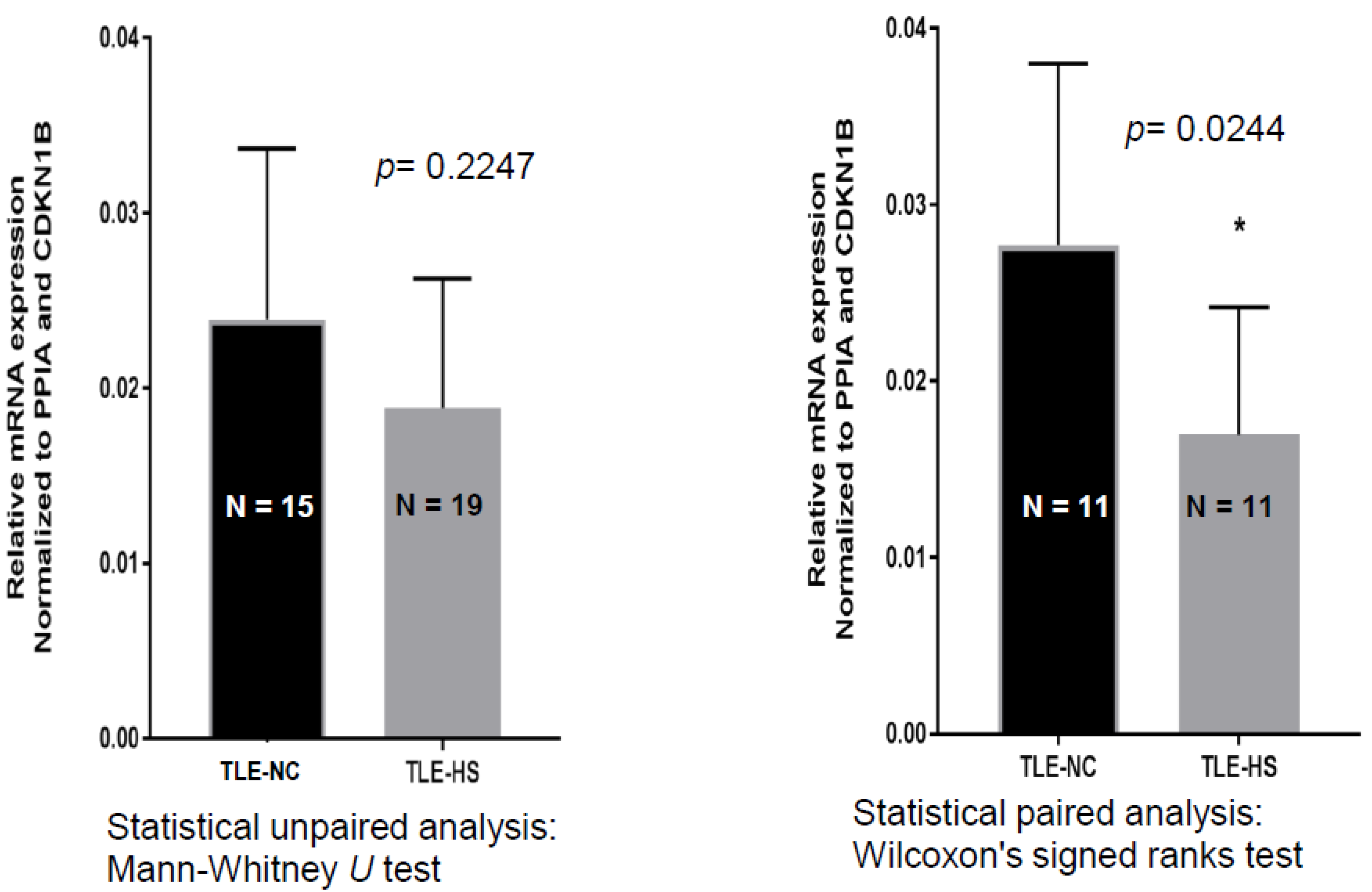
2.1. SCN4B Transcripts Expression in HS and NC Samples

In order to test the hypothesis that SCN4B is differentially expressed in hippocampus of pharmaco-resistant TLE-HS patients and adjacent (non-epileptic) neocortex tissue (designated as temporal lobe epilepsy neocortex (TLE-NC)) [28], real-time qPCR analysis was used, and SCN4B expression was normalized to the house-keeping genes (HKGs). The results from comparing SCN4B expression in TLE-NC and TLE-HS, by using unpaired analysis, show a decreased expression of SCN4B; however, this was not statistically significant (p = 0.2247) (Figure 1). When comparing TLE-HS and TLE-NC across the 11 patient samples, with a paired analysis, SCN4B downregulation in TLE-HS is statistically significant, with a 43% decrease in expression (p = 0.0244).

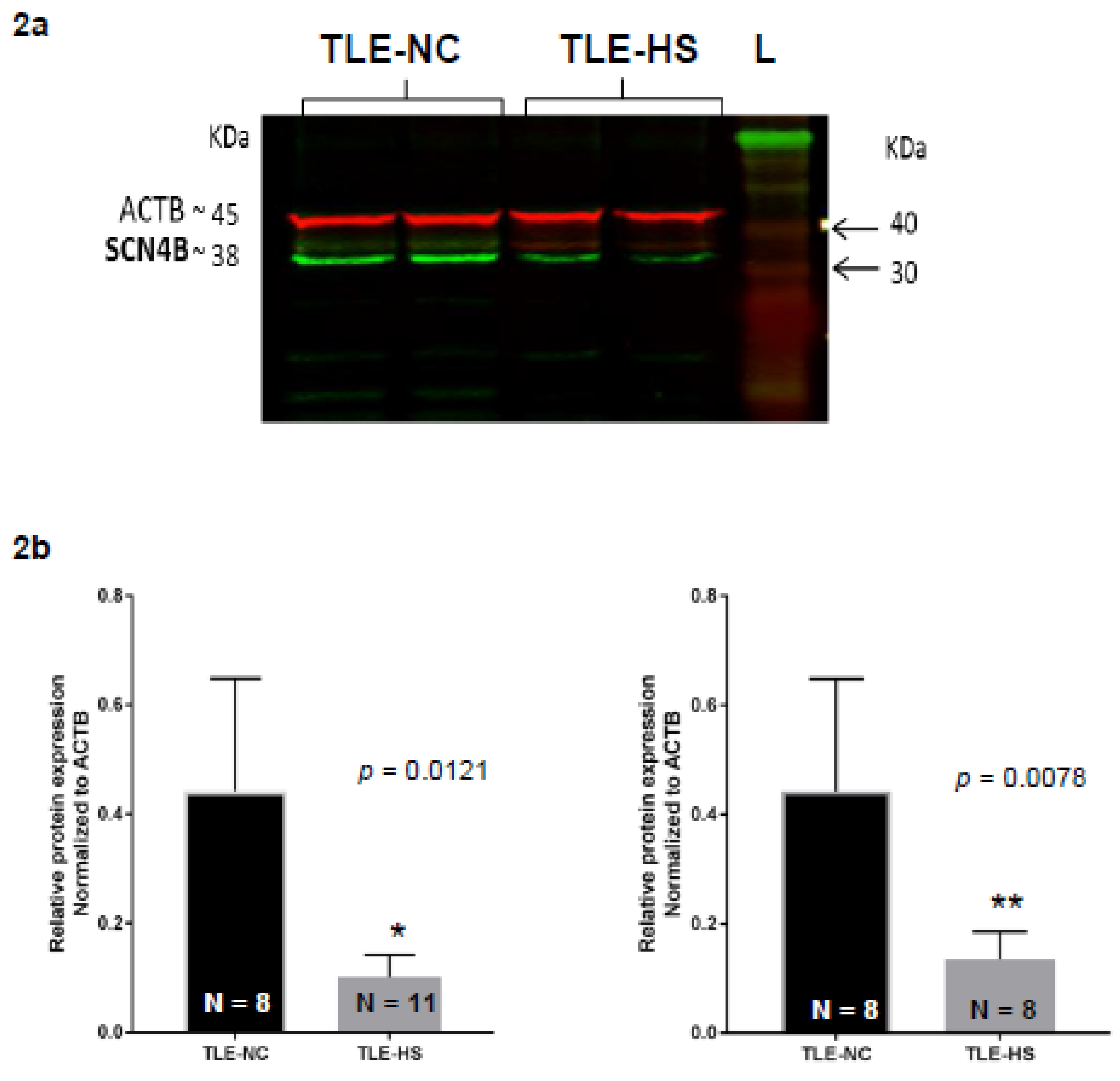
2.2. Western Blot Analysis of Nav β4 in HS and NC Samples

It is important to determine whether the transcriptional changes determined by qPCR are reflected in the protein levels of the SCN4B gene product Nav β4. Therefore, protein extracts from the hippocampal non-sclerotic and sclerotic tissue of each patient were subjected to Western blot analysis, staining for Nav β4 (Figure 2a). The normalized band densities were compared between the non-sclerotic and sclerotic tissue proteins for each patient to determine the relative protein changes in Nav β4 (Figure 2b). A reduction in the relative protein expression levels was observed.in sclerotic hippocampal tissue compared to non-sclerotic tissue resection. In this case, we used both unpaired and paired analysis, and the decrease in TLE-HS was statistically significant for both types of analysis (p = 0.0121 and p = 0.0078 for unpaired and paired, respectively).

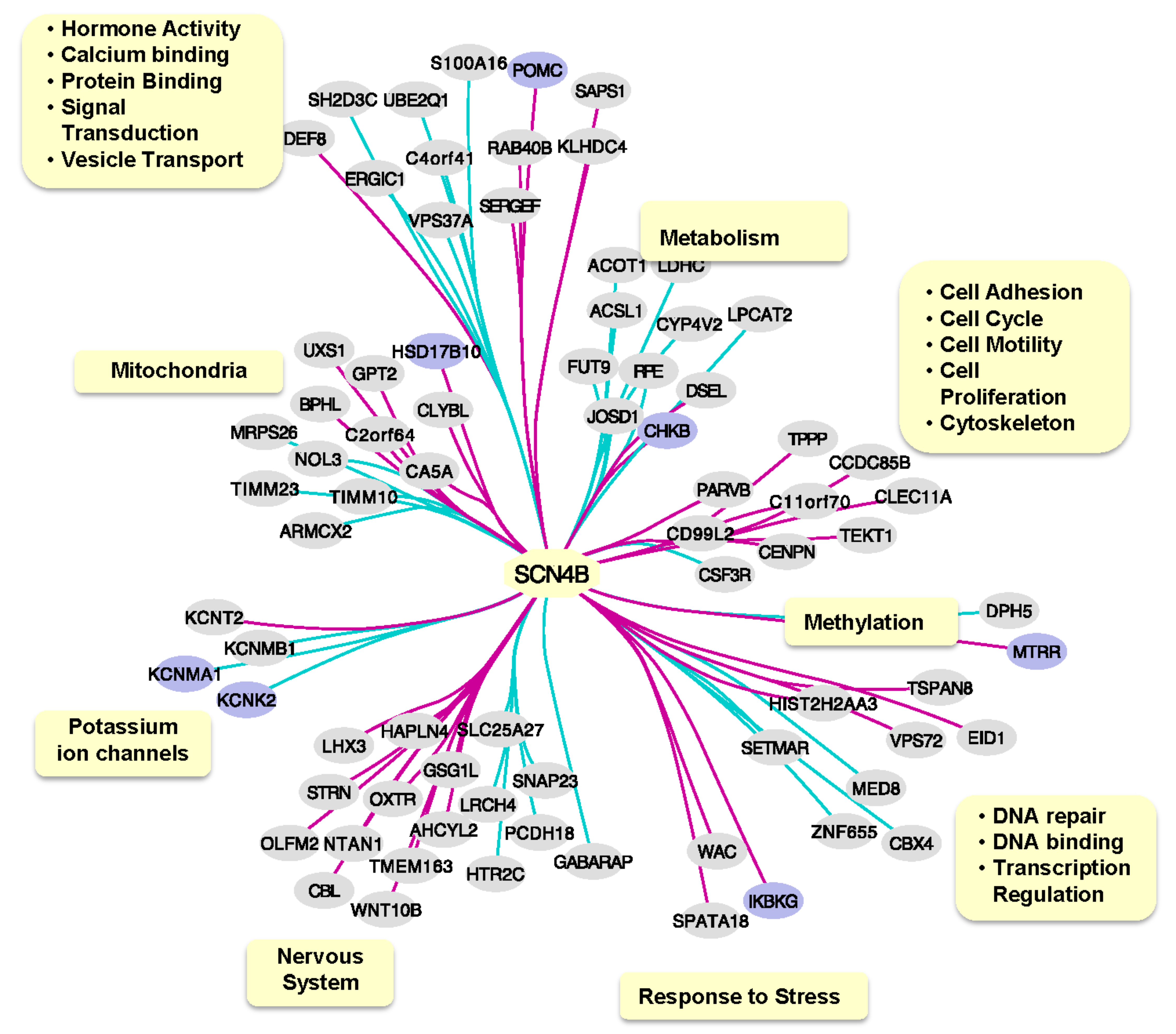
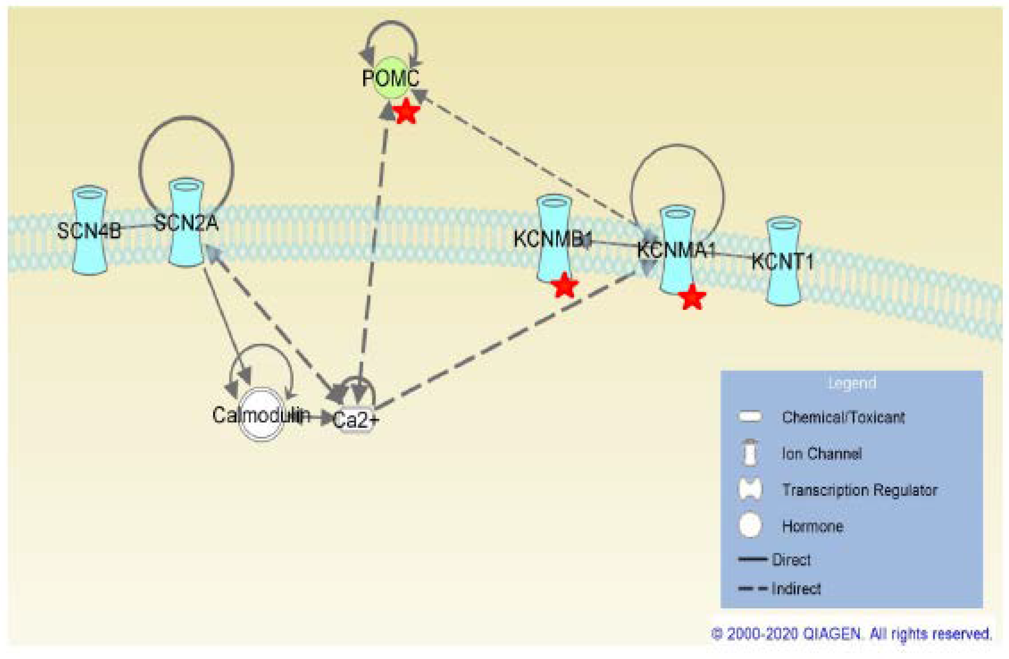
2.3. SCN4B Co-Expression Network Analysis in TLE Patients

After demonstrating that sclerotic hippocampus tissue in drug-resistant TLE patients is characterized by lower expression of SCN4B, both at the mRNA and protein level, we investigated the functional implications of this variability in SCN4B expression. This challenge was addressed by generating a co-expression network using a publicly available transcriptomics dataset from TLE hippocampus samples, followed by the investigation of functional aspects of genes significantly co-expressed with SCN4B. A total of 83 genes were found significantly co-expressed with SCN4B (i.e., FDR < 0.05 and posterior probability > 0.8), but only 77 genes contained functional information available.
Using information retrieved from both DAVID and AmiGO 2, we have annotated relevant GO terms associated to co-expressed genes and have identified several biological mechanisms and components associated to these genes, including components closely related to neuronal function such as potassium ion channels (Figure 3). Additionally, seven of the co-expressed genes were previously found associated to epilepsy, which seems to be associated to a diversity of biological mechanisms (hormone-based signaling, mitochondria, metabolism, potassium ion channels, methylation and response to stress). DAVID analysis found no enriched pathways for SCN4B co-expressed genes. In contrast, STRING analysis found that calcium-activated potassium channel activity GO term (GO:0015269) was significantly enriched (FDR = 0.0374) due to KCNMA1, KCNMB1 and KCNT2 genes.

3. Discussion

In this study, patients affected by mesial TLE were compared with an adjacent specimen from non-seizing medial temporal neocortex tissue, mesial TLE-NC. The experiments demonstrate a reduction in the expression profile, at a transcriptional and translational level, in the voltage-gated sodium channel β4-subunit in the sclerotic hippocampal tissue of AED-resistant TLE compared with TLE-NC. Changes in mRNA expression levels are not always reflected in the protein expression, but our results demonstrate a borderline correlation. This evidence potentially elucidates a key factor of AED resistance. The β4-subunit plays an important role in determining the gating of the VGSC and can therefore affect the binding sensitivities of AED, many of which act in a use-dependent manner.
A reduction in the Nav β4 could simply result in a loss of drug action on the target channel, contributing to AED resistance in TLE patients. However, the β4-subunit has already been shown in cell models to favor the open state of VGSCs and promote persistent currents with certain α-subunits [29]. Therefore, a reduction is more likely to result in a decrease in the frequency and duration of VGSC in the resting state, which would reduce the opportunity for the vast number of use-dependent VGSC-modulating AEDs to bind to their target.
The combination of β-subunits defines a range of VGSC current amplitudes. Co-expression of β1 with β4 can in fact suppress the gating effects of β4 mentioned previously. This is a result of β1-subunits’ own effect on VGSC gating, which instead stabilizes the closed states at hyperpolarized potentials and inactivated states at depolarized potentials [29]. Therefore, a pathology, like TLE-HS, that results in a reduction of β4 in an area of the brain that would usually express β4, would in fact have an enhanced effect. There would be a loss in the favor of the open/resting state of VGSCs caused by β4 association and an increase in the effects of β1 on the VGSC gating, which are usually partially suppressed through co-expression with β4. In turn, these increased effects of β1 would further reduce the binding of AEDs to VGSCs.
Despite understanding the possible influence that a reduction in β4 may have on AED resistance, it is important to identify the cause of this lower expression to help discover new molecular targets to manipulate β4 expression. These experiments do not allow us to determine why these changes occur. They could be a result of epilepsy or the sclerosis. Other types of epilepsy, with and without sclerotic tissue, and other pathologies associated with the formation of sclerotic tissue, like multiple sclerosis (MS), would need to be analyzed in order to develop our understanding of a potential cause for this reduction in β4.
Another approach that could help further direct this research is the determination of whether there are mutations in the SCN4B promoter site in TLE-HS patients. Methylation and acetylation patterns should also be investigated, as these may result in altered β4 expression levels, but they are yet to be investigated in any of the β-subunits. Nevertheless, increased expression patterns of DNA methyltransferase (DNMT) isoforms DNMT1 and DNMT3 [30] and histone deacetylases (HDAC2) [31] have been found in our analysis in TLE. Our data also show two more genes co-expressed with SCN4B: methionine synthase reductase (MTRR) and diphthine synthase (DPH5). They are both involved with the methylation process in neurons, and they show opposite co-expression with SCN4B, MTRR with a positive and DPH5 with a negative co-expression. MTRR was already investigated in epilepsy, [32], whereas DPH5 has been linked to the translation elongation factor 2 but has not been investigated yet in TLE, and this could lead to significant modulation of the translational regulatory machinery.
Mutations in other β-subunits have already been found. For example, heterozygous mutations in extracellular domain of SCN1B [33] and several additional mutations have all been associated with epilepsy [33,34,35,36,37]. Therefore, it appears reasonable to hypothesize that mutations affecting the structure and expression of β4 may be present in epilepsy, including TLE.
The diffused expression of Nav1.2 along some demyelinated axons supports recovery of action potential conduction. Polymorphisms in the SCN2A, which may reduce this recovery action, have been associated with epilepsy and AED resistance [18]. In contrast, Nav1.6 produces Na+ current that can drive the Na+–Ca2+ exchanger, potentially triggering injurious cascades and promoting neuronal atrophy [38]. Seizures also result in an increase in Na+ currents, which also drives neuronal atrophy: ‘seizure-induced neuronal atrophy’ [39]. Therefore, genetic and expressional changes in β4, along with other VGSC-subunits, in TLE could alter the gating of VGSCs and may alter the neuroprotective activity of the VGSCs in the neuronal atrophy seen in the formation of sclerosis. When considered along with the inflammatory abnormalities seen in TLE [40], this suggests that TLE drives the formation of HS in predisposed neuronal tissue.
Interestingly, the β-secretase, β-site APP-cleaving enzyme 1 (BACE1), has shown, in Purkinje neurons, to have a novel function in the regulation of neuronal excitability by impairing the regular processing of β4 [41,42,43]. The mechanism behind transcriptional regulation of SCN4B remains elusive. However, it is noted that BACE1 is known to have some transcriptional effects. Tts involvement in proteolytic cleaving of β1 is proposed to release a soluble intracellular domain of β1 which then enhances transcription of α-subunit genes, thus demonstrating the need to investigate any effects BACE1 may have on β4 transcription as well as protein cleavage. When the potential effects of BACE1 in sclerosis are combined with the loss of neuroprotective activity, the effects could result in the significant reduction of β4 and, hence, a reduction in the VGSCs susceptible to AED interaction. Alternatively, it may only be one of these mechanisms that results in a reduction of β4, but they would cause a reduction nonetheless.
AED resistance is likely to be multifactorial, which was demonstrated by VGSC mutations associated with AED resistance [17,18,21] and the altered expression of receptors [44]. This research elucidates the reduced expression of β4 as an important factor in the AED resistance seen in TLE-HS and could therefore direct both our current approach to the treatment of AED-resistant TLE and the development of novel AEDs in the future. The large number of AEDs with known VGSC-modulating properties, even in the small sample cohort used in this study, Table 1, helps signify the potential impact that a change in this molecular target might actually have on the approach to treating TLE.
Our network analysis using publicly available data revealed that important cellular (neuronal and glial) functions and pathways are indeed defining the phenotype of TLE in the AED-resistant cohort. This has allowed us to determine a principal element to be further investigated in order to improve the response to sodium channel blockers as therapeutics for TLE. Despite the analysis being more focused on the β1-subunit, it has been reported that sodium β channel subunits are multifunctional and act via multiple signaling pathways. This was shown in an animal model with induced mutations and by genetic characterization derived from epileptic patients affected by Dravet syndrome (DS), generalized epilepsy with febrile seizures plus (GEFS+), sudden infant death syndrome (SIDS) and sudden unexpected death in epilepsy (SUDEP) [8].
Differently, we have defined most, if not all, of these pathways focusing on sodium channel β4-subunit and in human brains affected by TLE (Figure 4).
The pathways that we have identified by SCN4B co-expression network analysis in TLE patients can be used to generate hypotheses of the mechanisms linked to impairment in the expression of SCN4B. These include the modulation, trafficking and localization of the sodium channel itself, the cellular migration and proliferation including the cytoskeleton, linking it to development phenomena; pathways which involve DNA regulation, repair/binding and transcription; and pathways involving protein binding and signal transduction.
Several SCN4B co-expressed genes are related to mitochondrial function, and its impairment has been identified as a potential cause of epileptic seizures [45]. One of these mitochondria-related genes is CA5A (carbonic anhydrase 5A), which has a positive co-expression with SCN4B. Several AEDs act through multiple mechanisms and simultaneously inhibit carbonic anhydrases and block VGSCs [44]. This example could point that other effects associated to the lower expression of SCN4B, in this case a correlated lower expression of CA5A, could simultaneously contribute to AED resistance. Mitochondrial disorders are often the cause of seizures, and diverse types of seizures have been reported to be associated with mitochondrial oxidative stress, which gradually disrupts the homeostasis of intracellular Ca2+. Mutations have been reported in HSD17B10, impairing the activities of the mitochondrial RNase P complex [46].
Interestingly, SCN4B was shown to be negatively co-expressed with two calcium-activated potassium channel genes (KCNMA1 and KCNMB1) and a two-pore potassium channel (KCNK2) and positively co-expressed with a sodium-activated potassium channel gene (KCNT2).
The KCNMA1, KCNMB1, and KCN2 genes have a main role in stopping or reducing excitatory stimuli in neurons [47]. Therefore, from these results, we can hypothesize that when SCN4B is dysregulated these sodium- and potassium-activated channel, they lose their inhibitory role, leading to the TLE.
There have been no previous associations of SCN4B with these potassium channel genes; therefore, the cause of their co-expression in TLE patients is unclear. Nevertheless, by using IPA to investigate possible pathways to link SCN4B to these potassium channel genes, we have found that SCN2A, which binds to SCN4B to form the sodium channel, also binds to Ca2+/calmodulin complexes. Mutations in SCN2A cause Ca2+ to dissociate from calmodulin, leading to higher intracellular Ca2+ concentration, and these changes could in turn have a downstream effect in the expression of calcium-activated potassium channels. One hypothesis is that the lower expression of SCN4B could also affect the ability of SCN2A to bind to Ca2+/calmodulin complexes. Interestingly, changes in KCNMA1 expression and in intracellular Ca2+ concentration also have an effect on POMC regulation and intra/extracellular concentrations of its products. POMC was also found to be co-expressed with SCN4B in our analysis, and it has been previously associated to epilepsy. The intracellular neuropeptide alpha-melanocyte stimulating hormone (alpha-MSH) POMC product increases Ca2+ level. Depletion of extracellular Ca2+ decreases adrenocorticotropic hormone (ACTH) POMC product release.
There is still much to uncover about biological mechanisms in which SCN4B is involved and its role in AED resistance. This functional analysis of putative systems affecting or affected by SCN4B expression, despite the limited sample size, points to avenues that can be explored for further understanding of AED resistance and potential candidate drug targets to be considered.

4. Materials and Methods

4.1. Tissue Collection

Tissue from the hippocampal formation was obtained from pharmaco-resistant mesial TLE-HS (n = 19) patients and adjacent (non-epileptic) medial temporal neocortex tissue TLE-NC (n = 15) [28], from Sheffield Hallamshire Hospital (Table 1). Informed consent was given by all participants for experimental use; approved by South Yorkshire NRES committee York and Humber (08-H1310-49).
TLE-HS and TLE-NC sample pairs were from the same patient, therefore eliminating inter-individual, age-related, postmortem-related and drug-related variabilities.

4.2. qRT-PCR Analysis of SCN4B

The quantification of mRNA levels of SCN4B was performed by qRT-PCR in TLE-NC (n = 11) and TLE-HS (n = 19) samples. The mRNA was extracted from the epileptic patient samples and purified using the SV Total RNA Isolation System (Promega, UK), and then the product was quantified using the Nano Assay (Nanodrop Analyzer, Thermofisher, Loughborough, UK). The reverse transcription of each mRNA sample was done using 1μg of RNA with the Superscript III First-stand kit (Qiagen, UK). Gene expression levels were then determined using the TaqMan Fast Universal MasterMix (Applied Biosystem, Life Technologies, Loughborough, UK) with the StepOnePlus Real-Time PCR System (Applied Biosystems, Thermo Fisher, UK). Real-time qPCR reactions were run over 50 cycles (initial heating of 95 °C for 20 s followed by cycles of 95 °C for 1 s and 20 s at 60 °C) with 20 ng of cDNA per reaction to amplify specific genetic sequences. The following primers were used to detect specific house-keeping genes (HKGs): peptidylprolyl isomerase A (PPIA) (Hs04194521_s1, Applied Biosystems, Life Technologies, UK) and cyclin-dependent kinase inhibitor 1B (CDKN1B) (Applied Biosystems, Life Technologies, UK). These results were utilized to normalize the gene expression levels of the target gene, SCN4B (Hs03681025_m1, Applied Biosystems, Life Technologies, UK). All samples were run in triplicate, and only the reactions that produced appropriate amplification curves were analyzed, as produced by the real-time qPCR analytical software (StepOne Software v2.0 (Applied Biosystems, Thermo Fisher, UK)). Relative expression of SCN4β compared to an average of the HKG expression was calculated using the 2−ΔCt and 2−ΔΔCt methods.

4.3. Western Blot Analysis of Nav β4

Tissue samples were lysed using 200 mg/mL CellLytic MT Mammalian Tissue Lysis/Extraction Regent (Sigma, Welwyn Garden City, UK) and 1 mg/mL stock protease inhibitor cocktail (Sigma, UK) to extract protein. Protein concentration was determined using a bicinchoninic acid (Sigma, UK) assay. The protein samples were then subjected to electrophoresis and separated on a 10% polyacrylamide gel, using 30 µg of protein per lane, alongside the molecular weight marker, Chameleon (Life Sciences, Applied Biosystems). The gel was then blotted onto a nitrocellulose blotting membrane, Amersham Protan supported 45 µm NC (GE Healthcare, Life Technologies, Little Chalfont, UK), using 75 V (~180 mA) (PowerPac Basic, BioRad, Deeside, UK) for 50 min in transfer buffer (25 mM Tris, 193 mM glycine, 10% methanol made up with water). Then, the blot was incubated overnight in blocking solution (5% non-fat powdered milk in TBST (10 mM Tris, pH 7.5, 50 mM NaCl and 0.05% Tween-20, (Sigma)). After blocking, the blots were incubated at room temperature for 1 h in the primary antibody solution; rabbit anti-SCN4B (1:500) (ab80539, Abcam, Cambridge, UK) and β-actin (8H10D10) mouse mAb (1:1000) (Santa Cruz, Harrogate, UK) diluted in blocking solution. The blots were washed three times, each for 10 min, in TBST. Primary antibody binding was detected by incubation with a solution of 1:10,000 dilution of infrared IRDye (800 CW)-conjugated donkey anti-rabbit (LI-COR Biosciences, Cambridge, UK) and 1:50,000 dilution of infrared IRDye (680 CW)-conjugated donkey anti-mouse (LI-COR biosciences, Cambridge, UK) in 5% powdered milk in TBST, for 1 h at room temperature. Again, the blots were washed three times, each for 10 min, in TBST. Blots were visualized and quantified using Odyssey LI-COR Scanner (LI-COR Biosciences, Cambridge, UK).

4.4. Western Blot Band Quantification

The protein band densities on the Western blots, as detected by the Odyssey LI-COR scanner (LI-COR Biosciences), were quantitated using the Image Studio software (Ver3.1, Applied Biosystems). The protein bands of interest in each sample, corresponding to the Nav β4 protein (37 kDa), were normalized to β-actin (42kDa) band density, and relative intensities were calculated to run comparative analysis between the sclerotic and control (non-sclerotic) samples.

4.5. Statistical Analysis of SCN4B Gene Expression and Nav β4 Band Intensities

The changes in SCN4B gene expression and Nav β4 protein expression between sclerotic and control samples were analyzed for statistical significance using Mann–Whitney U test for the unpaired data sets. Wilcoxon’s signed ranks test was used for statistical paired analysis.
Comparative analysis, investigating correlation between the SCN4B and Nav β4 expression level, was performed using Spearman’s rank test to produce a correlation coefficient (strong correlation when r ≥ 0.7).

4.6. SCN4B Co-Expression Network Analysis in TLE Patients

Microarray expression profiles from 129 TLE patients (GEO ID: GSE63808) were used for inferring co-expression networks [47]. To increase the power for detecting significant correlations, probes were filtered by expression level (probes showing a detection p-value < 0.05 in at least 20% of the samples) and expression variation (probes having a coefficient of variation (CV) of gene expression greater than the median CV calculated across all probes in the set). After filtering and collapsing probes to gene level, a total of 7475 remaining genes were used as input for co-expression network inference using Graphic Gaussian Models (GGM) [48] method through the “GeneNet” [49] package in R (http://www.r-project.org/). This method estimates partial correlations within the probe set, identifying co-expression between probe pairs, while removing effects from other probes. A mixture model is then fitted to the partial correlations for assessing significant edges in the network.

4.7. Functional Analysis of SCN4B Co-Expressed Genes

We used DAVID v6.8 [50] and STRING web tools to perform functional enrichment analysis [51]. We also used Ingenuity Pathwy Analysis (IPA) (QIAGEN Inc., https://www.qiagenbioinformatics.com/products/ingenuity-pathway-analysis) [52] to investigate putative pathways linking SCN4β to co-expressed genes. IPA built-in tools, such as ‘Connect’, ‘Path Explorer’ and ‘Grow’ search for connections between selected molecules based on prior knowledge. Genes previously associated with epilepsy were retrieved from DisGeNet [53] database v6.0 (disease ID: C0014544). We also searched AmiGO 2 [54,55] database v2.5.12 for gene ontology (GO) terms associated to co-expressed genes.

5. Conclusions

It may be possible to specifically target the β4-subunit of sodium channels; as it only shares 35% amino acid identity with the most closely associated β2-subunit, it would be more effective to induce an overall increasing expression of β4, rather than to modulate the relatively few β4-subunits that are still present. Investigations into mutations and the transcriptional regulation of SCN4B would be needed to elucidate potential targets for modulating SCN4B expression and the pathways it influences.

Author Contributions

M.A.S. designed and performed the experiments; L.Y.T. performed the bioinformatics analysis; E.J.S. designed and performed some experiments; F.F. helped to collect and analyze the data of M.A.S. and to critically review the manuscript; and A.P.P. designed the project and oversaw the reporting of this study. The manuscript was written by M.A.S., E.J.S., L.Y.T., F.F., and A.P.P. All authors have read and agreed to the published version of the manuscript.

Funding

This project has been mainly funded by Sheffield Hallam University Biomolecular Research Centre and partially funded by the Innovative Medicines Initiative 2 TransBioLine project (grant agreement No. 821283).

Acknowledgments

We are very grateful to Dev Bhattacharyya at Neurosurgery, Sheffield Hallamshire Hospital, Glossop Road, Sheffield, S10 2JF, UK, for having provided the human specimens.

Conflicts of Interest

The authors declare no conflict of interest.

Abbreviations

ATPAdenosine triphosphate
AEDsAntiepileptic drugs
ABCATP-binding cassete
BCABicinchonic acid
BACE1β-site APP-cleaving enzyme 1
DBSDeep brain stimulation
HSHippocampal sclerotic
MSMultiple sclerosis
TBSTris-buffered saline
TLETemporal lobe epilepsy
TLE-NCTemporal lobe epilepsy neocortical
TLE-HSTemporal lobe epilepsy hippocampal sclerotic
VGSCVoltage-gated sodium channel
VNSVagus brain stimulation

References

  1. Thom, M. Hippocampal sclerosis in epilepsy: A neuropathology review. Neuropathol. Appl. Neurobiol. 2014, 40, 520–543. [Google Scholar] [CrossRef] [PubMed]
  2. Harroud, A.; Bouthillier, A.; Weil, A.G.; Nguyen, D.K. Temporal lobe epilepsy surgery failures: A review. Epilepsy Res. Treat. 2012, 2012, 1–10. [Google Scholar] [CrossRef] [PubMed]
  3. Yardi, R.; Irwin, A.; Kayyali, H.; Gupta, A.; Nair, D.; Gonzalez-Martinez, J.; Bingaman, W.; Najm, I.M.; Jehi, L.E. Reducing versus stopping antiepileptic medications after temporal lobe surgery. Ann. Clin. Transl. Neurol. 2014, 1, 115–123. [Google Scholar] [CrossRef] [PubMed]
  4. Sherman, E.M.; Wiebe, S.; Fay-McClymont, T.B.; Téllez-Zenteno, J.; Metcalfe, A.; Hernandez-Ronquillo, L.; Jetté, N. Neuropsychological outcomes after epilepsy surgery: Systematic review and pooled estimates. Epilepsia 2011, 52, 857–869. [Google Scholar] [CrossRef]
  5. Macrodimitris, S.; Sherman, E.M.; Forde, S.; Téllez-Zenteno, J.F.; Metcalfe, A.; Hernández-Ronquillo, L.; Jetté, N. Psychiatric outcomes of epilepsy surgery: A systematic review. Epilepsia 2011, 52, 880–890. [Google Scholar] [CrossRef]
  6. Lason, W.; Chlebicka, M.; Rejdak, K. Research advances in basic mechanisms of seizures and antiepileptic drug action. Pharmacol. Rep. 2013, 65, 787–801. [Google Scholar] [CrossRef]
  7. Catterall, W.A. Signaling complexes of voltage-gated sodium and calcium channels. Neurosci. Lett. 2010, 486, 107–116. [Google Scholar] [CrossRef] [Green Version]
  8. O’Malley, H.A.; Isom, L.L. Sodium channel β subunits: Emerging targets in channelopathies. Annu. Rev. Physiol. 2015, 77, 481–504. [Google Scholar] [CrossRef] [Green Version]
  9. Ratcliffe, C.F.; Westenbroek, R.E.; Curtis, R.; Catterall, W.A. Sodium channel β1 and β3 subunits associate with neurofascin through their extracellular immunoglobulin-like domain. J. Cell Biol. 2001, 154, 427–434. [Google Scholar] [CrossRef] [Green Version]
  10. Yu, F.H.; Westenbroek, R.E.; Silos-Santiago, I.; McCormick, K.A.; Lawson, D.; Ge, P.; Ferriera, H.; Lilly, J.; DiStefano, P.S.; Catterall, W.A.; et al. Sodium channel beta4, a new disulfide-linked auxiliary subunit with similarity to beta2. J. Neurosci. 2003, 23, 7577–7585. [Google Scholar] [CrossRef] [Green Version]
  11. Semah, F.; Picot, M.C.; Adam, C.; Broglin, D.; Arzimanoglou, A.; Bazin, B.; Cavalcanti, D.; Baulac, M. Is the underlying cause of epilepsy a major prognostic factor for recurrence? Neurology 1998, 51, 1256–1262. [Google Scholar] [CrossRef] [PubMed]
  12. Siddiqui, A.; Kerb, R.; Weale, M.E.; Brinkmann, U.; Smith, A.; Goldstein, D.B.; Wood, N.W.; Sisodiya, S.M. Association of multidrug resistance in epilepsy with a polymorphism in the drug-transporter gene ABCB1. Brain 2003, 125, 22–31. [Google Scholar]
  13. Brand, C.; Bethmann, K.; Gastens, A.M.; Löscher, W. The multidrug transporter hypothesis of drug resistance in epilepsy: Proof-of-principle in a rat model of temporal lobe epilepsy. Neurobiol. Dis. 2006, 24, 202–211. [Google Scholar] [CrossRef]
  14. Bournissen, F.G.; Moretti, M.E.; Juurlink, D.; Koren, G.; Walker, M.; Finkelstein, Y. Polymorphisms of MDR1/ABC1B C3435T drug-transporter and resistance in anti-convulsant drugs: A meta-analysis. Epilepsia 2009, 50, 898–903. [Google Scholar] [CrossRef]
  15. Schmidt, D.; Löscher, W. Drug resistance in epilepsy: Putative neurobiologic and clinical mechanisms. Epilepsia 2005, 46, 858–877. [Google Scholar] [CrossRef]
  16. Remy, S.; Beck, H. Molecular and cellular mechanisms of pharmacoresistance in epilepsy. Brain 2006, 129, 18–35. [Google Scholar] [CrossRef] [PubMed] [Green Version]
  17. Kwan, P.; Poon, W.S.; Ng, H.K.; Kang, D.E.; Wong, V.; Ng, P.W.; Lui, C.H.; Sin, N.C.; Wong, K.S.; Baum, L. Multidrug resistance in epilepsy and polymorphisms in the voltage-gated sodium channel genes SCN1A, SCN2A, and SCN3A: Correlation among phenotype, genotype, and mRNA expression. Pharm. Genom. 2008, 18, 989–998. [Google Scholar] [CrossRef] [PubMed]
  18. Lakhan, R.; Kumari, R.; Misra, U.K.; Kalita, J.; Pradhan, S.; Mittal, B. Differential role of sodium channels SCN1A and SCN2A gene polymorphisms with epilepsy and multiple drug resistance in the north Indian population. Br. J. Clin. Pharmacol. 2009, 68, 214–220. [Google Scholar] [CrossRef] [Green Version]
  19. Amin, A.S.; Asghari-Roodsari, A.; Tan, H.L. Cardiac sodium channelopathies. Pflug. Arch.-Eur. J. Physiol. 2010, 460, 223–237. [Google Scholar] [CrossRef] [Green Version]
  20. George, A.L., Jr. Inherited disorders of voltage-gated sodium channels. J. Clin. Investig. 2005, 115, 1990–1999. [Google Scholar] [CrossRef]
  21. Lucas, P.T.; Meadows, L.S.; Nicholls, J.; Ragsdale, D.S. An epilepsy mutation in the beta 1 subunit of the voltage-gated sodium channel results in reduced channel sensitivity to phenytoin. Epilepsy Res. 2005, 64, 77–84. [Google Scholar] [CrossRef] [PubMed]
  22. Zhao, J.; O’Leary, M.E.; Chahine, M. Regulation of Nav1.6 and Nav1.8 peripheral nerve Na+ channels by auxiliary β-subunits. J. Neurophysiol. 2011, 106, 608–619. [Google Scholar] [CrossRef] [PubMed]
  23. Bean, B.P. The molecular machinery of resurgent sodium current revealed. Neuron 2005, 45, 185–187. [Google Scholar] [CrossRef] [Green Version]
  24. Bant, J.S.; Raman, I.M. Control of transient, resurgent, and persistent current by open-channel block by Na channel beta4 in cultured cerebellar granule neurons. Proc. Natl. Acad. Sci. USA 2010, 107, 12357–12362. [Google Scholar] [CrossRef] [PubMed] [Green Version]
  25. Oyama, F.; Miyazaki, H.; Sakamoto, N.; Becquet, C.; Machida, Y.; Kaneko, K.; Uchikawa, C.; Suzuki, T.; Kurosawa, M.; Ikeda, T.; et al. Sodium channel β4 subunit: Down-regulation and possible involvement in neuritic degeneration in Huntington’s disease transgenic mice. J. Neurochem. 2006, 98, 518–529. [Google Scholar] [CrossRef] [PubMed]
  26. Medeiros-Domingo, A.; Iturralde-Torres, P.; Ackerman, M.J. Clinical and genetic characteristics of long QT syndrome. Rev. Esp. Cardiol. 2007, 60, 739–752. [Google Scholar] [CrossRef] [PubMed]
  27. Li, R.G.; Wang, Q.; Xu, Y.J.; Zhang, M.; Qu, X.K.; Liu, X.; Fang, W.Y.; Yang, Y.Q. Mutations of the SCN4B-encoded sodium channel β4 subunit in familial atrial fibrillation. Int. J. Mol. Med. 2013, 32, 144–150. [Google Scholar] [CrossRef] [Green Version]
  28. Salman, M.M.; Sheilabi, M.A.; Bhattacharyya, D.; Kitchen, P.; Conner, A.C.; Bill, R.M.; Woodroofe, M.N.; Conner, M.T.; Princivalle, A.P. Transcriptome analysis suggests a role for the differential expression of cerebral aquaporins and the MAPK signaling pathway in human temporal lobe epilepsy. Eur. J. Neurosci. 2017, 46, 2121–2132. [Google Scholar] [CrossRef]
  29. Aman, T.K.; Grieco-Calub, T.M.; Chen, C.; Rusconi, R.; Slat, E.A.; Isom, L.L.; Raman, I.M. Regulation of persistent Na current by interactions between beta subunits of voltage-gated Na channels. J. Neurosci. 2009, 29, 2027–2042. [Google Scholar] [CrossRef] [Green Version]
  30. Zhu, Q.; Wang, L.; Zhang, Y.; Zhao, F.; Luo, J.; Xiao, Z.; Chen, G.; Wang, Z. Increased Expression of DNA methyltransferase 1 and 3a in Human Temporal Lobe Epilepsy. J. Mol. Neurosci. 2012, 46, 420–426. [Google Scholar] [CrossRef]
  31. Huang, Y.; Zhao, F.; Wang, L.; Yin, H.; Zhou, C.; Wang, X. Increased expression of histone deacetylases 2 in temporal lobe epilepsy: A study of epileptic patients and rat models. Synapse 2012, 66, 151–159. [Google Scholar] [CrossRef] [PubMed]
  32. Dean, J.C.S.; Robertson, Z.; Reid, V.; Wang, Q.; Hailey, H.; Moore, S.; Rasalam, A.D.; Turnpenny, P.; Lloyd, D.; Shaw, D.; et al. A high frequency of the MTHFR 677C>T polymorphism in Scottish women with epilepsy: Possible role in pathogenesis. Seizures 2008, 17, 269–275. [Google Scholar] [CrossRef] [PubMed] [Green Version]
  33. Wallace, R.H.; Wang, D.W.; Singh, R.; Scheffer, I.E.; George, A.L., Jr.; Phillips, H.A.; Saar, K.; Reis, A.; Johnson, E.W.; Sutherland, G.R.; et al. Febrile seizures and generalized epilepsy associated with a mutation in the Na+-channel beta1 subunit gene SCN1B. Nat. Genet. 1998, 19, 366–370. [Google Scholar] [CrossRef] [PubMed]
  34. Audenaert, D.; Claes, L.; Ceulemans, B.; Lofgren, A.; Van Broeckhoven, C.; De Jonghe, P. A deletion in SCN1B is associated with febrile seizures and early-onset absence epilepsy. Neurology 2003, 61, 854–856. [Google Scholar] [CrossRef]
  35. Yamakawa, K. Epilepsy and sodium channel gene mutations: Gain or loss of function? Neuroreport 2005, 16, 1–3. [Google Scholar] [CrossRef]
  36. Scheffer, I.E.; Harkin, L.A.; Grinton, B.E.; Dibbens, L.M.; Turner, S.J.; Zielinski, M.A.; Xu, R.; Jackson, G.; Adams, J.; Connellan, M.; et al. Temporal lobe epilepsy and GEFS+ phenotypes associated with SCN1B mutations. Brain 2007, 130, 100–109. [Google Scholar] [CrossRef] [Green Version]
  37. Patino, G.A.; Brackenbury, W.J.; Bao, Y.; Lopez-Santiago, L.F.; O’Malley, H.A.; Chen, C.; Calhoun, J.D.; Lafrenière, R.G.; Cossette, P.; Rouleau, G.A.; et al. Voltage-gated Na+ channel β1B: A secreted cell adhesion molecule involved in human epilepsy. J. Neurosci. 2011, 31, 14577–14591. [Google Scholar] [CrossRef] [Green Version]
  38. Waxman, S.G. Axonal conduction and injury in multiple sclerosis: The role of sodium channels. Nat. Rev. Neurosci. 2006, 7, 932–941. [Google Scholar] [CrossRef]
  39. Henshall, D.C. Apoptosis signaling pathways in seizure-induced neuronal death and epilepsy. Biochem. Soc. Trans. 2007, 35, 421–423. [Google Scholar] [CrossRef] [Green Version]
  40. Vezzani, A.; Friedman, A.; Dingledine, R.J. The role of inflammation in epileptogenesis. Neuropharmacology 2013, 69, 16–24. [Google Scholar] [CrossRef] [Green Version]
  41. Huth, T.; Rittger, A.; Saftig, P. β-Site APP-cleaving enzyme 1 (BACE1) cleaves cerebellar Na+ channel β4-subunit and promotes Purkinje cell firing by slowing the decay of resurgent Na+ current. Pflug. Arch. 2011, 461, 355–371. [Google Scholar] [CrossRef] [PubMed]
  42. Hu, X.; Zhou, X.; He, W.; Yang, J.; Xiong, W.; Wong, P.; Wilson, C.G.; Yan, R. BACE1 deficiency causes altered neuronal activity and neurodegeneration. J. Neurosci. 2010, 30, 8819–8829. [Google Scholar] [CrossRef] [PubMed]
  43. Hessler, S.; Zheng, F.; Hartmann, S.; Rittger, A.; Lehnert, S.; Völkel, M.; Nissen, M.; Edelmann, E.; Saftig, P.; Schwake, M.; et al. β-Secretase BACE1 regulates hippocampal and reconstituted M-currents in a β-subunit-like fashion. J. Neurosci. 2015, 35, 3298–3311. [Google Scholar] [CrossRef] [PubMed] [Green Version]
  44. Löscher, W.; Klotz, U.; Zimprich, F.; Schmidt, D. The clinical impact of pharmacogenetics on the treatment of epilepsy. Epilepsia 2009, 50, 1–23. [Google Scholar] [CrossRef]
  45. Bentley, P.; Sharma, P. Neurological disorders–epilepsy, Parkinson’s disease and multiple sclerosis. In Clinical Pharmacology, 11th ed.; Peter, N., Bennett, M.J., Brown, P.S., Eds.; Churchill Livingstone: London, UK, 2012; Chapter 21; pp. 349–370. ISBN 9780702040849. [Google Scholar]
  46. Falk, M.J.; Gai, X.; Shigematsu, M.; Vilardo, E.; Takase, R.; McCormick, E.; Christian, T.; Place, E.; Pierce, E.A.; Consugar, M.; et al. A novel HSD17B10 mutation impairing the activities of the mitochondrial RNase P complex causes X-linked intractable epilepsy and neurodevelopmental regression. RNA Biol. 2016, 13, 477–485. [Google Scholar] [CrossRef] [PubMed]
  47. Castillo, K.; Contreras, G.F.; Pupo, A.; Torres, Y.P.; Gonzáles, C.; Latorre, R.L. Molecular mechanism underlying β1 regulation in voltage- and calcium-activated potassium (BK) channels. Proc. Natl. Acad. Sci. USA 2015, 112, 4809–4814. [Google Scholar] [CrossRef] [Green Version]
  48. Johnson, M.R.; Behmoaras, J.; Bottolo, L.; Krishnan, M.L.; Pernhorst, K.; Santoscoy, P.; Rossetti, T.; Speed, D.; Srivastava, P.K.; Chadeau-Hyam, M.; et al. Systems genetics identifies Sestrin 3 as a regulator of a proconvulsant gene network in human epileptic hippocampus. Nat. Commun. 2015, 6, 6031. [Google Scholar] [CrossRef] [Green Version]
  49. Schäfer, J.; Strimmer, K. An empirical Bayes approach to inferring large-scale gene association networks. Bioinformatics (Oxf. Engl.) 2005, 21, 754–764. [Google Scholar] [CrossRef]
  50. Opgen-Rhein, R.; Strimmer, K. From correlation to causation networks: A simple approximate learning algorithm and its application to high-dimensional plant gene expression data. BMC Syst. Biol. 2007, 1, 37. [Google Scholar] [CrossRef] [Green Version]
  51. Jiao, X.; Sherman, B.T.; Huang, D.W.; Stephens, R.; Baseler, M.W.; Lane, H.C.; Lempicki, R.A. DAVID-WS: A stateful web service to facilitate gene/protein list analysis. Bioinformatics 2012, 28, 1805–1806. [Google Scholar] [CrossRef] [Green Version]
  52. Szklarczyk, D.; Gable, A.L.; Lyon, D.; Junge, A.; Wyder, S.; Huerta-Cepas, J.; Simonovic, M.; Doncheva, N.T.; Morris, J.H.; Bork, P.; et al. STRING v11: Protein-protein association networks with increased coverage, supporting functional discovery in genome-wide experimental datasets. Nucleic Acids Res. 2019, 47, D607–D613. [Google Scholar] [CrossRef] [PubMed] [Green Version]
  53. Krämer, A.; Green, J.; Pollard, J., Jr.; Tugendreich, S. Causal analysis approaches in Ingenuity Pathway Analysis. Bioinformatics 2014, 30, 523–530. [Google Scholar] [CrossRef] [PubMed]
  54. Piñero, J.; Ramírez-Anguita, J.M.; Saüch-Pitarch, J.; Ronzano, F.; Centeno, E.; Sanz, F.; Furlong, L.I. The DisGeNET knowledge platform for disease genomics: 2019 update. Nucleic Acids Res. 2020, 48, D845–D855. [Google Scholar] [CrossRef] [PubMed] [Green Version]
  55. Carbon, S.; Ireland, A.; Mungall, C.J.; Shu, S.; Marshall, B.; Lewis, S.; the AmiGO Hub; the Web Presence Working Group. AmiGO: Online access to ontology and annotation data. Bioinformatics 2009, 25, 288–289. [Google Scholar]
Figure 1. Relative expression level (2−ΔCt) of SCN4B gene, determine using RT-qPCR, in hippocampal sclerotic and non-sclerotic tissue samples of each patient. Left panel: unpaired samples. Right panel: paired samples. Gene expression levels were calculated using the Ct values of each sample and normalized to the house-keeping genes (PPIA and CDKN). Data are means ± SD, where n = 3 for control and epileptic samples of each patient.
Ijms 21 02955 g001
Figure 2. Qualitative example and quantitative Western blot analysis of β-actin and Nav β4 protein expression in sclerotic and non-sclerotic samples of patients with AED-resistant TLE. (a) A Western blot analysis of Nav β4 (37 kDa) and β-actin (45 kDa) in sclerotic and non-sclerotic hippocampal tissue. Protein extracts were run on SDS–polyacrylamide gels and analyzed by Western blotting. TLE-NC, non-sclerotic tissue; TLE-HS, hippocampal sclerotic tissue; L, Chameleon duo prestained protein ladder tissue. (b) Relative Nav β4 protein expression level, compared to the β-actin control, as determine by densitometry. Data are means ± SD, where n = 8 for TLE-HS patients and n = 11 for TLE-NC controls.
Ijms 21 02955 g002
Figure 3. Scheme summarizing 77 genes co-expressed with SCN4B (FDR < 0.05) and their functional annotation. An overview of their functional groups is presented on yellow boxes, based on associated gene ontology terms. Blue nodes indicate genes previously associated to epilepsy according to DisGeNET database. Green edges indicate negative correlation, while magenta edges indicate positive correlation. Blue lines: positive co-expression. Purple lines: negative co-expression.
Ijms 21 02955 g003
Figure 4. Putative mechanism involving SCN4B (IPA findings). Molecules with a red star are co-expressed with SCN4B. Figure produced using Ingenuity Pathway Analysis (IPA) (QIAGEN Inc., https://www.qiagenbioinformatics.com/products/ingenuity-pathway-analysis).
Ijms 21 02955 g004
Table 1. Clinical data of patient samples.
Patients Gender Age at Surgery (years)Duration of Epilepsy (years)Sample Side Current AEDPrevious AEDs
Pt. 13F2423TLE-HS (Lt)LCS, LEV CBZ *, LMT *
Pt. 18M4847TLE-HS (Rt)
TLE-NC (Rt)
PHT, LEV, LMT, GBP,
TPM, PGB, ZNS
PHT *, LEV,
LMT *, GBP,
TPM *, PGB, ZNS
Pt. 21M323TLE-HS (Rt)
TLE-NC (Rt)
LMT, OXCCBZ *, LEV
Pt 24F5453TLE-HS (Lt)
TLE-NC (Lt)
LMT, PGBGBP, VPA *,
PHT *, CBZ *,
PB, LEV, CNP, LCS
Pt 27F276TLE-HS (Lt)
TLE-NC (Lt)
NANA
Pt. 31 F44 10 TLE-HS (Lt)LCS, LEV, PHTOXC *, LMT *
Pt. 33 M35 14 TLE-HS (Lt)LMT, CLBVPA *, ZNS, CBZ *
Pt. 39M3331TLE-HS (Lt)LEV, LMT*CBZ*, VPA*, CLB
Pt. 41F3937TLE-HS (Rt)NoneGBP *, LEV, LMT *
CBZ *, LMT *, VPA *
Pt. 46M4841TLE-HS (Rt)NonePHT *, VGB, CLB,
TPM *, TGB
Pt. 47M48NATLE-HS (Lt)NAOXC *
CBZ, Carbamazepine; CNP, Clonazepam; GBP, Gabapentin; LEV, Levetiracetam; LMT, Lamotrigine; OXC, Oxcarbazepine; PB, Phenobarbital; PER, Perampanel; PGB, Pregabalin, PHT, Phenytoin; TGB, Tiagabine; TPM, Topiramate; VGB, Vigabatrin; VPA, Valproate. NA: not available. *AEDs with VGSC-modulating properties.

