Review

Retrotransposons Manipulating Mammalian Skeletal Development in Chondrocytes

Department of Biochemistry and Molecular Dentistry, Okayama University Graduate School of Medicine, Dentistry and Pharmaceutical Sciences, Okayama 700-8525, Japan
*
Author to whom correspondence should be addressed.
Int. J. Mol. Sci. 2020, 21(5), 1564; https://doi.org/10.3390/ijms21051564
Submission received: 31 January 2020 / Revised: 19 February 2020 / Accepted: 21 February 2020 / Published: 25 February 2020
(This article belongs to the Special Issue Molecular Processes in Chondrocyte Biology)

Abstract

:
Retrotransposons are genetic elements that copy and paste themselves in the host genome through transcription, reverse-transcription, and integration processes. Along with their proliferation in the genome, retrotransposons inevitably modify host genes around the integration sites, and occasionally create novel genes. Even now, a number of retrotransposons are still actively editing our genomes. As such, their profound role in the evolution of mammalian genomes is obvious; thus, their contribution to mammalian skeletal evolution and development is also unquestionable. In mammals, most of the skeletal parts are formed and grown through a process entitled endochondral ossification, in which chondrocytes play central roles. In this review, current knowledge on the evolutional, physiological, and pathological roles of retrotransposons in mammalian chondrocyte differentiation and cartilage development is summarized. The possible biological impact of these mobile genetic elements in the future is also discussed.

1. Introduction

Supported by remarkable advances in biotechnology and bioinformatics, the primary digital codes recorded in the genomes in creatures on Earth have been rapidly deciphered. As a result, a number of novel features of mammalian genomes were uncovered, most of which were against the expectation of scientists. For example, the number of protein-coding genes in the human genome turned out to be approximately 20,000 [1], which is comparable to, and even less than, that in the murine genome. Regarding the organization of the human genome, the area used for encoding those proteins is as much as 2%, whereas 55% of the whole genome is occupied by repetitive sequences [2]. Considering that primordial eukaryotic genomes are much smaller and contain much fewer repetitive elements, it is clear that the expansion of repetitive elements has played a critical role in the development of the human genome during the course of mammalian evolution.
Expansion of repetitive sequences can be achieved at the level of DNA replication. However, the contribution of such a process at a DNA level to human evolution seems to be limited, since most of the repetitive sequences in the human genome originate from mobile genetic elements entitled retrotransposons multiplying through the reverse-transcription process (Figure 1a) [2].
Retrotransposons are endogenous genetic elements that produce their copies and insert them randomly in the host genome. Following regular transcription to RNA, their DNA copies are produced by reverse transcription using the RNA as a template. This “DNA–RNA–DNA” replication cycle may not appear sophisticated, but it is a fundamental process that formed the present DNA world. Given that the original self-replicating genetic material on Earth was RNA [3,4], any RNA gene that has created the DNA world must have been reverse-transcribed to form a corresponding DNA copy, which was followed by RNA transcription. Naturally, the nascent RNA copy could be copied again to form another DNA copy. During the transitional era from the RNA world to the DNA world, the DNA–RNA–DNA replication cycle must have been the central process to form and enlarge the DNA genome. Following this classical genome manufacturing strategy, retrotransposons have constructed our genome over several hundred million years [5].
Reverse transcriptase was first discovered in the Rous sarcoma virus (RSV), an avian oncogenic retrovirus, in 1972 [6]. At that time, these retroviral enzymes were regarded as living fossils, and thus we failed to discover our reverse transcriptases produced from our own genomes for a long time. However, we now know that retrotransposons are still manipulating our genome, which may lead to further animal evolution, using our own reverse transcriptases. In spite of its biological significance, the roles of these internal gene-editing units in chondrogenesis and skeletal development are not widely recognized. Therefore, in this review article, current knowledge on the biological impact of these mobile genetic elements in chondrocytes of present mammals, which occasionally affects the structure and function of skeletal elements, is summarized and discussed, after a brief introduction of the retrotransposons still active in our genomes.

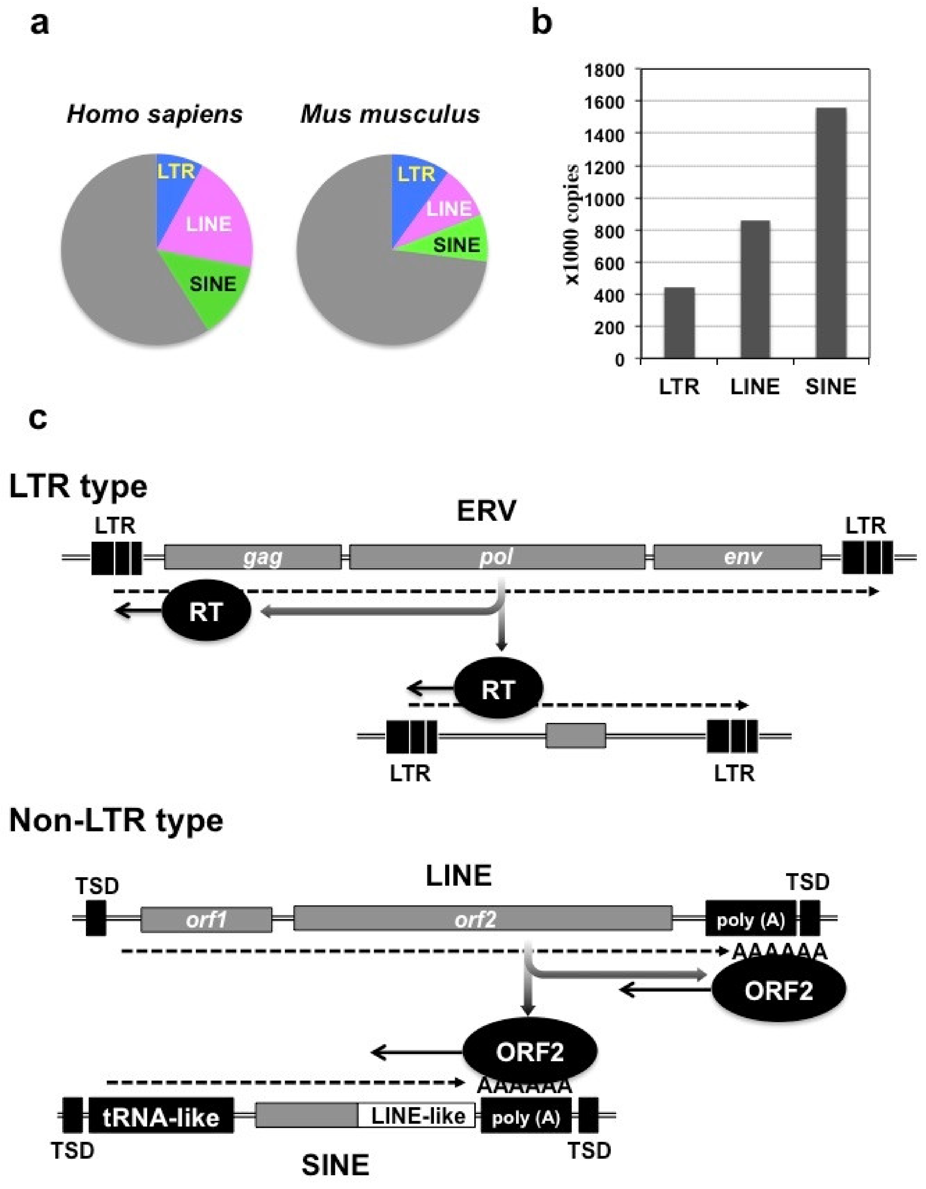
2. Retrotransposons in the Mammalian Genome

The mammalian genome contains two major classes of mobile genetic elements, and retrotransposons fall into the class 1 category [2]. Class 2 mobile genetic elements represent DNA transposons, which travel in the host genome by cutting and pasting themselves. Although the human genome contains 294,000 copies of DNA transposon-associated elements, they are no longer active and are regarded as genetic fossils [2]. In contrast, class 1 mobile genetic elements include a number of active retrotransposons that are able to modify our genome structure. These retrotransposons are classified into two major groups; one is long terminal repeat (LTR) type, and the other is non-LTR type [2,5]. Both groups contain autonomous retrotransposons that can copy and paste themselves using their own devices, and non-autonomous retrotransposons that can replicate only under the assistance of the autonomous ones. The autonomous non-LTR retrotransposons are represented by long interspersed nuclear elements (LINEs), whereas the non-autonomous ones are called short interspersed elements (SINEs). Among mammalian species, the human genome is particularly characterized by the involvement of a vast number of retrotransposons (Figure 1a). Indeed, more than 40% of the human genome is occupied by these mobile genetic elements, in which the LINE territory is the largest. In contrast, their occupancy in the murine genome is approximately 27%. This discrepancy suggests a critical role of these elements during the course of human evolution [2]. Regarding the copy number in the human genome, SINE stands out, represented by the Alu family that was actually regarded as a hallmark of the human genome (Figure 1b) [7].
The LTR-type retrotransposons are characterized by the two LTRs at both ends (Figure 1c). These elements evolved from a retroviral provirus integrated into the host genome at a certain time point during evolution, and thus are generally entitled endogenous retroviruses (ERVs). A prototypic ERV proviral genome retains gag, pol, and env genes, which are necessary for nucleocapsid formation, reverse transcription/integration, and infection/transmission, respectively. After transcription, genomic RNA is reverse transcribed from the primer binding site located immediately downstream of the LTR at the upstream end, using a host tRNA as a primer. Complete cDNA is synthesized from the genomic RNA through two rounds of jumping via the “R” region in the LTRs [8]. Reverse transcriptase produced by pol gene is also utilized for the propagation of non-autonomous LTR-type elements lacking protein-coding genes. Since retrotransposition is possible even without env, a significant number or ERV proviruses do not retain env at present [2]. The human genome holds a large family of human ERVs (HERVs) with known and unknown functions. Of note, expression of a number of HERV subfamily H (HERV-H) members was found induced transiently upon the establishment of induced pluripotent stem (iPS) cells from somatic cells [9]. Furthermore, a recent study indicated that these active HERV-H retrotransposons create topologically associating domains (TADs) in pluripotent stem cells [10]. Interestingly, mice also possess LTR-type retrotransposon fossils entitled mouse endogenous retrovirus type-L (MuERV-L) elements that are actively transcribed at early stages of embryonic development [11]. Together, these findings indicate the functional contribution of ERVs in mammalian development, as well as evolution.
The non-LTR autosomal LINEs contain two protein-coding regions, in which an internal ribosomal entry signal (IRES) is utilized for the translation of the second open reading frame (orf2). The orf2 encodes a protein (ORF2) with reverse transcriptase activity. Polyadenylated LINE transcripts are reverse transcribed by ORF2 from the poly (A) tail, while ORF2 also recognizes an AT-rich tract in the host genome and cuts it. As such, reverse transcription and integration of LINESs are both conducted by ORF2 protein (Figure 1c). Upon integration, duplication of the short nucleotide sequence at the integration site occurs, leaving target site duplication (TSD) at both ends [12]. SINEs probably share the same origin with tRNAs, since these elements retain the structural characteristics of tRNA and are transcribed by RNA polymerase III. These transcripts do not contain any open reading frames, but involve LINE-like polyadenylated regions to mimic LINE transcripts. Utilizing this region, SINE transcripts are able to summon ORF2 produced by LINE to exploit this protein for self-expansion [13]. The human genome is filled with nearly one million Alu elements, which are 7SL RNA-derived SINEs. Moreover, Alu elements are still increasing their de novo copies in our genome, for example, in the locus of breast cancer susceptibility gene 1 (BRCA1) [14]. This gene that encodes a tumor suppressor is already suffering from insult by a tremendous number of Alu elements. Similarly, the murine genome contains over a million SINE elements represented by 7SL RNA-derived SINE B1 and tRNA-derived SINE B2. Advances in molecular genomics would reveal more species-specific distribution of retrotransposons in the genomes of other mammalian species.

3. Creation and Modification of Genes by Retrotransposons

3.1. Modification of Pre-existing Host Genes by Retrotransposons

Retrotransposons, particularly LTR-type ones, insert their DNA copies anywhere in the host genome without preference in nucleotide sequences. However, the integration site may be delimited epigenetically by chromatin status. Namely, actively transcribed loci in euchromatin are more accessible for these mobile elements than those condensed in heterochromatin. Thus, unfortunately, active genes with significant biological functions are exposed to the risk of genetic manipulation by retrotransposons.
The insertion of mobile elements confers remarkable impact on the behavior and product of target genes at both transcriptional and post-transcriptional levels (Figure 2a) [15]. Destruction of the authentic promoter by such genetic intruders results in loss of function of the target gene at the transcriptional level. Conversely, insertion of enhancers and promoters, which are typically built in LTR, occasionally and differentially adds extra regulatory machinery to the target gene, depending upon the integration site. If a retrotransposon-derived promoter is added downstream of the open reading frame, novel proteins can be produced. This also happens when the mobile elements are inserted, destroying the initiation codon, causing a frame-shift of the reading frame, or inducing alternative splicing by the addition of extra splicing junctions. The stability of the host mRNA can also be modified by the addition of a retrotransposon-derived alternative poly (A) tail. As such, the genetic impact of retrotransposition is much more complex and can be more serious than DNA damage and subsequent mutation caused by errors and environmental carcinogens. It should be noted that naked moles, which only have fossil transposons and are lacking active ones at present, are characterized by extraordinary longevity without developing cancers [16].

3.2. Creation of Novel Genes by Retrotransposons

Along with the expansion in the host genome, retrotransposons accumulate mutations and gradually lose their proper gene products. During this process itself, new protein coding genes and, more likely, new long noncoding RNA (lncRNA) genes are occasionally created. In particular, the contribution of retrotransposon to lncRNA gene formation is prominent—for 75% of human lncRNA contains retrotransposon-derived sequences, at least in part [2]. These data are consistent with the fact that the human genome contains over 27,000 lncRNA genes, which is much more than that in the murine genome with less retrotransposon occupancy [17]. In addition to the genes directly evolved from retrotransposons, formation of chimeric genes containing both retroviral and host fragments is widely recognized. A classic example can be found in viral oncogenes created by animal retroviruses. RSV contains an oncogene known as v-src under the control of a strong viral promoter in the LTR, which transforms infected cells into sarcoma. The v-src gene is homologous to the host c-src gene and located downstream of the retroviral gag–pol–env cistron related to avian leukosis virus (ALV). This genomic organization suggests that RSV genomic RNA originated from a chimeric product of the parental ALV provirus with the cellular c-src gene located immediately downstream [18]. Finally, both retrotransposon-derived and host-derived portions of transcripts can create processed genes through the reverse transcription and integration of splicing variants of the original transcripts, adding more diversity to the host genome organization.
Apart from the genes constructed in cis by retrotransposons, which were introduced above, LINEs can manufacture novel genes with host mRNAs in trans, leaving only a slight trace of their work in the new genes. These resultant genes usually contain a processed cDNA of a host mRNA, and thus are called retrogenes. As described in Figure 1c, the ORF2 protein of LINEs reverse transcribes their RNAs recognizing their poly (A) tails. Therefore, theoretically, ORF2 could produce a cDNA from any poly (A)-containing host mRNA (Figure 2b). Nevertheless, this does not happen frequently for ORF2 strongly prefers its own mRNA (= LINE genomic RNA) as the template for reverse transcription [2]. Without this cis-preference, LINEs should have been serious threats to mammalian genome maintenance.

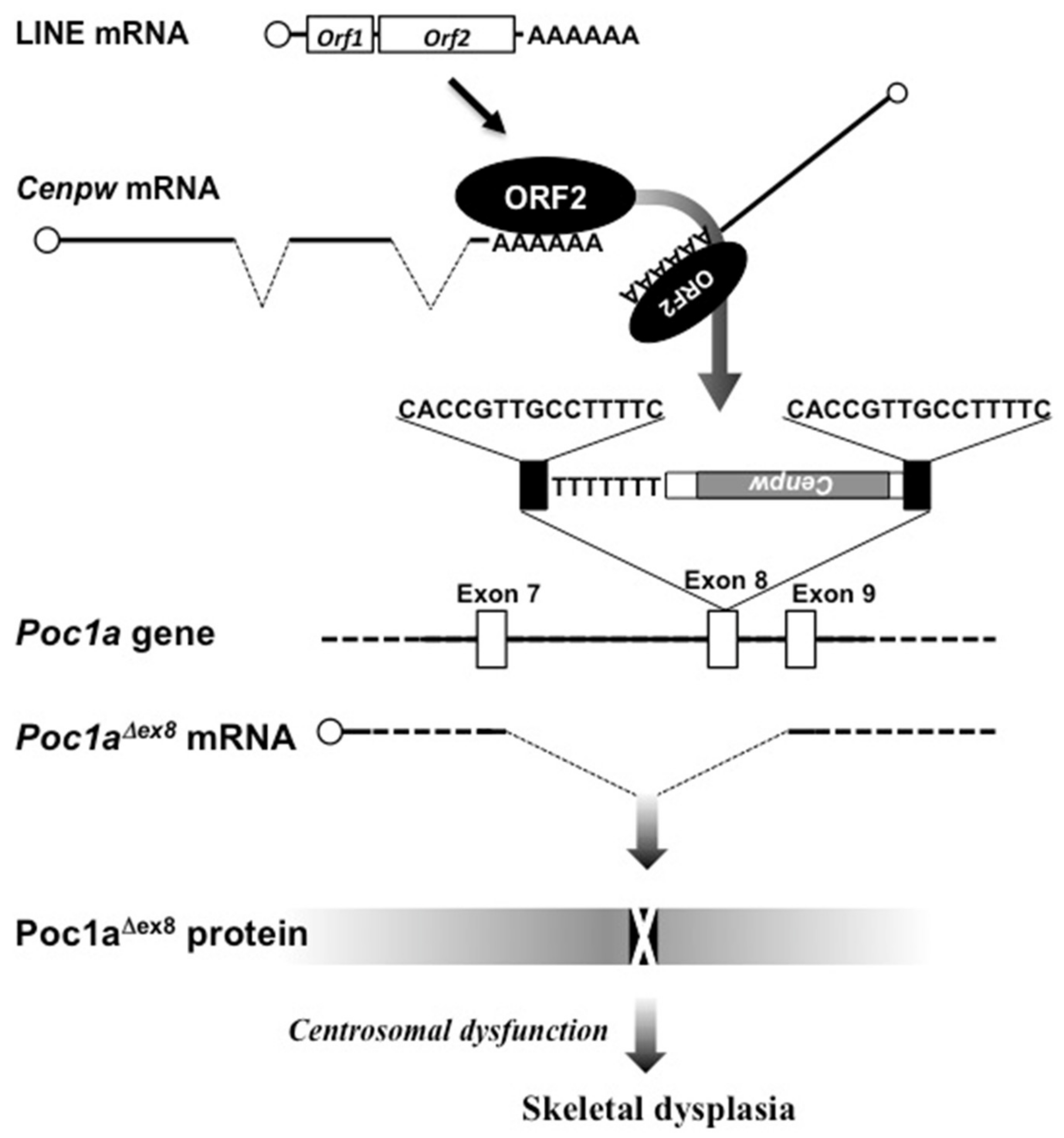
4. Gene Modification by a LINE Leading to Chondrodysplasia in Mice

In 2004, a mouse mutant with hereditable skeletal dysplasia and male infertility was first reported [19]. This mouse, named chagun, arose spontaneously and harbored the locus responsible for this phenotype in chromosome 9, which corresponds to human chromosome 3. In this mouse, disproportionate growth is observed in the skull and other skeletal parts. For example, the femora of chagun mice are significantly smaller, with disorganized growth plates. At birth, the mutant growth plate is apparently normal; however, disorganization in the growth plate progresses along with age. Abnormality in the growth plate is prominent in the proliferative zone. The columnar structure of flat proliferating chondrocytes becomes deranged with the morphological alteration in each chondrocyte. The intracellular position of Golgi apparatus in chagun chondrocytes suggests that cellular polarity is perturbed. Finally, a significant number of these chondrocytes undergo apoptosis, leading to growth insufficiency of long bones.
Subsequent genetic investigation identified the gene responsible for this chagun phenotype [20]—the Poc1a encoding protein of centriole 1A. As the name implies, this protein is a component of centrosome, suggesting its potential function in microtubule organization. Consistent with this anticipation, defects in the primary cilium and the mitotic spindle were evident in this mutant mouse, which led to craniofacial and skeletal abnormalities through impaired polarity of growth plate chondrocytes.
Of note, molecular genetic analysis revealed that Poc1a was destroyed by the insertion of a cDNA element in the middle of exon 8, which caused the skipping of this exon upon mRNA splicing to produce POCA1 protein lacking biological activity (Figure 3). The intruded DNA element was not of a LINE itself, but was a processed gene of centromere protein W (Cenpw) from chromosome 10. Nevertheless, clear traces of LINE-1 (a major group of LINEs)-mediated retrogene transposition were left. The first one was the poly (A) tail located downstream of the Cenpw cDNA, and second one was a set of TSDs at both ends of the integrated element. These findings indicate that ORF2 protein reverse transcribed and inserted the Cenpw cDNA into the Poc1a locus in chagun mice.
Several human pedigrees harbor mutations in POC1A loci, commonly characterized by growth insufficiency [20,21]. Therefore, the chagun mice phenocopying human cases can be a good model for medical investigation of these relevant human syndromes. This mouse also provides precious information about the role of the primary cilia in chondrocytes and skeletal development. Another interesting point is that chagun mutation affects the function of Sertoli cells, as well as that of chondrocytes. Although these two types of cells exert totally different missions in spermatogenesis and skeletal development, these are both SOX9-positive. Investigation on this SOX9–POCA1 axis may reveal novel aspects of the mechanism of skeletal growth and reproduction.

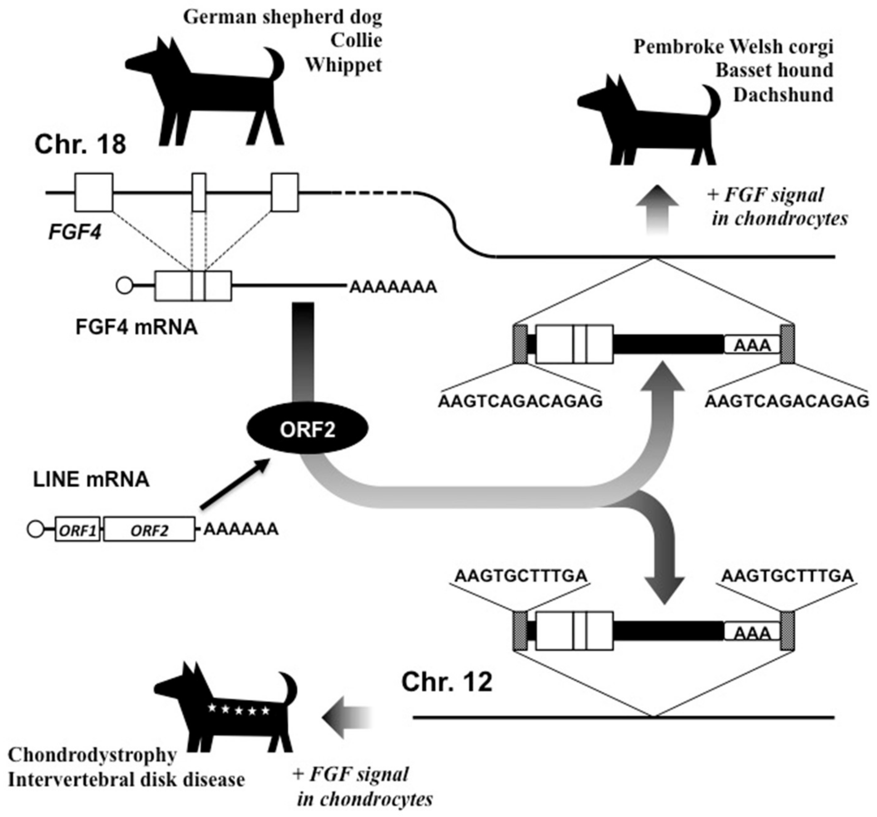
5. Creation of Short-legged Dogs: New FGF4 Gene Formation by LINE-1

Surprisingly, LINEs helped the evolution of dogs as partners of human beings. Dogs and cats are the two major partner animals for us, and particularly in city areas, small and short-legged breeds are often preferred, since they are relatively easy to control and require less space. Pembroke Welsh corgis, basset hounds, and dachshunds are widely known as typical examples of such dogs; however, it may not be recognized well that LINE-1 is responsible for at least 19 breeds of them.
Unlike the case of chagun mice, these short-legged dogs were created not by the destruction of a certain gene by LINE-associated intrusion, but by the addition of a processed host gene that encoded a functional growth factor [22]. In their genomes, an extra fibroblast growth factor (FGF) 4 cDNA containing all of the exons, including full coding regions and the 3’-UTR, was inserted in chromosome 18, in which the original FGF4 gene was present (Figure 4). The cDNA was followed by a poly (A) tail and was flanked by TSDs at both ends, clearly indicating the contribution of LINE-1 to its integration. This cDNA retains exactly the same protein-coding nucleotide sequence as that of the parental FGF4 gene, suggesting that it is required and conserved for the short-legged phenotype. This evolutional event caused by LINE-1 was confirmed, not only in the four breeds introduced above, but also in another 15 breeds in domestic dogs. In fact, FGF signaling is known to be closely associated with skeletal development. In humans, activating mutations in FGF receptor 3 gene (FGFR3), which is the major receptor transmitting FGF signals in chondrocytes, cause achondroplasia with typical dwarfism phenotype [22,23]. Therefore, LINE-1 may have done, or may do in the future, the same thing in humans, causing skeletal growth insufficiency. However, fortunately in this case, LINE-1 has established these major groups of domestic dogs that are widely accepted as excellent human partners through a single evolutionary event by adding an extra FGF signal emitter.
Another intriguing report suggests that breeds with additional FGF4 retrogenes were not necessarily established under a strong selective human pressure for preference of short-legged dogs. A genome-wide association study of skeletal dysfunction and intervertebral disc disease in Nova Scotia duck tolling retrievers (NSDTRs) uncovered a different FGF4 retrogene in chromosome 12 as a causative element of these diseases (Figure 4) [24]. This FGF4-inserted genotype was significantly associated with the height and intervertebral disc disease (IVDD) in these dogs, and FGF4 expression was nearly 220-fold higher in the neonatal intervertebral disc of dogs with homozygous FGF4 retrogene insertion than those without, clearly indicating its etiological role.
Since the TSD sequences of these FGF4 retrogenes in chromosomes 12 and 18 are distinct, these events were exerted independently by LINE-1 ORF2 capturing the same host mRNA, suggesting its remarkable activity in germ cells, or more possibly, in embryonic cells. Collectively, these two genetic events conducted by LINE-1 represent an awful property of a retrotransposon as a creator of endogenous pathogenic agents, using host mRNA as a template. Such an endogenous pathogen could even initiate hereditary diseases, as well as malignant neoplasms.

6. Hallmarks of Human Genome Regulating Chondrogenesis and Skeletal Development

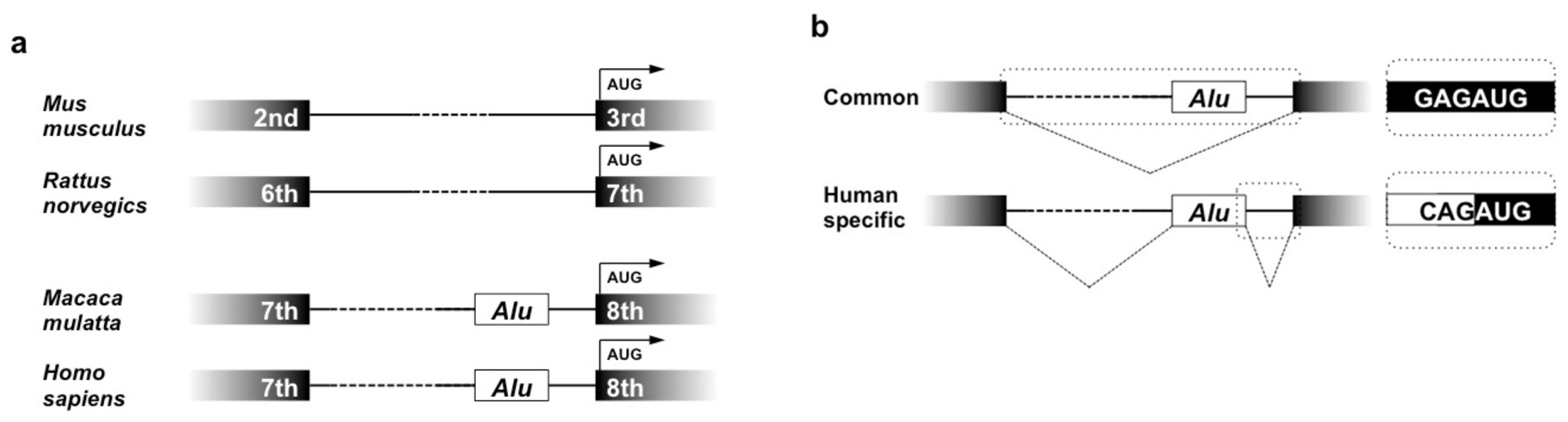
6.1. Height-related Gene Evolved along with Alu, a SINE Highly Specific to Primates

Owing to its exclusive inclusion in the human genome, with its absence in the genomes of rodents and other mammalian species, Alu retrotransposon was believed to be a hallmark of the human genome. However, comparative molecular genomics revealed that the Alu SINEs are widely shared by other primate species, suggesting its contribution to the evolution of primates rather than that of Homo sapiens. Among a number of genes evolved with Alu elements, the zinc finger and broad complex/tram track/bric-a-brac 38 (ZBTB38) gene is known to be expressed in chondrocytes and involved in the determination of height in humans. This gene encodes a transcriptional repressor and is conserved among vertebrates, and its murine orthologue, C-terminal binding protein-interacting BTB zinc finger protein (CIBZ) gene, is shown to regulate apoptosis. A strong association between height and ZBTB38 locus was reported both in European and Korean populations [25,26], which may be ascribed to its regulatory function of apoptosis, possibly in growth plate chondrocytes.
Alu integration into ZBTB38 occurred before the evolution of Hominidae, since it is already present at this particular locus in the genomes of the rhesus macaque (Macaca malatta) and the common marmoset (Calithrix jacchus) [27]. Although ZBT38 gene is conserved, the gene structure is different between primates and rodents (Figure 5a). The open reading frame starts from the 3rd and 7th exons of mouse and rat CIBZ genes, respectively, whereas primate ZBTB38 protein is encoded after the 8th exon. In the intron immediately upstream of the 8th one, an Alu copy is integrated. As such, the location of the Alu element in ZBTB38 is identical among primates. However, interestingly, these Alu elements behave differently, depending upon species. In the rhesus macaque and the common marmoset, splicing of ZBTB38 pre-mRNA is conducted as if the Alu element is not present, eliminating this element as a part of the intron. In contrast, in human cells, an alternative acceptor in the Alu element accepts the authentic donor of the 7th exon. The same Alu element also provides an alternative donor for the acceptor of the 8th exon. Consequently, the majority of the Alu sequence is added as an extra exon of 123 bases between exons 7 and 8 in the mRNA variant (Figure 5b) [27]. Since the Alu-derived new exon is located upstream of the translation initiation codon, it does not affect the primary structure of ZBTB38 protein. Instead, addition of this exon may alter the translation efficacy of mRNA by changing the proximity of the nucleotide sequence around the translation initiation codon to the Kozak’s consensus. It needs to be noted that the authentic mRNA with properly connected exons 7 and 8, as well as the Alu-containing one, is expressed in a variety of normal human cells. The impact of the Alu exonization on chondrocyte biology and skeletal development still remains unclear; however, the human-specific behavior of the Alu element implies a novel epigenetic mechanism of retrotransposon regulation in human cells and the human-specific role of such Alu elements.

6.2. Human Endogenous Retroviruses

As introduced in a previous subsection, the human genome is characterized by the involvement of a vast number of lncRNA genes that are even more abundant than the protein-coding classical genes. To date, although nearly 20,000 lncRNAs out of 27,000 in total are anticipated to play critical biological roles in the human body [17], the actual functionalities of most lncRNAs still remain to be clarified. HERVs produced a number of such lncRNA genes during their expansion along human evolution, which are transcribed under physiological and pathological status.
As is often the case with research regarding a novel class of molecules, an early advance in lncRNA research was also conducted in relation to cancer biology. Thus, a significant number of lncRNAs were found through cancer research and were defined as cancer-associated lncRNAs. Urothelial cancer-associated 1 (UCA1) is the most typical representative of such lncRNAs. Until today, UCA1 has been reported to be associated not only with bladder cancers [28,29,30], but also with other major human malignancies, including breast cancers [31,32,33]. The UCA1 gene is a typical LTR-type retrotransposon retaining gag and pol genes flanked by LTRs at both ends, lacking the env gene. This provirus is classified as part of the HERV-H subfamily and is located in the short arm of chromosome 19 in the human genome [34]. This HERV-H copy is hominid-specific, for orthologues can be found only in four species other than us—bonobos, chimpanzees, orangutans, and gorillas. Therefore, UCA1 is supposed to have contributed to human evolution and to have played a significant role in human-specific developmental events.
In spite of the structural retention of gag and pol genes, the UCA1 gene expresses lncRNAs only. This gene is able to produce several lncRNA variants, which are represented by two major ones of approximately 1400 and 2300 bases in length (Figure 6a). One should note that the human genome possesses several active lncRNA genes that are closely related to the UCA1 gene. All of the lncRNA genes shown in Figure 6b are confirmed to be expressed in human cells [35,36,37,38]. These lncRNAs share a high degree of homology with UCA1, especially in the LTR regions. It is assumed that these genes originate from the same retroviral progenitor, and thus they can be regarded as members of a gene family. Since the UCA1 gene retains gag and pol genes, it may have copied and pasted itself to generate another family member on the way to humans at present (Figure 6b).
UCA1 has been extensively investigated as a cancer-related lncRNA. However, recently, the physiological role of UCA1 in chondrocyte differentiation was uncovered [39,40]. During the process of differentiation from human bone marrow mesenchymal stem cells to chondrocytes, UCA1 was found to be induced rapidly and strongly prior to the induction of SOX9, a gene encoding the master transcription factor of chondrocytic differentiation. Furthermore, overexpression and silencing of the UCA1 gene resulted in the promotion and attenuation of chondrocyte differentiation, respectively, in human cells (Figure 7a). Interestingly, UCA1 could enhance the chondrogenesis, even in mouse chondroblasts that do not possess UCA1 genes. Considering the difference in skeletal sizes between hominids with UCA1 and rodents without UCA1, UCA1 may be acting as one of the determinants of skeletal size by regulating the endochondral ossification process.
Regarding the molecular function of UCA1, a vast number of recent reports have uncovered the functional complexity of this lncRNA (Figure 7b). As supposed by the predicted secondary structure (Figure 7b, center), UCA1 interacts with a variety of protein counterparts, using its secondary-structured functional elements as an interface. By interacting with these proteins, UCA1 occasionally acts as a scaffold for molecular events, sequesters them to prevent their works, or modifies their functions, conferring multiple biological outcomes [41,42,43,44,45,46,47,48]. Moreover, another function as a competitive endogenous RNA (ceRNA) may not be overlooked. Since UCA1 contains at least 32 experimentally verified binding sequences for distinct miRNAs, this single lncRNA can absorb these miRNAs away from the target mRNAs to de-repress the miRNA target genes [49,50,51,52,53,54,55,56,57,58,59,60,61,62,63,64,65,66,67,68,69,70,71,72,73,74,75,76,77,78,79,80,81,82,83,84,85]. Given that each miRNA in Figure 7b regulates thousands of target mRNAs, it is not easy to deduce the biological outcome caused by UCA1 via these miRNAs. However, it is strongly suggested that UCA1 should be playing significant roles in the process of endochondral ossification after chondrogenesis. Currently, it is at least clear that UCA1 is capable of promoting the maturation of proliferating chondrocytes via sponging miR-145 [86]. Indeed, HERV-H produced such a hominid-specific molecule with excessively multiple and context-dependent functions.

7. Retrotransposons in Human Joint Diseases

7.1. Osteoarthritis

Osteoarthritis (OA) is a multifactorial degenerative disease of the synovial joints and, currently, is a major medical issue in advanced countries with highly aged societies. Although OA itself is not life-threatening, progressive loss of locomotive function in patients severely impairs their quality of life. Unlike rheumatoid arthritis (RA; see the next subsection), OA is initiated from the loss of the integrity of articular cartilage in joints, most frequently in body weight-bearing knee and hip joints [87]. The fact that OA is usually developed in aged persons after a long time of mechanical loading on joints suggests continual mechanical stress as a critical etiological factor. Consistent with this notion, OA can quickly develop in young athletes, represented by soccer players, particularly after damage in the synovial ligaments, which allows traumatic movement of condyles. However, other associated etiological factors for OA development are being investigated as well, which include genetic background, systemic conditions, and viral infection.
In 2014, a Norwegian group attempted to determine if a virus was associated with OA, since a previous report [88] suggested human herpes virus 4 (HHV-4) infection might be specific to OA cartilage. As a result, they failed to detect any transcript from parvovirus B19, hepatitis C virus, or human herpes viruses in cartilage and cultured chondrocytes from OA patients. Nevertheless, they were able to detect several transcripts from HERV-W therein instead [89]. One of the transcripts corresponding to the env mRNA of HERV-WE2 was found expressed in 88% of advanced OA chondrocytes, while it was absent in normal and early OA chondrocytes. The etiological role of the activation of this particular subgroup of HERV is not yet clear. Since HERV expression is occasionally induced during the development of normal tissues, this finding may represent a regenerative response of cartilage after OA-inducing damage. In this context, it is of interest that the production of syncytin 1, which is the env gene product of HERV-W, was confirmed in these OA chondrocytes. Syncytin 1 is primarily a viral envelope glycoprotein that plays a role in infection, but it is expressed in the placenta and involved in trophoblast fusion. This protein is able to induce cell fusion; hence, its pathogenic activity may be suspected. Activation of HERV in OA chondrocyte was further supported by the viral particle formation confirmed by electron microscopy, even though its pathological role is questionable. Of note, recent studies suggest the involvement of lncRNAs, including the HERV-derived UCA1, in OA pathogenesis [90,91].

7.2. Rheumatoid Arthritis

Rheumatoid arthritis (RA) is another major disorder in synovial joints, with progressive destruction of joint tissues, including articular cartilage. In this systemic inflammatory disease with an autoimmune background, cartilage is degenerated as a result of inflammatory responses, in which synovial cells play a major role [92]. The etiological involvement of human retroviruses in RA was initially suspected, since transgenic mice with human T cell lymphotropic virus type I (HTLV-I) were shown to develop RA-like arthropathy [93]. In relation to this notion, retrotransposons attracted the interest of the researchers in this field as possible etiological factors for RA. With regard to the LTR type, a molecular genetic study found that a HERV-K LTR insertion into the HLA-DQB1 locus was associated with RA development [94]. Several other studies reported the traces of retroviral transcripts and particles in synovial fluids and tissues from RA patients [95]. Concerning the non-LTR type, it was once described that ORF1 protein and ORF2 transcripts from LINE-1 retrotransposons were detected in the synovial tissues of five out of six RA patients, whereas three OA and two normal synovial tissues showed no signals [96]. Overexpression of LINE-1 in RA synoviocytes caused the induction of several genes, including stress-induced protein kinase 2δ(SAPK2δ), which might provoke catabolic action of synoviocytes to harm articular cartilage [96]. However, mechanistic involvement of these LTR-type and non-LTR-type retrotransposons in RA pathogenesis has not been established thus far.

8. Conclusive Remarks

It is now obvious that retrotransposons have made a significant contribution to the generation of mammalian species by modifying and enlarging the genome. Particularly, considering their extraordinary genome occupancy, no humans with present shape, which is determined mostly by endochondral ossification, were present without HERV, LINEs, and SINEs. In addition, the fact that the genomes of crustacean and insects contain a number of retrotransposons suggests their contribution to intramembranous ossification—the older form of bone formation [97,98]. Thus, retrotransposons may be manipulating our craniofacial development by directly regulating osteoblasts as well. These mobile genetic elements are still actively editing our genome, indicating that these elements may still forward mammalian species toward further evolution.
Our cells, particularly germ cells, are armed by a protection system against the expansion of retrotransposons in the genome. Typically, using fossils of retrotransposons, small RNAs are produced that specifically recognize them, which lead to the silencing of relevant elements. These small RNAs consist of endogenous small interfering RNA (esiRNA) [99], miRNA, and PIWI-interacting RNA (piRNA) [100]. Among these RNAs, piRNAs are thought to be the most important in protecting our genome from inheritable assault by retrotransposons, as they are specifically produced in germ and embryonic cells. Despite such protection systems, mammalian genomes are still being edited by these endogenous parasites, as exemplified in this review.
In addition, we should stay alert to exogenous retroviruses as well, especially to human immunodeficiency viruses (HIVs). Considering the property of HIVs as lentiviruses, their genome-editing activity is evident. Although their primary targets are T-lymphocytes and macrophages, possible infection to other types of cells may not be ruled out. Indeed, direct interaction with sperm cells and HIV-1 virions via mannose receptor was observed [101], suggesting possible invasion of HIV into the genome of human embryo. In spite of the establishment of anti-retroviral therapy (ART) against AIDS, one should be careful to avoid viral contact itself, since our shape in the future with HIV in our genome is unpredictable.

Author Contributions

S.K. proposed the concept of this review article and constructed the draft. T.I., K.K., T.H., and T.N. provided the necessary information and reviewed the draft. All authors have read and agreed to the published version of the manuscript.

Funding

This research was funded by JSPS KAKENHI, Grant Numbers JP19K22716 and JP19K24119.

Acknowledgments

The authors thank Yoshiko Miyake for administrative support.

Conflicts of Interest

The authors declare no conflicts of interest.

Abbreviations

ERVEndogenous retrovirus
LINELong interspersed nuclear element
SINEShort interspersed nuclear element

References

  1. Moraes, F.; Góes, A. A decade of human genome project conclusion: Scientific diffusion about our genome knowledge. Biochem. Mol. Bio.l Educ. 2016, 44, 215–223. [Google Scholar] [CrossRef] [PubMed] [Green Version]
  2. Ganesh, S.; Svoboda, P. Retrotransposon-associated long non-coding RNAs in mice and men. Pflug. Arch. 2016, 468, 1049–1060. [Google Scholar] [CrossRef] [PubMed]
  3. Powner, M.W.; Gerland, B.; Sutherland, J.D. Synthesis of activated pyrimidine ribonucleotides in prebiotically plausible conditions. Nature 2009, 459, 239–242. [Google Scholar] [CrossRef] [PubMed]
  4. Wochner, A.; Attwater, J.; Coulson, A.; Holliger, P. Ribozyme-catalyzed transcription of an active ribozyme. Science 2011, 332, 209–212. [Google Scholar] [CrossRef] [PubMed]
  5. Kubiak, M.R.; Makałowska, I. Protein-coding genes’ retrocopies and their functions. Viruses 2017, 9, 80. [Google Scholar] [CrossRef] [Green Version]
  6. Temin, H.; Mizutani, S. RNA-dependent DNA Polymerase in Virions of Rous sarcoma virus. Nature 1972, 226, 1211–1213. [Google Scholar] [CrossRef]
  7. Deininger, P. Alu elements: Know the SINEs. Genome Biol. 2011, 12, 236. [Google Scholar] [CrossRef] [Green Version]
  8. Hughes, S.H. Reverse transcription of retroviruses and LTR retrotransposons. Microbiol. Spectr. 2015, 3, MDNA3-0027-2014. [Google Scholar]
  9. Ohnuki, M.; Tanabe, K.; Sutou, K.; Teramoto, I.; Sawamura, Y.; Narita, M.; Nakamura, M.; Tokunaga, Y.; Nakamura, M.; Watanabe, A.; et al. Dynamic regulation of human endogenous retroviruses mediates factor-induced reprogramming and differentiation potential. Proc. Natl. Acad. Sci. USA 2014, 111, 12426–12431. [Google Scholar] [CrossRef] [Green Version]
  10. Zhang, Y.; Li, T.; Preissl, S.; Amaral, M.L.; Grinstein, J.D.; Farah, E.N.; Destici, E.; Qiu, Y.; Hu, R.; Lee, A.Y.; et al. Transcriptionally active HERV-H retrotransposons demarcate topologically associating domains in human pluripotent stem cells. Nat. Genet. 2019, 51, 1380–1388. [Google Scholar] [CrossRef]
  11. Svoboda, P.; Stein, P.; Anger, M.; Bernstein, E.; Hannon, G.J.; Schultz, R.M. RNAi and expression of retrotransposons MuERV-L and IAP in preimplantation mouse embryos. Dev. Biol. 2004, 269, 276–285. [Google Scholar] [CrossRef] [PubMed] [Green Version]
  12. Bodak, M.; Yu, J.; Ciaudo, C. Regulation of LINE-1 in mammals. Biomol. Concepts 2014, 5, 409–428. [Google Scholar] [CrossRef] [PubMed] [Green Version]
  13. Dewannieux, M.; Esnault, C.; Heidmann, T. LINE-mediated retrotransposition of marked Alu sequences. Nat. Genet. 2003, 35, 41–48. [Google Scholar] [CrossRef] [PubMed]
  14. Teugels, E.; De Brakeleer, S.; Goelen, G.; Lissens, W.; Sermijn, E.; De Grève, J. De novo Alu element insertions targeted to a sequence common to the BRCA1 and BRCA2 genes. Hum. Mutat. 2005, 26, 284. [Google Scholar] [CrossRef] [PubMed]
  15. Cordaux, R.; Batzer, M.A. The impact of retrotransposons on human genome evolution. Nat. Rev. Genet. 2009, 10, 691–703. [Google Scholar] [CrossRef] [PubMed] [Green Version]
  16. Kim, E.B.; Fang, X.; Fushan, A.A.; Huang, Z.; Lobanov, A.V.; Han, L.; Marino, S.M.; Sun, X.; Turanov, A.A.; Yang, P.; et al. Genome sequencing reveals insights into physiology and longevity of the naked mole rat. Nature 2011, 479, 223–227. [Google Scholar] [CrossRef]
  17. Hon, C.C.; Ramilowski, J.A.; Harshbarger, J.; Bertin, N.; Rackham, O.J.; Gough, J.; Denisenko, E.; Schmeier, S.; Poulsen, T.M.; Severin, J.; et al. An atlas of human long non-coding RNAs with accurate 5’ ends. Nature 2017, 543, 199–204. [Google Scholar] [CrossRef] [Green Version]
  18. Vogt, P.K. Retroviral oncogenes: A historical primer. Nat. Rev. Cancer 2012, 12, 639–648. [Google Scholar] [CrossRef] [Green Version]
  19. Cha, K.B.; Karolyi, I.J.; Hunt, A.; Wenglikowski, A.M.; Wilkinson, E.; Dolan, D.F.; Dootz, G.; Finnegan, A.A.; Seasholtz, A.F.; Hankenson, K.D.; et al. Skeletal dysplasia and male infertility locus on mouse chromosome 9. Genomics 2004, 83, 951–960. [Google Scholar] [CrossRef]
  20. Geister, K.A.; Brinkmeier, M.L.; Cheung, L.Y.; Wendt, J.; Oatley, M.J.; Burgess, D.L.; Kozloff, K.M.; Cavalcoli, J.D.; Oatley, J.M.; Camper, S.A. LINE-1 mediated insertion into Poc1a (protein of centriole 1 a) causes growth insufficiency and male infertility in mice. Plos Genet. 2015, 11, e1005569. [Google Scholar] [CrossRef]
  21. Sarig, O.; Nahum, S.; Rapaport, D.; Ishida-Yamamoto, A.; Fuchs-Telem, D.; Qiaoli, L.; Cohen-Katsenelson, K.; Spiegel, R.; Nousbeck, J.; Israeli, S.; et al. Short stature, onychodysplasia, facial dysmorphism, and hypotrichosis syndrome is caused by a POC1A mutation. Am. J. Hum. Genet. 2012, 91, 337–342. [Google Scholar] [CrossRef] [PubMed] [Green Version]
  22. Parker, H.G.; VonHoldt, B.M.; Quignon, P.; Margulies, E.H.; Shao, S.; Mosher, D.S.; Spady, T.C.; Elkahloun, A.; Cargill, M.; Jones, P.G.; et al. An expressed fgf4 retrogene is associated with breed-defining chondrodysplasia in domestic dogs. Science 2009, 325, 995–998. [Google Scholar] [CrossRef] [PubMed] [Green Version]
  23. Ornitz, D.M.; Legeai-Mallet, L. Achondroplasia: Development, pathogenesis, and therapy. Dev. Dyn. 2017, 246, 291–309. [Google Scholar] [CrossRef] [PubMed] [Green Version]
  24. Brown, E.A.; Dickinson, P.J.; Mansour, T.; Sturges, B.K.; Aguilar, M.; Young, A.E.; Korff, C.; Lind, J.; Ettinger, C.L.; Varon, S.; et al. FGF4 retrogene on CFA12 is responsible for chondrodystrophy and intervertebral disc disease in dogs. Proc. Natl. Acad. Sci. USA 2017, 114, 11476–11481. [Google Scholar] [CrossRef] [Green Version]
  25. Weedon, M.N.; Lango, H.; Lindgren, C.M.; Wallace, C.; Evans, D.M.; Mangino, M.; Freathy, R.M.; Perry, J.R.; Stevens, S.; Hall, A.S.; et al. Genome-wide association analysis identifies 20 loci that influence adult height. Nat. Genet. 2008, 40, 575–583. [Google Scholar] [CrossRef] [Green Version]
  26. Cho, Y.S.; Go, M.J.; Kim, Y.J.; Heo, J.Y.; Oh, J.H.; Ban, H.J.; Yoon, D.; Lee, M.H.; Kim, D.J.; Park, M.; et al. A large-scale genome-wide association study of Asian populations uncovers genetic factors influencing eight quantitative traits. Nat. Genet. 2009, 41, 527–534. [Google Scholar] [CrossRef]
  27. Hong, K.W.; Shin, Y.B.; Jin, H.S.; Lim, J.E.; Choi, J.Y.; Chang, K.T.; Kim, H.S.; Oh, B. Alternative splicing of human height-related zinc finger and BTB domain-containing 38 gene through Alu exonization. Biochem. Genet. 2011, 49, 283–291. [Google Scholar] [CrossRef]
  28. Wang, F.; Li, X.; Xie, X.; Zhao, L.; Chen, W. UCA1, a non-protein-coding RNA up-regulated in bladder carcinoma and embryo, influencing cell growth and promoting invasion. FEBS Lett. 2008, 582, 1919–1927. [Google Scholar] [CrossRef] [Green Version]
  29. Xue, M.; Li, X.; Wu, W.; Zhang, S.; Wu, S.; Li, Z.; Chen, W. Upregulation of long non-coding RNA urothelial carcinoma associated 1 by CCAAT/enhancer binding protein α contributes to bladder cancer cell growth and reduced apoptosis. Oncol. Rep. 2014, 31, 1993–2000. [Google Scholar] [CrossRef] [Green Version]
  30. Eissa, S.; Matboli, M.; Essawy, N.O.; Shehta, M.; Kotb, Y.M. Rapid detection of urinary long non-coding RNA urothelial carcinoma associated one using a PCR-free nanoparticle-based assay. Biomarkers 2015, 20, 212–217. [Google Scholar] [CrossRef]
  31. Li, Z.; Yu, D.; Li, H.; Lv, Y.; Li, S. Long non-coding RNA UCA1 confers tamoxifen resistance in breast cancer endocrinotherapy through regulation of the EZH2/p21 axis and the PI3K/AKT signaling pathway. Int. J. Oncol. 2019, 54, 1033–1042. [Google Scholar] [PubMed]
  32. Liu, C.; Jin, J.; Shi, J.; Wang, L.; Gao, Z.; Guo, T.; He, Y. Long noncoding RNA UCA1 as a novel biomarker of lymph node metastasis and prognosis in human cancer: A meta-analysis. Biosci. Rep. 2019, 39, BSR20180995. [Google Scholar] [CrossRef] [PubMed]
  33. Ghafouri-Fard, S.; Taheri, M. UCA1 long non-coding RNA: An update on its roles in malignant behavior of cancers. Biomed. Pharm. 2019, 120, 109459. [Google Scholar] [CrossRef] [PubMed]
  34. Wang, X.S.; Zhang, Z.; Wang, H.C.; Cai, J.L.; Xu, Q.W.; Li, M.Q.; Chen, Y.C.; Qian, X.P.; Lu, T.J.; Yu, L.Z.; et al. Rapid identification of UCA1 as a very sensitive and specific unique marker for human bladder carcinoma. Clin. Cancer Res. 2006, 12, 4851–4858. [Google Scholar] [CrossRef] [PubMed] [Green Version]
  35. Mammalian Gene Collection (MGC) Program Team. Generation and initial analysis of more than 15,000 full-length human and mouse cDNA sequences. Proc. Natl. Acad. Sci. USA 2002, 99, 16899–16903. [Google Scholar] [CrossRef] [PubMed] [Green Version]
  36. Mlynarczyk, S.K.; Panning, B. X inactivation: Tsix and Xist as yin and yang. Curr Biol 2000, 10, R899–R903. [Google Scholar] [CrossRef] [Green Version]
  37. Wang, C.; Wang, G.; Zhang, Z.; Wang, Z.; Ren, M.; Wang, X.; Li, H.; Yu, Y.; Liu, J.; Cai, L.; et al. The downregulated long noncoding RNA DHRS4-AS1 is protumoral and associated with the prognosis of clear cell renal cell carcinoma. Onco Targets 2018, 11, 5631–5646. [Google Scholar] [CrossRef] [Green Version]
  38. Yang, Y.; Su, Z.; Song, X.; Liang, B.; Zeng, F.; Chang, X.; Huang, D. Enhancer RNA-driven looping enhances the transcription of the long noncoding RNA DHRS4-AS1, a controller of the DHRS4 gene cluster. Sci. Rep. 2016, 6, 20961. [Google Scholar] [CrossRef] [Green Version]
  39. Ishikawa, T.; Nishida, T.; Ono, M.; Takarada, T.; Nguyen, H.T.; Kurihara, S.; Furumatsu, T.; Murase, Y.; Takigawa, M.; Oohashi, T.; et al. Physiological role of urothelial cancer-associated one long noncoding RNA in human skeletogenic cell differentiation. J. Cell Physiol. 2018, 233, 4825–4840. [Google Scholar] [CrossRef]
  40. Shu, T.; He, L.; Wang, X.; Pang, M.; Yang, B.; Feng, F.; Wu, Z.; Liu, C.; Zhang, S.; Liu, B.; et al. Long noncoding RNA UCA1 promotes chondrogenic differentiation of human bone marrow mesenchymal stem cells via miRNA-145-5p/SMAD5 and miRNA-124-3p/SMAD4 axis. Biochem. Biophys. Res. Commun. 2019, 514, 316–322. [Google Scholar] [CrossRef]
  41. Kumar, P.P.; Emechebe, U.; Smith, R.; Franklin, S.; Moore, B.; Yandell, M.; Lessnick, S.L.; Moon, A.M. Coordinated control of senescence by lncRNA and a novel T-box3 co-repressor complex. eLife 2014, 3, e02805. [Google Scholar] [CrossRef] [PubMed]
  42. Liu, Y.; Feng, W.; Gu, S.; Wang, H.; Zhang, Y.; Chen, W.; Xu, W.; Lin, C.; Gong, A.; Xu, M. The UCA1/KRAS axis promotes human pancreatic ductal adenocarcinoma stem cell properties and tumor growth. Am. J. Cancer Res. 2019, 9, 496–510. [Google Scholar] [PubMed]
  43. Wang, Z.Q.; He, C.Y.; Hu, L.; Shi, H.P.; Li, J.F.; Gu, Q.L.; Su, L.P.; Liu, B.Y.; Li, C.; Zhu, Z. Long noncoding RNA UCA1 promotes tumour metastasis by inducing GRK2 degradation in gastric cancer. Cancer Lett. 2017, 408, 10–21. [Google Scholar] [CrossRef] [PubMed]
  44. Cai, Q.; Jin, L.; Wang, S.; Zhou, D.; Wang, J.; Tang, Z.; Quan, Z. Long non-coding RNA UCA1 promotes gallbladder cancer progression by epigenetically repressing p21 and E-cadherin expression. Oncotarget 2017, 8, 47957–47968. [Google Scholar] [CrossRef] [Green Version]
  45. Wang, X.; Gong, Y.; Jin, B.; Wu, C.; Yang, J.; Wang, L.; Zhang, Z.; Mao, Z. Long non-coding RNA urothelial carcinoma associated 1 induces cell replication by inhibiting BRG1 in 5637 cells. Oncol. Rep. 2014, 32, 1281–1290. [Google Scholar] [CrossRef] [Green Version]
  46. Huang, J.; Zhou, N.; Watabe, K.; Lu, Z.; Wu, F.; Xu, M.; XMo, Y.-Y. Long non-coding RNA UCA1 promotes breast tumor growth by suppression of p27 (Kip1). Cell Death Dis. 2014, 5, e1008. [Google Scholar] [CrossRef]
  47. Wang, Z.Q.; Cai, Q.; Hu, L.; He, C.Y.; Li, J.F.; Quan, Z.W.; Liu, B.Y.; Li, C.; Zhu, Z.G. Long noncoding RNA UCA1 induced by SP1 promotes cell proliferation via recruiting EZH2 and activating AKT pathway in gastric cancer. Cell Death Dis. 2017, 8, e2839. [Google Scholar] [CrossRef] [Green Version]
  48. Lin, X.; Spindler, T.J.; de Souza Fonseca, M.A.; Corona, R.I.; Seo, J.H.; Dezem, F.S.; Li, L.; Lee, J.M.; Long, H.W.; Sellers, T.A.; et al. Super-enhancer-associated lncRNA UCA1 interacts directly with AMOT to activate YAP target genes in epithelial ovarian cancer. iScience 2019, 17, 242–255. [Google Scholar] [CrossRef] [Green Version]
  49. Yang, Z.; Shi, X.; Li, C.; Wang, X.; Hou, K.; Li, Z.; Zhang, X.; Fan, Y.; Qu, X.; Che, X.; et al. Long non-coding RNA UCA1 upregulation promotes the migration of hypoxia-resistant gastric cancer cells through the miR-7-5p/EGFR axis. Exp. Cell Res. 2018, 368, 194–201. [Google Scholar] [CrossRef]
  50. Li, D.; Hao, S.; Zhang, J. Long non-coding RNA UCA1 exerts growth modulation by miR-15a in human thyroid cancer TPC-1 cells. Artif Cells Nanomed. Biotechnol. 2019, 47, 1815–1822. [Google Scholar] [CrossRef] [Green Version]
  51. Xiao, Y.; Jiao, C.; Lin, Y.; Chen, M.; Zhang, J.; Wang, J.; Zhang, Z. lncRNA UCA1 Contributes to Imatinib Resistance by Acting as a ceRNA Against miR-16 in Chronic Myeloid Leukemia Cells. DNA Cell Biol. 2017, 36, 8–25. [Google Scholar] [CrossRef] [PubMed]
  52. Zhu, H.Y.; Bai, W.D.; Ye, X.M.; Yang, A.G.; Jia, L.T. Long non-coding RNA UCA1 desensitizes breast cancer cells to trastuzumab by impeding miR-18a repression of Yes-associated protein 1. Biochem. Biophys. Res. Commun. 2018, 496, 1308–1313. [Google Scholar] [CrossRef] [PubMed]
  53. Tian, S.; Yuan, Y.; Li, Z.; Gao, M.; Lu, Y.; Gao, H. LncRNA UCA1 sponges miR-26a to regulate the migration and proliferation of vascular smooth muscle cells. Gene 2018, 673, 159–166. [Google Scholar] [CrossRef]
  54. Fang, Q.; Chen, X.; Zhi, X. Long non-coding RNA (LncRNA) urothelial carcinoma associated 1 (UCA1) increases multi-drug resistance of gastric cancer via downregulating miR-27b. Med. Sci. Monit. 2016, 22, 3506–3513. [Google Scholar] [CrossRef] [PubMed] [Green Version]
  55. Cui, M.; Chen, M.; Shen, Z.; Wang, R.; Fang, X.; Song, B. LncRNA-UCA1 modulates progression of colon cancer through regulating the miR-28-5p/HOXB3 axis. J. Cell Biochem. 2019. [Google Scholar] [CrossRef] [PubMed]
  56. Zhou, Y.; Chen, Y.; Ding, W.; Hua, Z.; Wang, L.; Zhu, Y.; Qian, H.; Dai, T. LncRNA UCA1 impacts cell proliferation, invasion, and migration of pancreatic cancer through regulating miR-96/FOXO3. Iubmb Life 2018, 70, 276–290. [Google Scholar] [CrossRef] [Green Version]
  57. Gong, J.; Lu, X.; Xu, J.; Xiong, W.; Zhang, H.; Yu, X. Coexpression of UCA1 and ITGA2 in pancreatic cancer cells target the expression of miR-107 through focal adhesion pathway. J. Cell Physiol. 2019, 234, 12884–12896. [Google Scholar] [CrossRef]
  58. Zhou, Y.; Meng, X.; Chen, S.; Li, W.; Li, D.; Singer, R.; Gu, W. IMP1 regulates UCA1-mediated cell invasion through facilitating UCA1 decay and decreasing the sponge effect of UCA1 for miR-122-5p. Breast Cancer Res. 2018, 20, 32. [Google Scholar] [CrossRef]
  59. Zhang, T.H.; Liang, L.Z.; Liu, X.L.; Wu, J.N.; Su, K.; Chen, J.Y.; Zheng, Q.Y. LncRNA UCA1/miR-124 axis modulates TGFβ1-induced epithelial-mesenchymal transition and invasion of tongue cancer cells through JAG1/Notch signaling. J. Cell Biochem. 2019, 120, 10495–10504. [Google Scholar] [CrossRef]
  60. Zhang, Y.; Liu, Y.; Xu, X. Knockdown of LncRNA-UCA1 suppresses chemoresistance of pediatric AML by inhibiting glycolysis through the microRNA-125a/hexokinase 2 pathway. J. Cell Biochem. 2018, 119, 6296–6308. [Google Scholar] [CrossRef]
  61. Sun, M.D.; Zheng, Y.Q.; Wang, L.P.; Zhao, H.T.; Yang, S. Long noncoding RNA UCA1 promotes cell proliferation, migration and invasion of human leukemia cells via sponging miR-126. Eur. Rev. Med. Pharm. Sci. 2018, 22, 2233–2245. [Google Scholar]
  62. Liu, Q.; Li, Y.; Lv, W.; Zhang, G.; Tian, X.; Li, X.; Cheng, H.; Zhu, C. UCA1 promotes cell proliferation and invasion and inhibits apoptosis through regulation of the miR129-SOX4 pathway in renal cell carcinoma. Onco. Targets 2018, 11, 2475–2487. [Google Scholar] [CrossRef] [PubMed] [Green Version]
  63. Zhang, X.; Gao, F.; Zhou, L.; Wang, H.; Shi, G.; Tan, X. UCA1 Regulates the Growth and Metastasis of Pancreatic Cancer by Sponging miR-135a. Oncol. Res. 2017, 25, 1529–1541. [Google Scholar] [CrossRef] [PubMed]
  64. Tuo, Y.L.; Li, X.M.; Luo, J. Long noncoding RNA UCA1 modulates breast cancer cell growth and apoptosis through decreasing tumor suppressive miR-143. Eur. Rev. Med. Pharm. Sci. 2015, 19, 3403–3411. [Google Scholar]
  65. Li, D.; Li, H.; Yang, Y.; Kang, L. Long Noncoding RNA Urothelial Carcinoma-Associated 1 Promotes the Proliferation and Metastasis of Human Lung Tumor Cells by Regulating MicroRNA-144. Oncol. Res. 2018, 26, 537–546. [Google Scholar] [CrossRef]
  66. Xue, M.; Pang, H.; Li, X.; Li, H.; Pan, J.; Chen, W. Long non-coding RNA urothelial cancer-associated 1 promotes bladder cancer cell migration and invasion by way of the hsa-miR-145-ZEB1/2-FSCN1 pathway. Cancer Sci. 2016, 107, 18–27. [Google Scholar] [CrossRef]
  67. He, Z.; You, C.; Zhao, D. Long non-coding RNA UCA1/miR-182/PFKFB2 axis modulates glioblastoma-associated stromal cells-mediated glycolysis and invasion of glioma cells. Biochem. Biophys. Res. Commun. 2018, 500, 569–576. [Google Scholar] [CrossRef]
  68. Zhou, G.; Li, C.; Feng, J.; Zhang, J.; Fang, Y. lncRNA UCA1 Is a Novel Regulator in Cardiomyocyte Hypertrophy through Targeting the miR-184/HOXA9 Axis. Cardiorenal Med. 2018, 8, 130–139. [Google Scholar] [CrossRef]
  69. Wu, H.; Zhou, C. Long non-coding RNA UCA1 promotes lung cancer cell proliferation and migration via microRNA-193a/HMGB1 axis. Biochem. Biophys. Res. Commun. 2018, 496, 738–745. [Google Scholar] [CrossRef]
  70. Li, H.J.; Sun, X.M.; Li, Z.K.; Yin, Q.W.; Pang, H.; Pan, J.J.; Li, X.; Chen, W. LncRNA UCA1 promotes mitochondrial function of bladder cancer via the miR-195/ARL2 signaling pathway. Cell Physiol. Biochem. 2017, 43, 2548–2561. [Google Scholar] [CrossRef]
  71. Zhang, J.; Zhang, C. Silence of long non-coding RNA UCA1 inhibits hemangioma cells growth, migration and invasion by up-regulation of miR-200c. Life Sci. 2019, 226, 33–46. [Google Scholar] [CrossRef] [PubMed]
  72. Xiao, J.N.; Yan, T.H.; Yu, R.M.; Gao, Y.; Zeng, W.L.; Lu, S.W.; Que, H.X.; Liu, Z.P.; Jiang, J.H. Long non-coding RNA UCA1 regulates the expression of Snail2 by miR-203 to promote hepatocellular carcinoma progression. J. Cancer Res. Clin. Oncol. 2017, 143, 981–990. [Google Scholar] [CrossRef]
  73. Bian, Z.; Jin, L.; Zhang, J.; Yin, Y.; Quan, C.; Hu, Y.; Feng, Y.; Liu, H.; Fei, B.; Mao, Y.; et al. LncRNA-UCA1 enhances cell proliferation and 5-fluorouracil resistance in colorectal cancer by inhibiting miR-204-5p. Sci. Rep. 2016, 6, 23892. [Google Scholar] [CrossRef] [PubMed] [Green Version]
  74. Yan, Q.; Tian, Y.; Hao, F. Downregulation of lncRNA UCA1 inhibits proliferation and invasion of cervical cancer cells through miR-206 expression. Oncol. Res. 2018. [CrossRef] [PubMed]
  75. Wang, C.J.; Zhu, C.C.; Xu, J.; Wang, M.; Zhao, W.Y.; Liu, Q.; Zhao, G.; Zhang, Z.Z. The lncRNA UCA1 promotes proliferation, migration, immune escape and inhibits apoptosis in gastric cancer by sponging anti-tumor miRNAs. Mol. Cancer 2019, 18, 115. [Google Scholar] [CrossRef] [Green Version]
  76. Wang, F.; Ying, H.Q.; He, B.S.; Pan, Y.Q.; Deng, Q.W.; Sun, H.L.; Chen, J.; Liu, X.; Wang, S.K. Upregulated lncRNA-UCA1 contributes to progression of hepatocellular carcinoma through inhibition of miR-216b and activation of FGFR1/ERK signaling pathway. Oncotarget 2015, 6, 7899–7917. [Google Scholar] [CrossRef] [Green Version]
  77. Guo, N.; Sun, Q.; Fu, D.; Zhang, Y. Long non-coding RNA UCA1 promoted the growth of adrenocortical cancer cells via modulating the miR-298-CDK6 axis. Gene 2019, 703, 26–34. [Google Scholar] [CrossRef]
  78. Zhu, G.; Liu, X.; Su, Y.; Kong, F.; Hong, X.; Lin, Z. Knockdown of urothelial carcinoma-associated 1 suppressed cell growth and migration through regulating miR-301a and CXCR4 in osteosarcoma MHCC97 cells. Oncol. Res. 2018, 27, 55–64. [Google Scholar] [CrossRef]
  79. Yang, Y.; Jiang, Y.; Wan, Y.; Zhang, L.; Qiu, J.; Zhou, S.; Cheng, W. UCA1 functions as a competing endogenous RNA to suppress epithelial ovarian cancer metastasis. Tumour Biol. 2016, 37, 10633–10641. [Google Scholar] [CrossRef]
  80. Lu, Y.; Liu, W.G.; Lu, J.H.; Liu, Z.J.; Li, H.B.; Liu, G.J.; She, H.Y.; Li, G.Y.; Shi, X.H. LncRNA UCA1 promotes renal cell carcinoma proliferation through epigenetically repressing p21 expression and negatively regulating miR-495. Tumour Biol. 2017, 39, 1010428317701632. [Google Scholar] [CrossRef] [Green Version]
  81. Wang, P.; Liu, X.; Han, G.; Dai, S.; Ni, Q.; Xiao, S.; Huang, J. Downregulated lncRNA UCA1 acts as ceRNA to adsorb microRNA-498 to repress proliferation, invasion and epithelial mesenchymal transition of esophageal cancer cells by decreasing ZEB2 expression. Cell Cycle 2019, 18, 2359–2376. [Google Scholar] [CrossRef] [PubMed]
  82. Guo, S.; Yang, P.; Jiang, X.; Li, X.; Wang, Y.; Zhang, X.; Sun, B.; Zhang, Y.; Jia, Y. Genetic and epigenetic silencing of mircoRNA-506-3p enhances COTL1 oncogene expression to foster non-small lung cancer progression. Oncotarget 2017, 8, 644–657. [Google Scholar] [CrossRef] [PubMed] [Green Version]
  83. Wei, Y.; Sun, Q.; Zhao, L.; Wu, J.; Chen, X.; Wang, Y.; Zang, W.; Zhao, G. LncRNA UCA1-miR-507-FOXM1 axis is involved in cell proliferation, invasion and G0/G1 cell cycle arrest in melanoma. Med. Oncol. 2016, 33, 88. [Google Scholar] [CrossRef] [PubMed]
  84. Wu, J.; Li, W.; Ning, J.; Yu, W.; Rao, T.; Cheng, F. Long noncoding RNA UCA1 targets miR-582-5p and contributes to the progression and drug resistance of bladder cancer cells through ATG7-mediated autophagy inhibition. Onco. Targets 2019, 12, 495–508. [Google Scholar] [CrossRef] [Green Version]
  85. Gu, L.; Lu, L.S.; Zhou, D.L.; Liu, Z.C. UCA1 promotes cell proliferation and invasion of gastric cancer by targeting CREB1 sponging to miR-590-3p. Cancer Med. 2018, 7, 1253–1263. [Google Scholar] [CrossRef]
  86. Razmara, E.; Bitaraf, A.; Yousefi, H.; Nguyen, T.H.; Garshasbi, M.; Cho, W.C.-S.; Babashah, S. Non-coding RNAs in cartilage development: An updated review. Int. J. Mol. Sci. 2019, 20, 18. [Google Scholar] [CrossRef] [Green Version]
  87. Martel-Pelletier, J.; Barr, A.J.; Cicuttini, F.M.; Conaghan, P.G.; Cooper, C.; Goldring, M.B.; Goldring, S.R.; Jones, G.; Teichtahl, A.J.; Pelletier, J.P. Osteoarthritis. Nat. Rev. Dis. Primers 2016, 2, 16072. [Google Scholar] [CrossRef] [Green Version]
  88. Rollín, R.; Alvarez-Lafuente, R.; Marco, F.; López-Durán, L.; Hoyas, J.A.; Jover, J.A.; Fernández-Gutiérrez, B. The ubiquitin-proteasome pathway and viral infections in articular cartilage of patients with osteoarthritis. Rheumatol. Int. 2009, 29, 969–972. [Google Scholar] [CrossRef]
  89. Bendiksen, S.; Martinez-Zubiavrra, I.; Tümmler, C.; Knutsen, G.; Elvenes, J.; Olsen, E.; Olsen, R.; Moens, U. Human endogenous retrovirus W activity in cartilage of osteoarthritis patients. Biomed. Res. Int. 2014, 2014, 698609. [Google Scholar] [CrossRef] [Green Version]
  90. Liu, Q.; Zhang, X.; Dai, L.; Hu, X.; Zhu, J.; Li, L.; Zhou, C.; Ao, Y. Long noncoding RNA related to cartilage injury promotes chondrocyte extracellular matrix degradation in osteoarthritis. Arthritis Rheumatol 2015, 66, 969–978. [Google Scholar] [CrossRef]
  91. Wang, G.; Bu, X.; Zhang, Y.; Zhao, X.; Kong, Y.; Ma, L.; Niu, S.; Wu, B.; Meng, C. LncRNA-UCA1 enhances MMP-13 expression by inhibiting miR- 204-5p in human chondrocytes. Oncotarget 2017, 8, 91281–91290. [Google Scholar] [CrossRef] [PubMed] [Green Version]
  92. Smolen, J.S.; Aletaha, D.; McInnes, I.B. Rheumatoid arthritis. Lancet 2016, 388, 2023–2038. [Google Scholar] [CrossRef]
  93. Iwakura, Y.; Saijo, S.; Kioka, Y.; Nakayama-Yamada, J.; Itagaki, K.; Tosu, M.; Asano, M.; Kanai, Y.; Kakimoto, K. Autoimmunity induction by human T cell leukemia virus type 1 in transgenic mice that develop chronic inflammatory arthropathy resembling rheumatoid arthritis in humans. J. Immunol. 1995, 155, 1588–1598. [Google Scholar] [PubMed]
  94. Seidl, C.; Donner, H.; Petershofen, E.; Usadel, K.H.; Seifried, E.; Kaltwasser, J.P.; Badenhoop, K. An endogenous retroviral long terminal repeat at the HLA-DQB1 gene locus confers susceptibility to rheumatoid arthritis. Hum. Immunol. 1999, 60, 63–68. [Google Scholar] [CrossRef]
  95. Stransky, G.; Vernon, J.; Aicher, W.K.; Moreland, L.W.; Gay, R.E.; Gay, S. Virus-like particles in synovial fluids from patients with rheuma- toid arthritis. Br. J. Rheumatol. 1993, 32, 1044–1048. [Google Scholar] [CrossRef]
  96. Neidhart, M.; Rethage, J.; Kuchen, S.; Künzler, P.; Crowl, R.M.; Billingham, M.E.; Gay, R.E.; Gay, S. Retrotransposable L1 elements expressed in rheumatoid arthritis synovial tissue: Association with genomic DNA hypomethylation and influence on gene expression. Arthritis Rheum. 2000, 43, 2634–2647. [Google Scholar] [CrossRef]
  97. Piednoël, M.; Gonçalves, I.R.; Higuet, D.; Bonnivard, E. Eukaryote DIRS1-like retrotransposons: An overview. BMC Genom. 2011, 12, 621. [Google Scholar]
  98. Nefedova, L.N.; Kim, A.I. Mechanisms of LTR-retroelement transposition: Lessons from Drosophila melanogaster. Viruses 2017, 9, 81. [Google Scholar] [CrossRef] [Green Version]
  99. Kim, V.N.; Han, J.; Siomi, M.C. Biogenesis of small RNAs in animals. Nat. Rev. Mol. Cell Biol. 2009, 10, 126–139. [Google Scholar] [CrossRef]
  100. Iwasaki, Y.W.; Siomi, M.C.; Siomi, H. PIWI-Interacting RNA: Its Biogenesis and Functions. Annu. Rev. Biochem. 2015, 84, 405–433. [Google Scholar] [CrossRef]
  101. Cardona-Maya, W.; Velilla, P.A.; Montoya, C.J.; Cadavid, Á.; Rugeles, M.T. In vitro human immunodeficiency virus and sperm cell interaction mediated by the mannose receptor. J. Reprod. Immunol. 2011, 92, 1–7. [Google Scholar] [CrossRef] [PubMed]
Figure 1. Retrotransposons in the human genomes. (a) Pie graphs showing the occupancy of retrotransposons in the human and murine genomes. (b) Copy numbers of three types of retrotransposons in the human genome. (c) Structure and mode of transcription (dotted lines with arrows) and reverse transcription (solid lines with arrows) of each type of retrotransposon. LTR, long terminal repeat; ERV, endogenous retrovirus; LINE, long interspersed nuclear element; SINE, short interspersed nuclear element; RT, reverse transcriptase; ORF2, open reading frame protein 2 of LINE; TSD, target site duplication. For ERV, a complete provirus retaining all of the essential retroviral genes, gag, pol, and env, is shown as a representative.
Ijms 21 01564 g001
Figure 2. Modification and creation of genes by retrotransposons. (a) Modification of pre-exiting host genes by the integration of transposable genetic elements. Transcriptional and post-transcriptional effects are summarized above and below the target host gene, respectively. Additional genetic elements provided by retrotransposons are noted, occasionally causing destruction of the target gene (KO). (b) Creation of novel genes by retrotransposons. RT and EN/IN represent proteins with reverse transcriptase and endonuclease/integrase activities, which are encoded by retrotransposon mRNAs, respectively.
Ijms 21 01564 g002
Figure 3. Exon skipping in Poc1a caused by LINE-1 in chagun mice. The nucleotide sequences of the target site duplications are shown in the middle. LINE, long interspersed nuclear element; ORF2, open reading frame protein 2 of LINE; Cenpw, centromere protein W gene; Poc1a, protein of centriole 1a gene. Polyadenylated Cenpw processed gene insertion occurred in a direction reverse to that of Poc1a, forming an apparently poly (T) nucleotide sequence.
Ijms 21 01564 g003
Figure 4. Creation of two new FGF4 retrogenes in distinct genomic context causing short legs and intervertebral disc diseases in dogs. Proper target site duplication sequences (stippled boxes) are shown for each integration site. Open and solid boxes denote FGF-4 encoding regions and untranslated regions, respectively, together with poly (A) tail (AAA).
Ijms 21 01564 g004
Figure 5. Alu insertion in primate ZBTB38 locus. (a) Structure of the rodent and the primate ZBTB38 gene and its orthologues around the translation initiation sites. Numbered exons and introns in between (lines) are illustrated with inserted Alu sequences (Open boxes). (b) Splicing patterns observed commonly among primates, and specifically in humans. The resultant variation in the nucleotide sequences around the translation initiation codon is shown on the right. The human-specific initiation codon displays a weaker Kozak’s consensus.
Ijms 21 01564 g005
Figure 6. Generation of UCA1 by a human endogenous retrovirus subfamily H (HERV-H) provirus. (a) Structure and transcripts of the UCA1 gene in human chromosome 19. Between the two long terminal repeats (LTRs), two (GAG and POL) out of three retroviral genes are structurally retained. Two major transcript variants are illustrated. (b) UCA1-associated lncRNAs and their evolution. As a result of homology search with the basic local alignment search tool (BLAST), four homologues that are confirmed to be expressed and contain segments displaying ≥ 200 BLAST scores were identified in human Reference RNA Sequences. A phylogenetic tree was constructed by the fast minimum evolution method. All of them are lncRNAs and were probably processed and created from the same HERV ancestor (octagon). Chromosomal locations (solid arrows) and possible expanding routes (dotted arrows) of these UCA1-associated genes are illustrated as well.
Ijms 21 01564 g006
Figure 7. UCA1 functions. (a) Expression and function of UCA1 during chondrocytic differentiation. SOX9, ACAN and COL2A1 are chondrocyte-specific genes. (b) Molecular counterparts supporting the multiple functions of UCA1. Around the predicted secondary structure of UCA1, miRNAs that are known to be under the regulation of UCA1 are clustered. The ones with their names in white were shown to directly bind to UCA1. At the bottom, proteins, which directly interact with UCA1, are summarized with their known targets. Regular arrows represent genetic targets of these proteins, whereas the bi-directional arrows indicate the direct molecular interactions.
Ijms 21 01564 g007

