Article

Overexpression of the Auxin Receptor AFB3 in Arabidopsis Results in Salt Stress Resistance and the Modulation of NAC4 and SZF1

by
Fernanda Garrido-Vargas
1,
Tamara Godoy
2,
Ricardo Tejos
2 and
José Antonio O’Brien
1,3,*
1
Departamento de Genética Molecular y Microbiología, Facultad de Ciencias Biológicas, Pontificia Universidad Católica de Chile, Santiago 8331150, Chile
2
Laboratorio de Biotecnología Celular, Facultad de Recursos Naturales Renovables, Universidad Arturo Prat, Iquique 1100000, Chile
3
Departamento de Fruticultura y Enología, Facultad de Agronomía e Ingeniería Forestal, Pontificia Universidad Católica de Chile, Santiago 8331150, Chile
*
Author to whom correspondence should be addressed.
Int. J. Mol. Sci. 2020, 21(24), 9528; https://doi.org/10.3390/ijms21249528
Submission received: 3 November 2020 / Revised: 18 November 2020 / Accepted: 19 November 2020 / Published: 15 December 2020

Abstract

:
Soil salinity is a key problem for crop production worldwide. High salt concentration in soil negatively modulates plant growth and development. In roots, salinity affects the growth and development of both primary and lateral roots. The phytohormone auxin regulates various developmental processes during the plant’s life cycle, including several aspects of root architecture. Auxin signaling involves the perception by specialized receptors which module several regulatory pathways. Despite their redundancy, previous studies have shown that their functions can also be context-specific depending on tissue, developmental or environmental cues. Here we show that the over-expression of Auxin Signaling F-Box 3 receptor results in an increased resistance to salinity in terms of root architecture and germination. We also studied possible downstream signaling components to further characterize the role of auxin in response to salt stress. We identify the transcription factor SZF1 as a key component in auxin-dependent salt stress response through the regulation of NAC4. These results give lights of an auxin-dependent mechanism that leads to the modulation of root system architecture in response to salt identifying a hormonal cascade important for stress response.

1. Introduction

In order to survive plants must be able to respond and adapt to different environmental changes [1]. Thus, challenging environmental conditions can be a limiting factor for plant growth and can negatively affect their development, reproduction, and in extreme cases, survival. These stimuli include extreme temperatures, light intensity, nutritional imbalance, heavy metals concentration, osmotic stress due to high or low water availability, and high salt concentration in soils [2]. In arid and semi-arid regions, drought and soil salinity are among the most detrimental problems in agriculture [3]. The impact of high salinity in the soil can be observed throughout the life cycle from seed germination to postembryonic development. At the root level, salinity induces a reprogramming of growth, thus modifying the root architecture allowing plants to circumvent salt-rich patches within the soil hence reducing potential damages [4,5]. These salt-induced morphological changes involve a concentration-dependent inhibition of primary root elongation under mild-salinity, while high salt concentrations strongly inhibit root growth and lateral root formation [6,7,8,9]. These adaptative responses are coordinated primarily by plant hormones such as abscisic acid (ABA) and auxins, among others [5,6,10,11,12].
Auxin has a key role in the modification of root architecture during adaptative responses in plants [11]. In the canonical auxin signaling pathway, the binding of auxin to their receptors activates the E3 ubiquitin ligase complex SCF, leading to degradation of AUX/IAA transcriptional repressors. This degradation allows AUXIN RESPONSE FACTORS (ARFs) to modulate the expression of auxin-responsive genes [13,14]. In Arabidopsis thaliana, a plant model species, the canonical auxin perception comprises six receptors that lead to the activation of target genes: TRANSPORT INHIBITOR RESPONSE 1 (TIR1) and five AUXIN F-BOX (AFB) proteins, AFB1 to AFB5 [15]. Interestingly, the accumulation and differential perception of auxins in the root has a fundamental role in the control of several types of abiotic stress, including salt stress [8,16,17,18,19].
It has been described that while TIR1 is up-regulated after 6 h treatment with NaCl [16], at the protein level TIR1 and AFB2 are downregulated after 4 h treatment of NaCl [20]. Moreover, the mechanism regulating TIR1/AFB2 expression in response to salt has been described to involve posttranscriptional modulation by the microRNA miR393 [16,20]. Interestingly, the over-expression of a resistant-to-degradation form of TIR1 (mTIR1) confers a salt-tolerant phenotype, increasing parameters such as lateral root density, germination rate, and Na+ exclusion [16]. Although the functions of the auxin receptors are similar and may be redundant, studies of single and multiple mutants of these receptors suggest that each receptor would have stimuli-specific functions [21]. Although the contribution of TIR1 within the regulation of auxin perception has been widely studied, little is known about the role of AFB proteins in other contexts. Likewise, AFB3 has been involved in the modulation of root architecture depending on availability of nutrients such as nitrate, thus, regulating the response of several genes involved in transport and assimilation of key metabolites [22]. Under limiting nitrate conditions it has been characterized that AFB3 has a key role and also downstream signaling components identified [23]. Accordingly, the transcription factor NAC4, a member of the NAM/ATAF/CUC (no apical meristem (NAM)/Arabidopsis transcription activation factor (ATAF)/cup-shaped cotyledon (CUC)) family, has been described as a signaling component downstream of AFB3 in response to nitrate [24]. Moreover, the AFB3-NAC4 module is possibly regulated through INDOLE-3-ACETIC ACID INDUCIBLE 14/SOLITARY ROOT (IAA14/SLR) [23].
While the mechanisms involved in auxin-mediated salt response had been studied, the role of auxin receptors in response to salt stress and their potential role in the modulation of root plasticity have not been further evaluated. Moreover, the TIR1/AFBs downstream signaling components in response to salt stress are yet not well-characterized. In this work, we showed that the auxin receptor AFB3 shows a differential response to salt stress. Furthermore, we proposed that the over-expression of AFB3 would promote salt stress resistance through the regulation of downstream response components such as NAC4 and SZF1 in Arabidopsis thaliana roots.

2. Results

2.1. AFB3 Is Regulated in Response to Salt Stress in the Root Meristem

Preliminary data from our laboratory suggested that while TIR1 and AFB2 are downregulated at the protein level in response to salt stress [20], AFB3 might be regulated in an opposite manner. In an effort to evaluate the role of AFB3 receptor in response to salt stress, we analyzed transcriptional and post-translational AFB3 lines fused to the β-Glucuronidase (GUS) reporter lines (pAFB3::GUS and pAFB3::AFB3-GUS/afb3-4 respectively) [25]. While AFB3 promoter is active throughout the root, and highly expressed in the columella and lateral root cap and very low in lateral roots (Supplementary Figure S1A,C), AFB3-GUS fusion protein is mainly expressed in the root apical meristem and lateral roots (Figure 1A,C) and completely absent from columella cells. In response to salt stress treatment, there is no significant up-regulation of pAFB3::GUS in the root meristem (Supplementary Figure S1B,D). This was further confirmed by qRT-PCR. Arabidopsis plants were grown for 7 days after sowing (DAS) in MS media and then transferred to MS plates (mock control) or MS plates supplemented with 150 mM NaCl. Under these experimental conditions, no significant changes in AFB3 expression were observed in whole roots (Supplementary Figure S1E). However, when we used the pAFB3::AFB3-GUS reporter line, we observed a clear up-regulation of the marker in the root apical meristem and lateral root primordia (Figure 1B,D), suggesting salinity modulate AFB3 at the protein level. These results are in contrast with the regulation observed for TIR1 and AFB2, which are negatively regulated at the protein level in the root meristem under salt stress [20]. These observations suggest that AFB3 might have a key role in salt stress responses despite its mild transcriptional regulation under this stimulus.

2.2. AFB3 Plays a Positive Role in Salt Stress Resistance

TIR1 and AFB2 over-expression has been linked to salt stress resistance [20]. To further investigate the role of AFB3 as a component of salt stress resistance, we generated AFB3 overexpression Arabidopsis transgenic plants harboring the p35S::AFB3 construct. We selected two independent lines that presented a 4-fold AFB3 overexpression level (Supplementary Figure S2). These lines did not show any evident root developmental phenotypic difference when compared to Arabidopsis Col-0 (wild type, Col-0 WT) when grown in MS media, however, a strong salt resistance phenotype was observed (Figure 2B). To further characterize this observation, Col-0 WT and p35S::AFB3 were grown for 3 days in MS plates and then transferred to MS plates (control) or MS plates supplemented with 100 or 150 mM NaCl. Two and five days after NaCl treatment we analyzed changes in root growth and lateral root primordia density (Figure 2A and Supplementary Figure S3). Remarkably, despite the discreet AFB3 overexpression levels, these lines showed a strong phenotype in terms of salt stress tolerance observed as reduced cotyledon chlorosis (Figure 2B). In Col-0 WT, we observed that primary root growth was strongly inhibited in a concentration-dependent manner (Figure 2C), while lateral root primordia density was maintained constant at 100 mM NaCl and reduced drastically at 150 mM NaCl (Figure 2D). While no changes were observed in control conditions, primary root growth in AFB3 over-expressor lines was less inhibited in response to 100 mM NaCl compared to Col-0 WT under the same treatment (Figure 2C). However, the stronger phenotype was in terms of lateral root primordia density (Figure 2D). When compared to Col-0 WT in control conditions, p35S::AFB3 over-expression lines showed a marked resistance to salt treatment when compared to Col-0 WT in lateral root primordia density at both 100 and 150 mM of NaCl (Figure 2D). Moreover, at 150 mM the difference with Col-0 WT is even more striking. While Col-0 WT lateral root primordia density is severely reduced at 150 mM, in p35S:AFB3 lines there is no significant change compared to Col-0 WT control treatments.
Considering the strong phenotype observed in the over-expressor lines, we also evaluated the AFB3 mutant line afb3-1 [22,26]. In the control condition, this line was affected in root growth and lateral root primordia formation, with a significant reduction of approximately 20% compared to WS (Wassileskaja, wild type) in both parameters (Figure 2E–G). Interestingly, the afb3-1 mutant was oversensitive at 150 mM NaCl in terms of lateral root primordia density when compared to WS under the same treatment (Figure 2G). This is consistent with the resistant phenotype observed in the over-expressor lines. Altogether, these results suggest a positive role of AFB3 during salt stress response in roots.
To evaluate whether the salt stress resistance was restricted to root architecture, we evaluated the seed germination rate over a seven-day period under 150 mM NaCl, which is considered an inhibitory salt concentration condition [27]. While no clear phenotype was observed in the afb3-1 mutant line (Supplementary Figure S4), a difference in the germination rate was observed between both AFB3 over-expressor lines and Col-0 WT at two DAS in control conditions (Figure 3A). While Col-0 WT only reached 60% germination at this point, the p35S::AFB3 lines showed over 75% germination. This phenotype was even more noticeable under salt stress conditions, where Col-0 WT seedlings exhibited a delay in the germination rate in 150 mM NaCl, reaching only 75% of germinated seeds after three DAS while p35S::AFB3 lines showed no significant alteration in the germination rate when compared to salt-free media (Figure 3B). Together, these results indicate that over-expressor lines are more resistant to high salt concentrations and are able to germinate normally under this stress condition suggesting a positive role of AFB3 in salt tolerance response.

2.3. AFB3-Downstream Signaling Components Are Differentially Regulated in Response to Salt Stress and in AFB3 Over-Expression Lines

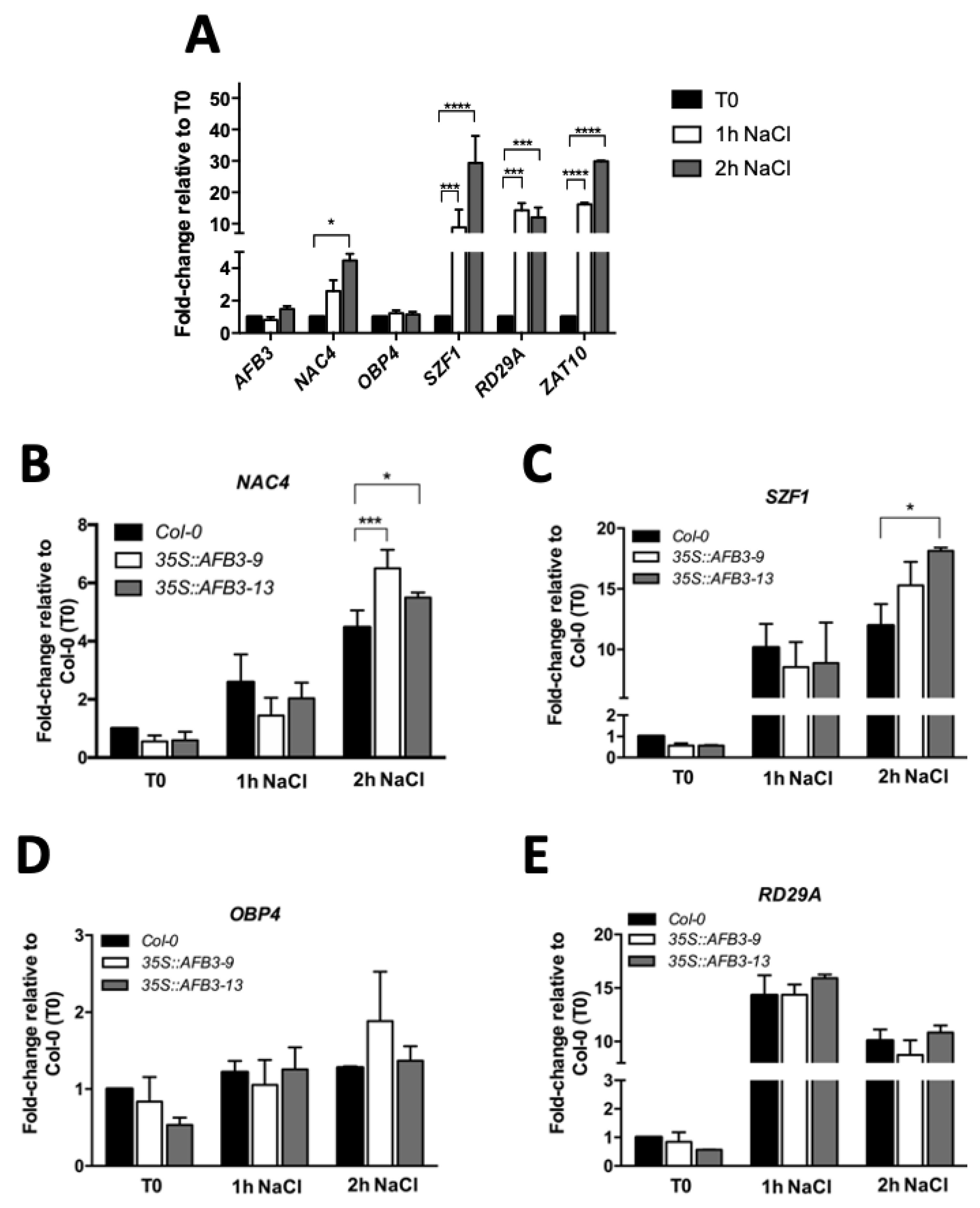
Using nitrate as a signaling molecule and a systems biology approach, a regulatory network downstream of AFB3 has been previously proposed [23]. The regulatory network suggested a key role of the transcription factors NAC4 (no apical meristem (NAM)/Arabidopsis transcription activation factor (ATAF)/cup-shaped cotyledon (CUC)), OBF BINDING PROTEIN 4 (OBP4) and SALT-INDUCIBLE ZING FINGER PROTEIN 1 (SZF1). A linear signaling cascade, where OBP4 acts downstream of NAC4, which acts downstream of AFB3 [23]. From these three genes, only SZF1 was previously described in salt stress responses [28]. To evaluate if NAC4 and OBP4 were also participating in salt stress responses, we quantified NAC4 and OBP4 transcript levels in response to salt stress (150 mM NaCl) for 1 or 2 h and compared them to SZF1 together with the previously characterized salt stress markers RESPONSIVE TO DESICCATION 29A (RD29A) and SALT TOLERANCE ZINC FINGER (STZ/ZAT10) [29,30]. As expected, there was a marked induction of at least 10 to 15-fold in both stress markers in response to salt stress after 1 and 2 h treatments (Figure 4A). Remarkably, NAC4 was up-regulated 4–5 fold at 2 h after salt stress treatment while OBP4 did not show any response suggesting that is not part of the signaling response. Moreover, SZF1 was highly up-regulated in response to salt stress at both 1 and 2 h of salt stress treatment (Figure 4A). This is in accordance with Sun et al. [28] and the up-regulation is comparable with the response level to salt observed in markers RD29A and STZ/ZAT10. These results might indicate a common response down-stream of AFB3 via NAC4 and possibly SZF1.
Since NAC4 and SZF1 transcripts are regulated in response to salt stress, we further evaluated whether AFB3 over-expression could lead to the modulation of NAC4 and SZF1 transcript levels, either directly or indirectly. Thus, Arabidopsis Col-0 WT and p35S::AFB3 seedlings were grown in MS plates for seven days and then transferred to MS plates with and without 150 mM NaCl and harvested after 1 or 2 h salt treatment. At the selected timepoints root tissue was collected and then qRT-PCR was performed to evaluate NAC4, SZF1, OBP4, and RD29A transcript levels. As shown in Figure 4, NAC4 showed induction at 2 h salt treatment in Col-0 WT and both over-expressor lines. Remarkably, there is also a stronger up-regulation response in the p35S::AFB3 when compared to Col-0 WT at 2 h NaCl treatments (Figure 4B). A similar situation was observed for SZF1 (Figure 4C). However, OBP4 showed no significant difference in either control or salt conditions (Figure 4D). The same was observed for p35S::AFB3 lines, indicating that OBP4 is not participating in the AFB3-dependent salt stress responses. Moreover, unlike the differential response observed with SZF1 and NAC4, RD29A did not show any differential expression between Col-0 WT and p35S::AFB3 lines after 1 and 2 h of NaCl treatment (Figure 4E). These results suggest that both NAC4 and SZF1 might be participating in the AFB3-dependent response to salt stress.

2.4. nac4 and szf1 Loss of Function Mutants Shows Altered Salt Stress Responses

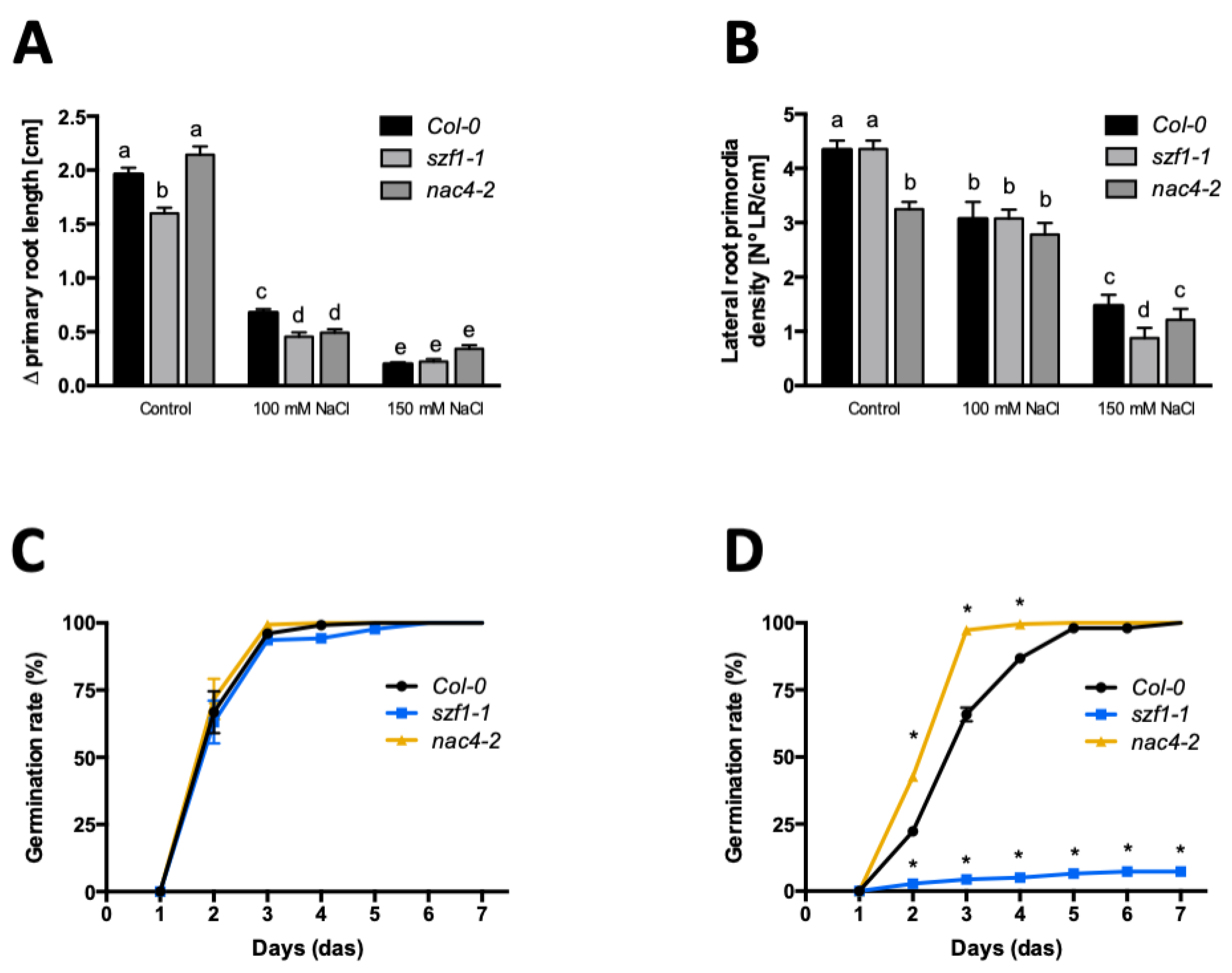
To characterize whether NAC4 and SZF1 have a role in salt stress responses we evaluated nac4 and szf1 loss of function mutant lines (Figure 5). Col-0 WT, nac4-2, and szf1-1 lines were germinated in MS plates for 3 days. At 3 DAS, seedlings were transferred to MS plates supplemented with or without 100 and 150 mM of NaCl. Plants were grown for 5 days under salt stress treatment. As shown in Figure 5, both nac4 and szf1-1 mutant lines were affected in their root architecture. While szf1-1 had shorter roots, nac4-2 lines showed a reduced lateral root primordia density (Figure 5A,B). Remarkably, both szf1-1 and nac4-2 mutant lines showed a mild but significant decrease in primary root growth compared to Col-0 WT in response to 100 mM NaCl treatment (Figure 5A), indicating an impaired sensitivity to salt in terms of primary root length in these mutants. Moreover, LR development was more affected in szf1-1 in response to 150 mM NaCl, also suggesting an oversensitivity to this stress (Figure 5B).
Constitutive overexpression of AFB3 showed a marked impact on germination under salt stress conditions (Figure 3). In order to evaluate whether SZF1 and NAC4 have a role modulating seed germination under salt stress conditions, szf1-1 and nac4-2 mutant lines were evaluated. The germination rate under MS media (control) or 150 mM of NaCl was evaluated for seven days. As shown in Figure 5, no differences were observed in any of the mutant lines compared to Col-0 WT in control conditions, reaching near 100% germination between two- and three-day after sowing (Figure 5C). However, a strong decrease in the germination rate was observed for szf1-1 in 150 mM of NaCl, reaching only 10% of germinated seeds after seven days after sowing (Figure 5D). Unexpectedly, an opposite phenotype was observed for nac4-2, was a salt-resistant phenotype was observed at 3 DAS with near a 100% germination while Col-0 WT only reached 60% (Figure 5D). The root architecture and germination results suggest that the main role of NAC4 and SZF1 in salt stress responses is not predominantly AFB3-dependent and possibly they are participating in other signaling pathways.

3. Discussion

Several studies have attributed the central role of auxin perception to TIR1 and AFB2, which act redundantly and show higher affinity for most auxins [16,21,31,32]. While TIR1 is up-regulated in response to salt stress [16], at the protein level TIR1 and AFB2 are down-regulated in response to salt, with TIR1 being more sensitive [20]. Interestingly, no significant changes were observed by qRT-PCR in AFB3 transcript accumulation or GUS expression in pAFB3::GUS lines after salt stress (Supplementary Figure S1). Moreover, in contrast with the down-regulation reported for TIR1 and AFB2 GUS protein fusion lines [20], pAFB3::AFB3-GUS lines revealed a tissue-specific protein accumulation in response to salt (Figure 1). This tissue-specific modulation strongly suggests a post-transcriptional regulation of the protein abundance. Moreover, this tissue-specific regulation of AFB3 in roots strongly suggests the key role of this auxin receptor in modulating root meristematic activity upon salt stress. The differences in salt stress response between auxin receptors are further supported by the analysis of mutant lines. It has been shown that double mutants tir1/afb1, tir1/afb2, and tir1/afb3 have differential responses to salt and oxidative stresses, with tir1/afb2 being more resistant than the other mutant combinations [17]. In this report, we showed that afb3 single mutants are hypersensitive to salt stress in terms of lateral root density, suggesting that each receptor also has developmental-specific roles [15,22,25]. Moreover, a higher order of complexity in this auxin perception has been attributed, since some studies have concluded that there are different affinities of these receptors for different auxin molecules and, at the same time, there are different specificities for AUX/IAA and ARF proteins that act downstream of auxin receptors [25,33,34]. All these differences and contrasting responses observed for TIR1 and AFBs reinforce the idea that auxins can exert differential responses depending on the receptors which might work in a highly complex regulatory network depending on specific environmental and developmental cues.
To further understand how auxin perception may work in the achievement of tolerance in a salt stress context, we made over-expressor lines of the AFB3 auxin receptor taking into consideration its opposite phenotype observed between afb3 mutant (Figure 2) when compared to tir1/afb2 [17]. The over-expression of AFB3 led to salt resistance in terms of root growth and particularly lateral root density when compared to Col-0 WT seedlings (Figure 2 and Supplementary Figure S4).
A previous report has shown that salt has a concentration- and exposure-dependent effect in root development [4]. According to this, an over-expression of AFB3 in roots could allow a suitable auxin perception even when the levels of this hormone are lower due to high salt concentrations. AFB3 overexpression also had a strong impact in seed germination, which is severely affected under salt stress conditions. While Arabidopsis seeds germinate between two to three days post-sowing in optimal conditions, under salt stress this is delayed by two days [35]. Surprisingly, p35S::AFB3 lines showed a normal germination rate under severe salt stress conditions (Figure 3). Although auxin by itself may not be determinant to induce seed germination under salt stress, it has been previously shown that the interaction between auxin and gibberellin may play an important role in several stages of germination and post-germination [36,37]. Thus, the activation of response elements that are responsible for the development of lateral roots under these stress conditions could be stimulated. Moreover, auxin can influence seed germination in the presence of ABA [10]. This may suggest that higher auxin sensitivity mediated by the over-expression of AFB3 receptor might stimulate hormonal changes in order to induce seed germination in stress conditions.
Although AFB3 transcript was not induced under salt stress in either 1 h or 2 h after salt treatment, NAC4 and SZF1, two putative downstream components described by Vidal et al. [23] were up-regulated in this context (Figure 4). In p35S::AFB3 lines, both NAC4 and SZF1 were up-regulated when compared to Col-0 WT after 2 h salt stress treatment (Figure 4). This suggests that these transcription factors could be downstream components of AFB3 and that they could have a role in tolerance to salt stress in p35S::AFB3 lines in terms of modulation of lateral root development.
As described before, NAC4 is part of the plant-specific NAC family involved in several developmental processes [38]. In addition, it has been previously described that several members of this family are in auxin signaling, such as NAC1 and NAC2 [39,40,41]. In terms of root modulation, it is shown that nac4-2 seedlings had a miss-regulation on formation of lateral roots in control conditions that were lost under salt stress conditions (Figure 5), suggesting that NAC4 has a role in the first stages of lateral root development. This is in accordance with a previous study related to NAC4 in Caragana intermedia (CiNAC4), a member of Fabaceae, where the over-expression of CiNAC4 led to an increase in lateral root number but not in primary root growth in long-term salt stress conditions [42]. Remarkably, nac4-2 mutants showed a resistant phenotype in terms of germination rate under salt stress in this study (Figure 5C,D). Several studies in salt-stress context have attributed a negative role of NAC proteins during seed germination, possibly by the down-regulation of gibberellins [38,43,44].
SZF1 is a member of the Cys3/Hys (CCCH) Zinc-finger proteins described in Arabidopsis [28]. This family has been widely characterized as growth and stress-response components [28,45,46]. Additionally, SZF1 has been described as a putative auxin-dependent [23] and salt-induced component of theses response pathways [28]. We show here that in AFB3 over-expressor lines there is a significant up-regulation of SZF1 when compared to Col-0 WT upon salt stress treatment (Figure 4). As expected szf1-1 mutant line exhibited a marked sensitivity to salt stress, which is in agreement with a previous study carried out by Sun et al. [28]. This suggests that salt-induction of SZF1 is partially AFB3-dependent and it may have a role in the modulation of salt-stress early response in Arabidopsis.
It has been described in several zinc-finger family members that their role in the acquisition of tolerance could be related to the modulation of the ABA-responsive genes [47]. This is further highlighted in previous studies where over-expression of zinc-finger proteins such as SZF1 or ZFP1 led to a decrease in salt-responsive gene expression and modulation of salt stress tolerance in Arabidopsis [28,48]. According to this, it has been proposed that this protein would be negatively regulated by ABA, in the same way as STZ/ZAT10, another transcription factor inducible by salt and drought [29]. Furthermore, SZF1 could act as a repressor of ABA-responsive genes in early stages of salt-stress. Both could provide resistance to salt-stress by modulating the excitatory stress response generated by ABA, enhancing the induction of early response genes responsible for ionic and osmotic adjustment as the activation of the salt-overly-sensitive pathway [8]. All together, these data suggest that in response to salt stress there is an accumulation of AFB3, which contributes to salt stress resistance. This is, at least in part, due to the direct or indirect regulation of NAC4 and SZF1.

4. Materials and Methods

4.1. Plant Material and Growth Conditions

Arabidopsis thaliana wild-type ecotype Columbia-0 (Col-0 WT) and Wassilewskija (WS) were used in this work and they are indicated in each section accordingly. Arabidopsis knockout mutants szf1-1 (SALK_141550), nac4-2 (SALK_006735), and afb3-1 (SALK_068787) were obtained from the Salk Institute Genomic Analysis Laboratory T-DNA insertion mutant seed collection (donated to the Arabidopsis Biological Resource Center). pAFB3::GUS (CS69678) and pAFB3::AFB3-GUS/afb3-4 (CS69682) lines were obtained from the Arabidopsis Biological Resource Center [25]. Primers used for genotyping each T-DNA line are listed in Supplementary Table S1. Arabidopsis thaliana seeds were incubated in water at 4 °C over-night. Seeds were then sterilized in Eppendorf tubes using a 2.5% Sodium hypochlorite, 0.1% Triton X-100 solution under constant mixing for 7 min. Then seeds were washed with sterile water at least three times for 3 min each time under constant mixing. After sterilization, seeds were sown on squared Petri dishes containing 0.5× Murashige and Skoog (MS) media supplemented with 0.05% MES, 1% sucrose, 0.8% agar. pH was adjusted at 5.8 using potassium hydroxide (KOH) 1 M. For plant growth, growth chambers were used with a long-day photoperiod (16 h light/8 h dark) and 22 °C constant temperature. For salt stress treatments, 100 or 150 mM of sodium chloride (NaCl) were added in previously described 0.5× MS medium.

4.2. Generation of Overexpression Lines

For the generation of AFB3 overexpression lines, the ORFs were amplified from a complementary DNA (cDNA) template with Pfu DNA Polymerase (Promega, Madison, WI, USA) using primers containing the Gateway attB sites and cloned into pDONR221 to generate the AFB3 ENTRY vector. The AFB3 ORG in the entry vector was cloned in the binary vector pK7m24GW using the CaMV p35S promoter, thus generating p35S::AFB3 lines. Overexpression lines were generated by floral dip method [49]. For the selection of p35S::AFB3 over-expression lines, seeds of 22 independent lines were sown in MS medium supplemented with 1% sucrose, 0.8% agar with 25 µg/mL of Kanamycin. p35S::AFB3 seedlings from resistant lines were grown in substrate and propagated for further experiments. Later, AFB3 gene expression levels of the previously selected lines were performed by using quantitative polymerase chain reaction.

4.3. Salt Treatment

For short-term salt treatments, Arabidopsis thaliana seeds were sterilized as described in Methods Section 1 and sown on 0.5× MS Medium pH 5.8. Seedlings were grown for 7 days in a growth chamber. Seven-day-old seedlings were transferred onto 0.5× MS medium with 0 or 150 mM NaCl for 1 or 2 h. For longer treatments, Arabidopsis thaliana seeds were grown for 3 days in growth chambers and then transferred to MS media supplemented with 0, 100, or 150 mM NaCl for 2, 5, or 10 days.

4.4. Phenotypic Analysis

Primary root growth was analyzed from seedling images recorded at given timepoints with the Perfection V700 Photo scanner (EPSON, Nagano, Japan). Roots were measured using ImageJ Software (National Institutes of Health, Bethesda, MD, USA; http://rsb.info.nih.gov/ij). To score lateral root primordia density, root clearing was performed according to Malamy and Benfey [50] with minor modifications. Briefly, seedlings were incubated in a 70% Ethanol solution overnight. Then a solution containing 20% Methanol and 4% HCl was added and seedlings were incubated for 40 min at 62 °C. This solution was replaced with a 60% Ethanol, 7% NaOH solution and incubated for 20 min at room temperature. Roots were re-hydrate in a succession of 40%, 20%, and 10% Ethanol solutions for 20 min each. Finally, an equal volume of 50% glycerol was added which made a 5% ethanol, 25% glycerol solution, and incubated for 2 h. Cleared seedlings were mounted in 25.4 × 76.2 mm microscope slides with 50% glycerol and covered with 24 × 50 mm cover glass [50]. The number of lateral roots was analyzed using a differential interference contrast microscope Zeiss (Berlin, Germany). Total lateral root density was calculated as the ratio between the total number of the lateral roots and the primary root length of each plant.
To evaluate germination potential under salt stress conditions, seeds were sown on MS medium with 0 or 150 mM of NaCl. Seed germination was scored daily for 7 days. Germination of the seed was established by root protrusion.

4.5. Histochemical Analysis

To detect β-glucuronidase (GUS) activity, 7-day-old seedlings were incubated in reaction buffer containing 0.1 M sodium phosphate buffer (pH 7), 1 mM ferricyanide, 1 mM ferrocyanide, 0.1% Triton X-100 and 1 mg mL−1 X-Gluc for 30 min up to 24 h in dark at 37 °C. Afterwards, chlorophyll was removed by incubation in 70% ethanol and seedlings were cleared as previously described [50]. GUS staining in roots was imaged by differential interference contrast microscopy Olympus BX51 (Tokyo, Japan).

4.6. DNA Purification

Genomic DNA was extracted from leaf tissue of 4-6-week-old plants. The purification of genomic DNA was performed according to a modified protocol, originally performed by Richards et al. [51]. Briefly, tissue samples were pulverized and 200 µL of High-salt TE extraction Buffer (200 mM Tris-HCl pH 8.0, 25 mM EDTA pH 8.0, 250 mM NaCl and 0.5% p/v SDS) was added. Samples were centrifugated at 12,000 RPM for 5 min. Supernatant was transferred onto a new microcentrifuge tube with 200 µL of Isopropanol to precipitate the DNA present in the samples. Then samples were mixed for 15 min and centrifugated at 12,000 RPM for 5 min. Supernatant was discarded and pellet was washed with 200 µL of 70% ethanol. To precipitate the DNA, samples were centrifugated at 12,000 RPM for 5 min and supernatant was discarded. Finally, DNA samples were dried and re-suspended in 100 µL of DNAse/RNAse free water [51].

4.7. RNA Extraction and cDNA Synthesis

Roots from 7-day-old or 10-day-old Arabidopsis thaliana seedlings were collected and frozen in liquid nitrogen. RNA purification was performed using the Purelink RNA Mini Kit (Thermo Fisher Scientific, Waltham, MA, USA) following manufacturer instructions. Then RNA samples were treated with DNase I Amplification Grade kit (Thermo Fisher Scientific, Waltham, MA, USA) to finalize RNA purification. RNA samples were treated with DNase I for 15 min at room temperature. To inactivate the enzyme, 1 μL of 25 mM EDTA was added and samples were incubated at 65 °C for 10 min. RNA samples were quantified and purity checked using the Nanodrop 2000 spectrophotometry system (Thermo Fisher Scientific, Waltham, MA, USA).
cDNA synthesis was performed by the incubation of 500 ng of each total RNA sample with 1 µL oligo dT (0.5 µg/µL) at 70 °C for 5 min to amplify only the mRNAs in the sample. Samples were retrotranscribed to DNA using “Improm II Reverse Transcriptase” include manufacturer instructions.

4.8. Quantitative Polymerase Chain Reaction (qPCR)

qPCR of cDNAs was performed using Brilliant III Ultra-Fast SYBR Green qPCR kit (Agilent Technologies, Santa Clara, CA, USA) following manufacturer instructions. cDNA of each gene was amplificated using primers listed in Supplementary Table S1. The reaction was performed in StepOnePlus Real-Time PCR System (Applied Biosystems, Waltham, MA, USA) in 0.1 mL MicroAmp® Fast 8-Tube Strip (Applied Biosystems, Waltham, MA, USA). The program used for the amplification went as follows: 95 °C for 3 min, 95 °C for 5 s and 60 °C for 10 s during 40 cycles and a final elongation for 15 s at 95 °C. Additionally, a melting curve was performed from 65 °C to 90 °C rising 0.3 °C each phase to confirm the amplification of only one transcript on each gene.
Data were normalized using clathrin adaptor complex subunit (AT5G46630) as housekeeping gene [52]. Finally, data were analyzed using LinReg program [53].

4.9. Statistical Analysis

All experiments were performed at least three times. For primary root growth, lateral root density, and qRT-PCR analysis, two-way ANOVA and Bonferroni a posteriori test were performed. Statistically significant p values were set to < 0.05. For germination rate analysis, One-way ANOVA and Bonferroni a posteriori test were performed. Statistically significant p values were set to < 0.05.

Supplementary Materials

The following are available online at https://www.mdpi.com/1422-0067/21/24/9528/s1.

Author Contributions

F.G.-V., T.G., R.T. and J.A.O. participated in the methodology, investigation and formal analysis. F.G.-V., R.T. and J.A.O. contributed to the writing—original draft preparation and writing—review and editing. R.T. and J.A.O. contributed to the resources and funding acquisition. All authors have read and agreed to the published version of the manuscript.

Funding

This work was supported by National Agency for Research and Development (ANID)/Fondo Nacional de Desarrollo Científico y Tecnológico (FONDECYT) No. 1181358 to J.A.O. and No. 1171442 to R.T.

Conflicts of Interest

The authors have no competing interest to declare.

References

  1. Žádníková, P.; Smet, D.; Zhu, Q.; Van Der Straeten, D.; Benková, E. Strategies of seedlings to overcome their sessile nature: Auxin in mobility control. Front. Plant. Sci. 2015, 6. [Google Scholar] [CrossRef] [Green Version]
  2. Fujita, M.; Fujita, Y.; Noutoshi, Y.; Takahashi, F.; Narusaka, Y.; Yamaguchi-Shinozaki, K.; Shinozaki, K. Crosstalk between abiotic and biotic stress responses: A current view from the points of convergence in the stress signaling networks. Curr. Opin. Plant. Biol. 2006, 9, 436–442. [Google Scholar] [CrossRef]
  3. Cabot, C.; Sibole, J.V.; Barceló, J.; Poschenrieder, C. Lessons from crop plants struggling with salinity. Plant. Sci. 2014, 226, 2–13. [Google Scholar] [CrossRef]
  4. Carillo, P.; Grazia, M.; Pontecorvo, G.; Fuggi, A.; Woodrow, P. Salinity Stress and Salt Tolerance. Abiotic Stress Plants Mech. Adapt. 2011. [Google Scholar] [CrossRef] [Green Version]
  5. Galvan-Ampudia, C.S.; Julkowska, M.M.; Darwish, E.; Gandullo, J.; Korver, R.A.; Brunoud, G.; Haring, M.A.; Munnik, T.; Vernoux, T.; Testerink, C. Halotropism Is a Response of Plant Roots to Avoid a Saline Environment. Curr. Biol. 2013, 23, 2044–2050. [Google Scholar] [CrossRef] [Green Version]
  6. Ribba, T.; Garrido-Vargas, F.; O’Brien, J.A. Auxin-mediated responses under salt stress: From developmental regulation to biotechnological applications. J. Exp. Bot. 2020, 71, 3843–3853. [Google Scholar] [CrossRef]
  7. He, F.; Xu, C.; Fu, X.; Shen, Y.; Guo, L.; Leng, M.; Luo, K. The MicroRNA390/TRANS-ACTING SHORT INTERFERING RNA3 Module Mediates Lateral Root Growth under Salt Stress via the Auxin Pathway. Plant. Physiol. 2018, 177, 775–791. [Google Scholar] [CrossRef] [Green Version]
  8. Ji, H.; Pardo, J.M.; Batelli, G.; Van Oosten, M.J.; Bressan, R.A.; Li, X. The salt overly sensitive (SOS) pathway: Established and emerging roles. Mol. Plant. 2013, 6, 275–286. [Google Scholar] [CrossRef] [Green Version]
  9. Potters, G.; Pasternak, T.P.; Guisez, Y.; Palme, K.J.; Jansen, M.A.K. Stress-induced morphogenic responses: Growing out of trouble? Trends Plant. Sci. 2007, 12, 98–105. [Google Scholar] [CrossRef]
  10. Brady, S.; Sarkar, S.F.; Bonetta, D.; McCourt, P. The ABSCISIC ACID INSENSITIVE 3 (ABI3) gene is modulated by farnesylation and is involved in auxin signaling and lateral root development in Arabidopsis. Plant J. 2003, 3, 67–75. [Google Scholar] [CrossRef]
  11. Kazan, K. Auxin and the integration of environmental signals into plant root development. Ann. Bot. 2013, 112, 1655–1665. [Google Scholar] [CrossRef] [Green Version]
  12. Verma, V.; Ravindran, P.; Kumar, P.P. Plant hormone-mediated regulation of stress responses. BMC Plant. Biol. 2016, 16, 1–10. [Google Scholar] [CrossRef] [Green Version]
  13. Woodward, A.W.; Bartel, B. Auxin: Regulation, action, and interaction. Ann. Bot. 2005, 95, 707–735. [Google Scholar] [CrossRef] [PubMed] [Green Version]
  14. Dos Santos Maraschin, F.; Memelink, J.; Offringa, R. Auxin-induced, SCFTIR1-mediated poly-ubiquitination marks AUX/IAA proteins for degradation. Plant. J. 2009, 59, 100–109. [Google Scholar] [CrossRef]
  15. Prigge, M.J.; Platre, M.; Kadakia, N.; Zhang, Y.; Greenham, K.; Szutu, W.; Pandey, B.K.; Bhosale, R.A.; Bennett, M.J.; Busch, W.; et al. Genetic analysis of the Arabidopsis TIR1/AFB auxin receptors reveals both overlapping and specialized functions. eLife 2020, 9. [Google Scholar] [CrossRef]
  16. Chen, Z.; Hu, L.; Han, N.; Hu, J.; Yang, Y.; Xiang, T.; Zhang, X.; Wang, L. Overexpression of a miR393-resistant form of transport inhibitor response protein 1 (mTIR1) enhances salt tolerance by increased osmoregulation and Na+ exclusion in Arabidopsis thaliana. Plant. Cell Physiol. 2015, 56, 73–83. [Google Scholar] [CrossRef] [Green Version]
  17. Iglesias, M.; Anahı, C. Auxin signaling participates in the adaptative response against oxidative stress and salinity by interacting with redox metabolism in Arabidopsis. Plant Mol. Biol. 2010, 74, 215–222. [Google Scholar] [CrossRef]
  18. Marrs, K.A. The Functions and Regulation of Glutathione S-Transferases in Plants. Annu. Rev. Plant Physiol. Plant Mol. Biol. 1996, 47, 127–158. [Google Scholar] [CrossRef]
  19. Park, J.-E.; Park, J.-Y.; Kim, Y.-S.; Staswick, P.E.; Jeon, J.; Yun, J.; Kim, S.-Y.; Kim, J.; Lee, Y.-H.; Park, C.-M. GH3-mediated auxin homeostasis links growth regulation with stress adaptation response in Arabidopsis. J. Biol. Chem. 2007, 282, 10036–10046. [Google Scholar] [CrossRef] [Green Version]
  20. Iglesias, M.J.; Terrile, M.C.; Windels, D.; Lombardo, M.C.; Bartoli, C.G.; Vazquez, F.; Estelle, M.; Casalongué, C.A. MiR393 Regulation of Auxin Signaling and Redox-Related Components during Acclimation to Salinity in Arabidopsis. PLoS ONE 2014, 9, e107678. [Google Scholar] [CrossRef] [Green Version]
  21. Parry, G.; Estelle, M. Auxin receptors: A new role for F-box proteins. Curr. Opin. Cell Biol. 2006, 18, 152–156. [Google Scholar] [CrossRef]
  22. Vidal, E.A.; Araus, V.; Lu, C.; Parry, G.; Green, P.J.; Coruzzi, G.M.; Gutierrez, R.A. Nitrate-responsive miR393/AFB3 regulatory module controls root system architecture in Arabidopsis thaliana. Proc. Natl. Acad. Sci. USA 2010, 107, 4477–4482. [Google Scholar] [CrossRef] [Green Version]
  23. Vidal, E.A.; Moyano, T.C.; Riveras, E.; Contreras-Lopez, O.; Gutierrez, R.A. Systems approaches map regulatory networks downstream of the auxin receptor AFB3 in the nitrate response of Arabidopsis thaliana roots. Proc. Natl. Acad. Sci. USA 2013, 110, 12840–12845. [Google Scholar] [CrossRef] [Green Version]
  24. Vidal, E.A.; Álvarez, J.M.; Gutiérrez, R.A. Nitrate regulation of AFB3 and NAC4 gene expression in Arabidopsis roots depends on NRT1.1 nitrate transport function. Plant. Signal. Behav. 2014, 9, e28501. [Google Scholar] [CrossRef] [Green Version]
  25. Parry, G.; Calderon-Villalobos, L.I.; Prigge, M.; Peret, B.; Dharmasiri, S.; Itoh, H.; Lechner, E.; Gray, W.M.; Bennett, M.; Estelle, M. Complex regulation of the TIR1/AFB family of auxin receptors. Proc. Natl. Acad. Sci. USA 2009, 106, 22540–22545. [Google Scholar] [CrossRef] [Green Version]
  26. Dharmasiri, N.; Dharmasiri, S.; Weijers, D.; Lechner, E.; Yamada, M.; Hobbie, L.; Ehrismann, J.S.; Jürgens, G.; Estelle, M. Plant development is regulated by a family of auxin receptor F box proteins. Dev. Cell 2005, 9, 109–119. [Google Scholar] [CrossRef] [PubMed] [Green Version]
  27. Blumwald, E. Sodium transport and salt tolerance in plants. Curr. Opin. Cell Biol. 2000, 12, 431–434. [Google Scholar] [CrossRef]
  28. Sun, J.; Jiang, H.; Xu, Y.; Li, H.; Wu, X.; Xie, Q.; Li, C. The CCCH-type zinc finger proteins AtSZF1 and AtSZF2 regulate salt stress responses in Arabidopsis. Plant. Cell Physiol. 2007, 48, 1148–1158. [Google Scholar] [CrossRef]
  29. Sakamoto, H.; Maruyama, K.; Sakuma, Y.; Meshi, T.; Iwabuchi, M. Arabidopsis Cys2/His2-type zinc-finger proteins function as transcription repressors under drought. Plant Physiol. 2004, 136, 2734–2746. [Google Scholar] [CrossRef] [Green Version]
  30. Yamaguchi-Shinozaki, K.; Shinozaki, K. A Novel cis-acting element in an Arabidopsis gene is involved in responsiveness to drought, low-temperature, or high-salt stress. Plant. Cell 1994, 6, 251–264. [Google Scholar]
  31. Lavy, M.; Estelle, M. Mechanisms of auxin signaling. Development 2016, 143, 3226–3229. [Google Scholar] [CrossRef] [Green Version]
  32. Sun, F.; Zhang, W.; Hu, H.; Li, B.; Wang, Y.; Zhao, Y.; Li, K.; Liu, M.; Li, X. Salt modulates gravity signaling pathway to regulate growth direction of primary roots in Arabidopsis. Plant. Physiol. 2007, 146, 178–188. [Google Scholar] [CrossRef] [PubMed] [Green Version]
  33. Shimizu-Mitao, Y.; Kakimoto, T. Auxin sensitivities of all Arabidopsis aux/IAAs for degradation in the presence of every TIR1/AFB. Plant. Cell Physiol. 2014, 55, 1450–1459. [Google Scholar] [CrossRef] [PubMed] [Green Version]
  34. Villalobos, L.I.A.C.; Lee, S.; De Oliveira, C.; Ivetac, A.; Brandt, W.; Armitage, L.; Sheard, L.B.; Tan, X.; Parry, G.; Mao, H.; et al. A combinatorial TIR1/AFB-Aux/IAA co-receptor system for differential sensing of auxin. Nat. Chem. Biol. 2012, 8, 477. [Google Scholar] [CrossRef] [Green Version]
  35. Page, D.R.; Grossniklaus, U. The art and design of genetic screens: Arabidopsis thaliana. Nat. Rev. Genet. 2002, 3, 124–136. [Google Scholar] [CrossRef] [PubMed]
  36. Fu, X.; Harberd, N.P. Auxin promotes Arabidopsis root growth by modulating gibberellin response. Nature 2003, 421, 2–5. [Google Scholar] [CrossRef]
  37. Chiwocha, S.D.S.; Cutler, A.J.; Abrams, S.R.; Ambrose, S.J.; Yang, J.; Ross, A.R.S.; Kermode, A.R. The etr1-2 mutation in Arabidopsis thaliana affects the abscisic acid, auxin, cytokinin and gibberellin metabolic pathways during maintenance of seed dormancy, moist-chilling and germination. Plant. J. 2005, 42, 35–48. [Google Scholar] [CrossRef] [Green Version]
  38. Park, C.; Park, J.; Kim, Y.; Kim, S.; Jung, J.; Woo, J.; Park, C. Integration of auxin and salt signals by the NAC transcription factor NTM2 during seed germination in Arabidopsis. Plant Physiol. 2011, 156, 537–549. [Google Scholar] [CrossRef] [Green Version]
  39. He, X.; Mu, R.; Cao, W.; Zhang, Z.; Zhang, J.; Chen, S. AtNAC2, a transcription factor downstream of ethylene and auxin signaling pathways, is involved in salt stress response and lateral root development. Plant J. 2005, 44, 903–916. [Google Scholar] [CrossRef]
  40. Xie, Q.; Frugis, G.; Colgan, D.; Chua, N. Arabidopsis NAC1 transduces auxin signal downstream of TIR1 to promote lateral root development. Genes Dev. 2000, 14, 3024–3036. [Google Scholar] [CrossRef] [Green Version]
  41. Youhua, W.; Liusheng, D.; Mengzhu, L.U.; Zhaohu, L.I.; Minjie, W.; Zhixi, Z. Expression of NAC1 up-stream regulatory region and its relationship to the lateral root initiation induced by gibberellins and auxins. Sci. China C Life Sci. 2006, 49, 429–435. [Google Scholar] [CrossRef]
  42. Han, X.; Feng, Z.; Xing, D.; Yang, Q.; Wang, R.; Qi, L.; Li, G. Two NAC transcription factors from Caragana intermedia altered salt tolerance of the transgenic Arabidopsis. BMC Plant. Biol. 2015, 1–12. [Google Scholar] [CrossRef] [Green Version]
  43. Kim, S.; Lee, A.; Yoon, H.; Park, C. A membrane-bound NAC transcription factor NTL8 regulates gibberellic acid-mediated salt signaling in Arabidopsis seed germination. Plant J. 2008, 55, 77–88. [Google Scholar] [CrossRef]
  44. Puranik, S.; Sahu, P.P.; Srivastava, P.S.; Prasad, M. NAC proteins: Regulation and role in stress tolerance. Trends Plant. Sci. 2012, 17, 369–381. [Google Scholar] [CrossRef]
  45. Han, G.; Wang, M.; Yuan, F.; Sui, N. The CCCH zinc finger protein gene AtZFP1 improves salt resistance in Arabidopsis thaliana. Plant Mol. Biol. 2014, 86, 237–253. [Google Scholar] [CrossRef]
  46. Bastola, D.R.; Pethe, V.V.; Winicov, I. Alfin1, a novel zinc-finger protein in alfalfa roots that binds to promoter elements in the salt-inducible MsPRP2 gene. Plant Mol. Biol. 1998, 38, 1123–1135. [Google Scholar] [CrossRef]
  47. Ciftci-Yilmaz, S.; Mittler, R. The zinc finger network of plants. Cell. Mol. Life Sci. 2008, 65, 1150–1160. [Google Scholar] [CrossRef]
  48. Huang, D.; Wu, W.; Abrams, S.R.; Cutler, A.J. The relationship of drought-related gene expression in Arabidopsis thaliana to hormonal and environmental factors. J. Exp. Bot. 2008, 59, 2991–3007. [Google Scholar] [CrossRef] [Green Version]
  49. Clough, S.J.; Bent, A.F. Floral dip: A simplified method for Agrobacterium-mediated transformation of Arabidopsis thaliana. Plant. J. 1998, 16, 735–743. [Google Scholar] [CrossRef] [Green Version]
  50. Malamy, J.E.; Benfey, P.N. Organization and cell differentiation in lateral roots of Arabidopsis thaliana. Development 1997, 124, 33–44. [Google Scholar]
  51. Richards, E.; Reichardt, M.; Rogers, S. Preparation of genomic DNA from plant tissue. Curr. Protoc. Mol. Biol. 2001, 1–7. [Google Scholar] [CrossRef]
  52. Czechowski, T.; Stitt, M.; Altmann, T.; Udvardi, M.K. Genome-wide identification and testing of superior reference genes for transcript normalization. Plant Physiol. 2005, 139, 5–17. [Google Scholar] [CrossRef] [Green Version]
  53. Ruijter, J.M.; Ramakers, C.; Hoogaars, W.M.H.; Karlen, Y.; Bakker, O.; van den Hoff, M.J.B.; Moorman, A.F.M. Amplification efficiency: Linking baseline and bias in the analysis of quantitative PCR data. Nucleic Acids Res. 2009, 37. [Google Scholar] [CrossRef] [Green Version]
Figure 1. Salinity induces AFB3 protein expression in root meristem of Arabidopsis thaliana seedlings. Three-day-old pAFB3::AFB3-GUS (AD) seedlings were transferred onto Murashige & Skoog (MS) medium supplemented with 0 (A,C) or 150 mM of NaCl (B,D) and cultivated for additional five days. At day 8 (DAS), seedlings were subjected to β-Glucuronidase (GUS) staining. A representative picture of each treatment is shown. (A,B) Meristematic/elongation zone, (C,D) Lateral root. Bar = 0.2 mm.
Ijms 21 09528 g001
Figure 2. AFB3 over-expression enhances salt stress tolerance in Arabidopsis thaliana roots. Three-day-old Col-0 WT, p35S::AFB3, WS, and afb3-1 were transferred onto MS medium supplemented with 0, 100, or 150 mM of NaCl and cultivated for additional five days. (A) Schematic representation of primary root growth quantification under salt stress (Δ primary root length). (B) Representative Col-0 WT and p35S::AFB3 in 150 mM NaCl are shown. (C,D) Primary root growth (C) and lateral root primordia density (D) of Col-0 WT and p35S::AFB3 lines after transfer to NaCl-containing media were quantified. (E) Representative Wassilewskija (WS) and afb3-1 seedlings in 150 mM NaCl are shown. (F,G) Primary root growth (F) and lateral root primordia density (G) of WS and afb3-1 seedlings after transfer to NaCl-containing media were quantified. Two-way ANOVA and Bonferroni a posteriori tests were performed. a, b, c, d represents statistically significant differences with p < 0.05. Error bars represent the standard error of the mean. Each experiment was performed at least three independent times, with 12 seedlings each replicate. Scale: 1 cm.
Ijms 21 09528 g002
Figure 3. AFB3 over-expression increases germination rates under control and salt stress conditions. Germination rates of Col-0 WT and p35S::AFB3 lines were determined on 0.5× MS medium (A) and 0.5× MS medium containing 150 mM NaCl (B). One-way ANOVA and Bonferroni a posteriori tests were performed. *: p < 0.05. Error bars represent the standard error of the mean. Each experiment was performed three times with at least 100 seeds per treatment.
Ijms 21 09528 g003
Figure 4. Putative AFB3-downstream signaling components NAC4 and SZF1, are induced under salt stress conditions and differentially regulated in Arabidopsis thaliana AFB3 over-expressor lines. 7-day-old Col-0 WT and p35S::AFB3 lines seedlings were transferred onto 0.5× MS medium supplemented with 150 mM of NaCl. At the time of transfer (T0), 1 h and 2 h after NaCl treatment, whole roots were collected. Transcript levels were analyzed by qRT-PCR using Clathrin adaptor complex subunit (AT5G46630) as house-keeping gene. (A) Col-0 WT, Fold-change was set for T0. (BE) Transcript levels from NAC4 (B), SZF1 (C), OBP4 (D), and RD29A (E) in Col-0 WT and p35S::AFB3 lines. Fold-change was set for T0 (Col-0 WT). Two-way ANOVA and Bonferroni a posteriori tests were performed. *: p < 0.05; ***: p = 0.001; ****: p < 0.001. Error bars represent the standard error of the mean. Each experiment was performed three times with at least 25 seedlings in each replicate.
Ijms 21 09528 g004
Figure 5. nac4-2 and szf1-1 mutant lines show an altered response to salt stress in Arabidopsis thaliana. (A,B) 3-day-old Col-0 WT, nac4-2, and szf1-1 lines were transferred to MS medium supplemented with 0, 100, or 150 mM of NaCl for five days. Primary root growth under NaCl treatment (A) and lateral root primordia density (B) were quantified. (C,D) Germination rates of Col-0 WT, nac4-1, and szf1-1 plants were determined on MS medium (C) and the same medium supplemented with 150 mM of NaCl (D). ANOVA and Bonferroni a posteriori tests were performed. *: p < 0.05. a, b, c, d represents statistically significant differences with p < 0.05. Error bars represent the standard error of the mean. Each experiment was performed in triplicates with at least 12 seedlings (A,B) or 100 seeds (C,D) per treatment.
Ijms 21 09528 g005
Publisher’s Note: MDPI stays neutral with regard to jurisdictional claims in published maps and institutional affiliations.