Article

The Regulatory Network of CMPG1-V in Wheat–Blumeria graminis f. sp. tritici Interaction Revealed by Temporal Profiling Using RNA-Seq

State Key Laboratory of Crop Genetics and Germplasm Enhancement, Cytogenetics Institute, Nanjing Agricultural University/JCIC-MCP, Nanjing 210095, China
*
Author to whom correspondence should be addressed.
These authors contribute equally to this work.
Int. J. Mol. Sci. 2020, 21(17), 5967; https://doi.org/10.3390/ijms21175967
Submission received: 16 July 2020 / Revised: 9 August 2020 / Accepted: 17 August 2020 / Published: 19 August 2020
(This article belongs to the Special Issue Wheat and Barley: Acclimatization to Abiotic and Biotic Stress)

Abstract

:
Wheat powdery mildew (Pm), caused by Blumeria graminis f. sp. tritici (Bgt), is a prevalent fungal disease. The diploid wheat relative Haynaldia villosa (H. villosa) showed broad-spectrum resistance (BSR) to Pm. A previous study reported an E3 ligase gene, CMPG1-V from H. villosa, showing BSR to Pm. To elucidate the regulatory network mediated by CMPG1-V, in this study, gene expression profiling of CMPG1-V transgenic plant (CMPG1-VOE) and its receptor Yangmai 158 was analyzed and compared after Bgt inoculation at four infection stages. GO and KEGG analysis revealed obvious reprogramming of SA and ABA signaling, starch/sucrose metabolism, and photosynthesis in CMPG1-VOE, compared with those in Yangmai 158. Transcripts of SA synthesis genes SARD1 and UGT, signaling factors TGA and PRs, and SnRKs in ABA signaling were specifically upregulated in CMPG1-VOE rather than Yangmai 158. Transcripts of LHCII in photosynthesis, GLUC and TPP in starch/sucrose metabolism were also induced distinctly in CMPG1-VOE. WGCNA analysis showed crucial regulatory candidates of CMPG1-V, involving serine/threonine-protein kinase in phosphorylation, glucosyltransferase in flavonoid biosynthesis, defense factor WRKYs, and peroxidase in oxidative stress. Our results facilitate the deciphering of the resistant regulatory network of CMPG1-V and the identification of key candidates which might be employed in breeding programs.

1. Introduction

When pathogens infect plants, the early defense signals mainly involve the hormone signaling pathways and oxidative homeostasis [1]. Ubiquitination regulated by E3 ubiquitin ligase plays an important role during the process [2]. SCFCOI1 type E3 ubiquitin ligase subunit F-box protein COI1 (Coronatine insensitive 1) is the first confirmed ubiquitination system against pathogen infection in JA signaling [3]. Arabidopsis (Arabidopsis thaliana) RING E3 ubiquitin ligase Keep On Going (KEG) interacting with SCFCOI1 E3 ubiquitin ligase positively regulates plant immunity [4]. A tobacco (Nicotiana benthamiana) RING-type E3 ubiquitin ligase, NtRFP1, has been shown recently to attenuate disease symptoms caused by bC1 protein through the ubiquitination of bC1 [5].
Salicylic acid (SA), considered to be an important endogenous signaling molecule in plant immunity, can promote the degradation of defense transcription repressors through E3 ubiquitin ligase. SA can promote E3 ubiquitin ligase BTBNPR1 degrading transcriptional repressors TGA2 and NIMIN1 to enhance disease resistance [6]. CUL4-DDB1-based ubiquitin ligase interacted with SA to enhance tomato’s resistance to nontumorigenic Agrobacterium tumefaciens [7]. AtSR1 interaction protein 1 (SR1IP1), a CUL3-based E3 ubiquitin ligase, positively regulates SA-related immunity by degrading the defense suppressor AtSR1 in Arabidopsis [8]. Several studies also confirmed the role of E3 ligases in ABA defense signaling. Overexpressing OsDRF1 in tobacco, an F-box type E3 ligase in rice, showed ABA sensitivity and increased resistance to viruses and bacteria [9]. Pepper (Capsicum annuum) RING E3 ubiquitin ligase CaRING1 induced by Xanthomonas oryzae acts as a positive regulator of defense responses [10,11].
Ubiquitin ligase also regulates plant primary metabolism after pathogen infection. Ubiquitin ligase ARABIDOPSIS TOXICOS EN LEVADURA31 (ATL31) and its closest homologue, ATL6, a membrane-associated ubiquitin ligase, are involved in the carbon/nitrogen (C/N) response by regulating the stability of 14-3-3 proteins through ubiquitination in plant immune response [12,13]. Overexpression of LeATL31 and LeATL6 enhanced the Pseudomonas syringae resistance in tomato [14].
Wheat (Triticum aestivum, AABBDD) makes a substantial contribution to human calorie intake, global agricultural sustainability, and food security [15]. Pathogens and pests cause wheat yield losses that account for approximately 21.5% of total losses, reaching up to 28.1% in food-deficit areas [16]. Wheat powdery mildew (Pm), a fungal disease caused by Blumeria graminis f. sp. tritici (Bgt), is one of the most destructive diseases of wheat, causing extensive yield losses worldwide [17]. Due to the host or race specificity of the wheat–Bgt interaction, new virulent races could rapidly evolve and emerge. Thus, the exploration and utilization of durable and broad-spectrum resistance (BSR) genes are of great significance for powdery mildew control.
Although there have many studies on the resistance mechanism of ubiquitin ligase genes, only a few studies performed the transcriptomic changes in response to Pm in a continuous time period. The related metabolism reprogramming in the disease resistance process is also indistinct. In a previous study, an E3 ligase gene, CMPG1-V, was cloned from Haynaldia villosa. Overexpression of CMPG1-V enhanced its BSR to Pm [18]. This provides an ideal system for investigating the temporal process during Bgt infection regulated by CMPG1-V. In this study, CMPG1-V transgenic line (CMPG1-VOE) and susceptible receptor Yangmai 158 were inoculated with Bgt isolate E26 for RNA-seq. CMPG1-V associated defense pathways were analyzed by the Gene Ontology (GO), Kyoto Encyclopedia of Genes and Genomes (KEGG) enrichment, and Weighted Gene Co-Expression Network (WGCNA). These findings provide insights into the molecular mechanisms of CMPG1-V-associated broad-spectrum powdery mildew resistance in wheat.

2. Results

2.1. Global Profiling of CMPG1-VOE in Response to Bgt Infection

When Bgt infects wheat, conidia forms rapidly and reaches the epidermal cells from 1 h after inoculation (hai) to 72 hai (Figure S1) [19]. Based on the above observation, the samples for RNA-seq of CMPG1-V transgenic line (CMPG1-VOE) and susceptible receptor Yangmai 158 were collected at 1, 8, 18, and 24 hai by moderately Bgt virulent race E26. The mock samples were collected simultaneously at the above corresponding time points. The Q20 and Q30 of RNA-seq sequences for all samples were more than 93% and 84% and the ratios of clean reads accounted for 89.41% to 95.17%, respectively (Table S1). To maximize the number of possible differentially expressed genes (DEGs), the selective parameter was set as Fold Change ≥ 2.00 and FDR ≤ 0.001. In CMPG1-VOE and Yangmai 158, 6363 and 8047 transcripts were upregulated, and 11,475 and 5114 were downregulated, respectively (Figure 1a). The number of upregulated DEGs in CMPG1-VOE was less than that in Yangmai 158 for all the analyzed infection time points. Venn diagram revealed 24% of the upregulated DEGs were specifically expressed in CMPG1-VOE, while for the downregulated DEGs, up to 60.8% were specifically expressed in CMPG1-VOE, and only 12% were specifically expressed in Yangmai 158 (Figure 1b). We have also tested the two materials with a more virulent race, E31, and harvested the samples for RNA-seq at the same time points after inoculation (Table S1 and Figure S2).

2.2. Functional Categories Enriched in CMPG1-VOE and Yangmai 158 in Response to Bgt Infection

To understand the biological function of the DEGs, GO enrichment was performed to classify the crucial biological processes and cardinal signaling pathways. Before Bgt infection, no significant difference was observed between CMPG1-VOE and Yangmai 158 (Figure S3a). The upregulated DEGs in CMPG1-VOE were enriched in normal physiological metabolism, such as “sulfur compound transport”, “aminoglycan metabolic process”, “glutathione metabolic process”, “oxoacid metabolic process”, and so on.
After Bgt inoculation, we observed distinct enriched DEGs in the two materials (Figure 2a,b). At 1 hai, most of the upregulated DEGs in the two materials focused on “photosynthesis” and “generation of precursor metabolites and energy”, and transcription levels increased in the succeeding infection timepoints. At later infection stages from 18 hai to 24 hai, the upregulated transcripts in “abscisic acid (ABA)-activated signaling pathway”, “regulation of salicylic acid (SA) metabolic process”, “glucose import and sucrose biosynthetic process”, “nitrogen compound”, and “nitric oxide metabolic process” were specifically enriched in CMPG1-VOE (Figure 2b). However, at the corresponding infectious timepoints in Yangmai 158, the upregulated transcripts were only enriched in “organonitrogen compound catabolic process”, “response to cytokinin”, and “carbohydrate metabolic process” (Figure 2a). Similarly, the hormone and energy biological processes were also observed in CMPG1-VOE challenged with E31 (Figure S4).
The special downregulated DEGs in CMPG1-VOE were mainly enriched in “glutathione metabolic process”, “toxin catabolic process”, and “organonitrogen compound catabolic process” (Figure 2b), while the transcripts in “ATP biosynthetic process”, “response to organic substance”, “fatty acid and lipid oxidation”, and “sulfide oxidation” were specially downregulated in Yangmai 158 (Figure 2a and Figure S4).
Common up- or downregulated DEGs were also present in the two materials after Bgt inoculation. The upregulated transcripts of common defense response in CMPG1-VOE and Yangmai 158 were enriched in “cell wall organization or biogenesis”, “glutathione metabolic process”, “toxin catabolic process”, and “sulfur compound transport” (Figure S3b). The downregulated transcripts were enriched in “carboxylic acid biosynthetic process”, “fatty acid biosynthetic process”, “response to acid chemical”, and “chitin metabolic process” (Figure S3c).

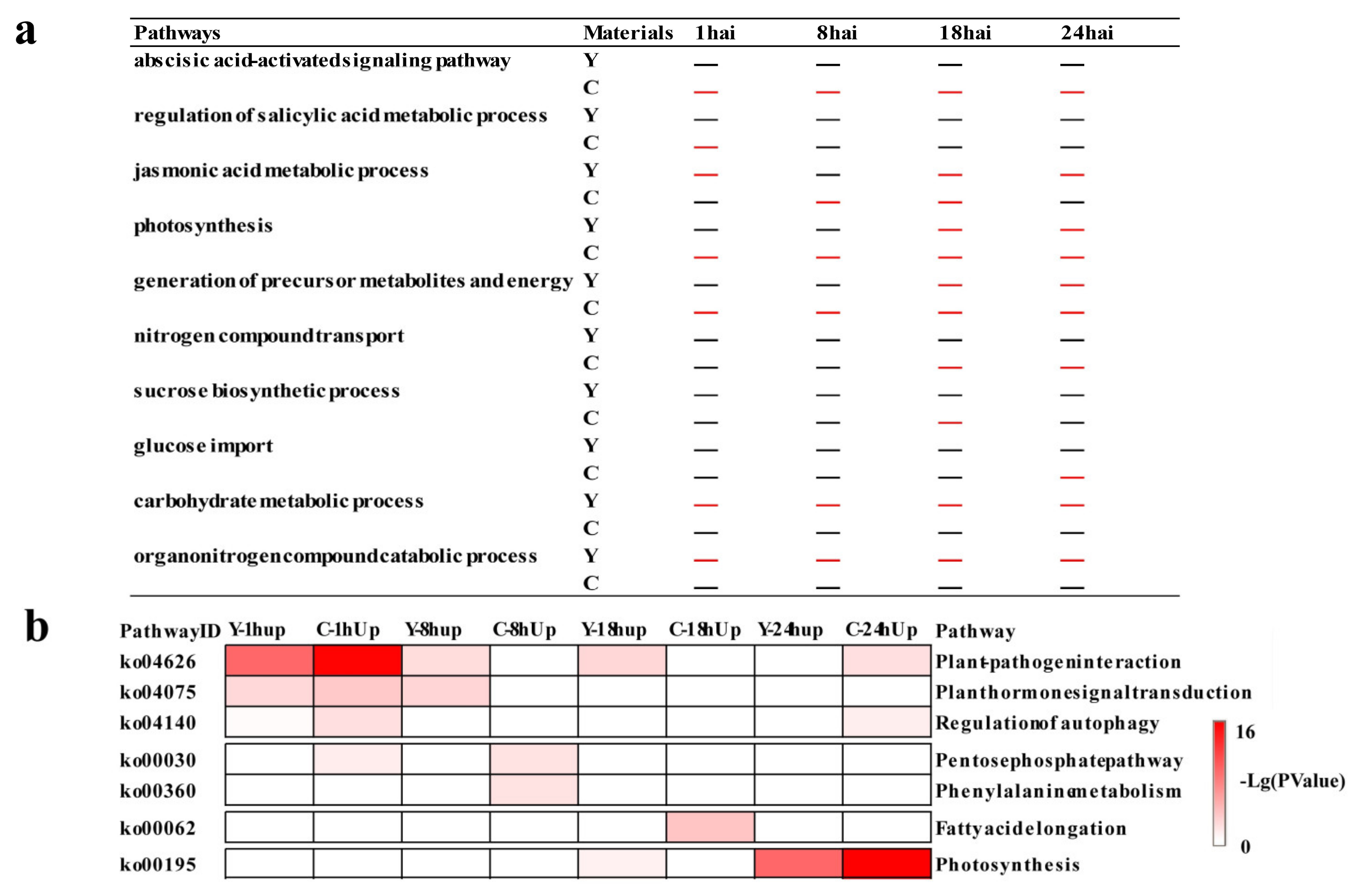
2.3. Temporal Specificity of CMPG1-VOE in Response to Bgt Infection

To characterize the pathways regulated by CMPG1-V at different time points, temporal specificity of CMPG1-VOE was analyzed (Figure 3a). At 1 hai, “ABA and SA pathways” firstly appeared in CMPG1-VOE. SA signaling pathway was only present in CMPG1-VOE at 1 hai, and ABA-associated processes were sustained during all the tested time points in CMPG1-VOE. “Jasmonic acid (JA) pathway” was induced in CMPG1-VOE at 8 hai, while it was induced at 1 hai and sustained from 18 to 24 hai in Yangmai 158 after Bgt infection. Moreover, transcripts in “photosynthesis” and “generation of precursor metabolites and energy” were significantly upregulated in CMPG1-VOE at 1 hai and thereafter. From 18 hai to 24 hai, the “nitrogen compound transport” was specifically enriched in CMPG1-VOE. The “sucrose biosynthetic process” and “glucose import” were enriched specifically in CMPG1-VOE at 18 hai and 24 hai, respectively. In Yangmai 158 at 18 and 24 hai, there was no significant enrichment of the above three pathways, but “photosynthesis” and “generation of precursor metabolites and energy” were specifically enriched. The “carbohydrate metabolic process” and “organonitrogen compound catabolic process” were activated in Yangmai 158, while not in CMPG1-VOE from 1 hai to 24 hai.
KEGG enrichment analysis was used to classify the crucial biological processes and cardinal signaling pathways (Figure 3b). At 1 hai, the “plant–pathogen interaction” and “plant hormone signal transduction” were obviously upregulated in CMPG1-VOE, and the transcription level in “regulation of autophagy” increased twofold more in CMPG1-VOE than in Yangmai 158. At 8 hai and the later infection stages, the “phenylalanine metabolism” was activated and increased up to threefold more in CMPG1-VOE than in Yangmai 158. Metabolic processes such as “fatty acid elongation” increased more than twofold in CMPG1-VOE. With the infection going on, “photosynthesis” was activated both in CMPG1-VOE and Yangmai 158 at 24 hai, but more significantly in CMPG1-VOE. The downregulated DEGs were mainly enriched in “glycine, serine, and threonine metabolism”, “ABC transporters”, “isoflavonoid biosynthesis”, and “glutathione metabolism” (Figures S5 and S6).

2.4. Phytohormone Signaling Was Rapidly Reprogrammed in CMPG1-VOE after Bgt Infection

Transcription analysis indicated obvious biosynthesis and signaling changes in ABA and SA pathways in CMPG1-VOE in response to E26 infection. For the ABA pathway, its degradation gene CYP707A2 decreased more than threefold, while its synthesis gene ABA3 increased more than 4.5-fold at 18 hai in CMPG1-VOE (Figure 4a). More, PP2C, a negative regulatory gene in ABA signaling, was downregulated in CMPG1-VOE for all the analyzed infection processes, while transcripts of SnRK2 and SnRK3 were activated evidently in CMPG1-VOE in all infectious time points (Figure 4a). SnRK2.1 increased more than twofold and SnRK2.5 increased more than fivefold in CMPG1-VOE than in Yangmai 158. Besides, CIPK23 (SnRK3) increased more than sevenfold. The qRT-PCR also validated the RNA-seq data. As shown in Figure 5, the expression level of synthesis gene ABA3 peaked at 18 hai in CMPG1-VOE, not in Yangmai 158; PP2C decreased in CMPG1-VOE from 18 hai to 24 hai, while SnRK2.1 increased to the maximum at 18 hai and SnRK2.5 peaked at 8 hai in CMPG1-VOE; and the expression level of CIPK23 maximized at 8 hai in CMPG1-VOE, not in Yangmai 158.
For the SA pathway, SA synthesis and transduction pathway were distinctly more upregulated in CMPG1-VOE. Transcript levels of critical genes for SA synthesis, SAR DEFICIENT1 (SARD1) and UDP-glycosyltransferase (UGT), were obviously increased in CMPG1-VOE when infected by Bgt. The UGT was threefold higher in CMPG1-VOE from 1 hai to 18 hai and SARD1 was threefold more in CMPG1-VOE than in Yangmai 158 from 1 hai to 18 hai (Figure 4b). The activation of the SA signal pathway was shown by the TGA1 expression increasing more than sixfold in CMPG1-VOE from 1 hai to 18 hai; the PR was more than fourfold in CMPG1-VOE at 8 hai and increased to ninefold at 24 hai (Figure 4b), while no obvious induction of these genes was observed in Yangmai 158 (Figure 4b). qRT-PCR of TaTGA1 and TaPR1 validated the results from the RNA-seq data (Figure 5). The expression level of TaTGA1 peaked at 24 hai in CMPG1-VOE and TaPR1 expression rose in CMPG1-VOE from 18 hai to 24 hai, which were more than those in Yangmai 158.
In addition, transcription levels of COI1 in JA signaling and ETR1 in ET signaling were upregulated in CMPG1-VOE (Figure 4c,d). The expression levels of COI1 and ETR1 maximized at 18 hai in CMPG1-VOE, which was more than those in Yangmai 158 (Figure 5). The upregulated hormone signaling and associated DEGs were also observed in CMPG1-VOE or Yangmai 158 tested with E31 (Figure S7).

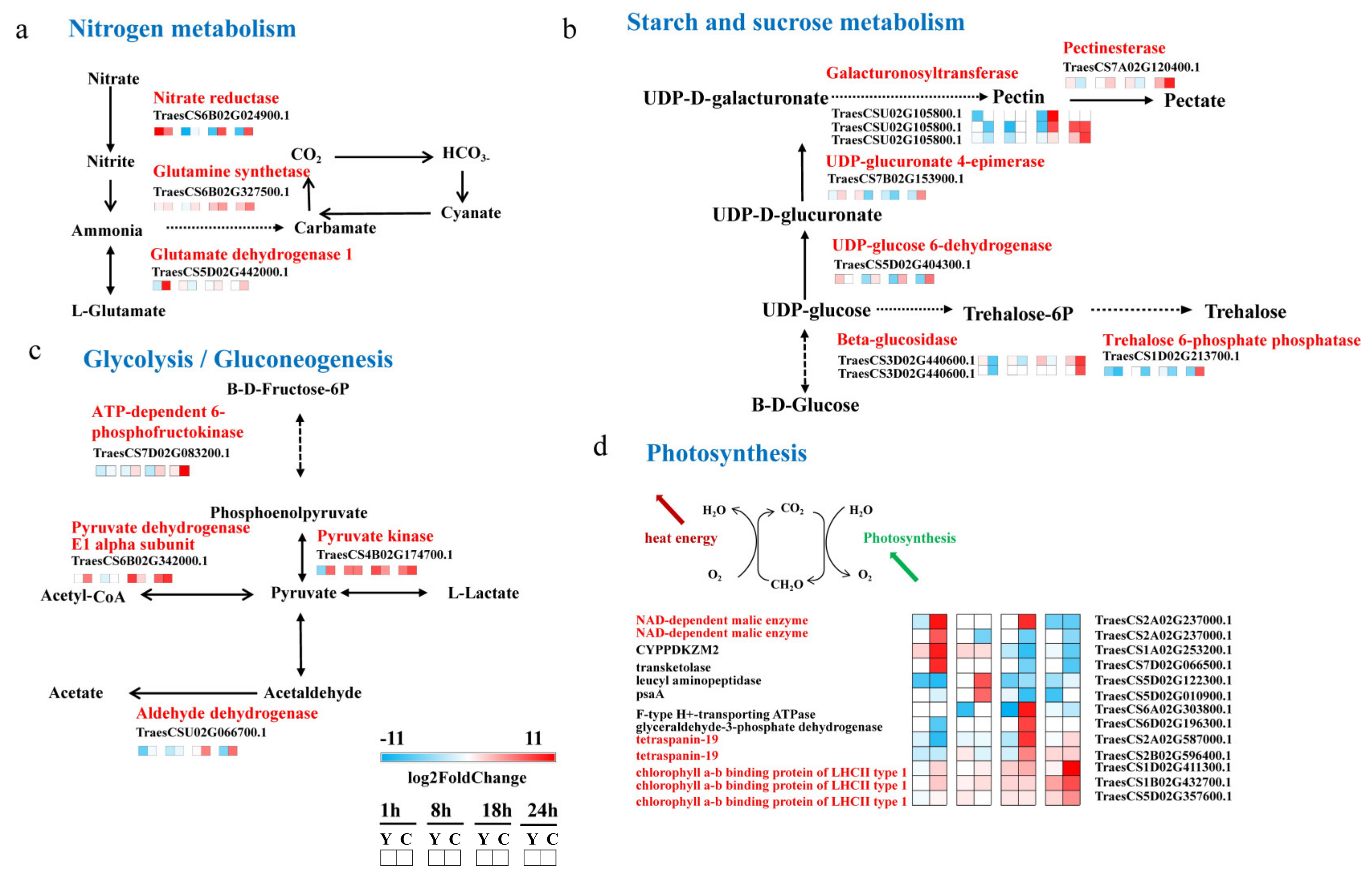
2.5. CMPG1-V Activates Conspicuous Energy Metabolic Signaling during Bgt Infection

Nitrogen assimilation genes nitrate reductase (NR) and glutamine synthetase (GS) were induced apparently in CMPG1-VOE (Figure 6a). The transcript levels of NR and GS increased more than fivefold and threefold in CMPG1-VOE, respectively. Expression levels of NR and GS increased to the maximum at 24 hai in CMPG1-VOE, which were more than those in Yangmai 158 (Figure 7). Glutamate dehydrogenase 1 (GDH1) transcripts rose up to more than sixfold in CMPG1-VOE compared with Yangmai 158. The starch and sucrose metabolism was enriched from 18 hai to 24 hai, and the transcripts of galacturonosyltransferase (GAUT) increased fivefold in CMPG1-VOE. This consisted of the pectinesterase, UDP-glucuronate 4-epimerase (GAE), UDP-glucose 6-dehydrogenase (UGDH), beta-glucosidase (GLUC), and trehalose 6-phosphatephosphatase (TPP), whose transcripts increased fourfold and reached their peaks at 24 hai (Figure 6b). As shown in Figure 7, expression levels of GLUC and TPP rose to the top at 24 hai in CMPG1-VOE, which were more than those in Yangmai 158 (Figure 7).
CMPG1-V activated the glycolysis/gluconeogenesis pathway. With the infection going on, the transcription levels of ATP-dependent 6-phosphofructokinase (pfkA), pyruvate kinase (PK), pyruvate dehydrogenase E1 alpha subunit (PDHA), and aldehyde dehydrogenase (ALDH) increased prominently in CMPG1-VOE (Figure 6c). The transcripts of PfkA increased fourfold in CMPG1-VOE, and those of PK and PDHA showed a similar pattern. The aldehyde dehydrogenase transcripts were fourfold more from 18 hai until 24 hai. The expression level of pfkA peaked at 24 hai in CMPG1-VOE, while ALDH maximized in CMPG1-VOE at 18 hai, not in Yangmai 158 (Figure 7). In addition, transcription of NAD-dependent malic enzyme (NAD-ME) and tetraspanin-19 (TSPAN) associated with photosynthesis were activated. Their expression levels were more than sixfold in CMPG1-VOE compared with those in Yangmai 158 (Figure 6d). The chlorophyll a-b binding protein (LHCII) increased sevenfold in CMPG1-VOE. The expression level of TSPAN maximized at 18 hai in CMPG1-VOE, LHCII increased to a maximum in CMPG1-VOE at 24 hai, not in Yangmai 158. The NAD-ME had a significant increase in CMPG1-VOE from 1 hai to 8 hai, which was more than that in Yangmai 158 (Figure 7). Similar results were observed in CMPG1-VOE or Yangmai 158 tested with E31 for the induction or inactivation of nitrogen and glycolysis/gluconeogenesis metabolism (Figure S8).

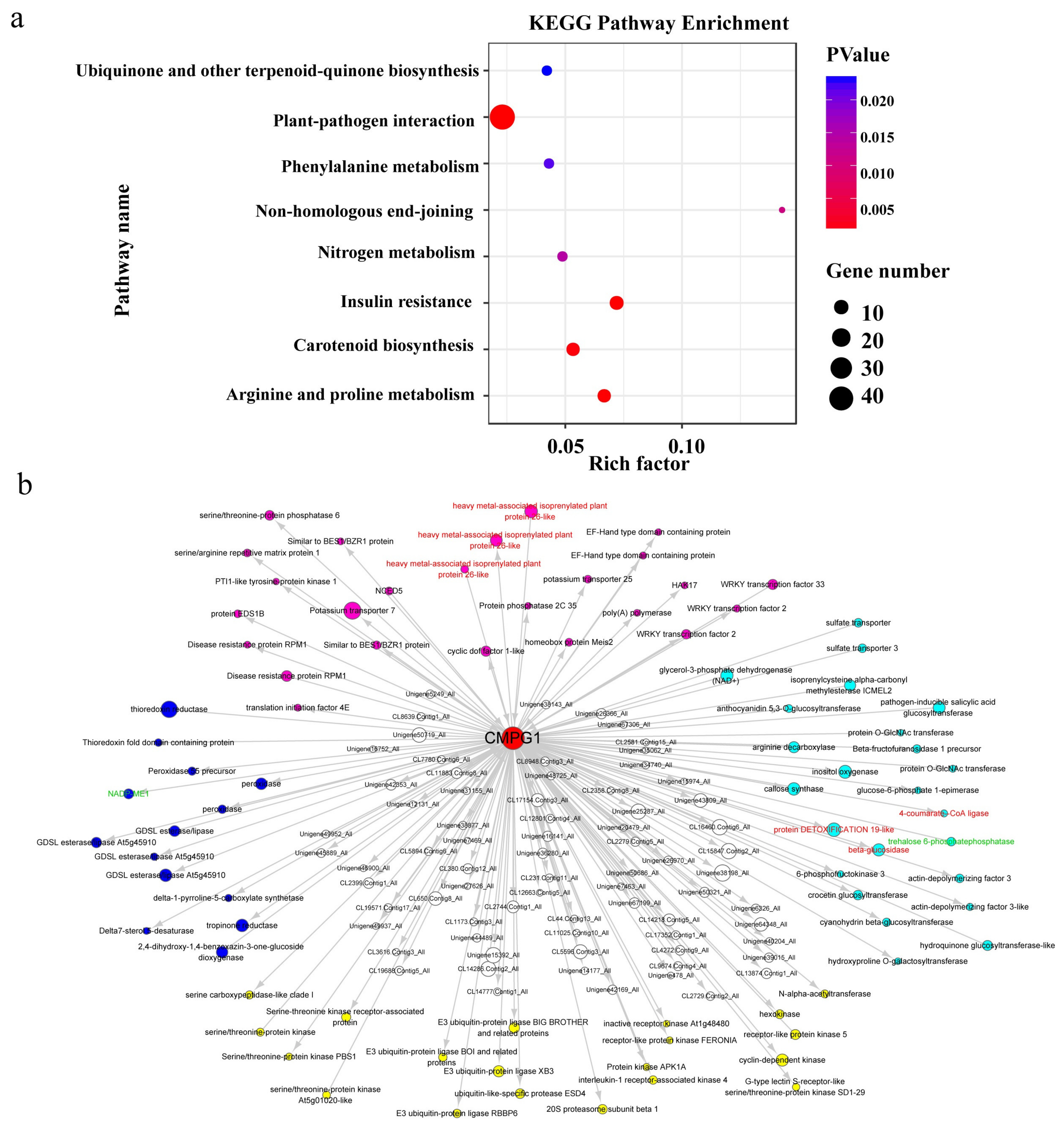
2.6. Weighted Gene Co-Expression Network (WGCNA) of Pm Resistance Regulated by CMPG1-V

To identify crucial regulatory factors or pathways in the CMPG1-V defense network, the co-expression data were analyzed from all DEGs in CMPG1-VOE and Yangmai 158 through WGCNA. The network contained 16,662 DEGs after filtering by the coefficient of variation. As shown in Figure S9, 28 module clusterings were set up and the samples collected at the four infectious time points were corresponded to each module (Figure S9a,b).
In total, 2259 DEGs enriched in all modules and CMPG1-V were classified into the pink module (Figure S9c). KEGG enrichment analysis revealed DEGs in the pink module were closely related to plant–pathogen interaction, nitrogen metabolism, phenylalanine metabolism, and ubiquinone and terpenoid-quinone biosynthesis (Figure 8a). Twenty-six DEGs shown as rose red nodes could be categorized into plant–pathogen interaction, including NCED5 and PP2C35 in the ABA pathway, EDS1B in the SA pathway, and RPM1 and PTI1-like tyrosine-protein kinase 1 in disease resistance (Figure 8b). Twenty-four DEGs shown as light blue nodes were associated with phytohormone metabolism, including beta-glucosidase (GLUC), 6-phosphofructokinase 3 (PFK3), and trehalose 6-phosphatephosphatase (TPP) of starch and sucrose metabolism, 4-coumarate-CoA ligase (4CL) of lignin metabolism, and anthocyanidin 5,3-O-glucosyltransferase (GT) of anthocyanin biosynthetic pathway (Figure 8b). In addition, 20 yellow nodes were associated with protein modification and 14 blue nodes involved in oxidation–reduction. Definitively, CMPG1-V interacting proteins previously identified by yeast two-hybrid assay were also identified, including 4-coumarate-CoA ligase-like, beta-glucosidase, protein DETOXIFICATION 19-like, and heavy metal-associated isoprenylated plant protein (Table S2). The DEGs including beta-glucosidase, trehalose 6-phosphatephosphatase, and NAD-ME were confirmed by qRT-PCR (Figure 7). Taken together, Table 1 lists the probable DEGs participating in the CMPG1-V defense network including protein phosphorylation modification, flavonoid metabolism, ABA/SA signaling, and oxidation homeostasis.

2.7. The Chromosomal Distribution of Candidate Genes

The chromosome distributions of the DEGs related to plant hormone pathways, energy metabolic signaling pathways, and in the network of the CMPG1-V module were surveyed through in silico mapping using Chinese Spring genomic sequences (http://www.wheatgenome.org/) as reference (Figure S10 and Table S3). In addition, all of them have homologous genes in Arabidopsis, with a similarity reached up to 80% (Table S3). As is shown in Figure S10, the 22 DEGs in plant hormone pathways were assigned to 11 different chromosomes; 29 DEGs in energy metabolism were assigned to 14 different chromosomes; and the remaining 23 DEGs in the network of CMPG1-V were assigned to 16 different chromosomes. Chromosome distribution analysis of the DEGs might facilitate the association study between these DEGs and previously reported genes in resistance to powdery mildew.

3. Discussion

Wheat powdery mildew is one of the most destructive diseases of wheat, causing extensive yield losses worldwide [17]. Due to the host or race specificity of the wheat–Bgt interaction, a new virulent race could rapidly evolve and emerge. Here, CMPG1-V, an E3 ligase gene from H. villosa, showed BSR to powdery mildew when overexpressed in wheat [18], thus the transgenic line CMPG1-VOE and its receptor variety Yangmai 158 provide an ideal system for studying BSR during wheat–Bgt interaction. We identified 3542 DEGs, which were only specifically upregulated in CMPG1-VOE after Bgt infection.

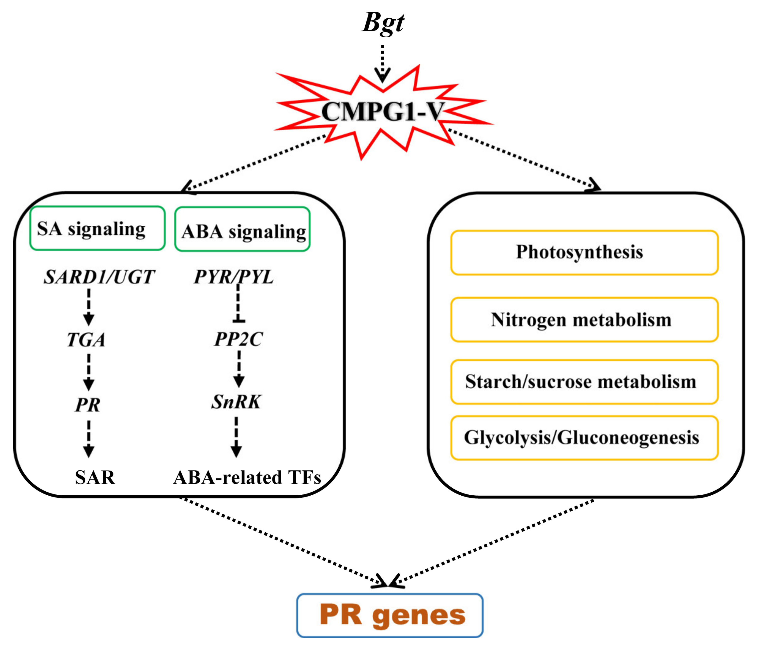
3.1. Phytohormone SA and ABA Play Important Roles in CMPG1-V-Associated Powdery Mildew Resistance

Plants are continuously exposed to diverse phytopathogenic microorganisms and have elaborated a variety of defense mechanisms to successfully avoid infection by limiting pathogen invasion and multiplication [35,36]. Plant hormone SA plays an important role in the interaction between plants and pathogens [37]. The exogenous application of SA conferred resistance against Magnaporthe oryzae [38]. SA-deficient NahG tobacco was hypersusceptible to powdery mildew (Euoidium longipes), as judged by significantly more severe powdery mildew symptoms and enhanced pathogen accumulation [39]. In a previous study, exogenous SA could increase the expression level of CMPG1-V, and TaPR1 and TaPR2 were upregulated significantly in CMPG1-VOE when infected by Bgt [18]. RNA-seq showed obvious transcription level increase of the SA synthesis genes SARD1 and UGT in CMPG1-VOE, hinting at the important role of the SA pathway in Pm resistance mediated by CMPG1-V. Arabidopsis AtSARD1 could enhance the resistance against Verticillium dahliae [40]. Overexpressing TaUGT3 enhanced fusarium head blight resistance of wheat [41]. What’s more, TGA TFs family positively regulates defense responses against biotrophic and necrotrophic pathogens [42], consistent with the transcription level of TGA accelerated in CMPG1-VOE. Thus, it will be important to elucidate the synthesis of SA during CMPG1-V regulating Bgt resistance. And what is more, the strategies targeting TAG TFs are more crucial for enhancing powdery mildew resistance during this process.
ABA, originally described for their function in response to abiotic stresses, has emerged as a crucial player in plant–pathogen interactions [43,44]. Exogenous ABA quickly induced CMPG1-V, suggesting that CMPG1-V may be involved in ABA-dependent defense response [18]. Here, we found transcripts of SnRK2 were upregulated in all infectious periods. In Arabidopsis, SnRK2.8 phosphorylated NPR1, enhancing systemic acquired resistance (SAR) [45]. OsSAPKs (SnRK2) play positive roles in response to pathogen infection in rice [46]. ABA-inducible SnRK2-type kinase OsSAPK10 phosphorylated WRKY72, released its suppression on AOS1, and enhanced resistance to Xanthomonas oryzae pv. oryzae in rice [47]. Besides, PP2C, a negative regulator in ABA signaling, was significantly downregulated in CMPG1-VOE after Bgt inoculation compared with Yangmai 158. Some rice PP2Cs subgroup K negatively regulate X. oryzae pv. oryzae infection [48]. Thus, PP2Cs are thought to play a specific role in CMPG1-V-associated defense pathways, and ABA-dependent defense signaling possibly plays a positively regulatory role in CMPG1-V resistance network.
Chromosome distribution analysis of the DEGs provided information to identify whether these DEGs were associated with previously identified genes or QTL. By gene expression comparison between resistant and susceptible durum wheat lines, a WAK2 gene in the mapped region of QFhb.mgb-2A was identified and responsible for Fusarium Head Blight resistance [49,50]. The DEGs which might be involved in CMPG1-V regulatory network have been mapped in silico onto chromosome regions of wheat (Table S3). We have found that a CIPK29 gene (Unigene49646_All) which was located in the terminal region 2BS might be associated with powdery-mildew-resistant gene Ml5323 in the interval of 2BS FL0.84-1.00 [49]. CIPKs were reported to play roles in the ABA signaling pathway [51]. This finding might provide clues to the cloning and elucidation of the resistant mechanism mediated by Ml5323.

3.2. CMPG1-V Reprogrammed Starch and Sucrose Metabolism/Photosynthesis in Response to Bgt Infection

When a plant defends against pathogen invasion, levels of resistance in the whole plant are influenced by systemic signals mediated by plant hormones [52]. Plant hormones can coordinate various signal pathways to mediate metabolism under pathogen infection [1]. NR-mediated NO generation plays a key role in protecting plants from abiotic stresses through activating antioxidant enzymes [53]. GS1 can be induced during development of the infectious process [54]. We observed that, in response to Bgt infection, the nitrogen assimilation genes NR and GS were upregulated apparently in CMPG1-VOE. What is more, typical stress metabolite trehalose plays an important role in regulation of defense response against pathogens. Some selected SlTPPs could be inducted by Botrytis cinerea and Pseudomonas syringae pv. tomato (Pst) DC3000 as well as defense signaling hormones [55]. We found TPP involving starch and sucrose metabolism and pfkA, PK, PDHA involved in glycolysis/gluconeogenesis were induced notably in CMPG1-VOE. The GAPDHs, multifunctional enzymes in glycolysis, have been proven to be involved in regulation of ROS, autophagy, and plant immune responses [56]. Photosynthesis also modulates plant defense responses induced by pathogen infection and abiotic signals such as light, circadian rhythm, and temperature [57]. During infection with Colletotrichum higginsianum, loss-of-function mutants of NADP-ME2 (nadp-me2) showed enhanced susceptibility [58]. The transcription levels of NAD-dependent malic enzyme, chlorophyll a-b binding protein, and tetraspanin-19 were upregulated remarkably in CMPG1-VOE, compared to those in Yangmai 158. Thus, the reprogramming of starch/sucrose metabolism and photosynthesis pathways played an important role in the CMPG1-V defense response to Bgt infection.

3.3. CMPG1-V Exerts Its Distinct Defense Response by Reprogramming a Specific Network

Plants have evolved innate immune systems that recognize the presence of potential pathogens and initiate effective defense responses. E3 ubiquitin ligase plays a crucial role in the plant immunity process [2]. There are, however, a few notable highlights or differences in resistance mechanisms against Bgt in CMPG1-V. WGCNA of CMPG1-V module revealed its close relation with phosphorylation modification, flavonoid metabolism, oxidation homeostasis, and WRKY. Crosstalk between ubiquitination and phosphorylation modification plays a key role in controlling the defense signaling events to ensure plant pathogen resistance [2]. Crosstalk between posttranslational modifications, such as ubiquitination and phosphorylation, play key roles in controlling the duration and intensity of signaling events to ensure cellular homeostasis [59]. Phosphorylation events contribute additively to the stabilization of PUB22 in response to the perception of pathogen-associated molecular patterns (PAMPs) [59]. Phosphorylation of NPR1 could switch autoubiquitination activity of BTBNPR1 to substrate ubiquitination by enhancing its affinity to targets such as TGA2 [60]. For oxidation homeostasis, E3 ubiquitin ligase can induce ROS to enhance the resistance level. Knockdown of stress inducible OsSRFP1, encoding an E3 ubiquitin ligase with transcriptional activation activity, conferred abiotic stress tolerance through enhancing antioxidant protection in rice [61]. Arabidopsis E3 ubiquitin ligase PUB13 regulated chitin receptor LYSIN MOTIF RECEPTOR KINASE5 and induced rapid responses, such as the production of ROS [62]. Moreover, other E3 ligases can interact with WRKY to enhance the resistance. The E3 ligase likely to be involved in plant immunity is UPL5, which was first identified by a yeast two-hybrid screen. UPL5 interacted with WRKY53, a transcription factor acting positively in leaf senescence [63]. The Chinese wild grapevine (Vitis pseudoreticulata) E3 ligase EIRP1 activated plant defense responses by inducing proteolysis of the VpWRKY11 transcription factor [64]. However, there are few studies about ubiquitination in plant immunity related with flavonoid metabolism. In this study, we found flavonoid metabolism may have an influence on ubiquitination in plant immunity. This will give us a new perspective to clarify the resistance function of CMPG1-V in wheat powdery mildew resistance. On all accounts, these candidates will provide extensive insights into molecular mechanisms of CMPG1-V-associated broad-spectrum powdery mildew resistance in wheat.

4. Materials and Methods

4.1. Plant Material and Fungal Isolates

Yangmai 158 is a moderate powdery-mildew-susceptible wheat variety and it is susceptible to Bgt isolates E26 or E31. Overexpression of CMPG1-V in Yangmai 158 enhanced its broad-spectrum resistance. Bgt isolates E26 and E31 were collected from Institute of Plant Protection, Chinese Academy of Agricultural Sciences, Beijing, China. They are maintained on seedlings of susceptible wheat variety Sumai 3 in a spore-proof greenhouse. All seedlings were grown in a growth chamber with 20 °C/16 °C (day/night), 16 h/8 h (light/dark). CMPG1-V transgenic line (CMPG1-VOE) and the receptor variety Yangmai 158 were inoculated with Bgt isolates E26 and E31 at the two-leaf stage and RNAs were isolated at 1 h, 8 h, 18 h, and 24 h before and after Bgt inoculation, followed by freezing in liquid nitrogen for subsequent RNA extraction.

4.2. RNA-Seq Library Construction and Sequencing

Total RNA extraction was performed using the Trizol reagent (Invitrogen, Waltham, MA, USA), according to the manufacturer’s instructions. RNA concentration was measured using a NanoDrop spectrophotometer and 1.2% agarose gel electrophoresis.
Sample detection and sequencing were performed by BGI-Shenzhen, Shenzhen Beijing. After extracting total RNA and treating with DNase I, Oligo(dT) was used to isolate mRNA. Mixed with the fragmentation buffer, the mRNA were fragmented. Then, cDNA was synthesized using the mRNA fragments as templates. Short fragments were purified and resolved with EB buffer for end reparation and single nucleotide A (adenine) addition. After that, the short fragments were connected with adapters. The suitable fragments were selected for the PCR amplification. During the QC steps, Agilent 2100 Bioanaylzer and ABI Step One Plus Real-Time PCR System were used in quantification and qualification of the sample library. Then, the library was sequenced using Illumina HiSeq 4000 (Illumina, San Diego, CA, USA). The raw data and processed data could be obtained from the NCBI website address. The initial short reads data sets were available at the NCBI Short Read Archive (SRA) with the accession number SRR12416499-SRR12416522.

4.3. Sequencing Reads Filtering and De Novo Assembly

To obtain clean reads, raw reads, including reads with adaptors, reads in which unknown bases represented more than 5% of the total bases, and low-quality reads (percentage of low-quality bases with a quality value ≤ 10 in more than 20% of a read), were removed. We used Trinity to perform de novo assembly with clean reads that PCR duplication removed (in order to improve the efficiency), then used Tgicl to cluster transcripts to Unigenes. Trinity: version: v2.0.6, parameters: --min_contig_length 150 --CPU 8 --min_kmer_cov 3 --min_glue 3 --bfly_opts ‘-V 5 --edge-thr=0.1 --stderr’. Tgicl: version: v2.0.6, parameters: -l 40 -c 10 -v 25 -O ‘-repeat_stringency 0.95 -minmatch 35 -minscore 35′.

4.4. Unigene Functional Annotation and Expression Calculation

NT (ftp://ftp.ncbi.nlm.nih.gov/blast/db), NR (ftp://ftp.ncbi.nlm.nih.gov/blast/db), GO (http://geneontology.org), COG (http://www.ncbi.nlm.nih.gov/COG), KEGG (http://www.genome.jp/kegg), Swiss Prot (http://ftp.ebi.ac.uk/pub/databases/swissprot), and Inter Pro (http://www.ebi.ac.uk/interpro) are functional databases. We used Blast [65] align Unigenes to NT, NR, COG, KEGG, and Swiss Prot to get the annotation, used Blast2GO [66] with NR annotation to get the GO annotation, and used InterProScan5 [67] to get the Inter Pro annotation. We mapped clean reads to Unigenes using Bowtie2 [68], and then calculated gene expression level with RSEM. Blast: version: v2.2.23, parameters: default, website: http://blast.ncbi.nlm.nih.gov/Blast.cgi. Blast2GO: version: v2.5.0, parameters: default, website: https://www.blast2go.com. InterProScan5: version: v5.11-51.0, parameters: default, website: https://code.google.com/p/interproscan/wiki/Introduction. Bowtie2: version: v2.2.5, parameters: -q --phred64 --sensitive --dpad 0 --gbar 99999999 --mp 1,1 --np 1 --score-min L,0,-0.1 -I 1 -X 1000 --no-mixed --no-discordant -p 1 -k 200, website: http://bowtie-bio.sourceforge.net/ Bowtie2 /index.shtml. RSEM: version: v1.2.12, parameters: default, website: http://deweylab.biostat.wisc.edu/RSEM.

4.5. Detection and Analysis of Differentially Expressed Gene

We detected DEGs with PossionDis as requested and parameters were set as Fold Change ≥ 2.00 and FDR ≤ 0.001. With the GO annotation result, we classified DEGs according to official classification, and we also performed GO functional enrichment using phyper, a function of R (version: v3.4.2). With the KEGG annotation result, we classified DEGs according to official classification, and we also performed pathway functional enrichment using phyper, a function of R. We calculated false discovery rate (FDR) for each p-value; in general, the terms with FDR not larger than 0.001 were defined as significantly enriched. Hierarchical clustering and heat map generation were performed in R. The gene expression data were log2-transformed and then quantile-normalized prior to generating the heat map for direct comparison of the data.

4.6. Weighted Gene Co-Expression Network Analysis and Visualization

Co-expression networks were constructed using the WGCNA package in R. We used the log2-transformed FPKM values, and replaced values smaller than one by zero. In total, 16,662 genes were used for the WGCNA analysis. The modules were obtained using the automatic network construction function block wise Modules with default settings. Parameters were set as follows: the power was 20, TOM-Type was unsigned, min Module Size was 30, and merge Cut Height was 0.25. We inferred an undirected, weighted network. Within the network, clusters of genes with similar expression patterns, called modules, were then inferred using a clustering of the Topological Overlay Matrix. The eigengene value was calculated for each module and 16,662 genes were clustered into 27 specific modules. The networks were visualized using Cytoscape _v.3.5.1.

4.7. Quantitative Real-Time Polymerase Chain Reaction (qRT-PCR) Assay

All selected genes were used for SYBR green real-time RT-PCR; primers are listed in Table S4. The Tubulin gene was used as the internal control gene. The qRT-PCR reaction for target gene transcript amplification was carried out in a final volume of 20 μL containing 20 ng cDNA, 0.2 μM of each primer, 2× AceQ qPCR SYBR Green Master Mix (Vazyme, Nanjing, China). The PCR reaction conditions were denaturation at 95 °C for 5 min followed by 40 cycles of 95 °C for 10 s, annealing at the appropriate temperature (from 57 to 61 °C) for 30 s, extension at 72 °C for 30 s, followed by 95 °C for 15 s, 60 °C for 1 min, then 95 °C for 15 s to obtain melt curves to ensure primer specificity. All reactions were done in triplicate. Reactions were performed on a Bio-Rad IQ single-color Real-Time PCR detection System (Bio-Rad, Hercules, CA, USA). The data were compiled from the mean Ct values of all the replicates after normalizing with the Ct values of the endogenous control. The relative expression level in terms of fold change was calculated using the 2ΔΔCT method [69]. All the qRT-PCRs had three biological replicates.

4.8. Statistical Analysis

All statistical analyses were performed using SPSS version 13.0, and one-way ANOVA was performed with a homogeneity of variance test, followed by an LSD test to check for quantitative differences between treatments. p < 0.05 was set as the significance cut-off.

5. Conclusions

In this study, we analyzed the temporal specificity of Pm resistance regulated by CMPG1-V at the whole genome level, represented by four time points after Bgt infection. We suspected that when the pathogen attacked, the plant activated CMPG1-V, then enhanced hormone defense signaling through SA and ABA signaling. Activation of C/N and photosynthesis metabolism provided energy during wheat–Bgt interaction, which affected the expression of downstream PR genes (Figure 9). Moreover, phosphorylation modification probably regulated different transcription of genes in defense signaling and related metabolisms. Our findings point to strengthening the utility of CMPG1-V in enhancing BSR to powdery mildew.

Supplementary Materials

Supplementary materials can be found at https://www.mdpi.com/1422-0067/21/17/5967/s1.

Author Contributions

X.W., J.X., H.W., Z.W., and J.L. conceived and designed the study; J.L. analyzed the data; J.L., Y.H., X.Z., H.Z., and M.L. collected the plant materials; J.L., Z.W., L.W., Y.C., and Z.Y. performed the experiments; J.L., L.S., and X.W. wrote the manuscript. All authors have read and agreed to the published version of the manuscript.

Funding

This work was supported by the National Key Research and Development program (2016YFD0101004, 2016YFD0102001-004), the National Science Foundation of China (Nos. 31471490, 31661143005, 31290213), the National Natural Science Foundation of China (No. 91935304 and No.31971943), the Program of Introducing Talents of Discipline to Universities (No. B08025), the 333 Talent Project of Jiangsu Province, the Shanghai Agriculture Applied Technology Development Program, China Grant (No. Z201502), the Special Fund of Jiangsu Province for the Transformation of Scientific and Technological Achievements (BA2017138), the Creation of Major New Agricultural Varieties in Jiangsu Province (PZCZ201706), the SAAS Program for Excellent Research Team, and the Science and Technology Service Programs of the Chinese Academy of Sciences (KFJ-STS-ZDTP-002), the Jiangsu Agricultural Technology System (JATS) (No. 2019429), the Key Research and Development Major Project of Ningxia Hui Autonomous Region (No. 2019BBF02022-04) and the‘948’ Project of Ministry of Agriculture (2015-Z41).

Conflicts of Interest

The authors declare no conflict of interest. The funders had no role in the design of the study; in the collection, analysis, or interpretation of the data; in the writing of the manuscript; and in the decision to publish the results.

Abbreviations

4CL4-coumarate-CoA ligase
ABAabscisic acid
AGTappressorial germ tube
ALDHaldehyde dehydrogenase
BgtBlumeria graminis f. sp. tritici
bpbase pair
BSRbroad-spectrum resistance
DEGdifferentially expressed gene
ETethylene
GAEUDP-glucuronate 4-epimeras
GAUTgalacturonosyltransferase
GDH1glutamate dehydrogenase 1
GLUCbeta-glucosidase
GOGene Ontology
GSglutamine synthetase
GTanthocyanidin 5,3-O-glucosyltransferase
HAhyphal appressoria
JAjasmonic acid
KEGGKyoto Encyclopedia of Genes and Genomes
LHCIIchlorophyll a-b binding protein
NAD-MENAD-dependent malic enzyme
NRnitrate reductase
PDHApyruvate dehydrogenase E1 alpha subunit
PFK36-phosphofructokinase 3
pfkAATP-dependent 6-phosphofructokinase
PGTprimary germ tube
PKpyruvate kinase
Pmpowdery mildew
PRpathogenesis-related gene
PUBplant U-box type E3 ubiquitin ligases
RNA-seqRNA-sequencing
SAsalicylic acid
SARD1SAR DEFICIENT1
SnRKSNF1-related protein kinase
TPPtrehalose 6-phosphatephosphatase
TSPANtetraspanin-19
UGDHUDP-glucose 6-dehydrogenase
UGTUDP-glycosyltransferase
WGCNAWeighted Gene Co-Expression Network Analysis

References

  1. Berens, M.L.; Berry, H.M.; Mine, A.; Argueso, C.T.; Tsuda, K. Evolution of hormone signaling networks in plant defense. Annu. Rev. Phytopathol. 2017, 55, 401–425. [Google Scholar] [CrossRef] [PubMed]
  2. Mithoe, S.C.; Menke, F.L. Regulation of pattern recognition receptor signalling by phosphorylation and ubiquitination. Curr. Opin. Plant. Biol. 2018, 45, 162–170. [Google Scholar] [CrossRef] [PubMed]
  3. Thines, B.; Katsir, L.; Melotto, M.; Niu, Y.; Mandaokar, A.; Liu, G.; Nomura, K.; He, S.Y.; Howe, G.A.; Browse, J. JAZ repressor proteins are targets of the SCF(COI1) complex during jasmonate signalling. Nature 2007, 448, 661–665. [Google Scholar] [CrossRef]
  4. Pauwels, L.; Ritter, A.; Goossens, J.; Durand, A.N.; Liu, H.; Gu, Y.; Geerinck, J.; Boter, M.; Vanden Bossche, R.; De Clercq, R.; et al. The RING E3 ligase KEEP ON GOING modulates JASMONATE ZIMDOMAIN12 stability. Plant. Physiol. 2015, 169, 1405–1417. [Google Scholar] [CrossRef] [Green Version]
  5. Shen, Q.; Hu, T.; Bao, M.; Cao, L.; Zhang, H.; Song, F.; Xie, Q.; Zhou, X. Tobacco RING E3 ligase NtRFP1 mediates ubiquitination and proteasomal degradation of a geminivirus-encoded betaC1. Mol. Plant. 2016, 9, 911–925. [Google Scholar] [CrossRef] [Green Version]
  6. Boyle, P.; Le Su, E.; Rochon, A.; Shearer, H.L.; Murmu, J.; Chu, J.Y.; Fobert, P.R.; Després, C. The BTB/POZ domain of the Arabidopsis disease resistance protein NPR1 interacts with the repression domain of TGA2 to negate its function. Plant. Cell 2009, 21, 3700–3713. [Google Scholar] [CrossRef] [Green Version]
  7. Liu, J.; Li, H.; Miao, M.; Tang, X.; Giovannoni, J.; Xiao, F.; Liu, Y. The tomato UV-damaged DNA-binding protein-1 (DDB1) is implicated in pathogenesis related (PR) gene expression and resistance to Agrobacterium tumefaciens. Mol. Plant. Pathol. 2012, 13, 123–134. [Google Scholar] [CrossRef]
  8. Zhang, L.; Du, L.; Shen, C.; Yang, Y.; Poovaiah, B.W. Regulation of plant immunity through ubiquitin-mediated modulation of Ca2+-calmodulin-AtSR1/CAMTA3 signaling. Plant J. 2014, 78, 269–281. [Google Scholar] [CrossRef]
  9. Cao, Y.; Yang, Y.; Zhang, H.; Li, D.; Zheng, Z.; Song, F. Overexpression of a rice defense-related F-box protein gene OsDRF1 in tobacco improves disease resistance through potentiation of defense gene expression. Physiol. Plant. 2008, 134, 440–452. [Google Scholar] [CrossRef]
  10. Lin, S.S.; Martin, R.; Mongrand, S.; Vandenabeele, S.; Chen, K.C.; Jang, I.C.; Chua, N.H. RING1 E3 ligase localizes to plasma membrane lipid rafts to trigger FB1-induced programmed cell death in Arabidopsis. Plant J. 2008, 56, 550–561. [Google Scholar] [CrossRef]
  11. Lee, D.H.; Choi, H.W.; Hwang, B.K. The pepper E3 ubiquitin ligase RING1 gene, CaRING1, is required for cell death and the salicylic acid-dependent defense response. Plant Physiol. 2011, 156, 2011–2025. [Google Scholar] [CrossRef] [PubMed] [Green Version]
  12. Sato, T.; Maekawa, S.; Yasuda, S.; Domeki, Y.; Sueyoshi, K.; Fujiwara, M.; Fukao, Y.; Goto, D.B.; Yamaguchi, J. Identification of 14-3-3 proteins as a target of ATL31 ubiquitin ligase, a regulator of the C/N response in Arabidopsis. Plant J. 2011, 68, 137–146. [Google Scholar] [CrossRef] [PubMed]
  13. Sato, T.; Maekawa, S.; Yasuda, S.; Sonoda, Y.; Katoh, E.; Ichikawa, T.; Nakazawa, M.; Seki, M.; Shinozaki, K.; Matsui, M.; et al. CNI1/ATL31, a RING-type ubiquitin ligase that functions in the carbon/nitrogen response for growth phase transition in Arabidopsis seedlings. Plant J. 2009, 60, 852–864. [Google Scholar] [CrossRef] [PubMed]
  14. Maekawa, S.; Sato, T.; Asada, Y.; Yasuda, S.; Yoshida, M.; Chiba, Y.; Yamaguchi, J. The Arabidopsis ubiquitin ligases ATL31 and ATL6 control the defense response as well as the carbon/nitrogen response. Plant. Mol. Biol. 2012, 79, 217–227. [Google Scholar] [CrossRef]
  15. Godfray, H.C.; Beddington, J.R.; Crute, I.R.; Haddad, L.; Lawrence, D.; Muir, J.F.; Pretty, J.; Robinson, S.; Thomas, S.M.; Toulmin, C. Food security: The challenge of feeding 9 billion people. Science 2010, 327, 812–818. [Google Scholar] [CrossRef] [Green Version]
  16. Savary, S.; Willocquet, L.; Pethybridge, S.J.; Esker, P.; McRoberts, N.; Nelson, A. The global burden of pathogens and pests on major food crops. Nat. Ecol. Evol. 2019, 3, 430–439. [Google Scholar] [CrossRef]
  17. Menardo, F.; Praz, C.R.; Wyder, S.; Ben-David, R.; Bourras, S.; Matsumae, H.; McNally, K.E.; Parlange, F.; Riba, A.; Roffler, S.; et al. Hybridization of powdery mildew strains gives rise to pathogens on novel agricultural crop species. Nat. Genet. 2016, 48, 201–205. [Google Scholar] [CrossRef] [Green Version]
  18. Zhu, Y.; Li, Y.; Fei, F.; Wang, Z.; Wang, W.; Cao, A.; Liu, Y.; Han, S.; Xing, L.; Wang, H.; et al. E3 ubiquitin ligase gene CMPG1-V from Haynaldia villosa L. contributes to powdery mildew resistance in common wheat (Triticum aestivum L.). Plant. J. 2015, 84, 154–168. [Google Scholar] [CrossRef] [Green Version]
  19. Zhang, Z.; Henderson, C.; Perfect, E.; Carver, T.L.; Thomas, B.J.; Skamnioti, P.; Gurr, S.J. Of genes and genomes, needles and haystacks: Blumeria graminis and functionality. Mol. Plant. Pathol. 2005, 6, 561–575. [Google Scholar] [CrossRef]
  20. Lalonde, S.; Sero, A.; Pratelli, R.; Pilot, G.; Chen, J.; Sardi, M.; Parsa, S.A.; Kim, D.Y.; Acharya, B.R.; Stein, E.V.; et al. A membrane protein/signaling protein interaction network for Arabidopsis version AMPv2. Front. Physiol. 2010, 1, 24. [Google Scholar] [CrossRef] [Green Version]
  21. Zhang, Y.; Feng, S.; Chen, F.; Chen, H.; Wang, J.; McCall, C.; Xiong, Y.; Deng, X.W. Arabidopsis DDB1-CUL4 ASSOCIATED FACTOR1 forms a nuclear E3 ubiquitin ligase with DDB1 and CUL4 that is involved in multiple plant developmental processes. Plant. Cell. 2008, 6, 1437–1455. [Google Scholar] [CrossRef] [PubMed] [Green Version]
  22. Benschop, J.J.; Mohammed, S.; O’Flaherty, M.; Heck, A.J.; Slijper, M.; Menke, F.L. Quantitative phosphoproteomics of early elicitor signaling in Arabidopsis. Mol. Cell Proteomics. 2007, 6, 1198–1214. [Google Scholar] [CrossRef] [PubMed] [Green Version]
  23. Russell, A.R.; Ashfield, T.; Innes, R.W. Pseudomonas syringae effector AvrPphB suppresses AvrB-induced activation of RPM1 but not AvrRpm1-induced activation. Mol. Plant. Microbe Interact. 2015, 28, 727–735. [Google Scholar] [CrossRef] [Green Version]
  24. Parsons, H.T.; Christiansen, K.; Knierim, B.; Carroll, A.; Ito, J.; Batth, S.T.; Smith-Moritz, A.M.; Morrison, S.; McInerney, P.; Hadi, M.Z.; et al. Isolation and proteomic characterization of the Arabidopsis Golgi defines functional and novel components involved in plant cell wall biosynthesis. Plant. Physiol. 2012, 159, 12–26. [Google Scholar] [CrossRef] [PubMed] [Green Version]
  25. Duan, Q.; Liu, M.J.; Kita, D.; Jordan, S.S.; Yeh, F.L.; Yvon, R.; Carpenter, H.; Federico, A.N.; Garcia-Valencia, L.E.; Eyles, S.J.; et al. FERONIA controls pectin- and nitric oxide-mediated male-female interaction. Nature 2020, 579, 561–566. [Google Scholar] [CrossRef]
  26. Tarutani, Y.; Sasaki, A.; Yasuda, M.; Nakashita, H.; Yoshida, S.; Yamaguchi, I.; Suzuk, Y. Identification of three clones which commonly interact with the kinase domains of highly homologous two receptor-like kinases, RLK902 and RKL1. Biosci. Biotechnol. Biochem. 2004, 68, 2581–2587. [Google Scholar] [CrossRef] [Green Version]
  27. Kobayashi, Y.; Fukuzawa, N.; Hyodo, A.; Kim, H.; Mashiyama, S.; Ogihara, T.; Yoshioka, H.; Matsuura, H.; Masuta, C.; Matsumura, T.; et al. Role of salicylic acid glucosyltransferase in balancing growth and defence for optimum plant fitness. Mol. Plant. Pathol. 2020, 21, 429–442. [Google Scholar] [CrossRef]
  28. Lin, J.S.; Huang, X.X.; Li, Q.; Cao, Y.; Bao, Y.; Meng, X.F.; Li, Y.J.; Fu, C.; Hou, B.K. UDP-glycosyltransferase 72B1 catalyzes the glucose conjugation of monolignols and is essential for the normal cell wall lignification in Arabidopsis thaliana. Plant J. 2016, 88, 26–42. [Google Scholar] [CrossRef] [Green Version]
  29. Zhang, G.Z.; Jin, S.H.; Jiang, X.Y.; Dong, R.R.; Li, P.; Li, Y.J.; Hou, B.K. Ectopic expression of UGT75D1, a glycosyltransferase preferring indole-3-butyric acid, modulates cotyledon development and stress tolerance in seed germination of Arabidopsis thaliana. Plant. Mol. Biol. 2016, 90, 77–93. [Google Scholar] [CrossRef]
  30. Woo, H.H.; Jeong, B.R.; Hirsch, A.M.; Hawes, M.C. Characterization of Arabidopsis AtUGT85A and AtGUS gene families and their expression in rapidly dividing tissues. Genomics 2007, 90, 143–153. [Google Scholar] [CrossRef] [Green Version]
  31. Gao, Q.M.; Venugopal, S.; Navarre, D.; Kachroo, A. Low oleic acid-derived repression of jasmonic acid-inducible defense responses requires the WRKY50 and WRKY51 proteins. Plant Physiol. 2011, 155, 464–476. [Google Scholar] [CrossRef] [PubMed] [Green Version]
  32. Jiang, W.; Yu, D. Arabidopsis WRKY2 transcription factor mediates seed germination and postgermination arrest of development by abscisic acid. BMC Plant. Biol. 2009, 9, 96. [Google Scholar] [CrossRef] [PubMed] [Green Version]
  33. Liu, S.; Kracher, B.; Ziegler, J.; Birkenbihl, R.P.; Somssich, I.E. Negative regulation of ABA signaling by WRKY33 is critical for Arabidopsis immunity towards Botrytis cinerea 2100. eLife 2015, 4, e07295. [Google Scholar] [CrossRef] [PubMed]
  34. Willmann, M.R.; Mehalick, A.J.; Packer, R.L.; Jenik, P.D. MicroRNAs regulate the timing of embryo maturation in Arabidopsis. Plant. Physiol. 2011, 155, 1871–1884. [Google Scholar] [CrossRef] [PubMed] [Green Version]
  35. Glazebrook, J. Contrasting mechanisms of defense against biotrophic and necrotrophic pathogens. Annu. Rev. Phytopathol. 2005, 43, 205–227. [Google Scholar] [CrossRef]
  36. Panstruga, R.; Parker, J.E.; Schulze-Lefert, P. SnapShot, plant immune response pathways. Cell 2009, 136, 1–3. [Google Scholar] [CrossRef] [Green Version]
  37. Robert-Seilaniantz, A.; Grant, M.; Jones, J.D. Hormone crosstalk in plant disease and defense: More than just jasmonate-salicylate antagonism. Annu. Rev. Phytopathol. 2011, 49, 317–343. [Google Scholar] [CrossRef]
  38. Zhang, K.L.; Liu, Q.S.; Kang, H.X.; Liu, X.M.; Chen, X.P.; Peng, Y.F.; Li, Y.H. Herbivore-induced rice resistance against rice blast mediated by salicylic acid. Insect Sci. 2020, 27, 49–57. [Google Scholar] [CrossRef]
  39. Künstler, A.; Kátay, G.; Gullner, G.; Király, L. Artificial elevation of glutathione contents in salicylic acid-deficient tobacco (Nicotiana tabacum cv. Xanthi NahG) reduces susceptibility to the powdery mildew pathogen Euoidium longipes. Plant. Biol. 2019, 22, 70–80. [Google Scholar] [CrossRef] [Green Version]
  40. Qin, J.; Wang, K.; Sun, L.; Xing, H.; Wang, S.; Li, L.; Chen, S.; Guo, H.S.; Zhang, J. The plant-specific transcription factors CBP60g and SARD1 are targeted by a verticillium secretory protein VDSCP41 to modulate immunity. eLife 2018, 7, e34902. [Google Scholar] [CrossRef]
  41. Xing, L.; Gao, L.; Chen, Q.; Pei, H.; Di, Z.; Xiao, J.; Wang, H.; Ma, L.; Chen, P.; Cao, A. Over-expressing a UDP-glucosyltransferase gene (Ta-UGT3) enhances Fusarium Head Blight resistance of wheat. Plant. Growth Regul. 2018, 84, 561–571. [Google Scholar] [CrossRef]
  42. Gatz, C. From pioneers to team players: TGA transcription factors provide a molecular link between different stress pathways. Mol. Plant. Microbe Interact. 2013, 26, 151–159. [Google Scholar] [CrossRef] [PubMed] [Green Version]
  43. Ton, J.; Flors, V.; Mauch-Mani, B. The multifaceted role of ABA in disease resistance. Trends Plant. Sci. 2009, 14, 310–317. [Google Scholar] [CrossRef] [PubMed]
  44. Mang, H.G.; Qian, W.; Zhu, Y.; Qian, J.; Kang, H.G.; Klessig, D.F.; Hua, J. Abscisic acid deficiency antagonizes high-temperature inhibition of disease resistance through enhancing nuclear accumulation of resistance proteins SNC1 and RPS4 in Arabidopsis. Plant. Cell 2012, 24, 1271–1284. [Google Scholar] [CrossRef] [PubMed] [Green Version]
  45. Lee, H.J.; Park, Y.J.; Seo, P.J.; Kim, J.H.; Sim, H.J.; Kim, S.G.; Park, C.M. Systemic immunity requires SnRK2.8-mediated nuclear import of NPR1 in arabidopsis. Plant Cell 2015, 27, 3425–3438. [Google Scholar] [CrossRef] [PubMed] [Green Version]
  46. Xu, M.R.; Huang, L.Y.; Zhang, F.; Zhu, L.H.; Zhou, Y.L.; Li, Z.K. Genome-wide phylogenetic analysis of stress-activated protein kinase genes in rice (OsSAPKs) and expression profiling in response to Xanthomonas oryzae pv. oryzicola Infection. Plant. Mol. Biol. Report. 2013, 31, 877–885. [Google Scholar] [CrossRef]
  47. Hou, Y.; Wang, Y.; Tang, L.; Tong, X.; Wang, L.; Liu, L.; Huang, S.; Zhang, J. SAPK10-mediated phosphorylation on WRKY72 releases its suppression on jasmonic acid biosynthesis and bacterial blight resistance. iScience 2019, 16, 499–510. [Google Scholar] [CrossRef] [Green Version]
  48. Akimoto-Tomiyama, C.; Tanabe, S.; Kajiwara, H.; Minami, E.; Ochiai, H. Loss of chloroplast-localized protein phosphatase 2Cs in Arabidopsis thaliana leads to enhancement of plant immunity and resistance to Xanthomonas campestris pv. campestris infection. Mol. Plant. Pathol. 2018, 19, 1184–1195. [Google Scholar] [CrossRef] [Green Version]
  49. Piarulli, L.; Gadaleta, A.; Mangini, G.; Signorile, M.A.; Pasquini, M.; Blanco, A.; Simeone, R. Molecular identification of a new powdery mildew resistance gene on chromosome 2BS from Triticum turgidum ssp. dicoccum. Plant. Sci. 2012, 196, 101–106. [Google Scholar] [CrossRef]
  50. Agata, G.; Pasqualina, C.; Stefania, L.G.; Antonio, B.; Angelica, G. Map-based cloning of QFhb.mgb-2A identifies a WAK2 gene responsible for Fusarium Head Blight resistance in wheat. Sci. Rep. 2019, 9, 6929. [Google Scholar]
  51. Cui, X.Y.; Du, Y.T.; Fu, J.D.; Yu, T.F.; Wang, C.T.; Chen, M.; Chen, J.; Ma, Y.Z.; Xu, Z.S. Wheat CBL-interacting protein kinase 23 positively regulates drought stress and ABA responses. BMC Plant. Biol. 2018, 18, 93. [Google Scholar] [CrossRef] [PubMed] [Green Version]
  52. Peng, Y.; van Wersch, R.; Zhang, Y. Convergent and divergent signaling in PAMP-triggered immunity and effector-triggered immunity. Mol. Plant. Microbe Interact. 2018, 31, 403–409. [Google Scholar] [CrossRef] [PubMed] [Green Version]
  53. Fu, Y.F.; Zhang, Z.W.; Yuan, S. Putative connections between nitrate reductase S-nitrosylation and NO synthesis under pathogen attacks and abiotic stresses. Front. Plant. Sci. 2018, 9, 474. [Google Scholar] [CrossRef] [PubMed] [Green Version]
  54. Olea, F.; Pérez-García, A.; Cantón, F.R.; Rivera, M.E.; Cañas, R.; Avila, C.; Cazorla, F.M.; Cánovas, F.M.; de Vicente, A. Up-regulation and localization of asparagine synthetase in tomato leaves infected by the bacterial pathogen Pseudomonas syringae. Plant. Cell Physiol. 2004, 45, 770–780. [Google Scholar] [CrossRef] [PubMed] [Green Version]
  55. Zhang, H.; Hong, Y.; Huang, L.; Liu, S.; Tian, L.; Dai, Y.; Cao, Z.; Huang, L.; Li, D.; Song, F. Virus-induced gene silencing-based functional analyses revealed the involvement of several putative trehalose-6-phosphate synthase/phosphatase genes in disease resistance against Botrytis cinerea and Pseudomonas syringae pv. tomato DC3000 in Tomato. Front. Plant. Sci. 2016, 47, 1176. [Google Scholar] [CrossRef]
  56. Henry, E.; Fung, N.; Liu, J.; Drakakaki, G.; Coaker, G. Beyond glycolysis: GAPDHs are multi-functional enzymes involved in regulation of ROS, autophagy, and plant immune responses. PLoS Genet. 2015, 11, e1005199. [Google Scholar] [CrossRef]
  57. Hua, J. Modulation of plant immunity by light, circadian rhythm, and temperature. Curr. Opin. Plant. Biol. 2013, 16, 406–413. [Google Scholar] [CrossRef]
  58. Voll, L.M.; Zell, M.B.; Engelsdorf, T.; Saur, A.; Wheeler, M.G.; Drincovich, M.F.; Weber, A.P.; Maurino, V.G. Loss of cytosolic NADP-malic enzyme 2 in Arabidopsis thaliana is associated with enhanced susceptibility to Colletotrichum higginsianum. New Phytol. 2012, 195, 189–202. [Google Scholar] [CrossRef]
  59. Furlan, G.; Nakagami, H.; Eschen-Lippold, L.; Jiang, X.; Majovsky, P.; Kowarschik, K.; Hoehenwarter, W.; Lee, J.; Trujillo, M. Changes in PUB22 ubiquitination modes triggered by MITOGEN-ACTIVATED PROTEIN KINASE3 dampen the immune response. Plant. Cell 2017, 29, 726–745. [Google Scholar] [CrossRef] [Green Version]
  60. Trujillo, M.; Shirasu, K. Ubiquitination in plant immunity. Curr. Opin. Plant. Biol. 2010, 13, 402–408. [Google Scholar] [CrossRef]
  61. Fang, H.; Meng, Q.; Xu, J.; Tang, H.; Tang, S.; Zhang, H.; Huang, J. Knock-down of stress inducible OsSRFP1 encoding an E3 ubiquitin ligase with transcriptional activation activity confers abiotic stress tolerance through enhancing antioxidant protection in rice. Plant. Mol. Biol. 2015, 87, 441–458. [Google Scholar] [CrossRef] [PubMed]
  62. Liao, D.; Cao, Y.; Sun, X.; Espinoza, C.; Nguyen, C.T.; Liang, Y.; Stacey, G. Arabidopsis E3 ubiquitin ligase PLANT U-BOX13 (PUB13) regulates chitin receptor LYSIN MOTIF RECEPTOR KINASE5 (LYK5) protein abundance. New Phytol. 2017, 214, 1646–1656. [Google Scholar] [CrossRef] [PubMed] [Green Version]
  63. Miao, Y.; Zentgraf, U. A HECT E3 ubiquitin ligase negatively regulates Arabidopsis leaf senescence through degradation of the transcription factor WRKY53. Plant J. 2010, 63, 179–188. [Google Scholar] [CrossRef] [PubMed]
  64. Yu, Y.; Xu, W.; Wang, J.; Wang, L.; Yao, W.; Yang, Y.; Xu, Y.; Ma, F.; Du, Y.; Wang, Y. The Chinese wild grapevine (Vitis pseudoreticulata) E3 ubiquitin ligase Erysiphe necator-induced RING finger protein 1 (EIRP1) activates plant defense responses by inducing proteolysis of the VpWRKY11 transcription factor. New Phytol. 2013, 200, 834–846. [Google Scholar] [CrossRef] [PubMed]
  65. Altschul, S.F.; Gish, W.; Miller, W.; Myers, E.W.; Lipman, D.J. Basic local alignment search tool. J. Mol. Biol. 1990, 215, 403–410. [Google Scholar] [CrossRef]
  66. Conesa, A.; Götz, S.; García-Gómez, J.M.; Terol, J.; Talón, M.; Robles, M. Blast2GO: A universal tool for annotation, visualization and analysis in functional genomics research. Bioinformatics 2005, 21, 3674–3676. [Google Scholar] [CrossRef] [Green Version]
  67. Quevillon, E.; Silventoinen, V.; Pillai, S.; Harte, N.; Mulder, N.; Apweiler, R.; Lopez, R. InterProScan: Protein domains identifier. Nucleic Acids Res. 2005, 33, W116–W120. [Google Scholar] [CrossRef] [Green Version]
  68. Langmead, B.; Salzberg, S. Fast gapped-read alignment with Bowtie 2. Nat. Methods 2012, 9, 357–359. [Google Scholar] [CrossRef] [Green Version]
  69. Livak, K.J.; Schmittgen, T.D. Analysis of relative gene expression data using real-time quantitative PCR and the 2-ΔΔCT Method. Methods 2011, 25, 402–408. [Google Scholar] [CrossRef]
Figure 1. Differentially expressed genes (DEGs) in CMPG1-VOE and susceptible receptor Yangmai 158 after Bgt infection. (a) DEGs of CMPG1-VOE and Yangmai 158 in different infection points. Purple and green represent upregulated and downregulated DEGs in Yangmai 158, red and blue represent upregulated and downregulated DEGs in CMPG1-VOE, respectively. (b) Venn diagram of upregulated and downregulated DEGs in CMPG1-VOE and Yangmai 158. (FDR ≤ 0.001; genes with the regulation ratio log ≥2 or ≤−2 were selected).
Ijms 21 05967 g001
Figure 2. Specificity categories in susceptible receptor Yangmai 158 (a) and CMPG1-VOE (b) after Bgt infection. Upregulated and downregulated DEGs from 1 hai to 24 hai were selected as follows: P value < 0.05; genes with the regulation ratio log ≥ 2 or ≤−2 were selected.
Ijms 21 05967 g002
Figure 3. GO classification and KEGG pathways enrichment in CMPG1-VOE and susceptible receptor Yangmai 158 from 1 hai to 24 hai after Bgt infection. (a) GO analysis of special upregulated genes in Yangmai 158 and CMPG1-VOE from 1 hai to 24 hai. (b) Heat map showing the P-value significance of enriched KEGG pathways of upregulated DEGs in CMPG1-VOE and Yangmai 158 from 1 hai to 24 hai. Y represents Yangmai 158, C represents CMPG1-VOE. The colors white, pink, and red represent low, medium, and high expression levels, respectively. Upregulated pathways were selected as follows: P value < 0.05; genes with the regulation ratio log ≥ 2 or ≤−2 were selected.
Ijms 21 05967 g003
Figure 4. Differentially expressed genes related to plant hormone pathways in CMPG1-VOE and susceptible receptor Yangmai 158 from 1 hai to 24 hai by Bgt infection. (a) ABA pathway; (b) SA pathway; (c) JA pathway; (d) ET pathway. Heat map showing the gene expression. The colors blue, white, and red represent low, medium, and high expression levels, respectively. Y represents Yangmai 158, C represents CMPG1-VOE. Genes with the regulation ratio log ≥ 2 or ≤−2 were selected.
Ijms 21 05967 g004
Figure 5. qRT-PCR and sequencing data of genes expression in CMPG1-VOE and susceptible receptor Yangmai 158 from 1 hai to 24 hai after Bgt infection. The histogram represents qRT-PCR of genes expression. Green represents Yangmai 158, red represents CMPG1-VOE. Line chart represents sequencing data of genes expression. The green line represents Yangmai 158, red line represents CMPG1-VOE.
Ijms 21 05967 g005
Figure 6. CMPG1-VOE and Yangmai 158 DEGs related to four energy metabolic signaling pathways. (a) Nitrogen metabolism; (b) starch and sucrose metabolism; (c) glycolysis/gluconeogenesis; (d) photosynthesis. Heat map showing the gene expression. The colors blue, white, and red represent low, medium, and high expression levels, respectively. Y represents Yangmai 158, C represents CMPG1-VOE. Genes with the regulation ratio log ≥ 2 or ≤−2 were selected.
Ijms 21 05967 g006
Figure 7. qRT-PCR and sequencing data of genes expression in CMPG1-VOE and susceptible receptor Yangmai 158 from 1 hai to 24 hai after Bgt infection. The histogram represents qRT-PCR of gene expression. Green represents Yangmai 158, red represents CMPG1-VOE. Line chart represents sequencing data of genes expression. The green line represents Yangmai 158, red line represents CMPG1-VOE.
Ijms 21 05967 g007
Figure 8. Coregulated genes expression network of CMPG1-V module. (a) KEGG enrichment analysis with all the genes in the pink module; (b) upstream and downstream network of CMPG1-V. Rose red nodes are related to signaling, light blue nodes are related to metabolism, blue nodes are related to oxidation–reduction, yellow nodes are related to protein modification. Red label font represents genes interacting with CMPG1-V obtained by yeast two-hybrid. Green label font represents genes having qRT-PCR results in previous study. Genes with the edge weight higher than 0.11 are visualized by Cytoscape.
Ijms 21 05967 g008
Figure 9. Proposed model for the molecular mechanism of powdery mildew resistance mediated by CMPG1-V.
Ijms 21 05967 g009
Table 1. Different expressed genes related with CMPG1-V Type.
Gene AnnotationTriticum Aestivum ID (Chinese Spring)Ortholog ID (Arabidopsis)Function
protein phosphorylationreceptor-like protein kinase 5TraesCS1D02G344700.1AT5G25930.1membrane signal transduction [20]
serine/threonine- kinase receptorTraesCS5A02G189200.1AT3G15610.1plant developmental [21]
hexokinaseTraesCS3D02G276200.1AT5G25930.1membrane signal transduction [20]
interleukin-1 receptor-associated kinaseTraesCS4D02G286100.1AT2G02800.1early elicitor signaling [22]
serine/threonine-protein kinaseTraesCS7D02G165300.1AT2G05940.1PTI [23]
serine/threonine-protein kinase SD1-29TraesCS2D02G217400.1AT4G21390.2early elicitor signaling [22]
protein kinase APK1ATraesCS7A02G163300.1AT2G05940.1PTI [23]
serine/threonine-protein kinase PBS1TraesCS2A02G348300.1AT3G59360.1cell wall biosynthesis [24]
receptor-like protein kinase FERONIATraesCS5D02G336300.1AT3G51550.1male–female interaction [25]
inactive receptor kinaseTraesCS4A02G267300.1AT1G48480.1pathogen infection [26]
flavonoid biosyntheticsalicylic acid glucosyltransferaseTraesCS5A02G149600.1AT1G05675.1SA/JA-mediated defense [27]
hydroquinone glucosyltransferase-likeTraesCS7A02G216000.1AT4G01070.1cell wall lignification [28]
crocetin glucosyltransferaseTraesCS3D02G120200.1AT4G15550.1seed germination [29]
cyanohydrin beta-glucosyltransferaseTraesCS5D02G324600.1AT1G22360.1cell cycle regulation [30]
anthocyanidin 5, 3-O-glucosyltransferaseTraesCS2B02G012000.1AT3G16520.3unknown
protein O-GlcNAc transferaseTraesCS1A02G351900.1AT3G18170.1unknown
protein O-GlcNAc transferaseTraesCS1D02G358500.1AT3G18170.1unknown
signaling
transcription
WRKY transcription factor 2TraesCS3A02G209800.1AT5G26170.1JA defense responses [31]
WRKY transcription factor 2TraesCS5B02G257300.1AT5G56270.1seed germination by ABA [32]
WRKY transcription factor 33TraesCS1D02G292700.1AT2G38470.1ABA signaling [33]
oxidative stressperoxidase 55 precursorTraesCS5B02G287600.1AT5G14130.1unknown
peroxidaseTraesCS7B02G381400.1AT4G39720.1unknown
peroxidaseTraesCS1B02G420500.1AT3G14180.1seedling development [34]

