Article

Molecular Analysis of the ABCA4 Gene Mutations in Patients with Stargardt Disease Using Human Hair Follicles

by
Aneta Ścieżyńska
1,2,3,
Marta Soszyńska
2,
Michał Komorowski
2,
Anna Podgórska
4,
Natalia Krześniak
5,
Aleksandra Nogowska
1,
Martyna Smolińska
1,
Kamil Szulborski
6,
Jacek P. Szaflik
6,
Bartłomiej Noszczyk
5,
Monika Ołdak
1,3 and
Jacek Malejczyk
1,2,3,*
1
Department of Histology and Embryology, Medical University of Warsaw, 02-004 Warsaw, Poland
2
Laboratory of Experimental Immunology, Military Institute of Hygiene and Epidemiology, 01-163 Warsaw, Poland
3
Center for Preclinical Research and Technology, Medical University of Warsaw, 02-097 Warsaw, Poland
4
Molecular Biology Laboratory, Department of Medical Biology, Cardinal Stefan Wyszyński Institute of Cardiology, 04-628 Warsaw, Poland
5
Department of Plastic Surgery, Medical Centre for Postgraduate Education, 00-416 Warsaw, Poland
6
Department of Ophthalmology, Medical University of Warsaw, 03-709 Warsaw, Poland
*
Author to whom correspondence should be addressed.
Int. J. Mol. Sci. 2020, 21(10), 3430; https://doi.org/10.3390/ijms21103430
Submission received: 3 April 2020 / Revised: 5 May 2020 / Accepted: 10 May 2020 / Published: 13 May 2020

Abstract

:
ABCA4 gene mutations are the cause of a spectrum of ABCA4 retinopathies, and the most common juvenile macular degeneration is called Stargardt disease. ABCA4 has previously been observed almost exclusively in the retina. Therefore, studying the functional consequences of ABCA4 variants has required advanced molecular analysis techniques. The aim of the present study was to evaluate whether human hair follicles may be used for molecular analysis of the ABCA4 gene splice-site variants in patients with ABCA4 retinopathies. We assessed ABCA4 expression in hair follicles and skin at mRNA and protein levels by means of real-time PCR and Western blot analyses, respectively. We performed cDNA sequencing to reveal the presence of full-length ABCA4 transcripts and analyzed ABCA4 transcripts from three patients with Stargardt disease carrying different splice-site ABCA4 variants: c.5312+1G>A, c.5312+2T>G and c.5836-3C>A. cDNA analysis revealed that c.5312+1G>A, c.5312+2T>G variants led to the skipping of exon 37, and the c.5836-3C>A variant resulted in the insertion of 30 nucleotides into the transcript. Our results strongly argue for the use of hair follicles as a model for the molecular analysis of the pathogenicity of ABCA4 variants in patients with ABCA4 retinopathies.

1. Introduction

The ABCA4 gene (OMIM 601691; GenBank:NG_009073.1) encodes an ATP-binding cassette transporter which translocates retinoid intermediates of the visual cycle across the disc membranes of photoreceptor cells [1]. Mutations of the ABCA4 gene are the cause of more than 95% of cases of Stargardt disease (STGD)—the most common form of inherited juvenile macular degeneration—as well as other monogenic retinal diseases, the so-called ABCA4 retinopathies [2].
Some of the ABCA4 mutations frequently occur in certain ethnic groups, e.g., European [3,4,5,6,7,8], Brazilian [9], Mexican [10] and South African [11] populations. According to the Human Gene Mutation Database (HGMD Professional version 2019.4), 1467 ABCA4 gene mutations have been identified so far, though novel, potentially pathogenic ABCA4 gene variants are still being detected. The ABCA4 gene carries a high number of non-canonical splice variants and protein-truncating mutations, which constitute the second highest type of genetic aberration, after missense mutations [12,13]. Residual activity of the mutant ABCA4 protein supposedly determines the severity of the disease [14]. Unfortunately, to date, the pathogenicity of many ABCA4 variants remains unclear, and some supposedly deleterious variants may influence the onset of Stargardt disease in different ways [15]. Additionally, some variants called “extremely” hypomorphic and modifier alleles, may result in different phenotypes when residing in cis or in trans with other pathogenic variants [16,17,18,19] as described in detail in the most recent review of Cremers et al. [20]. Therefore, identification of the ABCA4 mutations, as well as assessment of their pathogenicity, is essential for affected families and may be helpful in prediction of the disease’s severity and the introduction of precautions which may reduce the disease’s progression [21,22].
Thus far, functional studies have been limited due to difficulties in the development of functional assays to investigate the effects of ABCA4 mutations. The biological effect of mutations may differ from those predicted on the basis of bioinformatical analyses, therefore, the assessment of novel, rare, non-canonical splice-site variants cannot rely fully on in silico computations [23]. Assessment of the effects of non-canonical splice-site variants with the use of mini- or midigenes [24] requires an advanced, complex methodology [25], which may be expensive and laborious. Although the effects of ABCA4 splice-site variants have been evaluated in in vitro assays [16,24,26,27,28,29,30], these may not always mimic splice defects in vivo [24]. The analysis of deep intronic variants with induced pluripotent stem cell-derived photoreceptor cells (iPSC-derived PCs) is an even more tedious task [27,31,32,33]. Therefore, novel methods for assessment of ABCA4 gene mutations are highly desirable.
ABCA4 gene expression appears to be mostly retina-specific [34], which has made the assessment of the biological role of different ABCA4 variants almost impossible. Interestingly, a comprehensive analysis of ABCA4 expression levels in a broad range of tissues [35] revealed the presence of the gene in the epididymis, duodenum, small intestine and kidney. Recent findings show that the ABCA4 gene may also be expressed in human skin and hair follicles [36,37].
Analysis of RNA isolated from cultured human keratinocytes or dermal fibroblasts has revealed ABCA4 transcript alterations caused by splice-site mutations located mainly in the introns of the latter half of the ABCA4 gene [16,23,26,27,29,31,33,38]. It has been reported that normal human keratinocytes express an alternatively spliced truncated 70 kDa isoform of ABCA4 [36]. Nevertheless, knowledge about the presence of the full-length ABCA4 transcripts in human skin cells and hair follicles is incomplete and requires elucidation. Therefore, the present study investigated the presence and expression of the ABCA4 full-length transcript in human hair follicles and skin, in the context of their possible application for the molecular evaluation of ABCA4 splice-site variants. Additionally, using hair follicles from patients with Stargardt disease, we investigated the molecular consequences of selected ABCA4 variants on gene processing.

2. Results

2.1. ABCA4 Gene is Expressed in Human Hair Follicles and Skin

The ABCA4 gene was found to be expressed in human eyebrow hair follicles (n = 8) and skin explants (n = 8) (Figure 1A). Expression of the ABCA4 gene in hair follicles was over fifty-fold higher than in the skin (p-value < 0.0001 by Mann-Whitney U test). Some, but relatively very few, ABCA4 expressions were also detected in cultured human primary keratinocytes (n = 8), dermal fibroblasts (n = 8) and melanocytes (n = 8) (Figure 1B). There were no statistically significant differences in ABCA4 mRNA expression between keratinocytes, fibroblasts and melanocytes (p-value = 0.1562 by one-way ANOVA).
Additional evaluation of the particular ABCA4 exons in hair follicles and total skin showed that expression of exons 3–4 was lower as compared to the other exons (Table 1).

2.2. Hair Follicles and Skin Cells Express the Full-Length ABCA4 Transcript and Protein

The presence of all ABCA4 exons was confirmed in the analyzed hair follicle samples (n = 4), cultured keratinocytes (n = 4), fibroblasts (n = 4) and melanocytes (n = 4) by means of Sanger sequencing. Western blot analysis of hair follicles, skin protein extracts and cultured skin cells confirmed the presence of the main ABCA4 immunoreactive band, corresponding to a molecular mass of 250 kDa. In one skin sample, expression of the ABCA4 gene was not detected and this sample was used as a negative control. The result of the Western blot analysis of representative hair follicles and skin samples is presented in Figure 2.

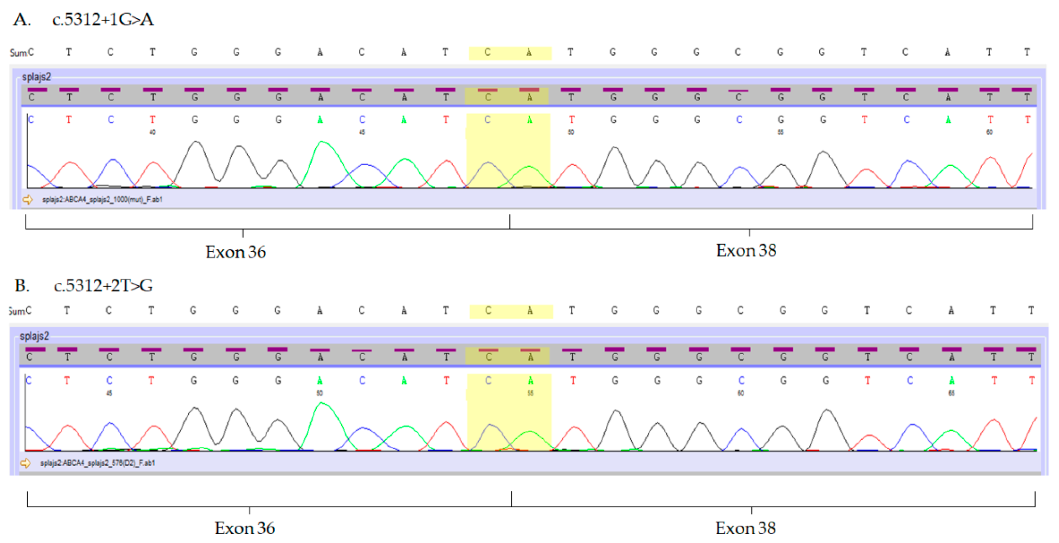
2.3. Analysis of the ABCA4 Splice-Site Variants in Hair Follicles

Considering that the expression of the ABCA4 gene in the skin samples was significantly lower than in the hair follicles, we used the latter for further experiments aimed at assessing the sequence of ABCA4 transcripts in patients’ samples.
In order to evaluate the usefulness of eyebrow hair follicles as a tool for the molecular analysis of ABCA4 variants, we investigated the consequences of the three ABCA4 splice-site variants c.5312+1G>A, c.5312+2T>G and c.5836-3C>A, previously detected in patients with Stargardt dystrophy [7]. In those patients, ABCA4 gene mutations were found in a compound heterozygous state with missense (NM_000350.2:c.[1622T>C;3113C>T], NP_000341.2:p.[Leu541Pro;Ala1038Val] or NM_000350.2:c.5882G>A, NP_000341.2:p.Gly1961Glu) or nonsense (NM_000350.2:c.4872G>A, NP_000341.2:p.Trp1624*) ABCA4 mutations [7]. Direct Sanger sequencing revealed that variants c.5312+1G>A and c.5312+2T>G result in the skipping of exon 37 (Figure 3A,B), whereas mutation c.5836-3C>A leads to the insertion of thirty nucleotides of the intron adjacent to exon 42 (Figure 3C).

2.4. Analysis of the ABCA4 Gene Expression in Hair Follicles of Patients with Stargardt Disease

In order to analyze the overall expression of the ABCA4 gene expression in patients with Stargardt disease we performed qRT-PCR. The results show that ABCA4 mRNA is also expressed in the hair follicles obtained from patients with Stargardt disease, however, this expression was significantly lower than in controls (p = 0.0001 by Mann-Whitney U test) (Figure 4).

3. Discussion

Here, we report that full-length ABCA4 transcript is highly expressed in human eyebrow hair follicles and may be thus considered useful for the molecular analysis of splice-site ABCA4 mutation consequences in patients with ABCA4 retinopathies. The applicability of the hair follicle model was confirmed by the analysis of three different splice-site ABCA4 variants from patients with Stargardt disease.
The ABCA4 gene was previously thought to be expressed at very low levels in non-ocular tissues. Although a previous high-throughput mRNA analysis of ABC transporters failed to detect ABCA4 expression in human total skin [39], nevertheless, we detected low levels of ABCA4 expression in human skin explants. On the other hand, a relatively high ABCA4 gene expression was observed in human hair follicles. This is in agreement with the observation made by Haslam et al., who also reported a high expression level of the ABCA4 gene throughout the epithelium of hair follicles [37]. Interestingly, we found that mRNA expression of the ABCA4 exons 3–4 was considerably lower as compared to other exons. This may implicate presence of alternate splicing in normal hair follicles, though one cannot exclude the possibility of an insufficient qPCR reaction for this particular probe. Nevertheless, we confirmed the presence of all ABCA4 exons of transcripts isolated from human hair follicles, cultured human keratinocytes, fibroblasts and melanocytes by means of direct Sanger sequencing. Presence of the ABCA4 full-length protein was also confirmed by the Western blot method.
In the hair follicles obtained from healthy controls, a high variability of the ABCA4 gene expression was observed. This was probably caused by individual differences of the ABCA4 gene expression in the population. However, it may be also associated with an unknown function of the ABCA4 in the hair follicles. It is also tempting to speculate that the slightly decreased expression of the ABCA4 gene in the control group (though significantly greater than that observed in the patients) may be related to existence of the pathogenic ABCA4 variants, as has been demonstrated in a canine model of Stargardt disease [40]. This, however, requires further analysis of ABCA4 gene expression in the probands’ parents.
Interestingly, ABCA4 mRNA expression in keratinocytes was very low compared to hair follicles, yet ABCA4 protein was detected at a relatively high level by the Western blot method. This difference may be due to variability between samples of different origins. However, this phenomenon may be also explained by the posttranscriptional regulation of the ABCA4 transporter in the skin, as has been observed for other ABCA transporters in tissues in which protein expression was much higher that would be expected based on their level of mRNA expression [41].
Variants potentially affecting RNA splicing can be directly tested with the use of qRT-PCR on accessible cells (e.g., skin cells, lymphoblasts), providing that they constitutively express a sufficient level of the gene of interest. To assess whether hair follicles are a suitable source of material for the study of the consequences of ABCA4 mutations, we analyzed hair follicle-derived mRNA obtained from patients with Stargardt disease carrying representative c.5312+1G>A, c.5312+2T>G and c.5836-3C>A ABCA4 variants [7]. Our findings are in concordance with the results of Schulz et al. who also observed skipping of the 37th exon caused by the c.5312+1G>A mutation using in vitro splicing assay [42]. Furthermore, our results concerning c.5836-3C>A mutation were also consistent with the observations of Sangermano et al. by means of midigene assays [24]. It would also be of great interest to analyze other ABCA4 gene splice variants, especially those located at the 5′ region of the ABCA4 gene. Unfortunately, due to lack of appropriate clinical material from patients, analysis of such variants could not be performed in the present study and awaits further investigation.
Analysis of the overall ABCA4 gene expression in patients with Stargardt disease showed a high reduction as compared to healthy controls. This is in agreement with results obtained from different cell types, which also showed a reduction of normally spliced full-length ABCA4 mRNA in fibroblasts of patients with Stargardt disease [23]. It needs to be further elucidated whether the expression level of the ABCA4 gene in patients with Stargardt disease correlates with disease severity.
Eyebrow hair follicles are regularly plucked by millions of women worldwide. Unlike a skin biopsy, no anesthetic is needed and no skin injuries are inflicted upon the patient. In addition, material can be obtained directly from patients and their families and sent to the laboratory without the need of patient arrival. About 10–15 eyebrow hair follicles yield an amount of RNA sufficient to conduct a preliminary molecular assessment of ABCA4 variant pathogenicity and ABCA4 gene expression analysis. This method appears to be simple and inexpensive, especially as it does not require cell culture or the highly advanced generation of mini-, midigenes or photoreceptor precursor cells (PPCs).
It should be stressed, however, that not all molecular and functional analyses can be performed on non-cultured cells from hair follicles. It is impossible to block nonsense-mediated decay in non-cultured hair follicle cells, therefore ABCA4 splice-site variant consequences can be missed in heterozygous patients. Also, reverse transcription reactions are difficult to conduct on large genes where 5′ of the gene may not be efficiently retrotranscribed, as compared with 3′ of the gene. For this purpose, the construction of mini- or midigenes is more favorable, even though mini-genes may result in the creation of splicing artefacts [13]. In order to improve the accuracy of these assays, interesting results regarding functional analysis of the ABCA4 splice-site variants were obtained with photoreceptor precursor cells generated from skin fibroblasts [24,27,32,33]. Still, the invasiveness of skin biopsies may impede these analyses. Hair follicles may be non-invasively obtained from patients multiple times and may also be a source of cultured epithelial stem cells [43]. Whether consequences of the ABCA4 splice-site variants in cultured hair follicle cells would lead to similar outcomes as those observed in PPCs needs further investigation.
In conclusion, analysis of mRNA from hair follicles appears to be a reliable method to assess the biological consequences of functional changes of the ABCA4 gene in patients with Stargardt disease and possibly other ABCA4 retinopathies. Furthermore, this method is non-invasive for the patients and is relatively simple and inexpensive as compared to other methods based on highly advanced molecular techniques.

4. Materials and Methods

4.1. Patients and Controls

Skin biopsies were acquired from eight healthy individuals subjected to aesthetic breast reduction or aesthetic abdominoplasty in the Department of Plastic Surgery, Medical Centre of Postgraduate Education, Orlowski Memorial Hospital in Warsaw. Up to 30 hair follicles were plucked with tweezers from eyebrows of eight healthy volunteers and three patients with Stargardt disease bearing splice-site ABCA4 gene mutations (NM_000350.3:c.5312+1G>A, NM_000350.3:c.5312+2T>G or NM_000350.3:c.5836-3C>A) who were diagnosed in the SPKSO Ophthalmic University Hospital, Medical University of Warsaw. Written informed consent was obtained from all participants. The study was approved by the Ethics Committee of Medical University of Warsaw (KB/191/2010 from 21 September 2010) and the Ethics Committee at the Medical Centre of Postgraduate Education, Orlowski Memorial Hospital in Warsaw (63/PB/2016 from 16 November 2016).

4.2. Culture of Primary Keratinocytes, Fibroblasts and Melanocytes

Keratinocyte cultures were established as described previously with minor modifications [44]. Briefly, the epidermis was separated from the dermis with 2.5 U/mL of dispase by an overnight digestion at 4 °C. Keratinocytes and melanocytes were released from the epidermis after 5 min of cleavage in 0.25% trypsin at 37 °C with subsequent pipetting. The cells were cultured in Medium 254 (Thermo Fisher Scientific, Waltham, MA, USA) supplemented with Human Melanocyte Growth Supplement-2, PMA-free (Thermo Fisher Scientific, Waltham, MA, USA). Following one-week culture, melanocytes were separated from keratinocytes with a short (up to 2 min) trypsinization. Keratinocytes were subsequently cultured in Keratinocyte Growth Medium (PromoCell, Heidelberg, Germany) supplemented with Keratinocyte Supplement Mix (PromoCell, Heidelberg, Germany). Dermal fibroblasts were released by 0.6 U/mL of Collagenase (Serva Electrophoresis GMbH, Heidelberg, Germany) digestion for 1 h at 37 °C with subsequent pipetting. Then they were cultured in DMEM (Thermo Fisher Scientific, Waltham, MA, USA) supplemented with 10% Fetal Bovine Serum (EURx, Gdansk, Poland). Cells in passages 1–2 were used in subsequent experiments.

4.3. RNA Isolation and Quantitative mRNA Measurements

Skin explants cut slightly below the basal layer of the epidermis with shallow fragments of the dermis and eyebrow hair follicles were submerged in the RNAlater Storage Solution (Thermo Fisher Scientific, Waltham, MA, USA) and stored at 4 °C prior to subsequent isolation of total RNA and protein within 6 h. Total RNA from skin explants, skin cells and hair follicles was extracted with NucleoSpin® TriPrep (with rDNase digestion) (Macherey-Nagel, Dürren, Germany) and transcribed into cDNA with RevertAid First Strand cDNA Synthesis Kit (Thermo Fisher Scientific, Waltham, MA, USA). For quantitative PCR, 20 ng of cDNA per reaction was used. All real-time PCR reactions were performed on the ABI 7500 Fast Real-time PCR system (Applied Biosystems, Inc., Thermo Fisher Scientific, Waltham, MA, USA). Expression levels of the ABCA4 gene in hair follicles, skin cells and skin explants from healthy donors and hair follicles from the patients were analyzed with Hs00979586_m1 TaqMan® probe (Thermo Fisher Scientific, Waltham, MA, USA). TBP gene (TATA-box binding protein, Hs00427620_m1) was used as a reference gene for further normalizations. All samples were run in triplicates. The relative level of mRNA expression was calculated by the ΔCt or ΔΔCt method.
qRT-PCR evaluation of particular ABCA4 exon mRNA expression in hair follicles and skin cells was performed with the use of TaqMan® probes presented in Table 2. PCR reactions were performed as described above and specific exon expression was presented as number of cycle threshold (Ct).

4.4. Sanger Sequencing of the ABCA4 Transcripts

PCR primers for the analysis of the full-length ABCA4 transcript were designed with Primer 3 Plus software [45] based on the NM_000350.3 reference sequence. The primers were designed to encompass an area of the five to six adjacent exons, therefore the presence of the full-length ABCA4 transcript was analyzed with 11 sets of primers, listed in Table 3.
PCR products were visualized on 2% agarose gel and excised with NucleoTrap® (Macherey-Nagel, Dürren, Germany). In order to visualize PCR products of keratinocytes, fibroblasts and melanocytes, re-amplification for 35 cycles have been performed. Purified products were sequenced with BigDye Terminator v3.1 Cycle Sequencing Kit (Applied Biosystems, Thermo Fisher Scientific Inc.) according to the manufacturer’s instructions and subsequently sequenced on 3500xL Genetic Analyzer (Applied Biosystems, Thermo Fisher Scientific Inc.). The results were analyzed with Variant Reporter v.3.1. (Thermo Fisher Scientific Inc.).

4.5. Protein Extract Isolation and Western Blot Analysis

Proteins were extracted from the hair follicles and skin explants, as well as cultured keratinocytes, dermal fibroblasts and melanocytes, with NucleoSpin® TriPrep (Macherey-Nagel), as described in detail by the manufacturer. Lysates were pelleted and resuspended in a buffer containing 150 mM NaCl, 1% Triton X-100, 0.5% sodium deoxycholate, 0.1% SDS and 50 mM Tris (pH 8.0). Protein concentration was evaluated with Pierce BCA Protein Assay Kit (Thermo Fisher Scientific Inc.) and 20 µg protein samples were separated by SDS–10% PAGE, transferred onto a nitrocellulose membrane (Thermo Fisher Scientific Inc.), blocked with Pierce Protein-Free Blocking Buffer (Thermo Fisher Scientific Inc.) and incubated at 4 °C overnight with 1:250 rabbit anti-ABCA4 (cat. no. PA5-87983, Thermo Fisher Scientific Inc.) or 1:500 mouse anti-β-actin (cat. no. A1978, Sigma-Aldrich, St. Louis, MO, USA). Secondary HRP-conjugated goat anti-rabbit IgG antibodies (cat. no. GTX213110-01, GeneTex) or rabbit anti-mouse IgG antibodies (cat. no. GTX213111-01, GeneTex, CA, USA) were used and visualized with SuperSignal West Pico Chemiluminescent Substrate (Thermo Fisher Inc.). As an ABCA4 positive control, 20 µg of Y79 human retinoblastoma cell lysate (sc-2240, Santa Cruz Biotechnology) was used. A skin sample in which ABCA4 mRNA expression was not detected by qRT-PCR served as a negative control. Broad range BlueEasy Prestained Protein Marker (Nippon Genetics Co., Dürren, Germany) was used as a protein standard.

4.6. Statistical Analysis

Statistical analyses and graphs were generated with GraphPad Prism 8 (GraphPad Software, San Diego, CA, USA). The nonparametric Mann Whitney U test and the one-way ANOVA were used to compare ABCA4 expression levels in different studied groups. p-value < 0.05 was considered significant.

Author Contributions

Conceptualization, A.Ś., J.M. and M.O.; material, N.K., M.S. (Martyna Smolińska), K.S., J.P.S. and B.N.; methodology, A.Ś., M.S. (Marta Soszyńska), M.K., A.N. and A.P.; data acquisition and data analyses A.Ś. and J.M.; writing—original draft preparation, A.Ś. and J.M.; writing—review and editing, A.Ś., M.O. and J.M. The funders had no role in the study design, data collection or preparation of the manuscript. All authors have read and agreed to the published version of the manuscript.

Funding

This study was supported by the National Science Centre, Poland grants no. 2016/23/N/NZ5/02588 and N N402 591640 (5916/B/P01/2011/40) and Medical University of Warsaw Grant no. 1M15/3G66.

Conflicts of Interest

The authors declare no conflict of interests regarding this paper.

References

  1. Tsybovsky, Y.; Molday, R.S.; Palczewski, K. The ATP-binding cassette transporter ABCA4: Structural and functional properties and role in retinal disease. Adv. Exp. Med. Biol. 2010, 703, 105–125. [Google Scholar] [CrossRef] [PubMed] [Green Version]
  2. Sheffield, V.C.; Stone, E.M. Genomics and the eye. N. Engl. J. Med. 2011, 364, 1932–1942. [Google Scholar] [CrossRef] [PubMed]
  3. Rivera, A.; White, K.; Stohr, H.; Steiner, K.; Hemmrich, N.; Grimm, T.; Jurklies, B.; Lorenz, B.; Scholl, H.P.; Apfelstedt-Sylla, E.; et al. A comprehensive survey of sequence variation in the ABCA4 (ABCR) gene in Stargardt disease and age-related macular degeneration. Am. J. Hum. Genet. 2000, 67, 800–813. [Google Scholar] [CrossRef] [PubMed] [Green Version]
  4. Valverde, D.; Riveiro-Alvarez, R.; Bernal, S.; Jaakson, K.; Baiget, M.; Navarro, R.; Ayuso, C. Microarray-based mutation analysis of the ABCA4 gene in Spanish patients with Stargardt disease: Evidence of a prevalent mutated allele. Mol. Vis. 2006, 12, 902–908. [Google Scholar]
  5. Maugeri, A.; Van Driel, M.A.; Van de Pol, D.J.; Klevering, B.J.; Van Haren, F.J.; Tijmes, N.; Bergen, A.A.; Rohrschneider, K.; Blankenagel, A.; Pinckers, A.J.; et al. The 2588G→C mutation in the ABCR gene is a mild frequent founder mutation in the Western European population and allows the classification of ABCR mutations in patients with Stargardt disease. Am. J. Hum. Genet. 1999, 64, 1024–1035. [Google Scholar] [CrossRef] [Green Version]
  6. Rosenberg, T.; Klie, F.; Garred, P.; Schwartz, M. N965S is a common ABCA4 variant in Stargardt-related retinopathies in the Danish population. Mol. Vis. 2007, 13, 1962–1969. [Google Scholar]
  7. Sciezynska, A.; Ozieblo, D.; Ambroziak, A.M.; Korwin, M.; Szulborski, K.; Krawczynski, M.; Stawinski, P.; Szaflik, J.; Szaflik, J.P.; Ploski, R.; et al. Next-generation sequencing of ABCA4: High frequency of complex alleles and novel mutations in patients with retinal dystrophies from Central Europe. Exp. Eye Res. 2016, 145, 93–99. [Google Scholar] [CrossRef]
  8. Nassisi, M.; Mohand-Said, S.; Dhaenens, C.M.; Boyard, F.; Demontant, V.; Andrieu, C.; Antonio, A.; Condroyer, C.; Foussard, M.; Mejecase, C.; et al. Expanding the Mutation Spectrum in ABCA4: Sixty Novel Disease Causing Variants and Their Associated Phenotype in a Large French Stargardt Cohort. Int. J. Mol. Sci. 2018, 19, 2196. [Google Scholar] [CrossRef] [Green Version]
  9. Salles, M.V.; Motta, F.L.; Martin, R.; Filippelli-Silva, R.; Dias da Silva, E.; Varela, P.; Costa, K.A.; Chiang, J.P.; Pesquero, J.B.; Sallum, J.F. Variants in the ABCA4 gene in a Brazilian population with Stargardt disease. Mol. Vis. 2018, 24, 546–559. [Google Scholar]
  10. Chacon-Camacho, O.F.; Granillo-Alvarez, M.; Ayala-Ramirez, R.; Zenteno, J.C. ABCA4 mutational spectrum in Mexican patients with Stargardt disease: Identification of 12 novel mutations and evidence of a founder effect for the common p.A1773V mutation. Exp. Eye Res. 2013, 109, 77–82. [Google Scholar] [CrossRef]
  11. September, A.V.; Vorster, A.A.; Ramesar, R.S.; Greenberg, L.J. Mutation spectrum and founder chromosomes for the ABCA4 gene in South African patients with Stargardt disease. Invest. Ophthalmol Vis. Sci. 2004, 45, 1705–1711. [Google Scholar] [CrossRef] [PubMed] [Green Version]
  12. Cornelis, S.S.; Bax, N.M.; Zernant, J.; Allikmets, R.; Fritsche, L.G.; Den Dunnen, J.T.; Ajmal, M.; Hoyng, C.B.; Cremers, F.P. In Silico Functional Meta-Analysis of 5,962 ABCA4 Variants in 3,928 Retinal Dystrophy Cases. Hum. Mutat. 2017, 38, 400–408. [Google Scholar] [CrossRef] [PubMed]
  13. Khan, M.; Cremers, F.P.M. ABCA4-Associated Stargardt Disease. Klin. Mon. Fur Augenheilkd. 2020, 237, 267–274. [Google Scholar] [CrossRef] [PubMed]
  14. Maugeri, A.; Klevering, B.J.; Rohrschneider, K.; Blankenagel, A.; Brunner, H.G.; Deutman, A.F.; Hoyng, C.B.; Cremers, F.P. Mutations in the ABCA4 (ABCR) gene are the major cause of autosomal recessive cone-rod dystrophy. Am. J. Hum. Genet. 2000, 67, 960–966. [Google Scholar] [CrossRef] [Green Version]
  15. D’Angelo, R.; Donato, L.; Venza, I.; Scimone, C.; Aragona, P.; Sidoti, A. Possible protective role of the ABCA4 gene c.1268A>G missense variant in Stargardt disease and syndromic retinitis pigmentosa in a Sicilian family: Preliminary data. Int. J. Mol. Med. 2017, 39, 1011–1020. [Google Scholar] [CrossRef] [Green Version]
  16. Bauwens, M.; Garanto, A.; Sangermano, R.; Naessens, S.; Weisschuh, N.; De Zaeytijd, J.; Khan, M.; Sadler, F.; Balikova, I.; Van Cauwenbergh, C.; et al. ABCA4-associated disease as a model for missing heritability in autosomal recessive disorders: Novel noncoding splice, cis-regulatory, structural, and recurrent hypomorphic variants. Genet. Med. Off. J. Am. Coll. Med. Genet. 2019, 21, 1761–1771. [Google Scholar] [CrossRef] [Green Version]
  17. Runhart, E.H.; Sangermano, R.; Cornelis, S.S.; Verheij, J.; Plomp, A.S.; Boon, C.J.F.; Lugtenberg, D.; Roosing, S.; Bax, N.M.; Blokland, E.A.W.; et al. The Common ABCA4 Variant p.Asn1868Ile Shows Nonpenetrance and Variable Expression of Stargardt Disease When Present in trans With Severe Variants. Investig. Ophthalmol. Vis. Sci. 2018, 59, 3220–3231. [Google Scholar] [CrossRef] [Green Version]
  18. Zernant, J.; Lee, W.; Collison, F.T.; Fishman, G.A.; Sergeev, Y.V.; Schuerch, K.; Sparrow, J.R.; Tsang, S.H.; Allikmets, R. Frequent hypomorphic alleles account for a significant fraction of ABCA4 disease and distinguish it from age-related macular degeneration. J. Med. Genet. 2017, 54, 404–412. [Google Scholar] [CrossRef]
  19. Zernant, J.; Lee, W.; Nagasaki, T.; Collison, F.T.; Fishman, G.A.; Bertelsen, M.; Rosenberg, T.; Gouras, P.; Tsang, S.H.; Allikmets, R. Extremely hypomorphic and severe deep intronic variants in the ABCA4 locus result in varying Stargardt disease phenotypes. Mol. Case Stud. 2018, 4. [Google Scholar] [CrossRef] [Green Version]
  20. Cremers, F.P.M.; Lee, W.; Collin, R.W.J.; Allikmets, R. Clinical spectrum, genetic complexity and therapeutic approaches for retinal disease caused by ABCA4 mutations. Prog. Retin. Eye Res. 2020. [Google Scholar] [CrossRef]
  21. Sciezynska, A.; Ozieblo, D.; Oldak, M. Experimenal studies On cell and gene therapies for retinal dystrophies with a particular focus On ABCA4 retinopathies. Klin Ocz. 2016, 118, 66–71. [Google Scholar]
  22. Sciezynska, A.; Ozieblo, D.; Oldak, M. Experimental studies on medical treatments of retinal dystrophies with a particular focus on ABCA4 retinopathies. Klin Ocz. 2016, 118, 59–65. [Google Scholar]
  23. Aukrust, I.; Jansson, R.W.; Bredrup, C.; Rusaas, H.E.; Berland, S.; Jorgensen, A.; Haug, M.G.; Rodahl, E.; Houge, G.; Knappskog, P.M. The intronic ABCA4 c.5461-10T>C variant, frequently seen in patients with Stargardt disease, causes splice defects and reduced ABCA4 protein level. Acta Ophthalmol. 2017, 95, 240–246. [Google Scholar] [CrossRef] [PubMed]
  24. Sangermano, R.; Khan, M.; Cornelis, S.S.; Richelle, V.; Albert, S.; Garanto, A.; Elmelik, D.; Qamar, R.; Lugtenberg, D.; Van den Born, L.I.; et al. ABCA4 midigenes reveal the full splice spectrum of all reported noncanonical splice site variants in Stargardt disease. Genome Res. 2018, 28, 100–110. [Google Scholar] [CrossRef] [PubMed] [Green Version]
  25. Desviat, L.R.; Perez, B.; Ugarte, M. Minigenes to confirm exon skipping mutations. Methods Mol. Biol. 2012, 867, 37–47. [Google Scholar] [CrossRef] [PubMed]
  26. Braun, T.A.; Mullins, R.F.; Wagner, A.H.; Andorf, J.L.; Johnston, R.M.; Bakall, B.B.; Deluca, A.P.; Fishman, G.A.; Lam, B.L.; Weleber, R.G.; et al. Non-exomic and synonymous variants in ABCA4 are an important cause of Stargardt disease. Hum. Mol. Genet. 2013, 22, 5136–5145. [Google Scholar] [CrossRef] [PubMed]
  27. Albert, S.; Garanto, A.; Sangermano, R.; Khan, M.; Bax, N.M.; Hoyng, C.B.; Zernant, J.; Lee, W.; Allikmets, R.; Collin, R.W.J.; et al. Identification and Rescue of Splice Defects Caused by Two Neighboring Deep-Intronic ABCA4 Mutations Underlying Stargardt Disease. Am. J. Hum. Genet. 2018, 102, 517–527. [Google Scholar] [CrossRef] [Green Version]
  28. Fadaie, Z.; Khan, M.; Del Pozo-Valero, M.; Cornelis, S.S.; Ayuso, C.; Cremers, F.P.M.; Roosing, S.; The Abca Study, G. Identification of splice defects due to noncanonical splice site or deep-intronic variants in ABCA4. Hum. Mutat. 2019, 40, 2365–2376. [Google Scholar] [CrossRef] [Green Version]
  29. Sangermano, R.; Garanto, A.; Khan, M.; Runhart, E.H.; Bauwens, M.; Bax, N.M.; Van den Born, L.I.; Khan, M.I.; Cornelis, S.S.; Verheij, J.; et al. Deep-intronic ABCA4 variants explain missing heritability in Stargardt disease and allow correction of splice defects by antisense oligonucleotides. Genet. Med. Off. J. Am. Coll. Med. Genet. 2019, 21, 1751–1760. [Google Scholar] [CrossRef] [Green Version]
  30. Khan, M.; Cornelis, S.S.; Pozo-Valero, M.D.; Whelan, L.; Runhart, E.H.; Mishra, K.; Bults, F.; AlSwaiti, Y.; AlTalbishi, A.; De Baere, E.; et al. Resolving the dark matter of ABCA4 for 1054 Stargardt disease probands through integrated genomics and transcriptomics. Genet. Med. Off. J. Am. Coll. Med. Genet. 2020. [Google Scholar] [CrossRef]
  31. Claassen, J.N.; Zhang, D.; Chen, S.C.; Moon, S.Y.; Lamey, T.; Thompson, J.A.; McLaren, T.; De Roach, J.N.; McLenachan, S.; Chen, F.K. Generation of the induced pluripotent stem cell line from a patient with autosomal recessive ABCA4-mediated Stargardt Macular Dystrophy. Stem Cell Res. 2019, 34, 101352. [Google Scholar] [CrossRef]
  32. Garanto, A.; Duijkers, L.; Tomkiewicz, T.Z.; Collin, R.W.J. Antisense Oligonucleotide Screening to Optimize the Rescue of the Splicing Defect Caused by the Recurrent Deep-Intronic ABCA4 Variant c.4539+2001G>A in Stargardt Disease. Genes 2019, 10, 452. [Google Scholar] [CrossRef] [PubMed] [Green Version]
  33. Sangermano, R.; Bax, N.M.; Bauwens, M.; Van den Born, L.I.; De Baere, E.; Garanto, A.; Collin, R.W.; Goercharn-Ramlal, A.S.; Den Engelsman-van Dijk, A.H.; Rohrschneider, K.; et al. Photoreceptor Progenitor mRNA Analysis Reveals Exon Skipping Resulting from the ABCA4 c.5461-10T-->C Mutation in Stargardt Disease. Ophthalmology 2016, 123, 1375–1385. [Google Scholar] [CrossRef] [PubMed]
  34. Wiszniewski, W.; Zaremba, C.M.; Yatsenko, A.N.; Jamrich, M.; Wensel, T.G.; Lewis, R.A.; Lupski, J.R. ABCA4 mutations causing mislocalization are found frequently in patients with severe retinal dystrophies. Hum. Mol. Genet. 2005, 14, 2769–2778. [Google Scholar] [CrossRef] [PubMed] [Green Version]
  35. The Human Protein Atlas. Available online: https://www.proteinatlas.org/ENSG00000198691-ABCA4/tissue (accessed on 2 April 2020).
  36. Wiley, L.; Kaalberg, E.; Penticoff, J.; Mullins, R.; Stone, E.; Tucker, B. Expression of the retina-specific flippase, ABCA4, in epidermal keratinocytes [version 1; peer review: 2 approved with reservations]. F1000Research 2016, 5. [Google Scholar] [CrossRef] [Green Version]
  37. Haslam, I.S.; El-Chami, C.; Faruqi, H.; Shahmalak, A.; O’Neill, C.A.; Paus, R. Differential expression and functionality of ATP-binding cassette transporters in the human hair follicle. Br. J. Derm. 2015, 172, 1562–1572. [Google Scholar] [CrossRef]
  38. Riera, M.; Patel, A.; Bures-Jelstrup, A.; Corcostegui, B.; Chang, S.; Pomares, E.; Corneo, B.; Sparrow, J.R. Generation of two iPS cell lines (FRIMOi003-A and FRIMOi004-A) derived from Stargardt patients carrying ABCA4 compound heterozygous mutations. Stem Cell Res. 2019, 36, 101389. [Google Scholar] [CrossRef]
  39. Takenaka, S.; Itoh, T.; Fujiwara, R. Expression pattern of human ATP-binding cassette transporters in skin. Pharm. Res. Perspect. 2013, 1, e00005. [Google Scholar] [CrossRef]
  40. Makelainen, S.; Godia, M.; Hellsand, M.; Viluma, A.; Hahn, D.; Makdoumi, K.; Zeiss, C.J.; Mellersh, C.; Ricketts, S.L.; Narfstrom, K.; et al. An ABCA4 loss-of-function mutation causes a canine form of Stargardt disease. PLoS Genet. 2019, 15, e1007873. [Google Scholar] [CrossRef] [Green Version]
  41. Pasello, M.; Giudice, A.M.; Scotlandi, K. The ABC subfamily A transporters: Multifaceted players with incipient potentialities in cancer. Semin. Cancer Biol. 2020, 60, 57–71. [Google Scholar] [CrossRef]
  42. Schulz, H.L.; Grassmann, F.; Kellner, U.; Spital, G.; Ruther, K.; Jagle, H.; Hufendiek, K.; Rating, P.; Huchzermeyer, C.; Baier, M.J.; et al. Mutation Spectrum of the ABCA4 Gene in 335 Stargardt Disease Patients From a Multicenter German Cohort-Impact of Selected Deep Intronic Variants and Common SNPs. Investig. Ophthalmol. Vis. Sci. 2017, 58, 394–403. [Google Scholar] [CrossRef] [Green Version]
  43. Guo, Z.; Draheim, K.; Lyle, S. Isolation and culture of adult epithelial stem cells from human skin. J. Vis. Exp. 2011. [Google Scholar] [CrossRef] [PubMed]
  44. Sciezynska, A.; Nogowska, A.; Sikorska, M.; Konys, J.; Karpinska, A.; Komorowski, M.; Oldak, M.; Malejczyk, J. Isolation and culture of human primary keratinocytes-a methods review. Exp. Derm. 2019, 28, 107–112. [Google Scholar] [CrossRef] [PubMed] [Green Version]
  45. Untergasser, A.; Cutcutache, I.; Koressaar, T.; Ye, J.; Faircloth, B.C.; Remm, M.; Rozen, S.G. Primer3—New capabilities and interfaces. Nucleic Acids Res. 2012, 40, e115. [Google Scholar] [CrossRef] [PubMed] [Green Version]
Figure 1. Expression of the ABCA4 mRNA in hair follicles, skin lysates, as well as keratinocytes, dermal fibroblasts and melanocytes as measured by qRT-PCR for TaqMan probe Hs00979586_m1 (ABCA4 exons 35–36). (A) Expression of the ABCA4 gene in the isolated hair follicles was significantly higher than in total skin lysates. (B) Relatively very low expression of the ABCA4 gene was detected in fibroblasts, keratinocytes and melanocytes. ∗∗∗∗ p < 0.0001 as calculated by Mann-Whitney U test.
Ijms 21 03430 g001
Figure 2. Expression of the ABCA4 gene in hair follicles, skin cells and skin lysates measured at the protein level with Western blot assay. HF, hair follicle; Skin, skin sample positive for ABCA4 mRNA; K, keratinocytes; F, fibroblasts; M, melanocytes; C(+), retinoblastoma lysate; C(–), skin sample negative for ABCA4 mRNA; Std, broad-range protein standard.
Ijms 21 03430 g002
Figure 3. Electropherograms of the mutated ABCA4 transcripts isolated from hair follicles. (A,B) Mutations c.5312+1G>A and c.5312+2T>G result in the exon 37 skipping. Boundaries of exon 36 and 38 are marked in yellow. (C) Mutation c.5836-3C>A results in an insertion of 30 nucleotides (marked purple) into the ABCA4 transcript between the 41st and 42nd exon of the ABCA4 transcript.
Ijms 21 03430 g003aIjms 21 03430 g003b
Figure 4. Expression of the ABCA4 gene in hair follicles (HF) of patients with Stargardt disease (n = 3) and healthy controls (n = 8) as measured by qRT-PCR for TaqMan probe Hs00979586_m1 (ABCA4 exons 35–36). ∗∗∗ p = 0.0001 as evaluated by Mann-Whitney U test.
Ijms 21 03430 g004
Table 1. Average Ct of ABCA4 gene expression in hair follicles, skin samples, keratinocytes, dermal fibroblasts and melanocytes.
Exon Number3–410–1120–2129–3035–3640–4149–50
Hair follicles (n = 8)33.7228.1828.5327.6527.2227.0527.28
Total skin samples (n = 4)37.7732.5133.17Not tested33.48Not tested31.92
Keratinocytes (n = 4)35.0734.4234.15Not tested33.02Not tested34.12
Fibroblasts (n = 4)33.6732.7532.54Not tested32.15Not tested31.48
Melanocytes (n = 4)37.8737.0737.01Not tested36.29Not tested38.46
Table 2. Localization of the TaqMan probes within the ABCA4 gene exons.
TaqMan® ProbesExon Boundary
Hs00979588_m13–4
Hs00979567_m110–11
Hs00979574_m120–21
Hs00979581_m129–30
Hs00979586_m135–36
Hs00979589_m140–41
Hs00979594_m149–50
Table 3. List of set of primers and annealing temperatures (Ta) used during ABCA4 transcript amplification.
ABCA4 ExonsForward Primer (5′–3′)Reverse Primer (5′–3′)Amplicon LengthTa
2–4GGAACTCGTGTGGCCTTTATCTTTATTGTCCATGAATTGGGACAAGATGTGTAG33158.2
4–8AGCTACACATCTTGTCCCAATTCATGGTGTCTTCATACCAGTTGAAGGAGAGCA64558.2
8–12CTCGGGTGCTCTCCTTCAACTGGTATTGGTAGAGAGCTGGTCCAGGGATACAT69861.3
12–15GTGCCCTCTCTCTACTGGAGGAAAACAAGACTGGCCTTGGAGAAGAAGGTGCT67061.3
15–20CACCTTCTTCTCCAAGGCCAGTCTGCAGGATGTTGTGCTGTGGACACATG78761
20–25CACAGCACAACATCCTGTTCCACGAAATTCCAAAACTGCTGAGACCAA76957.1
25–30GGCTGACCTTGGTCTCAGCAGTCCTGCAGGATGGTGAAGGGTTG70658.6
30–36CACAGGTCAACCCTTCACCATCCTGTTGGATTTGTTCACCCGCTCCTGGATCA69161
36–41ATCTGCGTGATTTTCTCCATGTCCTTATCATCTTCATCAACAATGGGCTCCT70856.4
40–46GAAGGGGTGGTGTACTTCCTCCTGACCCTTCTCTGATGATGCTCACGATGAC70762.7
44–49GTCCATCAAAATATGGGCTACTGTCCTCTGGTCCAGTGTGGTCTGTGTGACT70261

