Article

Cyclin-Dependent Kinase Inhibitors KRP1 and KRP2 Are Involved in Grain Filling and Seed Germination in Rice (Oryza sativa L.)

1
State Key Lab of Rice Biology, China National Rice Research Institute, Hangzhou 311400, China
2
Biotechnology Unit, National Cereals Research Institute, Badeggi, Bida 912101, Nigeria
3
College of Life Science, Yangtze University, Jingzhou 434025, China
*
Authors to whom correspondence should be addressed.
Int. J. Mol. Sci. 2020, 21(1), 245; https://doi.org/10.3390/ijms21010245
Submission received: 21 November 2019 / Revised: 26 December 2019 / Accepted: 26 December 2019 / Published: 30 December 2019
(This article belongs to the Special Issue Plant Cell and Organism Development)

Abstract

:
Cyclin-dependent kinase inhibitors known as KRPs (kip-related proteins) control the progression of plant cell cycles and modulate various plant developmental processes. However, the function of KRPs in rice remains largely unknown. In this study, two rice KRPs members, KRP1 and KRP2, were found to be predominantly expressed in developing seeds and were significantly induced by exogenous abscisic acid (ABA) and Brassinosteroid (BR) applications. Sub-cellular localization experiments showed that KRP1 was mainly localized in the nucleus of rice protoplasts. KRP1 overexpression transgenic lines (OxKRP1), krp2 single mutant (crkrp2), and krp1/krp2 double mutant (crkrp1/krp2) all exhibited significantly smaller seed width, seed length, and reduced grain weight, with impaired seed germination and retarded early seedling growth, suggesting that disturbing the normal steady state of KRP1 or KRP2 blocks seed development partly through inhibiting cell proliferation and enlargement during grain filling and seed germination. Furthermore, two cyclin-dependent protein kinases, CDKC;2 and CDKF;3, could interact with KRP1 in a yeast-two-hybrid system, indicating that KRP1 might regulate the mitosis cell cycle and endoreduplication through the two targets. In a word, this study shed novel insights into the regulatory roles of KRPs in rice seed maturation and germination.

1. Introduction

In eukaryotes, the cell cycle is strictly regulated by cyclin-dependent kinases (CDKs), a type of Ser-Thr protein kinases, together with specific cyclin (CYC) partners during plant growth and development [1,2]. As the central regulators of cell proliferation, the activity of CDKs is finely modulated by numerous molecular mechanisms, including post-translational modification (PTM) such as phosphorylation, proteolysis, and other regulatory proteins [3,4,5,6]. Among these regulatory proteins, CDK inhibitors (CKIs) directly bind to CDK/CYC complexes and inhibit their activities, thereby arresting the cell cycle in response to internal and external cues [7,8,9].
CKIs in mammals are classified into INK4(CKIs, such as p15INK4b and p16INK4a that bind the CDK4 and CDK6 proteins and reduce their binding affinity for cyclin D) and Kip/Cip (CKIs including p21 (Cip1) and p27 (Kip1), that inhibits CDK activity by interaction with both subunits of the CDK–CYC complex) families based on their structural and functional differences [8,10]. The INK4 proteins, containing four or five ankyrin repeats, exclusively bind G1(Gap 1) phase-specific CDKs with D-type cyclins such as CDK4 and CDK6. The Kip/Cip inhibitors, binding to both CDKs and cyclin through a conserved N-terminal domain, show a broader spectrum of inhibitory effects on CDKs with E or A-type cyclins, especially G1 and S (Synthesis) phase CDK/CYC complexes. The plant CDK inhibitors, harboring a conserved 40 amino acids CDK/CYC-binding domain located at the C-terminal region, are similar to the mammalian Kip/Cip family and are therefore designated as Kip-related proteins (KRPs) [11,12]. In Arabidopsis, seven KRP family members (Arath; KRP1–7) have been discovered through a yeast two-hybrid system, and they block the cell cycle by exclusively inhibiting CDKA and CYCD during the G1/S and G2(Gap 2/M(Mitotic) phases [9]. The overexpression of Arath;KRP1 showed dwarf phenotype inhibited leaf growth with a decreasing cell number and an up-regulating of the cell size, suggesting that Arath;KRP1 plays a key role in determining plant growth and development through regulating cell proliferation [13]. The functions of KRPs have also been characterized in tobacco and tomato in the past decade. NtKIS1a, a KRP family member identified in tobacco (Nicotiana tabacum), interacts with A-type CDK and D-type cyclins and inhibits the activity of the CDK/cyclin complexes. The overexpression of NtKIS1a in Arabidopsis inhibits cell division and blocks endoreduplication in nuclei with severe developmental abnormalities, including smaller/shorter vegetative and reproductive organs, strong leaf serration, and enlarged cells [14]. LeKRP1, another KRP inhibitor isolated in tomato (Lycopersicon esculentum), mainly accumulates in the jelly-like locular (gel) tissue and inhibits CDK/CYC kinase activities in endoreduplicating cells, thereby arresting the mitotic activities to determine the gel tissue development [15].
KRPs regulating cell proliferation/enlargement and plant morphogenesis have also been reported in rice [16,17,18]. Previous studies have identified seven KRP family members in Japonica rice (O. sativa subspecies japonica), which are designated as Orysa;KRP1 to Orysa;KRP7, respectively [16,17,19]. Among these KRP inhibitors, Orysa;KRP1 to Orysa;KRP6 contain the C-terminal CDK/CYC-binding region, which is missing in Orysa;KRP7, suggesting that Orysa;KRP1 to Orysa;KRP6 act as functional CDK inhibitors in rice [17,19]. The overexpression of Orysa;KRP1 down-regulates grain filling through inhibiting endosperm cell endoreduplications [16]. Orysa;KRP3 is highly expressed in the cellularized endosperm at two days after fertilization (DAF), while it is not detected in the caryopsis at 3 DAF, suggesting that Orysa;KRP3 regulates the cell cycle of syncytial endosperm development [18]. Interestingly, OsiICK1 (corresponding to Orysa;KRP1) and OsiICK6 (corresponding to Orysa;KRP4) are also reported as functional CDK inhibitors in Indica rice (O. sativa subspecies indica), and subcellular localization analysis shows that OsiICK1 and OsiICK6 are both mainly localized in the nucleus of transiently transfected tobacco cells, and they could interact with OsCYCD and CDKA through yeast two-hybrid assay [19]. The overexpression of OsiICK6 significantly reduces plant growth, pollen viability, and seed setting rate. The leaves of OsiICK6 overexpressing lines roll toward the abaxial side, suggesting that the maintenance of an even growth along the dorsal-ventral plane of leaf blades is at least partially regulated by OsiICK6-mediated cell proliferation [19]. Despite these observations, our understanding of monocot CDK inhibitors is still poor, especially in rice. Here, we characterized two rice cyclin-dependent kinase inhibitors, KRP1 and KRP2, to investigate their tissue-specific expression patterns, the function of the transgenic plants in seed development, and the target CDKs specifically regulated by KRP1, which would advance our understanding of the role of KRPs in rice seed maturation and germination.

2. Results

2.1. Tissue-Specific Expression Pattern of KRP1 and KRP2

To explore the tissue expression pattern of KRP1 (LOC_Os02g52480) and KRP2 (LOC_Os06g11050), quantitative RT-PCR (qRT-PCR) was performed to examine the transcriptional level of the two CDK inhibitors in eleven different rice tissues. As shown in Figure 1, KRP1 was predominantly expressed in roots and developing seeds (6, 12, 15 DAP, Day after Pollination), especially in 6 DAP, while extremely low in callus, panicle, and stem (Figure 1A).
KRP2 exhibited a similar tendency as KRP1 with higher mRNA accumulation in leaf, root, and 9 DAP during seed development (Figure 1B). However, the transcription levels of KRP1 and KRP2 in response to various exogenous phytohormones treatments (100 μM ABA, 100 μM GA, 10 μM BR, and 100 μM JA) at 0, 1, 3, 6, 9, 12, and 24 h were also examined (Figure 1C,D). Upon ABA treatment, the transcription level of KRP1 increased from one to three hours, then dropped at six hours and kept steady until nine hours, and sharply rose to about six and four-fold at 12 and 24 h, respectively (Figure 1C). In response to BR treatment, the transcription level of KRP1 increased to two-fold at three hours, then decreased back to a low level at 6 and 12 h and increased to six-fold at 24 h (Figure 1C). For the treatment of JA, KRP1 increased sharply to the highest point at three hours about five-times-fold, then gradually decreased to two-fold at six hours, and dropped to the basal level at 24 h (Figure 1C). However, KRP1 showed no response to GA treatment (Figure 1C). As for KRP2, all the applied phytohormones, except JA, significantly induced the transcription level of KRP2, especially in early stages (Figure 1D). For example, KRP2 was obviously induced by BR treatment to 15-fold at one hour, 4.1-fold at six hours in response to ABA treatment and 4.3 fold at six hours under GA treatment (Figure 1D). Notably, the high responses of KRP1 and KRP2 in transcription strongly indicated that the two KRPs are functionally relevant to ABA and BR. In addition, the subcellular location of KRP1 is likely in the nucleus of rice protoplast, as it co-localized with the nucleus marker D53 [20] (Figure 1E and Figure S1). Given that the plant cell cycle is widespread in the whole-life-cycle, the temporal and spatial expression pattern of KRP1 and KRP2 suggests that the two KPR members play important roles in the plant cell cycle, specifically in organ development and hormone-mediated morphogenesis processes.

2.2. Functional Characterization of KRP1 and KRP2 in Rice Grain Filling

The higher expression level of KRP1 and KRP2 in developing seeds intrigued us to speculate that the KRP1 and KRP2 might function in the grain filling process (Figure 1A,B). To verify this hypothesis, we generated KRP1 independent overexpression transgenic lines (OxKRP1) with a substantially increased transcription level, krp2 single mutant (crkrp2), as well as krp1/krp2 double mutant (crkrp1/krp2) by using the CRISPR/Cas9 technique (Figure 2A and Figure S2) [21].
Unfortunately, we did not obtain the krp1 single mutant due to the defects in the CRISPR/Cas9 design. krp2 single mutants were identified with a T insertion at the first exon of KRP2 in crkrp2-6, an A insertion at the first exon of KRP2 in crkrp2-12, and crkrp1/krp2 showed a T deletion at the first exon of KRP1 and a C insertion at the first exon of KRP2 in crkrp1/krp2-13, and three bases GTT deletion at the first exon of KRP1 and a T insertion at the first exon of KRP2 in crkrp1/krp2-20, resulting in premature termination by shifting the open reading frame (Figure S2). Though the transcriptional level of the mutated genes remained at the same level as native KRP1 or KRP2 in the wild type, the insertion/deletion or premature mutation should have disrupted the function of the resulting proteins (Figure 2B).
Under normal conditions, both the grain size of OxKRP1-3 and OxKRP1-6 lines were significantly reduced compared with that of the wild-type. The grain length of the wild type was about 7.7 mm, whereas the grain length of OxKRP1-3 and OxKRP1-6 decreased to 7.32 mm and 7.37 mm, respectively. Meanwhile, the seed width of OxKRP1-3 and OxKRP1-6 dropped to approximately 95% of the wild-type (Figure 2C,F,G). However, the grain size of crkrp2 and crkrp1/krp2 also displayed a similar tendency as KRP1 overexpression lines, though the seed length of crkrp2 exhibited no significant difference compared to that of the wild-type (Figure 2D–G). Consequently, the 1000-grain-weight of OxKRP1-3 and OxKRP1-6 were only 79.0%–82.0% that of the wild-type, and the 1000-grain-weight of the crkrp2 and crkrp1/krp2 also had a 12% and 18% reduction compared with the wild-type due to the smaller grain size (Figure 2H). Together, these results demonstrated that disturbing the normal steady state of KRP1 or KRP2 blocks grain filling and both the KRP inhibitors play important roles in seed cell proliferation and enlargement.

2.3. Functional Characterization of KRP1 and KRP2 in Seed Germination

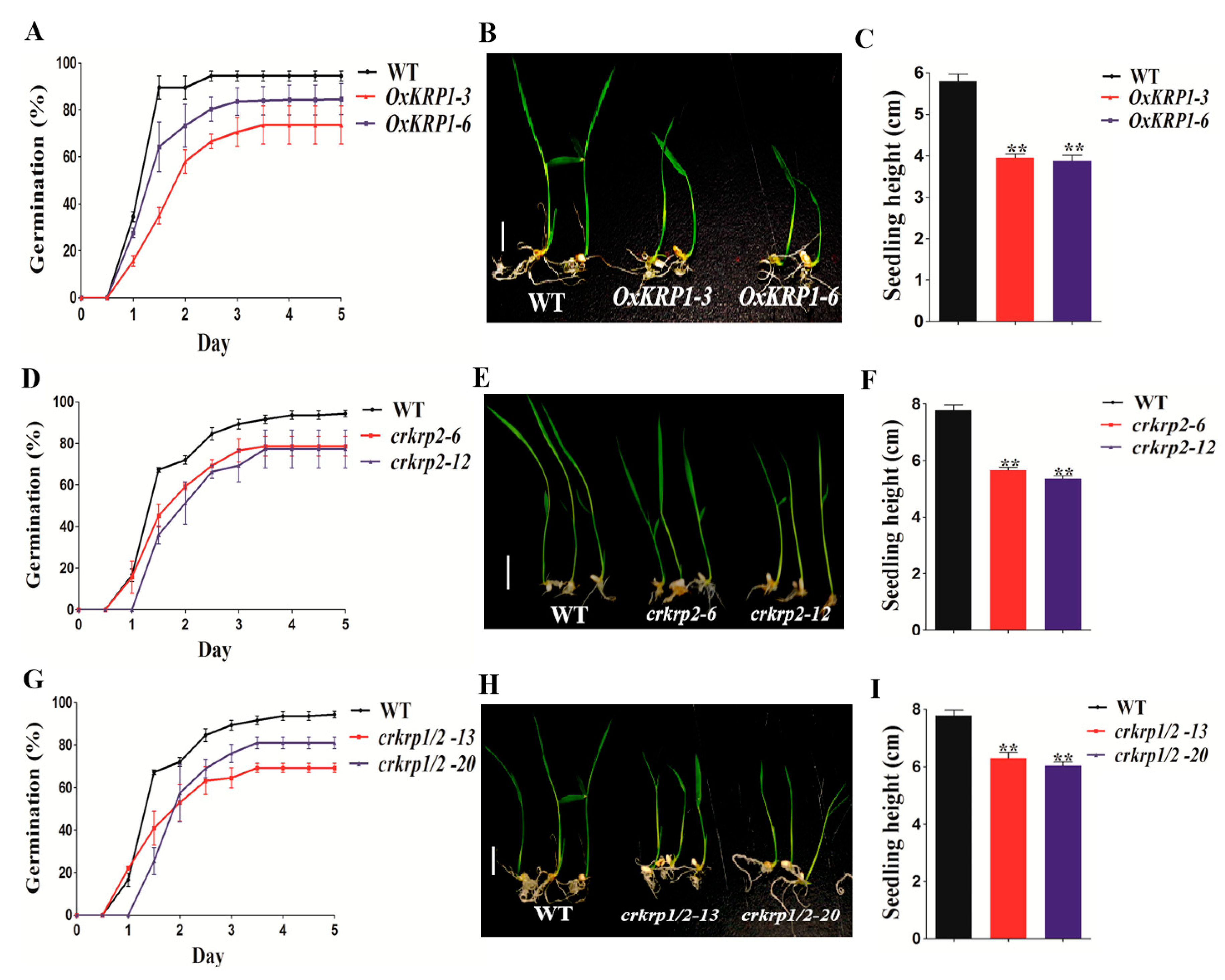
Since interrupting the equilibrium of KRP1 or KRP2 affects seed maturation, we further investigated their potential roles in seed germination. The seeds of OxKRP1, crkrp2, and crkrp1/krp2 mutants all exhibited a slower germination rate when grown on half-strength Murashige and Skoog medium (Figure 3). As shown in Figure 3A, 94.5% of the wild-type seeds germinated in 60 h, whereas 66.7% and 80.3% of the seeds of OxKRP1-3 and OxKRP1-6 germinated at the same time point, respectively (Figure 3A).
As for crkrp2 and crkrp1/krp2 mutants, the germination rates were 67.8% and 65.6% in 60 h, respectively (Figure 3D,G). Consistent with the seed germination tendency, the post-germination growth of OxKRP1, crkrp2, and crkrp1/krp2 were also significantly retarded compared with that of the wild type (Figure 3B,C,E,F,H,I). Additionally, OxKRP1, crkrp2, and crkrp1/crkrp2 mutants also displayed retarded early seedlings growth (Figure S3). Taken together, these results strongly suggested that KRP1 and KRP2 participate in normal seed germination and early seedling growth.

2.4. Grain Filling and Seed Germination Related Genes Regulated by KPR1

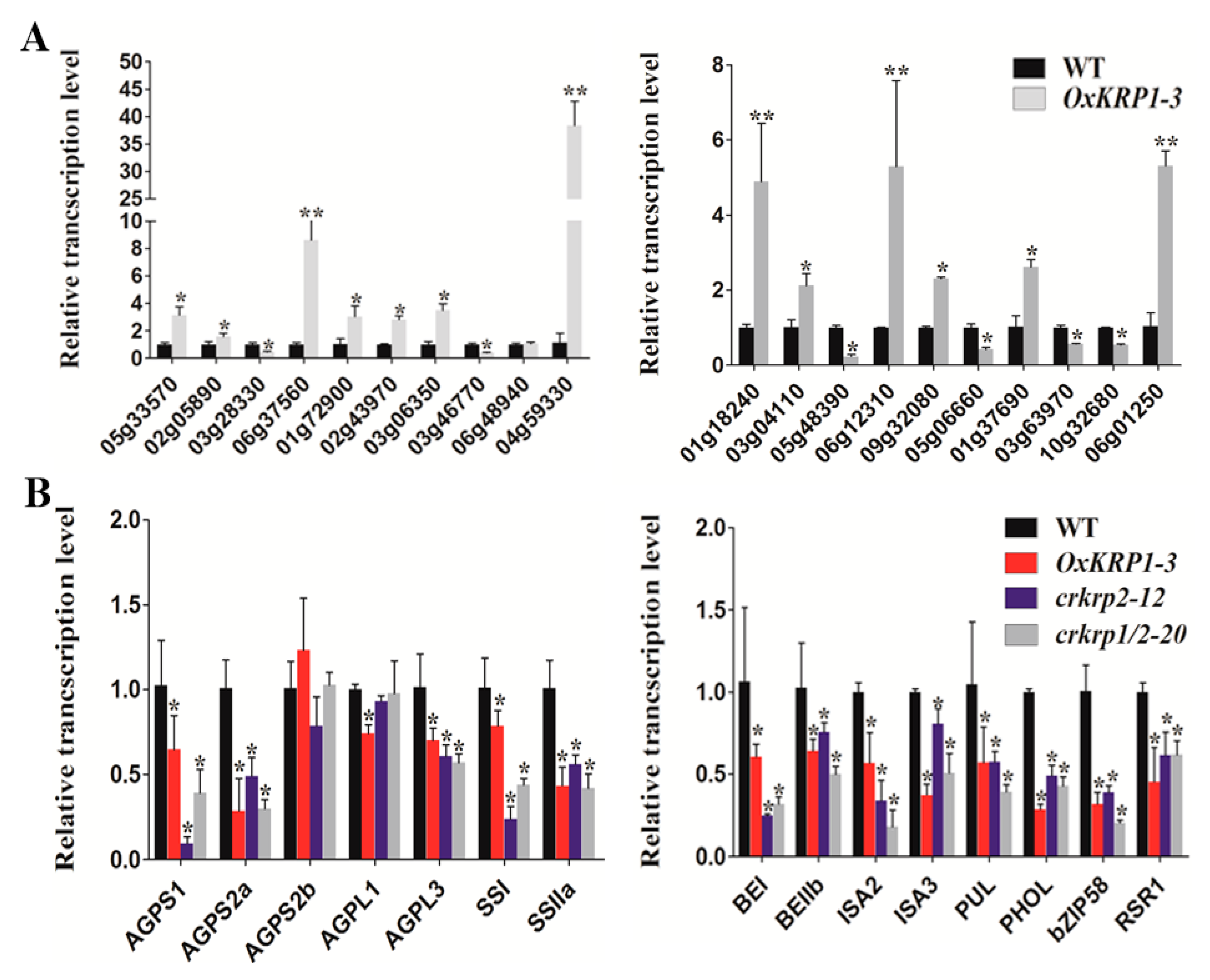
To figure out the genes regulated by KRP1, RNA-seq on the wild type and OxKRP1 developing seeds at 6 DAP (days after pollination) were carried out. As a result, a total of 1710 genes were found to be differentially expressed in OxKRP1, including 943 up-regulated genes and 767 down-regulated genes directly/indirectly mediated by KRP1 (|log 2 Ratio| ≥ 1; p value < 0.01) (Table S2). To validate the RNA-seq data, 20 DEGs (Differentially Expressed Genes) were selected to analyze the gene transcript abundance (Table S3). As shown in Figure 4A, most of the transcription levels of the chosen genes were consistent with the RNA-seq results, suggesting a high reliability of the transcriptomic data (Figure 4A).
Among these detected DEGs, several have been reported to be functionally involved in rice seed development. For example, ARAG1 (AP2/EREBP-type transcription factor) (LOC_Os02g43970), mainly expressed in germinating seeds and strongly induced by drought stress and ABA treatment [24], were up-regulated in OxKRP1 and exhibited co-expression patterns with KRP1 [24], indicating that ARAG1 could be involved in KRP1-mediated seed germination. OsPPDKB (White-core floury endosperm-4) (LOC_Os05g33570), regulating the carbon metabolism during grain filling [25,26], was also induced in OxKRP1, suggesting that KRP1 could partly modulate seed development through adjusting carbon distribution, such as starch and fatty acids. Given that some genes with minor, but statistically significant, differences may be undetected by RNA-seq, we further examined the transcription level of numerous key grain filling regulators in OxKRP1 6 DAP developing seeds, including ADP-glucose pyrophosphorylase (AGPase), soluble starch synthase (SS), starch branching enzyme (SBE), starch debranching enzyme (DBE), and starch phosphorylase L (PHOL) directly involved in amylose and amylopectin biosynthesis, and two transcription factors regulating rice starch biosynthesis such as RSR1 (Rice Starch Regulator1, a rice AP2/EREBP family transcription factor) and bZIP58 (RISBZ1, a basic leucine zipper transcription factor) during seed development [23,27]. As expected, most of these key regulators were down-regulated in OxKRP1, except for AGPS2b (Figure 4B). Interestingly, we also found the mRNA abundance of these regulators in crkrp2 and crkrp1/krp2, which exhibited a similar tendency to that of OxKRP1 (Figure 4B). These results well explained the significantly reduced seed size and grain weights in these KRPs related transgenic plants.
To functionally categorize these DEG, GO (gene ontology), and KEGG (Kyoto Encyclopedia of Genes and Genomes) pathway analyses were utilized to obtain an overview of the dynamic transcriptomic change in OxKRP1. GO analysis showed that the DEGs were preferentially catalogued into “cell part”, “organelle”, and “membrane”, whereas “symplast”, “cell junction”, and “extracellular region” were under-represented in terms of “cellular component” (Figure S4A and Table S4). From the perspective of “biological process”, these DEGs were majorly related to “cellular process”, “metabolic process”, and “regulation of biological process”, suggesting that metabolism and signal processes are required for KRP1 in plant cell cycle regulation. On the contrary, “pigmentation” and “rhythmic process” only took less than 1% (Figure S4A and Table S4). With respect to “molecular function”, we found that “catalytic activity” and “binding” were over-represented, indicating that numerous signal transduction factors or enzymes are involved in KRP1-mediated seed morphogenesis. However, only less than 1% of the DEGs were related to “transcription regulator activity” and “translation regulator activity” (Figure S4A and Table S4). However, KEGG analysis showed that DEGs related to metabolic pathways, starch, and sucrose metabolism, and the biosynthesis of secondary metabolites were highlighted (Figure S4B and Table S5). Since these pathways were tightly associated with storage compound synthesis and cell proliferation [28], the KEGG results strongly supported that KRP1 may affect seed morphogenesis and nutrient accumulation during grain filling. Additionally, CELLO (http://cello.life.nctu.edu.tw/) [29] was performed to predict the subcellular location of these DEGs. As a result, the DEGs preferentially occurred in the nucleus, plasma membrane, and chloroplast, which represented a proportion of 30.56%, 20.86%, and 15.13%, respectively, while other cellular compartments, such as peroxisomal, ER, and golgi only accounted for 0.46% of these DEGs (Figure S4C). The nuclear subcellular location of a large proportion of these DEGs indicate that KRP1 mainly performs its function in the nucleus, which was in consistent with the specific nuclear location of KRP1 in Figure 1E.

2.5. Determination of the Interactions of KRP1 with CDKs

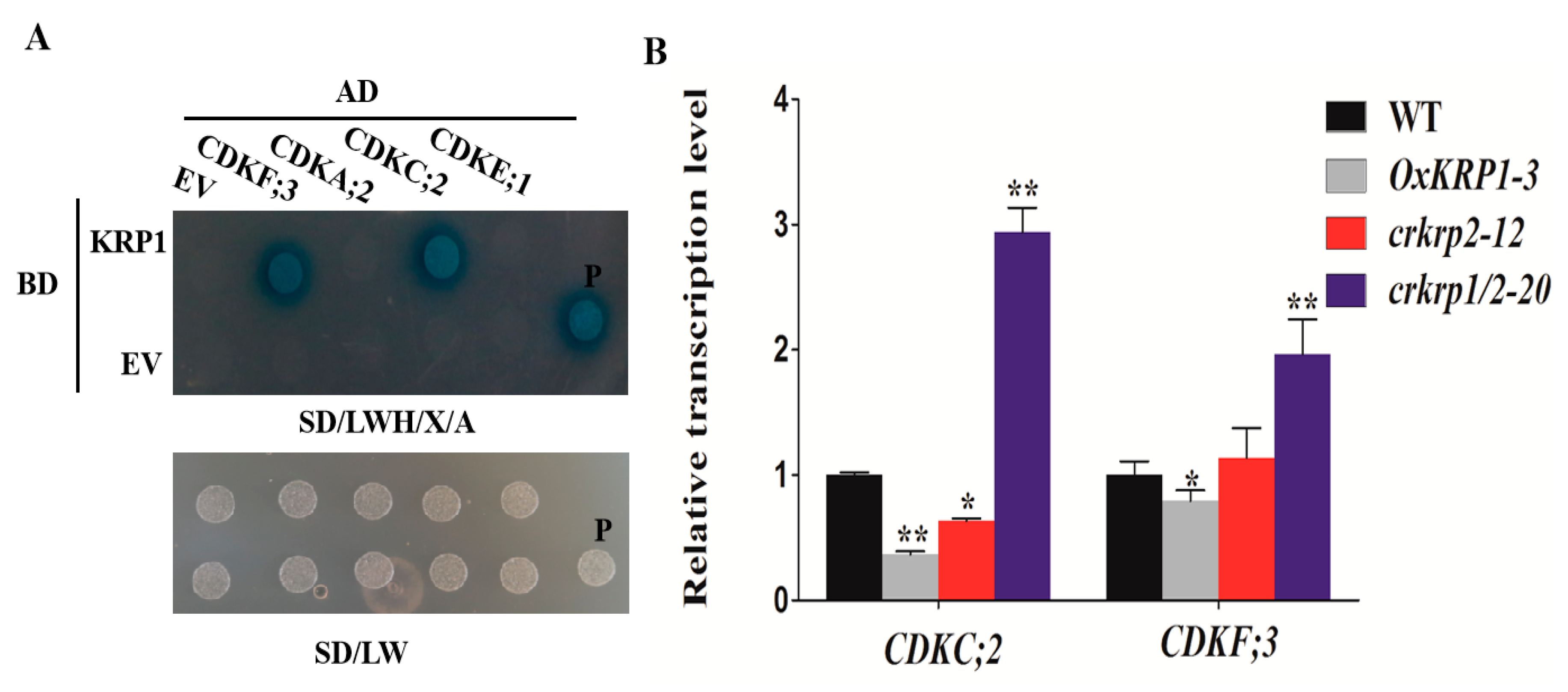
To search for the KRP1 interactive proteins, four different types of CDKs were selected to investigate their interaction with KRP1 by yeast two-hybrid assay. As shown in Figure 5A, KRP1 was able to interact with CDKC;2 (LOC_Os01g72790) and CDKF;3 (LOC_Os03g63020), but not with CDKA;2 (LOC_Os02g03060) and CDKE;1 (LOC_Os10g42950) (Figure 5A).
Furthermore, the transcription levels of these two CDK members were detected in the 6 DAP developing seeds of OxKRP1, crkrp2, and crkrp1/krp2. As shown in Figure 5B, the transcription level of CDKC;2 and CDKF;3 were both significantly down-regulated in OxKRP1, while slightly decreased in crkrp2 (Figure 5B). Interestingly, the transcription levels of CDKC;2 and CDKF;3 were drastically increased in crkrp1/krp2 double mutants (Figure 5B). These results demonstrated that the CDKC;2 and CDKF;3 are mainly inhibited by KRP1, while not KRP2 due to the functional difference of the two KRP inhibitors. However, CDKC;2 and CDKF;3 have not been functionally reported until now. Only spatial and temporal expression patterns have shown that CDKC;2 and CDKF;3 are expressed in developing seeds (0, 1, 3, 6 DAP, Day After Pollination), endosperms (9, 12, and 15 DAP) and embryos (9 and 15 DAP), as previously reported [17], indicating the potential role of the two CDK members participating in cell cycle regulation during seed development. Future work focusing on how CDKC;2 and CDKF;3 are involved in KRP1-governed grain filling and seed germination would clarify the finely regulated mechanisms of KRP1.

3. Discussion

In the past decades, several KRP inhibitors have been reported in connection with the cell cycle and plant morphogenesis. In Arabidopsis, ectopically overexpression of Arath;KRP1 under different organ-specific promoters, such as trichrome-specific GL2, petal-specific AP3, and pollen-specific Bgp1 promoters, usually inhibit the relative vegetative and reproductive organs development and lead to abnormal morphogenesis, such as impaired fertility, smaller size and reduced number of branches, and collapsed trichrome [30,31]. In monocotyledonous plants, overexpression of Orysa;KRP1 plants in Japonica rice decreased endoreduplication and thereby reduced grain filling, and overexpression of OsiICK6 in Indica rice also impaired vegetative growth and reduced seed production, similar to the phenotype observed in the overexpression plants of Arath;KRP1 driven by a constitutive promoter [12,13,14,16,19,32]. Two maize KRP inhibitors, Zeama;KRP1 and Zeama;KRP2, are characterized by the inhibiting the activities of CDK and endoreduplication during endosperm development [33]. However, the biological functions of these KRP inhibitors are mainly obtained through the phenotypic analysis of the overexpression transgenic plants, while far less was done on the mutants. In this study, KRP1 overexpression plants drastically reduced seed production through down-regulating the seed length and width (Figure 2C,F–H). Interestingly, we also discovered that the seed morphological characteristic of krp2 single (crkrp2) and krp1/krp2 double mutants (crkrp1/krp2) were largely the same as that of KRP1 overexpression plants (Figure 2D–H), suggesting that either overexpression or mutation of KRPs could disrupt the normal steady state of the two cell cycle regulators, thereby affecting cell proliferation during seed development. Moreover, most of the reported grain filling related genes were significantly down-regulated in either OxKRP1 or crkrp2 and crkrp1/krp2 developing seeds (Figure 4B), which explained well the reduced seed size and grain weight in these transgenic plants. However, slower seed germination and early retarded seedling growth were detected in OxKRP1, crkrp2, and crkrp1/krp2 mutants, possibly due to the seed morphogenesis activity blocked by KRP1 or KRP2 (Figure 3 and Figure S3). Notably, crkrp2 and crkrp1/krp2 both drastically reduced grain filling and seed germination, which reduced the functional redundancy between the two KRP members, though KRP1 and KRP2 displayed 51% amino acid sequence similarity and belonged to the same subgroup based on the phylogenetic analysis in a previous report [16].
Subcellular location is important for cell cycle regulators to perform their functions [34,35]. The tobacco CDK inhibitors NtKIS1a and NtKIS2 and all seven Arabidopsis KRP inhibitors are localized in the nucleus, and the punctuate pattern of sub-nuclear distribution is determined by the conserved protein motif ‘YLQLRSRRL’, in which the third residue is variable [19,36,37]. In this study, we first validated that KRP1 was specifically co-localized in the nucleus with the nuclear marker D53 [20] in rice protoplasts (Figure 1E), which strongly implied that KPR1 could act as a nuclear protein and interact with the downstream targets to mediate cell proliferation and plant organ development, which is also in consistent with the conserved nuclear localization of plant KRP proteins [19]. Additionally, KRP1 displayed a more homogeneous sub-nuclear punctuate pattern (Figure 1E), a partly conferred by the conserved sequence ‘YLQLRSRML’ located in KRP1, and a similar phenomenon is also detected in the sub-cellular localization of OsiICK1 in the genetic background of Indica rice [19].
Accumulating evidence has shown that CDK inhibitors could connect the phytohormones with cell cycle modulation in response to environmental cues. For example, previous studies have shown that KRP1 is induced by abscisic acid (ABA), while KRP2 involved in lateral root initiation is down-regulated by auxin in Arabidopsis [38,39,40]. KRP5 is up-regulated by auxin, while three KRP members, KRP1, 4, and 5, are inhibited by cytokinin [17]. OsiICK6 is also induced by ABA, which is similar to its Arabidopsis homology [38]. In this study, we determined that KRP1 and KRP2 were preferentially induced by ABA and BR (Figure 1C,D). Given that ABA and BR extensively regulate seed maturation and germination, as previously described [41,42,43,44,45,46], these results demonstrated that ABA and/or BR could affect the two cell cycle regulators by regulating cell proliferation during grain filling and seed germination processes.
In this study, KRP1 and KRP2 were highly expressed in 6 DAP and 9 DAP, respectively (Figure 1A,B). During seed developmental stages, the seed embryo differentiates into apical meristem and leaf primordium, and the endosperm cells gradually turn into aleurone cells and starch storage cells, and terminate cell division at around 9–10 DAP [16,47,48]. The spatial and temporal expression pattern showed that KRP1 and KRP2 were tightly involved in seed rapid cell proliferation and could act as key switches from the mitotic cell cycle to the endocycle. Additionally, previous studies have reported that the overexpression of KRP1 drastically inhibits endoreduplication by turning the number of nuclei from 12–24 C to 3 C ploidy level during endosperm formation, ultimately down-regulating seed nutrient absorption and yield [16,49]. Since crkrp2 also exhibits similar seed production as OxKRP1, it would be interesting to further figure out whether KRP2 also regulates endosperm development by hindering endoreduplication.
In the past decades, several reports have shown that KRPs directly bind and inhibit cyclin/CDK complexes and impair the latter’s activity in plants [9,14,15,18,19,32,33,38,50]. Based on the phylogenetic analysis of the catalytic site and cyclin-binding motifs, rice CDKs could be divided into eight different types (A–G-type and CKL-type), similar to CDKs in Arabidopsis [17,51]. Among these different types of CDKs, A-type CDKs (CDKA) are characterized by the PSTAIRE motif located in the cyclin-binding domain, C-type CDKs (CDKC) harbours the canonical PITAIRE motif, E-type CDKs (CDKE) is defined by its conserved SPTAIRE motif, and F-type CDKs (CDKF) could phosphorylate the threonine residue in the T-loop of other CDKs, as previously reported [2,52,53]. However, the target CDKs regulated by KRP1 in rice is still unclear. In this study, we selected four representatives CDKs to detect whether these CDK members could interact with KRP1. To our surprise, KRP1 directly interacts with CDKC;2 or CDKF;3, but not with CDKA;2 or CDKE;1 through yeast two-hybrid assay (Figure 5A). Previous studies have shown that plant KRP inhibitors mainly inhibit A-type CDK and D-type cyclins complexes [18,19,32,50]; the protein–protein interaction results suggest the functional differences of KRP1 with other KRP inhibitors in terms of the interaction with OsCDKs. Consistent with the yeast two-hybrid results, the transcription level of CDKC;2 and CDKF;3 were reduced in OxKRP1 developing seeds (Figure 5B). Additionally, CDKC;2 and CDKF;3 were detected in developing seeds (0, 3, and 6 DAP), as previously reported [17], which were overlapped with the tissue expression pattern of KRP1 (Figure 1A). These results suggest that KRP1 could down-regulate CDKC;2 or CDKF;3, and future work addressing how KRP1 regulating the activates of CDKC;2 and/or CDKF;3 will advance our understanding of the molecular mechanism of KRP1 involvement in rice grain filling and seed germination.

4. Materials and Methods

4.1. Subcellular Localization Analysis

To generate the rice protoplast, 5 g of rice leaf strips (grown in dark for two weeks) in 0.5 mm size were digested in 10 mL enzyme solution (1.5% cellulose R10, 0.75% macerozyme R10, 0.6 M mannitol, 10 mM MES pH = 7.5) for 6 h in the dark with gentle shaking (40 rpm) at 28 °C. The protoplasts were filtered and harvested by centrifugation, washed with 10 mL W5 solution (154 mM NaCl, 125 mM CaCl2, 2 mM KH2PO4, 2 mM MES, 5 mM glucose, pH = 5.7) twice, then the protoplasts were suspended in MMG solution (0.4 M mannitol, 15 mM MgCl2, 4 mM MES, pH = 5.8). The full-length the coding region of KRP1 without the stop codon was amplified and fused with eGFP in p35S-GFP vector and designated as 35S:KRP1-GFP. 35S:KRP1-GFP and 35S:D53-mKate (a nucleus marker) [20] were co-transiently expressed in the rice protoplasts and incubated in PEG (0.6 M mannitol, 100 mM CaCl2, 40% PEG4000) for 30 min at room temperature. The GFP signals were detected by observation using a Zeiss LSM710 laser confocal microscope (Carl Zeiss, AG, Jena, Germany).

4.2. Vector Construction and Plant Transformation

The CRISPR/Cas9 system was adopted, as previously described [22]. Annealed double-strand oligos of the gDNA sequences of KRP1 were ligated into the pYLgRNA-OsU3 using BsaI site (Thermo, Waltham, MA, USA). To make KPR1 overexpression construct, CDS of KRP1 were amplified and inserted between the KpnI and BamHI sites of vector pU1390, which was driven by a strong constitutive ubiquitin promoter. The primers used were listed in Table S1. All the constructed plasmids were transformed into the rice variety ‘Nipponbare’ (Oryza sativa L. cv. Nipponbare) callus using the Agrobacterium-mediated transformation method, as previously described [54].

4.3. RNA-Seq

The total RNA of 6 DAP rice seeds of the wild type (WT) and KPR1 overexpression (OxKRP1) plants were analyzed by Nanodrop 2000 spectrophotometer (Thermo, Waltham, MA, USA) and Agilent 2100 Bioanalyzer (Agilent, Santa Clara, CA, USA). Three biological replicates were used for each sample. The qualified RNA was processed for sequencing library construction, as previously described [55]. Briefly, the high-throughput sequencing was performed using the Illumina HiSeq™ 2000 platform (Illumina, Foster City, CA, USA) to obtain a standard quality library, reads with low-quality, adaptor-polluted, unknown base (N), and counts below 20 reads per million were deleted. The generated clean reads were aligned with the transcripts of rice genes in RGAP (http://rice.plantbiology.msu.edu/) using BOWTIE 2(http://bowtie-bio.sourceforge.net/bowtie2/index.shtml), and the gene expression level was calculated using RSEM (RNA-seq by expectation maximization) [56]. Differentially expressed genes (DEGs) between the three biological replicates of either the WT and OxKRP1 were identified using the EBSeq [57], and the cutoff value and fragments per kilobase of transcript per million mapped reads (|log 2 Ratio| ≥ 1; p value < 0.01) were used as thresholds to identify significant differences in gene expression.

4.4. RNA Isolation and qRT-PCR

The RNA of all the tissues, except developing seeds, was extracted by Trizol (Invitrogen, Carlsbad, CA, USA) according to the manufacturer’s instructions. For developing seeds, a modified SDS-trizol method was applied, as previously described [22]. Briefly, 0.5 g of ground seed powders were treated with 300 µL SDS RNA extraction buffer (50 mM Tris-HCl pH 8.0, 5 mM EDTA pH 8.0, 150 mM LiCl and 1% SDS), then 300 µL Phenol (pH 8.0): Chloroform = 1:1. After centrifugation was added; the RNA in the supernatant was extracted by Trizol according to the manufacturer’s instructions. The total RNA was quantified by nanodrop spectrometer (Thermo, Waltham, MA, USA) and 2 µg of each sample was used for reverse transcription using MMLV reverse transcriptase (Takara, Dalian, China) according to the manufacturer’s instructions. Quantitative RT-PCR (qRT-PCR) was performed with technical triplicates in a total of 10 μL reaction volume containing 5 µL THUNDERBIRD SYBR qPCR Mix (Toyobo, Shanghai, China), 1 µL cDNA, 0.2 µL primers, and 3.8 µL water using CFX96 touch real-time PCR detection system (Bio-rad, Hercules, CA, USA). The relative expression level of tested genes was normalized to the housekeeping gene ubiquitin (GenBank accession No. AF184280) detected in the same sample and was calculated by the 2−ΔΔCT method [58]. The primers used are listed in Table S1.

4.5. Yeast-Two-Hybrid Assay

The interactions of KRP1 with different CDKs were analysed using the Matchmaker™ Gold Yeast Two-Hybrid system (Clontech, Dalian, China) according to the manufacturer’s instructions. The CDS of KRP1 was amplified and cloned into the plasmid pGBKT7 (BD) as the bait and the CDSs of CDKA;2, CDKC;2, CDKE;1, and CDKF;3 were amplified and cloned into plasmid pGADT7 as the prey. The primers used are listed in Table S1. Different combinations of BD and AD constructs were co-transformed into the yeast strain Y2H Gold (Clontech, Dalian, China). The co-transformed yeast cell was cultured in SD/-Trp-Leu selective medium, and further screened in SD/-Trp-Leu-Ade-His selective medium with 0.04 mg/mL X-α-gal and 30 mM 3-AT to detect the interaction by visualizing the colour of the yeast colonies.

4.6. Phenotypic Analysis of Rice Transgenic Plant Seeds

The length, width, and thousand-grain-weight of the WT and all the KRP-related transgenic seeds were measured by the seed phenotyping system (Wangsheng, Hangzhou, China) and analyzed by graphpad prism 5 software (Graphpad software, San Diego, CA, USA) according to the manufacturer’s instructions with 50 biological replicates per sample. Seed germination assay was performed, as previously reported [59]. Briefly, the WT, KRP1 overexpression lines, krp2 single mutant (crkrp2), and krp1/krp2 double mutant (crkrp1/krp2) seeds were first surface sterilized in 70% ethanol for 1 min and soaked in 50% NaClO for 30 min. Then, the seeds were washed in sterilized water for five times and placed on half-strength Murashige and Skoog medium with 0.3% plant agar in a growth chamber (28 ± 2 °C, 12/12 h photoperiod with 60% relative humidity). A growth of 5 mm long coleoptile was considered a complete germination. The seed germination rate was recorded every 12 h for 5 days and triple biological replicates (each replicates containing 50 seeds) were performed for each sample.
In this study, we first systematically reported the biological function of two rice KRP inhibitors, KRP1 and KRP2, involved in grain filling and seed germination. KRP1 overexpression transgenic lines (OxKRP1), krp2 single mutant (crkrp2), and krp1/krp2 double mutant (crkrp1/krp2) all displayed poor seed production, together with retarded seed germination and impaired early seedling growth, suggesting that both KRP inhibitors play important roles in seed morphogenesis. Consistent with the phenotype of these transgenic lines, a series of grain filling key regulators, such as ADP-glucose pyrophosphorylase (AGPase), soluble starch synthase (SS), and Rice Starch Regulator1 (RSR1), were significantly down-regulated in OxKRP1, crkrp2, and crkrp1/krp2. Furthermore, we first showed that KRP1 was located in the nucleus of rice protoplasts and interacted with two cyclin-dependent protein kinases, CDKC;2 and CDKF;3, through the yeast two-hybrid system, indicating the two CDK members could participate in KRP1-mediated seed development. In a word, our work not only expands the knowledge on the finely regulated mechanism of KRPs, but also provides novel insights into the roles of KRPs in rice seed maturation and germination to facilitate plant functional genomics.

Supplementary Materials

Supplementary materials can be found at https://www.mdpi.com/1422-0067/21/1/245/s1. Table S1. Sequences of primers used in this study. Table S2. Differentially expressed genes between wild-type and OxKRP1 transgenic lines. Table S3. Selected DEGs used for RNA-seq verification. Table S4. GO term analysis for Gene Ontology (GO) in this study. Table S5. KEGG pathway analysis in this study. Figure S1. Subcellular localization of KRP1–GFP fusion protein in (A) or the nucleus marker D53 in (B) in rice protoplasts, respectively. Bar = 10 µM. Figure S2. Molecular characterization of crkrp2 and crkrp1/2 mutants. (A) Schematic presentation of the gene structure of KRP1, KRP2 and CRISPR-cas9 editing site. PAM: protospacer adjacent motif. Black boxes: untranslated regions; Brown boxes: exons; black line: intron; (B) Sanger sequencing chromatograph of the CRISPR-cas9 target site in homozygous mutants of crkrp2 and crkrp1/2, respectively. The letter in red represented the mutant sites. The letter in blue represented the PAM sequence. Figure S3. Early seedlings establishment of OxKRP1, crkrp2 and crkrp1/2 mutants. (AC) Growth phenotype of the WT, OxKRP1, crkrp2 and crkrp1/2 mutants after cultured in the hydroponic solution as described [60] for two weeks. Bar = 5 cm; (D) Seedlings height of OxKRP1, crkrp2 and crkrp1/2 mutants in correspondence to (AC). Error bars indicate SD with ten biological replicates (n = 10). Asterisks indicate the significance of differences between the WT and transgenic lines as determined by Student’s t-test analysis: ** p < 0.01. Figure S4. Functional categorization of the differentially expressed genes (DEGs) between the WT and OxKRP1 6 DAP developing seeds. (A) Enrichment analysis of the DEGs in Gene Ontology (GO) categories in terms of cellular components, biological processes and molecular functions; (B) Enrichment analysis of the DEGs in KEGG pathways; (C) Distribution of the DEGs in subcellular compartments.

Author Contributions

A.A.A., X.T., H.W., J.Z. (Juan Zhao), L.T., Z.L., Y.S., X.L., S.L., S.W. and W.L., S.M.T. performed the experiments and analyzed the data, Y.W. and J.Z. (Jian Zhang) conceived of the project, designed, coordinated the experiments, and wrote the manuscript. All authors have read and agreed to the published version of the manuscript.

Funding

This research was supported by National Natural Science Foundation of China (Grant No. 31701395), the special research funds for the Central Public Research Institute of the China National Rice Research Institute (Grant No. 2017RG002-5) and the special research funds of State Key Laboratory of Rice Biology (Grant No. 2017ZZKT10105).

Conflicts of Interest

The authors declare no competing financial interests.

References

  1. Morgan, D.O. The Cell Cycle: Principles of Control (Primers in Biology); New Science Press Ltd.: London, UK, 2007; p. 297. [Google Scholar]
  2. Joubes, J.; Chevalier, C.; Dudits, D.; Heberle-Bors, E.; Inze, D.; Umeda, M.; Renaudin, J.P. CDK-related protein kinases in plants. Plant Mol. Biol. 2000, 43, 607–620. [Google Scholar] [CrossRef] [PubMed]
  3. Torres Acosta, J.A.; Fowke, L.C.; Wang, H. Analyses of phylogeny, evolution, conserved sequences and genome-wide expression of the ICK/KRP family of plant CDK inhibitors. Ann. Bot. Lond. 2011, 107, 1141–1157. [Google Scholar] [CrossRef] [Green Version]
  4. Inze, D. Green light for the cell cycle. EMBO J. 2005, 24, 657–662. [Google Scholar] [CrossRef] [PubMed] [Green Version]
  5. Dewitte, W.; Murray, J.A. The plant cell cycle. Ann. Rev. Plant Biol. 2003, 54, 235–264. [Google Scholar] [CrossRef] [PubMed] [Green Version]
  6. De Veylder, L.; Joubes, J.; Inze, D. Plant cell cycle transitions. Curr. Opin. Plant Biol. 2003, 6, 536–543. [Google Scholar] [CrossRef] [PubMed] [Green Version]
  7. Lui, H.; Wang, H.; Delong, C.; Fowke, L.C.; Crosby, W.L.; Fobert, P.R. The Arabidopsis Cdc2a-interacting protein ICK2 is structurally related to ICK1 and is a potent inhibitor of cyclin-dependent kinase activity in vitro. Plant J. 2000, 21, 379–385. [Google Scholar] [CrossRef] [PubMed] [Green Version]
  8. Sherr, C.J.; Roberts, J.M. CDK inhibitors: Positive and negative regulators of G1-phase progression. Genes Dev. 1999, 13, 1501–1512. [Google Scholar] [CrossRef] [Green Version]
  9. Verkest, A.; Weinl, C.; Inze, D.; De Veylder, L.; Schnittger, A. Switching the cell cycle. Kip-related proteins in plant cell cycle control. Plant Physiol. 2005, 139, 1099–1106. [Google Scholar] [CrossRef] [Green Version]
  10. Sherr, C.J.; Roberts, J.M. Inhibitors of mammalian G1 cyclin-dependent kinases. Genes Dev. 1995, 9, 1149–1163. [Google Scholar] [CrossRef] [Green Version]
  11. Wang, H.; Fowke, L.C.; Crosby, W.L. A plant cyclin-dependent kinase inhibitor gene. Nature 1997, 386, 451–452. [Google Scholar] [CrossRef]
  12. De Veylder, L.; Beeckman, T.; Beemster, G.T.; Krols, L.; Terras, F.; Landrieu, I.; van der Schueren, E.; Maes, S.; Naudts, M.; Inze, D. Functional analysis of cyclin-dependent kinase inhibitors of Arabidopsis. Plant Cell 2001, 13, 1653–1668. [Google Scholar] [CrossRef] [PubMed] [Green Version]
  13. Wang, H.; Zhou, Y.; Gilmer, S.; Whitwill, S.; Fowke, L.C. Expression of the plant cyclin-dependent kinase inhibitor ICK1 affects cell division, plant growth and morphology. Plant J. 2000, 24, 613–623. [Google Scholar] [CrossRef] [PubMed]
  14. Jasinski, S.; Perennes, C.; Bergounioux, C.; Glab, N. Comparative molecular and functional analyses of the tobacco cyclin-dependent kinase inhibitor NtKIS1a and its spliced variant NtKIS1b. Plant Physiol. 2002, 130, 1871–1882. [Google Scholar] [CrossRef] [PubMed] [Green Version]
  15. Bisbis, B.; Delmas, F.; Joubes, J.; Sicard, A.; Hernould, M.; Inze, D.; Mouras, A.; Chevalier, C. Cyclin-dependent kinase (CDK) inhibitors regulate the CDK-cyclin complex activities in endoreduplicating cells of developing tomato fruit. J. Biol. Chem. 2006, 281, 7374–7383. [Google Scholar] [CrossRef] [PubMed] [Green Version]
  16. Barroco, R.M.; Peres, A.; Droual, A.M.; De Veylder, L.; Nguyen, L.S.L.; De Wolf, J.; Mironov, V.; Peerbolte, R.; Beemster, G.T.S.; Inze, D.; et al. The cyclin-dependent kinase inhibitor orysa; KRP1 plays an important role in seed development of rice. Plant Physiol. 2006, 142, 1053–1064. [Google Scholar] [CrossRef] [Green Version]
  17. Guo, J.; Song, J.; Wang, F.; Zhang, X.S. Genome-wide identification and expression analysis of rice cell cycle genes. Plant Mol. Biol. 2007, 64, 349–360. [Google Scholar] [CrossRef]
  18. Mizutani, M.; Naganuma, T.; Tsutsumi, K.; Saitoh, Y. The syncytium-specific expression of the Orysa;KRP3 CDK inhibitor: Implication of its involvement in the cell cycle control in the rice (Oryza sativa L.) syncytial endosperm. J. Exp. Bot. 2010, 61, 791–798. [Google Scholar] [CrossRef] [Green Version]
  19. Yang, R.F.; Tang, Q.C.; Wang, H.M.; Zhang, X.B.; Pan, G.; Wang, H.; Tu, J.M. Analyses of two rice (Oryza sativa) cyclin-dependent kinase inhibitors and effects of transgenic expression of OsiICK6 on plant growth and development. Ann. Bot. Lond. 2011, 107, 1087–1101. [Google Scholar] [CrossRef] [Green Version]
  20. Zhou, F.; Lin, Q.; Zhu, L.; Ren, Y.; Zhou, K.; Shabek, N.; Wu, F.; Mao, H.; Dong, W.; Gan, L.; et al. D14–SCFD3-dependent degradation of D53 regulates strigolactone signalling. Nature 2013, 504, 406–410. [Google Scholar] [CrossRef] [Green Version]
  21. Ma, X.; Zhang, Q.; Zhu, Q.; Liu, W.; Chen, Y.; Qiu, R.; Wang, B.; Yang, Z.; Li, H.; Lin, Y.; et al. A Robust CRISPR/Cas9 System for Convenient, High-Efficiency Multiplex Genome Editing in Monocot and Dicot Plants. Mol. Plant 2015, 8, 1274–1284. [Google Scholar] [CrossRef]
  22. Bello, B.K.; Hou, Y.X.; Zhao, J.; Jiao, G.A.; Wu, Y.W.; Li, Z.Y.; Wang, Y.F.; Tong, X.H.; Wang, W.; Yuan, W.Y.; et al. NF-YB1-YC12-bHLH144 complex directly activates Wx to regulate grain quality in rice (Oryza sativa L.). Plant Biotechnol. J. 2018, 17, 1–14. [Google Scholar]
  23. Fu, F.F.; Xue, H.W. Coexpression analysis identifies Rice Starch Regulator1, a rice AP2/EREBP family transcription factor, as a novel rice starch biosynthesis regulator. Plant Physiol. 2010, 154, 927–938. [Google Scholar] [CrossRef] [PubMed]
  24. Zhao, L.F.; Hu, Y.B.; Chong, K.; Wang, T. ARAG1, an ABA-responsive DREB gene, plays a role in seed germination and drought tolerance of rice. Ann. Bot. Lond. 2010, 105, 401–409. [Google Scholar] [CrossRef] [PubMed] [Green Version]
  25. Chastain, C.J.; Heck, J.W.; Colquhoun, T.A.; Voge, D.G.; Gu, X.Y. Posttranslational regulation of pyruvate, orthophosphate dikinase in developing rice (Oryza sativa) seeds. Planta 2006, 224, 924–934. [Google Scholar] [CrossRef]
  26. Kang, H.G.; Park, S.; Matsuoka, M.; An, G.H. White-core endosperm floury endosperm-4 in rice is generated by knockout mutations in the C-4-type pyruvate orthophosphate dikinase gene (OsPPDKB). Plant J. 2005, 42, 901–911. [Google Scholar] [CrossRef] [Green Version]
  27. Kawakatsu, T.; Yamamoto, M.P.; Touno, S.M.; Yasuda, H.; Takaiwa, F. Compensation and interaction between RISBZ1 and RPBF during grain filling in rice. Plant J. 2009, 59, 908–920. [Google Scholar] [CrossRef]
  28. Deng, Z.Y.; Gong, C.Y.; Wang, T. Use of proteomics to understand seed development in rice. Proteomics 2013, 13, 1784–1800. [Google Scholar] [CrossRef]
  29. Yu, C.S.; Chen, Y.C.; Lu, C.H.; Hwang, J.K. Prediction of protein subcellular localization. Proteins 2006, 64, 643–651. [Google Scholar] [CrossRef]
  30. Schnittger, A.; Weinl, C.; Bouyer, D.; Schobinger, U.; Hulskamp, M. Misexpression of the cyclin-dependent kinase inhibitor ICK1/KRP1 in single-celled Arabidopsis trichomes reduces endoreduplication and cell size and induces cell death. Plant Cell 2003, 15, 303–315. [Google Scholar] [CrossRef] [Green Version]
  31. Zhou, Y.; Wang, H.; Gilmer, S.; Whitwill, S.; Keller, W.; Fowke, L.C. Control of petal and pollen development by the plant cyclin-dependent kinase inhibitor ICK1 in transgenic Brassica plants. Planta 2002, 215, 248–257. [Google Scholar] [CrossRef]
  32. Zhou, Y.; Fowke, L.C.; Wang, H. Plant CDK inhibitors: Studies of interactions with cell cycle regulators in the yeast two-hybrid system and functional comparisons in transgenic Arabidopsis plants. Plant Cell Rep. 2002, 20, 967–975. [Google Scholar] [CrossRef]
  33. Coelho, C.M.; Dante, R.A.; Sabelli, P.A.; Sun, Y.J.; Dilkes, B.P.; Gordon-Kamm, W.J.; Larkins, B.A. Cyclin-dependent kinase inhibitors in maize endosperm and their potential role in endoreduplication. Plant Physiol. 2005, 138, 2323–2336. [Google Scholar] [CrossRef] [PubMed] [Green Version]
  34. Pines, J. Cyclins and cyclin-dependent kinases: A biochemical view. Biochem. J. 1995, 308, 697–711. [Google Scholar] [CrossRef] [PubMed]
  35. Pines, J. Four-dimensional control of the cell cycle. Nat. Cell Biol. 1999, 1, E73–E79. [Google Scholar] [CrossRef] [PubMed]
  36. Bird, D.A.; Buruiana, M.M.; Zhou, Y.; Fowke, L.C.; Wang, H. Arabidopsis cyclin-dependent kinase inhibitors are nuclear-localized and show different localization patterns within the nucleoplasm. Plant Cell Rep. 2007, 26, 861–872. [Google Scholar] [CrossRef]
  37. Zhou, Y.; Niu, H.; Brandizzi, F.; Fowke, L.C.; Wang, H. Molecular control of nuclear and subnuclear targeting of the plant CDK inhibitor ICK1 and ICK1-mediated nuclear transport of CDKA. Plant Mol. Biol. 2006, 62, 261–278. [Google Scholar] [CrossRef]
  38. Wang, H.; Qi, Q.; Schorr, P.; Cutler, A.J.; Crosby, W.L.; Fowke, L.C. ICK1, a cyclin-dependent protein kinase inhibitor from Arabidopsis thaliana interacts with both Cdc2a and CycD3, and its expression is induced by abscisic acid. Plant J. 1998, 15, 501–510. [Google Scholar] [CrossRef]
  39. Himanen, K.; Boucheron, E.; Vanneste, S.; de Almeida Engler, J.; Inze, D.; Beeckman, T. Auxin-mediated cell cycle activation during early lateral root initiation. Plant Cell 2002, 14, 2339–2351. [Google Scholar] [CrossRef] [Green Version]
  40. Richard, C.; Lescot, M.; Inze, D.; De Veylder, L. Effect of auxin, cytokinin, and sucrose on cell cycle gene expression in Arabidopsis thaliana cell suspension cultures. Plant Cell Tissue Organ Cult. 2002, 69, 167–176. [Google Scholar] [CrossRef]
  41. Shu, K.; Liu, X.D.; Xie, Q.; He, Z.H. Two Faces of One Seed: Hormonal Regulation of Dormancy and Germination. Mol. Plant 2016, 9, 34–45. [Google Scholar] [CrossRef] [Green Version]
  42. Xi, W.Y.; Liu, C.; Hou, X.L.; Yu, H. MOTHER OF FT AND TFL1 Regulates Seed Germination through a Negative Feedback Loop Modulating ABA Signaling in Arabidopsis. Plant Cell 2010, 22, 1733–1748. [Google Scholar] [CrossRef] [PubMed] [Green Version]
  43. Xi, W.; Yu, H. Mother Of ft and tfl1 regulates seed germination and fertility relevant to the brassinosteroid signaling pathway. Plant Signal Behav. 2010, 5, 1315–1317. [Google Scholar] [CrossRef] [PubMed] [Green Version]
  44. Hu, Y.; Yu, D. Brassinosteroid insensitive2 interacts with abscisic acid insensitive5 to mediate the antagonism of brassinosteroids to abscisic acid during seed germination in Arabidopsis. Plant Cell 2014, 26, 4394–4408. [Google Scholar] [CrossRef] [PubMed] [Green Version]
  45. Vaistij, F.E.; Gan, Y.B.; Penfield, S.; Gilday, A.D.; Dave, A.; He, Z.S.; Josse, E.M.; Choi, G.; Halliday, K.J.; Graham, I.A. Differential control of seed primary dormancy in Arabidopsis ecotypes by the transcription factor SPATULA. Proc. Natl. Acad. Sci. USA 2013, 110, 10866–10871. [Google Scholar] [CrossRef] [PubMed] [Green Version]
  46. Finkelstein, R.R.; Gampala, S.S.; Rock, C.D. Abscisic acid signaling in seeds and seedlings. Plant Cell 2002, 14, S15–S45. [Google Scholar] [CrossRef] [Green Version]
  47. Qiu, J.H.; Hou, Y.X.; Tong, X.H.; Wang, Y.F.; Lin, H.Y.; Liu, Q.; Zhang, W.; Li, Z.Y.; Nallamilli, B.R.; Zhang, J. Quantitative phosphoproteomic analysis of early seed development in rice (Oryza sativa L.). Plant Mol. Biol. 2016, 90, 249–265. [Google Scholar] [CrossRef]
  48. Bleckmann, A.; Alter, S.; Dresselhaus, T. The beginning of a seed: Regulatory mechanisms of double fertilization. Front. Plant Sci. 2014, 5, 452. [Google Scholar] [CrossRef] [Green Version]
  49. Larkins, B.A.; Dilkes, B.P.; Dante, R.A.; Coelho, C.M.; Woo, Y.M.; Liu, Y. Investigating the hows and whys of DNA endoreduplication. J. Exp. Bot. 2001, 52, 183–192. [Google Scholar] [CrossRef]
  50. Van Leene, J.; Hollunder, J.; Eeckhout, D.; Persiau, G.; Van De Slijke, E.; Stals, H.; Van Isterdael, G.; Verkest, A.; Neirynck, S.; Buffel, Y.; et al. Targeted interactomics reveals a complex core cell cycle machinery in Arabidopsis thaliana. Mol. Syst. Biol. 2010, 6, 397. [Google Scholar] [CrossRef]
  51. Menges, M.; de Jager, S.M.; Gruissem, W.; Murray, J.A. Global analysis of the core cell cycle regulators of Arabidopsis identifies novel genes, reveals multiple and highly specific profiles of expression and provides a coherent model for plant cell cycle control. Plant J. 2005, 41, 546–566. [Google Scholar] [CrossRef]
  52. Mironov, V.V.; De Veylder, L.; Van Montagu, M.; Inze, D. Cyclin-dependent kinases and cell division in plants—The nexus. Plant Cell 1999, 11, 509–522. [Google Scholar] [PubMed] [Green Version]
  53. Shimotohno, A.; Matsubayashi, S.; Yamaguchi, M.; Uchimiya, H.; Umeda, M. Differential phosphorylation activities of CDK-activating kinases in Arabidopsis thaliana. FEBS Lett. 2003, 534, 69–74. [Google Scholar] [CrossRef] [Green Version]
  54. Hiei, Y.; Ohta, S.; Komari, T.; Kumashiro, T. Efficient transformation of rice (Oryza sativa L.) mediated by Agrobacterium and sequence analysis of the boundaries of the T-DNA. Plant J. 1994, 6, 271–282. [Google Scholar] [CrossRef] [PubMed] [Green Version]
  55. Hou, Y.; Wang, L.; Wang, L.; Liu, L.; Li, L.; Sun, L.; Rao, Q.; Zhang, J.; Huang, S. JMJ704 positively regulates rice defense response against Xanthomonas oryzae pv. oryzae infection via reducing H3K4me2/3 associated with negative disease resistance regulators. BMC Plant Biol. 2015, 15, 286. [Google Scholar] [CrossRef] [Green Version]
  56. Li, B.; Dewey, C.N. RSEM: Accurate transcript quantification from RNA-Seq data with or without a reference genome. BMC Bioinform. 2011, 12. [Google Scholar] [CrossRef] [Green Version]
  57. Leng, N.; Dawson, J.A.; Thomson, J.A.; Ruotti, V.; Rissman, A.I.; Smits, B.M.G.; Haag, J.D.; Gould, M.N.; Stewart, R.M.; Kendziorski, C. EBSeq: An empirical Bayes hierarchical model for inference in RNA-seq experiments. Bioinformatics 2013, 29, 1035–1043. [Google Scholar] [CrossRef] [Green Version]
  58. Livak, K.J.; Schmittgen, T.D. Analysis of relative gene expression data using real-time quantitative PCR and the 2(-Delta Delta C(T)) Method. Methods 2001, 25, 402–408. [Google Scholar] [CrossRef]
  59. Lin, Q.B.; Wu, F.Q.; Sheng, P.K.; Zhang, Z.; Zhang, X.; Guo, X.P.; Wang, J.L.; Cheng, Z.J.; Wang, J.; Wang, H.Y.; et al. The SnRK2-APC/C-TE regulatory module mediates the antagonistic action of gibberellic acid and abscisic acid pathways. Nat. Commun. 2015, 6. [Google Scholar] [CrossRef] [Green Version]
  60. Wang, X.F.; Wang, Y.F.; Pineros, M.A.; Wang, Z.Y.; Wang, W.X.; Li, C.Y.; Wu, Z.C.; Kochian, L.V.; Wu, P. Phosphate transporters OsPHT1;9 and OsPHT1;10 are involved in phosphate uptake in rice. Plant Cell Environ. 2014, 37, 1159–1170. [Google Scholar] [CrossRef]
Figure 1. Expression pattern of KRP1 and KRP2. (A,B) qRT-PCR analysis of KRP1 and KRP2 transcription abundances in various rice tissues, respectively. Asterisks indicate the significant difference of other tissues compared with callus, as determined by Student’s t-test analysis: * p < 0.05. ** p < 0.01; (C,D) qRT-PCR analysis of KRP1 and KRP2 transcription abundances in response to various phytohormone treatments, respectively. ABA: abscisic acid; GA: gibberellic acid; BR: brassinosteroids; JA: Jasmonic acid; (E) subcellular localization of KRP1–GFP fusion protein in rice protoplasts. D53 fused with mKate was used as a nuclear marker as described [20]. Bar = 10 µM. Error bars indicated SD with biological triplicates (n = 3) in (AD). Primers used for qRT-PCR analysis were listed in Table S1.
Ijms 21 00245 g001
Figure 2. Phenotypic characteristics of OxKRP1, crkrp2, and crkrp1/2 mature seeds. (A) qRT-PCR analysis for transcript accumulation of KRP1 in the 6 DAP developing seeds of OxKRP1 transgenic lines. Error bars indicate the SD with biological triplicates (n = 3); (B) qRT-PCR analysis for transcript accumulation of KRP1 and KRP2 in the 6 DAP developing seeds of crkrp2 and crkrp1/2 mutants. Error bars indicate the SD with biological triplicates (n = 3); (C) comparison of mature seeds of the WT and OxKRP1 plants. OxKRP1-3 and OxKRP1-6 represent two independent KRP1-overexpressing lines, respectively; and (D) comparison of mature seeds of the WT and crkrp2 plants. crkrp2-6 and crkrp2-12 represent two independent crkrp2 mutants, respectively; (E) comparison of mature seeds of the WT and crkrp1/2 plants. crkrp1/2-13 and crkrp1/2-20 represent two independent crkrp1/2 double mutants, respectively; (FH) seed length, seed width and 1000-seed weight of the WT, OxKRP1, crkrp2, and crkrp1/2 mature seeds. Error bars indicate the SD with 50 biological replicates (n = 50). Asterisks indicate the significant difference between the WT and transgenic lines, as determined by Student’s t-test analysis: * p < 0.05, ** p < 0.01. WT: wild type, Bar = 1 cm in (CE).
Ijms 21 00245 g002
Figure 3. Seed germination characteristics of OxKRP1, crkrp2, and crkrp1/2 mutants. (A) Germination time courses of the WT and OxKRP1 on half-strength MS medium, respectively; (B) germination phenotypes of the WT and OxKRP1 growth on half-strength MS medium for 5 days, respectively; (C) seedling heights of the WT and OxKRP1 growth on half-strength MS medium for 5 days, respectively; (D) germination time courses of the WT and crkrp2 mutants on half-strength MS medium, respectively; (E) germination phenotypes of the WT and crkrp2 mutants growth on half-strength MS medium for 5 days, respectively; (F) seedling heights of the WT and crkrp2 mutants growth on half-strength MS medium for 5 days, respectively; (G) germination time courses of the WT and crkrp1/2 mutants on half-strength MS medium, respectively; (H) germination phenotypes of the WT and crkrp1/2 mutants growth on half-strength MS medium for 5 days, respectively; (I) seedling heights of the WT and crkrp1/2 mutants growth on half-strength MS medium for 5 days, respectively. Photographs were taken on day 5, Bar = 1 cm in (B,E,H). Error bars indicate SD with triple biological replicates (n = 3, each replicates containing 50 seeds) in (A,D,G). Error bars indicate SD with 50 biological replicates (n = 50) in (C,F,I). Asterisks indicate the significant difference between the WT and transgenic lines, as determined by Student’s t-test analysis: ** p < 0.01 in (C,F,I). WT: wild type, MS: Murashige and Skoog medium.
Ijms 21 00245 g003
Figure 4. Expression analysis of KPR1 regulated genes by qRT-PCR. (A) qRT-PCR validation of the DEGs revealed by RNA-seq experiments; (B) qRT-PCR analysis of the transcriptional abundances of grain filling related genes in the WT, OxKRP1, crkrp2, and crkrp1/2 mutants. 6 DAP seed cDNA were used as templates for this analysis. DEGs: Differentially Expressed Genes. Error bars indicate SD with biological triplicates (n = 3). Asterisks indicate the significant difference between the WT and transgenic lines, as determined by Student’s t-test analysis: * p < 0.05, ** p < 0.01. Primers used in (A) were listed in Table S1. Primers used in (B) were used according to previous reports [22,23].
Ijms 21 00245 g004
Figure 5. Yeast-two-hybrid assay of KRP1-CDKs interaction. (A) Yeast two-hybrid assays. Yeast cells co-transformed with CDKC;2 or CDKF;3 fused to the GAL4 activation domain (CDKC;2-AD or CDKF;3-AD) and KRP1 fused to the GAL4 binding domain (KRP1-BD) were grown on selective media. BD, pGBKT7; AD, pGADT7; EV, empty vector; SD/LW, -Leu-Trp; SD/LWH, -Leu-Trp-His; P, positive control, pGADT7-T+pGBKT7-53; X: X-α-Gal, 0.04 mg/mL; and A: Aureobasidin A, 100 ng/mL. (B) qRT-PCR analysis of the transcriptional abundances of CDKC;2 and CDKF;3 in the WT, OxKRP1, crkrp2, and crkrp1/2 mutants. Error bars indicate the SD with biological triplicates (n = 3). Asterisks indicate the significant difference between the WT and transgenic lines, as determined by Student’s t-test analysis: * p < 0.05, ** p < 0.01. WT: wild type. The primers used for qRT-PCR analysis were listed in Table S1.
Ijms 21 00245 g005