How to Make Anticancer Drugs Cross the Blood–Brain Barrier to Treat Brain Metastases


Abstract
1. Introduction
2. Molecular Biology of Brain Metastasis and Potential Targeted Therapies
3. Is the BBB Disrupted in the Case of Brain Metastases?
4. Limited Brain Delivery of Anticancer Drugs
- -
- (« Blood-brain barrier » [Mesh]) AND (« neoplasm » [Mesh] OR « Cancer » OR « malignant tumor ») AND (« Brain neoplasm » OR « Brain metastasis ») AND (« antineoplasic agents» [Mesh] OR « Chemotherapy » OR « Anticancer drug ») AND (« passage »),
- -
- (« antineoplasic agents» [MeSH Terms]) AND (“Blood–Brain Barrier/drug effects” [Majr]) AND (“pharmacokinetic”)
- -
- (“anticancer drugs”) AND (“blood brain barrier” (AND (“penetration”)
- -
- (“Cerebro Spinal Fluid penetration” (AND (“anticancer drugs” OR “antineoplasic agents”).
5. Enhancing the Therapeutic Delivery of Drugs from Blood to Brain
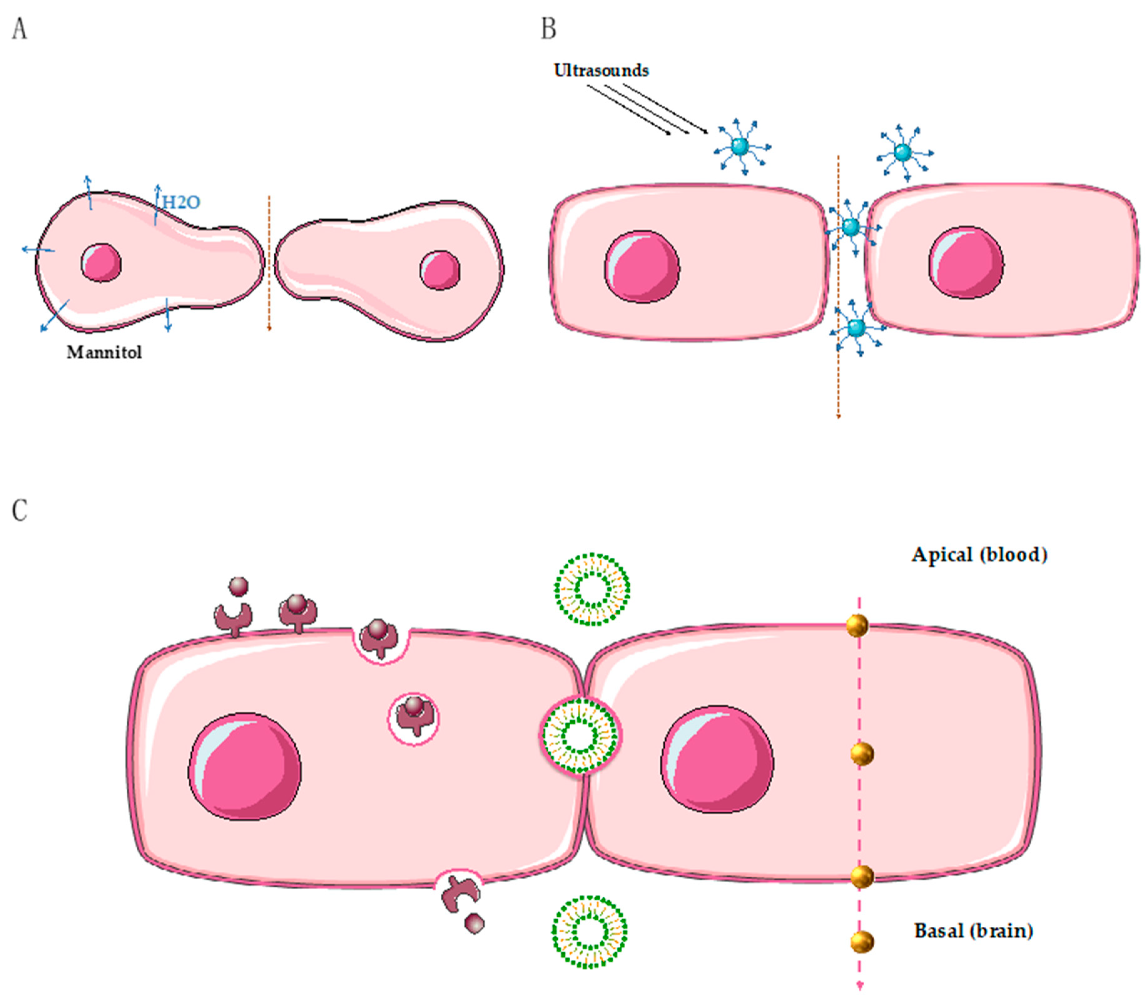
5.1. Mechanical Disruption of the Blood–Brain Barrier to Deliver Drugs to the Brain
5.2. Intranasal Delivery of Drugs
5.3. The Use of Nanoparticles to Cross the BBB
5.4. Combinations of Drugs Targeting Receptor-Mediated Transport
6. Conclusions
Author Contributions
Funding
Conflicts of Interest
Abbreviations
| BBB | Blood–brain barrier |
| BTB | Blood–tumor barrier |
| CSF | Cerebrospinal fluid |
| HER | Human epidermal receptor |
| EGFR | Epidermal growth factor receptor |
| TKI | Tyrosine kinase inhibitor |
References
- Riihimäki, M.; Hemminki, A.; Fallah, M.; Thomsen, H.; Sundquist, K.; Sundquist, J.; Hemminki, K. Metastatic sites and survival in lung cancer. Lung Cancer 2014, 86, 78–84. [Google Scholar] [CrossRef] [PubMed]
- Leyland-Jones, B. Human Epidermal Growth Factor Receptor 2–Positive Breast Cancer and Central Nervous System Metastases. J. Clin. Oncol. 2009, 27, 5278–5286. [Google Scholar] [CrossRef] [PubMed]
- Lowery, F.J.; Yu, D. Brain metastasis: Unique challenges and open opportunities. Biochim. Biophys. Acta Rev. Cancer 2017, 1867, 49–57. [Google Scholar] [CrossRef] [PubMed]
- Caroli, M.; di Cristofori, A.; Lucarella, F.; Raneri, F.A.; Portaluri, F.; Gaini, S.M. Surgical Brain Metastases: Management and Outcome Related to Prognostic Indexes: A Critical Review of a Ten-Year Series. ISRN Surg. 2011, 2011, 1–8. [Google Scholar] [CrossRef] [PubMed]
- Ramakrishna, N.; Temin, S.; Chandarlapaty, S.; Crews, J.R.; Davidson, N.E.; Esteva, F.J.; Giordano, S.H.; Gonzalez-Angulo, A.M.; Kirshner, J.J.; Krop, I.; et al. Recommendations on Disease Management for Patients With Advanced Human Epidermal Growth Factor Receptor 2–Positive Breast Cancer and Brain Metastases: American Society of Clinical Oncology Clinical Practice Guideline. J. Clin. Oncol. 2014, 32, 2100–2108. [Google Scholar] [CrossRef] [PubMed]
- Spadoni, I.; Fornasa, G.; Rescigno, M. Organ-specific protection mediated by cooperation between vascular and epithelial barriers. Nat. Rev. Immunol. 2017, 17, 761–773. [Google Scholar] [CrossRef]
- Wong, A.D.; Ye, M.; Levy, A.F.; Rothstein, J.D.; Bergles, D.E.; Searson, P.C. The blood-brain barrier: an engineering perspective. Front. Neuroeng. 2013, 6. [Google Scholar] [CrossRef]
- Enerson, B.E.; Drewes, L.R. The rat blood—Brain barrier transcriptome. J. Cereb. Blood Flow Metab. 2006, 26, 959–973. [Google Scholar] [CrossRef]
- Yachida, S.; Jones, S.; Bozic, I.; Antal, T.; Leary, R.; Fu, B.; Kamiyama, M.; Hruban, R.H.; Eshleman, J.R.; Nowak, M.A.; et al. Distant metastasis occurs late during the genetic evolution of pancreatic cancer. Nature 2010, 467, 1114–1117. [Google Scholar] [CrossRef]
- Gerlinger, M.; Rowan, A.J.; Horswell, S.; Math, M.; Larkin, J.; Endesfelder, D.; Grönroos, E.; Martinez, P.; Matthews, N.; Stewart, A.; et al. Intratumor heterogeneity and branched evolution revealed by multiregion sequencing. N. Engl. J. Med. 2012, 366, 883–892. [Google Scholar] [CrossRef]
- McPherson, A.; Roth, A.; Laks, E.; Masud, T.; Bashashati, A.; Zhang, A.W.; Ha, G.; Biele, J.; Yap, D.; Wan, A.; et al. Divergent modes of clonal spread and intraperitoneal mixing in high-grade serous ovarian cancer. Nat. Genet. 2016, 48, 758–767. [Google Scholar] [CrossRef] [PubMed]
- Yates, L.R.; Gerstung, M.; Knappskog, S.; Desmedt, C.; Gundem, G.; Van Loo, P.; Aas, T.; Alexandrov, L.B.; Larsimont, D.; Davies, H.; et al. Subclonal diversification of primary breast cancer revealed by multiregion sequencing. Nat. Med. 2015, 21, 751–759. [Google Scholar] [CrossRef] [PubMed]
- Dagogo-Jack, I.; Shaw, A.T. Tumour heterogeneity and resistance to cancer therapies. Nat. Rev. Clin. Oncol. 2018, 15, 81–94. [Google Scholar] [CrossRef] [PubMed]
- Tyran, M.; Carbuccia, N.; Garnier, S.; Guille, A.; Adelaïde, J.; Finetti, P.; Touzlian, J.; Viens, P.; Tallet, A.; Goncalves, A.; et al. A Comparison of DNA Mutation and Copy Number Profiles of Primary Breast Cancers and Paired Brain Metastases for Identifying Clinically Relevant Genetic Alterations in Brain Metastases. Cancers 2019, 11, 665. [Google Scholar] [CrossRef]
- Lee, J.Y.; Park, K.; Lim, S.H.; Kim, H.S.; Yoo, K.H.; Jung, K.S.; Song, H.-N.; Hong, M.; Do, I.-G.; Ahn, T.; et al. Mutational profiling of brain metastasis from breast cancer: Matched pair analysis of targeted sequencing between brain metastasis and primary breast cancer. Oncotarget 2015, 6, 43731–43742. [Google Scholar] [CrossRef]
- Lee, J.Y.; Park, K.; Lee, E.; Ahn, T.; Jung, H.H.; Lim, S.H.; Hong, M.; Do, I.-G.; Cho, E.Y.; Kim, D.-H.; et al. Gene Expression Profiling of Breast Cancer Brain Metastasis. Sci. Rep. 2016, 6, 28623. [Google Scholar] [CrossRef]
- Vareslija, D.; Priedigkeit, N.; Fagan, A.; Purcell, S.; Cosgrove, N.; O’Halloran, P.J.; Ward, E.; Cocchiglia, S.; Hartmaier, R.; A Castro, C.; et al. Transcriptome Characterization of Matched Primary Breast and Brain Metastatic Tumors to Detect Novel Actionable Targets. J. Natl. Cancer Inst. 2019, 111, 388–398. [Google Scholar] [CrossRef]
- Schrijver, W.A.; Selenica, P.; Lee, J.Y.; Ng, C.K.Y.; Burke, K.A.; Piscuoglio, S.; Berman, S.H.; Reis-Filho, J.S.; Weigelt, B.; Van Diest, P.J.; et al. Mutation Profiling of Key Cancer Genes in Primary Breast Cancers and Their Distant Metastases. Cancer Res. 2018, 78, 3112–3121. [Google Scholar] [CrossRef]
- Da Silva, L.; Simpson, P.T.; E Smart, C.; Cocciardi, S.; Waddell, N.; Lane, A.; Morrison, B.J.; Vargas, A.C.; Healey, S.; Beesley, J.; et al. HER3 and downstream pathways are involved in colonization of brain metastases from breast cancer. Breast Cancer Res. 2010, 12, R46. [Google Scholar] [CrossRef]
- Bollig-Fischer, A.; Michelhaugh, S.K.; Wijesinghe, P.; Dyson, G.; Kruger, A.; Palanisamy, N.; Choi, L.; Alosh, B.; Ali-Fehmi, R.; Mittal, S. Cytogenomic profiling of breast cancer brain metastases reveals potential for repurposing targeted therapeutics. Oncotarget 2015, 6, 14614–14624. [Google Scholar] [CrossRef]
- Schulten, H.-J.; Bangash, M.; Karim, S.; Dallol, A.; Hussein, D.; Merdad, A.; Al-Thoubaity, F.K.; Al-Maghrabi, J.; Jamal, A.; Al-Ghamdi, F.; et al. Comprehensive molecular biomarker identification in breast cancer brain metastases. J. Transl. Med. 2017, 15, 269. [Google Scholar] [CrossRef] [PubMed]
- Priedigkeit, N.; Hartmaier, R.J.; Chen, Y.; Vareslija, D.; Basudan, A.; Watters, R.J.; Thomas, R.; Leone, J.P.; Lucas, P.C.; Bhargava, R.; et al. Intrinsic Subtype Switching and Acquired ERBB2/HER2 Amplifications and Mutations in Breast Cancer Brain Metastases. JAMA Oncol. 2017, 3, 666–671. [Google Scholar] [CrossRef] [PubMed]
- Paik, P.K.; Shen, R.; Won, H.; Rekhtman, N.; Wang, L.; Sima, C.S.; Arora, A.; Seshan, V.; Ladanyi, M.; Berger, M.F.; et al. Next-Generation Sequencing of Stage IV Squamous Cell Lung Cancers Reveals an Association of PI3K Aberrations and Evidence of Clonal Heterogeneity in Patients with Brain Metastases. Cancer Discov. 2015, 5, 610–621. [Google Scholar] [CrossRef] [PubMed]
- Wang, H.; Ou, Q.; Li, D.; Qin, T.; Bao, H.; Hou, X.; Wang, K.; Wang, F.; Deng, Q.; Liang, J.; et al. Genes associated with increased brain metastasis risk in non-small cell lung cancer: Comprehensive genomic profiling of 61 resected brain metastases versus primary non-small cell lung cancer (Guangdong Association Study of Thoracic Oncology 1036). Cancer 2019, 125, 3535–3544. [Google Scholar] [CrossRef]
- Li, F.; Sun, L.; Zhang, S. Acquirement of DNA copy number variations in non-small cell lung cancer metastasis to the brain. Oncol. Rep. 2015, 34, 1701–1707. [Google Scholar] [CrossRef][Green Version]
- Chen, G.; Chakravarti, N.; Aardalen, K.; Lazar, A.J.; Tetzlaff, M.T.; Wubbenhorst, B.; Kim, S.-B.; Kopetz, S.; LeDoux, A.A.; Gopal, Y.V.; et al. Molecular profiling of patient-matched brain and extracranial melanoma metastases implicates the PI3K pathway as a therapeutic target. Clin. Cancer Res. 2014, 20, 5537–5546. [Google Scholar] [CrossRef]
- Fischer, G.M.; Jalali, A.; Kircher, D.A.; Lee, W.-C.; McQuade, J.L.; Haydu, L.E.; Joon, A.Y.; Reuben, A.; De Macedo, M.P.; Carapeto, F.C.L.; et al. Molecular Profiling Reveals Unique Immune and Metabolic Features of Melanoma Brain Metastases. Cancer Discov. 2019, 9, 628–645. [Google Scholar] [CrossRef]
- Ferguson, S.D.; Zheng, S.; Xiu, J.; Zhou, S.; Khasraw, M.; Brastianos, P.K.; Kesari, S.; Hu, J.; Rudnick, J.; Salacz, M.E.; et al. Profiles of brain metastases: Prioritization of therapeutic targets: Profiling brain metastases. Int. J. Cancer 2018, 143, 3019–3026. [Google Scholar] [CrossRef]
- Brastianos, P.K.; Carter, S.L.; Santagata, S.; Cahill, D.P.; Taylor-Weiner, A.; Jones, R.T.; Van Allen, E.M.; Lawrence, M.S.; Horowitz, P.M.; Cibulskis, K.; et al. Genomic Characterization of Brain Metastases Reveals Branched Evolution and Potential Therapeutic Targets. Cancer Discov. 2015, 5, 1164–1177. [Google Scholar] [CrossRef]
- Saunus, J.M.; Quinn, M.C.; Patch, A.-M.; Pearson, J.V.; Bailey, P.J.; Nones, K.; McCart, R.A.E.; Miller, D.; Wilson, P.J.; Al-Ejeh, F.; et al. Integrated genomic and transcriptomic analysis of human brain metastases identifies alterations of potential clinical significance: Integrated genomic and transcriptomic analysis of brain metastases. J. Pathol. 2015, 237, 363–378. [Google Scholar] [CrossRef]
- Janzer, R.C.; Raff, M.C. Astrocytes induce blood–brain barrier properties in endothelial cells. Nature 1987, 325, 253–257. [Google Scholar] [CrossRef] [PubMed]
- Pardridge, W.M. CNS drug design based on principles of blood-brain barrier transport. J. Neurochem. 2002, 70, 1781–1792. [Google Scholar] [CrossRef] [PubMed]
- Liu, S.; Agalliu, I.; Yu, C.; Fisher, M. The role of pericytes in blood-brain barrier function and stroke. Curr. Pharm. Des. 2012, 18, 3653–3662. [Google Scholar] [CrossRef] [PubMed]
- Abbott, N.J.; Rönnbäck, L.; Hansson, E.; R, L. Astrocyte–endothelial interactions at the blood–brain barrier. Nat. Rev. Neurosci. 2006, 7, 41–53. [Google Scholar] [CrossRef] [PubMed]
- Do, J.; Foster, D.; Renier, C.; Vogel, H.; Rosenblum, S.; Doyle, T.C.; Tse, V.; Wapnir, I. Ex vivo Evans blue assessment of the blood brain barrier in three breast cancer brain metastasis models. Breast Cancer Res. Treat. 2014, 144, 93–101. [Google Scholar] [CrossRef] [PubMed]
- Dréan, A.; Goldwirt, L.; Verreault, M.; Canney, M.; Schmitt, C.; Guehennec, J.; Delattre, J.-Y.; Carpentier, A.; Idbaih, A. Blood-brain barrier, cytotoxic chemotherapies and glioblastoma. Expert Rev. Neurother. 2016, 16, 1285–1300. [Google Scholar] [CrossRef] [PubMed]
- Liebner, S.; Fischmann, A.; Rascher, G.; Duffner, F.; Grote, E.-H.; Kalbacher, H.; Wolburg, H. Claudin-1 and claudin-5 expression and tight junction morphology are altered in blood vessels of human glioblastoma multiforme. Acta Neuropathol. 2000, 100, 323–331. [Google Scholar] [CrossRef]
- Papadopoulos, M.; Saadoun, S.; Binder, D.; Manley, G.; Krishna, S.; Verkman, A. Molecular mechanisms of brain tumor edema. Neuroscience 2004, 129, 1009–1018. [Google Scholar] [CrossRef]
- Papadopoulos, M.C.; Saadoun, S.; Woodrow, C.J.; Davies, D.C.; Costa-Martins, P.; Moss, R.F.; Krishna, S.; Bell, B.A. Occludin expression in microvessels of neoplastic and non-neoplastic human brain. Neuropathol. Appl. Neurobiol. 2001, 27, 384–395. [Google Scholar] [CrossRef]
- Lyle, L.T.; Lockman, P.R.; Adkins, C.E.; Mohammad, A.S.; Sechrest, E.; Hua, E.; Palmieri, D.; Liewehr, D.J.; Steinberg, S.M.; Kloc, W.; et al. Alterations in Pericyte Subpopulations Are Associated with Elevated Blood-Tumor Barrier Permeability in Experimental Brain Metastasis of Breast Cancer. Clin. Cancer Res. 2016, 22, 5287–5299. [Google Scholar] [CrossRef]
- Kevil, C.G.; Payne, D.K.; Mire, E.; Alexander, J.S. Vascular permeability factor/vascular endothelial cell growth factor-mediated permeability occurs through disorganization of endothelial junctional proteins. J. Biol. Chem. 1998, 273, 15099–15103. [Google Scholar] [CrossRef] [PubMed]
- Gril, B.; Paranjape, A.N.; Woditschka, S.; Hua, E.; Dolan, E.L.; Hanson, J.; Wu, X.; Kloc, W.; Izycka-Swieszewska, E.; Duchnowska, R.; et al. Reactive astrocytic S1P3 signaling modulates the blood–tumor barrier in brain metastases. Nat. Commun. 2018, 9, 2705. [Google Scholar] [CrossRef] [PubMed]
- Heideman, R.L.; E Cole, D.; Balis, F.; Sato, J.; Reaman, G.H.; Packer, R.J.; Singher, L.J.; Ettinger, L.J.; Gillespie, A.; Sam, J. Phase I and pharmacokinetic evaluation of thiotepa in the cerebrospinal fluid and plasma of pediatric patients: evidence for dose-dependent plasma clearance of thiotepa. Cancer Res. 1989, 49, 736–741. [Google Scholar] [PubMed]
- Ostermann, S.; Csajka, C.; Buclin, T.; Leyvraz, S.; Lejeune, F.; Decosterd, L.A.; Stupp, R. Plasma and Cerebrospinal Fluid Population Pharmacokinetics of Temozolomide in Malignant Glioma Patients. Clin. Cancer Res. 2004, 10, 3728–3736. [Google Scholar] [CrossRef] [PubMed]
- Csordás, K.; Hegyi, M.; Eipel, O.T.; Müller, J.; Erdélyi, D.J.; Kovacs, G.T. Comparison of pharmacokinetics and toxicity after high-dose methotrexate treatments in children with acute lymphoblastic leukemia. Anti-Cancer Drugs 2013, 24, 189–197. [Google Scholar] [CrossRef] [PubMed]
- Blaney, S.M.; E Cole, D.; Balis, F.M.; Godwin, K.; Poplack, D.G. Plasma and cerebrospinal fluid pharmacokinetic study of topotecan in nonhuman primates. Cancer Res. 1993, 53, 725–727. [Google Scholar] [PubMed]
- Blaney, S.M.; Takimoto, C.; Murry, D.J.; Kuttesch, N.; McCully, C.; Cole, D.E.; Godwin, K.; Balis, F.M. Plasma and cerebrospinal fluid pharmacokinetics of 9-aminocamptothecin (9-AC), irinotecan (CPT-11), and SN-38 in nonhuman primates. Cancer Chemother. Pharmacol. 1998, 41, 464–468. [Google Scholar] [CrossRef]
- Jacobs, S.; McCully, C.L.; Murphy, R.F.; Bacher, J.; Balis, F.M.; Fox, E. Extracellular fluid concentrations of cisplatin, carboplatin, and oxaliplatin in brain, muscle, and blood measured using microdialysis in nonhuman primates. Cancer Chemother. Pharmacol. 2010, 65, 817–824. [Google Scholar] [CrossRef]
- Relling, M.V.; Mahmoud, H.H.; Pui, C.H.; Sandlund, J.T.; Rivera, G.K.; Ribeiro, R.C.; Crist, W.M.; Evans, W.E. Etoposide achieves potentially cytotoxic concentrations in CSF of children with acute lymphoblastic leukemia. J. Clin. Oncol. 1996, 14, 399–404. [Google Scholar] [CrossRef]
- Warren, K.E.; Patel, M.C.; McCully, C.M.; Montuenga, L.M.; Balis, F.M. Effect of P-glycoprotein modulation with cyclosporin A on cerebrospinal fluid penetration of doxorubicin in non-human primates. Cancer Chemother. Pharmacol. 2000, 45, 207–212. [Google Scholar] [CrossRef]
- Berg, S.L.; Reid, J.; Godwin, K.; Murry, D.J.; Poplack, D.G.; Balis, F.M.; Ames, M.M. Pharmacokinetics and cerebrospinal fluid penetration of daunorubicin, idarubicin, and their metabolites in the nonhuman primate model. J. Pediatr. Hematol. 1999, 21, 26–30. [Google Scholar] [CrossRef]
- Widemann, B.C.; Balis, F.M.; Godwin, K.S.; McCully, C.; Adamson, P.C. The plasma pharmacokinetics and cerebrospinal fluid penetration of the thymidylate synthase inhibitor raltitrexed (Tomudex) in a nonhuman primate model. Cancer Chemother. Pharmacol. 1999, 44, 439–443. [Google Scholar] [CrossRef] [PubMed]
- Tije, A.J.T.; Loos, W.J.; Zhao, M.; Baker, S.D.; Enting, R.H.; Van Der Meulen, H.; Verweij, J.; Sparreboom, A. Limited cerebrospinal fluid penetration of docetaxel. Anti-Cancer Drugs 2004, 15, 715–718. [Google Scholar] [CrossRef] [PubMed]
- Kumthekar, P.; Grimm, S.A.; Avram, M.J.; Kaklamani, V.; Helenowski, I.; Rademaker, A.; Cianfrocca, M.; Gradishar, W.; Patel, J.; Mulcahy, M.; et al. Pharmacokinetics and efficacy of pemetrexed in patients with brain or leptomeningeal metastases. J. Neuro Oncol. 2013, 112, 247–255. [Google Scholar] [CrossRef]
- Stapleton, S.L.; Reid, J.M.; Thompson, P.A.; Ames, M.M.; McGovern, R.M.; McGuffey, L.; Nuchtern, J.; Dauser, R.; Blaney, S.M. Plasma and cerebrospinal fluid pharmacokinetics of pemetrexed after intravenous administration in non-human primates. Cancer Chemother. Pharmacol. 2007, 59, 461–466. [Google Scholar] [CrossRef]
- Arndt, C.; Balis, F.M.; McCully, C.L.; Colvin, O.M.; Poplack, D.G. Cerebrospinal fluid penetration of active metabolites of cyclophosphamide and ifosfamide in rhesus monkeys. Cancer Res. 1988, 48, 2113–2115. [Google Scholar]
- Kiewe, P.; Neumann, M.; Wagner, T.; SeyfertHeike, S.; Thiel, A.; Korfel, A. Penetration of ifosfamide and its active metabolite 4-OH-ifosfamide into cerebrospinal fluid of patients with CNS malignancies. Cancer Chemother. Pharmacol. 2011, 67, 27–33. [Google Scholar] [CrossRef]
- Kellie, S.J.; Barbaric, A.; Koopmans, P.; Earl, J.; Carr, D.J.; De Graaf, S.S.N. Cerebrospinal fluid concentrations of vincristine after bolus intravenous dosing: a surrogate marker of brain penetration. Cancer 2002, 94, 1815–1820. [Google Scholar] [CrossRef]
- Jackson, D.V.; Sethi, V.S.; Spurr, C.L.; McWhorter, J.M. Pharmacokinetics of vincristine in the cerebrospinal fluid of humans. Cancer Res. 1981, 41, 1466–1468. [Google Scholar]
- Kerr, J.Z.; Berg, S.L.; Dauser, R.; Nuchtern, J.; Egorin, M.J.; McGuffey, L.; Aleksic, A.; Blaney, S. Plasma and cerebrospinal fluid pharmacokinetics of gemcitabine after intravenous administration in nonhuman primates. Cancer Chemother. Pharmacol. 2001, 47, 411–414. [Google Scholar] [CrossRef]
- Tan, J.; Li, M.; Zhong, W.; Hu, C.; Gu, Q.; Xie, Y. Tyrosine kinase inhibitors show different anti-brain metastases efficacy in NSCLC: A direct comparative analysis of icotinib, gefitinib, and erlotinib in a nude mouse model. Oncotarget 2017, 8, 98771–98781. [Google Scholar] [CrossRef] [PubMed]
- Neville, K.; Parise, R.A.; Blaney, S.M.; Thompson, P.; Aleksic, A.; Egorin, M.J.; Balis, F.M.; McGuffey, L.; McCully, C.; Berg, S.L. Plasma and cerebrospinal fluid pharmacokinetics of imatinib after administration to nonhuman primates. Clin. Pharmacol. Ther. 2004, 75, P59. [Google Scholar] [CrossRef]
- Ballard, P.; Yates, J.W.; Yang, Z.; Kim, N.-W.; Cantarini, M.; Pickup, K.; Jordan, A.; Hickey, M.; Grist, M.; Box, M.; et al. Preclinical Comparison of Osimertinib with Other EGFR-TKIs in EGFR-Mutant NSCLC Brain Metastases Models, and Early Evidence of Clinical Brain Metastases Activity. Clin. Cancer Res. 2016, 22, 5130–5140. [Google Scholar] [CrossRef] [PubMed]
- Chang, H.-Y.; Morrow, K.; Bonacquisti, E.; Zhang, W.; Shah, D.K. Antibody pharmacokinetics in rat brain determined using microdialysis. mAbs 2018, 10, 1–11. [Google Scholar] [CrossRef]
- Harjunpää, A.; Wiklund, T.; Collan, J.; Janes, R.; Rosenberg, J.; Lee, D.; Grillo-López, A.; Meri, S. Complement Activation in Circulation and Central Nervous System after Rituximab (Anti-CD20) Treatment of B-Cell Lymphoma. Leuk. Lymphoma 2001, 42, 731–738. [Google Scholar] [CrossRef]
- Zylber-Katz, E.; Gomori, J.M.; Schwartz, A.; Lossos, A.; Bokstein, F.; Siegal, T. Pharmacokinetics of methotrexate in cerebrospinal fluid and serum after osmotic blood-brain barrier disruption in patients with brain lymphoma. Clin. Pharmacol. Ther. 2000, 67, 631–641. [Google Scholar] [CrossRef]
- Morikawa, N.; Mori, T.; Abe, T.; Kawashima, H.; Takeyama, M.; Hori, S. Pharmacokinetics of Etoposide and Carboplatin in Cerebrospinal Fluid and Plasma during Hyperosmotic Disruption of the Blood Brain Barrier and Intraarterial Combination Chemotherapy. Biol. Pharm. Bull. 1999, 22, 428–431. [Google Scholar] [CrossRef][Green Version]
- Brightman, M.W.; Hori, M.; Rapoport, S.I.; Reese, T.S.; Westergaard, E. Osmotic opening of tight junctions in cerebral endothelium. J. Comp. Neurol. 1973, 152, 317–325. [Google Scholar] [CrossRef]
- Knuutinen, O.; Kuitunen, H.; Alahuhta, S.; Isokangas, J.-M.; Sonkajarvi, E.; Turpeenniemi-Hujanen, T.; Kuittinen, O. Case Report: Chemotherapy in Conjunction With Blood–Brain Barrier Disruption for a Patient With Germ Cell Tumor With Multiple Brain Metastases. Clin. Genitourin. Cancer 2018, 16, e993–e996. [Google Scholar] [CrossRef]
- Fortin, D.; Gendron, C.; Boudrias, M.; Garant, M.-P. Enhanced chemotherapy delivery by intraarterial infusion and blood-brain barrier disruption in the treatment of cerebral metastasis. Cancer 2007, 109, 751–760. [Google Scholar] [CrossRef]
- Haluska, M.; Anthony, M.L. Osmotic Blood-Brain Barrier Modification for the Treatment of Malignant Brain Tumors. Clin. J. Oncol. Nurs. 2004, 8, 263–267. [Google Scholar] [CrossRef] [PubMed]
- Shang, X.; Wang, P.; Liu, Y.; Zhang, Z.; Xue, Y. Mechanism of low-frequency ultrasound in opening blood-tumor barrier by tight junction. J. Mol. Neurosci. 2011, 43, 364–369. [Google Scholar] [CrossRef] [PubMed]
- Sheikov, N.; McDannold, N.; Jolesz, F.; Zhang, Y.-Z.; Tam, K.; Hynynen, K. Brain arterioles show more active vesicular transport of blood-borne tracer molecules than capillaries and venules after focused ultrasound-evoked opening of the blood-brain barrier. Ultrasound Med. Biol. 2006, 32, 1399–1409. [Google Scholar] [CrossRef] [PubMed]
- Arvanitis, C.D.; Askoxylakis, V.; Guo, Y.; Datta, M.; Kloepper, J.; Ferraro, G.B.; Bernabeu, M.O.; Fukumura, D.; McDannold, N.; Jain, R.K. Mechanisms of enhanced drug delivery in brain metastases with focused ultrasound-induced blood–tumor barrier disruption. Proc. Natl. Acad. Sci. USA 2018, 115, E8717–E8726. [Google Scholar] [CrossRef]
- Carpentier, A.; Canney, M.; Vignot, A.; Reina, V.; Beccaria, K.; Horodyckid, C.; Karachi, C.; Leclercq, D.; Lafon, C.; Chapelon, J.-Y.; et al. Clinical trial of blood-brain barrier disruption by pulsed ultrasound. Sci. Transl. Med. 2016, 8, 343. [Google Scholar] [CrossRef] [PubMed]
- Thorne, R.; Pronk, G.; Padmanabhan, V.; Frey, W. Delivery of insulin-like growth factor-I to the rat brain and spinal cord along olfactory and trigeminal pathways following intranasal administration. Neuroscience 2004, 127, 481–496. [Google Scholar] [CrossRef]
- Sakane, T.; Yamashita, S.; Yata, N.; Sezaki, H. Transnasal Delivery of 5-Fluorouracil to the Brain in the Rat. J. Drug Target. 1999, 7, 233–240. [Google Scholar] [CrossRef]
- Shingaki, T.; Inoue, D.; Furubayashi, T.; Sakane, T.; Katsumi, H.; Yamamoto, A.; Yamashita, S. Transnasal Delivery of Methotrexate to Brain Tumors in Rats: A New Strategy for Brain Tumor Chemotherapy. Mol. Pharm. 2010, 7, 1561–1568. [Google Scholar] [CrossRef]
- League-Pascual, J.C.; Lester-McCully, C.M.; Shandilya, S.; Ronner, L.; Rodgers, L.; Cruz, R.; Peer, C.J.; Figg, W.D.; Warren, K.E. Plasma and cerebrospinal fluid pharmacokinetics of select chemotherapeutic agents following intranasal delivery in a non-human primate model. J. Neuro Oncol. 2017, 132, 401–407. [Google Scholar] [CrossRef]
- Peterson, A.; Bansal, A.; Hofman, F.; Chen, T.C.; Zada, G. A systematic review of inhaled intranasal therapy for central nervous system neoplasms: an emerging therapeutic option. J. Neuro Oncol. 2014, 116, 437–446. [Google Scholar] [CrossRef]
- Da Fonseca, C.O.; Schwartsmann, G.; Fischer, J.; Nagel, J.; Futuro, D.; Quirico-Santos, T.; Gattass, C.R. Preliminary results from a phase I/II study of perillyl alcohol intranasal administration in adults with recurrent malignant gliomas. Surg. Neurol. 2008, 70, 259–266. [Google Scholar] [CrossRef] [PubMed]
- Da Fonseca, C.O.; Simão, M.; Lins, I.R.; Caetano, R.O.; Futuro, D.; Quirico-Santos, T. Efficacy of monoterpene perillyl alcohol upon survival rate of patients with recurrent glioblastoma. J. Cancer Res. Clin. Oncol. 2011, 137, 287–293. [Google Scholar] [CrossRef] [PubMed]
- Da Fonseca, C.; Teixeira, R.M.; Silva, J.C.T.; Fischer, J.D.S.D.G.; Meirelles, O.C.; Landeiro, J.A.; Quirico-Santos, T. Long-term outcome in patients with recurrent malignant glioma treated with Perillyl alcohol inhalation. Anticancer. Res. 2013, 33, 5625–5631. [Google Scholar] [PubMed]
- Chen, T.C.; da Fonseca, C.O.; Schönthal, A.H. Intranasal Perillyl Alcohol for Glioma Therapy: Molecular Mechanisms and Clinical Development. Int. J. Mol. Sci. 2018, 19, 3905. [Google Scholar] [CrossRef] [PubMed]
- Kreuter, J. Nanoparticulate systems for brain delivery of drugs. Adv. Drug Deliv. Rev. 2001, 47, 65–81. [Google Scholar] [CrossRef]
- Patel, T.; Zhou, J.; Piepmeier, J.M.; Saltzman, W.M. Polymeric nanoparticles for drug delivery to the central nervous system. Adv. Drug Deliv. Rev. 2012, 64, 701–705. [Google Scholar] [CrossRef] [PubMed]
- Khaitan, D.; Reddy, P.L.; Ningaraj, N.S. Targeting Brain Tumors with Nanomedicines: Overcoming Blood Brain Barrier Challenges. Curr. Clin. Pharmacol. 2018, 13, 110–119. [Google Scholar] [CrossRef]
- Jose, S.; Juna, B.; Cinu, T.; Jyoti, H.; Aleykutty, N. Carboplatin loaded Surface modified PLGA nanoparticles: Optimization, characterization, and in vivo brain targeting studies. Colloids Surf. B Biointerfaces 2016, 142, 307–314. [Google Scholar] [CrossRef]
- Tahara, K.; Kato, Y.; Yamamoto, H.; Kreuter, J.; Kawashima, Y. Intracellular drug delivery using polysorbate 80-modified poly(D,L-lactide-co-glycolide) nanospheres to glioblastoma cells. J. Microencapsul. 2011, 28, 29–36. [Google Scholar] [CrossRef]
- Tian, X.-H.; Lin, X.-N.; Wei, F.; Feng, W.; Huang, Z.-C.; Wang, P.; Ren, L.; Diao, Y. Enhanced brain targeting of temozolomide in polysorbate-80 coated polybutylcyanoacrylate nanoparticles. Int. J. Nanomed. 2011, 6, 445–452. [Google Scholar]
- Gulyaev, A.E.; Gelperina, S.E.; Skidan, I.N.; Antropov, A.S.; Kivman, G.Y.; Kreuter, J. Significant transport of doxorubicin into the brain with polysorbate 80-coated nanoparticles. Pharm. Res. 1999, 16, 1564–1569. [Google Scholar] [CrossRef]
- Gelperina, S.; Khalansky, A.; Skidan, I.; Smirnova, Z.; Bobruskin, A.; Severin, S.; Turowski, B.; Zanella, F.; Kreuter, J. Toxicological studies of doxorubicin bound to polysorbate 80-coated poly(butyl cyanoacrylate) nanoparticles in healthy rats and rats with intracranial glioblastoma. Toxicol. Lett. 2002, 126, 131–141. [Google Scholar] [CrossRef]
- Nunes, T.; Pons, T.; Hou, X.; Van Do, K.; Caron, B.; Rigal, M.; Di Benedetto, M.; Palpant, B.; Leboeuf, C.; Janin, A.; et al. Pulsed-laser irradiation of multifunctional gold nanoshells to overcome trastuzumab resistance in HER2-overexpressing breast cancer. J. Exp. Clin. Cancer Res. 2019, 38, 306. [Google Scholar] [CrossRef]
- Gromnicova, R.; Davies, H.A.; Sreekanthreddy, P.; Romero, I.A.; Lund, T.; Roitt, I.M.; Phillips, J.B.; Male, D.K. Glucose-Coated Gold Nanoparticles Transfer across Human Brain Endothelium and Enter Astrocytes In Vitro. PLoS ONE 2013, 8, e81043. [Google Scholar] [CrossRef]
- Jensen, S.A.; Day, E.S.; Ko, C.H.; Hurley, L.A.; Luciano, J.P.; Kouri, F.M.; Merkel, T.J.; Luthi, A.J.; Patel, P.C.; Cutler, J.I.; et al. Spherical nucleic acid nanoparticle conjugates as an RNAi-based therapy for glioblastoma. Sci. Transl. Med. 2013, 5, 209ra152. [Google Scholar] [CrossRef]
- Karim, R.; Palazzo, C.; Evrard, B.; Piel, G. Nanocarriers for the treatment of glioblastoma multiforme: Current state-of-the-art. J. Control. Release 2016, 227, 23–37. [Google Scholar] [CrossRef]
- I Koukourakis, M.; Koukouraki, S.; Fezoulidis, I.; Kelekis, N.; Kyrias, G.; Archimandritis, S.; Karkavitsas, N. High intratumoural accumulation of stealth® liposomal doxorubicin (Caelyx®) in glioblastomas and in metastatic brain tumours. Br. J. Cancer 2000, 83, 1281–1286. [Google Scholar] [CrossRef]
- Shaw, T.K.; Mandal, D.; Dey, G.; Pal, M.M.; Paul, P.; Chakraborty, S.; Ali, K.A.; Mukherjee, B.; Bandyopadhyay, A.K.; Mandal, M. Successful delivery of docetaxel to rat brain using experimentally developed nanoliposome: a treatment strategy for brain tumor. Drug Deliv. 2017, 24, 346–357. [Google Scholar] [CrossRef]
- Mohammad, A.S.; Griffith, J.I.; Adkins, C.E.; Shah, N.; Sechrest, E.; Dolan, E.L.; Terrell-Hall, T.B.; Hendriks, B.S.; Lee, H.; Lockman, P.R. Liposomal Irinotecan Accumulates in Metastatic Lesions, Crosses the Blood-Tumor Barrier (BTB), and Prolongs Survival in an Experimental Model of Brain Metastases of Triple Negative Breast Cancer. Pharm. Res. 2018, 35, 31. [Google Scholar] [CrossRef]
- Lee, H.; Shields, A.F.; Siegel, B.A.; Miller, K.D.; Krop, I.; Ma, C.X.; Lorusso, P.M.; Munster, P.N.; Campbell, K.; Gaddy, D.F.; et al. 64Cu-MM-302 Positron Emission Tomography Quantifies Variability of Enhanced Permeability and Retention of Nanoparticles in Relation to Treatment Response in Patients with Metastatic Breast Cancer. Clin. Cancer Res. 2017, 23, 4190–4202. [Google Scholar] [CrossRef]
- Lajoie, J.M.; Shusta, E.V. Targeting Receptor-Mediated Transport for Delivery of Biologics Across the Blood-Brain Barrier. Annu. Rev. Pharmacol. Toxicol. 2015, 55, 613–631. [Google Scholar] [CrossRef]
- Tortorella, S.; Karagiannis, T.C. Transferrin Receptor-Mediated Endocytosis: A Useful Target for Cancer Therapy. J. Membr. Biol. 2014, 247, 291–307. [Google Scholar] [CrossRef]
- Laske, D.W.; Youle, R.J.; Oldfield, E.H. Tumor regression with regional distribution of the targeted toxin TF-CRM107 in patients with malignant brain tumors. Nat. Med. 1997, 3, 1362–1368. [Google Scholar] [CrossRef]
- Laske, D.W.; Muraszko, K.M.; Oldfield, E.H.; DeVroom, H.L.; Sung, C.; Dedrick, R.L.; Simon, T.R.; Colandrea, J.; Copeland, C.; Katz, D.; et al. Intraventricular immunotoxin therapy for leptomeningeal neoplasia. Neurosurgery 1997, 41, 1039–1051. [Google Scholar] [CrossRef]
- Wei, L.; Guo, X.-Y.; Yang, T.; Yu, M.-Z.; Chen, D.-W.; Wang, J.-C. Brain tumor-targeted therapy by systemic delivery of siRNA with Transferrin receptor-mediated core-shell nanoparticles. Int. J. Pharm. 2016, 510, 394–405. [Google Scholar] [CrossRef]
- Wyatt, E.A.; Davis, M.E. Method of establishing breast cancer brain metastases affects brain uptake and efficacy of targeted, therapeutic nanoparticles. Bioeng. Transl. Med. 2019, 4, 30–37. [Google Scholar] [CrossRef]
- Chang, J.; Paillard, A.; Passirani, C.; Morille, M.; Benoit, J.P.; Betbeder, D.; Garcion, E. Transferrin adsorption onto PLGA nanoparticles governs their interaction with biological systems from blood circulation to brain cancer cells. Pharm. Res. 2012, 29, 1495–1505. [Google Scholar] [CrossRef]
- Wagner, S.; Zensi, A.; Wien, S.L.; Tschickardt, S.E.; Maier, W.; Vogel, T.; Worek, F.; Pietrzik, C.U.; Kreuter, J.; Von Briesen, H. Uptake Mechanism of ApoE-Modified Nanoparticles on Brain Capillary Endothelial Cells as a Blood-Brain Barrier Model. PLoS ONE 2012, 7, e32568. [Google Scholar] [CrossRef]
- Molino, Y.; David, M.; Varini, K.; Jabès, F.; Gaudin, N.; Fortoul, A.; Bakloul, K.; Masse, M.; Bernard, A.; Drobecq, L.; et al. Use of LDL receptor–targeting peptide vectors forin vitroandin vivocargo transport across the blood-brain barrier. FASEB J. 2017, 31, 1807–1827. [Google Scholar] [CrossRef]
- Thomas, F.C.; Taskar, K.; Rudraraju, V.; Goda, S.; Thorsheim, H.R.; Gaasch, J.A.; Mittapalli, R.K.; Palmieri, D.; Steeg, P.S.; Lockman, P.R.; et al. Uptake of ANG1005, a novel paclitaxel derivative, through the blood-brain barrier into brain and experimental brain metastases of breast cancer. Pharm. Res. 2009, 26, 2486–2494. [Google Scholar] [CrossRef]
- Kurzrock, R.; Gabrail, N.; Chandhasin, C.; Moulder, S.; Smith, C.; Brenner, A.; Sankhala, K.; Mita, A.; Elian, K.; Bouchard, D.; et al. Safety, pharmacokinetics, and activity of GRN1005, a novel conjugate of Angiopep-2, a peptide facilitating brain penetration, and paclitaxel, in patients with advanced solid tumors. Mol. Cancer Ther. 2012, 11, 308–316. [Google Scholar] [CrossRef]
- Drappatz, J.; Brenner, A.; Wong, E.T.; Eichler, A.; Schiff, D.; Groves, M.D.; Mikkelsen, T.; Rosenfeld, S.; Sarantopoulos, J.; Meyers, C.A.; et al. Phase I Study of GRN1005 in Recurrent Malignant Glioma. Clin. Cancer Res. 2013, 19, 1567–1576. [Google Scholar] [CrossRef]
- Kumthekar, P.; Tang, S.; Brenner, A.J.; Kesari, S.; Anders, C.K.; Carrillo, J.A.; Chalasani, P.; Kabos, P.; Ahluwalia, M.S.; Ibrahim, N.K. OS7.2 A Phase II Study of ANG1005, a novel BBB/BCB Penetratant Taxane in Patients with Recurrent Brain Metastases and Leptomeningeal Carcinomatosis from Breast Cancer. Neuro-Oncology 2016, 18, iv16. [Google Scholar] [CrossRef][Green Version]
- Wang, X.; Xiong, Z.; Liu, Z.; Huang, X.; Jiang, X. Angiopep-2/IP10-EGFRvIIIscFv modified nanoparticles and CTL synergistically inhibit malignant glioblastoma. Sci. Rep. 2018, 8, 12827. [Google Scholar] [CrossRef]
- Regina, A.; Demeule, M.; Tripathy, S.; Lord-Dufour, S.; Currie, J.C.; Iddir, M.; Annabi, B.; Castaigne, J.P.; Lachowicz, J.E. ANG4043, a novel brain-penetrant peptide-mAb conjugate, is efficacious against HER2-positive intracranial tumors in mice. Mol. Cancer Ther. 2015, 14, 129–140. [Google Scholar] [CrossRef]


| Primary Localization | Histological Subtype | Patient Numbers/ BM Analyzed | Materials and Analyses | Examples of Driver mutations or CNV Acquired in BM | Reference |
|---|---|---|---|---|---|
| Breast | All | 14/14 | DNA | PIK3CA EGFR FGFR1 | Tyran 2019 [14] |
| All | 45/42 | DNA | TP53 (ns) | Lee JY 2015 [15] | |
| All | 61/61 | RNA Protein | SOX2 OLIG2 ERBB2 | Lee JY 2016 [16] | |
| All | 21/21 | RNA Protein | RET HER2 amplification | Varešlija 2019 [17] | |
| ER negative | 17/ 9 | DNA | TP53 PIK3CA SMAD4 RB1 | Schrijver 2018 [18] | |
| All | 78/52 | DNA RNA protein | PIK3CA HER3 EGFR HRAS/KRAS/NRAS | Da Silva 2010 [19] | |
| All | 10/10 | DNA Protein | PTEN FBXW7 ERBB2 KIT | Bollig-Fisher 2015 [20] | |
| All | 35/3 | DNA RNA Protein | TP53 ERBB2 BRCA1–2 IDH1 CDH1 | Schulten 2017 [21] | |
| All | 20/20 | DNA RNA Protein | ERBB2 FGFR4 EGFR ESR1 | Priedigkeit 2017 [22] | |
| Lung | Squamous | 79/9 | DNA RNA Protein | Total of 23 genes | Paik 2015 [23] |
| NSCLC | 61/61 | DNA | PIK3CA EGFR MET ROS1 VEGFA CCND1 CDKN2A/2B | Wang 2019 [24] | |
| NSCLC | 1/1 | DNA Protein | PTEN CDKN2A | Li 2015 [25] | |
| Melanoma | 16/16 | DNA RNA Protein | PI3K/AKT pathway genes | Chen G 2014 [26] | |
| 74/88 | DNA RNA Protein | Increase in oxidative phosphorylation gene expression | Fisher 2019 [27] | ||
| Both | Lung Breast Melanoma | NA/493 | DNA protein | TOP2A cMET (melanoma) HER2 (breast) EGFR | Ferguson 2018 [28] |
| Lung Breast Renal carcinoma | 86/86 | DNA | PTEN EGFR PI3K/AKT pathway genes HER2 amplification MCL1 amplification | Brastianos 2015 [29] | |
| Breast Lung Melanoma Esophagus | 36/36 | DNA RNA | MAP3K4 COL5A1 | Saunus 2015 [30] |
| Drug Family | CSF/Plasma Ratio (%) | Species Studied | Time | Ref |
|---|---|---|---|---|
| Chemotherapy | ||||
| Thiotepa | 100 (for thiotepa and metabolite) | Human (children) | AUC 0–24 h | [43] |
| Temozolomide | 20 | Human | AUC 0–5 h | [44] |
| Methotrexate | 2.8 | Human (children) | 24 h | [45] |
| Topotecan | 32 | RHM | AUC 0–60 min | [46] |
| Irinotecan | 9.6–16 Metabolite SN 38: <3 | RHM | AUC 0–48 h | [47] |
| Cisplatin | 3 | RHM | AUC 0–4 h | [48] |
| Carboplatin | 2.6 | RHM | AUC 0–4 h | [48] |
| Oxaliplatin | 1.2 | RHM | AUC 0–4 h | [48] |
| Etoposide | 0–3 | Human (children) | Mean 0–5 h | [49] |
| Doxorubicin | <5 | RHM | Mean 0–48 h | [50] |
| Idarubicin | 0–15 Metabolite idarubicinol: 1.9 | RHM | 1 h | [51] |
| Daunorubicin | 2.4 (Metabolite Daunorubicinol) | RHM | AUC 0–96 h | [51] |
| Tomudex | 0.6–2.0 | RHM | Mean 0–48 h | [52] |
| Docetaxel | 0.1–9 | Human | 72 h | [53] |
| Pemetrexed | 1–3 | Human | AUC 1–4 h | [54] |
| 0.76 | RHM | AUC 0–∞ | [55] | |
| Ciclofosphamid | 17 | RHM | AUC 0–240 min | [56] |
| Ifosphamid | 38 | RHM | AUC 0–240 min | [57] |
| Metabolite 4-OH-Ifo: 30 | ||||
| 13 | RHM | AUC 0–240 min | [56] | |
| Vincristin | 0 | Human (children) | Mean 8–46 min | [58] |
| 0 | Human | Mean 0–24 h | [59] | |
| Gemcitabin | 6.7 | RHM | NA | [60] |
| TKI | ||||
| Gefitinib | 1 | Mice | 1 h | [61] |
| Erlotinib | 1 | Mice | 1 h | [61] |
| Icotinib | 0.7 | Mice | 1 h | [61] |
| Imatinib | 5 | RHM | AUC 0–48 h | [62] |
| Osimertinib | >100 (brain/plasma ratio) | Mouse and monkey | AUC 0–90 min | [63] |
| Antibodies | ||||
| Trastuzumab | 0.5 | Rat | AUC 0–722 h | [64] |
| Rituximab | 0.2 | Human | Mean 0–15 days | [65] |
© 2019 by the authors. Licensee MDPI, Basel, Switzerland. This article is an open access article distributed under the terms and conditions of the Creative Commons Attribution (CC BY) license (http://creativecommons.org/licenses/by/4.0/).
Share and Cite
Angeli, E.; Nguyen, T.T.; Janin, A.; Bousquet, G. How to Make Anticancer Drugs Cross the Blood–Brain Barrier to Treat Brain Metastases. Int. J. Mol. Sci. 2020, 21, 22. https://doi.org/10.3390/ijms21010022
Angeli E, Nguyen TT, Janin A, Bousquet G. How to Make Anticancer Drugs Cross the Blood–Brain Barrier to Treat Brain Metastases. International Journal of Molecular Sciences. 2020; 21(1):22. https://doi.org/10.3390/ijms21010022
Chicago/Turabian StyleAngeli, Eurydice, Thuy T. Nguyen, Anne Janin, and Guilhem Bousquet. 2020. "How to Make Anticancer Drugs Cross the Blood–Brain Barrier to Treat Brain Metastases" International Journal of Molecular Sciences 21, no. 1: 22. https://doi.org/10.3390/ijms21010022
APA StyleAngeli, E., Nguyen, T. T., Janin, A., & Bousquet, G. (2020). How to Make Anticancer Drugs Cross the Blood–Brain Barrier to Treat Brain Metastases. International Journal of Molecular Sciences, 21(1), 22. https://doi.org/10.3390/ijms21010022