Communication

Alpha-Synuclein RNA Expression is Increased in Major Depression

Department of Psychiatry and Psychotherapy, Friedrich-Alexander-University Erlangen-Nürnberg (FAU), D-91054 Erlangen, Germany
*
Author to whom correspondence should be addressed.
Int. J. Mol. Sci. 2019, 20(8), 2029; https://doi.org/10.3390/ijms20082029
Submission received: 3 April 2019 / Revised: 18 April 2019 / Accepted: 20 April 2019 / Published: 25 April 2019
(This article belongs to the Special Issue Molecular Psychiatry)

Abstract

:
Alpha-synuclein (SNCA) is a small membrane protein that plays an important role in neuro-psychiatric diseases. It is best known for its abnormal subcellular aggregation in Lewy bodies that serves as a hallmark of Parkinson’s disease (PD). Due to the high comorbidity of PD with depression, we investigated the role of SNCA in patients suffering from major depressive disorder (MDD). SNCA mRNA expression levels were analyzed in peripheral blood cells of MDD patients and a healthy control group. SNCA mRNA expression was positively correlated with severity of depression as indicated by psychometric assessment. We found a significant increase in SNCA mRNA expression levels in severely depressed patients compared with controls. Thus, SNCA analysis could be a helpful target in the search for biomarkers of MDD.

Graphical Abstract

1. Introduction

Alpha-synuclein (SNCA) is a small membrane protein (~14 kDa) consisting of 140 amino acids encoded on chromosome 4q21 [1,2,3,4]. It was shown that SNCA is localized close to synaptic vesicles and interacts with the cell membrane. A specific role in the regulation of dopamine transmission was suggested [5]. SNCA was further found to localize at neuronal growth cones, which indicates a role in neuronal plasticity [6,7,8,9,10]. Abnormal subcellular SNCA aggregation is a hallmark of neurodegenerative diseases (Parkinson’s disease, dementia with Lewy bodies, and multiple system atrophy) that are recognized as alpha-synucleinopathies [11]. In addition, there is increasing evidence that SNCA could also be involved in the pathophysiology of major depressive disorder (MDD). MDD is a severe psychiatric disorder with a lifetime prevalence of approximately 10% that is characterized by depressed mood, a decline in motivation and the loss of feelings of pleasure and interest, resulting in increased suicide rates [12]. Due to the unclear pathogenesis of MDD it is suggested that environmental factors such as psychosocial stress and genetic characteristics trigger dysregulation of the cytokine system, the neurotransmitter systems, the hormonal systems and the circadian rhythm [13,14,15,16]. It has been shown that in all alpha-synucleinopathies, there is a 30–60% comorbidity with MDD [11]. An involvement of SNCA in psychiatric disorders was first detected in a study about eating disorders that correlated SNCA mRNA levels positively with the severity of depressive symptoms [17]. The connecting link between SNCA and MDD could be its modulating effect on monoamine transporters [18]. SNCA influences the expression and, thereby, the activity of dopamine, serotonin and norepinephrine transporters through direct binding and influence on trafficking, and helps to maintain the homeostasis of monoamine neurotransmitters in the brain [19,20,21]. SNCA was also associated with stress in a rat model of depression [22]. In addition, antidepressant therapy influences the SNCA system. Desipramine has been shown to modulate SNCA and the norepinephrine transporter in an animal model of depression [23]. Antidepressant therapy was found to influence SNCA mRNA expression in the hippocampus of rats [24]. Rats that were treated with paroxetine showed decreased protein expression of SNCA [25]. Several SNCA single nucleotide polymorphisms (SNPs) have been identified and seem to play an important role in the pathophysiology of psychiatric diseases. Alcohol craving, for example, was shown to be associated with an SNP in the SNCA gene [4] and higher SNCA protein levels in patients [26]. Moreover, significantly longer alleles of the repeat NACP-REP1 were detected in alcohol-dependent patients compared with healthy controls [27]. The NACP-REP1 length polymorphism was also found to correlate with depressive symptoms in healthy volunteers [15]. GWAS studies have identified SNCA as one of the top genes relevant to psychiatric disorders [28].
Therefore, we hypothesized a role for SNCA in MDD and investigated the link between SNCA mRNA expression in peripheral blood and depressive symptoms in depressed patients.

2. Results

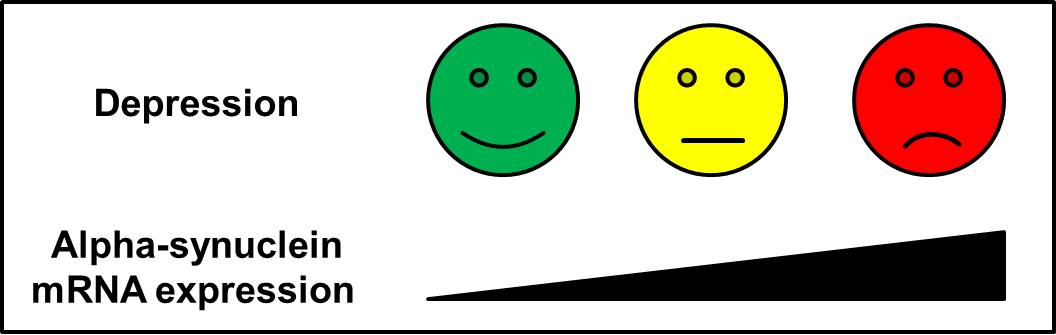
2.1. SNCA mRNA Expression Correlates Positively with the Severity of Depressive Symptoms

The mRNA expression levels of SNCA were determined in peripheral blood cells of MDD patients. The severity of depressive symptoms in patients was assessed using two psychometric scales: the Hamilton depression rating scale (HAM-D-17) for clinician-administered rating and Beck’s Depression Inventory—revised (BDI-II) for self-report rating. To investigate the relationship between SNCA mRNA expression and the severity of depressive symptoms in patients, we conducted a correlative analysis. Using Pearson correlation, we found a significant correlation between SNCA mRNA expression levels and BDI-II (r = 0.281, p = 0.026) and HAM-D-17 scores in the patients (r = 0.273, p = 0.028). Thus, the severity of depressive symptoms in MDD patients, as indicated by a higher psychometric score in the self-report rating as well as in the clinician-administered rating, was positively correlated with the measured SNCA mRNA expression in their blood cells.

2.2. SNCA mRNA Expression is Increased in Patients with Severe Depression

When analyzing the data of the MDD patients more closely, it turned out that the two patient subgroups, “ADT” (MDD patients recruited for the study “AntiDepressive Therapy” (ADT)) and “BLADe” (MDD patients participating in the study “Blood Lipid Alterations in Depression” (BLADe), Table 1) differed not only with regard to treatment, but also with regard to severity of symptoms. The patients of the ADT study, who were untreated, had an average HAM-D score of 17.7 ± 8.2 points, which characterizes this group as being moderately depressed. In contrast, the patients of the BLADe study, who were already treated at the beginning of the study, had an average HAM-D score of 21.4 ± 5.2 points, which falls into the category of severe depression. The difference in the severity of symptoms between both groups was statistically significant, as the ADT group exhibited significantly lower HAM-D scores than the BLADe group (t-test, df = 63, T = 2.2, p = 0.031; Table 2). There was no significant difference between females and males regarding the severity of depression (HAM-D score of 21.3 ± 6.6 and 18.0 ± 6.7, respectively; t-test, df = 63, T = −1.9, p > 0.05).
In a further analysis, we assessed the difference in SNCA mRNA expression between patients and controls. Due to the significant difference in age between the BLADe patient group and the healthy volunteers (Table 1), age was included as a covariate in all analyses. SNCA mRNA expression values were normally distributed. Compared with the control group, which had a mean normalized SNCA mRNA expression level of 17.4 ± 5.4 in their blood cells, MDD patients in the BLADe and ADT studies displayed increased SNCA mRNA expression levels (mean normalized expression of 31.9 ± 15.3 and 24.3 ± 13.8, respectively; analysis of variance (ANOVA) df = 2, F = 5.9, p = 0.004; Table 2). Pairwise comparison analysis revealed that the significant difference resulted from the comparison of the control group with the BLADe patient group (p = 0.001), but not with the ADT patient group (p = 0.114). Moreover, the comparison of SNCA mRNA expression between both patient groups revealed that the BLADe subgroup showed significantly higher SNCA mRNA expression levels than the ADT subgroup (p = 0.031). Even though females had a higher level of SNCA mRNA expression compared with males (mean normalized expression of 31.0 ± 17.2 and 21.3 ± 8.1, respectively; t-test, df = 63, T = −3.4, p = 0.001), there was no interaction effect between the groups and sex. Therefore, SNCA mRNA expression differs between healthy controls and depressed patients and seems to increase with the severity of depressive symptoms.

3. Discussion

Our study shows for the first time a significant increase in SNCA mRNA expression levels in severely depressed patients compared with healthy controls. This is in line with a parallel study in which increased SNCA protein levels were measured in the blood serum of depressed patients [29]. Our results showing a positive correlation between SNCA mRNA expression and BDI-II scores confirm a study by Frieling and colleagues in which this relationship was found in eating disorders [17]. Of note, other data have shown that patients who exhibited an early remission upon antidepressant treatment had increased SNCA mRNA expression levels at baseline compared with a non-responder group, but SNCA mRNA expression was not monitored during and after treatment [30]. The insights from clinical studies are derived from analyses of peripheral blood cells and do not address central mechanisms. In a murine study, the overexpression of SNCA in midbrain dopaminergic neurons resulted in depressive-like behavior [31]. The mediating effect of increased SNCA on the development of depression may be related to impaired adult neurogenesis in the hippocampus. In a mouse model overexpressing mutant A53T SNCA, adult neurogenesis in the dentate gyrus of the hippocampus was significantly impaired due to a reduction in proliferation of neural stem and precursor cells [32]. Another link could involve compromised neurotransmitter release associated with increased SNCA. In a stress model of depression in rats, several proteins were found to be differentially expressed and associated with deficits in synaptic vesicle release involving SNCA, synapsin I and the adaptor protein-3 complex, which were hypothesized to contribute to the pathomechanisms of psychiatric diseases [22].
Interestingly, increased mRNA expression of SNCA in patients were also detected in studies focusing on other neuro-psychiatric diseases: in neuronal disorders [3], in alcohol dependence [4] and cocaine dependence [33]. A common hallmark of these diseases is the impairment of cognition. The high comorbidity of PD with dementia and depression thus points to a common pathway. In 30–60% of PD patients, depressive symptoms occur and often precede motor symptoms [34]. The lack of studies investigating PD patients with depression makes further insights difficult. The treatment of depression in PD was investigated in two studies that found better outcomes for tricyclic antidepressants than for selective serotonin reuptake inhibitors (SSRIs) [35,36]. In murine studies, it was shown that treatment with fluoxetine did not influence SNCA mRNA expression levels [32], whereas paroxetine decreased SNCA mRNA levels [25]. It could be hypothesized that antidepressants in PD work via an influence on SNCA levels and that fluoxetine and paroxetine classified as SSRI are not optimized for this effect. Additionally, in a mouse model overexpressing SNCA, serotonergic projections in the hippocampus seem to be compromised, and the high protein levels of SNCA affected responsiveness to SSRIs [37]. These conflicting results indicate the need for human studies that monitor SNCA levels after antidepressant treatment.
One limitation of the present study may be that the number of patients in this study was relatively small, and we did not monitor treatment effects on SNCA mRNA expression. Moreover, the healthy volunteers differed from one of the patient groups regarding age.
In summary, we show a significant increase in SNCA mRNA expression levels in patients suffering from severe depression. Further studies with larger sample sizes and treatment monitoring are warranted to elucidate the clinical relevance of SNCA in MDD.

4. Materials and Methods

4.1. Ethics Statement

The collection of blood samples was approved by the Ethics Committee of the Friedrich-Alexander-University Erlangen-Nürnberg (FAU) (ID 4194, renewal of 3412, approval date: 20 April 2010) and conducted in concordance with the Declaration of Helsinki. Written informed consent was obtained from all participants.

4.2. Study Sample

All patients had an established diagnosis of MDD according to the International Statistical Classification of Diseases and Related Health Problems (ICD-10) and the Diagnostic and Statistical Manual of Mental Disorders (DSM-IV) criteria. After hospital admission, diagnosis was confirmed by conducting a diagnostic interview using the Strukturiertes Klinisches Interview für DSM-IV (SKID-I). Further, the following clinical scales were administered: the Hamilton depression rating scale (HAM-D-17) for clinician-administered rating and Beck’s Depression Inventory—revised (BDI-II) for self-report rating. All participants were carefully screened to rule out the existence of inflammatory, cardiac, endocrine, renal and hepatic disease by means of a structured medical history, physical examination, routine laboratory testing, and electrocardiography. Patients were excluded if comorbidity of alcohol or drug dependence was detected. MDD patients participating in the BLADe (Blood Lipid Alterations in DEpression) study (n = 39) were already treated with a standard antidepressant therapy at admission. MDD patients recruited for the ADT (AntiDepressive Therapy) study (n = 31) had not been treated with antidepressants, and standard antidepressant therapy was initiated after taking blood samples. A group of 18 healthy subjects without a personal history of psychiatric and somatic disorders served as a control group [38]. The BLADe patient group differed significantly from the healthy volunteers in terms of age (ANOVA, df = 2, F = 7.8, p = 0.001; post hoc analysis revealed significant difference only between controls and the BLADe group, p = 0.001; Table 1).

4.3. RNA Isolation and cDNA Synthesis

For patients in the ADT study, blood of fasting patients was taken for RNA isolation in the morning to secure for stable experimental conditions. For RNA isolation, the PAXgene system was employed (PreAnalytiX GmbH, Hombrechikon, Switzerland). PAXgene tubes containing blood samples were incubated at room temperature for 2 h, stored at −80 °C, and RNA was isolated according to manufacturer’s instructions. For patients in the BLADe study and for control samples, total RNA was extracted from whole blood in EDTA using Qiacube and the accordant protocol (QIAGEN GmbH, Hilden, Germany). RNA quality and quantity were analyzed using the Experion TM Automated Electrophoresis System and Nanodrop 1000 (PEQLAB, Erlangen, Germany). Reverse transcription was performed using the Bio-Rad Laboratories’ iScript cDNA Synthesis Kit (Bio-Rad, Munich, Germany).

4.4. Quantitative PCR

The expression of SNCA was analyzed by quantitative PCR using the LightCycler System (LightCycler® SW 1.5, Roche Diagnostics GmbH, Mannheim, Germany) as previously described [39]. Briefly, SNCA expression was assessed using SYBR green technology (Bio-Rad, Munich, Germany), and the mean of beta-actin (B-Actin), beta-2-microglobulin (B2M) and ornithine decarboxylase 1 (ODC1) expression values, assessed using specific probes of the Roche Universal Probe Library (Roche Diagnostics GmbH, Mannheim, Germany), served as reference values (Table 3). Mean normalized expression was calculated using the “Abs Quant/2nd Derivative Max” analysis method provided by Roche (Mannheim, Germany).

4.5. Statistical Analysis

Variables were tested for deviation from the normal distribution using the Kolmogorov-Smirnov test. Differences in sex distribution were calculated using the chi quadrat test. Correlative analyses were conducted using Pearson correlation coefficient. T-test and analysis of variance (ANOVA) were used to test for differences between the groups. A two-sided p-value ≤ 0.05 was considered indicative of statistical significance. The data were analyzed using SPSS TM for Windows 18.0 (SPSS Inc., Chicago, Ill., USA).

Author Contributions

Conceptualization, A.R., B.L., T.R.-S. and J.K.; Data curation, A.R., R.P. and T.R.-S.; Formal analysis, A.R., B.L., R.P., T.R.-S., J.K. and C.R.; Funding acquisition, J.K. and C.R.; Methodology, A.R., B.L., T.R.-S. and C.R.; Resources, A.R., B.L. and J.K.; Supervision, A.R. and C.R.; Validation, C.R.; Writing—original draft, A.R. and C.R.; Writing—review and editing, B.L. and J.K.

Funding

This research was funded by Forschungsstiftung Medizin at the University Hospital Erlangen, and the Scholarship Program ‘Equality for Women in Research and Teaching’ at the Friedrich-Alexander-University Erlangen-Nürnberg (FAU), to C.R.

Acknowledgments

We thank Alice Konrad for her excellent technical assistance.

Conflicts of Interest

The authors declare no conflict of interest. The funders had no role in the design of the study; in the collection, analyses, or interpretation of data; in the writing of the manuscript, or in the decision to publish the results.

References

  1. Iwai, A.; Masliah, E.; Yoshimoto, M.; Ge, N.; Flanagan, L.; de Silva, H.A.; Kittel, A.; Saitoh, T. The precursor protein of non-Aβ component of Alzheimer’s disease amyloid is a presynaptic protein of the central nervous system. Neuron 1995, 14, 467–475. [Google Scholar] [CrossRef]
  2. Iwai, A.; Yoshimoto, M.; Masliah, E.; Saitoh, T. Non-Aβ component of Alzheimer’s disease amyloid (NAC) is amyloidogenic. Biochemistry 1995, 34, 10139–10145. [Google Scholar] [CrossRef] [PubMed]
  3. Bayer, T.A.; Jakala, P.; Hartmann, T.; Egensperger, R.; Buslei, R.; Falkai, P.; Beyreuther, K. Neural expression profile of alpha-synuclein in developing human cortex. Neuroreport 1999, 10, 2799–2803. [Google Scholar] [CrossRef] [PubMed]
  4. Agrawal, A.; Wetherill, L.; Bucholz, K.K.; Kramer, J.; Kuperman, S.; Lynskey, M.T.; Nurnberger, J.I., Jr.; Schuckit, M.; Tischfield, J.A.; Edenberg, H.J.; et al. Genetic influences on craving for alcohol. Addict. Behav. 2013, 38, 1501–1508. [Google Scholar] [CrossRef]
  5. Pfefferkorn, C.M.; Lee, J.C. Tryptophan probes at the α-synuclein and membrane interface. J. Phys. Chem. B 2010, 114, 4615–4622. [Google Scholar] [CrossRef] [PubMed] [Green Version]
  6. Quilty, M.C.; Gai, W.-P.; Pountney, D.L.; West, A.K.; Vickers, J.C. Localization of α-, β-, and γ-synuclein during neuronal development and alterations associated with the neuronal response to axonal trauma. Exp. Neurol. 2003, 182, 195–207. [Google Scholar] [CrossRef]
  7. Madine, J.; Doig, A.J.; Middleton, D.A. A study of the regional effects of α-synuclein on the organization and stability of phospholipid bilayers. Biochemistry 2006, 45, 5783–5792. [Google Scholar] [CrossRef]
  8. Hsu, L.J.; Mallory, M.; Xia, Y.; Veinbergs, I.; Hashimoto, M.; Yoshimoto, M.; Thal, L.J.; Saitoh, T.; Masliah, E. Expression pattern of synucleins (non-β component of Alzheimer’s disease amyloid precursor protein/α-synuclein) during murine brain development. J. Neurochem. 1998, 71, 338–344. [Google Scholar] [CrossRef]
  9. George, J.M.; Jin, H.; Woods, W.S.; Clayton, D.F. Characterization of a novel protein regulated during the critical period for song learning in the zebra finch. Neuron 1995, 15, 361–372. [Google Scholar] [CrossRef] [Green Version]
  10. Gureviciene, I.; Gurevicius, K.; Tanila, H. Aging and α-synuclein affect synaptic plasticity in the dentate gyrus. J. Neural Transm. 2009, 116, 13–22. [Google Scholar] [PubMed]
  11. Stefanova, N.; Seppi, K.; Scherfler, C.; Puschban, Z.; Wenning, G.K. Depression in alpha-synucleinopathies: Prevalence, pathophysiology and treatment. J. Neural Transm. Suppl. 2000, 335–343. [Google Scholar]
  12. DeRubeis, R.J.; Siegle, G.J.; Hollon, S.D. Cognitive therapy versus medication for depression: Treatment outcomes and neural mechanisms. Nat. Rev. Neurosci. 2008, 9, 788–796. [Google Scholar] [CrossRef] [PubMed]
  13. Howren, M.B.; Lamkin, D.M.; Suls, J. Associations of depression with C-reactive protein, IL-1, and IL-6: A meta-analysis. Psychosom. Med. 2009, 71, 171–186. [Google Scholar] [CrossRef]
  14. Dowlati, Y.; Herrmann, N.; Swardfager, W.; Liu, H.; Sham, L.; Reim, E.K.; Lanctôt, K.L. A meta-analysis of cytokines in major depression. Biol. Psychiatry 2010, 67, 446–457. [Google Scholar] [CrossRef] [PubMed]
  15. Lenz, B.; Sysk, C.; Thuerauf, N.; Clepce, M.; Reich, K.; Frieling, H.; Winterer, G.; Bleich, S.; Kornhuber, J. Erratum to: NACP-Rep1 relates to Beck Depression Inventory scores in healthy humans. J. Mol. Neurosci. 2013, 50, 376–377. [Google Scholar] [CrossRef]
  16. Zhang, X.; Beaulieu, J.-M.; Sotnikova, T.D.; Gainetdinov, R.R.; Caron, M.G. Tryptophan hydroxylase-2 controls brain serotonin synthesis. Science 2004, 305, 217. [Google Scholar] [CrossRef]
  17. Frieling, H.; Gozner, A.; Römer, K.D.; Wilhelm, J.; Hillemacher, T.; Kornhuber, J.; de Zwaan, M.; Jacoby, G.E.; Bleich, S. Alpha-synuclein mRNA levels correspond to beck depression inventory scores in females with eating disorders. Neuropsychobiology 2008, 58, 48–52. [Google Scholar] [CrossRef]
  18. Oaks, A.W.; Sidhu, A. Synuclein modulation of monoamine transporters. FEBS Lett. 2011, 585, 1001–1006. [Google Scholar] [CrossRef]
  19. Jeannotte, A.M.; Sidhu, A. Regulation of the norepinephrine transporter by α-synuclein-mediated interactions with microtubules. Eur. J. Neurosci. 2007, 26, 1509–1520. [Google Scholar] [CrossRef]
  20. Wersinger, C.; Jeannotte, A.; Sidhu, A. Attenuation of the norepinephrine transporter activity and trafficking via interactions with α-synuclein. Eur. J. Neurosci. 2006, 24, 3141–3152. [Google Scholar] [CrossRef]
  21. Wersinger, C.; Rusnak, M.; Sidhu, A. Modulation of the trafficking of the human serotonin transporter by human alpha-synuclein. Eur. J. Neurosci. 2006, 24, 55–64. [Google Scholar] [CrossRef]
  22. Henningsen, K.; Palmfeldt, J.; Christiansen, S.; Baiges, I.; Bak, S.; Jensen, O.N.; Gregersen, N.; Wiborg, O. Candidate hippocampal biomarkers of susceptibility and resilience to stress in a rat model of depression. Mol. Cell Proteom. 2012, 11, M111 016428. [Google Scholar] [CrossRef]
  23. Jeannotte, A.M.; McCarthy, J.G.; Redei, E.E.; Sidhu, A. Desipramine modulation of α-, γ-synuclein, and the norepinephrine transporter in an animal model of depression. Neuropsychopharmacology 2009, 34, 987–998. [Google Scholar] [CrossRef]
  24. Lee, J.H.; Ko, E.; Kim, Y.E.; Min, J.Y.; Liu, J.; Kim, Y.; Shin, M.; Hong, M.; Bae, H. Gene expression profile analysis of genes in rat hippocampus from antidepressant treated rats using DNA microarray. BMC Neurosci. 2010, 11, 152. [Google Scholar] [CrossRef]
  25. McHugh, P.C.; Rogers, G.R.; Glubb, D.M.; Joyce, P.R.; Kennedy, M.A. Proteomic analysis of rat hippocampus exposed to the antidepressant paroxetine. J. Psychopharmacol 2010, 24, 1243–1251. [Google Scholar] [CrossRef]
  26. Bönsch, D.; Greifenberg, V.; Bayerlein, K.; Biermann, T.; Reulbach, U.; Hillemacher, T.; Kornhuber, J.; Bleich, S. α-Synuclein protein levels are increased in alcoholic patients and are linked to craving. Alcohol Clin. Exp. Res. 2005, 29, 763–765. [Google Scholar] [CrossRef]
  27. Bönsch, D.; Lederer, T.; Reulbach, U.; Hothorn, T.; Kornhuber, J.; Bleich, S. Joint analysis of the NACP-REP1 marker within the alpha synuclein gene concludes association with alcohol dependence. Hum. Mol. Genet. 2005, 14, 967–971. [Google Scholar] [CrossRef] [Green Version]
  28. Levey, D.F.; Le-Niculescu, H.; Frank, J.; Ayalew, M.; Jain, N.; Kirlin, B.; Learman, R.; Winiger, E.; Rodd, Z.; Shekhar, A.; et al. Genetic risk prediction and neurobiological understanding of alcoholism. Transl. Psychiatry 2014, 4, e391. [Google Scholar] [CrossRef]
  29. Ishiguro, M.; Baba, H.; Maeshima, H.; Shimano, T.; Inoue, M.; Ichikawa, T.; Yasuda, S.; Shukuzawa, H.; Suzuki, T.; Arai, H. Increased serum levels of α-synuclein in patients with major depressive disorder. Am. J. Geriatr. Psychiatry 2019, 27, 280–286. [Google Scholar] [CrossRef]
  30. Eyre, H.A.; Eskin, A.; Nelson, S.F.; Cyr, N.M. St.; Siddarth, P.; Baune, B.T.; Lavretsky, H. Genomic predictors of remission to antidepressant treatment in geriatric depression using genome-wide expression analyses: A pilot study. Int. J. Geriatr. Psychiatry 2016, 31, 510–517. [Google Scholar] [CrossRef]
  31. Caudal, D.; Alvarsson, A.; Bjorklund, A.; Svenningsson, P. Depressive-like phenotype induced by AAV-mediated overexpression of human α-synuclein in midbrain dopaminergic neurons. Exp. Neurol. 2015, 273, 243–252. [Google Scholar] [CrossRef]
  32. Kohl, Z.; Winner, B.; Ubhi, K.; Rockenstein, E.; Mante, M.; Münch, M.; Barlow, C.; Carter, T.; Masliah, E.; Winkler, J. Fluoxetine rescues impaired hippocampal neurogenesis in a transgenic A53T synuclein mouse model. Eur. J. Neurosci. 2012, 35, 10–19. [Google Scholar] [CrossRef] [Green Version]
  33. Brenz Verca, M.S.; Bahi, A.; Boyer, F.; Wagner, G.C.; Dreyer, J.L. Distribution of α- and γ-synucleins in the adult rat brain and their modification by high-dose cocaine treatment. Eur. J. Neurosci. 2003, 18, 1923–1938. [Google Scholar] [CrossRef]
  34. Gallagher, D.A.; Lees, A.J.; Schrag, A. What are the most important nonmotor symptoms in patients with Parkinson’s disease and are we missing them? Mov. Disord. 2010, 25, 2493–2500. [Google Scholar] [CrossRef]
  35. Devos, D.; Dujardin, K.; Poirot, I.; Moreau, C.; Cottencin, O.; Thomas, P.; Destée, A.; Bordet, R.; Defebvre, L. Comparison of desipramine and citalopram treatments for depression in Parkinson’s disease: A double-blind, randomized, placebo-controlled study. Mov. Disord. 2008, 23, 850–857. [Google Scholar] [CrossRef]
  36. Menza, M.; Dobkin, R.D.; Marin, H.; Mark, M.H.; Gara, M.; Buyske, S.; Bienfait, K.; Dicke, A. A controlled trial of antidepressants in patients with Parkinson disease and depression. Neurology 2009, 72, 886–892. [Google Scholar] [CrossRef]
  37. Deusser, J.; Schmidt, S.; Ettle, B.; Plötz, S.; Huber, S.; Müller, C.P.; Masliah, E.; Winkler, J.; Kohl, Z. Serotonergic dysfunction in the A53T alpha-synuclein mouse model of Parkinson’s disease. J. Neurochem. 2015, 135, 589–597. [Google Scholar] [CrossRef] [Green Version]
  38. Rotter, A.; Asemann, R.; Decker, A.; Kornhuber, J.; Biermann, T. Orexin expression and promoter-methylation in peripheral blood of patients suffering from major depressive disorder. J. Affect. Disord. 2011, 131, 186–192. [Google Scholar] [CrossRef]
  39. Lenz, B.; Klafki, H.W.; Hillemacher, T.; Frieling, H.; Clepce, M.; Gossler, A.; Thuerauf, N.; Winterer, G.; Kornhuber, J.; Bleich, S. ERK1/2 protein and mRNA levels in human blood are linked to smoking behavior. Addict. Biol. 2012, 17, 1026–1035. [Google Scholar] [CrossRef]
Table 1. Demographic overview. Differences in sex distribution were calculated using the chi quadrat test. Differences regarding age were calculated using analysis of variance (ANOVA). SD, standard deviation. ADT, MDD patients recruited for the study “AntiDepressive Therapy”; BLADe, MDD patients participating in the study “Blood Lipid Alterations in Depression”.
BLADeADTHealthy Controlsp-Value
N (male/female)39 (15/24)31 (15/16)18 (13/5)0.060
Age (years ± SD)46.3 ± 14.239.7 ± 16.530.4 ± 8.80.001
Table 2. Values for SNCA mRNA expression and psychometric scores. SNCA mRNA expression and Hamilton depression rating scale (HAM-D) scores differ significantly between groups (ANOVA). HAM-D was not conducted in the control group.
BLADeADTHealthy Controlsp-Value
SNCA expression ± SD31.9 ± 15.324.3 ± 13.817.4 ± 5.40.004
HAM-D scores ± SD21.4 ± 5.217.9 ± 8.2-0.034
Table 3. Sequences of oligonucleotides employed.
SNCA-F5′-CTC CTT TTC CTT CTT CTT TCC T-3′
SNCA-R5′-TGT TTG GTT TTC TCA GCA GC-3′
B-Actin-F5′-GTC TTC CCC TCC ATC GTG-3′
B-Actin-R5′-AGG TGT GGT GCC AGA TTT TC-3′
B-Actin-probe5′ Cy5-GAG CAA GAG AGG CAT CCT CAC CCT GAA GTA-Eclipse 3′
ODC1-F5′-CGC TTA CAC TGT TGC TGC TG-3′
ODC1-R5′-CAT CCT GTT CCT CTA CTT CGG G-3′
ODC1-probe5′ HEX-TCC AGA GGC CGA CGA TCT ACT ATG TGA TGT-BHQ1 3′
B2M-F5′-CGC TAC TCTC TCT TTC TGG C-3′
B2M-R5′-GTC AAC TTC AAT GTC GGA TGG AT-3′
B2M-probe#42 of Roche Universal Probe Library

