Article

Contribution of Inflammatory Cytokine Interleukin-18 Genotypes to Renal Cell Carcinoma

1
Terry Fox Cancer Research Laboratory, Translational Medicine Research Center, China Medical University Hospital, Taichung 40447, Taiwan
2
Institute of New Drug Development, China Medical University, Taichung 40402, Taiwan
3
School of Pharmacy, China Medical University, Taichung 40402, Taiwan
4
School of Medicine, China Medical University, Taichung 40402, Taiwan
5
Graduate Institute of Biomedical Sciences, China Medical University, Taichung 40402, Taiwan
6
Department of Bioinformatics and Medical Engineering, Asia University, Taichung 41354, Taiwan
*
Authors to whom correspondence should be addressed.
These authors contributed equally to this work.
Int. J. Mol. Sci. 2019, 20(7), 1563; https://doi.org/10.3390/ijms20071563
Submission received: 14 January 2019 / Revised: 26 March 2019 / Accepted: 26 March 2019 / Published: 28 March 2019
(This article belongs to the Special Issue Kidney Inflammation, Injury and Regeneration)

Abstract

:
Interleukin-18 (IL-18) is a multi-functional immuno-mediator in the development and progression of many types of infectious and inflammatory diseases. In this study, we evaluated the contribution of IL-18 genotypes to renal cell carcinoma (RCC) in Taiwan via the genotyping of IL-18 -656 (A/C), -607 (A/C), and -137 (G/C). Moreover, we analyzed their interactions with smoking, alcohol drinking, hypertension, and diabetes status. The results showed an association of the AC and CC genotypes of IL-18 −607 with a significant decrease in the risk of RCC compared with the AA genotype (odds ratio (OR) = 0.44 and 0.35, 95% confidence interval (CI) = 0.27–0.72 and 0.18–0.66, p = 0.0008 and 0.0010, respectively). Furthermore, a significantly lower frequency of the C allele at -607 was observed in the RCC group (35.3% vs. 49.8%; OR = 0.53; 95% CI = 0.35–0.71, p = 0.0003). However, IL-18 -656 and -137 did not exhibit a likewise differential distribution of these genotypes between the control and case groups. Stratifying the population according to smoking, alcohol drinking, hypertension, and diabetes status revealed a different distribution of IL-18 -607 genotypes among non-smokers, non-drinkers, and patients without diabetes, but not among smokers, drinkers, or patients with diabetes. These findings suggest that IL-18 -607 genotypes may play a role in the etiology and progression of RCC in Taiwan and may serve as a useful biomarker for early detection.

1. Introduction

Renal cell carcinoma (RCC) is the sixth most frequently diagnosed cancer in men (5%) and the tenth in women (3%) worldwide, thus posing a serious disease burden [1]. From an epidemiological viewpoint, RCC is the most common renal cancer and includes several subtypes that may be distinguished from each other by their histology, genetic background, clinical course, and response to treatment [2,3]. Moreover, there are several potential risk factors for RCC, including physical activity level, obesity, fruit and vegetable intake, cigarette smoking, and alcohol consumption. In addition, there are some common medical comorbidities for RCC, such as hypertension, diabetes, urinary stones, and other forms of chronic kidney diseases [3]. However, to date, no clinically practical genomic biomarker is available for RCC risk prediction. Unfortunately, many RCC patients, even those with advanced-stage tumors, remain asymptomatic [2,4], and the disease proceeds undetected. To make matters worse, up to 30% of RCC patients treated by radical nephrectomy suffer from many adverse effects and will relapse soon after their surgery [5]. Current personal prognostication of RCC is mainly based on histological validation, which may be labor intensive and time consuming, but frequently not useful for establishing a suitable course of treatment. Therefore, genomic molecular markers for early detection of RCC are urgently needed.
Interleukin-18 (IL-18), initially named IFN-γ inducing factor, is a proinflammatory cytokine encoded by the human IL-18 gene and produced by activated macrophages, epithelial cells, osteoblasts, keratinocytes, and most importantly, cancer cells [6,7]. In syngeneic mice models, supplementary IL-18 administration suppressed the growth of Meth A sarcoma and mouse glioma cells [7,8,9], suggesting that IL-18 plays an essential role in host mechanisms of defense against tumors. On a molecular level, IL-18 homeostasis is under the control of highly complex machinery involved in chronic inflammation and carcinogenesis, which is of great interest to both immunologists and oncologists. However, the promotive or suppressive effects of IL-18 on carcinogenesis are not yet fully understood. First, IL-18 may exert its tumor-suppressive influences by stimulating IFN-γ production, promoting Th1 differentiation, enhancing the cytotoxic capacities of CD8+ lymphocytes and natural killer cells [7], inducing cancer cells to undergo programmed cell death [10], and suppressing the angiogenesis [7,11]. Second, IL-18 can inhibit the recognition of cancer cells by immune cells, enhancing cancer cell adherence to the vascular wall, increasing the production of angiogenic and growth factors, and providing a pro-metastatic microenvironment [12,13]. Third, the serum levels of IL-18 were found to be higher in cancer patients than in healthy subjects, including bladder cancer [14], ovarian cancer [15], gastrointestinal cancer [16], and non-small cell lung cancer [17]. Fourth, the serum levels of IL-18 were much higher in those from breast cancer patients with metastasis than in those from patients without metastasis and non-cancer healthy subjects, supporting the hypothesis that elevated serum IL-18 levels can be used as non-invasive markers for suspected metastatic potential [18]. All the above findings support the hypothesis that the pleiotropic cytokine IL-18 can biphasically exert both anti-cancerous and pro-cancerous activities [13]. To summarize, the molecular interactions of IL-18 and other molecules are very complex and deeply involved in tumorigenesis.
From a genomic perspective, the expression level of IL-18 appears to be determined by at least two single nucleotide polymorphisms (SNPs) in the promoter at positions -607 (A/C) and -137 (G/C) of the human IL-18 gene. The former involves an A to C shift that disrupts a potential binding site for the cAMP responsive element binding protein, and the latter involves a G to C shift that abolishes the human histone H4 gene-specific transcription factor-1 (H4TF-1) nuclear factor binding site [19]. The alterations in transcription factor binding capacities determined by these two promoter polymorphisms may affect the overall activity of IL-18. Another SNP located in the IL-18 promoter is -656 (A/C); however, the effects of different genotypes at IL-18 -656 on cancer risk have not yet been well elucidated. That is to say, the contribution of -656 genotypes in human IL-18 to autoimmune diseases has started to be examined [15], but their involvement in any type of cancer has not.
As mentioned above, most of the previous genomic studies on human IL-18 were devoted to examining the association of IL-18 -607 and -137 polymorphisms with various types of cancer. Some of them revealed positive associations with cancer risk [20,21,22,23,24], while others identified negative ones [25,26]. However, only one such study investigated the association of the -137 and -607 genotypes of IL-18 with RCC [27]. In that paper, although the authors reported a negative association, they found that the -137 and -607 genotypes of IL-18 were correlated with more advanced stages of RCC, and the genotype related to a higher production of IL-18 was associated with a larger size and T stage of the tumor [27]. In the present study, the promoter SNPs at positions -656 (A/C, rs1946519), -607 (A/C, rs1946518), and -137 (G/C, rs187238) of the IL-18 gene were first examined and their genotype distributions analyzed in patients with RCC in Taiwan. In addition, we investigated the interaction of these IL-18 promoter genotypes with personal behavioral and clinical factors that contribute to RCC susceptibility.

2. Results

2.1. Comparison of Characteristics Among Patients with Renal Cell Carcinoma (RCC) and Controls

The frequency distributions in terms of age, gender, and personal behavioral habits for the 92 patients with RCC and the 580 cancer-free controls are summarized and compared in Table 1. Because the control subjects were already matched with patients with RCC for these factors, no difference was observed in terms of age and gender between these groups (p > 0.05). Moreover, there was no significant difference between the two groups in the frequency distributions in terms of personal behavioral habits, smoking, alcohol consumption, diabetes status, and family history (p > 0.05) (Table 1). An interesting finding was that there was a higher proportion of subjects with hypertension in the RCC group (66.3%) than in the cancer-free group (52.1%) (p = 0.0130). The percentage histologically identified as clear cell RCC patients is 77.2%. The percentages of “low” grade and “middle and high grade” are 52.2 and 47.8%, respectively (Table 1).

2.2. Analysis of the Association of IL-18 Promoter Genotypes and RCC Risk in Taiwan

The genotype frequencies of IL-18 -656 (A/C, rs1946519), -607 (A/C, rs1946518), and -137 (G/C, rs187238) for the 92 patients with RCC and the 580 age- and gender-matched healthy control subjects were determined, and the comparative results of codominant, dominant, and recessive models are presented in Table 2. The frequencies of IL-18 -656 and -137 genotypes in the control group, but not those of IL-18 -656 (p = 0.0206), were in agreement with the Hardy–Weinberg equilibrium.
For the first time, the genotypes at the IL-18 promoter -607 (A/C) polymorphic site were found to be differentially distributed between RCC cases and control groups (p for trend = 0.0004) (Table 2, middle panel). To explain in detail, the IL-18 -607 heterozygous AC and homozygous CC genotypes were associated with decreased risks for RCC (OR = 0.44 and 0.35, 95% CI = 0.27–0.72 and 0.18–0.66, p = 0.0008 and 0.0010, respectively) (Table 2, middle panel). After adjusting for the potential confounders, including age, gender, smoking, alcohol consumption, hypertension, diabetes status, and family history status, the significances still existed (Table 2, middle panel). In the dominant and recessive analyzing models, a significant association with the risk for RCC still persisted, as observed for the homozygous CC genotype (Table 2, middle panel). In contrast, none of the genotypes or alleles for IL-18, -656, and -137 demonstrated any correlation with RCC risk in any of the subgroups (Table 2).
We further performed allelic frequency analysis for these three IL-18 genotypes; the results are shown in Table 3. These results demonstrated that the variant allele C comprised only 35.3% in the RCC group, which was significantly less than that (49.8%) in the control group (adjusted OR = 0.53, 95% CI = 0.35–0.71, p = 0.0003), and these results fully confirmed the conclusion derived in Table 2. Consistently, the other two genotypes, IL-18 -656 and -137, showed no significant association with the risk for RCC (Table 3).

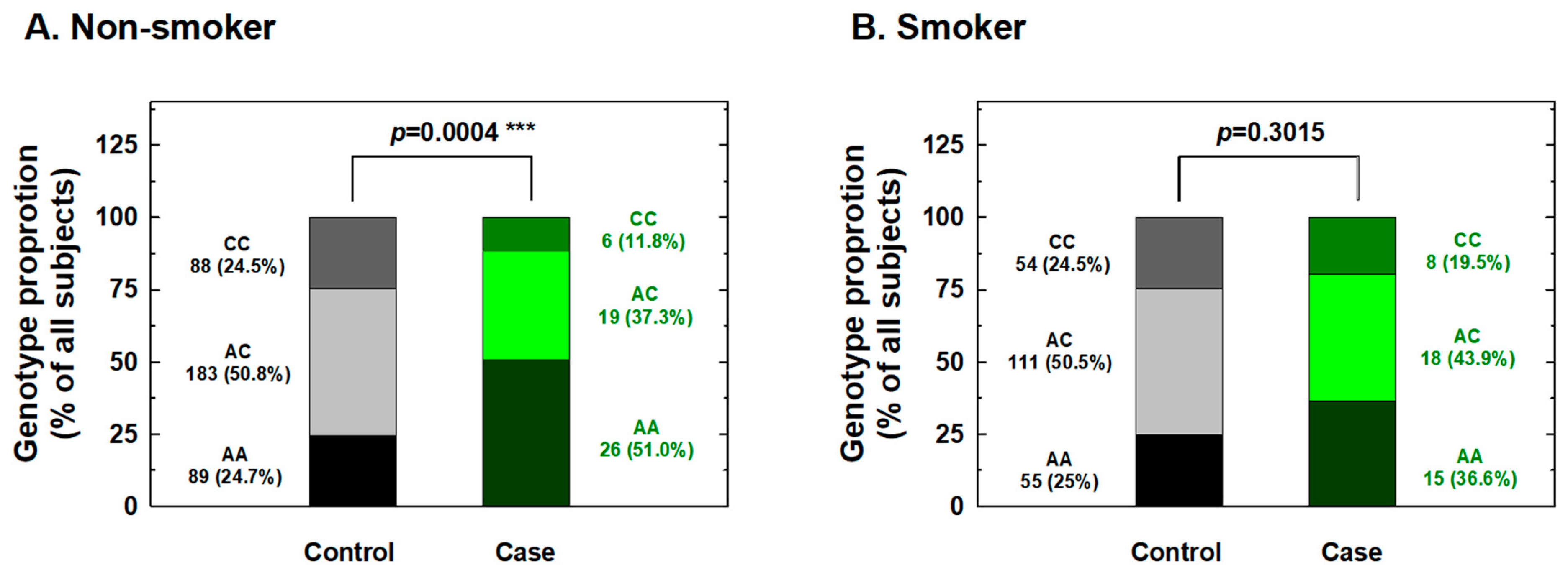
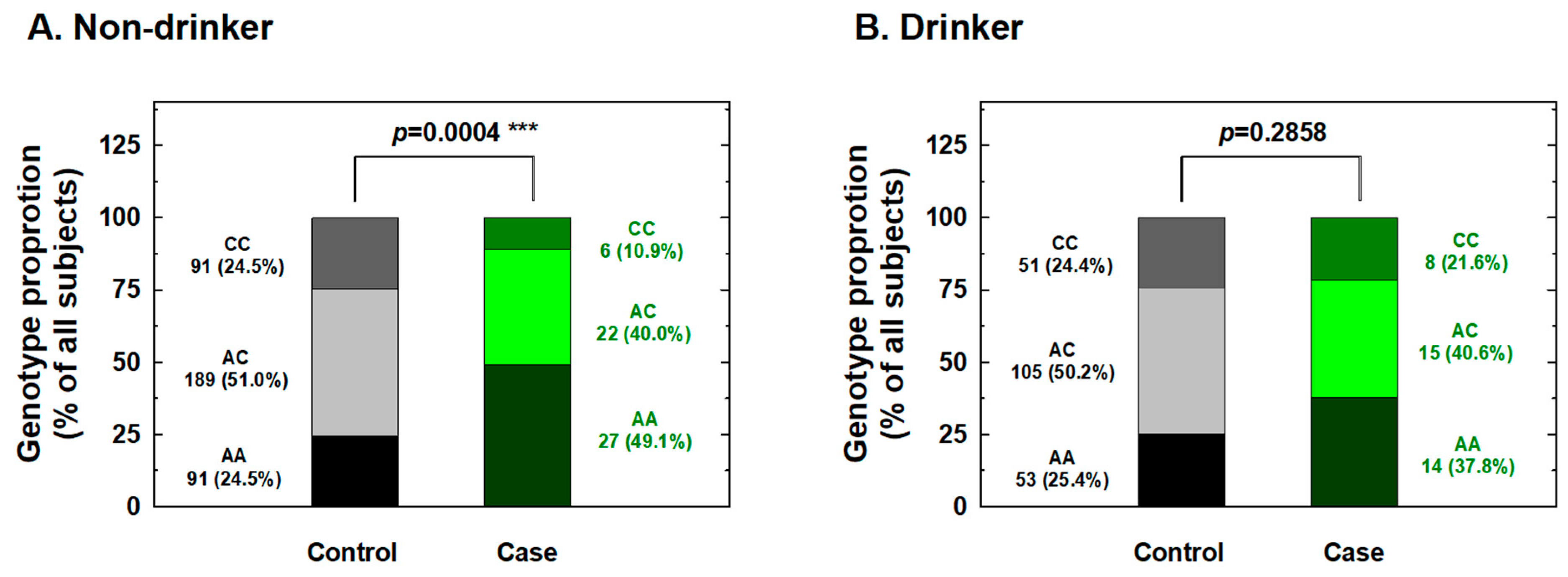
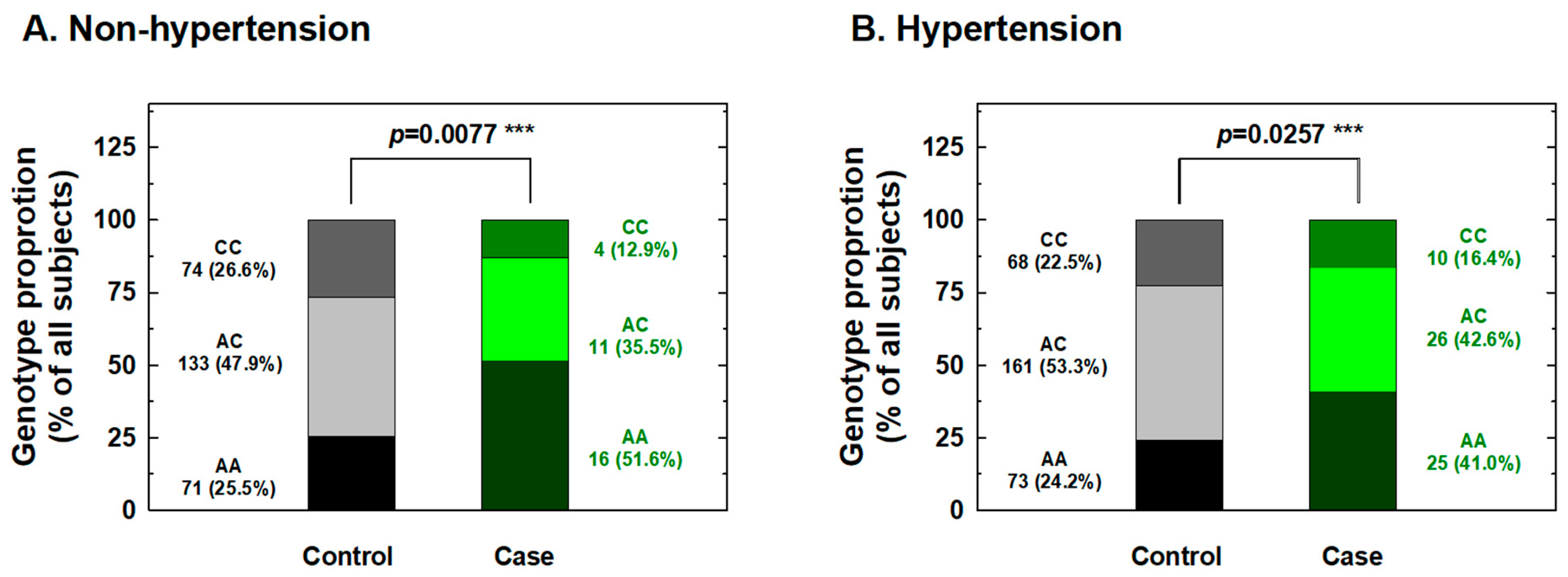
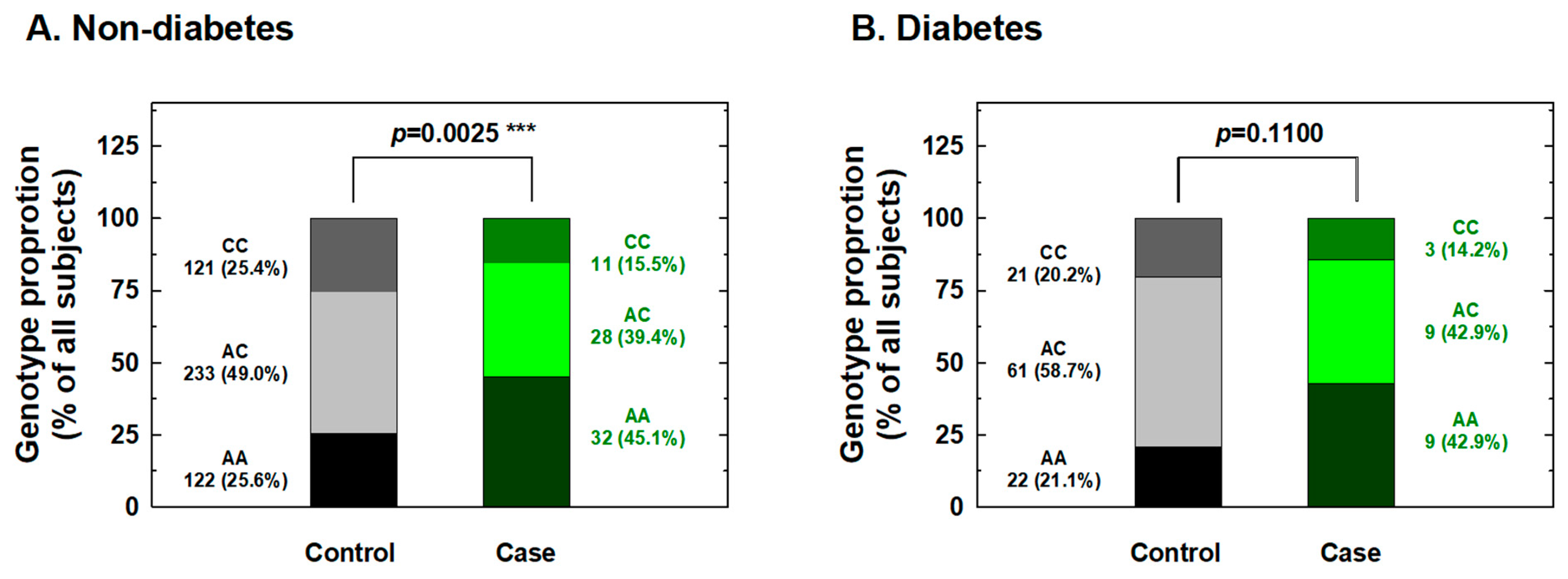
2.3. Stratified Analysis of IL-18 Genotypes According to Personal Behavioral and Clinical Factors

We further conducted stratification analysis of the association between IL-18 -607 genotypes and the risk for RCC based on potential personal behavioral and clinical risk factors among Taiwanese people, including cigarette smoking, alcohol consumption, hypertension, and diabetes status. First, the distributions of the genotype frequencies between the case and control groups among nonsmokers were significantly different, but showed similar proportions for cases and controls among smokers (Figure 1). The adjusted ORs for carriers with genotypes AC and CC at IL-18 -607 were 0.36 and 0.22 for nonsmokers (95% CI = 0.21–0.63 and 0.11–0.56, respectively) and 0.61 and 0.58 for smokers (95% CI = 0.33–1.31 and 0.24–1.35, respectively), respectively (Figure 1). It appeared that the protective effects of IL-18 -607 genotypes on the risk for RCC were obvious among nonsmokers, but not among smokers (Figure 1). Second, the distributions of the genotype frequencies between the case and control groups among nondrinkers were significantly different, but showed similar proportions for cases and controls among alcohol drinkers (Figure 2). The adjusted ORs for carriers with genotypes AC and CC at IL-18 -607 were 0.38 and 0.21 among nondrinkers (95% CI = 0.22–0.70 and 0.09–0.51, respectively) and 0.56 and 0.62 among alcohol drinkers (95% CI = 0.31–1.23 and 0.28–1.41, respectively), respectively (Figure 2). The protective effects of IL-18 -607 genotypes on the risk for RCC appeared to be obvious among nondrinkers, but not among alcohol drinkers (Figure 2). Third, the distributions of the genotype frequencies between case and control groups among non-hypertensive and hypertensive subjects were both significantly different (Figure 3). The adjusted ORs for carriers with genotypes AC and CC at IL-18 -607 were 0.39 and 0.24 among subjects without hypertension (95% CI = 0.18–0.81 and 0.11–0.69, respectively) and 0.41 and 0.39 among those with hypertension (95% CI = 0.26–0.84 and 0.18–0.94, respectively), respectively (Figure 3). The protective effects of IL-18 -607 genotypes on the risk for RCC appeared to be obvious among people with or without hypertension (Figure 3). Finally, the distributions of the genotype frequencies between the case and control groups among subjects without diabetes were significantly different, but presented similar proportions for cases and controls among subjects with diabetes (Figure 4). The adjusted ORs for carriers with genotypes AC and CC at IL-18 -607 were 0.44 and 0.35 among subjects without diabetes (95% CI = 0.28–0.81 and 0.21–0.68, respectively) and 0.38 and 0.36 among those with diabetes (95% CI = 0.18–1.02 and 0.13–1.58, respectively), respectively (Figure 4). The effects of IL-18 -607 genotypes on the risk for RCC appeared to be protective among subjects without diabetes, but not among those with diabetes (Figure 4). The sample size of those with a family history of cancer was too small for stratification analysis.
The levels of IL-18 in the serum of 10 RCC patients and 10 healthy controls were determined using ELISA. The results demonstrated that the basal IL-18 levels were significantly higher in RCC patients (212.80 ± 21.39 pg/mL) than those of control subjects (113.70 ± 9.94 pg/mL) (p = 0.0001) (Figure 5). According to their IL-18 -607 genotype distribution, the 10 RCC patients were divided into three subgroups: four patients with the AA genotype (216.25 ± 12.79 pg/mL), four with AC (206.25 ± 31.31 pg/mL), and two with CC (219.00 ± 19.80 pg/mL). There were no significant differences in serum IL-18 levels between different IL-18 -607 genotypes (AC versus AA: p = 0.5759; CC versus AA: p = 0.8412; and CC versus AC: p = 0.6369).

3. Discussion

The prevalence and death rates of RCC are not ranked as high as those of other cancers in Taiwan. Clinically, surgery is the major course of RCC treatment. However, the symptoms of early-stage RCC are not obvious, and early detection of RCC is not available. Thus, the findings of genomic biomarker(s), which are very useful in rapid and convenient screening, may contribute to early detection and prediction of RCC susceptibility and outcome. For many years, members of the Terry Fox Cancer Fox Cancer Research foundation, including translational scientists and surgeons, have devoted themselves to elucidating specific and practical genomic biomarkers for early detection and prediction in Taiwan, where RCC is a prevalent condition and the cause of many cancer deaths [28,29,30,31,32]. Cytokines play an essential but complex role in the initiation and progression of inflammation and tumorigenesis [33], which is currently still under investigation. The proinflammatory cytokine IL-18 confers protective effects against cancer proliferation, such as that of lung cancer, in several murine models [13,34], and the benefits of recombinant human IL-18 have been shown in preclinical trials for cancer treatment [35]. However, despite the conventional view of IL-18 as an anticancer agent, some studies have also proposed a procancerous behavior for IL-18 under specific conditions [13]. Recently, mounting studies have reported that various cytokine genotypes may influence the serum levels of their counterpart cytokines, which may be closely associated with susceptibility to certain human diseases [19,20,21,22,23,25,26]. Among the numerous SNPs in IL-18, three polymorphisms are present in the promoter region of this gene: -656 (G/T), -607 (C/A), and -137 (G/C), and they were reported to cause differences in the transcription factor binding capacity and expression level of IL-18 in serum [19]. The polymorphic genotypes of IL-18 promoter -607 and -137 were previously found to be associated with the risk of esophageal squamous cell carcinoma [20] and prostate cancer [21] in China, colorectal cancer in Greece [22], ovarian cancer in the USA (Hawaii) [23], and breast cancer in Iran [24]. On the contrary, there were also some negative associations reported between IL-18 polymorphisms and the risk of head and neck cancers in Iran [26], as well as oral cancer in Greece [25]. Reasonable explanations for these discrepant and diverse findings may involve three possibilities: the dual impact of IL-18 on tumor-immune responses [13], the different types of cancer investigated, and variation among the populations under study [26].
In the current study, the genotypes at polymorphic locations -656 (G/T), -607 (C/A), and -137 (G/C) of the IL-18 promoter region among RCC patients and healthy individuals in a Taiwan population were first determined and evaluated for their contribution to RCC risk. The results indicated a significantly lower risk for the heterozygous AC and homozygous CC variant genotypes and for the C allele at position -607 of the IL-18 gene than their counterparts about RCC susceptibility (Table 2 and Table 3), even after statistical adjustment for personal behavioral and clinical risk factors. In contrast, no significant association between any genotype or allelic type with RCC risk was found for -656 or -137 of IL-18 (Table 2 and Table 3). The positive findings indicating that genotypes of IL-18 -607 may be determinants for personal RCC susceptibility were inconsistent with previous findings from the only paper investigating the contribution of IL-18 genotypes to RCC [27], which returned negative findings. Once again, the inconsistency may be due to the location of Taiwan located in East Asia and the fact that it is an island with a conserved genetic, cultural, and environmental background, much different from the investigated Spanish population [27]. Although the sample size of the current study was similar to theirs (case:control = 92:580 vs. 158:506), we brought forward two novel findings: IL-18 -607 was a determinant of RCC susceptibility (Table 2 and Table 3), and there were positive interactions of this polymorphic site with personal behavioral and clinical factors (Figure 1, Figure 2, Figure 3 and Figure 4). In the near future, the significant contribution of IL-18 genotypes to RCC risk evaluation, especially those at IL-18 -607, should be validated in larger samples and other populations worldwide.
In detail, the stratification of RCC patients and non-cancer subjects according to personal behavior revealed that IL-18 -607 genotypes may play a significant role in the determination of susceptibility to RCC in non-smokers (Figure 1), non-alcohol drinkers (Figure 2), those with and without hypertension (Figure 3), and those without diabetes (Figure 4), but not in smokers, alcohol drinkers, or those with diabetes (Figure 1, Figure 3 and Figure 4). However, the undermined subtle mechanisms and signaling networks that are responsible for the interaction of IL-18 and other molecules related to the etiology of RCC require further investigation.
The genotype-phenotype association was performed after the measurement of serum levels of IL-18 in 10 RCC patients and 10 healthy controls. The results showed that: (a) the AA genotype at IL-18 -607 was higher in the RCC patients than the healthy controls (Table 2); (b) the IL-18 levels were higher in the RCC patients than the healthy controls (Figure 5), which is consistent with the previous findings [36]; and (c) there was no difference in the IL-18 levels among RC patients of different IL-18 -607 genotypes. This finding is consistent with the previous finding in lung cancer [37], but inconsistent with another [38]. The difference and similarity may due to the fact that different populations were investigated. Ours and the former were investigating Taiwanese and Chinese people, respectively, while the latter one was investigating people from Iran.
As for the perspective molecular mechanism, there is literature mentioning that the A to C shift at IL-18 -607 may disrupt the potential binding site for the cAMP responsive element binding protein, thus lowering the expression level of IL-18 [19]. That is to say, the differential genotype at IL-18 -607 may associate with elevated expression levels of IL-18, as the early detector for RCC, like we showed in Figure 5. The current study does not provide supporting evidence for the hypothesis that any genotype at IL-18 -607 may associate with elevated expression levels of IL-18, due to the limited samples examined, and confirmation in larger samples is an urgent need. We also have to notice that the alteration of IL-18 may not be the only indicator during RCC carcinogenesis. In 2015, Xu et al. reported that elevated IL-18 together with IL-1b were significantly associated with advanced RCC stages, an elevated recurrence rate, and a shortened survival period among patients with localized RCC [35].
To summarize, this pilot study indicated a significant association between the IL-18 -607 polymorphism and RCC in Taiwan. Furthermore, to the best of our knowledge, it is also the first to investigate the interaction of IL-18 genotypes and behavioral and clinical factors in RCC risk. Our results showed a significant association between the IL-18 -607 polymorphism and RCC, particularly in people without smoking or alcohol drinking behavior, and those without diabetes. However, it should be pointed out that the samples sizes of affected subjects (i.e., smokers, drinkers, and particularly patients with diabetes) were much smaller than those of un-affected subjects, which might be the main reason for the lack of statistically significant associations in the affected subjects. Future larger studies in various populations are needed to validate IL-18 genotypes as early detective and predictive determinants of RCC.

4. Materials and Methods

4.1. Selected Subjects

This case-control study was performed in the China Medical University Hospital and involved the collection of data from 92 patients with RCC and 580 cancer-free controls matched by age and gender; none of the participants were related to each other by any biological relationship. The diagnosis of RCC, and the grades and types of each patient were histopathologically confirmed by the surgeons and pathologists led by Hsi-Chin Wu. In addition, the age- and gender-matched cancer-free controls were genetically unrelated to any of the recruited participants and had no prior history of any cancer. Originally, we frequency matched seven controls, which were collected in the Health Examination Center of the China Medical University Hospital, for each RCC patient with the same gender and age at ±2 years. After the first-term matching, those with incomplete demographic data about smoking, alcohol drinking status, hypertension, diabetes, or family cancer history, were excluded. A further exclusion criterion for the control subjects was any symptom suggestive of RCC, such as hematuria. Finally, only 580 controls were collected in the study. After obtaining written informed consent, 3–5 mL of venous blood was collected from each participant for genotyping. The study was approved by the Institutional Review Board of China Medical University, and expert members of the Tissue Bank of China Medical University Hospital (DMR98-IRB-209 in 2009) provided their kind assistance. The overall agreement rate among the participants was >85%. Select characteristics of all the participants are summarized and compared in Table 1.

4.2. DNA Preparation and Storage

Genomic DNA from the leukocytes of each study subject was extracted using the QIAamp Blood Mini Kit (Qiagen, Valencia, CA, USA), stored for the long term at −80 °C, simultaneously diluted, and aliquoted and stored for genotyping as a working stock at −20 °C, as we have frequently performed previously [32,39,40].

4.3. IL-18 Genotype Discrimination Methodology

The genotype discrimination methodology for IL-18 -137, -607, and -656 genotypes was performed as we described in 2018 [41]. Briefly, -137 (G/C, rs187238) and -607 (A/C, rs1946518) genotyping was performed using the ABI StepOne™ Real-Time PCR System (Applied Biosystems, Foster City, CA, USA) and analyzed using the typical TaqMan assay. Regarding the genotyping of IL-18 -656 (A/C, rs1946519), the polymerase chain reaction-restriction fragment length polymorphism (PCR-RFLP) methodology was carried out using the primers originally reported in 2005 by Flowaczny et al. [42], with the forward primer being 5′-AGGTCAGTCTTTGCTATCATTCCAGG-3′ and the reverse primer being 5′-CTGCAACAGAAAGTAAGCTTGCGGAGAGG-3′, and a 120-bp fragment nearby the IL-18 -656 polymorphism was amplified. In detail, approximately 100 ng of genomic DNA of each sample was subjected to PCR, in which the reaction mixture of 25-μL contained 300 mM dNTP, 2 U of Taq DNA polymerase, 1× PCR buffer, 1.5 mM MgCl2, and 0.8 mM of each primer. After mixing up and briefly spinning down, the reaction mixture was heated to 94 °C for 4 min and amplified by 30 cycles using the My Cycler (Biorad, Hercules, CA, USA) with the following steps: denaturation at 94 °C for 60 s, annealing at 60 °C for 60 s, extension at 72 °C for 60 s for each cycle, and a final extension step at 72 °C for 5 min. The volume of the restriction assay was set at 12.5 μL, containing 8 μL of PCR products, 2 U Mwo I restriction enzyme, and 1× buffer. The reaction mixture was then incubated for 16 h or overnight at 60 °C. The resultant DNA fragments were subject to electrophoresis in 3.0% agarose gel at 100 V for 30 min. After electrophoresis, ethidium bromide staining was done to observe the DNA fragments under UV (260 nm) light. For the A allele of IL-18 -656, there was no digestion of the 120-bp PCR fragment, whereas for the C allele of IL-18 -656, two (96- and 24-bp) fragments were identified.

4.4. Enzyme-linked Immunosorbent Assay (ELISA) for Serum IL-18 Levels

Ten milliliters of blood samples were collected from 10 healthy controls and 10 RCC patients. The blood samples were collected in serum tubes with an accelerating agent for serum separation and kept at a room temperature for 30 min before their further centrifugation for 20 min at 1500× g. Serum was then isolated and stored at −80 °C until IL-18 measurement. The individual level of IL-18 in serum was measured by enzyme-linked immunosorbent assay (ELISA, Newwark, DE, USA) kits.

4.5. Statistical Analysis Methodology

The data of 580 cancer-free healthy controls and 92 patients with RCC who had complete genotypic and clinical details were finally included for statistical analysis, whose results are presented in the form of tables and figures. To ensure that the control subjects in this study were representative of the Taiwanese general population and to exclude the possibility of genotyping error, the deviation of the genotype frequencies of IL-18 SNPs in the control subjects from those expected under the Hardy–Weinberg equilibrium was assessed using the goodness-of-fit test. Pearson’s Chi-square test was used to compare the distribution of IL-18 genotypes between cases and control groups and in the stratification analysis. The comparison of the continuous factor age was performed and evaluated by the Student’s t-test. The contribution of IL-18 genotypes to the risk of developing RCC was estimated by the odds ratios (ORs) and their counterpart 95% confidence intervals (CIs) obtained through logistic regression analysis with adjustment for possible confounders. Any p value < 0.05 was considered to be statistically significant.

Author Contributions

Conceived and designed the experiments: W.-S.C., T.-C.S., and W.-L.Y. Performed the experiments: W.-S.C. and C.-W.T. Analyzed the data: C.-C.Y. and H.-Y.L. Contributed reagents/materials/analysis tools: W.-L.Y. and H.-C.W. Wrote the paper: C.-W.T. and D.-T.B.

Funding

This research was mainly funded by China Medical University Hospital (DMR107-175) to Shen.

Acknowledgments

We thank the Tissuebank and colleagues of the Urinary Department in the China Medical University Hospital for their excellent technical assistance. The genotyping and analyzing work was partially helped by Yun-Chi Wang, Hsin-Ting Li, and Huai-Mei Hsu. The statistical analyses were kindly double checked by Cheng-Li Lin (manpower under MOHW108-TDU-B-212-133004).

Conflicts of Interest

The authors declare no conflict of interest.

References

  1. Siegel, R.L.; Miller, K.D.; Jemal, A. Cancer statistics, 2018. CA Cancer J. Clin. 2018, 68, 7–30. [Google Scholar] [CrossRef] [PubMed]
  2. Ferlay, J.; Shin, H.R.; Bray, F.; Forman, D.; Mathers, C.; Parkin, D.M. Estimates of worldwide burden of cancer in 2008: GLOBOCAN 2008. Int. J. Cancer 2010, 127, 2893–2917. [Google Scholar] [CrossRef] [PubMed] [Green Version]
  3. Klaassen, Z.; Sayyid, R.K.; Wallis, C.J.D. Lessons learned from the global epidemiology of kidney cancer: A refresher in epidemiology 101. Eur. Urol. 2019, 75, 85–87. [Google Scholar] [CrossRef]
  4. Niedworok, C.; Dorrenhaus, B.; Vom Dorp, F.; Piotrowski, J.A.; Tschirdewahn, S.; Szarvas, T.; Rubben, H.; Schenck, M. Renal cell carcinoma and tumour thrombus in the inferior vena cava: Clinical outcome of 98 consecutive patients and the prognostic value of preoperative parameters. World J. Urol. 2015, 33, 1541–1552. [Google Scholar] [CrossRef]
  5. Cohen, H.T.; McGovern, F.J. Renal-cell carcinoma. N. Engl. J. Med. 2005, 353, 2477–2490. [Google Scholar] [CrossRef]
  6. Okamura, H.; Tsutsi, H.; Komatsu, T.; Yutsudo, M.; Hakura, A.; Tanimoto, T.; Torigoe, K.; Okura, T.; Nukada, Y.; Hattori, K.; et al. Cloning of a new cytokine that induces IFN-gamma production by T cells. Nature 1995, 378, 88–91. [Google Scholar] [CrossRef] [PubMed]
  7. Nakanishi, K.; Yoshimoto, T.; Tsutsui, H.; Okamura, H. Interleukin-18 is a unique cytokine that stimulates both Th1 and Th2 responses depending on its cytokine milieu. Cytokine Growth Factor Rev. 2001, 12, 53–72. [Google Scholar] [CrossRef]
  8. Golab, J. Interleukin 18--interferon gamma inducing factor--a novel player in tumour immunotherapy? Cytokine 2000, 12, 332–338. [Google Scholar] [CrossRef] [PubMed]
  9. Kikuchi, T.; Akasaki, Y.; Joki, T.; Abe, T.; Kurimoto, M.; Ohno, T. Antitumor activity of interleukin-18 on mouse glioma cells. J. Immunother. 2000, 23, 184–189. [Google Scholar] [CrossRef] [PubMed]
  10. Okano, F.; Yamada, K. Canine interleukin-18 induces apoptosis and enhances Fas ligand mRNA expression in a canine carcinoma cell line. Anticancer Res. 2000, 20, 3411–3415. [Google Scholar]
  11. Cao, R.; Farnebo, J.; Kurimoto, M.; Cao, Y. Interleukin-18 acts as an angiogenesis and tumor suppressor. FASEB J. 1999, 13, 2195–2202. [Google Scholar] [CrossRef] [PubMed]
  12. Park, C.C.; Morel, J.C.; Amin, M.A.; Connors, M.A.; Harlow, L.A.; Koch, A.E. Evidence of IL-18 as a novel angiogenic mediator. J. Immunol. 2001, 167, 1644–1653. [Google Scholar] [CrossRef] [PubMed]
  13. Vidal-Vanaclocha, F.; Mendoza, L.; Telleria, N.; Salado, C.; Valcarcel, M.; Gallot, N.; Carrascal, T.; Egilegor, E.; Beaskoetxea, J.; Dinarello, C.A. Clinical and experimental approaches to the pathophysiology of interleukin-18 in cancer progression. Cancer Metastasis Rev. 2006, 25, 417–434. [Google Scholar] [CrossRef] [PubMed]
  14. Jaiswal, P.K.; Singh, V.; Srivastava, P.; Mittal, R.D. Association of IL-12, IL-18 variants and serum IL-18 with bladder cancer susceptibility in North Indian population. Gene 2013, 519, 128–134. [Google Scholar] [CrossRef]
  15. Samsami Dehaghani, A.; Shahriary, K.; Kashef, M.A.; Naeimi, S.; Fattahi, M.J.; Mojtahedi, Z.; Ghaderi, A. Interleukin-18 gene promoter and serum level in women with ovarian cancer. Mol. Biol. Rep. 2009, 36, 2393–2397. [Google Scholar] [CrossRef]
  16. Haghshenas, M.R.; Hosseini, S.V.; Mahmoudi, M.; Saberi-Firozi, M.; Farjadian, S.; Ghaderi, A. IL-18 serum level and IL-18 promoter gene polymorphism in Iranian patients with gastrointestinal cancers. J. Gastroenterol. Hepatol. 2009, 24, 1119–1122. [Google Scholar] [CrossRef]
  17. Okamoto, M.; Azuma, K.; Hoshino, T.; Imaoka, H.; Ikeda, J.; Kinoshita, T.; Takamori, S.; Ohshima, K.; Edakuni, N.; Kato, S.; et al. Correlation of decreased survival and IL-18 in bone metastasis. Intern. Med. 2009, 48, 763–773. [Google Scholar] [CrossRef]
  18. Gunel, N.; Coskun, U.; Sancak, B.; Gunel, U.; Hasdemir, O.; Bozkurt, S. Clinical importance of serum interleukin-18 and nitric oxide activities in breast carcinoma patients. Cancer 2002, 95, 663–667. [Google Scholar] [CrossRef] [Green Version]
  19. Giedraitis, V.; He, B.; Huang, W.X.; Hillert, J. Cloning and mutation analysis of the human IL-18 promoter: A possible role of polymorphisms in expression regulation. J. Neuroimmunol. 2001, 112, 146–152. [Google Scholar] [CrossRef]
  20. Wei, Y.S.; Lan, Y.; Liu, Y.G.; Tang, H.; Tang, R.G.; Wang, J.C. Interleukin-18 gene promoter polymorphisms and the risk of esophageal squamous cell carcinoma. Acta. Oncol. 2007, 46, 1090–1096. [Google Scholar] [CrossRef] [Green Version]
  21. Liu, Y.; Lin, N.; Huang, L.; Xu, Q.; Pang, G. Genetic polymorphisms of the interleukin-18 gene and risk of prostate cancer. DNA Cell. Biol. 2007, 26, 613–618. [Google Scholar] [CrossRef] [PubMed]
  22. Nikiteas, N.; Yannopoulos, A.; Chatzitheofylaktou, A.; Tsigris, C. Heterozygosity for interleukin-18 -607 A/C polymorphism is associated with risk for colorectal cancer. Anticancer Res. 2007, 27, 3849–3853. [Google Scholar] [PubMed]
  23. Bushley, A.W.; Ferrell, R.; McDuffie, K.; Terada, K.Y.; Carney, M.E.; Thompson, P.J.; Wilkens, L.R.; Tung, K.H.; Ness, R.B.; Goodman, M.T. Polymorphisms of interleukin (IL)-1alpha, IL-1beta, IL-6, IL-10, and IL-18 and the risk of ovarian cancer. Gynecol. Oncol. 2004, 95, 672–679. [Google Scholar] [CrossRef] [PubMed]
  24. Khalili-Azad, T.; Razmkhah, M.; Ghiam, A.F.; Doroudchi, M.; Talei, A.R.; Mojtahedi, Z.; Ghaderi, A. Association of interleukin-18 gene promoter polymorphisms with breast cancer. Neoplasma 2009, 56, 22–25. [Google Scholar] [CrossRef]
  25. Vairaktaris, E.; Serefoglou, Z.C.; Yapijakis, C.; Agapi, C.; Vassiliou, S.; Nkenke, E.; Antonis, V.; Sofia, S.; Neukam, F.W.; Patsouris, E. The interleukin-18 -607A/C polymorphism is not associated with risk for oral cancer. Anticancer Res. 2007, 27, 4011–4014. [Google Scholar] [PubMed]
  26. Asefi, V.; Mojtahedi, Z.; Khademi, B.; Naeimi, S.; Ghaderi, A. Head and neck squamous cell carcinoma is not associated with interleukin-18 promoter gene polymorphisms: A case-control study. J. Laryngol. Otol. 2009, 123, 444–448. [Google Scholar] [CrossRef]
  27. Saenz-Lopez, P.; Carretero, R.; Vazquez, F.; Martin, J.; Sanchez, E.; Tallada, M.; Garrido, F.; Cozar, J.M.; Ruiz-Cabello, F. Impact of interleukin-18 polymorphisms-607 and -137 on clinical characteristics of renal cell carcinoma patients. Hum. Immunol. 2010, 71, 309–313. [Google Scholar] [CrossRef]
  28. Chang, W.S.; Ke, H.L.; Tsai, C.W.; Lien, C.S.; Liao, W.L.; Lin, H.H.; Lee, M.H.; Wu, H.C.; Chang, C.H.; Chen, C.C.; et al. The role of XRCC6 T-991C functional polymorphism in renal cell carcinoma. Anticancer Res. 2012, 32, 3855–3860. [Google Scholar]
  29. Chang, W.S.; Tsai, C.W.; Wang, S.M.; Wang, S.W.; Wu, H.C.; Ji, H.X.; Lin, C.H.; Wang, Z.H.; Chou, J.C.; Bau, D.T.; et al. Association of caveolin-1 genotypes with renal cell carcinoma risk in Taiwan. Chin. J. Physiol. 2014, 57, 220–226. [Google Scholar] [CrossRef]
  30. Chang, W.S.; Liao, C.H.; Miao, C.E.; Wu, H.C.; Hou, L.L.; Hsiao, C.L.; Ji, H.X.; Tsai, C.W.; Bau, D.T. The role of functional polymorphisms of cyclooxygenase 2 in renal cell carcinoma. Anticancer Res. 2014, 34, 5481–5486. [Google Scholar]
  31. Chang, W.S.; Liao, C.H.; Tsai, C.W.; Hu, P.S.; Wu, H.C.; Hsu, S.W.; Ji, H.X.; Hsiao, C.L.; Bau, D.T. The Role of IL-10 Promoter Polymorphisms in Renal Cell Carcinoma. Anticancer Res. 2016, 36, 2205–2209. [Google Scholar]
  32. Liao, C.H.; Chang, W.S.; Hu, P.S.; Wu, H.C.; Hsu, S.W.; Liu, Y.F.; Liu, S.P.; Hung, H.S.; Bau, D.T.; Tsai, C.W. The Contribution of MMP-7 Promoter Polymorphisms in Renal Cell Carcinoma. In Vivo 2017, 31, 631–635. [Google Scholar] [PubMed] [Green Version]
  33. Aggarwal, B.B.; Shishodia, S.; Sandur, S.K.; Pandey, M.K.; Sethi, G. Inflammation and cancer: How hot is the link? Biochem. Pharmacol. 2006, 72, 1605–1621. [Google Scholar] [CrossRef] [PubMed]
  34. Lian, H.; Jin, N.; Li, X.; Mi, Z.; Zhang, J.; Sun, L.; Li, X.; Zheng, H.; Li, P. Induction of an effective anti-tumor immune response and tumor regression by combined administration of IL-18 and Apoptin. Cancer Immunol. Immunother. 2007, 56, 181–192. [Google Scholar] [CrossRef] [PubMed]
  35. Herzyk, D.J.; Bugelski, P.J.; Hart, T.K.; Wier, P.J. Preclinical safety of recombinant human interleukin-18. Toxicol. Pathol. 2003, 31, 554–561. [Google Scholar] [CrossRef] [PubMed]
  36. Sozen, S.; Coskun, U.; Sancak, B.; Bukan, N.; Gunel, N.; Tunc, L.; Bozkirli, I. Serum levels of interleukin-18 and nitrite+nitrate in renal cell carcinoma patients with different tumor stage and grade. Neoplasma 2004, 51, 25–29. [Google Scholar] [PubMed]
  37. Jia, Y.; Zang, A.; Jiao, S.; Chen, S.; Yan, F. The interleukin-18 gene promoter -607 A/C polymorphism contributes to non-small-cell lung cancer risk in a Chinese population. Onco. Targets. Ther. 2016, 9, 1715–1719. [Google Scholar] [CrossRef] [Green Version]
  38. Xu, L.; Zhu, Y.; An, H.; Liu, Y.; Lin, Z.; Wang, G.; Xu, J. Clinical significance of tumor-derived IL-1β and IL-18 in localized renal cell carcinoma: Associations with recurrence and survival. Urol. Oncol. 2015, 33, 68.e9–68.e16. [Google Scholar] [CrossRef]
  39. Yueh, T.C.; Wu, C.N.; Hung, Y.W.; Chang, W.S.; Fu, C.K.; Pei, J.S.; Wu, M.H.; Lai, Y.L.; Lee, Y.M.; Yen, S.T.; et al. The Contribution of MMP-7 Genotypes to Colorectal Cancer Susceptibility in Taiwan. Cancer Genomics Proteomics 2018, 15, 207–212. [Google Scholar] [CrossRef]
  40. Shen, T.C.; Chang, W.S.; Tsai, C.W.; Chao, C.Y.; Lin, Y.T.; Hsiao, C.L.; Hsu, C.L.; Chen, W.C.; Hsia, T.C.; Bau, D.T. The Contribution of Matrix Metalloproteinase-1 Promoter Genotypes in Taiwan Lung Cancer Risk. Anticancer Res. 2018, 38, 253–257. [Google Scholar]
  41. Huang, C.Y.; Chang, W.S.; Tsai, C.W.; Hsia, T.C.; Shen, T.C.; Bau, D.T.; Shui, H.A. Interleukin-18 promoter genotype is associated with the risk of nasopharyngeal carcinoma in Taiwan. Cancer Manag. Res. 2018, 10, 5199–5207. [Google Scholar] [CrossRef] [PubMed]
  42. Folwaczny, M.; Glas, J.; Torok, H.P.; Tonenchi, L.; Paschos, E.; Bauer, B.; Limbersky, O.; Folwaczny, C. Polymorphisms of the interleukin-18 gene in periodontitis patients. J. Clin. Periodontol. 2005, 32, 530–534. [Google Scholar] [CrossRef] [PubMed]
Figure 1. Contribution of interleukin-18 (IL-18) promoter -607 genotype to the risk of renal cell carcinoma after stratification by smoking status. The distributions of AA, AC, and CC genotypes at IL-18 promoter -607 among nonsmokers (A) and smokers (B). *** Statistically significant between case and control groups.
Ijms 20 01563 g001
Figure 2. Contribution of interleukin-18 (IL-18) promoter -607 genotype to the risk of renal cell carcinoma after stratification by alcohol consumption status. The distributions of AA, AC, and CC genotypes at IL-18 promoter -607 among nondrinkers (A) and drinkers (B). *** Statistically significant between case and control groups.
Ijms 20 01563 g002
Figure 3. Contribution of interleukin-18 (IL-18) promoter -607 genotype to the risk of renal cell carcinoma after stratification by hypertension status. The distributions of AA, AC, and CC genotypes at IL-18 promoter -607 among non-hypertensive (A) and hypertensive subjects (B). *** Statistically significant between case and control groups.
Ijms 20 01563 g003
Figure 4. Contribution of interleukin-18 (IL-18) promoter -607 genotype to the risk of renal cell carcinoma after stratification by diabetes status. The distributions of AA, AC, and CC genotypes at IL-18 promoter -607 among subjects without (A) and with (B) diabetes. *** Statistically significant between case and control groups.
Ijms 20 01563 g004
Figure 5. Serum IL-18 levels in 10 RCC patients and 10 healthy control subjects. The serum IL-18 levels were measured by ELISA methodology. The basal levels of serum IL-18 were higher in RCC patients (212.80 ± 21.39 pg/mL) than those of control subjects (113.70 ± 9.94 pg/mL) (p = 0.0001). However, no association was found between serum IL-18 levels and specific genotypes. The values are shown as mean ± standard deviation. IL-18, interleukin-18; RCC, renal cell carcinoma; ELISA, enzyme-linked immunosorbent assay.
Ijms 20 01563 g005
Table 1. Distributions of the frequencies of selected characteristics among the renal cell carcinoma (RCC) cases and healthy controls.
CharacteristicsCases (n = 92) Controls (n = 580)p-Value
N% N%
Age (year) (mean ± SD)58.8 ± 11.7 58.3 ± 11.5 0.8971
≤604751.1% 30752.9%0.8223
>604548.9% 27347.1%
Gender
Male5964.1% 37164.0%1.0000
Female3335.9% 20936.0%
Smoking status
Smokers4144.6% 22037.9%0.2499
Non-smokers5155.4% 36062.1%
Alcohol drinking status
Drinkers3740.2% 20936.0%0.4848
Non-drinkers5559.8% 37164.0%
Hypertension
Yes6166.3% 30252.1%0.0130 *
No3133.7% 27847.9%
Diabetes
Yes2122.8% 10417.9%0.2523
No7177.2% 47682.1%
Family cancer history
Yes66.5% 172.9%0.1125
No8693.5% 56397.1%
Histological types
Clear cell7177.2%
Non-clear cell2122.8%
Histological grades
Low4852.2%
Middle and high4447.8%
* Statistically identified as significant based on Chi-square test without Yates’ correction.
Table 2. Distribution of interleukin-18 (IL-18) genotypes among the renal cell carcinoma patients and non-cancer healthy control subjects.
GenotypesControls Patients OR (95% CI) a aOR (95% CI) ap-Value
n% n%
IL-18 -656
AA22138.1% 3234.8% 1.00 (Reference) 1.00 (Reference)
AC25243.5% 4447.8% 1.21 (0.74–1.97) 1.20 (0.69–1.84)0.4535
CC10718.4% 1617.4% 1.03 (0.54–1.96) 1.04 (0.52–1.93)0.9218
Ptrend 0.7311
Carrier comparison
AA + AC473 81.6% 76 82.6% 1.00 (Reference) 1.00 (Reference)
CC10718.4% 1617.4% 0.93 (0.52–1.66) 0.96 (0.56–1.46)0.8076
AA22138.1% 3234.8% 1.00 (Reference) 1.00 (Reference)
AC + CC359 61.9% 60 65.2% 1.15 (0.73–1.83) 1.12 (0.70–1.79)0.5414
IL-18 -607
AA14424.8% 4144.6% 1.00 (Reference) 1.00 (Reference)
AC29450.7% 3740.2% 0.44 (0.27–0.72) 0.41 (0.22–0.64)0.0008 *
CC14224.5% 1415.2% 0.35 (0.18–0.66) 0.33 (0.15–0.58)0.0010 *
Ptrend 0.0004 *
Carrier comparison
AA + AC438 75.5% 7884.8% 1.00 (Reference) 1.00 (Reference)
CC14224.5% 1415.2% 0.55 (0.30-1.00) 0.51 (0.32–0.96)0.0505
AA14424.8% 4144.6% 1.00 (Reference) 1.00 (Reference)
AC + CC436 75.2% 5155.4% 0.41 (0.26–0.65) 0.39 (0.25–0.66)0.0001 *
IL-18 -137
GG46379.8% 7278.3% 1.00 (Reference) 1.00 (Reference)
GC10818.6% 1819.5% 1.07 (0.61-1.87) 1.06 (0.60-1.66)0.8074
CC 9 1.6% 22.2% 1.42 (0.30-6.75) 1.37 (0.35-6.23)0.6505
Ptrend 0.8825
Carrier comparison
GG + GC57198.4% 9097.8% 1.00 (Reference) 1.00 (Reference)
CC 9 1.6% 2 2.2% 1.41 (0.30-6.63) 1.38 (0.32-5.98)0.6622
GG46379.8% 7278.3% 1.00 (Reference) 1.00 (Reference)
GC + CC117 20.2% 20 21.7% 1.10 (0.64-1.88) 1.10 (0.65-1.85)0.7289
OR: odds ratio; a The ORs were estimated with multivariate logistic regression analysis after being adjusted with age, gender, smoking, alcohol drinking, hypertension, diabetes, and family history status; * Statistically identified as significant based on Chi-square test without Yates’ correction.
Table 3. Allelic frequency analysis for interleukin-18 (IL-18) polymorphisms and renal cell carcinoma.
AlleleControls
n (%)
Patients
n (%)
aOR (95% CI) ap-Value
IL-18 -656
G694 (59.8%)108 (58.7%)1.00 (Reference)
T466 (40.2%)76 (41.3%)1.06 (0.73-1.31)0.7712
IL-18 -607
A582 (50.2%)119 (64.7%)1.00 (Reference)
C578 (49.8%)65 (35.3%)0.53 (0.35-0.71)0.0003 *
IL-18 -137
G1034 (89.1%)162 (88.0%)1.00 (Reference)
C126 (10.9%)22 (12.0%)1.11 (0.69-1.83)0.6595
a The ORs were estimated with multivariate logistic regression analysis after being adjusted with age, gender, smoking, alcohol drinking, hypertension, diabetes, and family history status. * Statistically identified as significant based on chi-square test without Yates’ correction.

