Plasmatic Klotho and FGF23 Levels as Biomarkers of CKD-Associated Cardiac Disease in Type 2 Diabetic Patients

 , ,
, ,
Abstract
1. Introduction
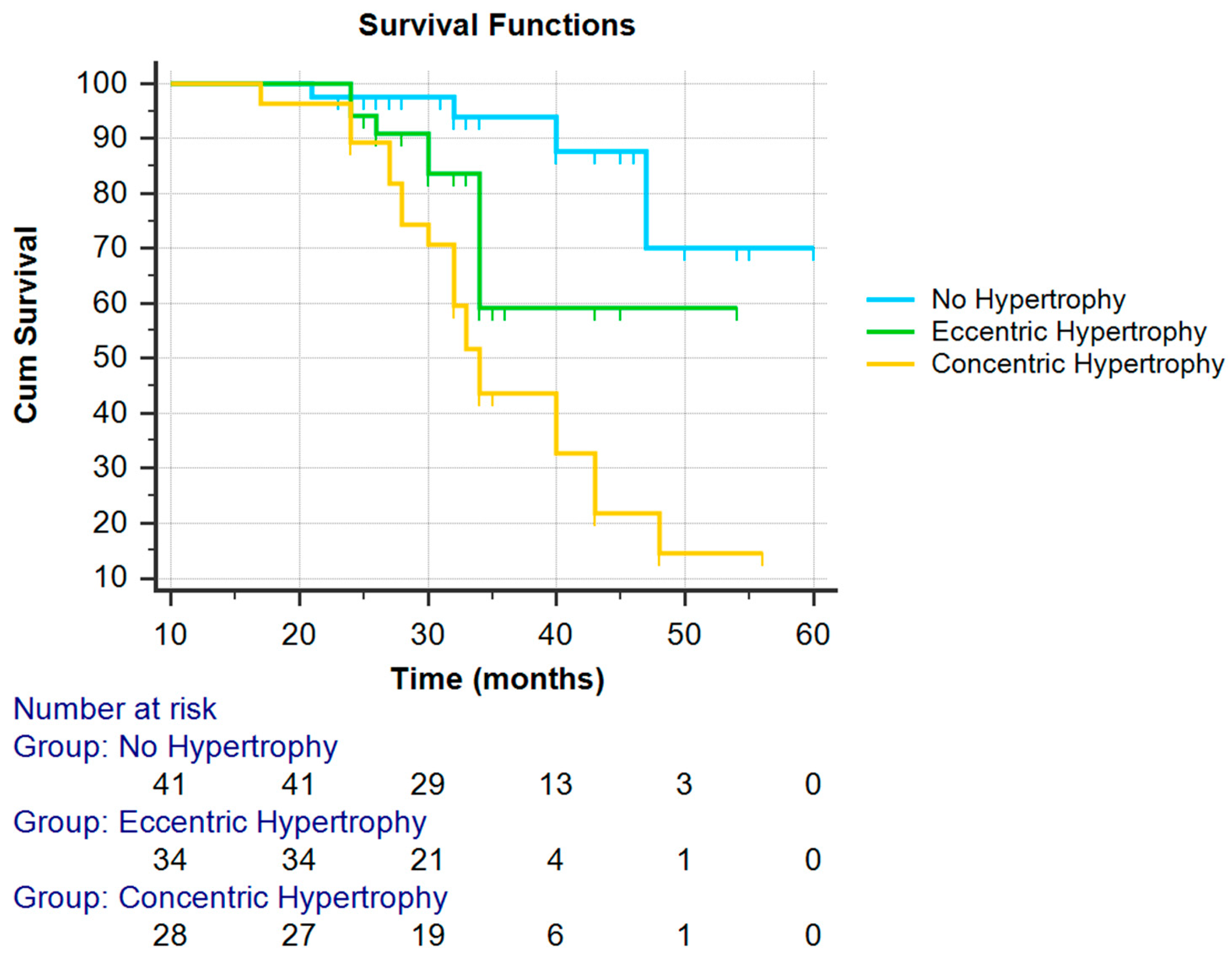
2. Results
3. Discussion
4. Materials and Methods
4.1. Subjects
4.2. BP and Echocardiographic Measurements
4.3. Follow-up
4.4. Blood Measurements
5.5. Urine Measurements
4.6. Cardiovascular Events
4.7. Statistical Analysis
5. Conclusions
Author Contributions
Funding
Conflicts of Interest
Abbreviations
| ARB | Angiotensin-converting inhibitor |
| ACR | Albumin/creatinine ratio |
| ACEI | Angiotensin-converting enzyme inhibitor |
| CKD | Chronic Kidney Disease |
| CVD | Cardiovascular Disease |
| DN | Diabetic Nephropathy |
| FGF-23 | Fibroblast growth factor 23 |
| FGFR | FGF receptor |
| eGFR | Estimated glomerular filtration rate |
| GFR | Glomerular Filtration Rate |
| GLM | Generalized linear model |
| IL-6 | Interleukin 6 |
| RAAS | Renin-angiotensin-aldosterone system |
References
- Levey, A.S.; Inker, L.A.; Coresh, J. GFR Estimation: From Physiology to Public Health. Am. J. Kidney Dis. 2014, 63, 820–834. [Google Scholar] [CrossRef]
- Elliott, S.; Tomita, D.; Endre, Z. Erythropoiesis stimulating agents and reno-protection: A meta-analysis. BMC Nephrol. 2017, 18, 14. [Google Scholar] [CrossRef]
- Liu, W.C.; Yen, J.F.; Lang, C.L.; Yan, M.T.; Lu, K.C. Bisphophonates in CKD Patients with Low Bone Mineral Density. Sci. World J. 2013, 2013, 837573. [Google Scholar] [CrossRef]
- Zoccali, C.; Benedetto, F.A.; Mallamaci, F.; Tripepi, G.; Giacone, G.; Cataliotti, A.; Seminara, G.; Stancanelli, B.; Malatino, L.S. Prognostic value of echocardiographic indicators of left ventricular systolic function in asymptomatic dialysis patients. J. Am. Soc. Nephrol. 2004, 15, 1029–1037. [Google Scholar] [CrossRef]
- Dubin, R.F.; Deo, R.; Bansal, N.; Anderson, A.H.; Yang, P.; Go, A.S.; Keane, M.; Townsend, R.; Porter, A.; Budoff, M.; et al. Associations of Conventional Echocardiographic Measures with Incident Heart Failure and Mortality: The Chronic Renal Insufficiency Cohort. Clin. J. Am. Soc. Nephrol. 2017, 12, 60–68. [Google Scholar] [CrossRef] [PubMed]
- Xu, J.; Murphy, S.L.; Kochanek, K.D.; Arias, E. Mortality in the United States, 2015. NCHS Data Brief 2016, 267, 1–8. [Google Scholar]
- Xie, J.; Cha, S.K.; An, S.W.; Kuro-O, M.; Birnbaumer, L.; Huang, C.L. Cardioprotection by Klotho through downregulation of TRPC6 channels in the mouse heart. Nat. Commun. 2012, 3, 1238. [Google Scholar] [CrossRef] [PubMed]
- Dalton, G.; An, S.W.; Al-Juboori, S.I.; Nischan, N.; Yoon, J.; Dobrinskikh, E.; Hilgemann, D.W.; Xie, J.; Luby-Phelps, K.; Kohler, J.J.; et al. Soluble klotho binds monosialoganglioside to regulate membrane microdomains and growth factor signaling. Proc. Natl. Acad. Sci. USA 2017, 114, 752–757. [Google Scholar] [CrossRef]
- Wolf, M.T.; An, S.W.; Nie, M.; Bal, M.S.; Huang, C.L. Klotho up-regulates renal calcium channel transient receptor potential vanilloid 5 (TRPV5) by intra- and extracellular N-glycosylation-dependent mechanisms. J. Biol. Chem. 2014, 289, 35849–35857. [Google Scholar] [CrossRef] [PubMed]
- Wu, Y.L.; Xie, J.; An, S.W.; Oliver, N.; Barrezueta, N.X.; Lin, M.H.; Birnbaumer, L.; Huang, C.L. Inhibition of TRPC6 channels ameliorates renal fibrosis and contributes to renal protection by soluble klotho. Kidney Int. 2016, 91, 830–841. [Google Scholar] [CrossRef]
- Tanaka, S.; Fujita, S.; Kizawa, S.; Morita, H.; Ishizaka, N. Association between FGF23, α-Klotho, and Cardiac Abnormalities among Patients with Various Chronic Kidney Disease Stages. PLoS ONE 2016, 11, e0156860. [Google Scholar] [CrossRef]
- Drew, D.A.; Katz, R.; Kritchevsky, S.; Ix, J.; Shlipak, M.; Gutiérrez, O.M.; Newman, A.; Hoofnagle, A.; Fried, L.; Semba, R.D.; et al. Association between Soluble Klotho and Change in Kidney Function: The Health Aging and Body Composition Study. J. Am. Soc. Nephrol. 2017, 28, 1859–1866. [Google Scholar] [CrossRef] [PubMed]
- Di Lullo, L.; House, A.; Gorini, A.; Santoboni, A.; Russo, D.; Ronco, C. Chronic kidney disease and cardiovascular complications. Heart Fail. Rev. 2015, 20, 259. [Google Scholar] [CrossRef] [PubMed]
- Lang, R.M.; Badano, L.P.; Mor-Avi, V.; Afilalo, J.; Armstrong, A.; Ernande, L.; Flachskampf, F.A.; Foster, E.; Goldstein, S.A.; Kuznetsova, T.; et al. Recommendations for cardiac chamber quantification by echocardiography in adults: An update from the American Society of Echocardiography and the European Association of Cardiovascular Imaging. Eur. Heart J. Cardiovasc. Imaging 2015, 16, 233–270. [Google Scholar] [CrossRef] [PubMed]
- Phan, D.; Aro, A.L.; Reinier, K.; Teodorescu, C.; Gunson, K.; Jui, J.; Chugh, S.S. Left Ventricular Geometry and Risk of Sudden Cardiac Arrest in Patients with Severely Reduced Ejection Fraction. J. Am. Heart Assoc. 2016, 5, e003715. [Google Scholar] [CrossRef]
- Neyra, J.A.; Hu, M.C. Potential application of klotho in human chronic kidney disease. Bone 2017, 100, 41–49. [Google Scholar] [CrossRef] [PubMed]
- Faul, C.; Amaral, A.P.; Oskouei, B.; Hu, M.C.; Sloan, A.; Isakova, T.; Gutiérrez, O.M.; Aguillon-Prada, R.; Lincoln, J.; Hare, J.M.; et al. FGF23 induces left ventricular hypertrophy. J. Clin. Investig. 2011, 121, 4393–4408. [Google Scholar] [CrossRef]
- Pavik, I.; Jaeger, P.; Ebner, L.; Wagner, C.A.; Petzold, K.; Spichtig, D.; Poster, D.; Wüthrich, R.P.; Russmann, S.; Serra, A.L. Secreted Klotho and FGF23 in chronic kidney disease Stage 1 to 5: A sequence suggested from a cross-sectional study. Nephrol. Dial. Transplant. 2013, 28, 352–359. [Google Scholar] [CrossRef]
- Sakan, H.; Nakatani, K.; Asai, O.; Imura, A.; Tanaka, T.; Yoshimoto, S.; Iwamoto, N.; Kurumatani, N.; Iwano, M.; Konishi, Y.N.; Saito, Y. Reduced Renal α-Klotho Expression in CKD Patients and Its Effect on Renal Phosphate Handling and Vitamin D Metabolism. PLoS ONE 2014, 9, e86301. [Google Scholar] [CrossRef]
- Krajisnik, T.; Olauson, H.; Mirza, M.A.; Hellman, P.; Akerstrom, G.; Westin, G.; Larsson, T.E.; Björklund, P. Parathyroid Klotho and FGF-receptor 1 expression decline with renal function in hyperparathyroid patients with chronic kidney disease and kidney transplant recipients. Kidney Int. 2010, 78, 1024–1032. [Google Scholar] [CrossRef]
- Takenaka, T.; Inoue, T.; Miyazaki, T.; Hayashi, M.; Suzuki, H. Xeno-Klotho Inhibits Parathyroid Hormone Signaling. J. Bone Miner. Res. 2016, 31, 455–462. [Google Scholar] [CrossRef]
- Ozeki, M.; Fujita, S.; Kizawa, S.; Morita, H.; Sohmiya, K.; Hoshiga, M.; Ishizaka, N. Association of serum levels of FGF23 and α-Klotho with glomerular filtration rate and proteinuria among cardiac patients. BMC Nephrol. 2014, 15, 147. [Google Scholar] [CrossRef]
- Bluemke, D.A.; Kronmal, R.A.; Lima, J.A.C.; Liu, K.; Olson, J.; Burke, G.L.; Folsom, A.R. The Relationship of Left Ventricular Mass and Geometry to Incident Cardiovascular Events: The MESA Study. Am. Coll. Cardiol. 2008, 52, 2148–2155. [Google Scholar] [CrossRef] [PubMed]
- Gerdts, E.; Cramariuc, D.; de Simone, G.; Wachtell, K.; Dahlof, B.; Devereux, R.B. Impact of left ventricular geometry on prognosis in hypertensive patients with left ventricular hypertrophy (the LIFE study). Eur. J. Echocardiogr. 2008, 9, 809–815. [Google Scholar] [CrossRef]
- Afshinnia, F.; Spitalewitz, S.; Chou, S.; Gunsburg, D.Z.; Chadow, H.L. Left Ventricular Geometry and Renal Function in Hypertensive Patients with Diastolic Heart Failure. Am. J. Kidney Dis. 2007, 49, 227–236. [Google Scholar] [CrossRef] [PubMed]
- Grabner, A.; Amaral, A.P.; Schramm, K.; Singh, S.; Sloan, A.; Yanucil, C.; Li, J.; Shehadeh, L.A; Hare, J.M.; David, V.; et al. Activation of cardiac fibroblast growth factor receptor 4 Causes left ventricular hypertrophy. Cell Metab. 2015, 22, 1020–1032. [Google Scholar] [CrossRef]
- Shibata, K.; Fujita, S.; Morita, H.; Okamoto, Y.; Sohmiya, K.; Hoshiga, M.; Hoshiga, M.; Ishizaka, N. Association between circulating fibroblast growth factor 23, alpha-Klotho, and the left ventricular ejection fraction and left ventricular mass in cardiology inpatients. PLoS ONE 2013, 8, e73184. [Google Scholar] [CrossRef]
- Kuro-O, M. The FGF23 and Klotho system beyond mineral metabolism. Clin. Exp. Nephrol. 2016, 21 (Suppl. 1), 64–69. [Google Scholar] [CrossRef]
- Xie, J.; Yoon, J.; An, S.W.; Kuro-o, M.; Huang, C.L. Soluble Klotho protects against uremic cardiomyopathy independently of fibroblast growth factor 23 and phosphate. J. Am. Soc. Nephrol. 2015, 26, 1150–1160. [Google Scholar] [CrossRef] [PubMed]
- Di Marco, G.S.; Reuter, S.; Kentrup, D.; Grabner, A.; Amaral, A.P.; Fobker, M.; Stypmann, J.; Pavenstädt, H.; Wolf, M.; Faul, C.; Brand, M. Treatment of established left ventricular hypertrophy with fibroblast growth factor receptor blockade in an animal model of CKD. Nephrol. Dial. Transpl. 2014, 29, 2028–2035. [Google Scholar] [CrossRef] [PubMed]
- Isakova, T.; Wahl, P.; Vargas, G.S.; Gutierrez, O.M.; Scialla, J.; Xie, H.; Appleby, D.; Nessel, L.; Bellovich, K.; Chen, J.; et al. Fibroblast growth factor 23 is elevated before parathyroid hormone and phosphate in chronic kidney disease. Kidney Int. 2011, 79, 1370–1378. [Google Scholar] [CrossRef] [PubMed]
- Yin, S.; Zhang, Q.; Yang, J.; Lin, W.; Li, Y.; Chen, F.; Cao, W. TGFβ-incurred epigenetic aberrations of miRNA and DNA methyltransferase suppress Klotho and potentiate renal fibrosis. Biochim. Biophys. Acta 2017, 1864, 1207–1216. [Google Scholar] [CrossRef]
- Silva, P.A.; Guedes, M.A.; Neves, L.P. Cardiovascular Risk Factors: The Old Ones and a Closer Look to the Mineral Metabolism. In Chronic Kidney Disease-From Pathophysiology to Clinic Improvements; Rath, T., Ed.; IntechOpen: London, UK, 2016; pp. 83–104. [Google Scholar] [CrossRef]
- Pi, M.; Ye, R.; Han, X.; Armstrong, B.; Liu, X. Cardiovascular Interactions between Fibroblast Growth Factor-23 and Angiotensin II. Sci. Rep. 2018. [Google Scholar] [CrossRef] [PubMed]
- Care, D. Classification and Diagnosis of Diabetes. Am. Diabetes Assoc. Diabetes Care 2016, 39 (Suppl. 1), S13–S22. [Google Scholar] [CrossRef]
- Savage, D.D.; Garrison, R.Y.; Kannel, W.B.; Levy, D.; Anderson, S.J.; Stokes, J., 3rd; Feinleib, M.; Castelli, W.P. The spectrum of left ventricular hypertrophy in a general population sample: The Framingham study. Circulation 1987, 75 (Suppl. I), 26–33. [Google Scholar]
- Chen, S.C.; Su, H.M.; Hung, C.C.; Chang, J.M.; Liu, W.C.; Tsai, J.C.; Lin, M.Y.; Hwang, S.I.; Chen, H.C. Echocardiographic parameters are independently associated with rate of renal function decline and progression to dialysis in patients with chronic kidney disease. Clin. J. Am. Soc. Nephrol. 2011, 6, 2750–2758. [Google Scholar] [CrossRef] [PubMed]
- Matthews, D.R.; Hosker, J.P.; Rudenski, A.S.; Naylor, B.A.; Treacher, D.F.; Turner, R.C. Homeostasis model assessment: Insulin resistance and beta-cell function from fasting plasma glucose and insulin concentrations in man. Diabetologia 1985, 28, 412–419. [Google Scholar] [CrossRef]
- Levey, A.S.; Stevens, L.A.; Schmid, C.H.; Zhang, Y.; Castro, A.F.; Feldman, H.I.; Kusek, J.W; Eggers, P.; Greene, T.; Coresh, J. CKD-EPI (chronic kidney disease epidemiology collaboration). A new equation to estimate glomerular filltration rate. Ann. Intern. Med. 2009, 150, 604–612. [Google Scholar] [CrossRef]
- Kavousi, M.; Leening, M.J.; Nanchen, D.; Greenland, P.; Graham, I.M.; Steyerberg, E.W.; Ikram, M.A.; Stricker, B.H.; Hofman, A.; Franco, O.H. Comparison of application of the ACC/AHA guidelines, Adult Treatment Panel III guidelines, and European Society of Cardiology guidelines for cardiovascular disease prevention in a European cohort. JAMA 2014, 311, 1416–1423. [Google Scholar] [CrossRef]


| Characteristics | Values |
|---|---|
| Number of patients, n | 107 |
| Gender (f/m) | 40/67 |
| BMI (Kg/m2) | 26.04 |
| Age (years) | 57.19 ± 7.05 |
| Hb (g/dL) | 12.97 ± 1.83 |
| Albumin (g/dL) | 4.27 ± 0.48 |
| eGFR (mL/min) | 52.89 ± 20.15 |
| ACR (µg/mg) | 181.89 ± 33.83 |
| Pi (mg/dL) | 3.99 ± 0.85 |
| PTH (pg/mL) | 113.11 ± 74.65 |
| Calcium (mg/dL) | 9.48 ± 0.68 |
| FGF-23 (RU/mL) | 135.04 ± 135.23 |
| 1,25 (OH)2 Vitamin D (pg/mL) | 21.21 ± 7.36 |
| α-Klotho (pg/mL) | 331.10 ± 171.06 |
| IL-6 (pg/mL) | 5.71 ± 3.80 |
| Oxidized LDL (U/L) | 39.91 ± 19.55 |
| HOMA-IR | 1.84 ± 1.67 |
| HbA1c (%) | 7.67 ± 1.47 |
| LVMI (g/m2) | 99.31 ± 23.45 |
| Systolic BP (mmHg) | 126.59 ± 16.53 |
| Diastolic BP (mmHg) | 78.58 ± 9.98 |
| Diabetes-related CKD evolution time (months) | 73.8 ± 8.7 |
| Group 1 Normal Geometry n = 41 | Group 2 Eccentric Hypertrophy n = 38 | Group 3 Concentric Hypertrophy n = 28 | p | |
|---|---|---|---|---|
| Age (years) | 56.12 ± 7.35 | 57.29 ± 7.27 | 58.61 ± 6.21 | NS |
| eGFR (mL/min/1.73 m2) | 62.89 ± 20.58 | 47.13 ± 15.18 | 46.06 ± 20.10 | <0.001 |
| BMI (Kg/m2) | 26.3 ± 1.08 | 26.8 ± 0.42 | 27.2 ± 0.64 | NS |
| Hb (g/dL) | 13.57 ± 1.73 | 12.98 ± 1.81 | 12.98 ± 1.81 | 0.003 |
| Pi (mg/dL) | 3.32 ± 0.64 | 4.76 ± 0.54 | 4.13 ± 0.75 | <0.001 |
| PTH (pg/mL) | 74.16 ± 49.67 | 104.07 ± 63.26 | 182.41 ± 73.07 | <0.001 |
| ACR (µg/mg) | 122.13 ± 90.60 | 193.50 ± 144.86 | 253.64 ± 91.30 | <0.001 |
| IL-6 (pg/mL) | 2.93 ± 1.82 | 8.47 ± 3.22 | 10.09 ± 2.42 | <0.001 |
| α-Klotho (pg/mL) | 399.17 ± 143.72 | 344.82 ± 53.52 | 160.82 ± 60.70 | <0.001 |
| FGF-23 (RU/mL) | 66.75 ± 39.66 | 113.06 ± 54.20 | 264.84 ± 72.96 | <0.001 |
| [1,25(OH)2D3] (pg/mL) | 25.78 ± 4.49 | 21.88 ± 6.80 | 18.60 ± 5.22 | <0.001 |
| Systolic BP (mmHg) | 124.2 ± 14.2 | 121.1 ± 15 | 125.5 ± 16.5 | 0.057 |
| Diastolic BP (mmHg) | 71.2 ± 10.4 | 71.9 ± 11.8 | 69.6 ± 11.6 | NS |
| Heart rate (bpm) | 67 ± 12 | 70 ± 13 | 69 ± 11 | NS |
| LV end-diastolic volume (mL) | 120 ± 17 | 162 ± 32 | 118 ± 20 | 0.005 |
| LV end-systolic volume (mL) | 42 ± 10 | 62 ± 23x | 45 ± 15 | <0.001 |
| LV mass/body surface area (g/m2) | 96 ± 11 | 132 ± 22 | 134 ± 20 | NS |
| RWT | 0.38 ± 0.3 | 0.37 ± 0.06 | 0.46 ± 0.03 | NS |
| RAS inhibitor/or ACEI (%) | 78.7 | 80.7 | 88.9 | NS |
| Calcium channel blockers with renoprotective action (%) | 35.6 | 48.6 | 52.3 | NS |
| Diabetes-related CKD evolution time (months) | 75.5 ± 9.6 | 71.7 ± 7.3 | 74.1 ± 8.7 | NS |
| Variable | R | p-Value |
|---|---|---|
| Gender | 0.741 | 0.354 |
| Age | 0.243 | 0.223 |
| BMI | 0.480 | 0.650 |
| eGFR | −0.227 | 0.060 |
| Hb | −0.317 | 0.973 |
| Pi | 0.672 | 0.005 |
| PTH | 0.520 | 0.199 |
| ACR | 0.427 | 0.075 |
| IL-6 | 0.737 | 0.087 |
| α-Klotho | −0.440 | 0.0001 |
| FGF-23 | 0.622 | 0.0001 |
| Variable | Eccentric Hypertrophy | Concentric Hypertrophy | ||||
|---|---|---|---|---|---|---|
| OR | 95% CI | p | OR | 95% CI | p | |
| α-Klotho | 1.005 | 0.97–1013 | 0.186 | 0.737 | 0.603–0.972 | 0.031 |
| Gender | 1.670 | 0.341–8.174 | 0.527 | 1.986 | 0.905–2.009 | 0.063 |
| Age | 1.095 | 0.970–1.235 | 0.142 | 1.053 | 0.821–1.349 | 0.685 |
| eGFR | 0.959 | 0.921–0.999 | 0.043 | 0.769 | 0.622–1.050 | 0.085 |
| Pi | 2.859 | 2.238–5.693 | 0.003 | 1.115 | 0.958–2.318 | 0.063 |
| PTH | 0.980 | 0.960–1.001 | 0.058 | 0.994 | 0.969–1.020 | 0.664 |
| ACR | 1.005 | 0.999–1.011 | 0.131 | 0.977 | 0.945–1.011 | 0.184 |
| FGF-23 | 1.008 | 0.996–1.020 | 0.219 | 1.031 | 1.008–1.205 | 0.009 |
| [1,25 (OH)2 D3] | 0.904 | 0.726–1.126 | 0.366 | 0.529 | 0.263–1.061 | 0.073 |
| Hb | 1.040 | 0.686–1.577 | 0.852 | 0.307 | 0.295–1.001 | 0.068 |
| IL-6 | 1.255 | 0.836–1.885 | 0.274 | 1.443 | 0.984–2.858 | 0.208 |
| Initial Model | Optimized Model | |||||
|---|---|---|---|---|---|---|
| ORa | 95%CI for ORa | p | ORa | 95%CI for ORa | p | |
| α-Klotho groups | ||||||
| <313 | 1.491 | 1.207–2.125 | 0.014 | 11.320 | 1.061–1.456 | 0.024 |
| ≥313 | Ref. | Ref. | ||||
| FGF-23 groups | ||||||
| ≥168 | 1.689 | 1.004–3.500 | 0.004 | 1.105 | 1.000–1.763 | 0.012 |
| <168 | Ref. | Ref. | ||||
| Concentric Hypertrophy | 4.889 | 3.372–5.512 | 0.023 | 2.284 | 1.970–4.720 | 0.1 |
| Eccentric Hypertrophy | 3.360 | 0.692–4.890 | 0.064 | 1.410 | 0.984–2.523 | 0.075 |
| No Hypertrophy | Ref. | Ref. | ||||
| p-value (model) | 0.007 | <0.001 | ||||
| Area under ROC (p-value) | - | 0.980 (p < 0.001) | ||||
| HRa | 95%CI for ORa | p-Value | |
|---|---|---|---|
| Pi | |||
| <3.6 | Ref. | ||
| ≥3.6 | 1.079 | 1.015–3.409 | 0.025 |
| α-Klotho groups | |||
| <313 | 2.377 | 1.488–11.585 | 0.044 |
| ≥313 | Ref. | ||
| FGF-23 groups | |||
| ≥168 | 2.046 | 1.008–8.249 | 0.014 |
| < 168 | Ref. | ||
| Concentric | 3.254 | 1.035–6.699 | 0.041 |
| Eccentric | 1.112 | 1.070–3.850 | 0.050 |
| No Hypertrophy | Ref. | ||
© 2019 by the authors. Licensee MDPI, Basel, Switzerland. This article is an open access article distributed under the terms and conditions of the Creative Commons Attribution (CC BY) license (http://creativecommons.org/licenses/by/4.0/).
Share and Cite
Silva, A.P.; Mendes, F.; Carias, E.; Gonçalves, R.B.; Fragoso, A.; Dias, C.; Tavares, N.; Café, H.M.; Santos, N.; Rato, F.; et al. Plasmatic Klotho and FGF23 Levels as Biomarkers of CKD-Associated Cardiac Disease in Type 2 Diabetic Patients. Int. J. Mol. Sci. 2019, 20, 1536. https://doi.org/10.3390/ijms20071536
Silva AP, Mendes F, Carias E, Gonçalves RB, Fragoso A, Dias C, Tavares N, Café HM, Santos N, Rato F, et al. Plasmatic Klotho and FGF23 Levels as Biomarkers of CKD-Associated Cardiac Disease in Type 2 Diabetic Patients. International Journal of Molecular Sciences. 2019; 20(7):1536. https://doi.org/10.3390/ijms20071536
Chicago/Turabian StyleSilva, Ana Paula, Filipa Mendes, Eduarda Carias, Rui Baptista Gonçalves, André Fragoso, Carolina Dias, Nelson Tavares, Hugo Mendonça Café, Nélio Santos, Fátima Rato, and et al. 2019. "Plasmatic Klotho and FGF23 Levels as Biomarkers of CKD-Associated Cardiac Disease in Type 2 Diabetic Patients" International Journal of Molecular Sciences 20, no. 7: 1536. https://doi.org/10.3390/ijms20071536
APA StyleSilva, A. P., Mendes, F., Carias, E., Gonçalves, R. B., Fragoso, A., Dias, C., Tavares, N., Café, H. M., Santos, N., Rato, F., Leão Neves, P., & Almeida, E. (2019). Plasmatic Klotho and FGF23 Levels as Biomarkers of CKD-Associated Cardiac Disease in Type 2 Diabetic Patients. International Journal of Molecular Sciences, 20(7), 1536. https://doi.org/10.3390/ijms20071536