Article

Identification of σE-Dependent Promoter Upstream of clpB from the Pathogenic Spirochaete Leptospira interrogans by Applying an E. coli Two-Plasmid System

by
Sabina Kędzierska-Mieszkowska
1,*,
Katarzyna Potrykus
2,
Zbigniew Arent
3 and
Joanna Krajewska
1
1
Department of General and Medical Biochemistry, University of Gdańsk, Faculty of Biology, 80-308 Gdańsk, Poland
2
Department of Bacterial Molecular Genetics, Faculty of Biology, University of Gdańsk, 80-308 Gdańsk, Poland
3
University Centre of Veterinary Medicine, University of Agriculture in Krakow, 30-059 Krakow, Poland
*
Author to whom correspondence should be addressed.
Int. J. Mol. Sci. 2019, 20(24), 6325; https://doi.org/10.3390/ijms20246325
Submission received: 14 November 2019 / Revised: 10 December 2019 / Accepted: 13 December 2019 / Published: 15 December 2019
(This article belongs to the Special Issue Molecular Chaperones 2.0)

Abstract

:
There is limited information on gene expression in the pathogenic spirochaete Leptospira interrogans and genetic mechanisms controlling its virulence. Transcription is the first step in gene expression that is often determined by environmental effects, including infection-induced stresses. Alterations in the environment result in significant changes in the transcription of many genes, allowing effective adaptation of Leptospira to mammalian hosts. Thus, promoter and transcriptional start site identification are crucial for determining gene expression regulation and for the understanding of genetic regulatory mechanisms existing in Leptospira. Here, we characterized the promoter region of the L. interrogans clpB gene (clpBLi) encoding an AAA+ molecular chaperone ClpB essential for the survival of this spirochaete under thermal and oxidative stresses, and also during infection of the host. Primer extension analysis demonstrated that transcription of clpB in L. interrogans initiates at a cytidine located 41 bp upstream of the ATG initiation codon, and, to a lesser extent, at an adenine located 2 bp downstream of the identified site. Transcription of both transcripts was heat-inducible. Determination of clpBLi transcription start site, combined with promoter transcriptional activity assays using a modified two-plasmid system in E. coli, revealed that clpBLi transcription is controlled by the ECF σE factor. Of the ten L. interrogans ECF σ factors, the factor encoded by LIC_12757 (LA0876) is most likely to be the key regulator of clpB gene expression in Leptospira cells, especially under thermal stress. Furthermore, clpB expression may be mediated by ppGpp in Leptospira.

1. Introduction

Leptospira interrogans is one of the many pathogenic species of the genus Leptospira and is composed of several strains that can cause leptospirosis in mammals, including humans. It has been reported that over 1 million human cases of severe leptospirosis occur worldwide each year, with approximately 60,000 deaths from this disease [1]. It has to be noted that leptospirosis also generates huge economic losses in a number of countries due to reproductive disorders in cattle, sheep, pigs, and horses [2,3]. Furthermore, recent serological and microbiological studies have indicated a high rate of leptospiral infections in domestic animals [2,4,5,6]. Despite a high risk of leptospirosis, especially in the tropical and subtropical countries, and its global importance, molecular mechanisms of both the leptospiral virulence and the disease pathogenesis currently remain largely unknown [7,8], mainly due to the historical lack of standard genetic tools for use in work with the pathogenic Leptospira species. Recent advances in genetic manipulation of these species have made it possible to identify several leptospiral virulence factors. However, many of them have turned out not to be required for virulence in animal models [7]. Unfortunately, the limitations of modern genetic tools available for pathogenic Leptospira spp. still have an enormous impact on the understanding of the molecular and cellular mechanisms involved in the pathogenesis of leptospirosis.
Like other pathogenic bacteria, L. interrogans is exposed to environmental stresses during infection of mammalian hosts. Bacteria fight various environmental stressors by altering the expression of genes involved in host adaptation and promoting their survival. Transcriptional regulation, and especially sigma factors controlling the promoter selectivity of bacterial RNA polymerase, play a crucial role in this stress-induced gene expression response. Unfortunately, there is limited information on gene regulation in Leptospira spp. Comparative genomics and genome-wide in silico analyses had shown that the Leptospira genome contains one basic sigma factor-encoding gene- rpoD70), and alternative sigma factor genes: rpoF28), rpoN54), and several (5–11) genes encoding extracytoplasmic function (ECF) sigma factors, referred to as rpoEE) in the literature [9,10] (Table 1).
According to these analyses, the housekeeping sigma factor, σ70, coordinates transcription of most genes in the leptospiral cells, while alternative sigma factors play key roles in environmental adaptation of bacteria and their virulence. In that light, it is intriguing that the Leptospira species differ in the number of ECF σ factors. For example, pathogenic L. interrogans encodes ten different ECF σ factors (Table 2), while saprophytic species encode only five [9].
On the other hand, the well-known bacterium E. coli possesses only two ECF σ factors [14] (Table 1). It is postulated that the ECF σ factors existing only in the pathogenic Leptospira species are important for the Leptospira life cycle within the host [9]. Unraveling regulation of virulence genes’ expression is a particularly important challenge because it is necessary for understanding the molecular basis of the disease caused by the pathogen and its underpinning. Molecular chaperone ClpB, a member of the Hsp100/Clp subfamily of the AAA + ATPases (ATPases associated with a variety of cellular activities), is among the known leptospiral virulence factors. It has been demonstrated that ClpB deficiency in L. interrogans resulted in bacterial growth defects under oxidative and heat stresses, and also in its loss of virulence [15]. Furthermore, in previous studies, we had shown that L. interrogans ClpB (ClpBLi) is not only synthesized but is also immunogenic during the infection process, further supporting its involvement in Leptospira pathogenicity [16]. Our recent studies suggest a possible role of ClpBLi, i.e., its aggregate-reactivation activity is necessary for maintaining the energy-generating metabolism of the Leptospira cell [17], again strongly supporting ClpB’s importance in leptospiral virulence and implying the importance of clpB gene expression regulation. Here, primer extension analysis combined with promoter activity assays using a genetic strategy revealed that clpBLi transcription is σE dependent. We postulate that factor encoded by LIC_12757 (LA0876) is a key ECF σE factor that promotes clpB transcription in pathogenic Leptospira, especially under stressful conditions. In addition, the ppGpp alarmone may act as a regulator of clpB expression in those cells. Our findings provide the first insight into Leptospira clpBLi transcription regulation. Still, further studies are needed to discover all regulatory elements affecting clpB expression in Leptospira cells.

2. Results and Discussion

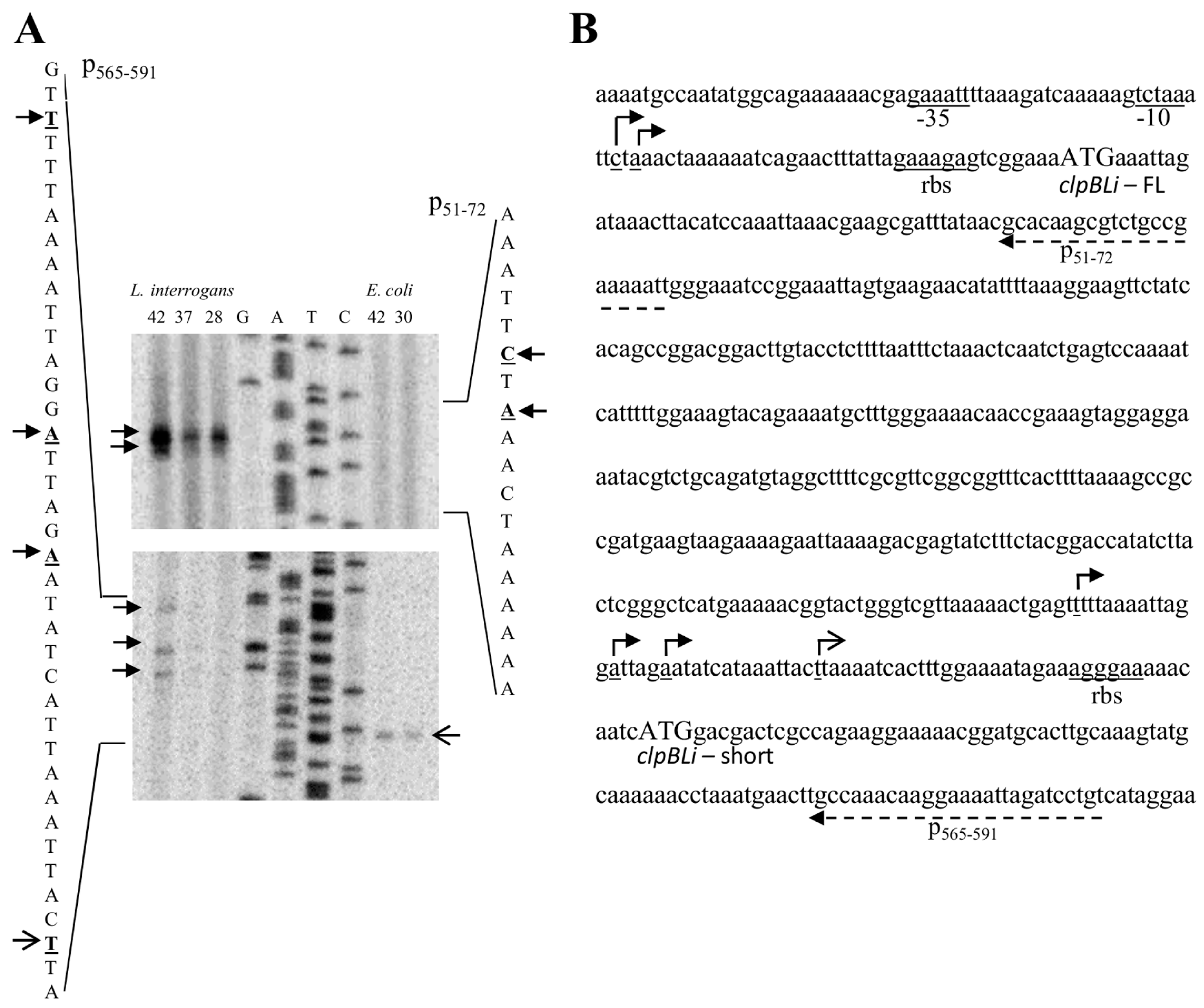
2.1. Identification of the clpB Transcriptional Start Sites by Primer Extension

In order to determine clpB transcriptional start sites and identify functional promoters responsible for its expression in Leptospira, primer extension assays were performed. Three synthetic oligonucleotides, p51–72, p290–321, p565–591 (see Table 3) were hybridized to RNA extracted from both, Leptospira interrogans and E. coli MC4100ΔclpB cells carrying the L. interrogans clpB gene (clpBLi) cloned into a low-copy pGB2 plasmid together with a 500-bp DNA fragment containing sequence located upstream of the gene (pClpBLi). A BLAST analysis did not show a potential cross-hybridization of the designed primers with regions of the E. coli genome, suggesting high specificity of the designed primers. To prepare RNA, the L. interrogans strain was grown at 28 °C for optimal in vitro growth and also under thermal stresses at 37 and 42 °C for 4 and 2 h, respectively, whereas E. coli cells were grown at 30 or 42 °C for 30 min (heat shock). Following reverse transcription and analysis on a sequencing gel, one major product was observed when the p51–72 primer was used in reactions with RNA isolated from Leptospira cells grown at all tested temperatures (Figure 1). This product corresponds to a transcriptional start site at a cytidine residue located 41 bp upstream of the first clpB ATG codon (−41 relative to ATG). In addition, a lesser band is observed at an adenosine residue located at the −39 position relative to ATG. It is interesting to note that this alternative start point of transcription (A-39) coincides with the primary transcriptional start site (tctAaac) previously predicted by [10] using differential RNA-sequencing (dRNA-seq). It is also worth mentioning that the secondary transcriptional start site (G-526 of the GenBank clpB gene sequence, accession number M28364) was found to be close to the primary transcriptional start site (A-525) in primer extension analysis of the E. coli clpB transcripts [18]. It is also known that Mycoplasma pneumoniae reference strain M129 uses two different transcriptional start sites (G-10 and A-7 relative to the ATG initiation codon) to produce ClpB [19]. Thus, the presence of the primary and secondary transcriptional start site in the clpBLi promoter region is not surprising. Further, the amount of both Leptospira clpB transcripts is increased after exposure of cells to a temperature of 42 °C for 2 h. This result indicates that clpBLi expression in Leptospira is induced by temperature stress, which is consistent with the findings of [15], who demonstrated that clpBLi transcription in Leptospira had increased by 3-folds after 2 h exposure to heat stress. One possible candidate regulating this expression could be RpoH (σ32; heat shock sigma factor); however, comparative genomics and genome-wide in silico analyses had shown no presence of a rpoH gene in the Leptospira genome [9,10]. Thus, Leptospira spp. does not possess σ32 that is required for heat shock gene expression and heat shock response in E. coli. On the other hand, as shown in Figure 1, for RNA extracted from the E. coli cells, no extension product was detectable with the p51-72 primer, indicating that E. coli either does not possess an appropriate factor for clpBLi expression or this factor is not induced in E. coli by temperature stress. Additional analysis has been performed with two other primers. Primer extension assay utilizing the p290–321 primer did not detect any transcripts either with Leptospira or with E. coli RNA (not shown), while the use of the p565–591 primer revealed a few weak heat-inducible transcripts for Leptospira and one transcript for E. coli RNA that was not induced by heat shock stress and did not coincide with any leptospiral transcription start sites (Figure 1). All of these transcripts initiate within the region that is 20–96 nt upstream of a potential internal translation initiation site of clpBLi with a good Shine–Dalgarno sequence (AGGGAA) and the ATG codon (see Figure 1). It is known that E. coli uses two translational start sites to produce two isoforms of ClpB, the full-length ClpB95 and shorter ClpB80, which does not contain the substrate-interacting N-terminal domain [18,20,21]. It has been demonstrated that the two isoforms of ClpB from E. coli cooperate in reactivation of aggregated proteins to form a highly efficient chaperone system [21]. It is very likely that the functional cooperation between ClpB isoforms arises from interactions between them because it was found that ClpB95 and ClpB80 associate into hetero-oligomers, which boost the aggregate-reactivation potential of the ClpB chaperone [21]. Thus, it was postulated that E. coli produces two isoforms of ClpB to optimize its disagregase activity. The presence of two forms of ClpB, a 96-kDa and an 80-kDa protein, was also demonstrated in L. interrogans [15]. Our results point to the occurrence of an additional transcriptional start site located within the clpBLi coding region in both E. coli and Leptospira. Still, it is intriguing that even though the internal clpBLi transcriptional start sites were detected in both instances, they differ depending on the host. It is also interesting to note the similarity between one of the internal transcriptional start sites (see Figure 1, gagtTtt) that we mapped by primer extension analysis and one of the sixteen internal start points (gagTttt) previously predicted by [10]. Thus, it is highly possible that clpBLi has an additional promoter. On the other hand, it cannot be excluded that the mapped minor transcripts result from the processing of the major transcript produced. Still, further studies are needed to examine the nature of those minor transcripts in Leptospira.

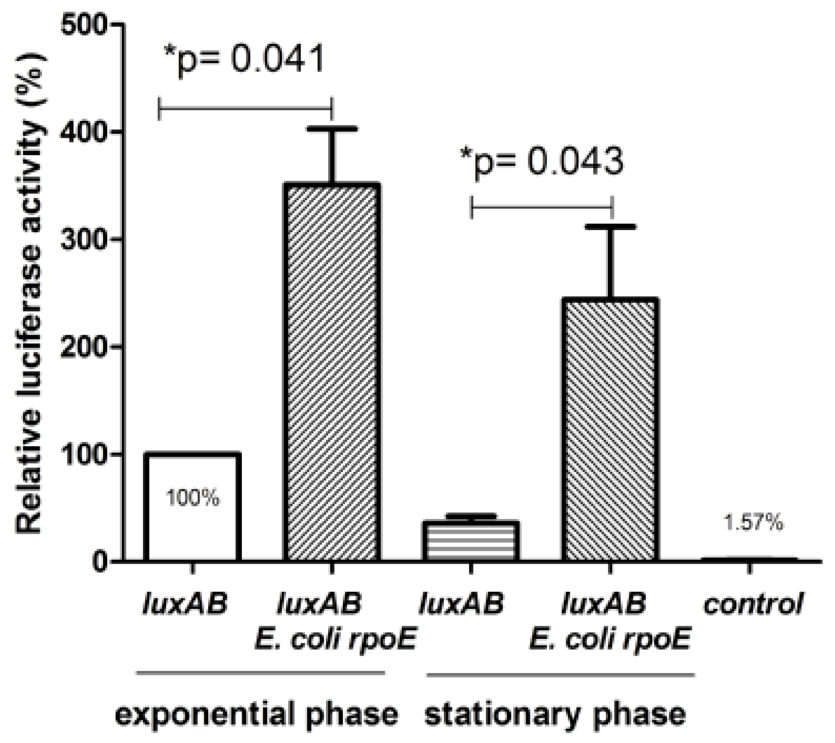
2.2. Presence of a Promoter Dependent on σE Upstream of the ClpBLi Gene and its Activity in E. coli Cells

After the mapping of the two clpBLi transcriptional start sites (both probably originating from the same promoter) in Leptospira (see Figure 1), the nucleotide sequence upstream of clpBLi was carefully investigated. A putative σE promoter element was successfully identified on the basis of sequence similarity to the well-characterized E. coli σE core promoters. As shown in Figure 2, sequences of the −10 and −35 motifs of the potential clpB promoter (clpBLiP1) exhibit a considerable homology to the known promoters controlled by E. coli σE.
To investigate whether the σE factor may be really involved in the clpBLi transcriptional regulation, promoter activity assay was carried out under σE-limiting (basal) and σE excess (overproduction) conditions. It is worth to emphasize that under normal growth conditions, the expression level of rpoE in E. coli cells is low. Therefore, it was of great interest to compare the activity of the potential clpBLi promoter (clpBLiP1) under basal and increased levels of σE. To our knowledge, there has been only one report to date, describing the construction and utility of the gfp reporter plasmid for assessing promoter activity in L. interrogans [22]. Therefore, due to the still existing limitations of genetic tools that could be easily utilized for examining gene expression in L. interrogans, experiments were performed in E. coli cells. A modified two-plasmid system has been employed that has been previously successfully used for the identification of many σE-cognate promoters from E. coli and also from other bacteria in studies that demonstrated the suitability of this system for assessing promoter activity in E. coli [23,24]. Briefly, this system uses two compatible plasmids. One of them is pAC-rpoE4 carrying the E. coli rpoE gene under the control of an arabinose inducible pBAD promoter, and the second plasmid carries a reporter gene fused to a potential σE promoter. We used the V. harveyi luxAB gene, encoding luciferase, as a reporter cloned into a low-copy pGB2 plasmid under control of the tested clpBLiP1 promoter (pclpBLiP1-luxAB). The method used assumes that shortly after arabinose addition σE factor overproduced from pAC-rpoE4 interacts with the E. coli RNA polymerase core enzyme forming functional RNA polymerase holoenzyme that is capable of recognizing σE -controlled promoters located upstream of a reporter gene carried on the second plasmid; in our study, it was the pclpBLiP1-luxAB plasmid carrying the luxAB reporter genes. As shown in Figure 3, the activity of luciferase in both, the exponential and stationary phase of bacterial growth was indeed significantly higher in the presence of the pAC-rpoE4 plasmid expressing the E. coli rpoE (i.e., under σE excess conditions) than that in cells transformed only with pclpBLiP1- luxAB and producing a basal level of σE.
Importantly, no significant luminescent signal was detected for cells carrying a construct with promoter-less luxAB genes. These results strongly suggested that the activity of clpBLiP1 is dependent on σE. Furthermore, we examined clpBLi expression driven by the clpBLiP1 promoter in E. coli cells under heat shock exposure at 42 °C and also under σE excess conditions (in the presence of pAC-rpoE4). Thus, the main aim of this experiment was to compare the clpBLiP1 promoter behavior under thermal stress and σE overproduction, by monitoring the amount of ClpBLi that was produced. Such an approach was impossible in case of experiments with pclpBLiP1-luxAB because luciferase is thermolabile and would not withstand the heat shock conditions. To this end, Western blotting analysis of whole-cell lysates of the E. coli MC4100ΔclpB strain carrying pClpBLi alone or together with pAC-rpoE4 was carried out using anti-ClpBLi158–334 serum [15]. Expression of E. coli rpoE was induced by the addition of 0.02% arabinose for 30–90 min, while the expression of pClpBLi was induced by exposure to 42 °C for 1 h. As shown in Figure 4A, thermal stress was not sufficient to induce the ClpBLi production (96-kDa protein) in the cells carrying the clpBLi gene under the control of the clpBLiP1 promoter (see lanes 5 and 6). This is in agreement with our results of primer extension analysis (see Figure 1A) that did not show the presence of the clpBLi transcripts in E. coli cells after heat shock stress. Consistent with our previous results [25], the use of pGB2-ClpBLi [25], which carries the heat-inducible promoter σ32 from E. coli located upstream of clpBLi, resulted in increased synthesis of ClpBLi after heat treatment (see lanes 3 and 4). The most interesting observation pertains to the clpBLi expression under σE excess conditions (Figure 4B), as the appearance of the 96-kDa ClpBLi in the E. coli cells 30–90 min after arabinose addition (see lanes 3–5) provides further evidence that clpBLiP1 is σE dependent. As can be seen in Figure 4B, there was one more protein that occurred at a higher amount after σE induction. It is likely that this protein corresponds to a shorter form of ClpBLi (80 kDa) that either represents transcription initiation from the internal promoter or exclusively translation initiation from the internal in-frame ATG codon.
Together, the results obtained with E. coli σE support that ECF σE factor may mediate the clpBLi transcription in L. interrogans cells.

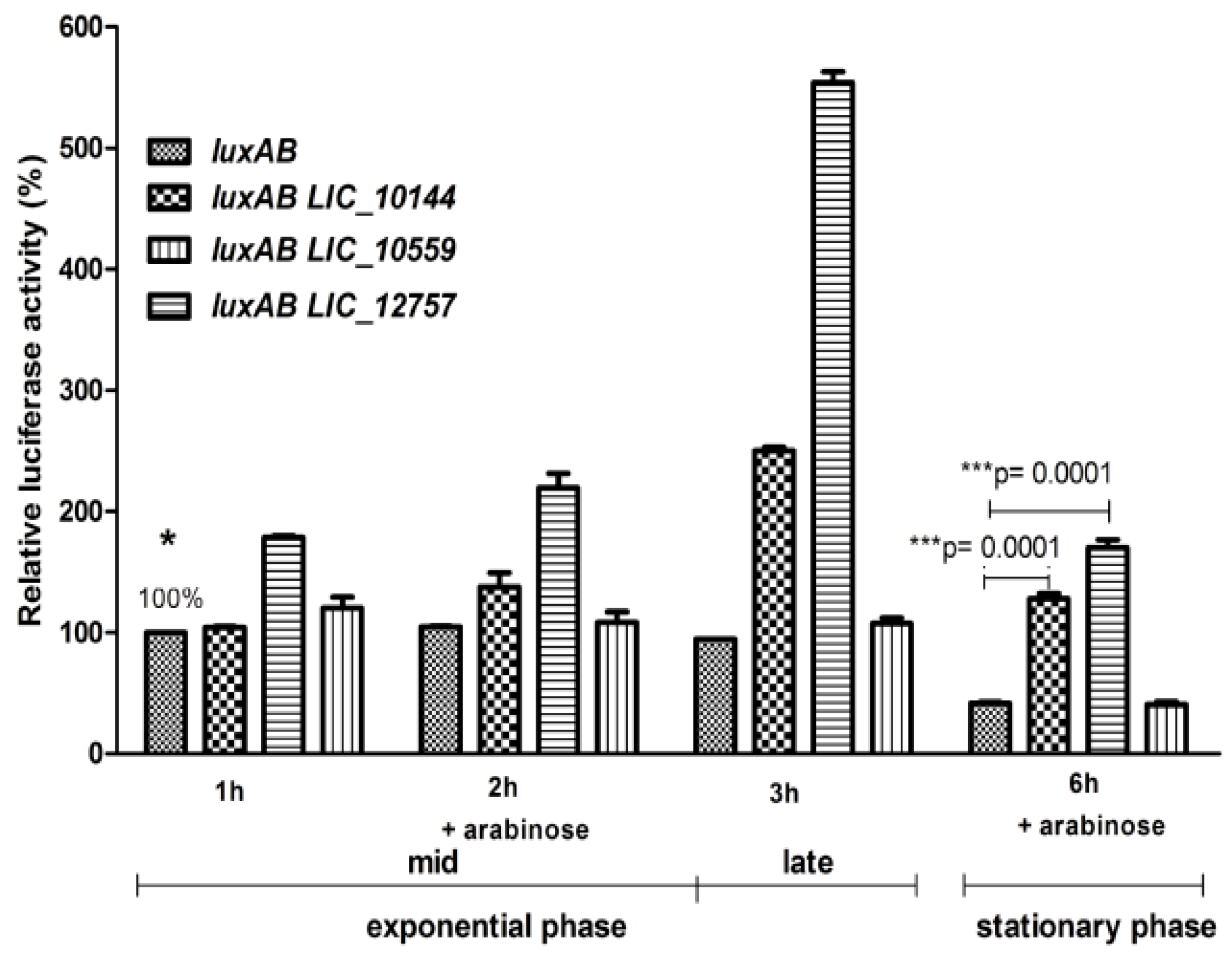
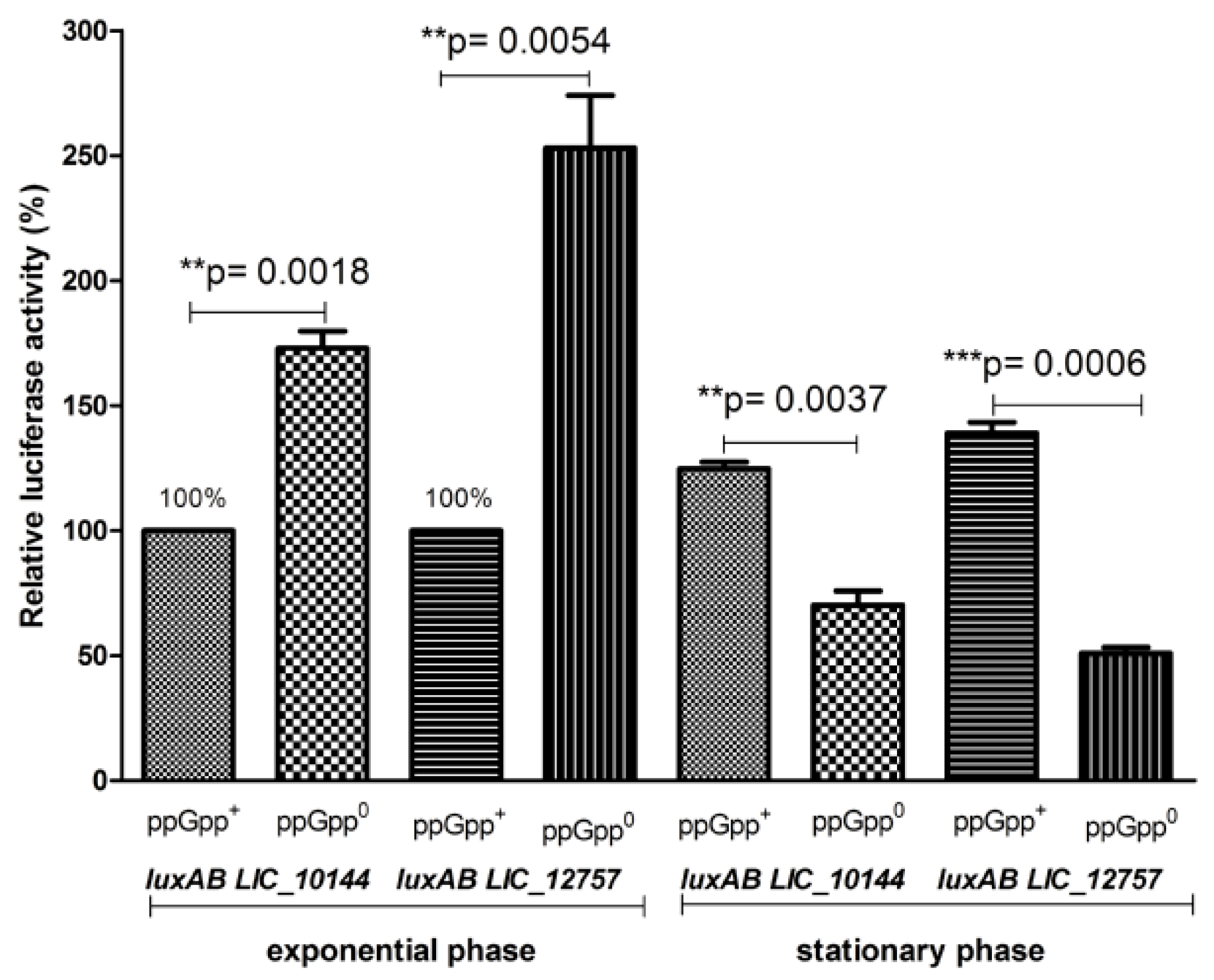
2.3. Role of Leptospiral ECFσ Factors in ClpBLi Gene Expression

It has been demonstrated that σE contributes to heat shock and oxidative stress response in a number of bacteria and is also involved in the regulation of virulence genes and virulence-associated genes in many bacterial pathogens as well [26]. Since L. interrogans rpoE expression was reported to be up-regulated in Leptospira cells exposed to elevated temperatures, it is possible that leptospiral σE factors also regulate expression of genes required for virulence or pathogenesis [13]. This correlates well with a specific function of ClpB in L. interrogans and ClpB’s importance during infections [15,17,25]. Of the ten genes encoding ECF σ (σE) factors listed in Table 2, LIC_12757 (LA0876), and LIC_10559 (LA3652) were found to be 1.5- and 2-fold up-regulated at higher temperatures, respectively [13]. Therefore, we considered their products as potential candidates ECF (σE) that transcribe clpBLi and we evaluated their effect on the transcriptional activity of clpBLiP1. To this end, we replaced the E. coli rpoE gene in the previously employed two-plasmid system with either LIC_12757 or LIC_10559 genes. Additionally, LIC_10144, which also encodes ECF σ factor (see Table 2) but is not up-regulated under heat shock, was also cloned and used in our assay. As shown in Figure 5, products of the LIC_12757 and LIC_10144 genes elevated the transcriptional activity of clpBLiP1 in both the exponential and stationary phase of bacterial growth, supporting our previous observations obtained for σE from E. coli and indicating that an ECF σE factor is indeed involved in clpBLi regulation (see Figure 3). Of note, clpBLiP1 displays a much higher activity in the presence of LIC_12757. This result suggests that its product may play a key role in clpBLi expression, especially under stressful conditions like high temperature. To our surprise, LIC_10559, whose expression level in response to thermal stress was reported to be more increased than that of LIC_12757 [13], did not significantly affect the transcriptional activity of clpBLiP1.
Interestingly, comparison of promoter transcriptional activity in cells harboring pclpBLiP1-luxAB alone or together with LIC_10144 or LIC_12757 genes, as well as with E. coli rpoE, revealed that clpBLiP1 is more active in the presence of excess σE in the stationary growth phase than in the mid-exponential phase when compared to basal σE level (see Figure 3 and Figure 5). Similar effect has been previously observed and carefully investigated for the E. coli rpoH promoter [27,28] and also for other σE-controlled promoters [29]. It has been proposed that an increase in activity of σE promoters during stationary phase results from elevation in the ppGpp level in this growth phase. To examine whether ppGpp could have an impact on the clpBLi promoter’s transcriptional activity and, therefore, on clpBLi expression, the activity of luciferase was determined in both the wild-type strain (MG1655) and its derivative, i.e., the ΔrelAΔspoT mutant strain lacking ppGpp. We observed (Figure 6) that in the ΔrelAΔspoT mutant strain, the luciferase activity was significantly lower in the stationary phase than in the wild-type strain, while the opposite was observed in the logarithmic phase of growth. This observation suggests that the clpBLiP1 promoter may be differently regulated by ppGpp during growth—it is repressed in the exponential phase and activated in the stationary phase. It is known that ppGpp is a key factor of the stringent response, which is a widespread response to changing environmental conditions found in all bacterial species tested so far [30]. Importantly, ppGpp is necessary for bacterial virulence and pathogenicity [31]. Presence of a single Rel-like bifunctional protein with (p) ppGpp-hydrolase/synthase activity, RelLin, in L. interrogans points to the existence of the stringent response in this bacterium [32] and corroborates our findings. Still, although ppGpp regulation of clpBLiP1 in Leptospira is very likely, it cannot be excluded that the promoter transcriptional activity observed in E. coli does not reflect regulation in Leptospira. The contribution of the stringent response to Leptospira zoonotic lifecycle is yet to be examined.

3. Materials and Methods

3.1. Bacterial Strains and Plasmids

The bacterial strains used were: E. coli MC4100ΔclpB:kan supplied by A. Toussaint (Université Libre de Bruxelles, Brussels, Belgium), E. coli MG1655, its relAspoT derivative (the ppGpp0 strain, CF) [33] and L. interrogans serovar Copenhageni strain B42 [26]. Plasmids pGB2 [34], pJET1.2 blunt vector (Fermentas, Vilnius, Lithuania), pAC7, pAC-rpoE4, pσ32-clpBLi (pGB2-clpBLi) and pLucVh containing the luxAB (luciferase) genes from Vibrio harveyi [21] were used. Plasmids pAC7 and its derivative pAC-rpoE4 were obtained from J. Kormanec (Institute of Molecular Biology, Slovak Academy of Sciences, Bratislava, Slovak Republic). Both plasmids bear a chloramphenicol resistance gene, and pAC-rpoE4 also carries the E. coli rpoE gene under the control of a pBAD promoter [23]. Plasmid pσ32-clpBLi is a pGB2 derivative containing the L. interrogans clpB gene under the control of the E. coli σ32-dependent promoter and bearing a spectinomycin resistance gene [25].

3.2. Cloning and PCR Methods

To generate a transcriptional fusion between the clpBLi promoter region (500 bp upstream of the first ATG codon) and luciferase reporter genes, the V. harveyi luxAB genes were amplified from pLucVh [21] by PCR using Pfu Turbo DNA polymerase (Agilent Technologies/Perlan Technologies, Warsaw, Poland) with appropriate primers (see Table 3). First, the PCR product was cloned into a pJET1.2 blunt vector, then digested with appropriate restriction enzymes and ligated with the linearized pGB2 NdeI-HindIII vector and the clpBLi promoter region to produce the clpBLiP1-luxAB fusion. The sequence of the resulting construct was confirmed by DNA sequencing (Genomed S.A., Warsaw, Poland). To construct a negative control reporter plasmid (pSDLiluxAB; promoter-less luxAB), V. harveyi luxAB genes were amplified together with the Shine–Dalgarno sequence of clpBLi from pclpBLiP1-luxAB by PCR using Pfu Turbo DNA polymerase (Agilent Technologies) with appropriate primers (see Table 3). Next, the same cloning procedure was used as described above.
To study the clpBLi expression in E. coli MC4100ΔclpB cells, clpBLi (2583 bp) and its promoter region (500 bp) were amplified by PCR from genomic DNA of L. interrogans (extracted with a QIAamp DNA mini kit (Qiagen, Germantown, MD, USA), by using Pfu Turbo DNA polymerase (Agilent Technologies) with appropriate primers (see Table 3). Subsequently, the same cloning procedure was used as described above for the construction of transcriptional fusion.
To clone leptospiral genes encoding ECF σ factors, LIC_10144, LIC_10559, and LIC_12757, into pAC7 under control of a pBAD promoter, the appropriate fragments of genomic DNA isolated from L. interrogans were amplified by PCR using Pfu Turbo polymerase and appropriate primers (see Table 3). Then, the same cloning procedure was used as described above for the construction of transcriptional fusion.

3.3. RNA Isolation and Primer Extension Assays

Total RNA was isolated from L. interrogans and E. coli MC4100ΔclpB cells carrying the clpBLi gene along with a region 500 bp upstream of the first ATG codon, using the Total RNA Mini Plus kit (A&A Biotechnology). L. interrogans strain was grown in liquid Ellinghausen McCollough Jonhson and Harris medium (EMJH), as described previously [17]. E. coli strain was cultured in the LB medium supplemented with 50 µg/mL spectinomycin and 30 µg/mL kanamycin at 30 °C to OD600 nm of 0.35, then transferred to 42 °C for 30 min (heat shock), whereas the thermally non-induced bacteria were further grown at 30 °C. The primers used for primer extension analyses are described in Table 3. Briefly, 2 μg of total RNA were combined with 0.6 pmol of a primer labeled at the 5’-terminus with [γ-32P]ATP (specific activity 6000 Ci/mmol; Hartmann) using polynucleotide kinase (Promega), denatured at 75 °C for 10 min, and annealed at 58 °C for 20 min. This was followed by the addition of extension buffer (1x AMV reverse transcriptase buffer (Promega, Medison, WI, USA), 1 mM dNTPs, 20 U RNase-In (Promega), and 10 U of the AMV reverse transcriptase (Promega). The final reaction volume was 25 μL. After incubation at 42 °C for 30 min, 2.5 μg of RNase H (Promega) were added, and incubation was continued at 37 °C for 15 min. cDNA was precipitated in ethanol with 0.35 M sodium acetate (pH 5.5). The samples were then resuspended in a loading buffer (95% formamide, 0.05% bromophenol blue, 0.05% xylene cyanol) and resolved on a 7 M urea, 8% polyacrylamide sequencing gel, run in parallel with a sequencing reaction performed with the same labeled primer and pClpBLi plasmid DNA (see Section 2.1), using a cycle sequencing kit (Jena Bioscience, Jena, Germany).

3.4. Luciferase Activity Assay

V. harveyi luciferase activity assay was performed as described previously [21], namely, 200 µL culture aliquots were withdrawn at the indicated times and mixed with 7 µL of 10% n-decanal (Sigma/Merck KGaA, Darmstadt, Germany) in ethanol for up to 1 min. Luminescence produced by the enzyme was monitored using a Berhold luminometer.

3.5. Detection of ClpBLi in E. coli MC4100ΔClpB Mutant Strain

The pClpBLi (pP1clpBLi) plasmid, alone or together with pAC-rpoE4, was introduced into E. coli MC4100ΔclpB mutant cells, and clpBLi expression was explored. Cells carrying pσ32-clpBLi (pGB2-ClpBLi) were used as a control for the heat-inducible expression. To detect ClpBLi in E. coli cultures, Western blotting was performed as described by [35] using anti-ClpBLi158-334 serum [15], a peroxidase-coupled goat anti-rabbit secondary antibody (Sigma), and visualized with a chromogenic substrate, 3,3’-diaminobenzidine tetrachloride (DAB, Sigma) and 30% H2O2.

4. Conclusions

To our knowledge, this is the first study providing insight into the transcriptional regulation of clpB in the pathogen L. interrogans. Primer extension analysis, in combination with promoter transcriptional activity assays permitted us to identify a major heat-inducible promoter located upstream of the clpB encoding sequence. Our results clearly indicate that the clpBLi transcription is under control of one of the σE-type factors, which have been also reported to regulate the biosynthesis of virulence factors in some bacterial pathogens. It has to be noted that ClpBLi is among the known leptospiral virulence factors. It is likely that the product of LIC_12757 plays a key role in clpB transcription in L. interrogans cells. Furthermore, we show that the ppGpp alarmone, which also allows expression of virulence genes in some bacterial pathogens, may mediate clpB expression in Leptospira cells. We believe that findings presented in this study help to improve our knowledge of regulatory mechanisms used by Leptospira and may also have an impact on the understanding of the leptospiral virulence mechanisms that allow this pathogen to adapt to its host organisms.

Author Contributions

S.K.-M. and K.P. designed and performed experiments, and also analyzed the data and wrote the manuscript. Z.A. performed experiments. J.K. assisted in data analyses. All authors read and approved final version of the manuscript.

Funding

This work was supported by the University of Gdańsk (grant no. DS-530/L136-D241-18-1A).

Acknowledgments

We are very grateful to Jan Kormanec from Institute of Molecular Biology, Slovak Academy of Science for the pAC-rpoE4 and pAC7 plasmids, and Mathieu Picardeau from Institut Pasteur, Unité de Biologie des Spirochètes for the generous gift of ClpBLi antiserum.

Conflicts of Interest

The authors declare no conflict of interest.

References

  1. Costa, F.; Hagan, J.E.; Calcagno, J.; Kane, M.; Torgerson, P.; Martinez-Silveira, M.S.; Stein, C.; Abela-Ridder, B.; Ko, A.I. Global morbidity and mortality of leptospirosis: A systematic review. PLoS Negl. Trop. Dis. 2015, 9, e0003898. [Google Scholar] [CrossRef] [PubMed]
  2. Ryan, E.G.; Nola, L.; O’Grady, L.; More, S.J.; Doherty, L.M. Seroprevalence of Leptospira Hardjo in the irish suckler cattle population. Ir. Vet. J. 2012, 65, 8. [Google Scholar] [CrossRef] [PubMed] [Green Version]
  3. Truszczyński, M.; Pejsak, Z. New data concerning diagnosis and control of swine leptospirosis. Życie Weterynaryjne 2012, 87, 369–372. [Google Scholar]
  4. Arent, Z.; Kędzierska-Mieszkowska, S. Seroprevalence study of leptospirosis in horses in northern Poland. Vet. Rec. 2013, 172, 269. [Google Scholar] [CrossRef] [PubMed]
  5. Arent, Z.J.; Andrews, S.; Adamama-Moraitou, K.; Gilmore, C.; Pardali, D.; Ellis, W.A. Emergence of novel Leptospira serovars: A need for adjusting vaccination policies for dogs? Epidemiol. Infect. 2013, 141, 1148–1153. [Google Scholar] [CrossRef] [PubMed]
  6. Arent, Z.; Frizzell, C.; Gilmore, C.; Mackie, D.; Ellis, W.A. Isolation of leptospires from genital tract of sheep. Vet. Rec. 2013, 173, 582. [Google Scholar] [CrossRef] [PubMed]
  7. Adler, B. Pathogenesis of leptospirosis: Cellular and molecular aspects. Vet. Microbiol. 2014, 172, 353–358. [Google Scholar] [CrossRef]
  8. Kumar, S.; Lata, K.S.; Sharma, P.; Bhairappanavar, S.B.; Soni, S.; Das, J. Inferring pathogen-host interactions between Leptospira interrogans and Homo sapiens using network theory. Sci. Rep. 2019, 9, 1434. [Google Scholar] [CrossRef] [Green Version]
  9. Fouts, D.E.; Matthias, M.A.; Adhikarla, H.; Adler, B.; Amorim-Santos, L.; Berg, D.E.; Bulach, D.; Buschiazzo, A.; Chang, Y.-F.; Galloway, R.L.; et al. What makes a bacterial species pathogenic?: Comparative genomic analysis of the genus Leptospira. PLoS Negl. Trop. Dis. 2016, 10, e0004403. [Google Scholar] [CrossRef] [Green Version]
  10. Zhukova, A.; Fernandes, L.G.; Hugon, P.; Pappas, C.J.; Sismeiro, O.; Coppée, J.Y.; Becavin, C.; Malabat, C.; Eshghi, A.; Zhang, J.J.; et al. Genome-wide transcriptional start site mapping and sRNA identification in the pathogen Leptospira interrogans. Front. Cell. Infect. Microbiol. 2017, 7, 1–14. [Google Scholar] [CrossRef] [Green Version]
  11. Bervoets, I.; Van Brempt, M.; Van Nerom, K.; Van Hove, B.; Maertens, J.; De Mey, M.; Charlier, D. A sigma factor toolbox for orthogonal gene expression in Escherichia coli. Nucleic Acids Res. 2018, 46, 2133–2144. [Google Scholar] [CrossRef] [PubMed] [Green Version]
  12. Nascimento, A.L.T.O.; Verjovski-Almeida, S.; Van Sluys, M.A.; Monteiro-Vitorello, C.B.; Camargo, L.E.A.; Digiampietri, L.A.; Harstkeerl, R.A.; Ho, P.L.; Marques, M.V.; Oliveira, M.C.; et al. Genome features of Leptospira interrogans serovar Copenhageni. Braz. J. Med. Biol. Res. 2004, 37, 459–477. [Google Scholar] [CrossRef] [PubMed] [Green Version]
  13. Lo, M.; Bulach, D.M.; Powell, D.R.; Haake, D.A.; Matsunaga, J.; Paustian, M.L.; Zuerner, R.L.; Adler, B. Effects of temperature on gene expression patterns in Leptospira interrogans serowar Lai as assessed by whole-genome microarrays. Infect. Immun. 2006, 74, 5848–5859. [Google Scholar] [CrossRef] [PubMed] [Green Version]
  14. Helmann, J.D. The extracytoplasmic function (ECF) sigma factors. Adv. Microb. Physiol. 2002, 46, 47–110. [Google Scholar] [PubMed]
  15. Lourdault, K.; Cerqueira, G.M.; Wunder, E.A., Jr.; Picardeau, M. Inactivation of clpB in the pathogen Leptospira interrogans reduces virulence and resistance to stress conditions. Infect. Immun. 2011, 79, 3711–3717. [Google Scholar] [CrossRef] [Green Version]
  16. Krajewska, J.; Arent, Z.; Więckowski, D.; Zolkiewski, M.; Kędzierska-Mieszkowska, S. Immunoreactivity of the AAA+ chaperone ClpB from Leptospira interrogans with sera from Leptospira-infected animals. BMC Microbiol. 2016, 16, 151–158. [Google Scholar] [CrossRef]
  17. Krajewska, J.; Arent, Z.; Zolkiewski, M.; Kędzierska-Mieszkowska, S. Isolation and identification of putative protein substrate of the AAA+ chaperone chaperone ClpB from pathogenic spirochaete Leptospira interrogans. Int. J. Mol. Sci. 2018, 19, 1234. [Google Scholar] [CrossRef] [Green Version]
  18. Squires, C.L.; Pedersen, S.; Ross, B.M.; Squires, C. ClpB is the Escherichia coli heat shock protein F84.1. J. Bacteriol. 1991, 173, 4254–4262. [Google Scholar] [CrossRef] [Green Version]
  19. Weiner, J., III; Herrmann, R.; Browning, G.F. Transcription in Mycoplasma pneumonia. Nucleic Acids Res. 2000, 28, 4488–4496. [Google Scholar] [CrossRef] [Green Version]
  20. Park, S.K.; Kim, K.I.; Woo, K.M.; Seol, J.H.; Tanaka, K.; Ichihara, A.; Ha, D.B.; Chung, C.H. Site-directed mutagenesis of the dual translational initiation sites of the clpB gene of Escherichia coli and characterization of its gene products. J. Biol. Chem. 1993, 268, 20170–20174. [Google Scholar]
  21. Nagy, N.; Guenther, I.; Akoyev, V.; Barnett, M.E.; Zavodszky, M.I.; Kedzierska-Mieszkowska, S.; Zolkiewski, M. Synergistic cooperation between two ClpB isoforms in aggregate reactivation. J. Mol. Biol. 2010, 396, 697–707. [Google Scholar] [CrossRef] [PubMed] [Green Version]
  22. Matsunaga, J.; Haake, D.A. Cis-Acting determinant limiting expression of sphingomyelinase gene sph2 in Leptospira interrogans, identified with a gfp reporter plasmid. Appl. Environ. Microbiol. 2018, 84, e02068-1. [Google Scholar] [CrossRef] [PubMed] [Green Version]
  23. Rezuchova, B.; Kormanec, J. A two-plasmid system for identification of promoters recognized by RNA polymerase containing extracytoplasmic stress response σE in Escherichia coli. J. Microbiol. Methods 2001, 45, 103–111. [Google Scholar] [CrossRef]
  24. Rezuchova, B.; Miticka, H.; Homerova, D.; Roberts, M.; Kormanec, J. New members of the Escherichia coli σE regulon identified by a two-plasmid system. FEMS Microbiol. Lett. 2003, 225, 1–7. [Google Scholar] [CrossRef] [Green Version]
  25. Krajewska, J.; Modrak-Wójcik, A.; Arent, Z.; Więckowski, D.; Zolkiewski, M.; Bzowska, A.; Kędzierska-Mieszkowska, S. Characterization of the molecular chaperone ClpB from the pathogenic spirochaete Leptospira interrogans. PLoS ONE 2017, 12, e0181118. [Google Scholar] [CrossRef] [Green Version]
  26. Kazmierczak, M.J.; Wiedmann, M.; Boor, K.J. Alternative sigma factors and their roles in bacterial virulence. Microbiol. Mol. Biol. Rev. 2005, 69, 527–543. [Google Scholar] [CrossRef] [Green Version]
  27. Costanzo, A.; Ades, S.E. Growth phase-Dependent regulation of the extracytoplasmatic stress factor, σE, by guanosine 3’, 5’-bispyrophosphate (ppGpp). J. Bacteriol. 2016, 188, 4627–4634. [Google Scholar] [CrossRef] [Green Version]
  28. Costanzo, A.; Nicoloff, H.; Barchinger, E.; Banta, A.B.; Gourse, R.L.; Ades, S.E. ppGpp and DskA likely regulate the activity of the extracytoplasmatic stress factor σE in Escherichia coli by both direct and indirect mechanisms. Mol. Microbiol. 2008, 67, 619–632. [Google Scholar] [CrossRef]
  29. Mutalik, V.K.; Nonaka, G.; Ades, S.E.; Rhodius, V.A.; Gross, C.A. Promoter strength properties of the complete sigma E regulon of Escherichia coli and Salmonella enterica. J. Bacteriol. 2009, 191, 7279–7287. [Google Scholar] [CrossRef] [Green Version]
  30. Potrykus, K.; Cashel, M. (p)ppGpp: Still magical? Annu. Rev. Microbiol. 2008, 62, 35–51. [Google Scholar] [CrossRef] [Green Version]
  31. Dalebroux, Z.D.; Svensson, S.L.; Gaynor, E.C.; Swanson, M.S. ppGpp conjures bacterial virulence. Microbiol. Mol. Biol. Rev. 2010, 74, 171–199. [Google Scholar] [CrossRef] [PubMed] [Green Version]
  32. He, P.; Deng, C.; Liu, B.; Zeng, L.; Zhao, W.; Zhang, Y.; Jiang, X.; Guo, X.; Qin, J. Characterization of a bifunctional enzyme with (p)ppGpp-hydrolase/synthase activity in Leptospira interrogans. FEMS Microbiol. Lett. 2013, 348, 133–142. [Google Scholar] [CrossRef] [PubMed] [Green Version]
  33. Vinella, D.; Potrykus, K.; Murphy, H.; Cashel, M. Effects on growth by changes of the balance between GreA, GreB, and DksA suggest mutual competition and functional redundancy in Escherichia coli. J. Bacteriol. 2012, 194, 261–273. [Google Scholar] [CrossRef] [PubMed] [Green Version]
  34. Churchward, G.; Belin, D.; Nagamine, Y. A pSC101-derived plasmid which shows no sequence homology to other commonly used cloning vectors. Gene 1984, 31, 165–171. [Google Scholar] [CrossRef]
  35. Harlow, E.; Lane, D. Antibodies: A Laboratory Manual; CSH: Cold Spring Harbor, NY, USA, 1988. [Google Scholar]
Figure 1. Determination of clpBLi transcriptional start sites by primer extension assays. (A) Primer extension results for the p565–591 (left) and p51–72 (right) primers, and the corresponding sequencing ladders. Arrows indicate mapped transcriptional start sites; closed arrowheads—L. interrogans, opened arrowheads—E. coli. The temperature at which bacteria were grown prior to RNA isolation is indicated (28, 30, 37, or 42 °C). (B) clpBLi promoter and coding region with the clpBLiP1 promoter region underlined; transcriptional start sites are indicated by arrows as in (A), primer annealing sites and putative rbs sequences are also annotated; clpBLi-FL: start codon yielding ClpB95; clpBLi-short: internal ATG site producing ClpB80.
Ijms 20 06325 g001
Figure 2. Nucleotide sequence alignment of the well-characterized E. coli σE-dependent promoters and the potential clpBLi promoter (clpBLiP1). The two transcriptional start sites are marked in bold and underlined, and the major transcriptional start site is denoted by +1. The consensus sequence for the σE-regulated promoter element is shown below the alignment.
Ijms 20 06325 g002
Figure 3. Activity of the clpBLiP1 promoter in E. coli cells during the exponential and stationary phase of growth. E. coli MG1655 cultures carrying the clpBLiP1-luxAB (luxAB) transcriptional fusion, or a two-plasmid system (pclpBLiP1- luxAB and pAC-rpoE4E. coli rpoE) were grow in LB at 30 °C to OD600 nm of 0.45–0.5 and expression of rpoE was induced by addition of 0.02% arabinose for 1 h (for mid-exponential-phase culture) or 6 h (for early-stationary-phase culture), and the luciferase reporter assay was carried out. E. coli MG1655 cells carrying pSDLiluxAB (promoter-less luxAB) together with pAC-rpoE4 were used as a negative control. The luciferase activity (relative luminescence units/OD600 nm) in the E. coli strain without pAC-rpoE4 is taken as 100%; bars represent the percentage of luciferase activity normalized to that in the E. coli strain without rpoE overexpression; the results are presented as the average of three independent experiments, each performed with duplicate cultures, with standard deviations indicated. The paired t-test result: * p < 0.05 calculated with GraphPad Prism software.
Ijms 20 06325 g003
Figure 4. ClpBLi expression in E. coli MC4100ΔclpB[pClpBLi] cells both under heat shock and σE excess conditions. (A) Immunodetection of ClpBLi with specific antibodies in bacterial lysates of the E. coli ΔclpB cells carrying pClpBLi (marked as pP1clpBLi) and grown at 30 °C, and after 1 h of heat shock at 42 °C. Cells carrying pσ32-clpBLi (pGB2-ClpBLi; [25]) were used as the control for the heat-inducible expression, while the ΔclpB mutant cells without a plasmid were the negative control. (B) Immunodetection of ClpBLi in the lysates of E. coli ΔclpB [pClpBLi, pAC-rpoE4] cells grown at 30 °C both, in the absence and presence of 0.02% arabinose. An asterisk indicates ClpBLi (96 kDa), while a short form of ClpBLi (80 kDa) is marked by a square. The positions of protein size markers (M), the PageRuler prestained protein ladder (Thermo Scientific), are also shown.
Ijms 20 06325 g004
Figure 5. Transcriptional activity of the clpBLiP1 promoter in the presence of leptospiral ECF σ (σE) factors during the exponential and stationary phase of growth. E. coli MG1655 cultures carrying the clpBLiP1-luxAB (luxAB) transcriptional fusion or a two-plasmid system (pclpBLiP1-luxAB and an appropriate LIC gene coding σE factor) were grow in LB at 30 °C to OD600 nm of 0.45–0.5 and expression of LIC genes was induced by addition of 0.02% arabinose for 1–3 h (for exponential-phase culture) or 6 h (for early-stationary-phase culture), then the luciferase reporter assay was carried out. The luciferase activity (relative luminescence units/OD600 nm) in the E. coli strain without LIC genes and 1 h after arabinose addition is taken as 100% (*); bars represent the percentage of luciferase activity normalized to that in the E. coli strain without σE overexpression (*); the results are presented as the average of three independent experiments, each performed with duplicate cultures, with standard deviations indicated. The paired t-test result: *** p < 0.001 calculated with GraphPad Prism software.
Ijms 20 06325 g005
Figure 6. Transcriptional activity of the clpBLi gene promoter is dependent on ppGpp during the entry of bacteria into the stationary phase. E. coli wild-type (MG1655; ppGpp+) and ΔrelAΔspoT mutant (ppGpp0) cultures carrying pclpBLiP1-luxAB along with LIC_10144 or LIC_12757 genes were grow in LB at 30 °C to OD600 nm of 0.45–0.5 and expression of the LIC genes encoding ECF σE was induced by addition of 0.02% arabinose for 1 h (for mid-exponential-phase culture) or 6 h (for early-stationary-phase culture), and then the luciferase reporter assay was carried out. The luciferase activity (relative luminescence units/OD600nm) in the E. coli ppGpp+ cells during exponential phase is taken as 100%; bars represent the percentage of luciferase activity normalized to that of E. coli ppGpp+ cells during exponential phase; results are presented as the average of three independent experiments, each performed with duplicate cultures, with standard deviations indicated. The paired t-test result: ** p < 0.01 and *** p < 0.001 calculated with GraphPad Prism software.
Ijms 20 06325 g006
Table 1. Sigma factors identified in E. coli and predicted in L. interrogans (based on [10,11]).
Species.σ FactorFunction/Controlled Genes
L. interrogansσ70housekeeping genes (>1000 genes)
σ28genes encoding components of the endoflagellum and the flagellin-specific chaperone FliS
σ24Ε (ECF σ factor)extracytoplasmic function (469 putative binding sites in the Leptospira genome), stress response and virulence (clpB, this study)
σ54 genes encoding putative lipoproteins and the ammonium transporter AmtB
E. coliσ70housekeeping genes
σS38stationary phase gene expression
σ32Ηheat shock response
σ28F flagellar genes/motility genes
σ24Ε (ECF σ factor) extracytoplasmic function, cell surface stress response, resistance to heat shock and other environmental stresses
σ19FecI (ECF σ factor) extracytoplasmic function
σ54Νnitrogen metabolism genes
Table 2. ECF σ factors (σE) from Leptospira interrogans.
Gene ID aProtein Accession NumberNumber of Amino AcidsIdentity/Similarity b (%)
LIC_10144AAS6877717426.0/50.3
LIC_10225AAS6885330120.8/51.6
LIC_10386AAS6900918220.1/55.6
LIC_10559AAS6918018128.3/58.7
LIC_10644AAS6926517426.8/61
LIC_11817AAS7040518426.1/55.7.9
LIC_12490AAS7105520632.7/70.8
LIC_12757AAS7131418024.0/55.0
LIC_13266AAS7181019231.1/63.3
LIC_13285AAS7182916927.6/62.9
a Gene ID was based on ORFs of the genome sequence of L. interrogans serovar Copenhageni deposited in GenBank under accession numbers AE016823 (chromosome I) and AE016824 (chromosome II) [12]. b Identity/similarity scores were determined from sequence alignment of the ECF σ factors from L. interrogans serovar Copenhageni and E. coli σE (in GenBank under accession number CDJ72918) using Clustal software. Genes in bold were found to be up-regulated at elevated temperatures [13].
Table 3. Oligonucleotides used in this study.
OligonucleotideSequence (5’ to 3’)Purpose
p51–72ATTTTTCGGCAGACGCTTGTGCprimer extension
p290–321AAGATACTCGTCTTTTAATTCTTTTCTTACTTprimer extension
p565–591ACAGGATCTAATTTTCCTTGTTTGGCprimer extension
luxANdeICATATGAAATTTGGAAACTTCCTTCTCcloning of luciferase reporter genes
luxBHindIIICGACCAAAGCTTACAGTGGTATTTGACGATGcloning of luciferase reporter genes
SDLiLuxAXmaICCCGGGAACTTTATTAGAAAGAGTCcloning of luciferase reporter genes
clpBLiNdeICATATGAAATTAGATAAACTTACATCCAAATTcloning of clpBLi
clpBLiHindIIIAAGCTTTTAAACTACAACAACTACCTTTCCCTcloning of clpBLi
prLiXmaICCCGGGATAAAATTTCCGAGTCCGATTcloning of clpBLi promoter region
prLIC_10144NdeICATATGGTTCAATCTGATTCTGCcloning of LIC_10144
prLIC_10144HindIIIAAGCTTAGAATTGAAATCCTTGTAGcloning of LIC_10144
prLIC_10559NdeICATATGATGCTGAATCCGAATTGCcloning of LIC_10559
prLIC_10559HindIIIAAGCTTTCATTCTTCATAAAATTTCTCCcloning of LIC_10559
prLIC_12757NdeICATATGAGCCAAAATTCCGAAACcloning of LIC_12757
prLIC_12757HindIIIAAGCTTCTATATACTCTCAAAGTCGcloning of LIC_12757
DNA primers were synthesized by Genomed S.A. (Warsaw, Poland) or Sigma-Aldrich.

